Review

Individual and Interactive Effects of Elevated Ozone and Temperature on Plant Responses

Department of Environmental Horticulture, University of Seoul, Seoul 02504, Korea
*
Author to whom correspondence should be addressed.
Horticulturae 2022, 8(3), 211; https://doi.org/10.3390/horticulturae8030211
Submission received: 8 February 2022 / Revised: 23 February 2022 / Accepted: 24 February 2022 / Published: 28 February 2022

Abstract

:
From the preindustrial era to the present day, the tropospheric ozone (O3) concentration has increased dramatically in much of the industrialized world due to anthropogenic activities. O3 is the most harmful air pollutant to plants. Global surface temperatures are expected to increase with rising O3 concentration. Plants are directly affected by temperature and O3. Elevated O3 can impair physiological processes, as well as cause the accumulation of reactive oxygen species (ROS), leading to decreased plant growth. Temperature is another important factor influencing plant development. Here, we summarize how O3 and temperature elevation can affect plant physiological and biochemical characteristics, and discuss results from studies investigating plant responses to these factors. In this review, we focused on the interactions between elevated O3 and temperature on plant responses, because neither factor acts independently. Temperature has great potential to significantly influence stomatal movement and O3 uptake. For this reason, the combined influence of both factors can yield significantly different results than those of a single factor. Plant responses to the combined effects of elevated temperature and O3 are still controversial. We attribute the substantial uncertainty of these combined effects primarily to differences in methodological approaches.

1. Introduction

Climate change, including global warming, has been occurring over the last several decades, and is now recognized as the most significant threat to human and ecosystem health [1]. Recent studies have reported that future global surface temperatures are expected to increase steadily due to increasing atmospheric carbon dioxide (CO2) levels from greenhouse gas emissions. According to the Fifth Assessment Report of the Intergovernmental Panel on Climate Change (IPCC), the global mean surface temperature is projected to increase relative to current conditions by 1.5 °C to 4.8 °C by the end of the 21st century [2].
Additionally, the tropospheric ozone (O3) concentration is expected to rise along with the global surface temperature, because the emissions that cause O3 formation are also predicted to increase [3]. These increases may be especially significant in East Asia, where the average O3 concentration is projected to increase by 20% from the current level by the end of this century [4]. Ambient O3 concentrations have risen from less than 10 nL L−1 before the industrial revolution to midday summer concentration in many parts of the Northern Hemisphere, exceeding 60 nL L−1 [5]. O3 is one of the most widespread atmospheric pollutants and is a subject of considerable concern worldwide [6,7]. As an atmospheric secondary pollutant, O3 is formed by photochemical reactions involving nitrogen dioxide (NO2) and volatile organic compounds (VOCs) [8,9]. The main chemical reaction in O3 formation is NO2 photolysis, which generates nitrogen oxide (NO) and a single oxygen atom (O). Under atmospheric conditions, the O atoms react with oxygen (O2) molecules to generate O3. In other reactions, VOCs combine with hydroxyl radicals (OH) to form water vapor (H2O). Subsequently, the organic radical (R) from the previous chemical reaction is chemically converted to a peroxyl radical (RO2), which can generate NO2 by combining with O2. O3 formation generally occurs via NO2 photolysis, as shown in Figure 1 [10].
Plants are directly affected by elevated temperatures and O3 concentrations. Thus, future vegetation will be sensitive to changes in both environmental parameters [11]. Temperature is among the major factors controlling plant development; it affects photosynthesis and respiration rates, which in turn, affect plant growth [12]. Depending on the species, plants have different optimal temperature ranges. A temperature rise above the tolerance threshold of a plant species can cause irreversible damage to the plant and its growth. O3 is regarded as a phytotoxic gas that negatively affects plant development [13,14]. The plant leaf surface is the point of first contact by air pollutants such as O3. O3 usually enters the leaf via the stomata and then produces reactive oxygen species (ROS), which can cause considerable damage to the plant [15].

2. Plant Responses to Ozone

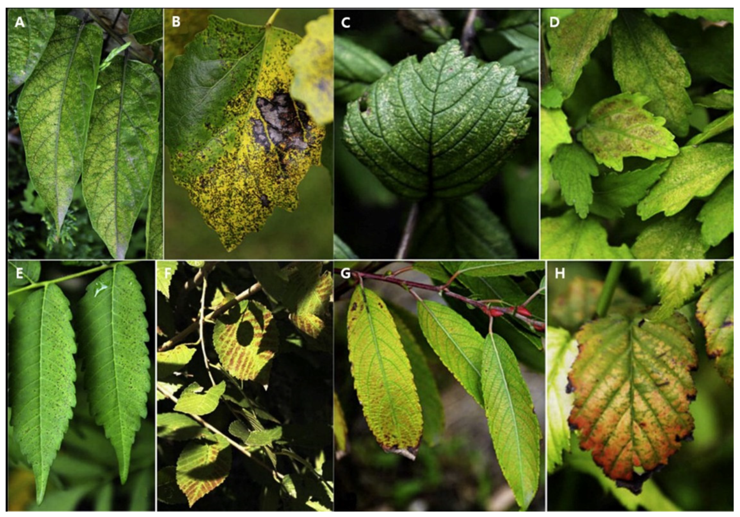
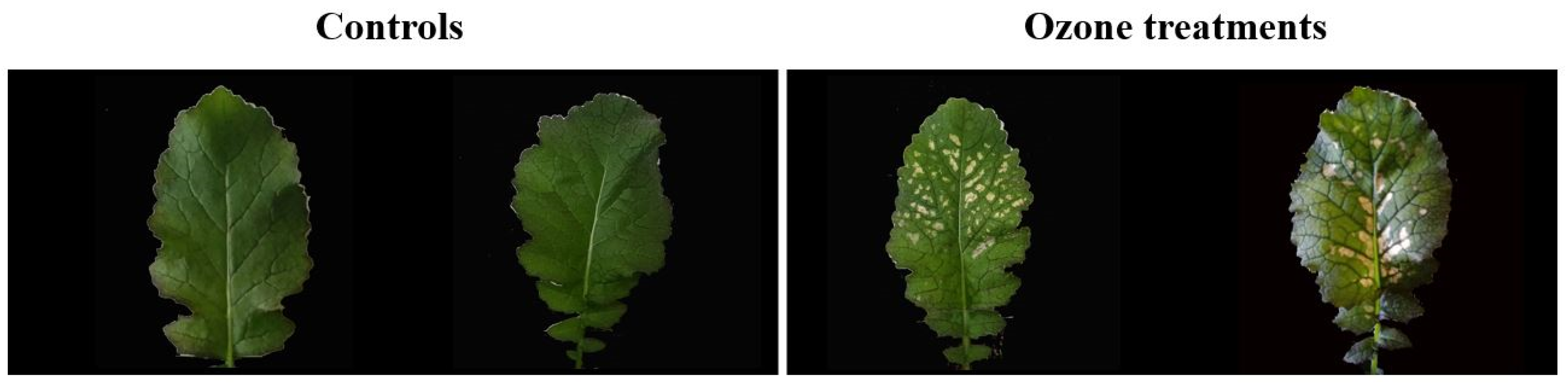
2.1. Visible Symptoms of Sensitive Plants to Ozone

Recent studies have demonstrated that high O3 concentrations may cause visible foliar symptoms in plants. Visible symptoms are generally caused by abiotic and biotic stresses that may include air pollutants, drought, insects, and fungal infections. The observation of visible symptoms in leaves is considered a particularly important tool for assessing O3 effects on vegetation [15]. The presence and extent of O3-induced visible foliar symptoms may signify biological damage to vegetation, although it is less biologically significant than changes in growth and biomass [16]. The effects of O3 on crops and tree seedlings have been studied in experiments using open-top chambers [17]. The older leaves of O3-sensitive plants tend to show more symptoms of injury than younger leaves. Additionally, it is well known that O3-induced visible symptoms can generally occur on sunlit foliage because light-induced oxidative stress leads to visible injury. Nevertheless, visible symptoms can also appear on shaded leaves [18]. When exposure to elevated O3 levels occurs during leaf formation, leaves may be less affected and acclimate to later O3 exposure [19,20]. Visible foliar O3 injury symptoms can be observed as whitish or light-green coloring, and bleaching, reddening, or bronzing patterns in interveinal patches [21]. Wan et al. [22] studied visible symptoms indicating O3 injury on O3-sensitive trees and shrubs around Beijing (Figure 2). Wilting occurred in Medicago truncatula under an O3 concentration of 70 nL L−1, and necrotic spots appeared on the leaves within 6 days [23]. Lee et al. [24] studied Brassica juncea L., which is widely cultivated in East Asia. Under an O3 concentration of 100 nL L−1 in an O3 fumigated growth chamber, B. juncea L. displayed visible symptoms (bleaching) indicating O3 injury (Figure 3). This study demonstrated that exposure to elevated O3 causes foliar injury. Visible injury to O3-sensitive leafy vegetables may negatively affect the quality of agricultural products and may, therefore, have financial impacts on farm markets [25].

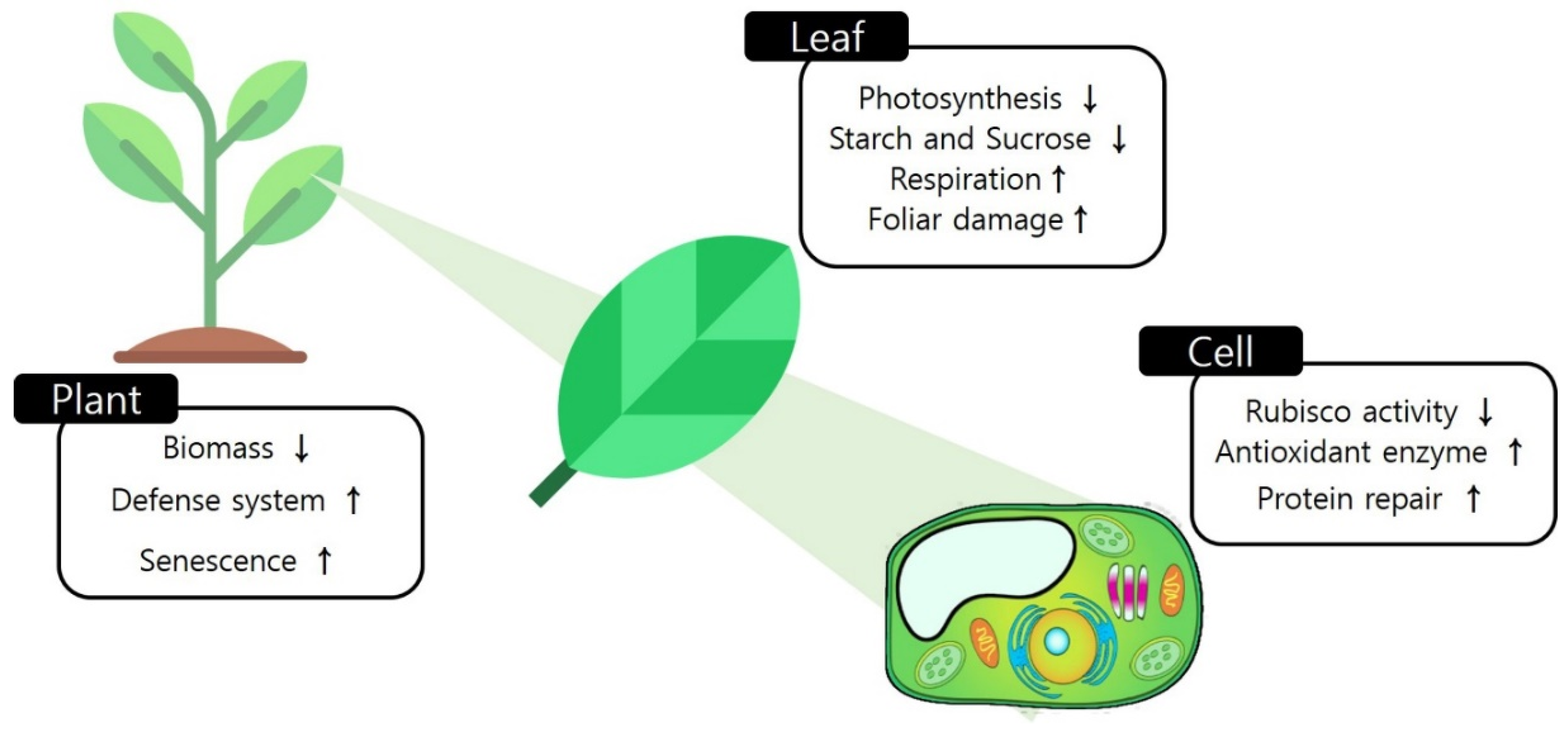
2.2. Physiological Changes in Response to Ozone

Tropospheric O3 has many adverse effects on plants and is the most impactful form of air pollution. Elevated O3 concentrations can impair plant physiological processes within plants, such as carbon assimilation. Chronic O3 exposure reduces photosynthesis and total biomass, and accelerates senescence [26]. Several studies have reported that net photosynthesis rates in broad-leaved trees, wheat, soybean, and rice were considerably decreased by high ambient O3 concentrations [27,28,29]. The degradation of photosynthesis by O3 exposure has generally been attributed to decreased carboxylation efficiency, impacts on the photosynthetic electron transport system, and effects on the stomata [30]. Based on a study of ribulose-1,5-bisphosphate carboxylase/oxygenase (Rubisco) content and activity, O3-induced alterations to physiological capacity were found to correlate with decreased CO2 fixation [5]. Moreover, O3 can cause decreased electron transfer between photosystems (PS) I and PS II. Chlorophyll a fluorescence is a practical indicator of photoinhibition [31]. The Fv/Fm ratio, which indicates the maximum quantum yield of PS II, is generally reduced under elevated O3. The reduction in the PS II quantum yield is known to decrease photosynthetic electron transport. ATP and NADPH production are also decreased, due to reduced demand from the Calvin cycle, in plants exposed to O3 [32]. Furthermore, the decreased photosynthetic aperture leads to the production of nonstructural carbohydrates such as sucrose and starch (Figure 4) [5].
The rate of O3 influx into leaves is controlled by the stomatal aperture. Stomatal closure is generally recognized as a response that limits O3 uptake [30]. Acute O3 exposure causes a notable decrease in stomatal conductance by causing reactive oxygen species (ROS) to accumulate in guard cells. In Arabidopsis, higher O3 concentrations caused a rapid transient decrease in stomatal conductance within 3–6 min of exposure [33]. Several studies using open-top chamber experiments have reported that O3 usually decreases stomatal conductance, consequently limiting CO2 inflow to the leaves [34]. However, this process was not supported by the results of all experiments [35]. Some studies have reported that stomata were unable to close rapidly when impaired by exposure to elevated O3 levels [36]. Regardless of the mechanisms involved, however, O3 concentration is clearly an important determinant of plant physiological conditions. Plants can enhance their O3 stress tolerance by regulating physiological mechanisms such as stomatal closure and an increase in antioxidants. This acclimation mechanism enables stomata to remain partly open and facilitates photosynthesis-related gas exchange, avoiding drastic plant growth decreases [37,38].

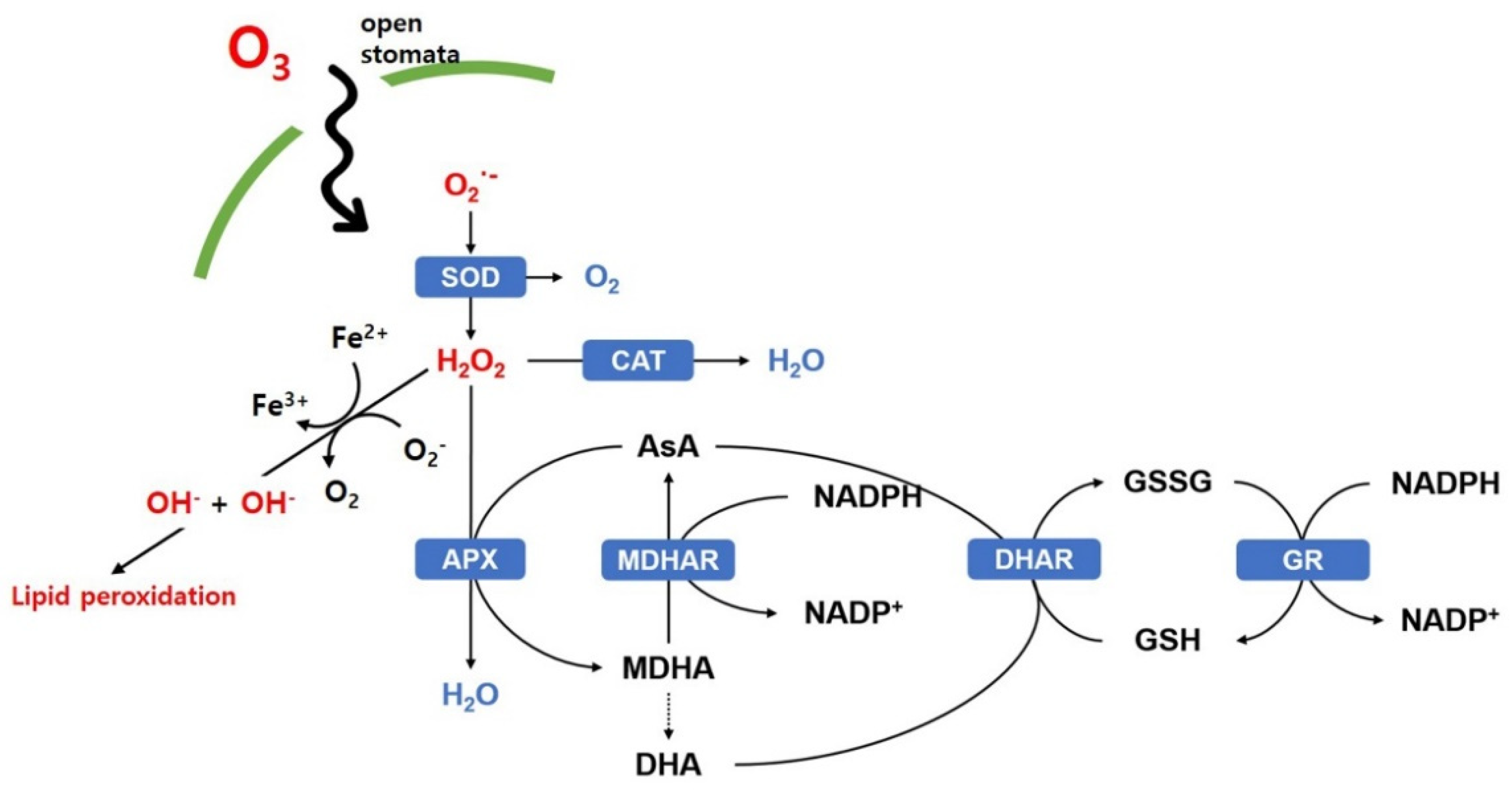
2.3. Biochemical Changes Caused by Ozone

During exposure to elevated O3 concentrations, O3 induces changes in plant characteristics at the biochemical and molecular levels [39,40]. O3 enters leaves through the stomata and decomposes within the apoplastic space. This produces reactive oxygen species (ROS), such as the superoxide anion (O2), hydrogen peroxide (H2O2), and hydroxyl radicals (OH) [41]. Although O2 and H2O2 are among the less reactive types of ROS, they are still recognized as dangerous ROSs because they can diffuse into cellular compartments [42]. OH has been thought to damage cell membranes because of its strong oxidation potential [43]. These apoplastic ROSs cause oxidative damage to cell membranes, proteins, and DNA molecules and can cause changes in enzyme activities, thereby leading to cell destruction [44]. ROS concentrations are mitigated by enzymatic antioxidants such as those involved in the ascorbate–glutathione cycle (AsA-GSH cycle), which include catalase (CAT), superoxide dismutase (SOD), ascorbate peroxidase (APX), dehydroascorbate reductase (DHAR), and glutathione reductase (GR) [45]. The detoxification of O2 by SOD produces H2O2, which is then removed by CAT or APX. Peroxidase (POX) requires the phenolic compound guaiacol as an electron donor, which can then be used to reduce the amount of H2O2. APX uses a reduced form of AsA to protect cells against oxidative damage caused by H2O2. The oxidized AsA formed by APX activity is regenerated through other components of the AsA–GSH cycle, including dehydroascorbate reductase (DHAR) and monodehydroascorbate reductase (MDHAR). GR reduces the oxidized form of glutathione (GSSG) by reducing NADPH activity [46,47] (Figure 5).
The accumulation of ROS limits stomatal movements and abscisic acid (ABA) levels independently of CO2 [48]. Plant responses to O3 involve the regulation of guard–cell ion channels. The accumulation of ROS in the extracellular space activates a still-unknown calcium channel protein and increases cytosolic calcium accumulation, thus activating calcium-dependent protein kinases (CPKs) [49]. Calcium is a second messenger that contributes to diverse signaling cascades that regulate plant stress responses. The activation of two types of anion channels—the slow anion channel 1 (SLAC1) and the quickly activating anion channel 1 (QUAC1)—then causes stomatal closure [50,51]. ROSs activate the protein kinase open stomata 1 (OST 1), which controls the outward rectifying SLAC1 and QUAC1 channels, as well as the inward rectifying K+ channel KAT1 [52,53]. OST1 activity is limited by ABA-insensitive 1 (ABI 1) and ABI 2 protein phosphatase 2Cs, which are inactivated by the abscisic acid receptor PYR/PYL. SLAC1 is also activated by CPKs and the ROS-induced guard cell hydrogen peroxide-resistant 1 (GHR1) protein [48] (Figure 6). ROSs are thus involved in the regulation of stomatal movements, as they affect various signaling pathways.

3. Plant Responses to Elevated Temperatures

3.1. Physiological Changes Caused by Elevated Temperatures

Heat stress due to prolonged exposure to elevated temperatures is a critical threat to vegetation and crop production worldwide [54]. Elevated ambient temperatures can positively influence plant growth and metabolism. Plants respond differently to elevated temperatures depending on their developmental stages and species characteristics [55]. However, temperatures above the upper tolerance threshold of a plant species decrease their net photosynthetic rate and total biomass [56]. Constantly elevated temperatures also cause physiological changes in plants. The plant characteristic most susceptible to elevated temperatures is water status [57]. Elevated temperatures increase transpiration and stomatal conductance. Therefore, elevated temperature stress is usually related to reduced water availability [58]. Under elevated temperatures, plants usually grow smaller leaves, and extend their root systems to increase water uptake and decrease water loss from leaves through transpiration [59]. Wahid et al. [57] showed that increased temperatures greatly reduced the leaf-water potential in treated plants, compared to the control group. Elevated temperatures also negatively impacted plant height, leaf area, and total biomass of Brassica napus due to reduced CO2 assimilation [59]. Changes in CO2 assimilation under elevated temperatures are good indicators of plant responses to high temperatures. The photochemical reactions in the thylakoid lamellae have been regarded as the prime sites of injury at elevated temperatures [60]. The functioning of PS II is highly decreased or partly stopped at elevated temperatures due to it being thermolabile [61]. Additionally, the photosynthetic ability of plants at elevated temperatures is limited by the decreased activity of Rubisco, an enzyme involved in carbon fixation [12]. Further, an imbalance between photosynthesis and respiration impairs plant growth under elevated temperature conditions, because high temperatures generally decrease the rate of photosynthesis in plants while increasing the rates of photo- and dark respiration [57]. These physiological changes in plants at higher temperatures result in decreased growth and development, thus reducing total biomass and crop production.

3.2. Biochemical Changes in Response to Elevated Temperatures

Temperature is the primary environmental factor affecting plant growth and development. Generally, the biochemical characteristics of plants are more negatively affected by air temperatures above 5 °C than the optimum [55]. Elevated temperature stress may cause the excess production of reactive oxygen species (ROS), leading to oxidative stress [62]. ROSs, including singlet oxygen (1O2), the superoxide radical (O2), hydrogen peroxide (H2O2), and hydroxyl radicals (OH), can cause lipid peroxidation, protein oxidation, changes in enzyme activities, and oxidative damage to membranes, all of which lead to cell death [62,63]. Plants have a variety of enzymatic and non-enzymatic antioxidant systems to diminish ROS levels in their tissues, thereby protecting their cell membranes from oxidative damage [64].
Elevated environmental temperatures increased leaf temperatures, which degraded the activity of enzymatic antioxidant systems that are responsible for the malondialdehyde (MDA) content in rice leaves in [65]. Measuring MDA content usually indicates the occurrence level of lipid peroxidation, which is a sign of damage in living organisms [66]. In a study with wheat kept at 33 °C, oxidative stress significantly increased membrane peroxidation by 28%, greatly increasing solute leakage [67].
Heat stress due to elevated temperatures induces the expression of stress proteins that are not produced under normal conditions [68]. Heat-shock proteins (HSPs) are exclusively involved in the heat-stress response. HSPs prevent heat-stress-induced protein denaturation and mediate protein homeostasis [69]. HSPs are classified into five families based on their molecular masses: HSP100, HSP90, HSP70, HSP60, and sHSPs [70]. The diversity and abundance of HSPs that can be expressed, indicates the heat tolerance of a plant species. Heat-stress factors (HSFs) and HSPs play important roles in the plant heat-stress response [71] (Figure 7). HSFs located in the cytoplasm mainly control the transcription of HSP genes [72]. At least 21 HSF members cloned in plants have been found to cooperate in all steps of their heat-stress responses. Therefore, HSFs are regarded as transcriptional activators of heat-shock responses [73]. HSF proteins are grouped into three conserved classes (A, B, and C) based on the structural features of their oligomeric domains. HSFA1a is the master regulator for the heat-stress-induced synthesis of HSFA2, a major heat-stress factor [74]. Heat-induced gene expression resulting in HSP synthesis is initiated by mechanisms that can sense and transduce signals of heat stress to HSFs [75,76]. HSFs then bind to a heat-shock promoter element (HSE) in the promoter region of the HSP gene and initiate transcription [75]. HSPs mainly stabilize partially unfolded proteins to help maintain assembly and reduce protein degradation. However, while these proteins contain no specific information about the correct folding of any specific proteins, they do prevent unproductive interactions that result in unfolding and loss of structure and function [77].

4. Plant Responses to Ozone under Elevated Temperatures

To date, researchers have studied the effects of single abiotic stress parameters on plants. However, the interactive effects of stress factors on plants remain unclear, because different plant responses can be deduced from combinations of single factors [78]. In particular, the considerable variety of plant responses to elevated O3 is most likely related to the interactive effects of other co-occurring environmental variables including temperature, solar radiation, drought, and increased NOx in the atmosphere [79]. In this review, we focus on the plant response on the interactive effect of elevated temperature and O3. Because temperature directly affects chemical kinetics and VOC emissions, both of which are associated with O3 production, temperature is known to be a strong predictor of tropospheric O3 concentration levels. Furthermore, elevated temperatures may promote O3 accumulation by influencing sunny, dry, and stagnant atmospheres [80]. For these reasons, high temperature and elevated O3 conditions generally co-occur [11].
Because O3 and temperature can each independently affect the physiological processes of plants, the interplay of factors must be investigated. Many studies have focused on the interaction of these factors to study possible ways in which temperature affects plants exposed to O3. Plants exposed to modest temperature elevation (1–5 °C) showed increased photosynthesis, growth, and biomass in various experiments [24,81,82]. Increased O3 concentration, however, can have a variety of detrimental effects on plants, including reduced photosynthesis, stomatal conductance, and growth [83]. Elevated O3 conditions are thought to suppress plant development and growth. At the same time, elevated ambient temperatures are expected to enhance plant development and growth. Nevertheless, the final effect of elevated ambient temperature and O3 on plant responses may be determined by how these factors interact with the plant’s physio–biochemical processes. Increased temperatures above the optimum may decrease the uptake of ozone by reducing stomatal conductance [55,84]. However, due to increased stomatal conductance, moderately elevated temperatures (+5 °C) can enhance ozone uptake [24,85]. Lee et al. [24,85] reported that +5 °C above an optimal temperature increased O3-induced foliar damage and reduced photosynthesis in chamber experiments with constant humidity. Conversely, O3 accelerated leaf senescence in silver birch (Betula pendula), while a +1.2 °C-temperature increase delayed leaf senescence in field experiments [86]. Elevated temperatures in natural conditions are frequently related to increased vapor pressure deficits (VPD), which significantly affect stomatal movement [79,87] as well as stomatal O3 flux [88]. Many studies have shown that stomatal O3 uptake, rather than the O3 level, is related to O3 deposition and damage to plants [88,89,90]. Pea cultivars with slower stomatal closure under elevated O3, and thus higher O3 flux in the interior leaf tissue, had more O3 damage [91]. Stomatal O3 flux can be determined by leaf boundary layer resistance and stomatal resistance; both factors are affected by heat flux and VPD [79]. Without confounding changes in humidity, the stomatal response to higher ambient temperatures obviously showed increased stomatal opening in relation to O3 flux [92,93]. Presently, the plant physiological characteristics resulting from the combined effects and interacting mechanisms of elevated O3 and temperature are debatable [24,85,87,88,94,95].

5. Conclusions

In this paper we summarized the plant responses to the combined and individual effects of elevated O3 and temperature, including physiological and biochemical changes. Plant responses to abiotic stressors, such as O3 and temperature, are dynamic and complicated. The inevitable impacts of O3 and temperature on terrestrial vegetation may exceed those of any other abiotic stress factors.
As demonstrated in Section 2, Plant Responses to Ozone, tropospheric O3 is a critical air pollutant with many negative consequences for plants. At the physiological, biochemical, and anatomical levels, the mechanisms of O3 effects on plants are relatively well characterized. Elevated O3 has been correlated with photosynthesis degradation due to lower carboxylation efficiency, effects on photosynthetic electron transport, and effects on stomata. Because O3 enters the leaf through the stomata, differences in stomatal opening may potentially cause O3 sensitivity. Because O3 can decompose within the apoplastic space in the cell after penetrating through stomatal pores, it can form reactive oxygen species (ROS).
The examples discussed in Section 3 clearly show that extremely and moderately high temperatures can cause physiological and biochemical changes in plants in different ways. Moderately elevated temperatures may positively impact plant growth and metabolism. Extremely high temperatures, however, may decrease plant growth due to an imbalance between photosynthesis and respiration. Extremely high temperatures also increase leaf temperature, thereby reducing antioxidant system function.
Increased temperature and O3 conditions frequently co-occur because elevated temperatures may increase O3 concentrations in nature. The combined influence of both factors can produce results that are remarkably different from those produced by a single factor. Stomatal movement, which can be highly determined by temperature, may be an important factor in the plant response to O3 exposure. Stomatal O3 flux can be determined by the leaf boundary layer, vapor pressure deficit (VPD), and relative humidity, which can be changed by temperature. The leaf boundary layer, vapor pressure deficit (VPD), and relative humidity are also factors that influence stomatal O3 flux. As a result, the plant response to a combination of elevated O3 and temperature is still a subject of debate.
To obtain a comprehensive understanding of how plants respond to both parameters, more extensive methodological approaches, such as long-term eddy-covariance, open top chambers, and free-air enrichment systems, are needed. Plant response assessments at the organ, tissue, and cellular levels must also be extended to studies of proteomics and enzyme activity.

Author Contributions

S.Y.W., as the corresponding author, developed the concept, edited the manuscript, and acquired funding. J.K.L. performed the literature review and wrote the manuscript. M.J.K. performed the literature review and edited the manuscript. S.G.J. performed the visualization. All authors have read and agreed to the published version of the manuscript.

Funding

This study was carried out with the support of ‘A Study on Mechanism and Function Improvement of Plants for Reducing Air Pollutants’ (Grant No. FE0000-2018-01-2021) from the National Institute of Forest Science (NIFoS), Republic of Korea.

Institutional Review Board Statement

Not applicable.

Informed Consent Statement

Not applicable.

Data Availability Statement

Not applicable.

Conflicts of Interest

The authors declare no conflict of interest.

References

  1. Goldstein, A.; Turner, W.R.; Spawn, S.A.; Anderson-Teixeira, K.J.; Cook-Patton, S.; Fargione, J.; Gibbs, H.K.; Griscom, B.; Hewson, J.H.; Howard, J.F.; et al. Protecting irrecoverable carbon in Earth’s ecosystems. Nat. Clim. Chang. 2020, 10, 287–295. [Google Scholar] [CrossRef]
  2. Pachauri, R.K.; Allen, M.R.; Barros, V.R.; Broome, J.; Cramer, W.; Christ, R.; Church, J.A.; Clarke, L.; Dahe, Q.; Dasgupta, P. Climate Change 2014: Synthesis Report. Contribution of Working Groups I, II and III to the Fifth Assessment Report of the Intergovernmental Panel on Climate Change; IPCC: Geneva, Switzerland, 2014. [Google Scholar]
  3. Fuhrer, J. Ozone risk for crops and pastures in present and future climates. Naturwissenschaften 2009, 96, 173–194. [Google Scholar] [CrossRef] [PubMed]
  4. Ainsworth, E.A.; Rogers, A.; Leakey, A.D.B. Targets for crop biotechnology in a future high-CO2 and high-O3 world. Plant Physiol. 2008, 147, 13–19. [Google Scholar] [CrossRef] [PubMed] [Green Version]
  5. Ainsworth, E.A.; Yendrek, C.R.; Sitch, S.; Collins, W.J.; Emberson, L.D. The effects of tropospheric ozone on net primary productivity and implications for climate change. Annu. Rev. Plant Biol. 2012, 63, 637–661. [Google Scholar] [CrossRef] [Green Version]
  6. Tammam, A.; Badr, R.; Abou-Zeid, H.; Hassan, Y.; Bader, A. Nickel and ozone stresses induce differential growth, antioxidant activity and mRNA transcription in Oryza sativa cultivars. J. Plant Interact. 2019, 14, 87–101. [Google Scholar] [CrossRef] [Green Version]
  7. Karnosky, D.F.; Pregitzer, K.S.; Zak, D.R.; Kubiske, M.E.; Hendrey, G.R.; Weinstein, D.; Nosal, M.; Percy, K.E. Scaling ozone responses of forest trees to the ecosystem level in a changing climate. Plant Cell Environ. 2005, 28, 965–981. [Google Scholar] [CrossRef] [Green Version]
  8. Atkinson, R. Atmospheric chemistry of VOCs and NOx. Atmos. Environ. 2000, 34, 2063–2101. [Google Scholar] [CrossRef]
  9. Cape, J.N. Surface ozone concentrations and ecosystem health: Past trends and a guide to future projections. Sci. Total Environ. 2008, 400, 257–269. [Google Scholar] [CrossRef] [PubMed] [Green Version]
  10. Fitzky, A.C.; Sandén, H.; Karl, T.; Fares, S.; Calfapietra, C.; Grote, R.; Saunier, A.; Rewald, B. The Interplay between ozone and urban vegetation—BVOC emissions, ozone deposition, and tree ecophysiology. Front. For. Glob. Chang. 2019, 2, 50. [Google Scholar] [CrossRef]
  11. Tai, A.P.K.; Martin, M.V.; Heald, C.L. Threat to future global food security from climate change and ozone air pollution. Nat. Clim. Chang. 2014, 4, 817–821. [Google Scholar] [CrossRef] [Green Version]
  12. Sage, R.F.; Kubien, D.S. The temperature response of C3 and C4 photosynthesis. Plant Cell Environ. 2007, 30, 1086–1106. [Google Scholar] [CrossRef]
  13. Booker, F.; Muntifering, R.; Mcgrath, M.; Burkey, K.; Decoteau, D.; Fiscus, E.; Manning, W.; Krupa, S.; Chappelka, A.; Grantz, D. The ozone component of global change: Potential effects on agricultural and horticultural plant yield, product quality and interactions with invasive species. J. Integr. Plant Biol. 2009, 51, 337–351. [Google Scholar] [CrossRef] [PubMed]
  14. Mills, G.; Sharps, K.; Simpson, D.; Pleijel, H.; Broberg, M.; Uddling, J.; Jaramillo, F.; Davies, W.J.; Dentener, F.; Van den Berg, M.; et al. Ozone pollution will compromise efforts to increase global wheat production. Glob. Chang. Biol. 2018, 24, 3560–3574. [Google Scholar] [CrossRef] [PubMed]
  15. Choquette, N.E.; Ainsworth, E.A.; Bezodis, W.; Cavanagh, A.P. Ozone tolerant maize hybrids maintain Rubisco content and activity during long-term exposure in the field. Plant. Cell Environ. 2020, 43, 3033–3047. [Google Scholar] [CrossRef]
  16. Feng, Z.; Sun, J.; Wan, W.; Hu, E.; Calatayud, V. Evidence of widespread ozone-induced visible injury on plants in Beijing, China. Environ. Pollut. 2014, 193, 296–301. [Google Scholar] [CrossRef] [PubMed]
  17. Paoletti, E.; Materassi, A.; Fasano, G.; Hoshika, Y.; Carriero, G.; Silaghi, D.; Badea, O. A new-generation 3D ozone FACE (Free Air Controlled Exposure). Sci. Total Environ. 2017, 575, 1407–1414. [Google Scholar] [CrossRef] [PubMed]
  18. Günthardt-Goerg, M.S.; Vollenweider, P. Linking stress with macroscopic and microscopic leaf response in trees: New diagnostic perspectives. Environ. Pollut. 2007, 147, 467–488. [Google Scholar] [CrossRef]
  19. Baek, S.G.; Park, J.H.; Na, C.S.; Lee, B.; Cheng, H.C.; Woo, S.Y. The morphological characteristics of Pterocarpus indicus induced by elevated ozone under well-watered and drought conditions. Forest Sci. Technol. 2018, 14, 105–111. [Google Scholar] [CrossRef] [Green Version]
  20. Pääkkönen, E. Ageing-related anatomical and ultrastructural changes in leaves of birch (Betula pendula Roth.) clones as affected by low ozone exposure. Ann. Bot. 1995, 75, 285–294. [Google Scholar] [CrossRef]
  21. Vollenweider, P.; Günthardt-Goerg, M.S.; Menard, T.; Baumgarten, M.; Matyssek, R.; Schaub, M. Macro- and microscopic leaf injury triggered by ozone stress in beech foliage (Fagus sylvatica L.). Ann. For. Sci. 2019, 76, 71. [Google Scholar] [CrossRef]
  22. Wan, W.; Manning, W.J.; Wang, X.; Zhang, H.; Sun, X.; Zhang, Q. Ozone and ozone injury on plants in and around Beijing, China. Environ. Pollut. 2014, 191, 215–222. [Google Scholar] [CrossRef]
  23. Iyer, N.J.; Tang, Y.; Mahalingam, R. Physiological, biochemical and molecular responses to a combination of drought and ozone in Medicago truncatula. Plant Cell Environ. 2013, 36, 706–720. [Google Scholar] [CrossRef]
  24. Lee, J.K.; Woo, S.Y.; Kwak, M.J.; Park, S.H.; Kim, H.D.; Lim, Y.J.; Park, J.H.; Lee, K.A. Effects of elevated temperature and ozone in Brassica juncea L.: Growth, physiology, and ROS accumulation. Forests 2020, 11, 68. [Google Scholar] [CrossRef] [Green Version]
  25. Sharps, K.; Hayes, F.; Harmens, H.; Mills, G. Ozone-induced effects on leaves in African crop species. Environ. Pollut. 2021, 268, 115789. [Google Scholar] [CrossRef]
  26. Fiscus, E.L.; Booker, F.L.; Burkey, K.O. Crop responses to ozone: Uptake, modes of action, carbon assimilation and partitioning. Plant Cell Environ. 2005, 28, 997–1011. [Google Scholar] [CrossRef]
  27. Wittig, V.E.; Ainsworth, E.A.; Long, S.P. To what extent do current and projected increases in surface ozone affect photosynthesis and stomatal conductance of trees? A meta-analytic review of the last 3 decades of experiments. Plant Cell Environ. 2007, 30, 1150–1162. [Google Scholar] [CrossRef]
  28. Feng, Z.; Kobayashi, K.; Ainsworth, E.A. Impact of elevated ozone concentration on growth, physiology, and yield of wheat (Triticum aestivum L.): A meta-analysis. Glob. Chang. Biol. 2008, 14, 2696–2708. [Google Scholar] [CrossRef]
  29. Ainsworth, E.A. Rice production in a changing climate: A meta-analysis of responses to elevated carbon dioxide and elevated ozone concentration. Glob. Chang. Biol. 2008, 14, 1642–1650. [Google Scholar] [CrossRef]
  30. Paoletti, E.; Grulke, N.E. Does living in elevated CO2 ameliorate tree response to ozone? A review on stomatal responses. Environ. Pollut. 2005, 137, 483–493. [Google Scholar] [CrossRef]
  31. Murata, N.; Takahashi, S.; Nishiyama, Y.; Allakhverdiev, S.I. Photoinhibition of photosystem II under environmental stress. Biochim. Biophys. Acta Bioenerg. 2007, 1767, 414–421. [Google Scholar] [CrossRef] [PubMed] [Green Version]
  32. Liu, X.; Sui, L.; Huang, Y.; Geng, C.; Yin, B. Physiological and visible injury responses in different growth stages of winter wheat to ozone stress and the protection of spermidine. Atmos. Pollut. Res. 2015, 6, 596–604. [Google Scholar] [CrossRef] [Green Version]
  33. Kollist, T.; Moldau, H.; Rasulov, B.; Oja, V.; Rämma, H.; Hüve, K.; Jaspers, P.; Kangasjärvi, J.; Kollist, H. A novel device detects a rapid ozone-induced transient stomatal closure in intact Arabidopsis and its absence in abi2 mutant. Physiol. Plant. 2007, 129, 796–803. [Google Scholar] [CrossRef]
  34. Kollist, H.; Moldau, H.; Mortensen, L.; Rasmussen, S.K.; Jørgensen, L.B. Ozone flux to plasmalemma in barley and wheat is controlled by stomata rather than by direct reaction of ozone with cell wall ascorbate. J. Plant Physiol. 2000, 156, 645–651. [Google Scholar] [CrossRef]
  35. Pell, E.J.; Eckardt, N.; Enyedi, A.J. Timing of ozone stress and resulting status of ribulose bisphosphate carboxylase/oxygenase and associated net photosynthesis. New Phytol. 1992, 120, 397–405. [Google Scholar] [CrossRef]
  36. Seifikalhor, M.; Aliniaeifard, S.; Shomali, A.; Azad, N.; Hassani, B.; Lastochkina, O.; Li, T. Calcium signaling and salt tolerance are diversely entwined in plants. Plant Signal. Behav. 2019, 14, 1665455. [Google Scholar] [CrossRef]
  37. Held, A.A.; Mooney, H.A.; Gorham, J.N. Acclimation to ozone stress in radish: Leaf demography and photosynthesis. New Phytol. 1991, 118, 417–423. [Google Scholar] [CrossRef]
  38. Droutsas, I.; Challinor, A.J.; Arnold, S.R.; Mikkelsen, T.N.; Hansen, E.M.Ø. A new model of ozone stress in wheat including grain yield loss and plant acclimation to the pollutant. Eur. J. Agron. 2020, 120, 126125. [Google Scholar] [CrossRef]
  39. Agrawal, G.K.; Rakwal, R.; Yonekura, M.; Kubo, A.; Saji, H. Proteome analysis of differentially displayed proteins as a tool for investigating ozone stress in rice (Oryza sativa L.) seedlings. Proteomics 2002, 2, 947–959. [Google Scholar] [CrossRef]
  40. Cho, K.; Shibato, J.; Kubo, A.; Kohno, Y.; Satoh, K.; Kikuchi, S.; Agrawal, G.K.; Sarkar, A.; Rakwal, R. Genome-wide mapping of the ozone-responsive transcriptomes in rice panicle and seed tissues reveals novel insight into their regulatory events. Biotechnol. Lett. 2013, 35, 647–656. [Google Scholar] [CrossRef]
  41. Hoshika, Y.; Haworth, M.; Watanabe, M.; Koike, T. Interactive effect of leaf age and ozone on mesophyll conductance in Siebold’s beech. Physiol. Plant. 2020, 170, 172–186. [Google Scholar] [CrossRef]
  42. Di Baccio, D.; Castagna, A.; Paoletti, E.; Sebastiani, L.; Ranieri, A. Could the differences in O3 sensitivity between two poplar clones be related to a difference in antioxidant defense and secondary metabolic response to O3 influx? Tree Physiol. 2008, 28, 1761–1772. [Google Scholar] [CrossRef] [Green Version]
  43. Wu, H.; Sun, P.; Feng, H.; Zhou, H.; Wang, R.; Liang, Y.; Lu, J.; Zhu, W.; Zhang, J.; Fang, J. Reactive oxygen species in a non-thermal plasma microjet and water system: Generation, conversion, and contributions to bacteria inactivation—An analysis by electron spin resonance spectroscopy. Plasma Process. Polym. 2012, 9, 417–424. [Google Scholar] [CrossRef]
  44. Choudhury, F.K.; Rivero, R.M.; Blumwald, E.; Mittler, R. Reactive oxygen species, abiotic stress and stress combination. Plant J. 2017, 90, 856–867. [Google Scholar] [CrossRef]
  45. Akram, N.A.; Shafiq, F.; Ashraf, M. Ascorbic acid-a potential oxidant scavenger and its role in plant development and abiotic stress tolerance. Front. Plant Sci. 2017, 8, 613. [Google Scholar] [CrossRef] [PubMed]
  46. Foyer, C.H.; Halliwell, B. The presence of glutathione and glutathione reductase in chloroplasts: A proposed role in ascorbic acid metabolism. Planta 1976, 133, 21–25. [Google Scholar] [CrossRef]
  47. Caverzan, A.; Passaia, G.; Rosa, S.B.; Ribeiro, C.W.; Lazzarotto, F.; Margis-Pinheiro, M. Plant responses to stresses: Role of ascorbate peroxidase in the antioxidant protection. Genet. Mol. Biol. 2012, 35, 1011–1019. [Google Scholar] [CrossRef] [Green Version]
  48. Vainonen, J.P.; Kangasjärvi, J. Plant signalling in acute ozone exposure. Plant Cell Environ. 2015, 38, 240–252. [Google Scholar] [CrossRef]
  49. Short, E.F.; North, K.A.; Roberts, M.R.; Hetherington, A.M.; Shirras, A.D.; McAinsh, M.R. A stress-specific calcium signature regulating an ozone-responsive gene expression network in Arabidopsis. Plant J. 2012, 71, 948–961. [Google Scholar] [CrossRef]
  50. Kollist, H.; Jossier, M.; Laanemets, K.; Thomine, S. Anion channels in plant cells. FEBS J. 2011, 278, 4277–4292. [Google Scholar] [CrossRef] [Green Version]
  51. Joshi-Saha, A.; Valon, C.; Leung, J. A brand new START: Abscisic acid perception and transduction in the guard cell. Sci. Signal. 2011, 4, re4. [Google Scholar] [CrossRef]
  52. Kudla, J.; Becker, D.; Grill, E.; Hedrich, R.; Hippler, M.; Kummer, U.; Parniske, M.; Romeis, T.; Schumacher, K. Advances and current challenges in calcium signaling. New Phytol. 2018, 218, 414–431. [Google Scholar] [CrossRef] [PubMed]
  53. Hua, D.; Wang, C.; He, J.; Liao, H.; Duan, Y.; Zhu, Z.; Guo, Y.; Chen, Z.; Gong, Z. A plasma membrane receptor kinase, GHR1, mediates abscisic acid- and hydrogen peroxide-regulated stomatal movement in Arabidopsis. Plant Cell 2012, 24, 2546–2561. [Google Scholar] [CrossRef] [PubMed] [Green Version]
  54. Teixeira, F.K.; Menezes-Benavente, L.; Galvão, V.C.; Margis-Pinheiro, M. Multigene families encode the major enzymes of antioxidant metabolism in Eucalyptus grandis L. Genet. Mol. Biol. 2005, 28, 529–538. [Google Scholar] [CrossRef] [Green Version]
  55. Hasanuzzaman, M.; Nahar, K.; Alam, M.; Roychowdhury, R.; Fujita, M. Physiological, biochemical, and molecular mechanisms of heat stress tolerance in plants. Int. J. Mol. Sci. 2013, 14, 9643–9684. [Google Scholar] [CrossRef]
  56. Posch, B.C.; Kariyawasam, B.C.; Bramley, H.; Coast, O.; Richards, R.A.; Reynolds, M.P.; Trethowan, R.; Atkin, O.K. Exploring high temperature responses of photosynthesis and respiration to improve heat tolerance in wheat. J. Exp. Bot. 2019, 70, 5051–5069. [Google Scholar] [CrossRef]
  57. Wahid, A.; Gelani, S.; Ashraf, M.; Foolad, M.R. Heat tolerance in plants: An overview. Environ. Exp. Bot. 2007, 61, 199–223. [Google Scholar] [CrossRef]
  58. Fahad, S.; Bajwa, A.A.; Nazir, U.; Anjum, S.A.; Farooq, A.; Zohaib, A.; Sadia, S.; Nasim, W.; Adkins, S.; Saud, S.; et al. Crop production under drought and heat stress: Plant responses and management options. Front. Plant Sci. 2017, 8, 1147. [Google Scholar] [CrossRef] [Green Version]
  59. Qaderi, M.M.; Kurepin, L.V.; Reid, D.M. Effects of temperature and watering regime on growth, gas exchange and abscisic acid content of canola (Brassica napus) seedlings. Environ. Exp. Bot. 2012, 75, 107–113. [Google Scholar] [CrossRef]
  60. Wise, R.R.; Olson, A.J.; Schrader, S.M.; Sharkey, T.D. Electron transport is the functional limitation of photosynthesis in field-grown Pima cotton plants at high temperature. Plant Cell Environ. 2004, 27, 717–724. [Google Scholar] [CrossRef]
  61. Camejo, D.; Rodríguez, P.; Morales, M.A.; Dell’Amico, J.M.; Torrecillas, A.; Alarcón, J.J. High temperature effects on photosynthetic activity of two tomato cultivars with different heat susceptibility. J. Plant Physiol. 2005, 162, 281–289. [Google Scholar] [CrossRef]
  62. Liu, X.; Huang, B. Heat stress injury in relation to membrane lipid peroxidation in creeping bentgrass. Crop Sci. 2000, 40, 503–510. [Google Scholar] [CrossRef]
  63. Medina, E.; Kim, S.-H.; Yun, M.; Choi, W.-G. Recapitulation of the function and role of ROS generated in response to heat stress in plants. Plants 2021, 10, 371. [Google Scholar] [CrossRef]
  64. Ott, M.; Gogvadze, V.; Orrenius, S.; Zhivotovsky, B. Mitochondria, oxidative stress and cell death. Apoptosis 2007, 12, 913–922. [Google Scholar] [CrossRef]
  65. Akter, N.; Rafiqul Islam, M. Heat stress effects and management in wheat. A review. Agron. Sustain. Dev. 2017, 37, 37. [Google Scholar] [CrossRef]
  66. Gill, S.S.; Tuteja, N. Reactive oxygen species and antioxidant machinery in abiotic stress tolerance in crop plants. Plant Physiol. Biochem. 2010, 48, 909–930. [Google Scholar] [CrossRef]
  67. Savicka, M.; Škute, N. Effects of high temperature on malondialdehyde content, superoxide production and growth changes in wheat seedlings (Triticum aestivum L.). Ekologija 2010, 56, 26–33. [Google Scholar] [CrossRef]
  68. Balogi, Z.; Multhoff, G.; Jensen, T.K.; Lloyd-Evans, E.; Yamashima, T.; Jäättelä, M.; Harwood, J.L.; Vígh, L. Hsp70 interactions with membrane lipids regulate cellular functions in health and disease. Prog. Lipid Res. 2019, 74, 18–30. [Google Scholar] [CrossRef]
  69. Scharf, K.D.; Berberich, T.; Ebersberger, I.; Nover, L. The plant heat stress transcription factor (Hsf) family: Structure, function and evolution. Biochim. Biophys. Acta Gene Regul. Mech. 2012, 1819, 104–119. [Google Scholar] [CrossRef] [PubMed]
  70. Sun, W.; Van Montagu, M.; Verbruggen, N. Small heat shock proteins and stress tolerance in plants. Biochim. Biophys. Acta Gene Struct. Expr. 2002, 1577, 1–9. [Google Scholar] [CrossRef]
  71. Asthir, B. Mechanisms of heat tolerance in crop plants. Biol. Plant. 2015, 59, 620–628. [Google Scholar] [CrossRef]
  72. Qu, A.L.; Ding, Y.F.; Jiang, Q.; Zhu, C. Molecular mechanisms of the plant heat stress response. Biochem. Biophys. Res. Commun. 2013, 432, 203–207. [Google Scholar] [CrossRef]
  73. Hu, W.; Hu, G.; Han, B. Genome-wide survey and expression profiling of heat shock proteins and heat shock factors revealed overlapped and stress specific response under abiotic stresses in rice. Plant Sci. 2009, 176, 583–590. [Google Scholar] [CrossRef]
  74. Kotak, S.; Larkindale, J.; Lee, U.; von Koskull-Döring, P.; Vierling, E.; Scharf, K.D. Complexity of the heat stress response in plants. Curr. Opin. Plant Biol. 2007, 10, 310–316. [Google Scholar] [CrossRef]
  75. Schöffl, F.; Prändl, R.; Reindl, A. Regulation of the heat-shock response. Plant Physiol. 1998, 117, 1135–1141. [Google Scholar] [CrossRef] [Green Version]
  76. Larkindale, J.; Hall, J.D.; Knight, M.R.; Vierling, E. Heat stress phenotypes of Arabidopsis mutants implicate multiple signaling pathways in the acquisition of thermotolerance. Plant Physiol. 2005, 138, 882–897. [Google Scholar] [CrossRef] [PubMed] [Green Version]
  77. Port, M.; Tripp, J.; Zielinski, D.; Weber, C.; Heerklotz, D.; Winkelhaus, S.; Bublak, D.; Scharf, K.D. Role of Hsp17.4-CII as coregulator and cytoplasmic retention factor of tomato heat stress transcription factor HsfA2. Plant Physiol. 2004, 135, 1457–1470. [Google Scholar] [CrossRef] [Green Version]
  78. Cotrozzi, L.; Remorini, D.; Pellegrini, E.; Landi, M.; Massai, R.; Nali, C.; Guidi, L.; Lorenzini, G. Variations in physiological and biochemical traits of oak seedlings grown under drought and ozone stress. Physiol. Plant. 2016, 157, 69–84. [Google Scholar] [CrossRef] [Green Version]
  79. Juráň, S.; Grace, J.; Urban, O. Temporal changes in ozone concentrations and their impact on vegetation. Atmosphere 2021, 12, 82. [Google Scholar] [CrossRef]
  80. Li, K.; Jacob, D.J.; Liao, H.; Shen, L.; Zhang, Q.; Bates, K.H. Anthropogenic drivers of 2013–2017 trends in summer surface ozone in China. Proc. Natl. Acad. Sci. USA 2019, 116, 422–427. [Google Scholar] [CrossRef] [Green Version]
  81. Peltola, H.; Kilpeläinen, A.; Kellomäki, S. Diameter growth of Scots pine (Pinus sylvestris) trees grown at elevated temperature and carbon dioxide concentration under boreal conditions. Tree Physiol. 2002, 22, 963–972. [Google Scholar] [CrossRef] [PubMed] [Green Version]
  82. Kivimäenpää, M.; Sutinen, S.; Valolahti, H.; Häikiö, E.; Riikonen, J.; Kasurinen, A.; Ghimire, R.P.; Holopainen, J.K.; Holopainen, T. Warming and elevated ozone differently modify needle anatomy of Norway spruce (Picea abies) and Scots pine (Pinus sylvestris). Can. J. For. Res. 2017, 47, 488–499. [Google Scholar] [CrossRef] [Green Version]
  83. Agathokleous, E.; Feng, Z.; Oksanen, E.; Sicard, P.; Wang, Q.; Saitanis, C.J.; Araminiene, V.; Blande, J.D.; Hayes, F.; Calatayud, V.; et al. Ozone affects plant, insect, and soil microbial communities: A threat to terrestrial ecosystems and biodiversity. Sci. Adv. 2020, 6, eabc1176. [Google Scholar] [CrossRef] [PubMed]
  84. Wieser, G.; Matyssek, R. Linking ozone uptake and defense towards a mechanistic risk assessment for forest trees. New Phytol. 2007, 174, 7–9. [Google Scholar] [CrossRef]
  85. Lee, J.K.; Kwak, M.J.; Park, S.H.; Kim, H.D.; Lim, Y.J.; Jeong, S.G.; Choi, Y.S.; Woo, S.Y. Ozone response of leaf physiological and stomatal characteristics in Brassica juncea L. at supraoptimal temperatures. Land 2021, 10, 357. [Google Scholar] [CrossRef]
  86. Kasurinen, A.; Biasi, C.; Holopainen, T.; Rousi, M.; Mäenpää, M.; Oksanen, E. Interactive effects of elevated ozone and temperature on carbon allocation of silver birch (Betula pendula) genotypes in an open-air field exposure. Tree Physiol. 2012, 32, 737–751. [Google Scholar] [CrossRef] [PubMed] [Green Version]
  87. Urban, O.; Klem, K.; Ač, A.; Havránková, K.; Holišová, P.; Navrátil, M.; Zitová, M.; Kozlová, K.; Pokorný, R.; Šprtová, M.; et al. Impact of clear and cloudy sky conditions on the vertical distribution of photosynthetic CO2 uptake within a spruce canopy. Funct. Ecol. 2012, 26, 46–55. [Google Scholar] [CrossRef]
  88. Juráň, S.; Šigut, L.; Holub, P.; Fares, S.; Klem, K.; Grace, J.; Urban, O. Ozone flux and ozone deposition in a mountain spruce forest are modulated by sky conditions. Sci. Total Environ. 2019, 672, 296–304. [Google Scholar] [CrossRef] [PubMed]
  89. Jurán, S.; Edwards-Jonášová, M.; Cudlín, P.; Zapletal, M.; Šigut, L.; Grace, J.; Urban, O. Prediction of ozone effects on net ecosystem production of Norway spruce forest. iForest 2018, 11, 743. [Google Scholar] [CrossRef] [Green Version]
  90. Matyssek, R.; Bytnerowicz, A.; Karlsson, P.E.; Paoletti, E.; Sanz, M.; Schaub, M.; Wieser, G. Promoting the O3 flux concept for European forest trees. Environ. Pollut. 2007, 146, 587–607. [Google Scholar] [CrossRef]
  91. Barnes, J.D.; Reiling, K.; Davison, A.W.; Renner, C.J. Interaction between ozone and winter stress. Environ. Pollut. 1988, 53, 235–254. [Google Scholar] [CrossRef]
  92. Kostaki, K.-I.; Coupel-Ledru, A.; Bonnell, V.C.; Gustavsson, M.; Sun, P.; McLaughlin, F.J.; Fraser, D.P.; McLachlan, D.H.; Hetherington, A.M.; Dodd, A.N.; et al. Guard cells integrate light and temperature signals to control stomatal aperture. Plant Physiol. 2020, 182, 1404–1419. [Google Scholar] [CrossRef] [PubMed] [Green Version]
  93. Mills, G.; Hayes, F.; Simpson, D.; Emberson, L.; Norris, D.; Harmens, H.; Büker, P. Evidence of widespread effects of ozone on crops and (semi-)natural vegetation in Europe (1990–2006) in relation to AOT40- and flux-based risk maps. Glob. Chang. Biol. 2011, 17, 592–613. [Google Scholar] [CrossRef] [Green Version]
  94. Wang, Y.; Xu, S.; Zhang, W.; Li, Y.; Wang, N.; He, X.; Chen, W. Responses of growth, photosynthesis and related physiological characteristics in leaves of Acer ginnala Maxim. to increasing air temperature and/or elevated O3. Plant Biol. 2021, 23, 221–231. [Google Scholar] [CrossRef] [PubMed]
  95. Changey, F.; Bagard, M.; Souleymane, M.; Lerch, T.Z. Cascading effects of elevated ozone on wheat rhizosphere microbial communities depend on temperature and cultivar sensitivity. Environ. Pollut. 2018, 242, 113–125. [Google Scholar] [CrossRef]
Figure 1. Diagram of the main chemical reactions involved in O3 formation. Adapted from Fitzky et al. [10].
Horticulturae 08 00211 g001
Figure 2. Visible symptoms of O3 damage in O3-sensitive trees and shrubs around Beijing. (A) Ailanthus altissima; (B) Populus tomentosa; (C) Amygdalus triloba; (D) Hibiscus syriacus; (E) Rhus typhina; (F) Ulmus pumila; (G) Salix leucopithecia; (H) Kerria japonica var. pleniflora. Photos from Wan et al. [22].
Horticulturae 08 00211 g002
Figure 3. The visible symptoms of O3 damage under an O3 concentration of 100 nL L−1 at 14 days after exposure. Photos from Lee et al. [24].
Horticulturae 08 00211 g003
Figure 4. Plant changes caused by elevated O3 at the plant, foliar, and cellular levels. Figure modified from Ainsworth et al. [5].
Horticulturae 08 00211 g004
Figure 5. Reactive oxygen species detoxification systems in plants under ozone stress conditions. APX: ascorbate peroxidase; AsA: ascorbate; CAT: catalase; DHAR: dehydroascorbate reductase; DHA: dehydroascorbate; GR: glutathione reductase; GSH: reduced glutathione; GSSG: oxidized glutathione; H2O2: hydrogen peroxide; MDHA: monodehydroascorbate; MDHAR: monodehydroascorbate reductase; O2: superoxide anion; OH: hydroxyl radical; SOD: superoxide dismutase. Adapted from Foyer and Halliwell [46].
Horticulturae 08 00211 g005
Figure 6. Diagram showing the regulation of guard cell ion channels in response to ozone. ABA: abscisic acid; ABI1: ABA-insensitive 1; ABI2: ABA-insensitive 2; CPKs: calcium-dependent protein kinases; GHR1: guard cell hydrogen peroxide-resistant 1; KAT1: inward rectifying K+ channel; OST1: open stomata 1; PYR/PYL: pyrabactin resistance/PYR-like; QUAC1: quickly activating anion channel. Figure from [47].
Horticulturae 08 00211 g006
Figure 7. Diagram of the heat-shock protein pathway. HSFA1, one of the heat-stress factors (HSFs), triggers a heat-stress response by inducing the expression of HSFA2, which forms co-activators. HSFA2 binds to a heat-shock promoter element (HSE), which then induces the expression of various heat-shock proteins (HSPs). HSP101, HSP70, and sHSP help to repair damaged proteins. Adapted from Asthir [71].
Horticulturae 08 00211 g007
Publisher’s Note: MDPI stays neutral with regard to jurisdictional claims in published maps and institutional affiliations.

