Article

Impact of Blue Light on Plant Growth, Flowering and Accumulation of Medicinal Flavones in Scutellaria baicalensis and S. lateriflora

1
Department of Environmental Horticulture, University of Florida, Gainesville, FL 32606, USA
2
Department of Environmental Horticulture, Mid-Florida Research and Education Center, University of Florida, Apopka, FL 32703, USA
3
Plant Molecular and Cellular Biology Program, University of Florida, Gainesville, FL 32606, USA
*
Author to whom correspondence should be addressed.
Horticulturae 2022, 8(12), 1141; https://doi.org/10.3390/horticulturae8121141
Submission received: 4 November 2022 / Revised: 28 November 2022 / Accepted: 29 November 2022 / Published: 3 December 2022
(This article belongs to the Special Issue Bioactive Compounds in Horticultural Plants)

Abstract

:
Scutellaria baicalensis Georgi (Baikal skullcap) and S. lateriflora (American skullcap) are two ethnobotanical medicinal plants used to treat gastrointestinal, respiratory, and inflammatory disorders, in addition to demonstrated anti-cancer properties. The predominant bioactive compounds produced in these species are unique 4′-deoxyflavones, in roots of S. baicalensis and leaves of S. lateriflora, making these two species suitable to study the effects of light quality on flavone accumulation in aerial and underground tissues. Light emitting diodes were used to study the impact of blue-dominated spectrum on the accumulation of bioactive flavones. Eight major flavones, including 4′-deoxyflavones baicalein, wogonin, baicalin, wogonoside and chrysin, along with 4′-hydroxyflavones scutellarein, scutellarin and apigenin, were quantified using HPLC in high flavone accumulating tissues. Aerial tissues directly exposed to blue light in S. lateriflora showed an increase in the concentrations of scutellarein by 18.7%, scutellarin by 296%, and baicalin by 31.6%. While the roots in S. baicalensis also had significant increases in baicalein by 154% and wogonin by 76% in response to blue light, there was a slight reduction in their respective glycosides baicalin and wogonoside as well as a decrease in total flavone content. Blue light resulted in compact skullcap plants with early flowering and modified flavone profiles.

1. Introduction

The plant growth and development are highly impacted by properties of light, including photosynthetic photon flux density (PPFD or light intensity) defined as number of photosynthetically active radiation (PAR) photons received per unit time in unit area measured in µmol·m−2·s−1, daily light integral (DLI), the amount of PAR received by the plants in a day (mol·m−2·d−1), and light quality, the spectral distribution of light (nm). PAR ranging from 400–700 nm, is the spectrum of light that plants use for photosynthesis and growth. Spectrum of light outside this range, including ultraviolet (UV, 100–400 nm) and far-red light (700–800 nm), may also influence vegetative or reproductive plant growth and the synthesis of specialized metabolites, dependent upon species [1,2]. The red spectrum (600–700 nm) had the highest relative quantum yield of CO2 assimilation compared to green (500–600 nm) and blue (400–500 nm) light, up to 500 µmol·m−2·s−1 PPFD as observed in lettuce [3]. However, at higher light intensities, green light had a similar relative quantum yield of CO2 due to its ability to penetrate deeper into the leaf and excite the deeper chlorophyll [3]. In contrast, a fraction of blue light gets absorbed by non-photosynthetic pigments, such as anthocyanins or photosynthetic carotenoids, resulting in lower photosynthetic efficiency than red spectrum [3,4,5,6,7].
In addition to primary metabolic pathways, including photosynthesis, light quality and intensity impact the synthesis of major classes of chemically diverse plant specialized metabolites [8,9]. Plants produce these metabolites mainly to combat biotic and abiotic stresses and for ecological adaptations [10,11,12]. One such class is phenylpropanoids, a complex group of phytochemicals that have numerous physiological functions critical for plant growth, development, and interactions with the environment [13,14,15]. Within the flavonoids, flavones are one of the largest subgroups of compounds widely found in plants and may be differentiated from other subgroups by oxygenation and desaturation status in their chemical structures [16]. Flavones have a variety of biological activities, including serving as pigments, UVB protectants, and natural pesticides against insects and fungal diseases of plants [17,18,19]. Moreover, they have also garnered interest of medicinal chemists for their various pharmacological activities [20,21].
Roots of Scutellaria baicalensis Georgi, traditionally known as Huang Qin or Baikal skullcap, have been used to treat gastrointestinal, respiratory, inflammatory, and liver related problems [22]. S. baicalensis root extracts have also shown potential to be used in anti-tumor, anti-viral, neuroprotective, and hepatoprotective therapeutic applications in modern medicine [23,24,25]. The predominant bioactive metabolites in this species are unique 4′-deoxyflavones, including baicalein, baicalin, wogonin, and wogonoside, accumulated in the roots, for which the biosynthetic pathway has been deciphered [22]. In addition, aerial tissues of S. lateriflora (American skullcap), have been historically used for medicinal applications [26,27]. The unique 4′-deoxyflavones, baicalein, wogonin, baicalin, wogonoside in S. lateriflora accumulate primarily in the aerial leaves [28,29,30].
Blue-enriched light quality has been shown to increase flavonoid production within Cyclocarya paliurus (wheel wingnut), the leaves of which are used to make tea given their demonstrated hypoglycemic and antioxidant activities [31]. Similar results were found in the medicinal plant Ginkgo biloba L., due to increased expression of key phenylpropanoid and flavonoid enzymes after treatment with blue monochromatic lights [32]. Blue light has also been found to increase concentrations of phenolic compounds, especially flavonoids, in pea sprouts, kale, mustard, basil, arugula, and red cabbage [33,34,35]. Previous studies conducted on the impact of blue light quality in S. baicalensis and S. lateriflora have shown higher levels of flavones in in vitro calli, shoot cultures or seedlings; however, research on developed Scutellaria plants is lacking [36,37,38]. We hypothesized that blue light positively affects the phenylpropanoid pathway through which flavones are synthesized in plants. The objective of the present work was to evaluate the impact of blue light on plant growth, flowering, and flavone accumulation in aerial tissues of S. lateriflora and underground roots of S. baicalensis.

2. Materials and Methods

2.1. Plant Material and Growth Conditions

Seeds of Baikal skullcap (S. baicalensis) and American skullcap (S. lateriflora) were purchased from Floral Encounters (https://www.floralencounters.com (accessed on 2 November 2022)) and Strictly Medicinal Seeds (https://strictlymedicinalseeds.com/ (accessed on 2 November 2022)), respectively. The germinated seedlings from both species were transplanted into 2-gallon containers filled with PRO-MIX BX (Premier Tech Horticulture, Quakertown, PA, USA) soil and fertilized with Osmocote 18N-6P-12K control release fertilizer (Scotts, Marysville, OH, USA) at the labeled rate of 109 g·L−1. Plants were grown in controlled growth room where temperature and relative humidity were recorded every 5 min using an Onset HOBO UX100 Indoor Temperature/Relative Humidity (RH) Data Logger (Onset Computer Corporation, Bourne, MA, USA). Plant growth conditions including temperature of 22.5 °C and relative humidity of 46% were maintained during the period of study. Post germination, plants were grown under a full-spectrum white control light treatment under white RAY66 PhysioSpec Indoor LED fixtures (Fluence Bioengineering, Austin, TX, USA) for one month before subjecting half the plants to the RAY44 custom blue LED fixtures (Fluence Bioengineering, Austin, TX, USA), and the remaining half continued under the control treatment. LED fixtures were standardized at 300 µmol·m−2·s−1 PPFD the first month and were lowered to 200 µmol·m−2·s−1 for the second month of treatment. Light measurements were recorded on top of the canopy with a SS-110 Spectroradiometer (Apogee Instruments, Logan, UT, USA). A 16 h light and 8 h dark photoperiod was set up for a DLI of 17.28 mol·m−2·d −1 for the first month and 11.52 mol·m−2·d −1 for the second. A Solunar dimmer controlled the light output to achieve desired light intensity (Fluence Bioengineering, Austin, TX, USA).
Four plants of each species per replication, and three replications each of blue and full-spectrum white light treatment were grown on two leveled shelves in the growth room, using a randomized block design. The growth room was divided into compartments using a black curtain to prevent light penetration and interference. Plants were rotated clockwise every two days to minimize location effects within the shelf for each replication.

2.2. Morphological and Plant Growth Data Collection

Beginning 12 days after treatment initiation, plant growth indices (PGI = height × width1 × width2) were measured at weekly intervals for a total of 8 weeks. Plant height was measured from the soil surface to the tip of the tallest leaf. Plant width1 and width2 were measured as the distance between outermost leaves from the widest point and directly perpendicular. Lateral branching was measured as branches initiated from the main stem. Relative flowering was recorded on a scale of 1–5, where 1 = no flowering, 2 = at least 10 flower buds present, 3 = at least 10 flowers open, 4 = mostly open flowers, 5 = almost all open flowers (peak) [39].

2.3. Extraction and Identification of Flavonoids

Leaves from S. lateriflora and leaf and root tissues from S. baicalensis were harvested from each plant at the termination of the experiment by flash-freezing the samples in liquid-N2, ground to a fine powder using mortar and pestles and stored at −80 °C until further use. Samples were lyophilized until dry for extractions. Thirty milligrams of ground powder from each tissue type were combined with 1 mL of 70% HPLC grade methanol (30,000 ppm). Ultrasonic water bath was used to sonicate all the samples at room temperature for 1.5 h and then centrifuged at 15,000 rpm at 4 °C for 5 min. The remaining supernatant was filtered using a 0.2 µm filter (Fisher Scientific, Waltham, MA, USA). Samples were diluted to 5000 ppm, and 200 µL of each sample were aliquoted into an HPLC vial for analysis [22].
Scutellarin, baicalin, wogonoside, wogonin, and apigenin standards were obtained from Biosynth Carbosynth (Newbury, UK). Scutellarin, baicalein, and chrysin standards were acquired from Millipore Sigma (Burlington, MA, USA). The standard stock solutions were prepared by dissolving the compounds into dimethyl sulfoxide (DMSO) (except baicalin) and then made up to volume using HPLC grade methanol according to [30]. 50 ppm (scutellarein and baicalin) and 30 ppm (scutellarin, wogonoside, apigenin, baicalein, wogonin, and chrysin) standard mixtures were used for the identification and quantification of the metabolites.
Thermo Scientific UltiMate 3000 (Waltham, MA, USA) HPLC system was used to analyze the flavone extracts. 3 × 100 mm Acclaim RSLC 120 C18 column was used to inject the samples for reverse-phase separation. Two different methods were used to separate metabolites for quantification according to [30]. Briefly, scutellarin, wogonoside, wogonin, apigenin, chrysin, and baicalein metabolites were separated by setting the column temperature to 40 °C. 10 µL of sample was injected at a flow rate of 0.5 mL·min−1. A mixture of 0.1% formic acid (solvent A) and 100% acetonitrile (solvent B) with the following gradient, 0–6 min, 25% B; 6–9 min, 25–50% B, 9–11 min, 50% B; 11–16 min, 50–95% B; 16–20 min, 95% B; 20–21 min, 95–25% B; 21–24 min, 25% B (modified from [40]) was used for elution. Scutellarein and baicalin were separated by setting the column temperature to 30 °C. A flow rate of 0.4 mL·min−1 was used for each sample. A mixture of 0.1% formic acid (solvent A) and 100% acetonitrile (solvent B) with the following gradient: 0–2 min, 30% B; 2–6 min, 30–60% B; 6–11 min, 60–85% B; 11–11.25 min, 85–99% B; 11.25–12.25 min, 99% B; 12.25–12.50 min, 99–30% B; 12.5–16 min, 30% B was used. An injection volume of 1 µL was used for sample separation. A standard curve was generated by injecting 8.0, 1.0, and 0.2 µL of 30 ppm standard mixes and 8.0, 1.0, and 0.1 µL of 50 ppm standard mixes for quantifying the metabolite concentration.

2.4. Statistical Analysis

Data are presented as means and standard errors unless stated otherwise. Statistical analyses were performed using Student’s t test (p ≤ 0.05) in JMP Pro 15.0.0 (SAS Institute, Cary, NC, USA).

3. Results and Discussion

3.1. Blue Light Impacts Plant Growth and Flowering

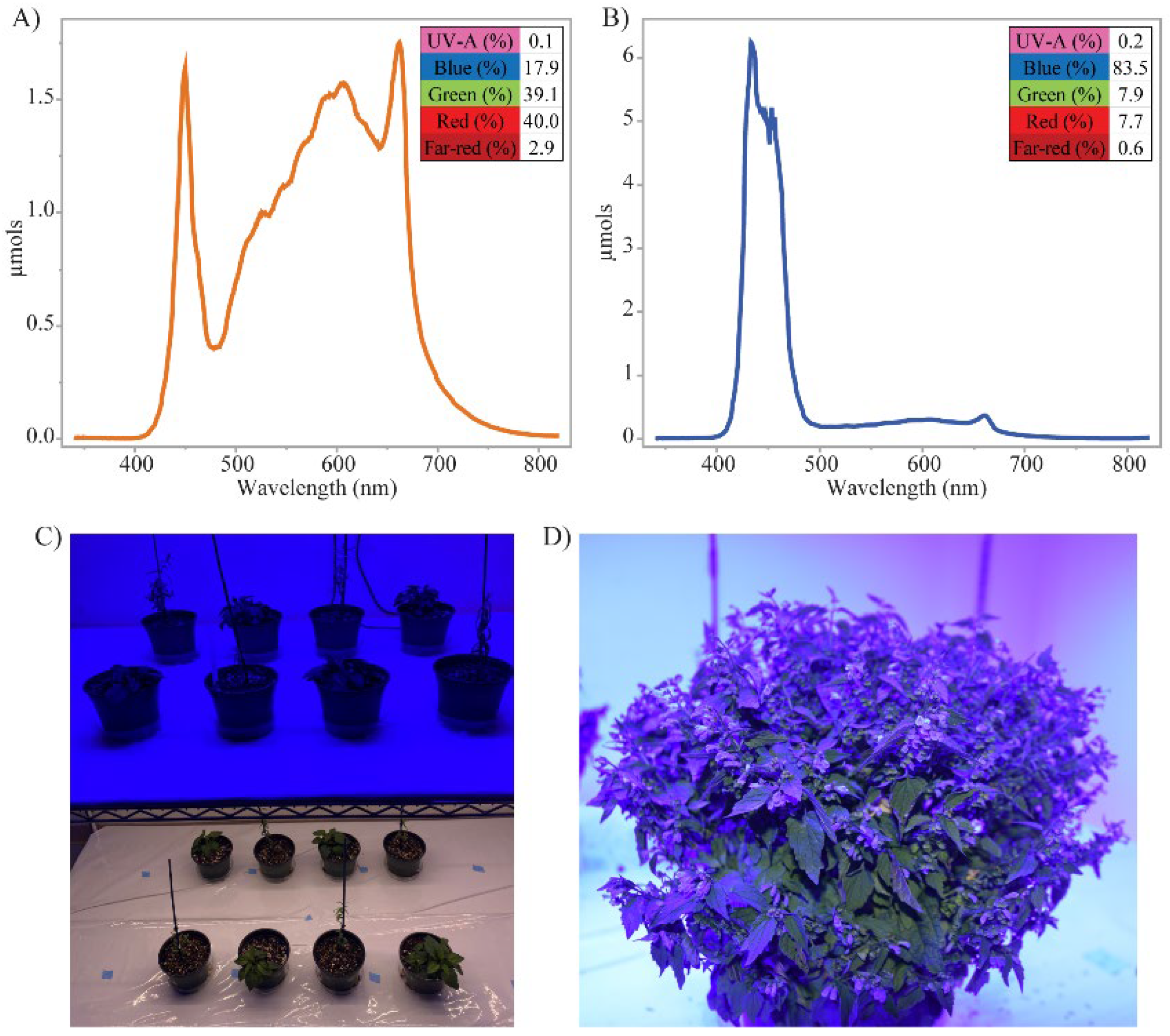
Phenylpropanoid metabolism is the principal pathway that generates a wide range of plant specialized metabolites, including flavones produced in skullcaps (Figure 1). Initial light intensity of both treatments was standardized to a DLI of 17.3 mol·m−2·d−1 (Figure 2A–D). S. baicalensis and S. lateriflora grown under blue light treatment showed significantly reduced PGI compared to the control full spectrum light treatment (Figure 3A,B). Lateral branching was not significantly different in response to blue light in either species. S. baicalensis had an average of 10.5 lateral branches for the blue treatment and 12.2 for white, while S. lateriflora averaged at 24.0 lateral branches under both conditions. The reduction in plant size with no significant variation in lateral branching is consistent with previous studies showing the ability of light qualities with a higher composition of blue light to result in more compact plants [41,42,43]. This variation in PGI is likely due to reduction in stem and petiole height, and leaf size [44,45]. Cryptochromes play a critical role in plant growth and various developmental processes, including circadian rhythms, photomorphogenesis, flowering time regulation, etc. [43,46,47,48]. The linear reduction in plant height, as observed in this study under predominant blue light, may be likely attributed to cryptochrome regulated response resulting in inhibition of hypocotyl and epicotyl elongation.
The progression of flowering under blue light was consistently faster in both species, however, more pronounced in S. lateriflora than in S. baicalensis (Figure 3C, D). Blue light has previously been reported to be important for the onset and progression of flowering [49,50,51]. One month after the experimental period, leaf curling and drying in the tips were observed in both species under both light treatments. Based on the plant diagnostic center, it was confirmed that no plant pests or diseases caused the observed symptoms. Therefore, we hypothesized that high light intensity caused these physiological disorders. Accordingly, light intensities for both light treatments were reduced to 200 µmol·m−2·s−1 for the same 16 h photoperiod. This reduction dropped total DLI from 17.28 to 11.52 mol·m−2·d−1, a 33.4% decrease in total accumulated light over 24 h. This change in light intensity resulted in improved plant health and thus these conditions were maintained for the remaining experimental period.

3.2. Blue Light Impact on Flavone Profiles

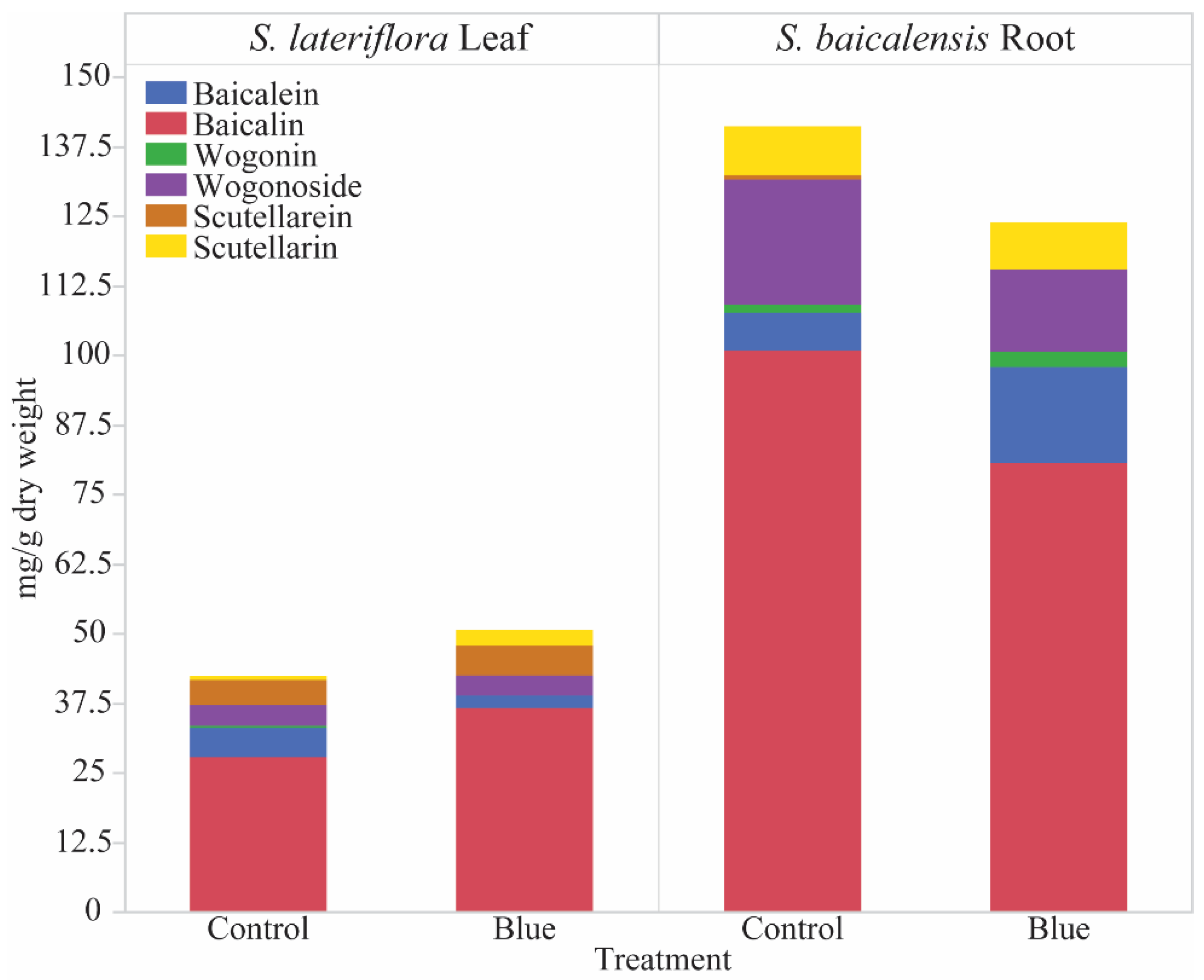
HPLC analyses of leaves of S. lateriflora and leaves and roots of S. baicalensis, the main flavone accumulating tissues in these species, revealed different responses among tissue types when exposed to blue light. Significant increases in predominant flavones were observed in S. lateriflora leaf tissue, while only a slight increase of major 4′-hydroxy flavone scutellarin was observed in S. baicalensis leaf tissue (Figure 4, Table 1). Underground roots of S. baicalensis had significantly increased 4′-deoxyflavone aglycones baicalein and wogonin by 154.4% and 76.0%, respectively, under blue light. However, concentrations of their respective flavone glycosides baicalin and wogonoside decreased by 20.1% and 33.6%, respectively (Figure 4). Since baicalin is the predominant flavone in all tissues, followed by wogonoside, the total flavone accumulation in S. baicalensis roots showed a reduction under the blue light treatment (Figure 4 and Table 1). Previous studies conducted on the impacts of blue light quality on flavone profiles in S. baicalensis used in vitro calli produced from hairy roots and two- to four-week-old seedlings [37,38]. The in vitro calli showed reductions in wogonoside and increases in baicalein and wogonin under blue light consistent with our findings from the roots of S. baicalensis with the exception of baicalin [37]. However, the two-week-old seedlings under blue light had lower concentrations of baicalin and wogonin in the root tissues with no changes in baicalein [38]. Although our study indicated a similar trend for baicalin, we found significantly higher concentrations of both baicalein and wogonin under blue light. The variation in results may be due to the difference in the plant materials used for these studies.
The blue light response is consistent in S. lateriflora, where in vitro plants had high flavone accumulation under blue light, while 4′-deoxyflavone baicalein was reduced to undetectable levels [36]. Our study obtained similar results, where baicalein was reduced by 86.6% under the blue light treatment. This decrease could potentially be associated with a 31.6% increase in downstream baicalin. A similar increase was also observed in scutellarein by 296% and its glycoside, scutellarin by 18.6% under blue light. Although there is a general trend of enhanced flavone profiles under blue light, specific differences in metabolite concentrations between different tissue types suggest that there may be developmental variation in the flavone profiles, thus warranting further investigation.
These results point to the potential stress response of skullcap plants by accumulating flavones specifically in tissues that perceive blue light to protect against oxidative stress [52]. Aerial tissues of both skullcap species had an increase in overall flavone content, while a decrease in specific 4′-deoxy flavones was observed in underground S. baicalensis, the tissues that do not perceive blue light directly (Table 1). This is concurrent to previous root calli studies in S. baicalensis, indicating overall increases in flavones when directly exposed to blue-dominated light spectra [37]. This enhanced accumulation of flavones in tissues directly exposed to blue light could have caused preferential increase in 4′-hydroxyflavones, scutellarein and scutellarin which are predominantly produced in aerial tissues of S. baicalensis. Since S. lateriflora retains the ability to produce 4′-deoxyflavones within its aerial tissues, a drastic increase in baicalin, the dominant flavone, was observed, along with 4′-hydroxyflavones (Table 1). Cryptochromes are blue or UV-A light photoreceptors in plants controlling photomorphogenesis which synthesize reactive oxygen species in response to light which are stress signaling molecules induced by various biotic and abiotic stresses in plants. Plant growth and development of these skullcap species in response to blue-dominated light spectra along with enhanced flavone profiles may be a cryptochrome mediated stress response in these plants [52,53]. Moreover, results provide foundational knowledge documenting plant response to production light environment that may help explain variability of specialized metabolite concentrations found in natural product medicines. Understanding these relationships allows for manipulation of plant production environment to enhance accumulation of flavones and influence induction of flowering, two important production factors to producers of S. baicalensis and S. lateriflora.

4. Conclusions

This study demonstrated that blue light significantly altered the accumulation of flavones in both aerial and underground tissues along with early induction of flowering and reduction in plant growth index in both Scutellaria species. Findings help establish fundamental knowledge regarding relationships between Scutellaria and its production environment and may help explain variability in flavone concentrations found in commercial natural product medicines. Controlled production systems with a light quality-based enhancement of bioactive metabolites facilitate safe and efficient means of producing plant-derived medicinal compounds. These results are expected to be applied to growing skullcaps to produce compact plants and optimize the medicinal flavones of interest to maximize plant value and application.

Author Contributions

Conceptualization, S.S.N.; methodology, S.S.N., B.C., M.Z. and B.P.; formal analysis, S.S.N., B.C., M.Z. and B.P.; investigation, S.S.N., B.C., M.Z. and B.P.; writing—original draft preparation, S.S.N. and B.C.; writing—review and editing, S.S.N., B.C., M.Z. and B.P.; visualization, B.C.; supervision, S.S.N. and B.P.; project administration, S.S.N.; funding acquisition, S.S.N. All authors have read and agreed to the published version of the manuscript.

Funding

This work is supported by Satya Swathi Nadakuduti USDA-NIFA Hatch project #006010, a startup fund from the Environmental Horticultural Department and by the Support for Emerging Enterprise Development Integration Teams (SEEDIT) program from Institute of Food and Agricultural Sciences at the University of Florida.

Data Availability Statement

The data presented in this study are available on request from the corresponding author.

Acknowledgments

We would like to thank Celina Gomez for the blue LEDs that were used in the study.

Conflicts of Interest

The authors declare no conflict of interest.

References

  1. Alexandru Suchar, V.; Robberecht, R. Integration and scaling of UV-B radiation effects on plants: The relative sensitivity of growth forms and interspecies interactions. J. Plant Ecol. 2018, 11, 656–670. [Google Scholar] [CrossRef] [Green Version]
  2. Kalaitzoglou, P.; van Ieperen, W.; Harbinson, J.; van der Meer, M.; Martinakos, S.; Weerheim, K.; Nicole, C.C.S.; Marcelis, L.F.M. Effects of continuous or end-of-day far-red light on tomato plant growth, morphology, light absorption, and fruit production. Front. Plant Sci. 2019, 10, 322. [Google Scholar] [CrossRef] [PubMed] [Green Version]
  3. Liu, J.; van Iersel, M.W. Photosynthetic Physiology of Blue, Green, and Red Light: Light Intensity Effects and Underlying Mechanisms. Front. Plant Sci. 2021, 12, 328. [Google Scholar] [CrossRef] [PubMed]
  4. McCree, K.J. The action spectrum, absorptance and quantum yield of photosynthesis in crop plants. Agric. Meteorol. 1971, 9, 191–216. [Google Scholar] [CrossRef]
  5. Inada, K. Action spectra for photosynthesis in higher plants. Plant Cell Physiol. 1976, 17, 355–365. [Google Scholar] [CrossRef]
  6. Evans, J.R. The Dependence of Quantum Yield on Wavelength and Growth Irradiance. Funct. Plant Biol. 1987, 14, 69–79. [Google Scholar] [CrossRef]
  7. Hogewoning, S.W.; Wientjes, E.; Douwstra, P.; Trouwborst, G.; van Ieperen, W.; Croce, R.; Harbinson, J. Photosynthetic Quantum Yield Dynamics: From Photosystems to Leaves. Plant Cell 2012, 24, 1921–1935. [Google Scholar] [CrossRef] [Green Version]
  8. Zhang, S.; Zhang, L.; Zou, H.; Qiu, L.; Zheng, Y.; Yang, D.; Wang, Y. Effects of Light on Secondary Metabolite Biosynthesis in Medicinal Plants. Front. Plant Sci. 2021, 12, 2892. [Google Scholar] [CrossRef]
  9. Dou, H.; Niu, G.; Gu, M.; Masabni, J.G. Effects of Light Quality on Growth and Phytonutrient Accumulation of Herbs under Controlled Environments. Horticulturae 2017, 3, 36. [Google Scholar] [CrossRef] [Green Version]
  10. Ramakrishna, A.; Ravishankar, G.A. Influence of abiotic stress signals on secondary metabolites in plants. Plant Signal. Behav. 2011, 6, 1720–1731. [Google Scholar] [CrossRef]
  11. Stevenson, P.C.; Nicolson, S.W.; Wright, G.A. Plant secondary metabolites in nectar: Impacts on pollinators and ecological functions. Funct. Ecol. 2017, 31, 65–75. [Google Scholar] [CrossRef]
  12. Zandalinas, S.I.; Sales, C.; Beltrán, J.; Gómez-Cadenas, A.; Arbona, V. Activation of Secondary Metabolism in Citrus Plants Is Associated to Sensitivity to Combined Drought and High Temperatures. Front. Plant Sci. 2017, 7, 1954. [Google Scholar] [CrossRef] [PubMed] [Green Version]
  13. Biała, W.; Jasiński, M. The phenylpropanoid case—It is transport that matters. Front. Plant Sci. 2018, 9, 1610. [Google Scholar] [CrossRef] [PubMed] [Green Version]
  14. Yang, L.; Wen, K.S.; Ruan, X.; Zhao, Y.X.; Wei, F.; Wang, Q. Response of Plant Secondary Metabolites to Environmental Factors. Molecules 2018, 23, 762. [Google Scholar] [CrossRef] [Green Version]
  15. Shalaby, S.; Horwitz, B.A. Plant phenolic compounds and oxidative stress: Integrated signals in fungal-plant interactions. Curr. Genet. 2015, 61, 347–357. [Google Scholar] [CrossRef]
  16. Martens, S.; Mithöfer, A. Flavones and flavone synthases. Phytochemistry 2005, 66, 2399–2407. [Google Scholar] [CrossRef]
  17. Lattanzio, V.; Lima, G.; Cicco, V. De Antifungal activity of phenolics against fungi commonly encountered during storage. Ital. J. Food Sci. 1994, 6, 23–30. [Google Scholar]
  18. McNally, D.J.; Wurms, K.V.; Labbé, C.; Quideau, S.; Bélanger, R.R. Complex C-glycosyl flavonoid phytoalexins from Cucumis sativus. J. Nat. Prod. 2003, 66, 1280–1283. [Google Scholar] [CrossRef]
  19. Hostetler, G.L.; Ralston, R.A.; Schwartz, S.J. Flavones: Food sources, bioavailability, metabolism, and bioactivity. Adv. Nutr. 2017, 8, 423–435. [Google Scholar] [CrossRef] [Green Version]
  20. Singh, M.; Kaur, M.; Silakari, O. Flavones: An important scaffold for medicinal chemistry. Eur. J. Med. Chem. 2014, 84, 206–239. [Google Scholar] [CrossRef]
  21. Sharma, A.; Tuli, H.S.; Kashyap, D.; Sharma, A.K. Flavones: Flavonoids Having Chemico-Biological Properties with a Preview into Anticancer Action Mechanism. In Bioactive Natural Products for the Management of Cancer: From Bench to Bedside; Springer Nature: Berlin, Germany, 2019; pp. 71–89. [Google Scholar] [CrossRef]
  22. Zhao, Q.; Zhang, Y.; Wang, G.; Hill, L.; Weng, J.-K.; Chen, X.-Y.; Xue, H.; Martin, C. A specialized flavone biosynthetic pathway has evolved in the medicinal plant, Scutellaria baicalensis. Sci. Adv. 2016, 2, e1501780. [Google Scholar] [CrossRef] [PubMed]
  23. Shen, J.; Li, P.; Liu, S.; Liu, Q.; Li, Y.; Sun, Y.; He, C.; Xiao, P. Traditional uses, ten-years research progress on phytochemistry and pharmacology, and clinical studies of the genus Scutellaria. J. Ethnopharmacol. 2021, 265, 113198. [Google Scholar] [CrossRef] [PubMed]
  24. Cathcart, M.C.; Useckaite, Z.; Drakeford, C.; Semik, V.; Lysaght, J.; Gately, K.; O’Byrne, K.J.; Pidgeon, G.P. Anti-cancer effects of baicalein in non-small cell lung cancer in-vitro and in-vivo. BMC Cancer 2016, 16, 707. [Google Scholar] [CrossRef] [PubMed] [Green Version]
  25. Tao, Y.; Zhan, S.; Wang, Y.; Zhou, G.; Liang, H.; Chen, X.; Shen, H. Baicalin, the major component of traditional Chinese medicine Scutellaria baicalensis induces colon cancer cell apoptosis through inhibition of oncomiRNAs. Sci. Rep. 2018, 8, 14477. [Google Scholar] [CrossRef] [Green Version]
  26. Zhang, Z.; Lian, X.Y.; Li, S.; Stringer, J.L. Characterization of chemical ingredients and anticonvulsant activity of American skullcap (Scutellaria lateriflora). Phytomedicine 2009, 16, 485–493. [Google Scholar] [CrossRef]
  27. Millspaugh, C.F. American Medicinal Plants Charles F Millspaugh; Dover Publications: New York, NY, USA, 1974; ISBN 0486230341. [Google Scholar]
  28. Upton, R.; Dayu, R.H. Skullcap Scutellaria lateriflora L.: An American nervine. J. Herb. Med. 2012, 2, 76–96. [Google Scholar] [CrossRef]
  29. Cole, I.B.; Alan, A.R.; Saxena, P.K.; Murch, S.; Cao, J.; Murch, S.J. Comparisons of Scutellaria baicalensis, Scutellaria lateriflora and Scutellaria racemosa: Genome Size, Antioxidant Potential and Phytochemistry. Planta Med. 2008, 74, 474–481. [Google Scholar] [CrossRef]
  30. Costine, B.; Zhang, M.Z.; Chhajed, S.; Pearson, B.; Chen, S.X.; Nadakuduti, S.S. Exploring native Scutellaria species provides insight into differential accumulation of flavones with medicinal properties. Sci. Rep. 2022, 12, 13201. [Google Scholar] [CrossRef]
  31. Liu, Y.; Fang, S.; Yang, W.; Shang, X.; Fu, X. Light quality affects flavonoid production and related gene expression in Cyclocarya paliurus. J. Photochem. Photobiol. B Biol. 2018, 179, 66–73. [Google Scholar] [CrossRef]
  32. Zhang, L.; Wang, G.; Wang, G.; Cao, F. Ginkgo biloba l. Responds to red and blue light: Via phenylpropanoid and flavonoid biosynthesis pathway. Forests 2021, 12, 1079. [Google Scholar] [CrossRef]
  33. Taulavuori, K.; Pyysalo, A.; Taulavuori, E.; Julkunen-Tiitto, R. Responses of phenolic acid and flavonoid synthesis to blue and blue-violet light depends on plant species. Environ. Exp. Bot. 2018, 150, 183–187. [Google Scholar] [CrossRef] [Green Version]
  34. Liu, H.K.; Chen, Y.Y.; Hu, T.T.; Zhang, S.J.; Zhang, Y.H.; Zhao, T.Y.; Yu, H.E.; Kang, Y.F. The influence of light-emitting diodes on the phenolic compounds and antioxidant activities in pea sprouts. J. Funct. Foods 2016, 25, 459–465. [Google Scholar] [CrossRef]
  35. Ying, Q.; Jones-Baumgardt, C.; Zheng, Y.; Bozzo, G. The Proportion of Blue Light from Light-emitting Diodes Alters Microgreen Phytochemical Profiles in a Species-specific Manner. HortScience 2021, 56, 13–20. [Google Scholar] [CrossRef]
  36. Kawka, B.; Kwiecień, I.; Ekiert, H. Influence of Culture Medium Composition and Light Conditions on the Accumulation of Bioactive Compounds in Shoot Cultures of Scutellaria lateriflora L. (American Skullcap) Grown In Vitro. Appl. Biochem. Biotechnol. 2017, 183, 1414–1425. [Google Scholar] [CrossRef] [Green Version]
  37. Stepanova, A.; Solov’yova, A.; Salamaikina, S. Influence of spectral light composition on flavones formation in callus culture of Scutellaria baicalensis georgi. Pharmacogn. Mag. 2020, 16, 156. [Google Scholar] [CrossRef]
  38. Yeo, H.J.; Park, C.H.; Park, S.Y.; Chung, S.O.; Kim, J.K.; Park, S.U. Metabolic analysis of root, stem, and leaf of scutellaria baicalensis plantlets treated with different led lights. Plants 2021, 10, 940. [Google Scholar] [CrossRef]
  39. Wilson, S.B.; Steppe, C.; Deng, Z.; Druffel, K.; Knox, G.W.; van Santen, E. Landscape Performance, Flowering, and Female Fertility of Eight Trailing Lantana Varieties Grown in Central and Northern Florida. HortScience 2020, 55, 1737–1743. [Google Scholar] [CrossRef]
  40. Askey, B.C.; Liu, D.; Rubin, G.M.; Kunik, A.R.; Song, Y.H.; Ding, Y.; Kim, J. Metabolite profiling reveals organ-specific flavone accumulation in Scutellaria and identifies a scutellarin isomer isoscutellarein 8-O-β-glucuronopyranoside. Plant Direct 2021, 5, e372. [Google Scholar] [CrossRef]
  41. Wollaeger, H.M.; Runkle, E.S. Growth of Impatiens, Petunia, Salvia, and Tomato Seedlings under Blue, Green, and Red Light-emitting Diodes. HortScience 2014, 49, 734–740. [Google Scholar] [CrossRef]
  42. Cosgrove, D.J. Rapid Suppression of Growth by Blue Light. Plant Physiol. 1981, 67, 584–590. [Google Scholar] [CrossRef]
  43. Kalaitzoglou, P.; Taylor, C.; Calders, K.; Hogervorst, M.; van Ieperen, W.; Harbinson, J.; de Visser, P.; Nicole, C.C.S.; Marcelis, L.F.M. Unraveling the effects of blue light in an artificial solar background light on growth of tomato plants. Environ. Exp. Bot. 2021, 184, 104377. [Google Scholar] [CrossRef]
  44. Kang, W.H.; Park, J.S.; Park, K.S.; Son, J.E. Leaf photosynthetic rate, growth, and morphology of lettuce under different fractions of red, blue, and green light from light-emitting diodes (LEDs). Hortic. Environ. Biotechnol. 2016, 57, 573–579. [Google Scholar] [CrossRef]
  45. Mu, R.; Lyu, X.; Ji, R.; Liu, J.; Zhao, T.; Li, H.; Liu, B. GmBICs Modulate Low Blue Light-Induced Stem Elongation in Soybean. Front. Plant Sci. 2022, 13, 803122. [Google Scholar] [CrossRef] [PubMed]
  46. Fraser, D.P.; Hayes, S.; Franklin, K.A. Photoreceptor crosstalk in shade avoidance. Curr. Opin. Plant Biol. 2016, 33, 1–7. [Google Scholar] [CrossRef] [Green Version]
  47. Pooam, M.; Arthaut, L.D.; Burdick, D.; Link, J.; Martino, C.F.; Ahmad, M. Magnetic sensitivity mediated by the Arabidopsis blue-light receptor cryptochrome occurs during flavin reoxidation in the dark. Planta 2019, 249, 319–332. [Google Scholar] [CrossRef] [Green Version]
  48. Kharshiing, E.V.; Ibapalei, O.; Mawphlang, L.; Lama, V.; Bhattacharjee, R.; Sahoo, L. Manipulation of light environment for optimising photoreceptor activity towards enhancing plant traits of agronomic and horticultural importance in crops. J. Hortic. Sci. Biotechnol. 2022, 97, 535–551. [Google Scholar] [CrossRef]
  49. Eskins, K. Light-quality effects on Arabidopsis development. Red, blue and far-red regulation of flowering and morphology. Physiol. Plant. 1992, 86, 439–444. [Google Scholar] [CrossRef]
  50. Shibuya, T.; Takahashi, T.; Hashimoto, S.; Nishiyama, M.; Kanayama, Y. Effects of overnight radiation with monochromatic far-red and blue light on flower budding and expression of flowering-related and light quality-responsive genes in Eustoma grandiflorum. J. Agric. Meteorol. 2019, 75, 160–165. [Google Scholar] [CrossRef] [Green Version]
  51. Park, Y.G.; Jeong, B.R. How Supplementary or Night-Interrupting Low-Intensity Blue Light Affects the Flower Induction in Chrysanthemum, a Qualitative Short-Day Plant. Plants 2020, 9, 1694. [Google Scholar] [CrossRef]
  52. El-Esawi, M.; Arthaut, L.D.; Jourdan, N.; D’Harlingue, A.; Link, J.; Martino, C.F.; Ahmad, M. Blue-light induced biosynthesis of ROS contributes to the signaling mechanism of Arabidopsis cryptochrome. Sci. Rep. 2017, 7, 13875. [Google Scholar] [CrossRef]
  53. Consentino, L.; Lambert, S.; Martino, C.; Jourdan, N.; Bouchet, P.E.; Witczak, J.; Castello, P.; El-Esawi, M.; Corbineau, F.; d’Harlingue, A.; et al. Blue-light dependent reactive oxygen species formation by Arabidopsis cryptochrome may define a novel evolutionarily conserved signaling mechanism. New Phytol. 2015, 206, 1450–1462. [Google Scholar] [CrossRef] [PubMed]
Figure 1. Flavone biosynthetic pathway in Scutellaria baicalensis based on (Zhao et al., 2016). Underlined flavones have been quantified by HPLC in this study. Flavones encircled in green (left) indicate 4′-hydroxyflavones of interest. Flavones encircled in yellow (right) indicate 4′-deoxyflavones of interest. Listed enzymes are phenylalanine ammonia lyase (PAL), cinnamate 4-hydroxylase (C4H), cinnamate-coA ligase (4CL 1-2), chalcone synthase (CHS 1-2), chalcone isomerase (CHI), flavone synthase II (FNSII 1-2), flavone 6-hydroxylase (F6H), flavone 8-hydroxylase (F8H), 8-O-methyl transferase (8-OMT), and UDP-glucuronosyl transferase (UGT).
Horticulturae 08 01141 g001
Figure 2. Spectral distribution and percentage wavelength measured at total photosynthetic photon flux density (PPFD) of 300 μmol m−2 s−1. Each lighting treatment utilized a 16 h photoperiod. (A) Full spectrum control (B) Blue light treatment is defined as blue light fraction of PPFD to be 83.5% supplemented with other wavelengths as indicated. (C) Light treatments implemented. Top: Blue light treatment, Bottom: Full spectrum control. (D) S. lateriflora plant under blue light treatment. Spectra are categorized as: UV-A (315–400 nm), blue (400–500 nm), green (500–600 nm), red (600–700 nm) and far-red (700–800 nm). Both light treatments have same PPFD and photoperiod.
Horticulturae 08 01141 g002
Figure 3. Blue light quality impacts plant growth and flowering. (A,B) Plant growth indices (PGI) of S. lateriflora (A), and S. baicalensis (B). (C,D) Flower scale ratings of S. lateriflora (C), and S. baicalensis (D). Relative flowering on a scale of 1–5 (1 = No flowering; 2 = A few flower buds present; 3 = A few flowers open; 4 = mostly open flowers; 5 = almost all open flowers (peak)). Data presented as means (S. lateriflora n = 12, S. baicalensis n = 11) ± standard error of the mean. Means with * are significantly different (Student’s t test, p ≤ 0.05).
Horticulturae 08 01141 g003
Figure 4. Blue light quality impacts flavone accumulation in primary localization tissues of Scutellaria species. Data presented as means of flavone concentrations (S. lateriflora n = 12, S. baicalensis n = 11).
Horticulturae 08 01141 g004
Table 1. Quantification of 4′-deoxy and 4′-hydroxy flavones under blue light treatment.
S. baicalensis LeavesS. baicalensis RootsS. lateriflora Leaves
FlavoneControlBlueControlBlueControlBlue
WogonosideNDND22.38 ± 4.0214.86 ± 2.043.71 ± 0.23.62 ± 0.4
WogoninNDND1.54 ± 0.32.71 ± 0.3 *0.35 ± 0.04ND
Baicalin1.19 ± 0.41.08 ± 0.3100.77 ± 6.780.54 ± 10.227.81 ± 1.736.6 ± 2.03 *
BaicaleinNDND6.8 ± 1.317.3 ± 2.5 *5.29 ± 0.8 *2.29 ± 0.5
ChrysinNDNDND0.1 ± 0.010.11 ± 0.020.07 ± 0.01
Scutellarin15.63 ± 2.621.66 ± 4.08.71 ± 0.68.41 ± 0.70.72 ± 0.22.85 ± 0.2 *
Scutellarein1.38 ± 0.40.83 ± 0.020.85 ± 0.02ND4.5 ± 0.25.34 ± 0.2 *
ApigeninNDNDNDND0.41 ± 0.070.43 ± 0.05
Data are presented as means ± standard errors reported in mg·g−1 of dry weight. * Indicates values that are significantly different (Students t test, p ≤ 0.05). Statistical analysis was performed for each metabolite within species and tissue types. ND = not detected as levels are below detectable thresholds.
Publisher’s Note: MDPI stays neutral with regard to jurisdictional claims in published maps and institutional affiliations.