Article

Partial Elimination of Viruses from Traditional Potato Cultivar ‘Brinjak’ by Chemotherapy and Its Impact on Physiology and Yield Components

by
Snježana Kereša
1,*,
Darko Vončina
2,
Boris Lazarević
3,
Anita Bošnjak Mihovilović
1,
Milan Pospišil
4,
Marina Brčić
4,
Ana Matković Stanković
1 and
Ivanka Habuš Jerčić
1
1
Department of Plant Breeding, Genetics and Biometrics, Faculty of Agriculture, University of Zagreb, Svetošimunska cesta 25, 10000 Zagreb, Croatia
2
Department of Plant Pathology, Faculty of Agriculture, University of Zagreb, Svetošimunska cesta 25, 10000 Zagreb, Croatia
3
Department of Plant Nutrition, Faculty of Agriculture, University of Zagreb, Svetošimunska cesta 25, 10000 Zagreb, Croatia
4
Department of Field Crops, Forage and Grassland, Faculty of Agriculture, University of Zagreb, Svetošimunska cesta 25, 10000 Zagreb, Croatia
*
Author to whom correspondence should be addressed.
Horticulturae 2022, 8(11), 1013; https://doi.org/10.3390/horticulturae8111013
Submission received: 29 September 2022 / Revised: 27 October 2022 / Accepted: 28 October 2022 / Published: 1 November 2022
(This article belongs to the Special Issue In Vitro Technology and Micropropagated Plants)

Abstract

:
Viruses are responsible for more than 50% of annual potato tuber yield losses and cause great economic damage. The traditional Croatian potato cultivar ‘Brinjak’ is important for local growers because of its economically profitable production and as a gene pool for future breeding programs. However, the full genetic potential of the cultivar cannot be exploited due to virus infection. In this study, we attempted to eliminate potato virus M (PVM) and potato virus S (PVS) from potato cultivar ‘Brinjak’ and to evaluate the effects on physiological parameters and yield. Shoot apices were isolated from PVM + PVS-infected sprouts and cultivated for six weeks on MS medium with the addition of 50 or 100 mg L−1 ribavirin. The surviving shoot apices were micropropagated. The in vitro post-eradication period lasted 200 days. DAS-ELISA and RT-PCR were performed on R0 and R1 plants 90 days after acclimatization to determine the sanitary status of the plants. Chlorophyll fluorescence and multispectral imaging were performed on the R0 plants at the same time. The success of PVS elimination was 33% at both ribavirin concentrations. However, neither concentration was successful in eliminating PVM. Plants with mixed infection (PVM + PVS) had more severe disease symptoms compared to PVM-infected plants, affecting photochemistry and multispectral parameters and, consequently, yield. PVM + PVS plants had significantly lower number and weight of tubers per plant and lower average tuber weight than plants with single PVM infection in most of the generations studied. The results indicate a strong negative impact of PVS in mixed infections with PVM and show the importance of its elimination from potato plants.

1. Introduction

Potato (Solanum tuberosum L.) is one of the five most important crops in the world today and is currently grown on over 20.7 million hectares across all continents with an estimated global production of 437 million tons [1]. In 2020, potato production in Croatia amounted to 174,280 tons of tubers from approximately 9330 ha [2]. Commercial production is entirely based on a limited number of foreign varieties that have replaced traditional cultivars/ecotypes. The intensive substitution of traditional by modern high-yielding varieties in agricultural production during the last century resulted in some erosion of genes [3], as the traditional cultivars are a source of genetic variability for many traits [4]. This is also the case with the traditional cultivar ‘Brinjak’, which is resistant to Phytophthora infestans and Streptomyces scabies, according to reports from local producers in Lika (a region in Croatia), where the cultivar ‘Brinjak’ has been grown for many generations. Therefore, traditional varieties are of great importance for biodiversity conservation, as a gene pool for future plant breeding programs [5], but also for economically profitable production once the variety is included in the List of Varieties of the Republic of Croatia as a conservation variety. It is expected that the cultivar 'Brinjak' will soon be included in that list, which will enable its commercial production and sale in the Republic of Croatia. Viruses are important agricultural pathogens with an estimated economic loss of >USD 30 billion annually [6] and are responsible for more than 50% of yield losses of potato tubers [7]. While there are no effective measures for controlling viruses and curing plants once they are infected [8,9], virus-free plants are required for commercial potato production and maintenance of potato germplasm [10]. Commercially important potato viruses cause a reduction in the starch content of tubers [11] and other biochemical and physiological changes manifested by a reduction in dry matter and vitamin content, starch granules, a decrease in amylase content, and starch acidity [11,12]. In addition to poor vegetative growth (stunting, dwarfing) and significant yield losses in tubers, viruses in potato plants can cause symptoms such as leaf curling, mosaic, leaf drop, chlorosis, necrosis, reduced product quality, undersized tubers, and sometimes death of the entire plant [11,12,13,14,15]. Potato leafroll virus (PLRV), potato virus A, M, S, X, and Y (PVA, PVM, PVS, PVX, and PVY) are usually the most important viral pathogens associated with significant economic impact and production loss [9,15,16]. PVM and PVS can be transmitted by aphids, especially the green peach aphid (Myzus persicae), by vegetative propagation (via tubers), or mechanically (e.g., through contaminated tools and wounds) [17,18]. Plants infected with PVM and PVS do not always show symptoms, depending on the cultivar and virus isolates [9,15,19]. However, PVM can reduce tuber yield by 11–45% [11,20], and PVS by 10–20% [21,22]. Mixed infection results in a yield reduction of 20–30% [22]. Symptoms of virus infections leading to changes in plant morphology, anatomy, physiology, and biochemistry can be quantified using spectral reflectance analysis. Since plant diseases interact with their host and lead to disruption of metabolic processes and development of specific symptoms, various optical sensors have been used for non-destructive diagnosis and detection of plant diseases [23,24]. Virus infection can reduce the photosynthetic efficiency of potato by decreasing the assimilation area, photosynthetic rate, ribulose-1,5-diphosphate carboxylase activity, chlorophyll and xanthophyll cycle pigment content, and PSII concentration and activity [25,26]. The sensitivity of PSII to biotic factors has made chlorophyll fluorescence one of the most widely used techniques in the study of plant disease interactions [27,28]. In addition, visible and multispectral imaging can assess the spectral information of infected leaves and can be used to calculate various vegetation indices [23,24,29].
The production of certified seed potatoes always starts with the micropropagation of virus-free stock plants, followed by the production of minitubers, and then tubers through several generations until the production of certified seed potatoes supplied to potato growers for commercial production [16]. In this process, potato growers receive healthy seed potatoes, or seed potatoes infected with viruses at a very low percentage, which is permissible. Various in vitro virus eradication methods such as meristem tip culture, thermotherapy, chemotherapy, cryotherapy, electrotherapy, and somatic embryogenesis [30,31,32,33,34] have been successfully used in different species. The efficiency of each method depends on the virus to be eliminated, the size of the explant, the duration of treatment, and also the variety [9,35,36]. Successful eradication of viruses from infected potato plants through chemotherapy or in combination with other methods, and the efficiency of ribavirin are well documented in the literature [9,10,32,35,36,37,38,39,40,41,42,43,44,45,46,47]. Ribavirin is a synthetic purine nucleoside analog with a structure closely related to guanosine [48]. It is a broad-spectrum antiviral nucleoside that is active against a variety of RNA and DNA viruses. Several possible mechanisms have been proposed for the antiviral activity of ribavirin, including the possibility that ribavirin negatively affects the synthesis of the RNA cap structure of viral RNA transcripts [49]. Exogenous applications of the antivirus agent ribavirin in chemotherapy inhibits viral RNA synthesis [35,50,51].
The aim of this study was to eliminate potato viruses M (PVM) and S (PVS) in the traditional potato cultivar ‘Brinjak’ by applying ribavirin in a culture medium and to quantify the effects of single (PVM) and mixed (PVM + PVS) infection on potato photochemical and multispectral parameters and yield.

2. Materials and Methods

2.1. Plant Material

The tubers of the traditional potato cultivar ‘Brinjak’ were obtained from the field collection of the University of Zagreb, Faculty of Agriculture, which is maintained within the framework of the Croatian National Program for Conservation and Sustainable Use of Plant Genetic Resources Important for Food and Agriculture. Plants of the cultivar ‘Brinjak’ are short, upright, stem-type, and with late maturity. The plants produce mostly smaller tubers that are round-shaped with a light beige color skin. Flesh has a medium yellow color. At maturity, the tubers have a high dry matter content.

2.2. Culture Establishment on Media Containing Ribavirin and Micropropagation

The tubers of the two mother potato plants of the cultivar ‘Brinjak‘ were kept in the dark at a temperature of approximately 18 °C for five months. The 2–6 cm long sprouts grown on the tubers were used as the starting material for setting up the experiments. The sprouts were washed under running tap water for 20 minutes and then disinfected in 70% ethanol for 1 minute. They were then shaken for 15 minutes in a 4% solution of sodium dichloroisocyanurate dihydrate (Izosan® G, Pliva Hrvatska, Zagreb, Croatia) supplemented with 3 drops 100 mL−1 of surfactant Tween-20. The sprouts were then washed three times for 3 min in sterile distilled water.
Shoot apices > 0.5–1 mm in size were aseptically isolated from the terminal and lateral buds of the elongated sprouts under a stereomicroscope. Shoot apices were placed in 12-cm-high tubes on the surface of 10 mL of the Murashige and Skoog medium [52] containing vitamins (Duchefa Biochemie, Haarlem, The Netherlands) supplemented with 0.04 mg L−1 Kinetin (Kin), 0.5 mg L−1 Gibberellic acid (GA), 3% sucrose, 0.8% BD bacto agar (Becton, Dickinson and Company, NJ, USA), and 50 or 100 mg L−1 ribavirin (Duchefa Biochemie, Haarlem, The Netherlands). Ribavirin was prepared as a stock solution of 50 mg mL−1, filter-sterilized, and added to the autoclaved medium before solidification. The pH of the medium was adjusted to 5.8. For each ribavirin treatment (50 or 100 mg L−1), 24 shoot apices were placed. After 6 weeks, the number of surviving shoot apices was determined, and the live shoot apices were subcultured on hormone-free MS medium (HFMS), which was the same medium used to establish the culture, except that it did not contain plant growth regulators and ribavirin. Shoots formed from shoot apices were cut into single-nodal segments without leaves and subcultured in Magenta boxes on the same medium in several cycles until the production of a sufficient number of shoots from the surviving explants. Therefore, each surviving shoot apex was micropropagated to produce a clone. Post-eradication period in vitro lasted 200 days, and shoots that passed this stage were used for all subsequent steps. Culture establishment and micropropagation were performed in a growth chamber at 23 °C with a 16 h photoperiod of cool white light (40 μmol m−2 s−1).

2.3. Microtuberization

Twenty single-nodal segments with a leaf of each micropropagated potato clone were placed vertically on the medium for microtuberization in two Sterivent high containers (Duchefa Biochemie, The Netherlands). The medium for microtuberization consisted of MS medium with vitamins (Duchefa Biochemie, Haarlem, The Netherlands) supplemented with 4 mg L−1 Kin, 1 mg L−1 6-benzylaminopurine (BAP), 8% sucrose, 0.7% plant agar (Duchefa Biochemie, Haarlem, The Netherlands). The pH was adjusted to 5.8. Explants were grown for 20 days at a photoperiod of 16/8 day/night with cool white light (40 μmol m−2 s−1) at a temperature of 22 °C. Then, they were moved to short-day conditions, i.e., a photoperiod of 8/16 day/night at the same light intensity and temperature, and grown for four months. To determine the effect of viruses on microtuberization, the percentage of shoots with microtubers, the number of microtubers, and the weight of microtubers were determined.

2.4. Acclimatization and Minituber Production of R0 Plants

Three-week-old rooted shoots were planted in sterile Kekkila TSM 2 substrate (Kekkilä Professional, Vantaa, Finland) in small pots (3 cm × 3 cm × 7 cm) and covered by a transparent plastic cover for 7 days for acclimatization. After 10 days, the plants were transplanted into larger pots (10 cm × 10 cm × 20 cm) containing sterile Kekkila TSM 3 substrate, in which they were grown to maturity. The number of transplanted plants per clone varied from 6 to 10. The substrate was sterilized by gamma irradiation (Ruđer Bošković Institute, Zagreb, Croatia). Irradiation was performed with a 60Co gamma ray source and an absorbed dose of 50 kGy. R0 plants were grown for 150 days in a growth chamber at a photoperiod of 16/8 day/night of LED tubes (Valoya L35 NS12) and a light intensity of 300 μmol m−2 s−1 at a temperature of 22 °C. Plants were watered twice a week and fertilized once a month with organic liquid fertilizer “Organomex 6-2-4” (Omex Agrifluids Limited, King’s Lynn, UK).

2.5. Production of R1 Clones from Microtubers and Minitubers

Three and a half months after microtuberization and storage at room temperature, two microtubers from each clone were planted in plastic pots (10 cm × 10 cm × 20 cm) in sterile Kekkila TSM 3 substrate, in which they were grown to maturity. Similarly, minitubers, the largest from each R0 plant, were planted in the same way. The plants were grown to maturity under the same conditions as the R0 clones. At harvest, the number and weight of tubers per plant were determined for all three generations of plants.

2.6. Verification of Virus Status by DAS-ELISA and RT-PCR

To gain insight into the virus sanitary status of the mother plants and, later, their progeny, DAS-ELISA was performed on six different viruses: PLRV, PVA, PVM, PVS, PVX, and PVY. Four randomly selected plants per clone in the R0 generation, one plant per clone in the R1 generation produced from microtubers, and two randomly selected plants per clone in the R1 generation produced from minitubers were analyzed. Lower leaves from different sides of each plant studied were collected 90 days after acclimatization. The collected leaves from each plant were mixed and used to prepare an average sample of 0.1 g, which served as a potential antigen source. Commercially available ELISA kits from Bioreba (Reinach, Switzerland) were used for virus detection according to the manufacturer’s instructions. Viruses detected by DAS-ELISA in the mother plants that were not detected by the same method in the progeny (R0 micropropagated plants, R1 from microtubers, and R1 from minitubers) were verified by reverse transcription-polymerase chain reaction (RT-PCR). For this purpose, RNA was isolated from leaf samples collected 90 days after acclimatization using the RNeasy Plant Mini Kit (Qiagen, Hilden, Germany). Leaves taken from different sides of each plant were mixed into an average sample consisted of 0.1 g of plant tissue that was minced in MiniG 1600, SpexSample Prep (Metuchen, NJ, USA) using grinding balls. The quality and quantity of the isolated RNA were checked spectrophotometrically (A260/A280) using a NanoPhotometer P330 (Implen, Munich, Germany). One-step RT-PCR reactions were performed in the Mastercycler (Eppendorf, Hamburg, Germany) under the following conditions: reverse transcription at 50 °C for 30 min, initial activation step at 95 °C for 15 min, followed by 35 cycles of denaturation at 94 °C for 30 s, annealing at 53 °C for 30 s, and elongation at 72 °C for 30 s. The final elongation step was performed at 72 °C for 10 min. For virus detection, the primers described in [7] were combined with the one-step RT-PCR kit (Qiagen, Germany) in a reaction volume of 10 µL consisting of: 0.5 µL of each primer (10 µM), 0.4 µL dNTP mix (10 µM), 2 µL Qiagen OneStep RT-PCR enzyme mix, 2 µL Qiagen OneStep RT-PCR buffer, 2 µL Q-Solution, 3.2 µL RNase-free water, and 1 µL template RNA. To visualize the RT-PCR products, a 1.5% agarose gel was prepared in a 1X TBE buffer with the addition of GelRed (CareDx AB, Stockholm, Sweden) and subjected to horizontal gel electrophoresis (Bio-Rad, Hercules, CA, USA).

2.7. Chlorophyll Fluorescence and Multispectral Analysis

Chlorophyll fluorescence and multispectral imaging were conducted on all R0 plants 90 days after planting using CropReporterTM (PhenoVation B.V., Wageningen, The Netherlands), while image processing and analysis were performed using DA software (PhenoVation B.V., Wageningen, The Netherlands). Plants were imaged from a distance of 70 cm, and measurements were made according to the protocol described in [53]. For the chlorophyll fluorescence imaging, plants were first dark-adapted for 30 min. On dark-adapted plants, photosynthesis was excited using 4000 μmol m−2 s−1 red LED light flash. Minimum chlorophyll fluorescence (F0) was measured after ten (10) µs and maximum chlorophyll fluorescence (Fm) after 800 ms. Following these measurements, plants were relaxed in the dark for 15 s and then were light-adapted for 5 min using actinic light of 250 μmol m−2 s−1. Saturating pulse (4000 μmol m−2 s−1) was again applied for photosynthetic excitation of the light-adapted plants. The steady-state fluorescence yield (Fs′) was measured before the onset of the saturation pulse, and the maximum chlorophyll fluorescence (Fm′) of the light-adapted leaves was measured at saturation. After the measurement, the actinic light was turned off, and in the presence of far-red light, the minimal fluorescence yield of the illuminated plant (F0′) was estimated. These measured parameters were used for the calculation of different chlorophyll fluorescence parameters, which are shown in Table 1.
Following the chlorophyll fluorescence imaging, actinic light 250 μmol m−2 s−1 was again switched on and multispectral imaging was performed. A list of all measured and multispectral parameters and calculated vegetation indices are given in Table 2.

2.8. Data Analysis

All experiments were set up in a completely randomized design. Data from all measurements were subjected to ANOVA. Bonferroni post hoc test at p ≤ 0.05 was used for means comparison. The statistical analysis of the data was carried out by the SAS/STAT® [60] program package.

3. Results and Discussion

3.1. Culture Establishment and Micropropagation

Out of 24 shoot apices placed on medium supplemented with 50 mg L−1 of ribavirin, 6 (25%) survived, whereas only 3 (12.5%) survived on medium supplemented with 100 mg L−1 of ribavirin. There was no contamination. The cause of the low percentage of surviving shoot apices may be ribavirin, but also their physiological condition, because the sprouts for shoot apices isolation were used after the tubers had been kept in the dark for five months. Most of the surviving explants (Figure 1a,b) grew very slowly or were even inhibited in growth 100 days after placement in in vitro culture, i.e., two months after subcultivation on HFMS medium without ribavirin (Figure 1c,d). The strength of ribavirin treatment in the previous medium did not affect inhibition; surviving shoots derived from both ribavirin concentrations, whether 50 or 100 mg L−1, were equally inhibited. Phytotoxicity of ribavirin to potato regeneration and plantlet growth was also reported by [44], who found that plant height and fresh weight generally showed a decreasing tendency with increasing ribavirin concentration in a culture medium. Singh [61] reported that the percentage of shoot regeneration and plantlet development decreased when the concentration of antiviral chemicals, including ribavirin, was increased from 5 to 30 mg L−1.
Eight months after the start of the experiment, all nine surviving shoot apices that developed into shoots were micropropagated, but with different efficiency. Some shoots were still partially growth-inhibited and micropropagation resulted in a small number of plants per clone, while other shoots grew well after initial inhibition (Figure 1e) and were micropropagated into a large number of plants by single node culture. The slow growth of individual shoots that we observed according to ELISA results not only did not depend on ribavirin concentration (50 or 100 mg L−1), but also did not depend on virus infection (single or mixed). Micropropagated plants rooted spontaneously on HFMS medium in a very high percentage, and no special step was required for rooting.

3.2. The Influence of The Sanitary Status of Plants on Microtuberization

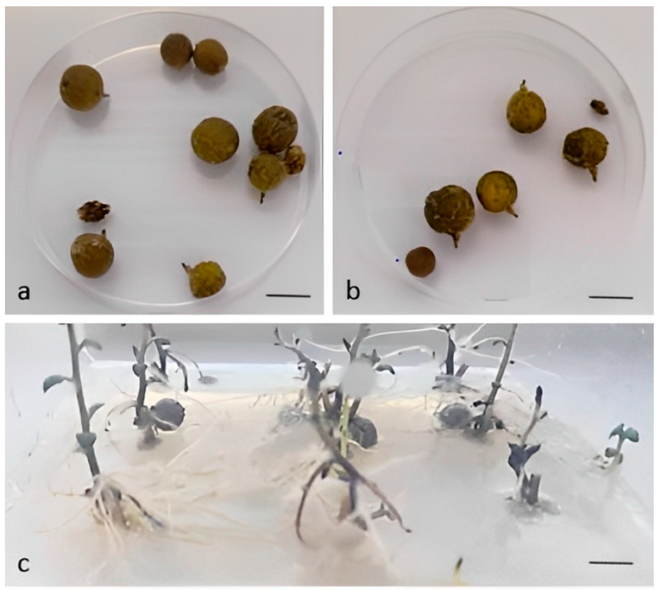
The sanitary status of the plants affected the number of microtubers per plant, which was higher on average in plants infected with PVM only (Table 3, Figure 2a,b). The data show that mixed infection (PVM + PVS) reduced the number of microtubers. Bettoni et al. [9] reported a significantly lower number of microtubers in plants infected with PVS compared to virus-free plants in the cultivar ‘Duncle’, but mixed infection with PVS and PVM in other cultivars or PVS and PVA did not affect the number of microtubers. We could not assess the effect of PVM on microtuberization or mixed infections with PVM + PVS compared to healthy plants because none of the plants were virus-free. The percentage of plants that formed microtubers under short-day conditions (Figure 2c) was similar and was 82% in plants infected with both viruses and 78% in plants infected with PVM only. The average microtuber weight was not significantly different between PVM- and PVM- + PVS-infected plants (Table 3). This is in agreement with the results of Zhang et al. [10], who found that infection with viruses had little effect on microtuber production compared to healthy plants, except in the case of triple infection with PVX, PVS, and PVY viruses.

3.3. Acclimatization and Plant Growth

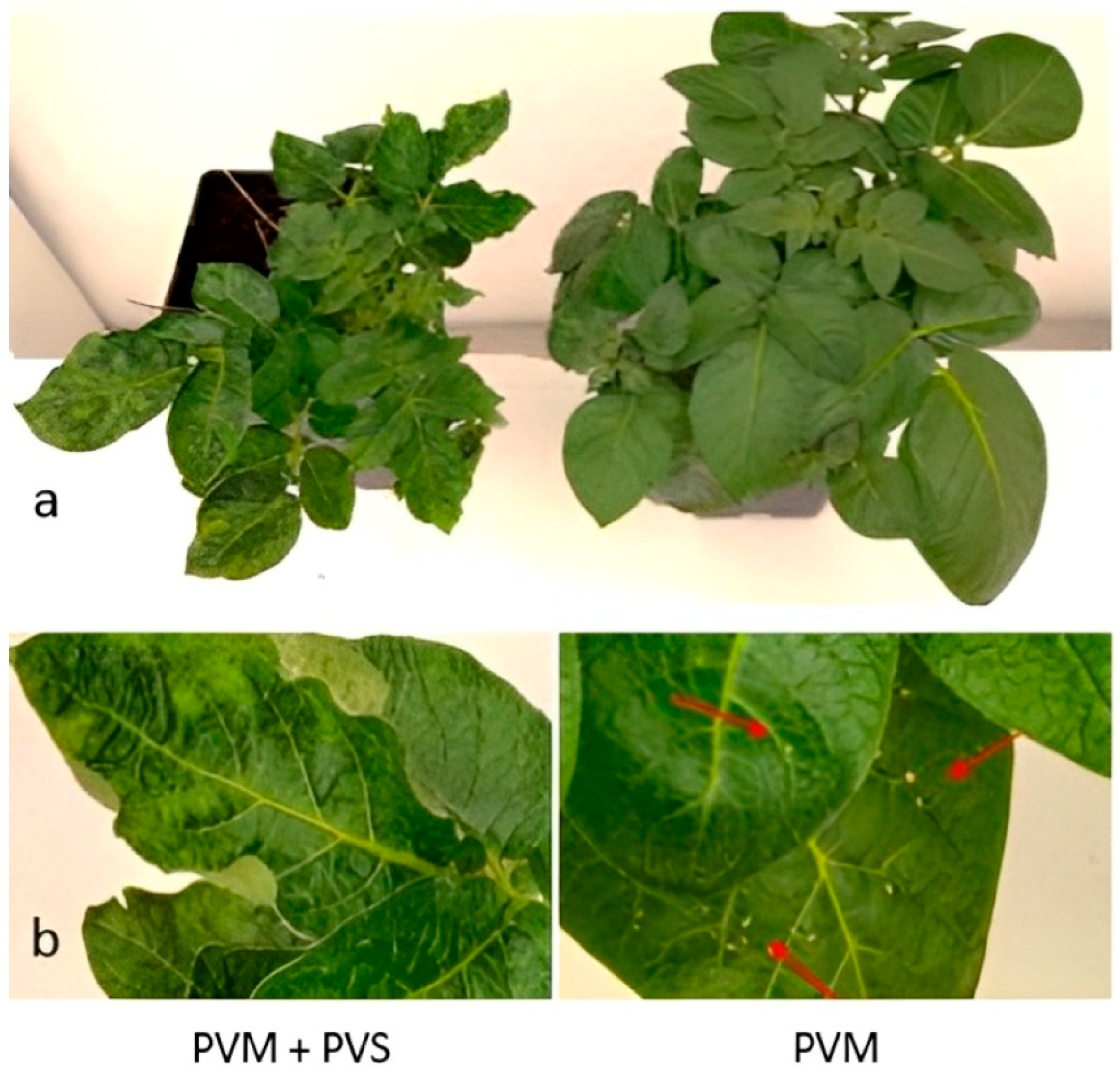
Micropropagated plants planted in a sterile substrate acclimatized with 100% success, and after transplanting into larger pots, they developed quickly under optimal conditions. The difference in symptoms between plants infected with both (PVM + PVS) or only one virus (PVM) in the R0 generation of plants produced by micropropagation, although noticeable, was not very pronounced. However, differences in viral disease symptoms were evident in R1 plants produced from microtubers and minitubers. In contrast to the pronounced symptoms in PVS + PVM-infected plants (reduced growth, wrinkling, twisting, and mosaicism), plants infected only with PVM had, on some leaves, very small lesions resembling a hypersensitivity reaction (Figure 3a,b).

3.4. Virus Detection and Efficiency of Virus Elimination

Initial virus screening of the mother plants for the presence of six viruses confirmed infection with only two viruses: PVM and PVS (Supplementary Table S1). This finding was interesting because the traditional potato cultivar Brinjak has long been grown with tubers without any sanitary selection. Despite that, other common viruses frequently detected in imported cultivars (e.g., PLRV, PVX, and PVY) were not confirmed. According to the ELISA results, PVS was successfully eliminated by six weeks of chemotherapy with ribavirin in three (14-1, 14-3, and 14-4) of nine R0 clones, and PVS-free status was also confirmed in the progeny (R1 from microtubers and minitubers). However, the same treatments were ineffective in eliminating PVM (Supplementary Table S1). Successful elimination of PVS was also confirmed by RT-PCR, where only the mother plants reacted positively, resulting in an amplicon of 885 base pairs, while selected three ELISA-negative PVS plants were also negative in molecular tests (Figure 4). These results are in partial agreement with the results of [36], who found that ribavirin at a concentration of 100 mg L−1 was not successful in eliminating either PVM or PVS after one subculture of 45 days, but only after the second or third subculture on ribavirin medium, and even then, elimination was genotype-dependent. The combination of ribavirin in one subculture with cryotherapy eliminated PVS in a certain percentage of plants, but not PVM. However, two subcultures with ribavirin + cryotherapy achieved 71–100% success in eliminating virus S and virus M, depending on the genotype [36].
Bettoni et al. [9] found that the effectiveness of virus eradication varied depending on the type of virus; PVS and PVM were more difficult to eliminate than PVA regardless of the method used. Using chemotherapy alone, Bettoni et al. [9] obtained an eradication success of 20–50% for PVS and 20% for PVM after a four-week treatment of shoot segments with ribavirin at a concentration of 100 mg L−1. Even with a combination of different consecutive and simultaneous treatments, e.g., chemotherapy + (chemotherapy + thermotherapy) + cryotherapy, PVM was eliminated in 70% of the plants, while the elimination of PVS with this combination of treatments was 100% [9]. Yang et al. [44] achieved 17% successful elimination of PVS and PVM with ribavirin (75 and 100 mg L−1) with one subcultivation of 45 days. At the second 45-day subcultivation, the percentage of virus elimination was higher, and after the third subcultivation, i.e., after 135 days, the plants were free from both PVS and PVM.
There was no difference in the efficiency of PVS elimination between the different ribavirin concentrations (Table 4). Ribavirin at a lower concentration (50 mg L−1) eliminated PVS from 33% of the surviving shoot apices, as did ribavirin at a concentration of 100 mg L−1. Oana et al. [35] also found no differences between the results obtained when 35 or 50 mg L−1 ribavirin was added to the culture medium and claimed that even 35 mg L−1 of ribavirin was sufficient for potato virus eradication, but with longer treatment. They also noted that successful eradication seemed to depend on the duration of treatment and the cultivar rather than the virus. Therefore, for complete elimination of the viruses in the traditional cultivar ‘Brinjak’, additional cycles of ribavirin chemotherapy or another method or combination of methods should be used.
Bougie and Bisaillon [49] found that ribavirin acts as a substrate for a viral RNA capping enzyme; however, such RNA transcripts blocked with ribavirin are not efficiently translated. Consequently, ribavirin prevents replication of a large number of DNA and RNA viruses [62].
Sometimes, as reported by [9], a plant tested for a particular virus may be negative (e.g., when a sample of plant tissue is tested from in vitro conditions), and upon subsequent inspection, e.g., after growing in a greenhouse, the same plant is found to be positive for the tested virus. In this study, except for the very long post-eradication period of 200 days + 90 days of cultivation in the growth chamber, three generations of clones obtained by treatment with ribavirin were used for virus detection by the methods DAS-ELISA and RT-PCR, eliminating the possibility of false-negative results. Accordingly, Yang et al. [44], who examined the plants by qPCR after a six-month post-eradication period, proved that ribavirin can thoroughly remove the viruses from the infected plants and not only suppress their replication. The same authors, using SSR markers, showed that ribavirin treatment did not cause genetic variation in plants.

3.5. The Influence of Sanitary Status of Plants on Chlorophyll Fluorescence and Multispectral Parameters

Mixed infection with both viruses (PVM + PVS) significantly decreased the effective quantum yield of PSII (Fq′/Fm′) and increased non-photochemical quenching (NPQ) (Table 5), which indicates that less light is used for photochemistry and more energy is lost through heat dissipation in plants with mixed infection. Zhou et al. [26] hypothesized that virus infection affects photosynthesis mainly by interfering with the Calvin cycle, leading to down-regulation of the efficiency of excitation energy capture by open PSII reaction centers (Fq′/Fm′). Although Fv/Fm is the most common fluorescence parameter used in plant stress studies [27,28], the results of this study show that Fv/Fm ranged from 0.79 to 0.81 for both PVM and PVM + PVS plants, indicating good fitness of plants. As noted by [15,19], plants infected with PVM and PVS do not always show symptoms, which depends on the variety and virus isolates.
Considering the multispectral parameters, mixed infection significantly decreases the chlorophyll index (CHI) and increases the reflectance in the far-red (RFarRed) (Table 6). These results are consistent with [63,64], who found that mixed infection can increase symptoms severity and virus accumulation. Decreased chlorophyll content affects photosynthetic light absorption and increases its reflection, as evidenced by increased (although not significant) reflection of RRed, RGreen, and RSpcGrn in plants with mixed infection. Thus, besides decreased Fq′/Fm′ and increased NPQ, plants with mixed infection will have lower light absorption, likely resulting in decreased sugar production in photosynthesis.

3.6. The Influence of the Sanitary Status of Plants on Yield Components

The sanitary status of R0 clones produced from micropropagated plants affected tuber weight per plant and average tuber weight (Table 7). Mixed infection with both viruses (PVM + PVS) reduced tuber weight per plant by 31% and average tuber weight by 64% compared to plants infected with PVM only. In R1 clones obtained from microtubers, mixed infection resulted in a significantly lower number of tubers per plant (59%), as well as lower tuber weight per plant (44%) (Table 8). An even greater difference was observed in R1 clones produced from minitubers, where the decrease in tuber weight per plant was 86% in plants with mixed infection compared to those infected with PVM alone (Table 9). The results obtained are in agreement with previous studies by [22,65], which found that mixed infection with two viruses resulted in a much greater yield loss than single infection. A statistically significant increase in the number of tubers per plant in PVS-free R1 clones obtained from microtubers and minitubers could be due to the higher number of stems per plant compared to plants with mixed infection. However, in the R0 clones obtained from micropropagated plants, each plant had only one main stem, and there was no significant difference in the number of tubers per plant between differently infected plants. These results are in agreement with those of [66], who reported that the number of main stems correlated positively with tuber number and tuber yield and negatively with average tuber weight.
Yield reduction in plants infected with both viruses (PVM + PVS) is the result of changes in metabolic processes in the affected tissues. Phytopathogenic viruses often cause physical deformation of leaves [11]. This leads to a decrease in the intensity of photosynthesis, which is very important for plant productivity.
Reduced growth, wrinkling, twisting, and mosaic structure of leaves caused by mixed infection were not observed in plants infected only with PVM, which had only small necrotic lesions. Severe phenotypic changes in plants with mixed infection are the cause of lower chlorophyll index (CHI) and lower effective PSII quantum yield (Fq′/Fm′), which ultimately resulted in large differences in potato yield between plants infected with two (PVM + PVS) compared to those infected with only one (PVM) virus.
The traditional potato cultivar ‘Brinjak‘ is resistant to the fungi Phytophthora infestans and Streptomyces scabies (personal communication), thus providing valuable germplasm for breeding programs and commercial growing. Infection of plants that have never been subjected to virus elimination before with only two—PVM and PVS—of the six viruses tested (PVX, PVY, PVA, PLRV, PVM, and PVS) may indicate that this cultivar possesses resistance to these viruses, as suggested by [10] for resistance to PVY in Norwegian cultivars. Elimination of these two viruses and use of virus-free planting material would probably increase yield and bring out the full genetic potential of this cultivar, in which growers are interested, especially in the mountainous regions of Croatia, where it is adapted.

4. Conclusions

The goal we set at the beginning of the research was only partially achieved, as ribavirin treatment at both concentrations (50 or 100 mg L−1) eliminated PVS from 33% of surviving shoot apices, but these treatments were ineffective in eliminating PVM. Mixed infection with PVM + PVS significantly decreased the effective quantum yield of PSII (Fq′/Fm′) and increased non-photochemical quenching (NPQ) compared to single infection with PVM. The multispectral parameters show that the mixed infection affected photosynthesis also at the chlorophyll level by significantly decreasing the chlorophyll index (CHI) and increasing far-red reflection (RFarRed). Plants infected only with PVM, compared to those with mixed infection, had 36–59% higher number of tubers per plant, 31–86% higher tuber weight per plant, and up to 64% higher average tuber weight, depending on the generation. The results indicate a strong negative impact of PVS in mixed infection with PVM and highlight the importance of removing PVS from potato plants.

Supplementary Materials

The following supporting information can be downloaded at: https://www.mdpi.com/article/10.3390/horticulturae8111013/s1, Table S1: DAS-ELISA and RT-PCR results for mother plants and their progeny tested on the presence of potato virus M (PVM) and potato virus S (PVS).

Author Contributions

Conceptualization, S.K. and I.H.J.; methodology, S.K., D.V. and B.L.; formal analysis, S.K, B.L.; investigation, S.K, I.H.J., D.V. and B.L.; resources, M.P, S.K. and B.L.; data curation, S.K.; writing—original draft preparation, S.K, D.V., B.L., I.H.J., M.B. and A.M.S.; writing—review and editing, M.P. and A.B.M.; visualization, A.M.S.; supervision, S.K. All authors have read and agreed to the published version of the manuscript.

Funding

This research was funded by the University of Zagreb’s financial support for scientific research.

Data Availability Statement

Data are contained within the article or Supplementary Materials.

Conflicts of Interest

The authors declare no conflict of interest.

References

  1. FAO. 2022. Available online: https://www.fao.org/3/nj015en/nj015en.pdf (accessed on 15 September 2022).
  2. FAOSTAT. 2022. Available online: https://www.fao.org/faostat/en/#data/QCL (accessed on 15 September 2022).
  3. Raggi, L.; Pacicco, L.C.; Caproni, L.; Álvarez-Muñiz, C.; Annamaa, K.; Barata, A.M.; Batir-Rusu, D.; Díez, M.J.; Heinonen, M.; Holubec, V.; et al. Analysis of landrace cultivation in Europe: A means to support in situ conservation of crop diversity. Biol. Conserv. 2022, 267, 109460. [Google Scholar] [CrossRef]
  4. Jovović, Z.; Stešević, D.; Meglič, V.; Dolničar, P. Old Potato Varieties in Montenegro; University of Montenegro Biotechnical Faculty Podgorica: Podgorica, Montenegro, 2013. [Google Scholar]
  5. Bradshaw, J.E. Review and Analysis of Limitations in Ways to Improve Conventional Potato Breeding. Potato Res. 2017, 60, 117–193. [Google Scholar] [CrossRef]
  6. Jones, R.A.C.; Naidu, R.A. Global Dimensions of Plant Virus Diseases: Current Status and Future Perspectives. Annu. Rev. Virol. 2019, 6, 387–409. [Google Scholar] [CrossRef] [PubMed]
  7. Rashid, M.-O.; Li, J.H.; Liu, Q.; Wang, Y.; Han, C.G. Molecular detection and identification of eight potato viruses in Gansu province of China. Curr. Plant Biol. 2021, 25, 100184. [Google Scholar] [CrossRef]
  8. Rubio, L.; Galipienso, L.; Ferriol, I. Detection of plant viruses and disease management: Relevance of genetic diversity and evolution. Front. Plant Sci. 2020, 11, 1092. [Google Scholar] [CrossRef]
  9. Bettoni, J.C.; Mathew, L.; Pathirana, R.; Wiedow, C.; Hunter, D.A.; McLachlan, A.; Khan, S.; Tang, J.; Nadarajan, J. Eradication of Potato Virus S, Potato Virus A, and Potato Virus M From Infected in vitro Grown Potato Shoots Using in vitro Therapies. Front. Plant Sci. 2022, 13, 878733. [Google Scholar] [CrossRef]
  10. Zhang, Z.; Wang, Q.C.; Spetz, C.; Blystay, D.R. In vitro therapies for virus elimination of potato-valuable germplasm in Norway. Sci. Hortic. 2019, 249, 7–14. [Google Scholar] [CrossRef]
  11. Kolychikhina, M.S.; Beloshapkina, O.O.; Phiri, C. Change in potato productivity under the impact of viral diseases. Earth Environ. Sci. 2021, 663, 012035. [Google Scholar] [CrossRef]
  12. Oyesola, O.L.; Aworunse, O.S.; Oniha, M.I.; Obiazikwor, O.H.; Bello, O.; Atolagbe, O.M.; Sobowale, A.A.; Popoola, J.O.; Obembe, O.O. Impact and Management of Diseases of Solanum tuberosum. In Solanum Tuberosum: A Promising Crop for Starvation Problem; Yildiz, M., Ozgen, Y., Eds.; IntechOpen: London, UK, 2021. [Google Scholar]
  13. Jayashige, U.; Chuquilllanqui, C.; Salazar, L.F. Modified expression of virus resistance in potato in mixed virus infections. Am. J. Potato Res. 1989, 66, 137–144. [Google Scholar]
  14. Onditi, J.; Nyongesa, M.; Vlugt, R. Prevalence, distribution and control of six major potato viruses in Kenya. Trop. Plant Pathol. 2021, 46, 311–323. [Google Scholar] [CrossRef]
  15. Kreuze, J.F.; Souza-Dias, J.A.C.; Jeevalatha, A.; Figueira, A.R.; Valkonen, J.P.T.; Jones, R.A.C. Viral diseases in potato. In The Potato Crop; Campos, H., Ortiz, O., Eds.; Springer: Cham, Switzerland, 2020; pp. 389–430. [Google Scholar]
  16. Wang, B.; Ma, Y.; Zhang, Z.; Wu, Z.; Wu, Y.; Wang, Q.; Li, M. Potato viruses in China. Crop Prot. 2011, 30, 1117–1123. [Google Scholar] [CrossRef]
  17. Hlaoui, A.; Boukhris-Bouhachem, S.; Sepúlveda, D.A.; Correa, M.C.G.; Briones, L.M.; Souissi, R.; Figueroa, C.C. Spatial and Temporal Genetic Diversity of the Peach Potato Aphid Myzus persicae (Sulzer) in Tunisia. Insects 2019, 10, 330. [Google Scholar] [CrossRef] [PubMed] [Green Version]
  18. Bragard, C.; Dehnen-Schmutz, K.; Gonthier, P.; Jacques, M.; Miret, J.A.J.; Justesen, A.F.; MacLeod, A.; Magnusson, C.S.; Milonas, P.; Navas-Cortes, J.A.; et al. Pest categorisation of potato virus M (non-EU isolates). EFSA J. 2020, 18, e05854. [Google Scholar] [PubMed]
  19. Bradshaw, J.E. Improving resistance to diseases and pests: A dynamic situation. In Potato Breeding: Theory and Practice; Bradshaw, J.E., Ed.; Springer Nature: Cham, Switzerland, 2021; pp. 247–337. [Google Scholar]
  20. Mosahebi, G.; Koohi-Habibi, M.; Okhovvat, S.M. Study on potato virus M (PVM) occurrence in potato fields in Iran. Commun. Agric. Appl. Biol. Sci. 2005, 70, 441–443. [Google Scholar]
  21. Wang, X.M.; Jin, L.P.; Yin, J. Progress in Breeding for Virus Resistance in Potatoes. Chin. Potato J. 2005, 19, 285–289. [Google Scholar]
  22. Huang, P.; Yan, Q.; Ding, Y. Occurrence and Control of Potato Virus S in Guizhou. Guizhou Agricul. Sci. 2009, 37, 88–89. [Google Scholar]
  23. Mutka, A.M.; Bart, R.S. Image-based phenotyping of plant disease symptoms. Front. Plant Sci. 2015, 5, 734. [Google Scholar] [CrossRef] [Green Version]
  24. Kuska, M.T.; Mahlein, A.K. Aiming at decision making in plant disease protection and phenotyping by the use of optical sensors. Eur. J. Plant. Pathol 2018, 152, 987–992. [Google Scholar] [CrossRef]
  25. Synkova, H.; Semoradova, S.; Schnablova, R.; Muller, K.; Pospísilova, J.; Ryslava, H.; Malbeck, J.; Serovska, N. Effects of biotic stress caused by Potato virus Y on photosynthesis in ipt transgenic and control Nicotiana Tableacum L. Plant Sci. 2006, 171, 607–616. [Google Scholar] [CrossRef]
  26. Zhou, Y.H.; Peng, Y.H.; Lei, J.L.; Zou, L.Y.; Zheng, J.H.; Yu, J.Q. Effects of potato virus YNTN infection on gas exchange and Photosystem II function in leaves of Solanum tuberosum L. Photosynthetica 2004, 42, 417–423. [Google Scholar] [CrossRef]
  27. Maxwell, M.; Johnson, G.N. Chlorophyll fluorescence—A practical guide. J. Exp. Bot. 2000, 51, 659–668. [Google Scholar] [CrossRef] [PubMed]
  28. Murchie, E.H.; Lawson, T. Chlorophyll fluorescence analysis: A guide to good practice and understanding some new applications. J. Exp. Bot. 2013, 64, 3983–3998. [Google Scholar] [CrossRef] [PubMed] [Green Version]
  29. Li, L.; Zhang, Q.; Huang, D. A Review of Imaging Techniques for Plant Phenotyping. Sensors 2014, 14, 20078–20111. [Google Scholar] [CrossRef] [PubMed]
  30. Panattoni, A.; Luvisi, A.; Triolo, E. Review: Elimination of viruses in plants: Twenty years of progress. Span. J. Agric. Res. 2013, 11, 173–188. [Google Scholar] [CrossRef]
  31. Wang, B.; Wang, R.R.; Li, J.W.; Ma, Y.L.; Sheng, W.M.; Li, M.F.; Wang, Q.C. Development of Three-vitrification-based cryopreservation procedures for shoot tips of China’s potato. Cryoletters 2013, 34, 369–380. [Google Scholar]
  32. Kidulile, C.E.; Ateka, E.M.; Alankonya, A.E.; Ndunguru, J.C. Efficacy of chemotherapy and thermotherapy in elimination of East African cassava mosaic virus from Tanzanian cassava landrace. J. Phytopathol. 2018, 166, 739–745. [Google Scholar] [CrossRef] [Green Version]
  33. Gong, H.; Igiraneza, C.; Dusengemungu, L. Major in vitro techniques for potato virus elimination and post eradication detection methods. A review. Am. J. Potato Res. Vol. 2019, 96, 379–389. [Google Scholar] [CrossRef] [Green Version]
  34. Kereša, S.; Kurtović, K.; Goreta Ban, S.; Vončina, D.; Habuš Jerčić, I.; Bolarić, S.; Lazarević, B.; Godena, S.; Ban, D.; Bošnjak Mihovilović, A. Production of Virus-Free Garlic Plants through Somatic Embryogenesis. Agronomy 2021, 11, 876. [Google Scholar] [CrossRef]
  35. Danci, O.; Erdei, L.; Vidacs, L.; Danci, M.; Anca, B.; Baciu, A.; David, I.; Berbentea, F. Influence of ribavirin on potato plants regeneration and virus eradication. J. Hortic. For. Biotechnol. 2009, 13, 421–425. [Google Scholar]
  36. Kushnarenko, S.; Romadanova, N.; Aralbayeva, M.; Zholamanova, S.; Alexandrova, A.; Karpova, O. Combined ribavirin treatment and cryotherapy for efficient Potato virus M and Potato virus S eradication in potato (Solanum tuberosum L.) in vitro shoots. Vitr. Cell. Dev. Biol. Plant 2017, 53, 425–432. [Google Scholar] [CrossRef]
  37. Wambugu, F.M.; Secor, G.A.; Gudmestad, N.C. Eradication of potato virus Y and S from potato by chemotherapy of cultured axillary bud tips. Am. Potato J. Vol. 1985, 62, 667–672. [Google Scholar] [CrossRef]
  38. Bittner, H.; Schenk, G.; Schuster, G.; Kluge, S. Elimination by chemotherapy of potato virus S from potato plants grown in vitro. Potato Res. Vol. 1989, 32, 175–179. [Google Scholar] [CrossRef]
  39. Zapata, C.; Miller, J.C.; Smith, R.H. An In vitro Procedure to Eradicate Potato Viruses X, Y, and S from Russet Norkotah and Two of Its Strains. Vitr. Cell. Dev. Biol. Plant 1995, 31, 153–159. [Google Scholar] [CrossRef]
  40. Faccioli, G. Control of Potato Viruses using Meristem and Stem-cutting Cultures, Thermotheraphy and Chemotherapy. In Virus and Viruslike Diseases of Potatoes and Production of Seed-Potatoes; Loebenstein, G., Berger, P.H., Brunt, A.A., Lawson, R.H., Eds.; Springer: Dordrecht, The Netherlands, 2001; pp. 365–390. [Google Scholar]
  41. Faccioli, G.; Colalongo, M.C. Eradication of potato virus Y and potato leafroll virus by chemotherapy of infected potato stem cuttings. Phytopathol. Mediterr. 2002, 41, 76–78. [Google Scholar]
  42. Nascimento, L.C.; Pio-Ribeiro, G.; Willadino, L.; Andrade, G.P. Stock indexing and potato virus y elimination from potato plants cultivated in vitro. Sci. Agric. 2003, 60, 525–530. [Google Scholar] [CrossRef] [Green Version]
  43. Gopal, J.; Garg, I.D. An efficient protocol of chemo-cum-thermotherapy for elimination of potato (Solanum tuberosum L.) virus by meristem-tip culture. Indian J. Agric. Sci. 2011, 81, 544–549. [Google Scholar]
  44. Yang, L.; Nie, B.; Liu, J.; Song, B. A Reexamination of the Effectiveness of Ribavirin on Eradication of Viruses in Potato Plantlets in vitro Using ELISA and Quantitative RT-PCR. Am. J. Potato Res. 2014, 91, 304–311. [Google Scholar] [CrossRef]
  45. Smyda-Daymund, P. Virus elimination from in vitro potato plants. Plant Breed. Seed Sci. 2017, 78, 81–85. [Google Scholar] [CrossRef]
  46. Shoala, T.; Eid, K.E.; El-Fiki, I.A.I. Impact of Chemotherapy and Thermotherapy Treatments on The Presence of Potato Viruses Pvy, Pvx and Plrv in Tissue-Cultured Shoot Tip Meristem. J. Plant Prot. Pathol. 2019, 10, 581–585. [Google Scholar] [CrossRef]
  47. Karjadi, A.K.; Kardjadi; Gunaeni, N. The Effect Of Antiviral Ribavirin, Explant Size, Varieties On Growth And Development In Potato Meristematic. IOP Conf. Ser. Earth Environ. Sci. 2021, 985, 012022. [Google Scholar] [CrossRef]
  48. Prusiner, P.; Sundaralingam, M. A New Class of Synthetic Nucleoside Analogues with Broad-spectrum Antiviral Properties. Nat. New Biol. 1973, 244, 116–118. [Google Scholar] [CrossRef] [PubMed]
  49. Bougie, I.; Bisaillon, M. The Broad Spectrum Antiviral Nucleoside Ribavirin as a Substrate for a Viral RNA Capping Enzyme. J. Biol. Chem. 2004, 279, 22124–22130. [Google Scholar] [CrossRef] [PubMed] [Green Version]
  50. Wang, M.-R.; Cui, Z.-H.; Li, J.-W.; Hao, X.-Y.; Zhao, L.; Wang, Q.-C. In vitro thermotherapy-based methods for plant virus eradication. Plant Methods 2019, 14, 87. [Google Scholar] [CrossRef] [Green Version]
  51. Crotty, S.; Cameron, C.; Andino, R. Ribavirin’s antiviral mechanism of action: Lethal mutagenesis? J. Mol. Med. 2002, 80, 86–95. [Google Scholar] [CrossRef]
  52. Murashige, T.; Skoog, F. A revised medium for rapid growth and bioassay with tobacco tissue culture. Physiol. Plant 1962, 15, 473–497. [Google Scholar] [CrossRef]
  53. Lazarević, B.; Šatović, Z.; Nimac, A.; Vidak, M.; Gunjača, J.; Politeo, O.; Carović-Stanko, K. Application of phenotyping methods in detection of drought and salinity stress in basil (Ocimum basilicum L.). Front. Plant Sci. 2021, 12, 174. [Google Scholar] [CrossRef]
  54. Kitajima, M.; Butler, W. Quenching of chlorophyll fluorescence and primary photochemistry in chloroplasts by dibromothymoquinone. Biochim. Biophys. Acta. 1975, 376, 105–115. [Google Scholar] [CrossRef]
  55. Genty, B.; Briantais, J.M.; Baker, N.R. The relationship between the quantum yield of photosynthetic electron transport and quenching of chlorophyll fluorescence. Biochim. Biophys. Acta Gen. Subj. 1989, 990, 87–92. [Google Scholar] [CrossRef]
  56. Bilger, W.; Björkman, O. Role of the xanthophyll cycle in photoprotection elucidated by measurements of light-induced absorbance changes, fluorescence and photosynthesis in leaves of Hedera canariensis. Photosynth. Res. 1990, 25, 173–185. [Google Scholar] [CrossRef]
  57. Gitelson, A.A.; Merzlyak, M.N.; Chivkunova, O.B. Optical Properties and Non-destructive Estimation of Anthocyanin Content in Plant Leaves. Photochem. Photobiol. 2001, 74, 38–45. [Google Scholar] [CrossRef]
  58. Gitelson, A.A.; Gritz, Y.; Merzlyak, M.N. Relationships between leaf chlorophyll content and spectral reflectance and algorithms for non-destructive chlorophyll assessment in higher plant leaves. J. Plant Physiol. 2003, 160, 271–282. [Google Scholar] [CrossRef] [PubMed]
  59. Rouse, J.W.; Haas, R.H.; Schell, J.A.; Deering, D.W. Monitoring vegetation systems in the Great Plains with ERTS. In NASA. Goddard Space Flight Center 3d ERTS-1 Symp; NASA: Washington, DC, USA, 1974; Volume 1, pp. 309–317. [Google Scholar]
  60. SAS/STAT®Software, version [9.4] 2002–2012; SAS Institute Inc.: Cary, NC, USA, 2013.
  61. Singh, B. Effect of antiviral chemicals on in vitro regeneration response and production of PLRV-free plants of potato. J. Crop. Sci. Biotechnol. 2015, 18, 341–348. [Google Scholar] [CrossRef]
  62. De Clercq, E. Antiviral Agents: Characteristic Activity Spectrum Depending on the Molecular Target With Which They Interact. Adv. Virus Res. 1993, 42, 1–55. [Google Scholar] [PubMed]
  63. Nyalugwe, E.P.; Wilson, C.R.; Coutts, B.A.; Jones, R.A.C. Biological properties of potato virus X in potato: Effects of mixed infection with potato virus S and resistance phenotypes in cultivars from three continents. Plant Dis. 2012, 96, 43–54. [Google Scholar] [CrossRef] [Green Version]
  64. Moreno, A.B.; López-Moya, J.J. When viruses play team sports: Mixed infections in plants. Phytopathology 2020, 110, 29–48. [Google Scholar] [CrossRef] [PubMed]
  65. Liu, W.P. Synergistic effect of potato virus Y (PVY) and potato spindle tuber viroid (PSTV) on tuber yield of potato. J. Heilongjiang August First Land Reclam. Uni. 2007, 19, 40–43. [Google Scholar]
  66. Lemaga, B.; Caesar, K. Relationships between numbers of main stems and yield components of potato (Solanum tuberosum L. cv. Erntestolz) as influenced by different daylengths. Potato Res. 1990, 33, 257–267. [Google Scholar] [CrossRef]
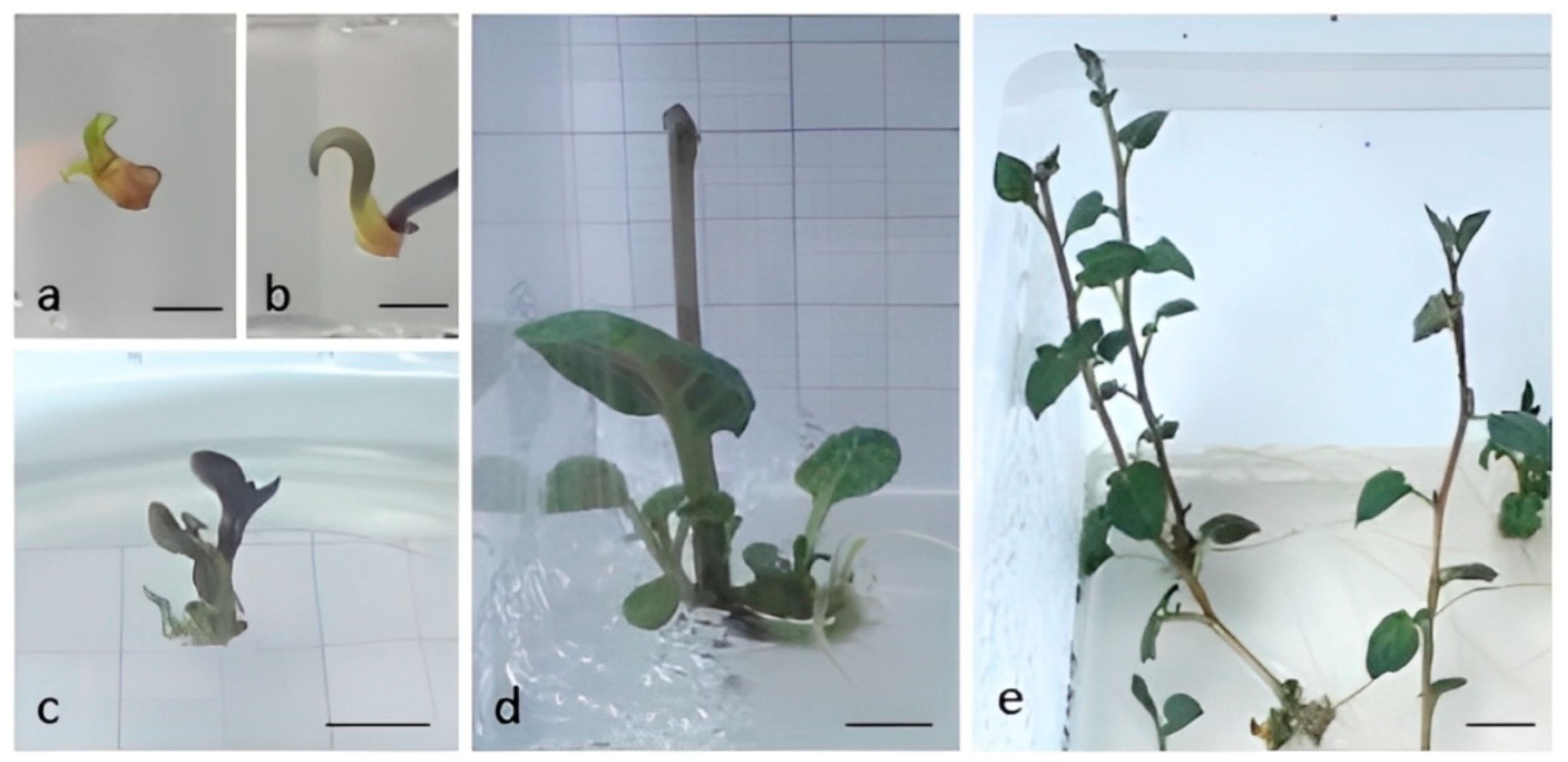
Figure 1. Surviving explants after 6 weeks of growth on ribavirin (a,b); inhibited growth on HFMS medium 100 days from the start of the experiment—shoot originating from RIB 50 (c) and RIB 100 (d); micropropagation by single node culture (e). Bars: (a,b) = 2 mm; (c,d) = 5 mm; (e) = 10 mm.
Horticulturae 08 01013 g001
Figure 2. Microtubers produced in one container of a clone infected with PVM (a) or with both viruses, PVM + PVS (b); microtuberization under short-day conditions (c); bar = 1 cm.
Horticulturae 08 01013 g002
Figure 3. Symptoms in mixed infection with PVM + PVS compared to a plant infected only with PVM; view on the whole plant (a) and details on the leaf (b). Arrows show small lesions.
Horticulturae 08 01013 g003
Figure 4. RT-PCR results for potato virus S (PVS) with an expected amplicon size of 885 base pairs (bps) performed on two PVS-infected mother plants (P1 and P2) and PVS-free plants obtained by ribavirin treatment B 14-1 RIB 50, B 14-3 RIB 100, and B14-4 RIB 50 in the following order: lines 1–3—R0 plants, lines 4–6—R1 plants from microtubers, lines 7–9—R1 plants form minitubers; N—negative control, M—marker (GelPilot 100 bp Plus Ladder, Qiagen, Hilden, Germany). For further details, please refer to Supplementary Table S1.
Horticulturae 08 01013 g004
Table 1. List of calculated chlorophyll fluorescence parameters with abbreviations, the equation for calculation, and the reference.
AbbrevTraitWavelength/Equation
Fv/FmMaximum Efficiency of Photosystem Two Fv/Fm = (Fm − F0)/Fm
[54]
Fq′/FmEffective Quantum Yield of Photosystem Two Fq′/Fm′ = (Fm′ − Fs′)/Fm
[55]
ETRElectron Transport RateETR = Fq′/Fm′ × PPFD × (0.5)
[55]
NPQNon-Photochemical QuenchingNPQ = (Fm − Fm′)/Fm
[56]
Table 2. List of analyzed multispectral parameters with abbreviations, wavelength for measurement or equation for calculation, and the reference if appropriate.
Abbrev.Trait Wavelength/Equation
RRed, Reflectance in Red640 nm
RGreenReflectance in Green 550 nm
RBlueReflectance in Blue 475 nm
RSpcGrnReflectance in Specific Green 510–590 nm
RFarRedReflectance in Far-Red710 nm
RNIRReflectance in Near Infra-Red769 nm
RChlReflectance Specific to Chlorophyll 730 nm
HUEHue (0–360°)HUE = 60 × (0 + (RGreen—RBlue)/(max-min)), if max = RRed;
HUE = 60 × (2 + (RBlue—RRed)/(max-min)), if max = RGreen;
HUE = 60 × (4 + (RRed—RGreen)/(max-min)) if max = RBlue;
360 was added in case HUE < 0
SATSaturation (0–1)SAT = (max—min)/(max + min) if VAL > 0.5,
or SAT = (max—min)/(2.0—max—min) if VAL < 0.5,
where max and min are selected from the RRed, RGreen, RBlue
VALValue (0–1) VAL = (max + min)/2;
where max and min are selected from the RRed, RGreen, RBlue
ARIAnthocyanin Index ARI = (R550)−1—(R700)−1
[57]
CHIChlorophyll Index CHI = (R700)−1—(R769)−1
[58]
NDVINormalized Differential Vegetation IndexNDVI = (RNIR − RRed)/(RNIR + RRed)
[59]
Table 3. Microtuber yield components, as affected by the sanitary status of the plants.
Virus
Infection
Number of Microtubers
Per Plant
Average Microtuber
Weight (mg)
PVM1.3 a207 a
PVM + PVS1.0 b199 a
Values within the column followed by the same letter are not significantly different at p < 0.05.
Table 4. The efficiency of virus elimination by ribavirin from potato cultivar ‘Brinjak’.
Ribavirin
Concentration (mg L−1)
Plants Free of Virus after Chemotherapy
PVMPVS
500/6 (0%)2/6 (33%)
1000/3 (0%)1/3 (33%)
Table 5. Chlorophyll fluorescence parameters as affected by the sanitary status of the plants.
Virus InfectionFv/FmFq′/FmETRNPQ
PVM0.81 a0.48 a5581 a0.38 b
PVM + PVS0.79 a0.42 b5234 a0.50 a
Values within the column followed by the same letter are not significantly different at p < 0.05.
Table 6. Multispectral parameters as affected by the sanitary status of the plants.
Virus InfectionRRedRGreenRBlueRFarRedRNIRRSpcGrnHUESATVALCHIARINDVI
PVM1975 a3019 a1581 a6046 b2831 a3394 a105 a0.05 a0.46 a3.8 a3.9 a0.85 a
PVM + PVS2001 a3148 a1563 a6403 a2849 a3558 a104 a0.05 a0.49 a3.5 b3.8 a0.85 a
Values within the column followed by the same letter are not significantly different at p < 0.05.
Table 7. Minituber yield components of R0 clones produced from micropropagated plants, as affected by the sanitary status of the plants.
Virus
Infection
Number of Tubers
per Plant
Tuber Weight
per Plant (g)
Average Tuber
Weight (g)
PVM4.0 a52.0 a15.6 a
PVM + PVS4.7 a39.8 b9.5 b
Values within the column followed by different letters are significantly different at p < 0.05.
Table 8. Mini tuber yield components of R1 clones produced from microtubers, as affected by the sanitary status of the plants.
Virus
Infection
Number of Tubers
per Plant
Tuber Weight
per Plant (g)
Average Tuber
Weight (g)
PVM7.3 a65.0 a9.6 a
PVM + PVS4.6 b45.1 b10.1 a
Values within the column followed by the same letter are not significantly different at p < 0.05.
Table 9. Tuber yield components of R1 clones produced from minitubers, as affected by the sanitary status of the plants.
Virus
Infection
Number of Tubers
per Plant
Tuber Weight
per Plant (g)
Average Tuber
Weight (g)
PVM9.8 a133.9 a16.1 a
PVM + PVS7.2 b72.1 b12.1 a
Values within the column followed by the same letter are not significantly different at p < 0.05.
Publisher’s Note: MDPI stays neutral with regard to jurisdictional claims in published maps and institutional affiliations.