Article

Analysis of Light-Independent Anthocyanin Accumulation in Mango (Mangifera indica L.)

1
College of Horticulture, Hainan University, Haikou 570228, China
2
Engineering Research Center of Selecting and Breeding New Tropical Crops Varieties, Ministry of Education, Haikou 570228, China
3
Key Laboratory of Quality Regulation of Tropical Horticultural Crop in Hainan Province, Haikou 570228, China
4
Ministry of Agriculture Key Laboratory of Tropical Fruit Biology, South Subtropical Crops Research Institute, Chinese Academy of Tropical Agricultural Sciences, Zhanjiang 524091, China
5
Chinese Academy of Tropical Agricultural Sciences & Ministry of Agriculture Key Laboratory of Crop Gene Resources and Germplasm Enhancement in Southern China, Tropical Crops Genetic Resources Institute, Haikou 571100, China
*
Authors to whom correspondence should be addressed.
These authors contributed equally to this work.
First correspondence.
Horticulturae 2021, 7(11), 423; https://doi.org/10.3390/horticulturae7110423
Submission received: 8 October 2021 / Revised: 12 October 2021 / Accepted: 12 October 2021 / Published: 21 October 2021

Abstract

:
Light dependent anthocyanin accumulation contributes to the red pigmentation of the fruit skin of mango (Mangifera indica L.). Light-independent pigmentation has also been reported, but remains poorly characterized. In this study, the pigmentation patterns in the skin of two red mango cultivars, ‘Ruby’ and ‘Sensation’, were evaluated. Metabolomic profiling revealed that quercetin-3-O-glucoside, cyanidin-3-O-galactoside, procyanidin B1, and procyanidin B3 are the predominant flavonoid compounds in the skin of ‘Ruby’ and ‘Sensation’ fruit. Young fruit skin mainly accumulates flavonol and proanthocyanidin, while anthocyanin is mainly accumulated in the skin of mature fruit. Bagging treatment inhibited the biosynthesis of flovonol and anthocyanin, but promoted the accumulation of proanthocyanidin. Compared with ‘Sensation’, matured ‘Ruby’ fruit skin showed light red pigmentation at 120 days after full bloom (DAFB), showing a light-independent anthocyanin accumulation pattern. However, the increase of anthocyanin concentration, and the expression of key anthocyanin structural and regulatory genes MiUFGT1, MiUFGT3, and MiMYB1 in the skin of bagged ‘Ruby’ fruit versus ‘Sensation’ at 120 DAFB was very limited. There was no mutation in the crucial elements of MiMYB1 promoter between ‘Ruby’ and ‘Sensation’. We hypothesize that the light-independent anthocyanin accumulation in the skin of mature ‘Ruby’ fruit is regulated by plant hormones, and that ‘Ruby’ can be used for breeding of new more easily pigmented red mango cultivars.

1. Introduction

Mango (Mangifera indica L.) is one of the most popular tropical fruits in the world and is often referred to as the ‘king of fruits’ [1]. Fruit color is an important index of fruit quality, and normally red-colored mango fruits are preferred by the customers [2]. The red color of mango fruit is due to the accumulation of anthocyanin, and the major components of anthocyanin in mango are cyanidin 3-O-galactoside and 7-O-methylcyanidin 3-O-β-D galactopyranoside [3,4]. Anthocyanin is water soluble pigment, derived from a branch of the flavonoid biosynthesis pathway. So far, a number of anthocyanin biosynthetic genes in mango have been isolated, including PAL (phenylalanine ammonia-lyase), CHS (chalcone synthase), CHI (chalcone isomerase), F3H (flavanone 3-hydroxylase), F3H (flavonoid 3’-hydroxylase), DFR (dihydroflavonol 4-reductase), ANS (anthocyanidin synthase), and UFGT (UDP-glucose: flavonoid 3-O-glucosyltransferase) [5,6]. The expression of anthocyanin biosynthetic genes is regulated through the MYB-bHLH-WD40 complex, and among these R2R3-MYB has been showed to be the most important transcription factor (TF) [7,8]. The best known R2R3-MYB is MYB75/PAP1 (Production of Anthocyanin Pigment 1) which was identified in Arabidopsis [9]. Its homologs called MYB1 or MYB10 were isolated from diverse fruit species including apple [10,11,12], grape [13], orange [14], pear [15], strawberry [16] and peach [17]. In addition, a R2R3-MYB TF, PyMYB114 has been reported to regulate anthocyanin biosynthesis in pear [18]. Recently, a MYB TF controlling anthocyanin biosynthesis in mango was isolated from mango cv. ‘Irwin’, and named MiMYB1 [19].
Bagging, i.e., covering young fruits with paper bags, can improve fruit pigmentation and protect the fruit from diseases and pests, and this practice is widely used during fruit production including for pear [20], apple [21], grape [22], and mango [19,23]. Since the anthocyanin accumulation in fruit skin (also in mango) is usually dependent on sunlight, bags should be removed before harvest to re-expose the fruit to sunlight and induce anthocyanin biosynthesis and the consequent red pigmentation. Unlike other mango cultivars, cv. ‘Ruby’ fruit skin can still accumulate anthocyanin at the later stages of development when bagged (Figure 1). So, this light-independent cultivar is of particular interest for commercial reasons (labor saving by skipping of bag removal) as well as scientific reasons (elucidation of the molecular mechanism of light-independent anthocyanin biosynthesis).
Light-independent anthocyanin accumulation has also been found in grape berry [24,25,26], the flesh of apple [27], blood orange [14], peach [28], tuberous roots of purple-fleshed sweet potato [29], and the purple head of Chinese cabbage [30]. In grape berries, bagging changed the concentration of specific anthocyanin compounds but had no effect on the total concentration of anthocyanin in ‘Shiraz’ [24,26], and ‘Crimson Seedless’ [25]. Comparing light-independent grape berry cv. ‘Jingyan’ with light-dependent cv. ‘Jingxiu’, researchers found that the key structural gene UFGT and the regulatory gene MYB-A1 were highly expressed in ‘Jingyan’ with or without sunlight, although expression of these genes was down-regulated by bagging in light-dependent ‘Jingxiu’ [22]. No differences were found in the sequences of these two genes between the two cultivars. In the red-fleshed apple cultivar ‘Red Field’, an allelic rearrangement in the promoter of MYB10 has generated an autoregulatory locus, which increases the transcription of MYB10, and subsequently leads to the anthocyanin accumulation in flesh [27]. In blood orange, anthocyanin accumulation in flesh is due to the high expression of Ruby gene (encoding a MYB TF) caused by a retrotransposon insertion in the Ruby promoter region [14]. The red-fleshed trait in peach cultivar ‘Dahongpao’ is controlled by a BLOOD (BL) gene encoding a NAC transcription factor, which acts as a heterodimer with PpNAC1 activating the transcription of PpMYB10.1, resulting in anthocyanin pigmentation [28]. Anthocyanin accumulation in the tuberous roots of purple-fleshed sweet potato is regulated by IbMYB1, which shows the specific expression in tuberous roots, and over-expression of IbMYB1 could up-regulate the expression of all the anthocyanin biosynthetic genes and cause anthocyanin accumulation [29]. In purple head Chinese cabbage cultivar 11S91, a large deletion in intron 1 of gBrMYB2 increases the expression of BrMYB2, causing purple pigmentation in the head of Chinese cabbage [30]. All these results indicated light-independent anthocyanin biosynthesis in plants is highly relevant to MYB. So far, there is no report about the light-independent accumulation of anthocyanin in mango, and it is worthy to see the role of MYB during this process.
In this study, concentration of different flavonoid components (mainly anthocyanins) in fruit skin was compared between two red mango cultivars with similar developmental period (blooming in early March, and maturing in late June), i.e., light-independent cv. ‘Ruby’ and light-dependent cv. ‘Sensation’. Bagged and un-bagged fruit peel from these two cultivars was sampled at different developmental stages, i.e., 50 days after full bloom (DAFB), 80 DAFB, and 120 DAFB. Expression of anthocyanin biosynthetic genes and regulatory genes was analyzed, and the promoter sequence of MiMYB1 was cloned and compared between these two cultivars. This study will provide novel insight in light independent anthocyanin biosynthesis in fruits.

2. Materials and Methods

2.1. Fruit Materials and Experimental Treatment

The experiment was carried out at the South Subtropical Crops Research Institute (SSCRI) in Zhanjiang, China. Three mature ‘Ruby’ trees and three mature ‘Sensation’ trees were selected, and each tree was regarded as one biological replicate. These trees were similar in size and number of fruit and had uniform exposure to sunlight. In each tree, about 50 fruitlets were covered with double layers of yellow-black paper bag (Qingdao Kobayashi Co., Ltd., Qingdao, China) to block out all the light at 20 DAFB. The remaining non-bagged fruitlets were set as controls. Per tree, 10 fruits were harvested at 50, 80, and 120 DAFB, stored in an ice box, and carried back to the lab as soon as possible. Fruit skin was peeled, instantly frozen in liquid nitrogen and stored at −80 °C for assays.

2.2. Metabolomic Profiling

Skin of bagged and natural grown ‘Ruby’ and ‘Sensation’ mango fruits at three stages of development was used for metabolomic analysis. Extraction and measurement of flavonoid metabolites was performed by Metware Biotechnology Co. Ltd. (Wuhan, China). Briefly, the freeze-dried sample was ground to powder for 1.5 min at 30 Hz using a mill (MM400, Retsch, Germany). Next, 0.05 g powder was added to 500 µL extraction solution containing 50% methanol and 0.1% HCl. After two 5 min vortexes, 5 min ultrasound, and 3 min centrifuge (12,000 r/min, 4 °C), the supernatant was filtered through a microporous membrane (0.22 μm) and stored in a vial for subsequent analysis on a high-performance liquid chromatography with tandem mass Spectrometric (HPLC−MS/MS) system (UPLC, ExionLC™ AD, https://sciex.com.cn/ (accessed on 2 October 2021); MS, QTRAP® 6500+, https://sciex.com.cn/ (accessed on 2 October 2021). The HPLC conditions were as follows: column, Waters ACQUITY BEH C18 (1.7 µm, 2.1 mm × 100 mm); mobile phase, water (0.1% formic acid): methanol (0.1% formic acid); gradient program, 95:5 v/v at 0 min, 50:50 v/v at 6 min, 5:95 v/v at 12 min, hold for 2 min, 95:5 v/v at 14 min, hold for 2 min; flow rate, 0.35 mL/min; temperature, 40 °C; injection volume: 2 µL. An electrospray ion source (ESI) was used for positive ion detection. The spray voltage was 5.5 kV, the ion source temperature was 550 °C, and the curtain gas pressure was 35 psi.
Different flavonoid compounds detected by mass spectrometry were identified based on Metware Database (MWDB). The quantitative analysis of anthocyanins was performed by multiple reaction monitoring (MRM) of triple quadrupole mass spectrometry. Different concentrations of standard solutions (93 anthocyanins, 6 Procyanidins, and 9 other types of flavonoid) including 0.01 ng/mL, 0.02 ng/mL, 0.05 ng/mL, 0.1 ng/mL, 0.5 ng/mL, 1 ng/mL, 5 ng/mL, 10 ng/mL, 50 ng/mL, 100 ng/mL, 500 ng/mL, 1000 ng/mL, 2000 ng/mL, 50,000 ng/mL were prepared to obtain the mass spectrum peak intensity data for the corresponding quantitative signals of each concentration standard. The standard curve linear equation was obtained with standard concentration as abscissa and peak area as ordinate, which is used to calculate the concentration of detected substance.
The concentration of metabolites in the sample (μg/g) = C × V/1,000,000/m.
C is the concentration value (ng/ml) calculated by substituting the integral peak area of the sample into the standard curve; V is the volume of extraction solution (μL); m is the dry weight of the sample (g).

2.3. DNA Isolation, RNA Extraction and cDNA Synthesis

Genomic DNA was extracted from fruit skin of ‘Ruby’ and ‘Sensation’ mango harvested at 50 DAFB using Super Plant Genomic DNA Kit (Tiangen, DP360, Beijing, China), and DNA concentration was measured by NanoDrop Lite Spectrophotometer (Thermo Scientific, Waltham, MA, USA). Total RNA was extracted using an RNA prep pure plant kit special for plant tissues rich in polysaccharides and polyphenolics (Tiangen, DP441, Beijing, China). The concentration of total RNA was measured by NanoDrop Lite Spectrophotometer (Thermo Scientific, Waltham, USA) after genomic DNA had been digested by DNase I supplied with the RNA extraction kit. First-strand cDNA was synthesized from 1 µg of DNA-free RNA using HiScript IIQ RT SuperMix (Vazyme, R223-01, Nanjing, China). The cDNA was diluted 20 fold and 5.5 µL of the diluted cDNA was used as the template for real-time quantitative PCR (Q-PCR) analysis.

2.4. Q-PCR Analysis

The Q-PCR reaction solution (total volume 15 µL) was composed of 7.5 µL of SYBR Premix Ex TaqTM II (Takara, Japan), 1 µL of each primer (10 μM), and 5.5 µL of cDNA. The reaction, performed on a real-time PCR machine (qTOWER3 G, Jena, Germany), was initiated with a preliminary step of 30 seconds at 95 °C followed by 40 cycles of 95 °C for 5 seconds and 60 °C for 30 seconds. Templateless controls (5.5 µL ddH2O) for each primer pair were necessary for each run. The Q-PCR primers for MiMYB114 were as follows: MiMYB114-F: 5′-GAAGCTGCCCTTCAAGACAC-3′ and MiMYB114-R: 5′-CCGTAGGATCCTTCAGTGGA-3′. The Q-PCR primers for the rest structural and regulatory genes were used as reported by Kanzaki et al. [19]. All Q-PCR reactions were normalized using the Ct value corresponding to the mango actin gene [31]. Analysis was performed in three biological replicates.

2.5. Cloning of Promoter Regions of MiMYB1

The primers used to amplify the MiMYB1 promoter region in ‘Ruby’ and ‘Sensation’ were based on the genomic sequence obtained from the mango genome database (BIG Genome Sequence Archive database, accession number: PRJCA002248), and the primers were as follows: forward primer: 5′-TAAGAATGGTAGACAAAGGATG-3′, reverse primers: 5′-TCTTAGGGGAACTTGATGCC-3′. PCR products were analyzed on 1.0% agarose gels, and a single fragment was recovered from gels and purified using a DNA purification kit (Takara). The fragment was then ligated into the plasmid pMD18-T vector, transformed into Escherichia coli DH5α competent cells (Takara), and then sequenced (Sangong, Shanghai, China).

2.6. Statistical Analysis

Data were subjected to a one-way Analysis of Variance (ANOVA), with mean values separated by Tukey’s multiple range test using SPSS 19.0 (SPSS, Chicago, IL, USA). Probability values of <0.05 were considered statistically significant.

3. Results

3.1. Fruit Color and Metabolomic Profiling

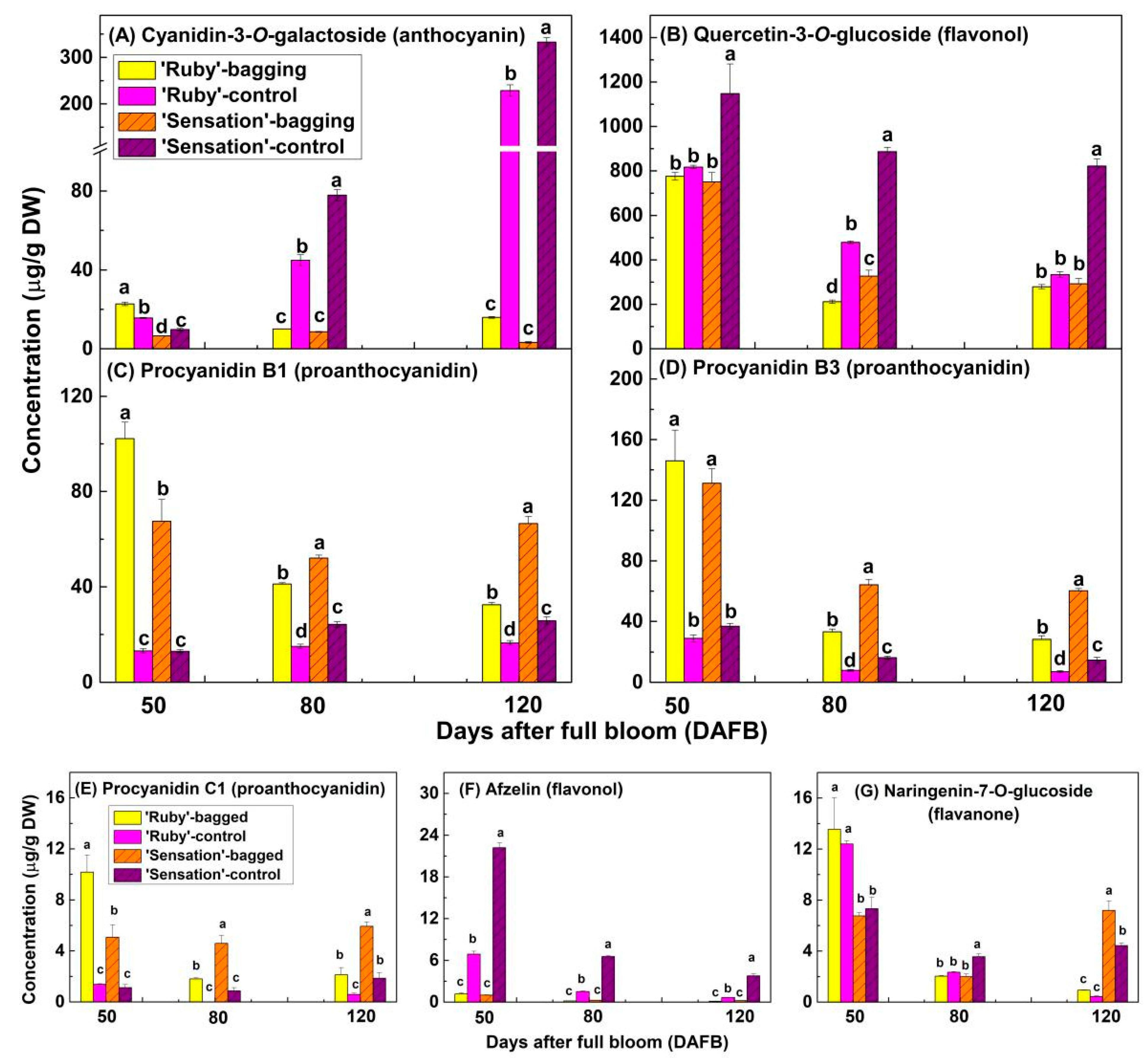
Under the natural condition of sunlight exposure, both ‘Ruby’ and ‘Sensation’ fruits exhibited dark red skin color (Figure 1). The bagged fruits of ‘Sensation’ showed no red pigmentation during any developmental stage. In contrast, bagged, mature ‘Ruby’ fruit showed bright red pigmentation at 120 DAFB (Figure 1). According to the standard of 93 anthocyanins, 6 Procyanidins, and 9 other types of flavonoid, major substances including cyanidin-3-O-galactoside (anthocyanin, Figure 2A), quercetin-3-O-glucoside (flavonol, Figure 2B), procyanidin B1 (proanthocyanidin, Figure 2C), and procyanidin B3 (proanthocyanidin, Figure 2D), and minor substances including procyanidin C1 (proanthocyanidin, Figure 2E), afzelin (flavonol, Figure 2F), and Naringenin-7-O-glucoside (flavonone, Figure 2G), were identified in the fruit skin of ‘Ruby’ and ‘Sensation’ mangoes. Apart from seven substances listed in Figure 2, all the other substances showed very low concentration, trace amount, or could not even be detected.
The concentration of anthocyanin increased with development (Figure 2A), while the concentration of flavonol, and proanthocyanidin, and flavonone decreased during the development of fruits (Figure 2B–G). Sunlight exposure significantly induced the accumulation of anthocyanin and flavonol in the fruit skin of mango but repressed the biosynthesis of proanthocyanidin (Figure 2A–F). The cyanidin-3-O-galactoside concentration in the fruit skin of bagged ‘Ruby’ was around five times higher than that of bagged ‘Sensation’ (15.87 vs. 3.22 μg/g DW) at 120 DAFB (Figure 2A), yet the difference was not statistically significant.

3.2. Expression of Anthocyanin Biosynthetic and Regulatory Genes in Fruit Skin

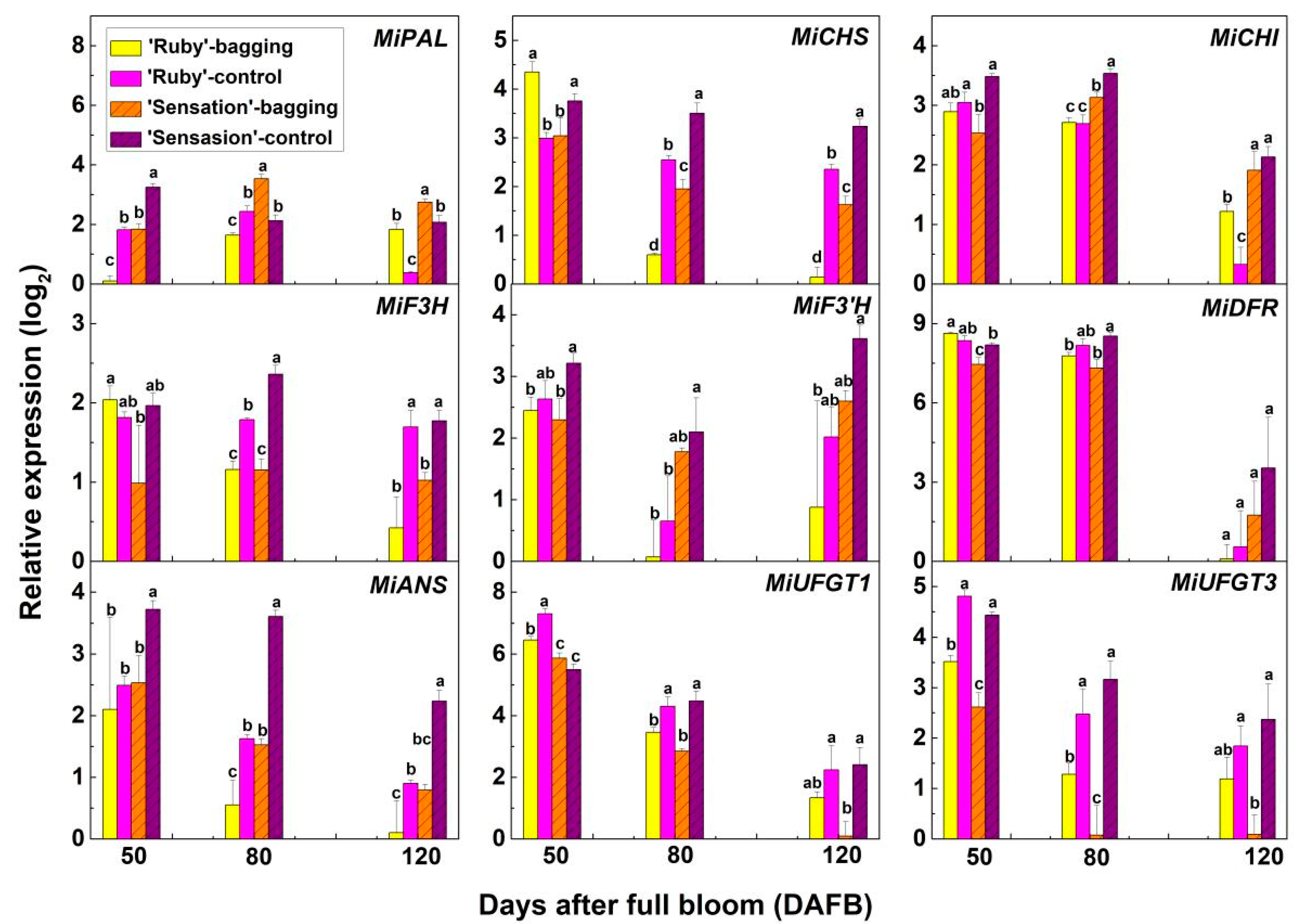
In general, anthocyanin biosynthetic genes were highly expressed at the early stages of development, i.e., 50 DAFB and 80 DAFB, especially the late biosynthetic genes including MiDFR, MiANS, MiUFT1, and MiUFT3 (Figure 3). Moreover, compared with bagged fruit, control (non-bagged) fruit skin showed a higher expression level, particularly for late biosynthetic genes (Figure 3). It is remarkable that two family members of UFGT, which encode the enzyme catalyzing the last step of anthocyanin biosynthesis and is regarded as the most crucial structural gene, MiUFGT1 and MiUFGT3, showed relatively higher expression in bagged ‘Ruby’ fruit skin than in bagged ‘Sensation’ fruit at 120 DAFB, although this effect was not significant (Figure 3). Among three regulatory genes, MiMYB1 was also highly expressed in the skin of fruitlet or sunlight-exposed fruit (Figure 4). The expression of MiMYB1 in ’Ruby’ bagged fruit skin at 120 DAFB was 2.6 times higher than in ’Sensation’ bagged fruit, without significant difference (Figure 4). The expression of MiMYB114 and MibHLH2 was not correlated to anthocyanin concentration (Figure 4).

3.3. MiMYB1 Promoter Sequence Isolation and Comparison in ’Ruby’ and ‘Sensation’

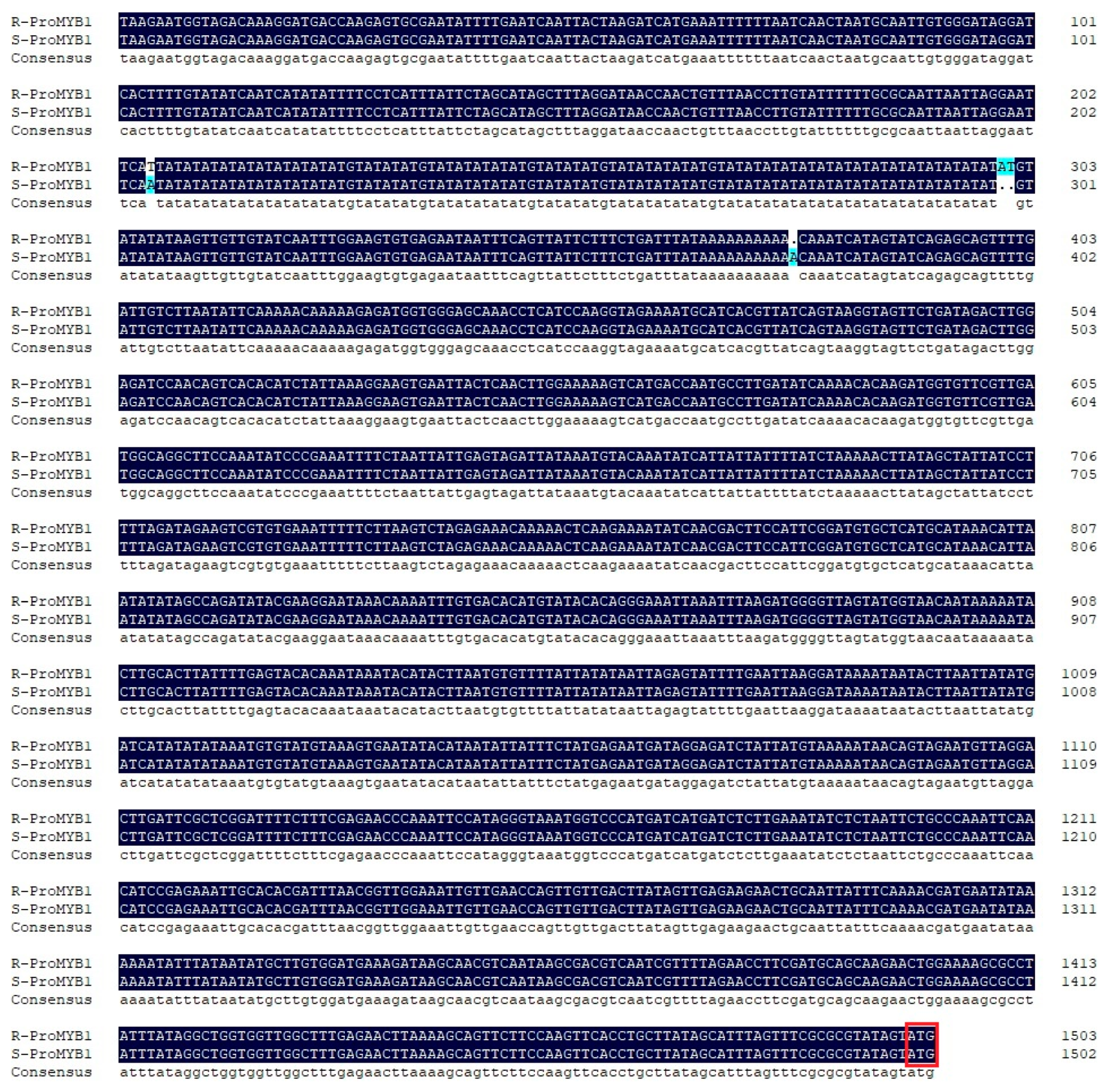
Due to the important role of MiMYB1 (MYB10 homolog) in regulating anthocyanin biosynthesis, the promoter region of MiMYB1 was obtained from the mango genome database, and a circa 1500 bp promoter sequence was cloned for both ’Ruby’ and ‘Sensation’. The obtained sequence was designated as proMYB1. A number of light-responsive elements including AE-box, Box4, GATA-motif, G-box, and GT1-motif, and plant hormone-responsive elements such as ABRE (abscisic acid), CGTCA-motif (MeJA), TCA-element (salicylic acid), and TGA-element (auxin) were identified in proMYB1 (Table 1). Also, three MYB binding sites (MBS) were found in proMYB1 (Table 1). The proMYB1 in ’Ruby’ and ‘Sensation’ shared 99.73% identity, but two SNPs and one 2-bp mutation were found between two cultivars, although none of these were located on any of the identified motifs (Figure 5).

4. Discussion

In many fruits, flavonol, and proanthocyanidin are the major flavonoid compounds at the early stage of fruit development, while the flavonoid pathway switches to anthocyanin biosynthesis when the ripening begins [32,33]. In the present study, the same phenomenon of flavonoid accumulation pattern was detected in the fruit skin of ‘Ruby’ and ‘Sensation’ (Figure 2). It has been indicated that the astringent taste of proanthocyanidin in unripe fruits could protect the fruits against early feeding [34], while anthocyanin is needed in ripe fruits to attract seed dispersers including insects and animals [35]. In addition, non-bagged mango fruit skin accumulated high levels of anthocyanin (Figure 2A) and low levels of proanthocyanidin (Figure 2C–E), while bagged fruit skin showed the opposite accumulation pattern (Figure 2A,C–E), suggesting competition between proanthocyanidin and anthocyanin pathways. Proanthocyanidin and anthocyanin are derived from the same precursor, anthocyanidin, by the activity of ANR and UFGT, respectively [36,37]. Ectopic expression of both Arabidopsis ANR gene (BANYULS) and apple MdANR genes in tobacco promoted and inhibited the accumulation of proanthocyanidin and anthocyanin in tobacco, respectively [37,38]. High concentration of proanthocyanidin in the skin of bagged ‘Ruby’ and ‘Sensation’ also indicated sufficient accumulation of precursor anthocyanidin in the fruit skin under darkness, which means UFGT, rather than the other structural genes, is the limiting factor for anthocyanin accumulation in bagged mango fruit skin. Due to low expression of UFGT gene and subsequent low level of UFGT protein, most anthocyanidin was converted to proanthocyanidin instead of anthocyanin in bagged fruit skin.
Fruit bagging is a safe and eco-friendly technique widely used in Asian countries (China, Japan, and Korea), Australia and The USA for producing high quality fruits. This practice can protect fruit from fungi, bacteria, insects, and even birds, and minimize of the need for pesticide applications [39]. In the case of fruits whose commercial value is determined by anthocyanin accumulation (e.g., apple, grape, pear, and mango), bags should be removed before harvest to ensure light-dependent fruit pigmentation [20,21,23]. Bag treatment and bag removal is labor intensive, so the light-independent pigmentation phenotype reported in this study is regarded as an excellent horticultural trait (bag-removal skipping and labor saving).
Although bagged ‘Ruby’ fruit skin exhibited visible red pigmentation at 120 DAFB (Figure 1), the anthocyanin concentration (Figure 2A), and the expression of MiUFGT1, MiUFGT3, and MiMYB1 (Figure 3A,B) were still highly repressed by the bagging treatment. The relatively higher anthocyanin concentration and gene expression in the skin of bagged ‘Ruby’ compared to ‘Sensation’ at 120 DAFB was not statistically significant (Figure 2A and Figure 3). Since UFGT expression is well-known to be regulated by R2R3-MYB [40,41], low expression of MYB is fundamental reason for low levels of anthocyanin in bagged mango fruit skin. Several light-responsive elements in the promoter region of MiMYB1 (Table 1) indicate light is essential for activating the expression of MiMYB1. Meanwhile, MYB protein undergoes COP1-mediated ubiquitination and degradation in darkness [42]. Mutations in the promoter region of R2R3-MYB, changing the MYB expression into almost a constitutive pattern, could lead to high level of MYB mRNA and protein in darkness, and subsequently result in the light-independent anthocyanin accumulation in plants [14,27], which is obviously not the case in the present study (Figure 5).
Interestingly, the bagged ‘Ruby’ fruit skin accumulated anthocyanin at 120 DAFB rather than 50 and 80 DAFB, indicating the possible role of ripening-related plant hormones regulating this process. The ripening of climacteric fruits is associated with ethylene production and respiratory burst [43]. Ethylene can induce the anthocyanin accumulation of most climacteric fruits. For example, in apple the key transcription factor involved in ethylene signal transduction, EIN3-LIKE1 (EIL1), directly binds to the promoter region of MdMYB1 and activates its expression, subsequently leading to anthocyanin accumulation. Furthermore, MdMYB1 can up-regulate the expression of MdERF3, a key regulator of ethylene biosynthesis, to produce more ethylene, thereby providing a regulatory loop of ethylene-induced anthocyanin biosynthesis mediated by MdEIL1-MdMYB1-MdERF3 module [44]. ABA can also induce the anthocyanin accumulation. In apple the expression of a bZIP regulatory gene MdbZIP44 is up-regulated by ABA treatment, and MdbZIP44 can interact with MdMYB1 to enhance the MdMYB1-mediated expression regulation of anthocyanin structural genes, leading to the anthocyanin accumulation [45]. Jasmonates (JAs) have also been reported to promote anthocyanin accumulation in apple [46], pear [47], and mango [48], via two key JA transduction pathway proteins, JAZ and COI1 [49,50].
So, our hypothesis for light-independent anthocyanin accumulation in ‘Ruby’ mango fruit skin is that ripening-related plant hormones such as ethylene, ABA or JA are produced at the onset of ripening, triggering the expression of hormone responsive up-stream transcription factors, which interact with MiMYB1 to promote anthocyanin biosynthesis. On the other hand, MiMYB1 expression is strongly repressed by darkness, and undergoes COP1-mediated degradation, overall resulting in the limited anthocyanin accumulation in the skin of bagged ‘Ruby’ fruit.

5. Conclusions

Quercetin-3-O-glucoside, cyanidin-3-O-galactoside, procyanidin B1, and procyanidin B3 are the dominant flavonoid compounds in the skin of ‘Ruby’ and ‘Sensation’ mango fruit. Flavonol and proanthocyanidin strongly accumulate in the skin of young fruit, while anthocyanin is mainly accumulated in the skin of mature fruit. Bagging treatment represses the biosynthesis of flovonol and anthocyanin, but promotes the accumulation of proanthocyanidin. Compared with ‘Sensation’, Bagged ‘Ruby’ fruit skin accumulates anthocyanin at the late developmental stage and exhibits bright-red color, showing a light-independent pigmentation pattern. The increase of anthocyanin concentration, and the expression of MiUFGT1, MiUFGT3, and MiMYB1 in the skin of bagged ‘Ruby’ fruit versus in ‘Sensation’ at 120 DAFB was very limited, and was strongly inhibited by bagging treatment. The promoter sequence of MiMYB1 in ‘Ruby’ and ‘Sensation’ was cloned and compared, and there was no mutation in the element of ProMiMYB1 between these two cultivars. The present study reported a light-independent anthocyanin accumulation mango germplasm, ‘Ruby’, which will be an excellent parent for breeding new more easily pigmented red mango cultivars.

Author Contributions

K.Z., A.G. and M.Q. conceived the study. B.S., H.W., K.Z., A.G. and M.Q. designed the experimental procedures. B.S., H.W. and B.Z. carried out experimental work. B.S., H.W., K.Z., A.G. and M.Q. wrote the manuscript. All authors have read and agreed to the published version of the manuscript.

Funding

This research was supported by the Natural Science Foundation of Hainan Province (320QN192), and the National Key Research and Development Plan of China (2018YFD1000504; 2019YFD1000504).

Data Availability Statement

Not applicable.

Acknowledgments

We thank Marcel A.K. Jansen from University College Cork for his excellent help in polishing and revising the manuscript.

Conflicts of Interest

The authors declare that they have no competing interest.

References

  1. Tharanathan, R.N.; Yashoda, H.M.; Prabha, T.N. Mango (Mangifera indica L.), “the king of fruits”—An overview. Food Rev. Int. 2006, 22, 95–123. [Google Scholar] [CrossRef]
  2. Medlicott, A.P.; Bhogal, M.; Reynolds, S.B. Changes in peel pigmentation during ripening of mango fruit (Mangifera indica var. Tommy Atkins). Ann. Appl. Biol. 1986, 109, 651–656. [Google Scholar] [CrossRef]
  3. Berardini, N.; Fezer, R.; Conrad, J.; Beifuss, U.; Carle, R.; Schieber, A. Screening of mango (Mangifera indica L.) cul-tivars for their contents of flavonol O- and xanthone C-glycosides, anthocyanins, and pectin. J. Agric. Food Chem. 2005, 53, 1563–1570. [Google Scholar] [CrossRef]
  4. Berardini, N.; Schieber, A.; Klaiber, I.; Beifuss, U.; Carle, R.; Conrad, J. 7-O-Methylcyanidin 3-O-β-D-Galactopyranoside, a novel anthocyanin from mango (Mangifera indica L. cv. ‘Tommy Atkins’) peels. Z. Nat. B 2005, 60, 801–804. [Google Scholar] [CrossRef]
  5. Bajpai, A.; Khan, K.; Muthukumar, M.; Rajan, S.; Singh, N.K. Molecular analysis of anthocyanin biosynthesis pathway genes and their differential expression in mango peel. Genome 2018, 61, 157–166. [Google Scholar] [CrossRef] [PubMed]
  6. Kanzaki, S.; Kamikawa, S.; Ichihi, A.; Tanaka, Y.; Shimizu, K.; Koeda, S.; Utsunomiya, N. Isolation of UDP:flavonoid 3-O-glycosyltransferase (UFGT)-like genes and expression analysis of genes associated with anthocyanin accumulation in mango ‘Irwin’ skin. Hortic. J. 2019, 88, 435–443. [Google Scholar] [CrossRef] [Green Version]
  7. Allan, A.C.; Hellens, R.; Laing, W. MYB transcription factors that colour our fruit. Trends Plant Sci. 2008, 13, 99–102. [Google Scholar] [CrossRef] [PubMed]
  8. Xu, W.; Dubos, C.; Lepiniec, L. Transcriptional control of flavonoid biosynthesis by MYB–bHLH–WDR complexes. Trends Plant Sci. 2015, 20, 176–185. [Google Scholar] [CrossRef]
  9. Borevitz, J.O.; Xia, Y.; Blount, J.; Dixon, R.A.; Lamb, C. Activation tagging identifies a conserved MYB regulator of phenylpropanoid biosynthesis. Plant Cell 2000, 12, 2383–2393. [Google Scholar] [CrossRef] [PubMed] [Green Version]
  10. Ban, Y.; Honda, C.; Hatsuyama, Y.; Igarashi, M.; Bessho, H.; Moriguchi, T. Isolation and Functional Analysis of a MYB Transcription Factor Gene that is a Key Regulator for the Development of Red Coloration in Apple Skin. Plant Cell Physiol. 2007, 48, 958–970. [Google Scholar] [CrossRef]
  11. Espley, R.V.; Hellens, R.P.; Putterill, J.; Stevenson, D.E.; Kutty-Amma, S.; Allan, A.C. Red colouration in apple fruit is due to the activity of the MYB transcription factor, MdMYB10. Plant J. 2007, 49, 414–427. [Google Scholar] [CrossRef] [Green Version]
  12. Takos, A.M.; Jaffé, F.W.; Jacob, S.R.; Bogs, J.; Robinson, S.; Walker, A.R. Light-induced expression of a MYB gene regulates anthocyanin biosynthesis in red apples. Plant Physiol. 2006, 142, 1216–1232. [Google Scholar] [CrossRef] [Green Version]
  13. Kobayashi, S.; Goto-Yamamoto, N.; Hirochika, H. Retrotransposon-induced mutations in grape skin color. Science 2004, 304, 982. [Google Scholar] [CrossRef]
  14. Butelli, E.; Licciardello, C.; Zhang, Y.; Liu, J.; Mackay, S.; Bailey, P.; Reforgiato-Recupero, G.; Martin, C. Retrotransposons control fruit-specific, cold-dependent accumulation of anthocyanins in blood oranges. Plant Cell 2012, 24, 1242–1255. [Google Scholar] [CrossRef] [PubMed] [Green Version]
  15. Feng, S.; Wang, Y.; Yang, S.; Xu, Y.; Chen, X. Anthocyanin biosynthesis in pears is regulated by a R2R3-MYB transcription factor PyMYB10. Planta 2010, 232, 245–255. [Google Scholar] [CrossRef] [PubMed]
  16. Wang, K.L.; Bolitho, K.; Grafton, K.; Kortstee, A.; Karunairetnam, S.; McGhie, T.K.; Espley, R.V.; Hellens, R.P.; Allan, A.C. An R2R3 MYB transcription factor associated with regulation of the anthocyanin biosynthetic pathway in Rosaceae. BMC Plant Biol. 2010, 10, 50. [Google Scholar] [CrossRef] [Green Version]
  17. Rahim, A.; Busatto, N.; Trainotti, L. Regulation of anthocyanin biosynthesis in peach fruits. Planta 2014, 240, 913–929. [Google Scholar] [CrossRef] [PubMed]
  18. Yao, G.; Ming, M.; Allan, A.C.; Gu, C.; Li, L.; Wu, X.; Wang, R.; Chang, Y.; Qi, K.; Zhang, S.; et al. Map-based cloning of the pear gene MYB114 identifies an interaction with other transcription factors to coordinately regulate fruit anthocyanin biosynthesis. Plant J. 2017, 92, 437–451. [Google Scholar] [CrossRef] [PubMed] [Green Version]
  19. Kanzaki, S.; Ichihi, A.; Tanaka, Y.; Fujishige, S.; Koeda, S.; Shimizu, K. The R2R3-MYB transcription factor MiMYB1 regulates light dependent red coloration of ‘Irwin’ mango fruit skin. Sci. Hortic. 2020, 272, 109567. [Google Scholar] [CrossRef]
  20. Qian, M.; Zhang, D.; Yue, X.; Wang, S.; Li, X.; Teng, Y. Analysis of different pigmentation patterns in ‘Mantianhong’ (Pyrus pyrifolia Nakai) and ‘Cascade’ (Pyrus communis L.) under bagging treatment and postharvest UV-B/visible irradiation conditions. Sci. Hortic. 2013, 151, 75–82. [Google Scholar] [CrossRef]
  21. Bai, S.; Tuan, P.A.; Saito, T.; Honda, C.; Hatsuyama, Y.; Ito, A.; Moriguchi, T. Epigenetic regulation of MdMYB1 is associated with paper bagging-induced red pigmentation of apples. Planta 2016, 244, 573–586. [Google Scholar] [CrossRef] [PubMed]
  22. Zheng, Y.; Li, J.H.; Xin, H.P.; Wang, N.; Guan, L.; Wu, B.H.; Li, S.H. Anthocyanin profile and gene expression in berry skin of two red Vitis vinifera grape cultivars that are sunlight dependent versus sunlight independent. Aust. J. Grape Wine R. 2013, 19, 238–248. [Google Scholar] [CrossRef]
  23. Karanjalker, G.R.; Ravishankar, K.V.; Shivashankara, K.S.; Dinesh, M.R. Influence of bagging on color, anthocyanin and anthocyanin biosynthetic genes in peel of red colored mango cv. ‘Lily’. Erwerbs-Obstbau 2018, 60, 281–287. [Google Scholar] [CrossRef]
  24. Downey, M.O.; Harvey, J.S.; Robinson, S.P. The effect of bunch shading on berry development and flavonoid accumulation in Shiraz grapes. Aust. J. Grape Wine R. 2004, 10, 55–73. [Google Scholar] [CrossRef]
  25. Human, M.; Bindon, K. Interactive effect of ethephon and shading on the anthocyanin composition of Vitis vinifera L. cv. Crimson Seedless. S. Afr. J. Enol. Vitic. 2016, 28, 50–58. [Google Scholar] [CrossRef] [Green Version]
  26. Ristic, R.; Downey, M.O.; Iland, P.G.; Bindon, K.; Francis, I.L.; Herderich, M.; Robinson, S.P. Exclusion of sunlight from Shiraz grapes alters wine colour, tannin and sensory properties. Aust. J. Grape Wine Res. 2007, 13, 53–65. [Google Scholar] [CrossRef]
  27. Espley, R.V.; Brendolise, C.; Chagné, D.; Kutty-Amma, S.; Green, S.; Volz, R.; Putterill, J.; Schouten, H.J.; Gardiner, S.E.; Hellens, R.P.; et al. Multiple repeats of a promoter segment causes transcription factor autoregulation in red apples. Plant Cell 2009, 21, 168–183. [Google Scholar] [CrossRef] [Green Version]
  28. Zhou, H.; Lin-Wang, K.; Wang, H.; Gu, C.; Dare, A.P.; Espley, R.; He, H.; Allan, A.C.; Han, Y. Molecular genetics of blood-fleshed peach reveals activation of anthocyanin biosynthesis by NAC transcription factors. Plant J. 2015, 82, 105–121. [Google Scholar] [CrossRef]
  29. Mano, H.; Ogasawara, F.; Sato, K.; Higo, H.; Minobe, Y. Isolation of a regulatory gene of anthocyanin biosynthesis in tuberous roots of purple-fleshed sweet potato. Plant Physiol. 2007, 143, 1252–1268. [Google Scholar] [CrossRef] [Green Version]
  30. He, Q.; Wu, J.; Xue, Y.; Zhao, W.; Li, R.; Zhang, L. The novel gene BrMYB2, located on chromosome A07, with a short intron 1 controls the purple-head trait of Chinese cabbage (Brassica rapa L.). Hortic. Res. 2020, 7, 1–19. [Google Scholar] [CrossRef]
  31. Zhang, Z.; Zhu, Q.; Hu, M.; Gao, Z.; An, F.; Li, M.; Jiang, Y. Low-temperature conditioning induces chilling tolerance in stored mango fruit. Food Chem. 2017, 219, 76–84. [Google Scholar] [CrossRef]
  32. Boss, P.; Davies, C.; Robinson, S. Analysis of the expression of anthocyanin pathway genes in developing Vitis vinifera L. cv Shiraz grape berries and the implications for pathway regulation. Plant Physiol. 1996, 111, 1059–1066. [Google Scholar] [CrossRef] [PubMed] [Green Version]
  33. Jaakola, L.; Maatta, K.; Pirttila, A.M.; Torronen, R.; Karenlampi, S.; Hohtola, A. Expression of genes involved in anthocyanin biosynthesis in relation to anthocyanin, proanthocyanidin, and flavonol levels during bilberry fruit development. Plant Physiol. 2002, 130, 729–739. [Google Scholar] [CrossRef] [PubMed] [Green Version]
  34. Harborne, J.B. Phytochemistry of fruit and vegetables: An ecological overview. In Phytochemistry of Fruits and Vegetables; Tomas-Barberan, F., Ed.; Oxford University Press: New York, NY, USA, 1996; pp. 353–367. [Google Scholar]
  35. Harborne, J.B.; Williams, C.A. Advances in flavonoid research since 1992. Phytochemistry 2000, 55, 481–504. [Google Scholar] [CrossRef]
  36. Tohge, T.; Nishiyama, Y.; Hirai, M.Y.; Yano, M.; Nakajima, J.; Awazuhara, M.; Inoue, E.; Takahashi, H.; Goodenowe, D.B.; Kitayama, M.; et al. Functional genomics by integrated analysis of metabolome and transcriptome of Arabidopsis plants over-expressing an MYB transcription factor. Plant J. 2005, 42, 218–235. [Google Scholar] [CrossRef] [PubMed]
  37. Xie, D.Y.; Sharma, S.B.; Paiva, N.L.; Ferreira, D.; Dixon, R.A. Role of anthocyanidin reductase, encoded by BANYULS in plant flavonoid biosynthesis. Science 2003, 299, 396–399. [Google Scholar] [CrossRef] [Green Version]
  38. Han, Y.; Vimolmangkang, S.; Soria-Guerra, R.E.; Korban, S.S. Introduction of apple ANR genes into tobacco inhibits expression of both CHI and DFR genes in flowers, leading to loss of anthocyanin. J. Exp. Bot. 2012, 63, 2437–2447. [Google Scholar] [CrossRef] [Green Version]
  39. Ali, M.; Anwar, R.; Yousef, A.; Li, B.; Luvisi, A.; Bellis, L.; Aprile, A.; Chen, F. Influence of bagging on the development and quality of fruits. Plants 2021, 10, 358. [Google Scholar] [CrossRef]
  40. Bai, S.; Sun, Y.; Qian, M.; Yang, F.; Ni, J.; Tao, R.; Li, L.; Shu, Q.; Zhang, D.; Teng, Y. Transcriptome analysis of bagging-treated red Chinese sand pear peels reveals light-responsive pathway functions in anthocyanin accumulation. Sci. Rep. 2017, 7, 63. [Google Scholar] [CrossRef] [Green Version]
  41. Qian, M.; Sun, Y.; Allan, A.C.; Teng, Y.; Zhang, D. The red sport of ‘Zaosu’ pear and its red-striped pigmentation pattern are associated with demethylation of the PyMYB10 promoter. Phytochemistry 2014, 107, 16–23. [Google Scholar] [CrossRef]
  42. Li, Y.Y.; Mao, K.; Zhao, C.; Zhao, X.Y.; Zhang, H.L.; Shu, H.R.; Hao, Y.J. MdCOP1 ubiquitin E3 ligases interact with MdMYB1 to regulate light-induced anthocyanin biosynthesis and red fruit coloration in apple. Plant Physiol. 2012, 160, 1011–1022. [Google Scholar] [CrossRef] [PubMed] [Green Version]
  43. Giovannoni, J.J. Genetic Regulation of fruit development and ripening. Plant Cell 2004, 16, S170–S180. [Google Scholar] [CrossRef] [PubMed] [Green Version]
  44. An, J.P.; Wang, X.F.; Li, Y.Y.; Song, L.Q.; Zhao, L.L.; You, C.X.; Hao, Y.J. EIN3-LIKE1, MYB1, and ETHYLENE RE-SPONSE FACTOR3 act in a regulatory loop that synergistically modulates ethylene biosynthesis and anthocyanin accumulation. Plant Physiol. 2018, 178, 808–823. [Google Scholar] [CrossRef]
  45. An, J.P.; Yao, J.F.; Xu, R.R.; You, C.X.; Wang, X.F.; Hao, Y.J. Apple bZIP transcription factor MdbZIP44 regulates abscisic acid-promoted anthocyanin accumulation. Plant Cell Environ. 2018, 41, 2678–2692. [Google Scholar] [CrossRef]
  46. Rudell, D.; Mattheis, J.; Fan, X.; Fellman, J. Methyl jasmonate enhances anthocyanin accumulation and modifies production of phenolics and pigments in `Fuji’ apples. J. Am. Soc. Hortic. Sci. 2002, 127, 435–441. [Google Scholar] [CrossRef]
  47. Qian, M.; Yu, B.; Li, X.; Sun, Y.; Zhang, N.; Teng, Y. Isolation and expression analysis of anthocyanin biosynthesis genes from the red Chinese sand pear, Pyrus pyrifolia Nakai cv. Mantianhong, in response to methyl jasmonate treatment and UV-B/VIS conditions. Plant Mol. Biol. Rep. 2014, 32, 428–437. [Google Scholar] [CrossRef]
  48. Kumar, S.P.; Maurer, D.; Feygenberg, O.; Love, C.; Alkan, N. Improving the red color and fruit quality of ‘Kent’ mango fruit by pruning and preharvest spraying of prohydrojasmon or abscisic acid. Agronomy 2020, 10, 944. [Google Scholar] [CrossRef]
  49. Qi, T.; Song, S.; Ren, Q.; Wu, D.; Huang, H.; Chen, Y.; Fan, M.; Peng, W.; Ren, C.; Xie, D. The Jasmonate-ZIM-domain proteins interact with the WD-Repeat/bHLH/MYB complexes to regulate jasmonate-mediated anthocyanin accumulation and trichome initiation in Arabidopsis thaliana. Plant Cell 2011, 23, 1795–1814. [Google Scholar] [CrossRef] [Green Version]
  50. Shan, X.; Zhang, Y.; Peng, W.; Wang, Z.; Xie, D. Molecular mechanism for jasmonate-induction of anthocyanin accumulation in Arabidopsis. J. Exp. Bot. 2009, 60, 3849–3860. [Google Scholar] [CrossRef] [PubMed] [Green Version]
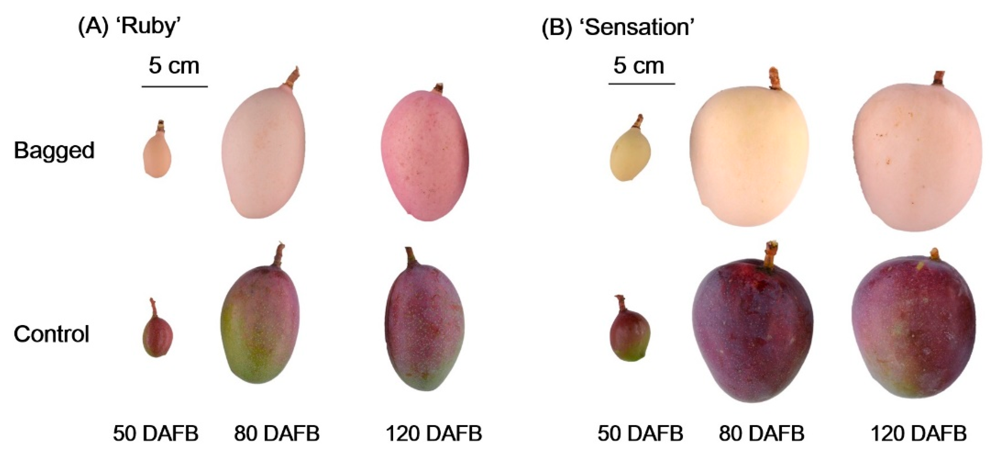
Figure 1. Representative images of bagged and control (un-bagged) ‘Ruby’ (A) and ‘Sensation’ (B) mango fruits during different developmental stages (DAFB is Days after full bloom).
Horticulturae 07 00423 g001
Figure 2. Concentration of main (AD) and minor (EG) flavonoid compounds detected in the fruit skin of bagged and control (non-bagged) ‘Ruby’ and ‘Sensation’ mango during different developmental stages. Each value represents the mean ± standard deviation of three biological replicates. Mean values were compared within the same day after full bloom, and values with the same letter are not significant-ly different, p > 0.05 according to Tukey tests.
Horticulturae 07 00423 g002
Figure 3. log2 expression levels of anthocyanin biosynthetic genes in bagged and control (non-bagged) ‘Ruby’ and ‘Sensation’ mango fruit skin at different developmental stages. Each value represents the mean ± standard deviation of three biological replicates. Mean values were compared within the same day after full bloom, and values with the same letter are not significant-ly different, p > 0.05 according to Tukey tests.
Horticulturae 07 00423 g003
Figure 4. log2 expression levels of anthocyanin regulatory genes in bagged and control (non-bagged) ‘Ruby’ and ‘Sensation’ mango fruit skin at different developmental stages. Each value represents the mean ± standard deviation of three biological replicates. Mean values were compared within the same day after full bloom, and values with the same letter are not significant-ly different, p > 0.05 according to Tukey tests.
Horticulturae 07 00423 g004
Figure 5. Sequence comparison of MiMYB1 promoter between ‘Ruby’ and ‘Sensation’.
Horticulturae 07 00423 g005
Table 1. Cis-acting elements within the promoter region of MiMYB1.
MotifSequenceDistance from ATGStrandFunction
ABREACGTG1030cis-acting element involved in the abscisic acid responsiveness
AE-boxAGAAACAA756+part of a module for light response
Box 4ATTAAT1311+part of a conserved DNA module involved in light responsiveness
ATTAAT697
CGTCA-motifCGTCA149+cis-acting regulatory element involved in the MeJA-responsiveness
CGTCA136+
GATA-motifGATAGGA1407+part of a light responsive element
GATAGGA433+
G-boxCACGAC783cis-acting regulatory element involved in light responsiveness
CACGTT1030+
GT1-motifGGTTAA1333light responsive element
LTRCCGAAA876+cis-acting element involved in low-temperature responsiveness
MBSCAACTG1340+MYB binding site involved in drought-inducibility
CAACTG244
CAACAG991+
TCA-elementCCATCTTTTT1074cis-acting element involved in salicylic acid responsiveness
TGA-elementAACGAC731+auxin-responsive element
Publisher’s Note: MDPI stays neutral with regard to jurisdictional claims in published maps and institutional affiliations.