Article

Boron Mitigates Cadmium Toxicity by Reducing Cadmium Accumulation, Enhancing Cell Wall Immobilization and Regulating Gene Expression in Malus Rootstock Under Hydroponic Conditions

1
College of Horticulture, Shenyang Agricultural University, Shenyang 110866, China
2
Key Lab of Fruit Quality Development and Regulation of Liaoning Province, Shenyang 110866, China
3
Northeast Germplasm Resources Innovation and Utilization Research Center (Analysis and Testing Center), Shenyang Agricultural University, Shenyang 110866, China
*
Author to whom correspondence should be addressed.
Horticulturae 2026, 12(9), 1163; https://doi.org/10.3390/horticulturae12091163
Submission received: 5 August 2026 / Revised: 6 September 2026 / Accepted: 12 September 2026 / Published: 15 September 2026
(This article belongs to the Special Issue Response of Horticultural Crops to Abiotic Stress)

Highlights

What are the main findings?
  • Treatment with 50 μM boron reduced Cd accumulation in apple rootstock and was accompanied by the downregulation of ZIP6/IRT1.
  • Boron enhanced Cd immobilization in root cell walls via increased pectin content and pectin methylesterase activity.
  • Boron alleviated oxidative damage by activating antioxidant enzymes and non-enzymatic defenses.
What are the implications of the main findings?
  • This study provides a physiological and molecular basis for using boron to mitigate Cd contamination in orchards.
  • It offers mechanistic insights that may support future strategies toward improved food safety and sustainable fruit production.

Abstract

To investigate the mitigating role and underlying mechanisms of exogenous boron (B) in cadmium (Cd)-stressed woody fruit trees, a hydroponic study was conducted using Malus hupehensis Rehd. seedlings treated with different B concentrations (0, 12.5, 50, and 150 μM H3BO3). Cd stress significantly inhibited plant growth and reduced photosynthetic parameters, pigment content, biomass, and root activity, but induced reactive oxygen species (ROS) accumulation. In contrast, the 50 μM B treatment (B2) effectively alleviated Cd toxicity. This treatment significantly decreased Cd accumulation, bioconcentration factor, and translocation factor across tissues. The B2 treatment enhanced Cd immobilization in root cell walls by increasing pectin content and pectin methylesterase activity. Additionally, it shifted Cd chemical forms toward lower-toxicity forms—increasing pectin- and protein-bound, phosphate-bound, and oxalate-bound Cd—while reducing inorganic and water-soluble Cd fractions. The B2 treatment elevated the activities of superoxide dismutase and peroxidase, and increased free proline and ascorbic acid, thereby reducing ROS and malondialdehyde accumulation. The B2 treatment also downregulated key genes including ZIP6 and IRT1 involved in Cd uptake. In conclusion, the most effective concentration among those tested (50 μM) alleviated Cd stress in Malus hupehensis Rehd., accompanied by modified Cd uptake and translocation, enhanced cell wall fixation, altered Cd chemical forms, improved antioxidant responses, and changes in stress-related gene expression.

1. Introduction

Heavy metal pollution is a severe environmental challenge for the survival of animals and plants, among which cadmium (Cd) poses the most serious threat to plant growth and human health [1]. In China, about 7% of arable land has been polluted by Cd (MEP 2014) [2]. Recently, inappropriate management and fertilization have led to Cd accumulation in orchards. For example, a study conducted in orchards across Shaanxi Province revealed that 10.0% of soil samples contained detectable levels of Cd, whereas 52.5% of fruit samples surpassed the national maximum permissible level of 0.03 mg kg−1 dry weight (DW) [3]. Cd is highly biotoxic and difficult to degrade, and can be easily absorbed and accumulated by plants. Cd accumulation in fruit trees can damage photosynthetic organs, decrease photosynthetic pigments, inhibit photosynthesis, induce oxidative stress, damage cell membrane structure and function, inactivate enzymes and proteins, restrain plant growth, and lower crop yield [4,5,6,7,8]. Therefore, it is practically meaningful to explore feasible strategies for alleviating Cd-induced phytotoxicity in apple rootstock.
Boron (B) is a trace element required for many physiological and metabolic functions in plants. Previous studies have demonstrated that B is involved in cell division and elongation, nucleic acid metabolism, biofilm structure and function maintenance, enzyme activation, nitrogen and carbohydrate metabolism, and sugar transport [9,10]. Furthermore, B is proposed in many plants to affect Cd accumulation and resistance. For example, exogenous B can significantly inhibit the absorption and transport of Cd in wheat, especially in the seedling stage and the growth stage [11]. Similarly, B addition significantly decreased Cd translocation from root to shoot in rice [12]. However, a narrow range exists between B deficiency and toxicity. For example, appropriate B supply promotes plant growth, whereas B concentrations above 50 μM may induce gradual growth-inhibitory responses [13]. Therefore, appropriate B concentrations must be screened for each experimental system before application to mitigate Cd toxicity. Recent studies have shown that appropriate B application alleviated Cd toxicity by activating the defense mechanisms in rape [10], wheat [14], and rice [15]. However, no information is available on whether appropriate B application could alleviate Cd stress in apples.
Exogenous B enhances Cd detoxification in herbaceous plants via several physiological mechanisms [16]. First, B can affect the cell wall’s ability to fix Cd by regulating the components of the plant cell wall (such as cellulose, hemicellulose, lignin and pectin) and their structure. The active groups, such as carboxyl (COO-), hydroxyl (-OH), and thiol (-SH) groups, in these cell wall components are the key sites for chelating Cd [17,18]. When B is deficient or excessive, it may change the physicochemical properties of the cell wall, thereby weakening its blocking effect on Cd through ion binding and fixation mechanisms [19]. Under Cd stress, the application of B increased the pectin content of the cell wall, improved the pectin methylesterase activity, reduced the degree of pectin methyl ester, and then enhanced the chelation of Cd by the root cell wall, thus reducing the level of Cd entering the organelles in Brassica napus [20]. Wu et al. [21] also found that the exogenous addition of 0.25 mg·L-1B increased the contents of pectin and cellulose in young stems of Brassica napus, promoted the fixation of Cd in the cell wall, enhanced the resistance of young stems to Cd, and reduced the toxicity of Cd.
Second, B affects the vacuolar compartmentalization capacity for heavy metals, with vacuoles playing a critical role in the immobilization, inactivation and detoxification of heavy metals [22]. B regulates the activity of vacuolar membrane transport proteins, which utilize proton gradients or ATP hydrolysis to compartmentalize Cd into vacuoles [23,24]. B status may modify membrane integrity, and this property could further affect vacuolar-mediated heavy metal detoxification under Cd stress [25]. Additionally, B influences the chelation of Cd by modulating the synthesis of phytochelatins (PCs). Since PCs are synthesized from GSH, it can be inferred that B-regulated GSH-dependent thiol metabolism may further affect Cd-PC complex formation and vacuolar sequestration efficiency [26].
Third, B affects plant Cd tolerance by regulating cell ROS homeostasis [26,27]. The appropriate concentration of B improves Cd resistance by regulating antioxidant enzymes and non-enzymatic antioxidants, eliminating excessive accumulated ROS in rice [28]. Fourth, B affects the expression of genes involved in Cd absorption and detoxification processes. When B is deficient, the expressions of iron-regulated transporter 1 (IRT1) and Zrt-/Irt-like protein 6 (ZIP6) are upregulated, promoting Cd to enter root cells; however, an appropriate supply of B can inhibit these transporters and reduce Cd accumulation [29]. B can also influence vacuolar Cd sequestration by affecting the expression of tonoplast-localized transporters such as metal tolerance protein 1 (MTP1) and magnesium-proton exchanger protein (MHX) [30]. To date, the B-mediated Cd stress response and its underlying mechanisms remain poorly characterized in woody fruit species, particularly in Malus hupehensis.
Soil Cd pollution in orchard restricts the healthy and sustainable development of the fruit industry. Appropriate B application could be a viable candidate in the mitigation of Cd toxicity. However, the alleviating effect of B on Cd stress in woody fruit trees, especially apple plants, and its mechanism have not been reported. This experiment employed Malus hupehensis Rehd., a commonly used rootstock known from previous research to exhibit poor Cd tolerance [31]. To examine whether appropriate B would alleviate Cd toxicity and the underlying physiological mechanism of B affecting Cd uptake, accumulation and detoxification in M. hupehensis, we exposed it to either 0 or 50 μM Cd together with 0, 12.5, 50 or 150 μM B for 30 days. We hypothesized that (i) appropriate B would reduce Cd migration and accumulation, while enhancing Cd tolerance in apple plants, and (ii) B would alleviate Cd toxicity in apple plants by regulating the physiological basis and the expression of genes related to Cd absorption and vacuolar compartmentalization. To test these hypotheses, we measured plant growth parameters, the antioxidant defense system, and Cd adsorption of the cell wall (CW), and studied the variation in the CW functional groups using FTIR analysis. The present findings advance our mechanistic understanding of B-mediated Cd tolerance in apple rootstock seedlings under hydroponic conditions. Further investigations are required to translate these laboratory observations into practical approaches for apple and other fruit trees.

2. Materials and Methods

2.1. Plant Material Cultivation and Treatment

The seedlings of M. hupehensis Rehd. were stratified in moist sand at 0–4 °C for 40 d. After germination, the seeds were cultivated for 40 days in a greenhouse under natural light (day/night temperature: 26/18 °C, 50–60% RH) using nursery plates with seedling matrices at Shenyang Agricultural University, Shenyang, China (41°49′ N, 123°34′ E). After 30 days, plants of similar height were transferred to plastic pots (20 cm × 20 cm × 18 cm) containing 4 kg clean sand. The plants in each pot were given 100 mL distilled water every morning, and were irrigated every other day with 50 mL 1/4 Hoagland nutrient solution.
When the seedlings grew to 20 cm high, 96 seedlings with the same growth were selected and transferred to a hydroponic tank for further cultivation. The experiment used a 4 × 2 factorial design with four boron (B) levels (0, 12.5, 50, and 150 μM H3BO3, designated B0, B1, B2, and B3) and two cadmium (Cd) levels (0 and 50 μM CdCl2), giving eight treatment combinations. Each treatment was replicated in three independent hydroponic tanks (the hydroponic tank was the experimental unit), with four seedlings grown per tank. Thus, the experiment comprised 24 tanks and 96 seedlings in total. All tanks were randomly arranged inside the greenhouse and re-randomized upon each solution renewal to minimize positional effects. They were then divided into 8 groups (12 plants per group) on average. Each tank was equipped with a 7 W ventilation pump, and the groups B0 (0 μM H3BO3), B1 (12.5 μM H3BO3), B2 (50 μM H3BO3), and B3 (150 μM H3BO3) were set by changing the amount of H3BO3 in the nutrient solution. Then, the seedlings were treated with 0 or 50 μM CdCl2, the nutrient solution was changed every 2 days, and the test materials were harvested after 30 days of treatment.
Before harvest, gas-exchange and chlorophyll fluorescence parameters were measured on individual plants. At harvest, the roots were rinsed with 0.05 mM CaCl2 to displace surface-adsorbed Cd, followed by three washes with distilled water. Each plant was separated into roots, stems, and leaves; plant height, basal stem diameter, root length, and fresh mass of each tissue were recorded on an individual-plant basis. Tissues were then frozen in liquid nitrogen and ground to fine powder using a ball mill (MM400; Retsch GmbH, Haan, Germany). Equal amounts of ground tissue from the four plants within the same hydroponic tank were pooled to form one composite sample representing that tank. Therefore, each treatment had three composite samples, one from each independent tank, which served as three biological replicates for all subsequent biochemical and molecular analyses.
The hydroponic nutrient solution was prepared from complete full-strength Hoagland stock solution, which contained inherent H3BO3. The full-strength stock contained (in mg L−1) KNO3 606, Ca(NO3)2·4H2O 944, NH4H2PO4 115, MgSO4·7H2O 246, Fe-EDTA 40, MnCl2·4H2O 7.2, ZnSO4·7H2O 0.44, CuSO4·5H2O 0.16, H2MoO4·H2O 0.18, and H3BO3 2.86. For plant cultivation, the stock solution was diluted with deionized water to obtain quarter-strength working solution, which provided a low background basal B concentration. H3BO3 was further supplemented into the diluted working solution to establish final total B concentrations of 0, 12.5, 50, and 150 μM. The “0 μM” B treatment represented only the background B derived from quarter-strength Hoagland solution. Each tank held 15 L of working nutrient solution. Solution pH was initially adjusted to 6.0 ± 0.1 using 0.1 M KOH and monitored daily. No obvious pH difference was observed between B-free and B-supplemented solutions. The solutions were continuously aerated using 7 W air pumps and completely renewed every two days to maintain stable nutrient concentrations and pH status.

2.2. Determination of Photosynthetic and Foliar Parameters, and Growth Characteristics

The net photosynthetic rate (Pn), stomatal conductance (Gs), and transpiration rate (Tr) of fully mature, upper-third leaves were measured using a CIRAS-2 portable photosynthesis system (PP Systems, USA). Measurements were performed between 9:00 and 11:00 a.m. under controlled leaf-chamber conditions. The leaf temperature was maintained at 25 °C, photosynthetic photon flux density (PPFD) was set at 800 μmol m−2 s−1, reference CO2 concentration was maintained at 400 μmol mol−1, and relative humidity inside the leaf chamber was controlled at 50–60%. The flow rate through the leaf chamber was set to 200 mL min−1. Before recording data, the leaves were acclimated inside the chamber for 2–3 min; data were logged only after Pn and Gs maintained stable values for at least 30 s. For each treatment, gas-exchange measurements were conducted on three plants per independent hydroponic tank, with three independent tanks serving as three biological replicates, consistent with the replication structure adopted for biochemical analyses.
The concentrations of chlorophylls and carotenoids in the leaves were analyzed as previously described [32]. After the sample was completely decolorized, the pigment content was determined by an ultraviolet spectrophotometer (UV-3802, Unico Instruments Co., Ltd., Shanghai, China) at 663, 646 and 470 nm, respectively.
Fresh subsamples (100 mg) were collected from each tissue sample of an individual plant. These subsamples were oven-dried at 60 °C for 72 h to constant weight to obtain the fresh-to-dry mass ratio. The total dry biomass of each tissue per plant was calculated by multiplying the total fresh weight of the corresponding tissue by the dry-to-fresh ratio derived from the 100 mg subsample. Fresh mass and dry mass were measured for each individual plant before any sample pooling for subsequent biochemical analyses.
Root morphological parameters were quantified using the WinRHIZO Root Analyzer System (WinRHIZO 2012b, Regent Instruments Canada Inc., Montreal, QC, Canada). Cleaned root samples were spread evenly in a water-filled transparent tray to avoid root overlapping, and scanned at 400 dpi resolution. Total root length, root surface area, and root volume were exported for subsequent analysis.
Root activity was determined by the TTC reduction assay. Briefly, 0.5 g fresh root tip segments were immersed in 5 mL of 0.4% (w/v) TTC solution mixed with an equal volume of phosphate buffer (pH 7.0). The samples were incubated at 37 °C in darkness for 2 h. The reaction was terminated by adding 2 mL 1 mol L−1 H2SO4. The roots were then transferred to a new tube, and formazan was extracted with 10 mL ethyl acetate by shaking. The absorbance of the extract was measured at 485 nm. Root activity was expressed as formazan production per gram fresh root per hour [33].

2.3. Determination of Cd Concentration, Cd Bio-Concentration Factor (BCF) and Cd Translocation Factor (Tf)

Fresh powders (500 mg) of each tissue sample were added to 7 mL HNO3 and 1 mL HClO4. After standing overnight, the samples were placed on a constant-temperature digestion plate at 170 °C for digestion according to the method described by Ma et al. [34]. After digestion, the concentration of Cd was determined by a flame atomic absorption spectrophotometer (Hitachi 180-80, Hitachi, Tokyo, Japan).
The Cd bio-concentration factor (BCF) and Cd translocation factor (Tf) were calculated using the following equations, respectively:
BCF = Croot/stem/leaf/Cs
Tfstem = Cstem/Croot
Tfleaf = Cleaf/Croot
Croot/stem/leaf represents the Cd concentration in the roots, stems, and leaves, and Cs represents the Cd content in the solution. BCF was calculated separately for the roots, stems and leaves. Tf values for stem-to-root and leaf-to-root translocation were computed independently.

2.4. Analysis of Subcellular Distribution and Chemical Forms of Cd

2.4.1. Subcellular Fractionation of Cd

The subcellular distribution of Cd was analyzed via differential-speed centrifugation following the modified procedure from Yan et al. [35]. Plant tissues were separated into four operationally defined fractions: cell wall fraction, organelle-enriched fraction, membrane-containing fraction, and soluble fraction. In short, frozen tissue samples were homogenized in pre-chilled extraction buffer (4 °C) consisting of 50 mM HEPES, 500 mM sucrose, 1.0 mM DTT, 5.0 mM ascorbic acid, and 1.0% (w/v) Polyclar AT PVPP, adjusted to pH 7.5. The homogenate was filtered through 100 μm nylon mesh; the retained residue corresponded to the cell wall fraction (F I). The obtained filtrate was centrifuged at 10,000× g for 30 min at 4 °C, and the pellet was collected as the organelle-enriched fraction (F II). The resulting supernatant was further centrifuged at 100,000× g for 30 min under 4 °C. The pellet from this step represented the membrane-containing fraction (F III), while the final supernatant was the soluble fraction (F IV). Each pellet fraction was resuspended using the same extraction buffer. All four subcellular fractions were dried and digested individually, and Cd concentrations were quantified by flame atomic-absorption spectrometry (Hitachi 180-80, Hitachi Ltd., Tokyo, Japan). Fraction recovery was calculated as the sum of Cd amounts recovered from all four fractions divided by the total Cd amount measured in the original tissue sample.

2.4.2. Sequential Extraction of Cd Chemical Forms

The determination of different chemical forms of Cd was performed following the method described by Zhou et al. [31]. Sequential extraction was conducted using five distinct extractants in fixed order to separate Cd with varied chemical properties: (1) 80% ethanol, for inorganic Cd species including nitrate-, chloride- and aminophenol-bound Cd; (2) deionized water, to recover water-soluble Cd-organic acid complexes and Cd(H2PO4)2; (3) 1 M NaCl, targeting Cd bound to pectate substances and proteins; (4) 2% acetic acid (HAC), for poorly soluble Cd phosphate such as CdHPO4 and Cd3(PO4)2; and (5) 0.6 M HCl, to extract oxalate-combined Cd.
Frozen plant samples were homogenized with corresponding extractant at a solid-to-liquid ratio of 1:100 (w/v). The mixture was shaken at 25 °C for 22 h, followed by centrifugation at 5000× g for 10 min, and the supernatant was retained. The residual pellet was re-extracted twice using the same extractant; each re-extraction lasted 2 h under 25 °C with shaking, and then centrifuged (5000× g, 10 min). All supernatants from one extraction step were pooled together. The remaining pellet was subjected to the subsequent extractant and the whole extraction workflow was repeated. The combined supernatant from each fraction was evaporated to dryness at 70 °C, digested with HNO3 at 145 °C, and Cd concentrations were measured by flame atomic-absorption spectrometry (Hitachi 180-80, Hitachi Ltd., Tokyo, Japan).

2.5. Fourier-Transform Infrared (FTIR) Spectroscopy Analysis

FTIR measurement was performed using an FTIR spectrometer (VERTEX70, Bruker Crop, Billerica, MA, USA). Briefly, 2 mg root-cell-wall isolates were fully ground with 200 mg KBr powder and pressed into pellets for infrared spectrum acquisition. Spectra were recorded at a resolution of 4 cm−1 over the wavenumber range of 4000–400 cm−1.
Three independent biological replicates were prepared per treatment. Two technical replicate spectra were acquired for each biological replicate and averaged to minimize instrumental noise. Raw spectra were pre-processed with OPUS software (Version 7.5, Bruker Optik GmbH, Ettlingen, Germany). Baseline correction was implemented to remove baseline drift caused by light scattering. Spectra were further normalized against the reference absorption band near 2920 cm−1 (C-H stretching vibration) to reduce deviations from uneven pellet thickness and sample loading. Savitzky–Golay smoothing was applied for noise reduction.
The peak intensities of characteristic functional group absorption bands were quantified as peak-height absorbance derived from baseline-corrected and normalized spectra. Relative variations in peak intensity under different Cd treatments were used for subsequent statistical comparisons.

2.6. Cell Wall Analysis

2.6.1. Determination of Cd Content in Each Component of Cell Wall

Root cell wall samples were isolated according to Kang et al. [36] with minor modifications. Briefly, 1.0 g frozen root tissues were homogenized in pre-cooled 75% (v/v) ethanol and centrifuged at 5000× g for 10 min at 4 °C; this extraction step was repeated three times. The pellets were sequentially washed with ice-cold acetone, 1:1 (v/v) methanol–chloroform mixture, and methanol (20 min for each washing step). Supernatants were discarded after each centrifugation, and the final pellet represented crude cell wall material. The crude cell walls were freeze-dried and stored at 4 °C for subsequent fractionation, Cd quantification and FTIR measurement. FTIR spectroscopy was performed as described in Section 2.5.
The separated cell wall components were oven-dried and digested with HNO3:HClO4 (7:1, v/v) on a heating plate. Cd concentrations were determined using a flame atomic absorption spectrophotometer.

2.6.2. Cell Wall Component Content Determination

The concentrations of pectin were determined by the m-hydroxybiphenyl method according to the method of Chudzik et al. [37].
The content of hemicellulose 1 (HC1), hemicellulose 2 (HC2) and cellulose was determined according to Zhu et al. [38], with components referring to previous descriptions with some modifications.

2.6.3. Assay of Cell Wall-Related Enzyme Activity

CW enzymes were extracted according to the method of Wu et al. [20]. The 0.02 g sample was weighed and 2 mL 1 mol/L NaCl (containing 20 mmol/L Tris–HCl, pH 4.8) was used as the extraction solution, centrifuged at 10,000× g for 15 min at 4 °C, and the separated supernatant was used as the crude enzyme extraction solution. After that, the activities of β-glucosaccharase and methylcellulase were measured at 540 nm. The activity of pectin methylesterase was measured at 450 nm.

2.7. Determination of O2, H2O2, MDA Content

The concentrations of O2 and H2O2 in the root and leaf tissues were determined spectrophotometrically at 530 and 410 nm, respectively, as suggested by He et al. [39]. Fresh tissue samples were homogenized with corresponding extraction buffer under ice-bath conditions, followed by centrifugation to obtain supernatant for subsequent color reaction according to the described protocol. The concentrations of MDA in plant tissues were determined with a spectrophotometer as described by He et al. [39].

2.8. Analysis of Non-Enzymatic Antioxidants and Enzymatic Antioxidant Activity

The content of free proline was determined according to Tamás et al. [40]. The soluble phenol content was determined using Folin–Ciocalteu reagent according to the method of Luo et al. [41]. The contents of AsA and total thiols (T-SH) were determined according to the methods of He et al. [42].
The determination of soluble protein to calculate antioxidant enzyme activity was made according to Luo et al. [41]. All enzymatic antioxidant measurements (SOD, POD, CAT, APX) were performed on root and leaf tissues. The enzyme activities of superoxide dismutase (SOD), peroxidase (POD), catalase (CAT), and ascorbate peroxidase (APX) were assayed according to Wang et al. [43]. Enzyme activities were expressed as U mg−1 soluble protein.

2.9. Quantitative Gene Expression Analysis

Total RNA was isolated from root tissues using the modified CTAB method [44]. Genomic DNA contamination was eliminated by DNase I treatment during RNA purification. RNA integrity and quality were verified by agarose gel electrophoresis and spectrophotometry (A260/A280 = 1.8–2.1). A fixed amount of total RNA (e.g., 1 μg) was used for first-strand cDNA synthesis using a reverse transcription kit (RR047A, Takara, China). Synthesized cDNA was 10-fold-diluted prior to qRT-PCR analysis [45].
qRT-PCR was performed in a total reaction volume of 10 μL, containing 5 μL 2 × SYBR Green Premix EX Taq II (DRR820A, Takara, Dalian, China), 1 μL diluted cDNA, and 1 μL of forward and reverse primers (10 μM stock, final concentration 0.2 μM for each primer). The thermal cycling program was 95 °C for 10 min; 40 cycles of 95 °C for 15 s; 60 °C for 60 s. Melting curve analysis was performed after amplification to confirm primer-specific single-product amplification. No-template negative controls (NTCs) were included in each run.
β-actin was selected as reference gene; its expression stability under B- and Cd-stress treatments was validated across all samples. Each sample included three biological replicates and two technical replicates. Amplification efficiencies of target genes and β-actin were comparable and within an acceptable range for the 2−ΔΔCT method. Relative gene expression levels were calculated using the2−ΔΔCT approach. Gene-specific primer sequences are listed in Table S1.

2.10. Data Statistics and Analysis

Statistical analyses were performed using Statgraphics (STN, St. Louis, MO, USA). Before analysis, dataset normality (Shapiro–Wilk test) and homogeneity of variance (Levene’s test) were examined to verify the assumptions for two-way ANOVA. No data transformation was applied in this study. Two-way factorial ANOVA was implemented, with B and Cd as the two fixed factors. The experimental unit for ANOVA was an individual plant, with n = 3 biological replicates per treatment.
The main effects of B, Cd, and their B × Cd interaction effect were evaluated. When ANOVA indicated significant effects (p ≤ 0.05), the Tukey HSD post hoc multiple comparison test was applied for pairwise comparisons, with multiplicity correction. Lower-case letters in figures denote significant differences among treatments. Figures were plotted using Origin 2021. The heatmap was generated using OmicStudio (https://www.omicstudio.cn/, accessed on 10 May 2025).

3. Results

3.1. Physiological and Growth Indicators

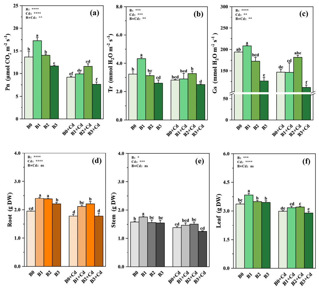
In the absence of Cd, Pn, Tr, Gs and photosynthetic pigments of M. hupehensis increased first and then decreased with the increases in B concentration (Figure 1 and Table S2). Cd stress significantly decreased Pn, Gs and photosynthetic pigments, regardless of B addition, with the exception of Gs, Chl(a) and Car under the B2 condition. The detrimental effects of Cd on those above parameters were alleviated after the application of 50 μM B, but aggravated with the supplement of 150 μM B (Figure 1 and Table S2).
Consistent with Cd-induced photosynthetic inhibition, Cd exposure significantly reduced the root, stem and leaf biomass of seedlings, except for root biomass under the B0 condition and stem biomass under the B2 and B3 conditions (Figure 1d–f). Under Cd exposure treatment, tissue biomass initially increased but then declined with rising B concentrations, reaching its peak in the B2 + Cd treatment (Figure 1d–f).
In the absence of Cd, with the increase in B concentration, the total root length, total root volume, total root surface and root activity of plants first increased and then decreased (Table S3). Under Cd stress, B application significantly increased root surface area, total root volume and root vitality compared to the B0 + Cd treatment, with the most pronounced effects observed under B2 + Cd treatment (Table S3).

3.2. Cd Concentration, BCF and Tf

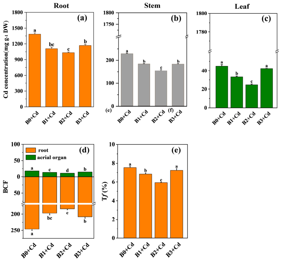
Under Cd treatment, B application significantly reduced Cd concentration in the roots, stems and leaves compared to the B-free treatment, with B2 + Cd treatment displaying the most pronounced effects (Figure 2a–c). Specifically, the B2 + Cd treatment reduced Cd concentration in the roots, stems and leaves by 25.23%, 32.60% and 44.72%, respectively, relative to the B0 + Cd treatment (Figure 2a–c). The BCF in the roots and aerial organ and Tf of M. hupehensis showed an initial decrease followed by an increase with rising concentration of B, reaching its minimum under the B2 + Cd treatment (Figure 2d,e). Compared to the B0 + Cd treatment, the B2 + Cd treatment reduced the BCF by 38.52% in roots and 25.23% in aerial parts, and decreased the Tf by 21.25% (Figure 2d,e).

3.3. Different Forms and Subcellular Distribution of Cd

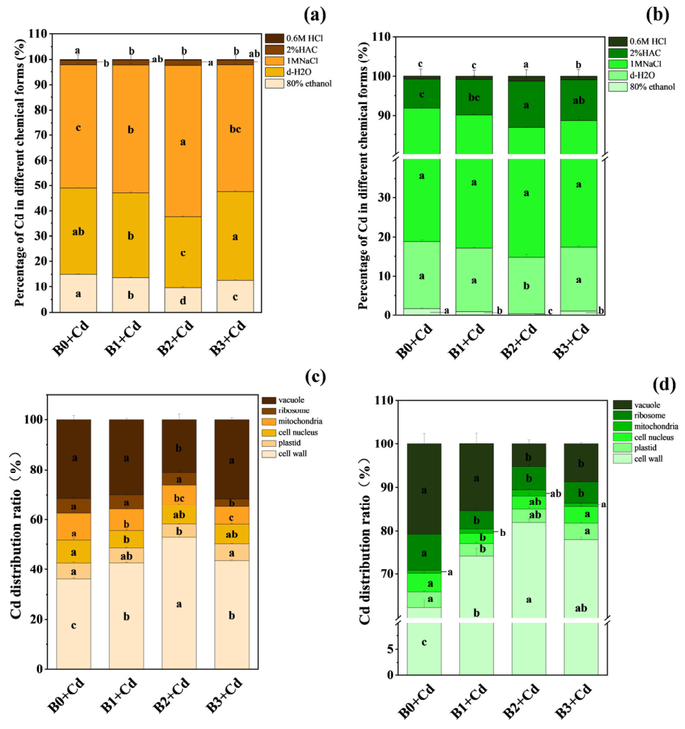
In both roots and leaves of M. hupehensis, Cd proportion in pectates and protein-integrated forms (extracted by 1 M NaCl) were highest, followed by water-soluble fraction (extracted by deionized water and corresponded to the most toxic form of metals) (Figure 3a,b). Under Cd stress, the proportion of Cd in inorganic (extracted by 80% ethanol) and water-soluble Cd in roots decreased first and then increased with the increase in B concentration, with B2 + Cd exhibiting the lowest value (Figure 3a). On the contrary, the proportion of pectin and protein-bound Cd and undissolved Cd phosphate form (extracted by 2% HAC) in the roots showed an initial increase followed by a decrease with rising concentration of B, with B2 + Cd treatment showing the highest value (Figure 3a). Compared with the B0 + Cd treatment, the addition of different concentrations of exogenous B reduced the proportion of Cd in the oxalate state (Figure 3a).
In the leaves, B2 + Cd treatment resulted in the lowest levels of inorganic and water-soluble Cd compared to B-free and other B treatments (Figure 3b). There was no significant difference in the proportion of pectates and protein-bound Cd among different B treatments (Figure 3b). Compared with B0 + Cd treatment, both B2 + Cd and B3 + Cd treatments increased the proportion of insoluble phosphate Cd and oxalate Cd, with the most pronounced increase observed under B2 + Cd (Figure 3b).
In the roots, the addition of various B concentrations increased the proportion of Cd in the cell wall compared with the B0 + Cd treatment, with the B2 + Cd treatment being the highest (Figure 3c). On the contrary, B2 + Cd treatment significantly decreased the proportions of Cd in the plastid, mitochondria and vacuole of roots compared to B0 + Cd (Figure 3c).
In the leaves, the proportion of Cd in the cell wall were increased by 19.06%, 31.47% and 24.9%, respectively, in the B1 + Cd, B2 + Cd and B3 + Cd treatment than those in the B0 + Cd treatment (Figure 3d). Compared with B0 + Cd, the proportion of Cd in plastids and nucleus was only decreased in the B1 + Cd treatment. The addition of all B concentrations decreased the proportion of Cd in ribosomes relative to the B0 + Cd treatment, but no significant difference was observed among the three treatments (Figure 3d). The Cd proportions in the vacuoles were relative lower in the B2 + Cd and B3 + Cd treatments than those in the B0 + Cd and B1 + Cd treatments (Figure 3d).

3.4. Analysis of Cd Content in Each Component of Root Cell Wall, FTIR, Root Cell Wall Components and Cell Wall Metabolic Enzyme Activities

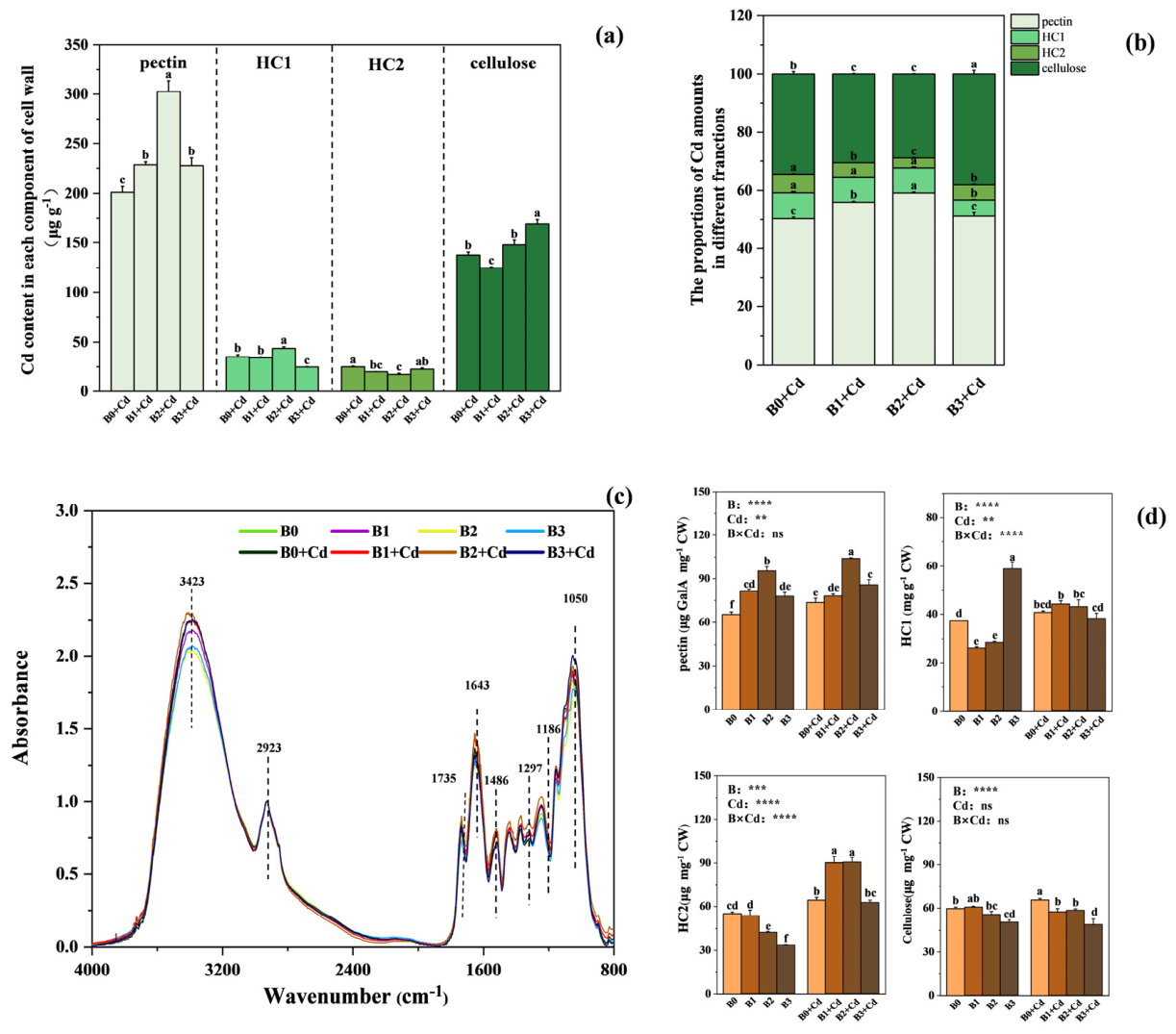
Compared with B0 + Cd treatment, the addition of B significantly increased the Cd content in pectin, with B2 + Cd showing the most pronounced effects. Moreover, the B2 + Cd treatments significantly increased the Cd content in HC1 and cellulose, respectively, relative to the B0 + Cd treatment (Figure 4a). Surprisingly, the addition of B reduced the Cd content in HC2, except for B3 + Cd treatment in comparison with no B treatment (Figure 4a). In general, the proportion of Cd in pectin was the highest, followed by cellulose, and the Cd proportion in HC1 and HC2 was relatively low in the roots of M. hupehensis (Figure 4b). Consistent with the results for the Cd content in various cell wall components, compared with the B-free treatment, the three B concentration treatments significantly increased the proportion of Cd in pectin and cellulose, except for in pectin under the B3 + Cd treatment (Figure 4b).
FTIR analysis was conducted to characterize the functional groups in the root cell of M. hupehensis subjected to B and Cd treatments (Figure 4c). A peak at ~3413 cm−1 represented the O-H or N-H stretching vibration of fatty acids, proteins, pectin, and hemicellulose. A peak at ~2924 cm−1 represented the C-H stretching vibration of a lipid carbon chain (-CH3, =CH2, =CH-) derived mainly from hydrophilic lipid molecules. A peak at ~1738 cm−1 represented the vibrations of -C=O in pectin. A peak at 1639 cm−1 represented the vibrations of C-N in proteins. Numerous absorption peaks were detected in the 1517–1253 cm−1 range and represented the symmetric bending vibration of N-H in proteins, the unsymmetrical stretching vibration of carboxylate COO-, the stretching vibration of sulfate -C-O-S, the vibration of phosphate in C-O-P, and the stretching vibration of carboxyl -C-O. A peak at 1154 cm−l represented the polysaccharide ring structure of C-C or C-O. A peak at 1041 cm−1 represented the -CH bending or -C-C, -C-O stretching vibration peak of insoluble sugars, cellulose sugar chains, and hemicellulose. Regardless of B and Cd stress, the spectral peak shape of the plant root cell walls did not change significantly. Only the characteristic absorption peaks occurred to different degrees. Cd stress led to higher intensity for each absorption peak of the root cell wall, regardless of B concentration. Under Cd stress, greater absorbance intensity was observed for each absorption peak under B2 + Cd treatment, except for the peak at 1050 cm−1 (Figure 4c).
With the increase in B concentration, the pectin content first increased and then decreased, reaching its peak after the application of 50 μM B, regardless of Cd treatment (Figure 4d). In general, Cd stress increased the content of pectin in all treatments, with the exception of 12.50 μM B, and the highest value was observed in the B2 + Cd treatment. After Cd exposure, the contents of HC1 and HC2 significantly increased in all treatments except for HC1 under the B-free and 150 μM B treatments (Figure 4d). Surprisingly, Cd stress had no significant effects on the cellulose content of the roots after the addition of B (Figure 4d).
In the absence of Cd, β-glucosaccharase, methylcellulase and pectin methylesterase activities were significantly increased after the addition of B except for the first two enzymes under B1 treatment (Figure S1). Under Cd stress, the activities of β-glucosaccharase and pectin methylesterase significantly increased and then decreased with the increase in B concentrations, with the B2 + Cd treatment showing the highest values (Figure S1). There was no significant difference in methylcellulase activity among the different treatments under Cd stress (Figure S1).

3.5. Reactive Oxygen Species and Antioxidants in Seedlings

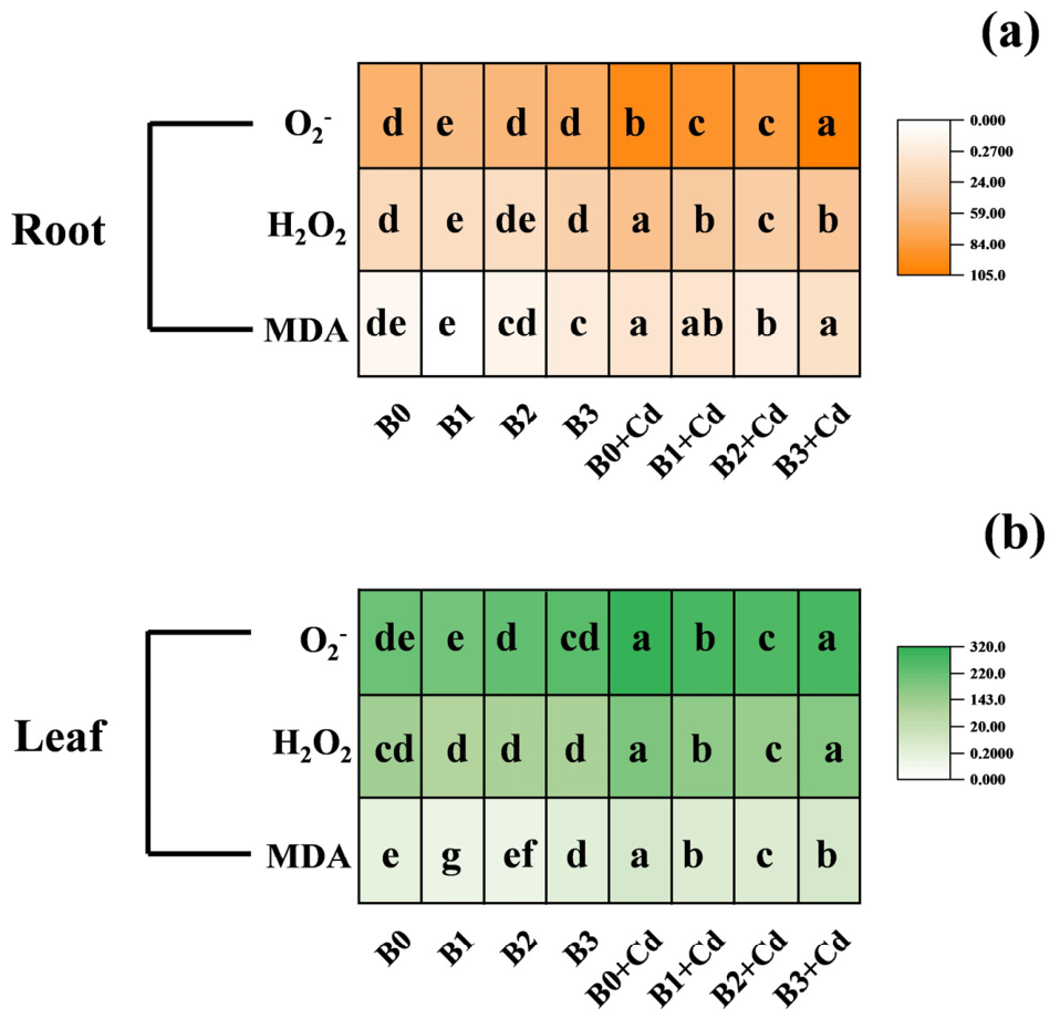
Cd stress significantly increased the contents of O2, H2O2 and MDA in the roots and leaves of M. hupehensis (Figure 5). However, under Cd stress, the contents of O2, H2O2 and MDA in the roots and leaves of M. hupehensis were significantly lower in the B2 + Cd treatment than the other three treatments (Figure 5). In contrast, the O2, H2O2 and MDA levels in the B0 + Cd treatment were generally higher than in the other treatments.
In general, compared with the Cd-free controls, Cd stress significantly decreased free proline contents in roots and leaves under the B0 and B1 treatments (Figure S2). However, under Cd exposure, the application of 50 μM B (B2 + Cd) significantly increased root proline content compared with B0 + Cd (Figure S2). Compared with Cd-free controls, Cd stress significantly decreased T-SH content in roots but increased T-SH content in leaves, irrespective of B treatment. Under Cd exposure, T-SH content was highest in the B2 + Cd treatment in both roots and leaves. Compared with their respective Cd-free controls, Cd stress only significantly increased root soluble phenolics under the B2 + Cd treatment, and leaf soluble phenolics under the B3 + Cd treatment. Under Cd stress, compared with B0 + Cd, the B3 + Cd treatment significantly increased the ASC content in roots, and all three B treatments (B1 + Cd, B2 + Cd, B3 + Cd) significantly elevated the ASC content in leaves (Figure S2).
Compared with their respective Cd-free controls, Cd stress exerted no consistent significant effect on root and leaf SOD activity, except that leaf SOD activity decreased significantly in the B3 + Cd treatment (Figure S3). Under Cd stress, SOD activity in both roots and leaves was higher in the B1 + Cd and B2 + Cd treatments than in B0 + Cd and B3 + Cd (Figure S3). Under Cd stress, POD activity in the roots and leaves was significantly higher in the B2 + Cd treatment than for B0 + Cd (Figure S3). Under Cd stress, compared with B0 + Cd, B1 + Cd significantly increased root CAT activity. By contrast, B2 + Cd and B3 + Cd reduced leaf CAT activity relative to B0 + Cd. Compared with Cd-free controls, Cd stress significantly increased root APX activity in all B treatments except B0 + Cd. However, Cd stress significantly decreased leaf APX activity, irrespective of B treatment (Figure S3).

3.6. Gene Expression Analysis

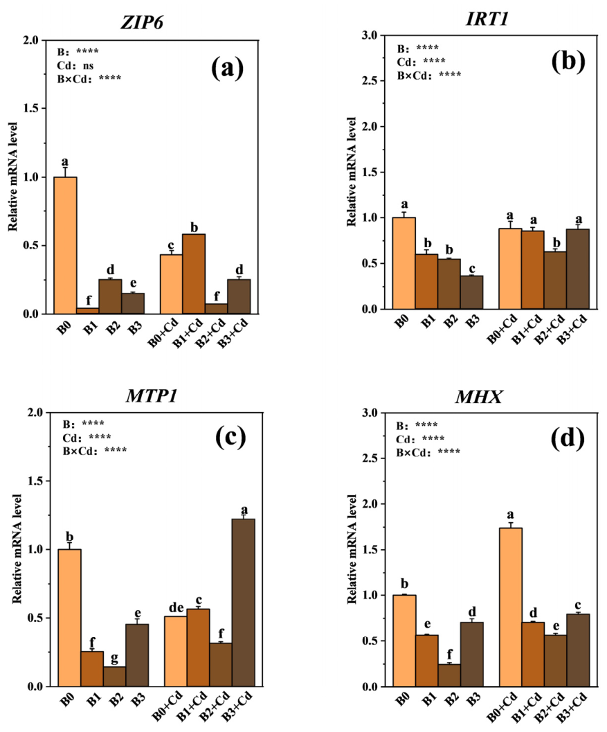
In the absence of Cd stress, the expression of ZIP6 was significantly downregulated in all B addition treatments compared with B0 (Figure 6). Under Cd stress, the relative expression of ZIP6 decreased to approximately 1/6.14 (0.16-fold) and 1/1.72 (0.58-fold) of the B0 + Cd level under the B2 + Cd and B3 + Cd treatments respectively compared with the B0 + Cd treatment (Figure 6). Similarly, under Cd stress, the expression of IRT1 in the B2 + Cd treatment was also significantly lower than that in the other three treatments. Compared to the Cd-free controls, Cd exposure increased MTP1 transcript levels in the roots under all B treatments except for the B0 condition. Irrespective of Cd treatment, MTP1 expression remained lower under the 50 µM B treatment (Figure 6c). Similarly, compared to the Cd-free controls, Cd exposure generally upregulated MHX transcript levels. In parallel with the trend observed for MTP1, MHX expression was also consistently lower under 50 µM B treatment regardless of Cd exposure (Figure 6d).

4. Discussion

4.1. Appropriate B Application Reduced Cd Accumulation and Enhanced Cd Tolerance in Apple Rootstock

The toxic effects of Cd stress on plants are primarily characterized by a significant inhibition of photosynthetic systems and root growth [46]. Under Cd stress, Malus hupehensis Rehd. exhibited suppressed photosynthetic pigment levels in leaves and severe impairment of root growth and activity, which is consistent with Cd-induced damage reported in previous plant studies [46].
The exogenous application of B can effectively alleviate the toxicity of Cd, but this alleviating effect has a concentration effect. In herbaceous crops, optimal B supply restrains Cd root-to-shoot translocation and mitigates Cd-caused growth inhibition, while insufficient or excessive B fails to provide protection or even aggravates stress damage [11,15]. In our study, 50 μM B exerted the most prominent protective effect against Cd stress. This dosage suppressed Cd accumulation in roots and lowered Cd translocation to shoots, thereby restoring photosynthetic performance and root morphology. However, compared with the 50 μM B treatment, a lower B concentration (12.5 µM) exhibited a reduced alleviative effect on Cd stress, whereas a higher B concentration (150 µM) even exacerbated Cd toxicity. These results suggest that 50 µM B represents the most effective concentration in this study for effectively mitigating Cd stress in M. hupehensis Rehd. This concentration may activate specific plant defense mechanisms, thereby reducing Cd mobility and enhancing plant tolerance.

4.2. Appropriate B Application Enhanced Cd Tolerance by Modulating Cd Chemical Forms and Cell Wall-Binding Capacity

The toxicity degree and mobility of Cd in plants are dependent on its chemical forms inside cells. Overall, inorganic and organic Cd (extracted by 80% ethanol and deionized water) have stronger migration ability and greater toxicity to plant cells than other Cd chemical forms [47,48]. In our experiment, the appropriate concentration of B treatment (50 μM) significantly reduced the proportion of highly mobile and toxic inorganic and water-soluble Cd in roots and leaves, while increasing the proportion of less mobile and less toxic pectin protein-bound and insoluble phosphate-bound Cd. This shift indicates that 50 μM B alleviates Cd toxicity by reducing the proportion of highly mobile and phytotoxic Cd species and promoting their conversion into less bioavailable immobilized forms. The increased proportion of pectate/protein-integrated Cd further indicates that cell wall components play a critical role in Cd immobilization, which is consistent with findings in Cd-stressed watercress plants [49].
At the subcellular level, root cell walls serve as the primary barrier preventing Cd from entering the cytoplasm. Under Cd stress, treatment with 50 μM B significantly increased the content and distribution ratio of Cd in the cell wall components of roots and leaves. This pattern is consistent with reduced Cd transport from the cell wall into the cytoplasm, thereby protecting organelles and limiting Cd translocation to aerial parts of the plant. The present study indicates that 50 μM B increased the proportion of cell wall-bound Cd and decreased the Cd distribution in plastids and organelles. In agreement with Riaz et al. [12], B reduced intracellular Cd transport by enhancing cell wall adsorption in rice. These results corresponded to the finding that the appropriate concentration of 50 μM B decreased Cd accumulation in aerial organs and alleviated Cd toxicity in M. hupehensis Rehd. Seedlings (Figure 2). Therefore, the enhanced Cd-binding capacity of root cell walls may be recognized as an effective mechanism for improving Cd tolerance under 50 μM B exposure.
The Cd binding capacity of CWs depends on their composition (cellulose, pectin, and hemicellulose) and component properties. Under Cd stress, the 50 μM B significantly increased both the content and proportion of Cd in the pectin and HC1 fractions, suggesting that pectin and HC1 play important roles in Cd immobilization. This result aligns with the findings of Wu et al. [21], in which B application enhanced Cd sequestration in the cell wall of Brassica napus by promoting its binding to pectin and cellulose. These results suggested that pectin-mediated Cd sequestration may constitute a key mechanism by which 50 μM B enhances the Cd-binding capacity of the cell wall. The CW metal binding capacity depends on the abundance of functional groups (−COO, −OH, and C=O). FTIR analysis showed that 50 μM B increased the absorption intensities of these key groups. This increase in the number of reactive sites may likely have augmented the affinity of the CWs for Cd, further consistent with the increased pectin−Cd binding [50]. Additionally, 50 μM B further enhanced the Cd-induced increases in the contents of pectin, contributing to improved Cd-binding ability. More importantly, root PME activity was significantly increased under 50 μM B treatment in the presence of Cd. PME catalyzes the demethylation of pectin, generating negatively charged carboxyl groups that enhance the capacity for Cd2+ binding [51]. Consistent with this mechanism, our results suggested that 50 μM B may promote the demethylation of highly methyl-esterified pectin under Cd exposure by enhancing root PME activity, thereby potentially increasing the abundance of free carboxyl groups available for Cd binding.

4.3. Appropriate B Application Enhanced Tolerance to Cd Stress in Apple Rootstock by Activating the Antioxidant Defense System and Regulating Related Gene Expression

Cd stress induces reactive oxygen species (ROS) accumulation and membrane lipid peroxidation in plants, leading to oxidative stress [52]. In this study, the application of 50 μM B significantly decreased the levels of O2, H2O2, and MDA in both Cd-exposed roots and leaves. These results demonstrated that exogenous B application effectively mitigates Cd-induced oxidative damage. Thus, the appropriate concentration of B plays a critical role in enhancing the oxidative stress tolerance of M. hupehensis Rehd. under Cd stress.
Plants alleviate heavy metal toxicity by activating both enzymatic and non-enzymatic antioxidant systems [53]. In the present study, the reduced levels of ROS and MDA under 50 μM B treatment were associated with a marked increase in the contents of free proline and T-SH in roots and leaves. Elevated root AsA was observed under higher-dose B3 + Cd treatment. For antioxidant enzymes, enhanced POD activity was detected in roots and leaves, together with increased APX activity in roots. No consistent significant changes in SOD activity were observed across most comparisons. The results indicated that M. hupehensis Rehd. seedlings treated with 50 μM B exhibited greater antioxidant capacity compared with other B concentrations. These findings are consistent with earlier reports in rice [28], where exogenous B alleviated Cd-induced oxidative stress by strengthening the antioxidant defense system.
B application reduced Cd accumulation and enhanced Cd tolerance in M. hupehensis Rehd., which is probably associated with the B-modulated transcription of genes regulating Cd uptake and detoxification. IRT1 has been shown to enhance Cd accumulation when overexpressed in Arabidopsis thaliana [54]. Under Cd stress, the expression of ZIP6 and IRT1 was markedly downregulated in plants treated with 50 μM B, suggesting that B2 + Cd treatment may reduce Cd uptake. This finding aligns with the observed decrease in root Cd concentration under Cd stress following B2 + Cd treatment. Desbrosses-Fonrouge et al. reported that the tonoplast-localized MTP1 participates in the vacuolar sequestration of divalent heavy metal cations including Cd [55].
In the present study, Cd stress significantly upregulated MTP1 and MHX expression except for B-free treatment, but their expression levels were always lowest under B2 + Cd treatment. These results suggest that, under Cd stress, vacuolar compartmentalization significantly mitigates Cd toxicity in M. hupehensis Rehd. However, appropriate B application does not appear to boost vacuolar Cd compartmentalization, suggesting that B likely reduces Cd toxicity primarily by strengthening the cell wall rather than via vacuolar sequestration. This hypothesis is consistent with the subcellular distribution results described above, which showed that under B2 treatment, the proportion of Cd in root vacuoles decreased while that in the cell wall increased. Together, these findings point to a putative molecular mechanism by which B modulates Cd detoxification.
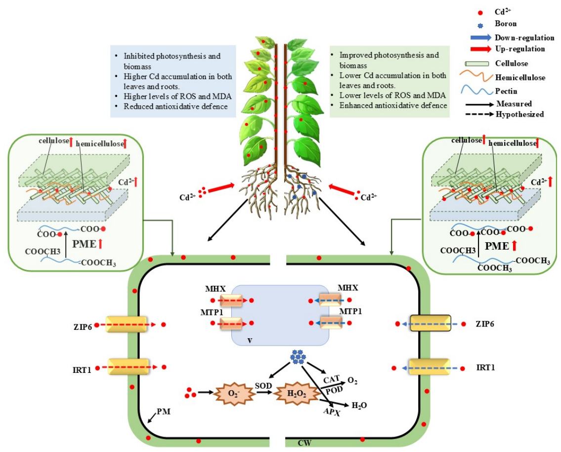
As summarized in Figure 7, collectively, the present work illustrates how the appropriate concentration of B treatment confers Cd tolerance in M. hupehensis Rehd. At the whole-plant level, appropriate B supply improves photosynthetic performance and plant biomass, reduces Cd accumulation in both leaves and roots, lowers ROS and MDA contents, and enhances antioxidant defense capacity under Cd stress. Different from many previous studies that regarded vacuolar compartmentalization as one of the pathways for B-mediated heavy metal detoxification [30,56], our results suggest that cell-wall-driven Cd immobilization acts as the dominant pathway. The appropriate concentration of B modified cell wall components and promoted pectin-associated Cd sequestration, restricting Cd translocation from root cell walls into intracellular compartments. At the molecular level, B downregulated the transcription of Cd-uptake-related genes (IRT1, ZIP6), as well as vacuolar-compartmentation-related genes (MHX, MTP1). These multilayered physiological and molecular responses jointly contribute to Cd resistance of apple rootstock seedlings, providing new insights into B-alleviated Cd toxicity in woody fruit tree rootstock. It should be acknowledged that the present study was conducted using young seedlings under hydroponic conditions. The obtained findings cannot be directly extrapolated to mature fruit-bearing trees grown in field orchard soils. Nevertheless, this seedling-based model system provides preliminary mechanistic insights into B-mediated Cd detoxification in woody fruit tree rootstock, and further validation under field conditions is warranted in future studies.

5. Conclusions

The application of B at the most effective concentration among those tested (50 μM) effectively alleviates Cd toxicity in M. hupehensis Rehd. seedlings under hydroponic conditions. The mitigation is achieved through multiple interconnected mechanisms: (1) B supply enhances Cd immobilization in the root cell wall by increasing pectin content and pectin methylesterase activity, thereby promoting the binding of Cd to cell wall components and altering its subcellular distribution toward less mobile and less toxic forms; (2) B activates both enzymatic and non-enzymatic antioxidant systems, reducing reactive oxygen species (ROS) accumulation and lipid peroxidation, thus improving cellular redox homeostasis under Cd stress; and (3) B reduces Cd uptake by downregulating the expression of Cd influx transporters (ZIP6 and IRT1). These findings suggest the pivotal role of B in enhancing Cd tolerance in apple rootstock through coordinated physiological responses under the tested hydroponic conditions, providing a mechanistic basis for further investigation of B in mitigating Cd contamination in orchards and ensuring the sustainable production of fruit trees. Nevertheless, these results were obtained from hydroponic seedling experiments; field-based validation is required before extending such conclusions to practical orchard production.

Supplementary Materials

The following supporting information can be downloaded at https://www.mdpi.com/article/10.3390/horticulturae12091163/s1, Table S1: Primers used for qRT-PCR. Table S2: Photosynthetic pigments (mg g-1dry weight) in leaves of Malus hupehensis exposed to 0 or 50 μM CdCl2 combined with 0, 12.5, 50 or 150 μM B for 30 days. Data are means ± standard error (SE; n = 3). Different letters after values indicate significant differences between treatments; p-values for ANOVA of CdCl2 (Cd), B, and their interactions are shown. **: p ≤ 0.01; ***: p ≤ 0.001; ****: p ≤ 0.0001; ns: not significant. Chla, chlorophyll a; Chl b, chlorophyll b; Chl (a + b), sum of chlorophyll a and b; Car, carotenoid. Table S3: Root configuration and vitality of Malus hupehensis exposed to 0 or 50 μM CdCl2 combined with 0, 12.5, 50 or 150 μM B for 30 days. Data are means ± standard error (SE; n = 3). Different letters after values indicate significant differences between treatments; p-values for ANOVA of CdCl2 (Cd), B, and their interactions are shown. *: p ≤ 0.05; **: p ≤ 0.01; ***: p ≤ 0.001; ****: p ≤ 0.0001; ns: not significant. Figure S1: Activities of β-glucosaccharase, methylcellulase and pectin methylesterase in the root of Malus hupehensis exposed to 0 or 50 μM CdCl2 combined with 0, 12.5, 50 or 150 μM B for 30 days. Data are means ± standard error (SE; n = 3). Different letters on the bars indicate significant differences between treatments. p-values for ANOVA of CdCl2 (Cd), B, and their interactions are shown. *: p ≤ 0.05; **: p ≤ 0.01; ***: p ≤ 0.001; ****: p ≤ 0.0001; ns: not significant. Figure S2: Free proline, soluble phenolics, ascorbate (ASC), and total thiols (T-SH) in the roots and leaves of Malus hupehensis exposed to 0 or 50 μM CdCl2 combined with 0, 12.5, 50 or 150 μM B for 30 days. Data are means ± standard error (SE; n = 3). Different letters on the bars indicate significant differences between treatments; p-values for ANOVA of CdCl2 (Cd), B, and their interactions are shown. *: p ≤ 0.05; **: p ≤ 0.01; ***: p ≤ 0.001; ****: p ≤ 0.0001; ns: not significant. Figure S3: Superoxide dismutase (SOD), peroxidase (POD), catalase (CAT), ascorbate peroxidase (APX) and glutathione reductase (GR) in the roots and leaves of Malus hupehensis exposed to 0 or 50 μM CdCl2 combined with 0, 12.5, 50 or 150 μM B for 30 days. Data are means ± standard error (SE; n = 3). Different letters on the bars indicate significant differences between treatments; p-values for ANOVA of CdCl2 (Cd), B, and their interactions are shown. *: p ≤ 0.05; **: p ≤ 0.01; ***: p ≤ 0.001; ****: p ≤ 0.0001; ns: not significant. Figure S4: Representative phenotypes of Malus hupehensis exposed to 0 or 50 μM CdCl2 combined with 0, 12.5, 50 or 150 μM B for 30 days. References [57,58] are cited in the Supplementary Materials.

Author Contributions

Y.T.: Writing—review and editing, Writing—original draft, Software, Methodology, Formal analysis, Data curation, Conceptualization. X.L.: Writing—review and editing, Methodology. M.X.: Writing—review and editing. S.Q.: Writing—review and editing, Funding acquisition. D.L.: Writing—review and editing, Resources, Project administration, Methodology, Funding acquisition, Conceptualization. J.H.: Writing—review and editing, Supervision, Methodology, Investigation, Funding acquisition, Conceptualization. All authors have read and agreed to the published version of the manuscript.

Funding

This work was funded by the earmarked fund for China Agriculture Research System [Grant No. CARS-25], the Liaoning Science and Technology Plan Project [Grant No. 2023-MSLH-282], and the Scientific Research Foundation of Talent Introduction of Shenyang Agricultural University [20153007].

Institutional Review Board Statement

Not applicable.

Informed Consent Statement

Not applicable.

Data Availability Statement

All data produced or examined throughout this study are contained within this article. For additional inquiries, please contact the corresponding author.

Conflicts of Interest

The authors declare no conflicts of interest.

References

  1. Liu, H.; Wu, Y.; Cai, J.; Chen, Y.; Zhou, C.; Qiao, C.; Wang, Y.; Wang, S. Effect of Auxin on Cadmium Toxicity-Induced Growth Inhibition in Solanum lycopersicum. Toxics 2024, 12, 374. [Google Scholar] [CrossRef] [Scilit] [PubMed]
  2. MEP; MLR. Report on the National General Survey on Soil Contamination; Ministry of Environmental Protection: Beijing, China, 2014.
  3. Guo, J.; Yue, T.; Li, X.; Yuan, Y. Heavy metal levels in kiwifruit orchard soils and trees and its potential health risk assessment in Shaanxi, China. Environ. Sci. Pollut. Res. 2016, 23, 14560–14566. [Google Scholar] [CrossRef] [Scilit] [PubMed]
  4. Mittler, R. ROS are good. Trends Plant Sci. 2017, 22, 11–19. [Google Scholar] [CrossRef] [Scilit] [PubMed]
  5. Haider, F.U.; Liqun, C.; Coulter, J.A.; Alam Cheema, S.; Wu, J.; Zhang, R.; Wenjun, M.; Farooq, M. Cadmium toxicity in plants: Impacts and remediation strategies. Ecotoxicol. Environ. Saf. 2021, 211, 111887. [Google Scholar] [CrossRef] [Scilit] [PubMed]
  6. Huang, D.; Wang, Q.; Zou, Y.; Ma, M.; Jing, G.; Ma, F.; Li, C. Silencing MdGH3-2/12 in apple reduces cadmium resistance via the regulation of AM colonization. Chemosphere 2021, 269, 129407. [Google Scholar] [CrossRef] [Scilit] [PubMed]
  7. Zhuang, X.L.; Liu, S.Y.; Xu, S.Z.; Qin, S.; Lyu, D.; He, J.; Zhou, J. Arbuscular Mycorrhizal Fungi Alleviate Cadmium Phytotoxicity by Regulating Cadmium Mobility, Physiological Responses, and Gene Expression Patterns in Malus hupehensis Rehd. Int. J. Mol. Sci. 2025, 26, 1418. [Google Scholar] [CrossRef] [Scilit] [PubMed]
  8. Zhou, L.; Zhou, L.; Wu, H.; Kong, L.; Li, J.; Qiao, J.; Chen, L. Analysis of Cadmium Contamination in Lettuce (Lactuca sativa L.) Using Visible-Near Infrared Reflectance Spectroscopy. Sensors 2023, 23, 9562. [Google Scholar] [CrossRef] [Scilit] [PubMed]
  9. Shireen, F.; Nawaz, M.A.; Chen, C.; Zhang, Q.; Zheng, Z.; Sohail, H.; Sun, J.; Cao, H.; Huang, Y.; Bie, Z. Boron: Functions and approaches to enhance its availability in plants for sustainable agriculture. Int. J. Mol. Sci. 2018, 19, 1856. [Google Scholar] [CrossRef] [Scilit] [PubMed]
  10. Matthes, M.S.; Robil, J.M.; Mcsteen, P. From element to development: The power of the essential micronutrient boron to shape morphological processes in plants. J. Exp. Bot. 2020, 71, 1681–1693. [Google Scholar] [CrossRef] [Scilit] [PubMed]
  11. Qin, S.; Xu, Y.; Nie, Z.; Liu, H.; Gao, W.; Li, C.; Wang, L.; Zhao, P. Effect of boron on cadmium uptake and expression of Cd transport genes at different growth stages of wheat (Triticum aestivum L.). Ecotoxicol. Environ. Saf. 2022, 241, 113834. [Google Scholar] [CrossRef] [Scilit] [PubMed]
  12. Riaz, M.; Kamran, M.; Fang, Y.; Yang, G.; Rizwan, M.; Ali, S.; Zhou, Y.; Wang, Q.; Deng, L.; Wang, Y.; et al. Boron supply alleviates cadmium toxicity in rice (Oryza sativa L.) by enhancing cadmium adsorption on cell wall and triggering antioxidant defense system in roots. Chemosphere 2012, 266, 128938. [Google Scholar]
  13. Dinh, A.Q.; Naeem, A.; Mühling, K.H. Growth and Distribution of Boron in Oilseed Rape (Brassica napus L.) as Affected by Boron Supply. Plants 2022, 11, 2746. [Google Scholar] [CrossRef] [Scilit] [PubMed]
  14. Qin, S.; Liu, H.; Rengel, Z.; Gao, W.; Nie, Z.; Li, C.; Hou, M.; Cheng, J.; Zhao, P. Boron inhibits cadmium uptake in wheat (Triticum aestivum) by regulating gene expression. Plant Sci. 2020, 297, 110522. [Google Scholar] [CrossRef] [Scilit] [PubMed]
  15. Huang, Y.Y.; Fei, G.; Yu, S.L.; Liu, Y.-F.; Fu, H.-L.; Liao, Q.; Huang, B.-F.; Liu, X.-Y.; Xin, J.-L.; Shen, C. Molecular and biochemical mechanisms underlying boron-induced alleviation of cadmium toxicity in rice seedlings. Ecotoxicol. Environ. Saf. 2021, 225, 112776. [Google Scholar] [CrossRef] [Scilit] [PubMed]
  16. Huang, Y.Y.; Shen, C.; Wang, X.S.; Fu, H.; Huang, B.; Li, Y.; Wen, H.; Wang, Y.; Zhou, W.; Xin, J. Boron decreases cadmium accumulation in water spinach by enhancing cadmium retention in the root cell walls. Environ. Sci. Pollut. Res. 2023, 30, 101168–101177. [Google Scholar] [CrossRef] [Scilit] [PubMed]
  17. Kushwaha, A.; Rani, R.; Kumar, S.; Gautam, A. Heavy metal detoxification and tolerance mechanisms in plants: Implications for phytoremediation. Environ. Rev. 2015, 24, 39–51. [Google Scholar] [CrossRef] [Scilit]
  18. Wan, H.; Feng, Y.Y.; Xiao, L.Z.; Cao, Y.; He, J.; Li, H.; Qin, S.; Lyu, D. Malus rootstocks affect copper accumulation and tolerance in trees by regulating copper mobility, physiological responses, and gene expression patterns. Environ. Pollut. 2021, 287, 117610. [Google Scholar] [CrossRef] [Scilit] [PubMed]
  19. Krzesłowska, M. The cell wall in plant cell response to trace metals: Polysaccharide remodeling and its role in defense strategy. Acta Physiol. Plant. 2011, 33, 35–51. [Google Scholar] [CrossRef] [Scilit]
  20. Wu, X.; Song, H.; Guan, C.; Zhang, Z. Boron alleviates cadmium toxicity in Brassica napus by promoting the chelation of cadmium onto the root cell wall components. Sci. Total Environ. 2020, 728, 138833. [Google Scholar] [CrossRef] [Scilit] [PubMed]
  21. Wu, X.; Song, H.; Guan, C.; Zhang, Z. Boron mitigates cadmium toxicity to rapeseed (Brassica napus) shoots by relieving oxidative stress and enhancing cadmium chelation onto cell walls. Environ. Pollut. 2020, 263, 114546. [Google Scholar] [CrossRef] [Scilit] [PubMed]
  22. Sharma, S.S.; Yamamoto, K.; Hamaji, K.; Ohnishi, M.; Anegawa, A.; Sharma, S.; Thakur, S.; Kumar, V.; Uemura, T.; Nakano, A.; et al. Cadmium-induced changes in vacuolar aspects of Arabidopsis thaliana. Plant Physiol. Biochem. 2017, 114, 29–37. [Google Scholar] [CrossRef] [Scilit] [PubMed]
  23. Sharma, S.S.; Dietz, K.J.; Mimura, T. Vacuolar compartmentalization as indispensable component of heavy metal detoxification in plants. Plant Cell Environ. 2016, 39, 1112–1126. [Google Scholar] [CrossRef] [Scilit] [PubMed]
  24. Park, J.; Song, W.Y.; Ko, D.; Eom, Y.; Hansen, T.H.; Schiller, M.; Lee, T.G.; Martinoia, E.; Lee, Y. The phytochelatin transporters AtABCC1 and AtABCC2 mediate tolerance to cadmium and mercury. Plant J. 2012, 69, 278–288. [Google Scholar] [CrossRef] [Scilit] [PubMed]
  25. Aiguo, Y.; Baifei, H.; Jiayi, X.; Huang, Y.; Shen, C.; Xin, J. Boron decreases cadmium influx into root cells of Capsicum annuum by altering cell wall components and plasmalemma permeability. Environ. Sci. Pollut. Res. Int. 2021, 28, 52587–52597. [Google Scholar] [CrossRef] [Scilit] [PubMed]
  26. Yingying, H.; Baifei, H.; Chuang, S.; Zhou, W.; Liao, Q.; Chen, Y.; Xin, J. Boron supplying alters cadmium retention in root cell walls and glutathione content in Capsicum annuum. J. Hazard. Mater. 2022, 432, 128713. [Google Scholar] [CrossRef] [Scilit] [PubMed]
  27. Jiang, P.L.; Zi, L.; Liu, F.X.; Liu, X.F.; Liu, Z.-X.; Zhong, L.F.; Xue, X.X. Programmed cell death induced by cadmium stress and/or boron deprivation in tobacco (Nicotiana tabacum L.) cultivar Bright Yellow 2 cells. Vitr. Cell. Dev. Biol.-Plant 2019, 55, 15–25. [Google Scholar] [CrossRef] [Scilit]
  28. Riaz, M.; Kamran, M.; Rizwan, M.; Ali, S.; Zhou, Y.; Núñez-Delgado, A.; Wang, X. Boron application mitigates Cd toxicity in leaves of rice by subcellular distribution, cell wall adsorption and antioxidant system. Ecotoxicol. Environ. Saf. 2021, 222, 112540. [Google Scholar] [CrossRef] [Scilit] [PubMed]
  29. Xin, J.L.; Yuan, H.W.; Yang, L.; Liao, Q.; Luo, J.; Wang, Y.; Ye, Z.; Huang, B. Effect of boron supply on the uptake and translocation of cadmium in Capsicum annuum. Ecotoxicol. Environ. Saf. 2023, 257, 114925. [Google Scholar] [CrossRef] [Scilit] [PubMed]
  30. Xin, J.L. Boron-Mediated Inhibition of Cadmium Uptake in Crops: Mechanisms of Apoplastic Barrier Reinforcement and Transmembrane Transport Regulation. J. Agric. Food Chem. 2026, 74, 9064–9075. [Google Scholar] [CrossRef] [Scilit] [PubMed]
  31. Zhou, J.; Zhou, J.; Wan, H.; Lyu, D.; Li, H. Integration of Cadmium Accumulation, Subcellular Distribution, and Physiological Responses to Understand Cadmium Tolerance in Apple Rootstocks. Front. Plant Sci. 2017, 8, 966. [Google Scholar] [CrossRef] [Scilit] [PubMed]
  32. Alan, R.W. The spectral determination of chlorophyll a and b, as well as total carotenoids, using various solvents with spectrophotometers of different resolution. Plant Physiol. 1994, 144, 307–313. [Google Scholar] [CrossRef] [Scilit]
  33. Richter, A.K.; Frossard, E.; Brunner, I. Polyphenols in the woody roots of Norway spruce and European beech reduce TTC. Tree Physiol. 2007, 27, 155–160. [Google Scholar] [CrossRef] [Scilit] [PubMed]
  34. Ma, Y.; He, J.; Ma, C.; Luo, J.; Li, H.; Liu, T.; Polle, A.; Peng, C.; Luo, Z.B. Ectomycorrhizas with Paxillus involutus enhance cadmium uptake and tolerance in Populus × canescens. Plant Cell Environ. 2014, 37, 627–642. [Google Scholar] [CrossRef] [Scilit] [PubMed]
  35. Yan, L.; Li, S.; Cheng, J.; Liu, Y.; Liu, J.; Jiang, C. Boron contributes to excessive aluminum tolerance in trifoliate orange (Poncirus trifoliata (L.) Raf.) by inhibiting cell wall deposition and promoting vacuole compartmentation. J. Hazard. Mater. 2022, 437, 129275. [Google Scholar] [CrossRef] [Scilit] [PubMed]
  36. Kang, W.; Bao, J.G.; Zheng, J.; Hu, H.; Du, J. Distribution and chemical forms of copper in the root cells of castor seedlings and their tolerance to copper phytotoxicity in hydroponic culture. Environ. Sci. Pollut. Res. 2015, 22, 7726–7734. [Google Scholar] [CrossRef] [Scilit] [PubMed]
  37. Chudzik, B.; Szczuka, E.; Leszczuk, A.; Strubińska, J. Modification of pectin distribution in sunflower (Helianthus annuus L.) roots in response to lead exposure. Environ. Exp. Bot. 2018, 155, 251–259. [Google Scholar] [CrossRef] [Scilit]
  38. Zhu, X.F.; Wang, Z.W.; Dong, F.; Lei, G.J.; Shi, Y.Z.; Li, G.X.; Zheng, S.J. Exogenous auxin alleviates cadmium toxicity in Arabidopsis thaliana by stimulating synthesis of hemicellulose 1 and increasing the cadmium fixation capacity of root cell walls. J. Hazard. Mater. 2013, 263, 398–403. [Google Scholar] [CrossRef] [Scilit] [PubMed]
  39. He, J.; Li, H.; Ma, C.; Zhang, Y.; Polle, A.; Rennenberg, H.; Cheng, X.; Luo, Z.B. Overexpression of bacterial gamma-glutamylcysteine synthetase mediates changes in cadmium influx, allocation and detoxification in poplar. New Phytol. 2015, 205, 240–254. [Google Scholar] [CrossRef] [Scilit] [PubMed]
  40. Tamás, L.; Dudíková, J.; Ďurčeková, K.; Halušková, L.; Huttová, J.; Mistrík, I.; Ollé, M. Alterations of the gene expression, lipid peroxidation, proline and thiol content along the barley root exposed to cadmium. J. Plant Physiol. 2008, 165, 1193–1203. [Google Scholar] [CrossRef] [Scilit] [PubMed]
  41. Luo, Z.; Calfapietra, C.; Scarascia-mugnozza, G.; Liberloo, M.; Polle, A. Carbon-based secondary metabolites and internal nitrogen pools in Populus nigra under Free Air CO2 Enrichment (FACE) and nitrogen fertilisation. Plant Soil 2008, 304, 45–57. [Google Scholar] [CrossRef] [Scilit]
  42. He, J.; Li, H.; Luo, J.; Ma, C.; Li, S.; Qu, L.; Gai, Y.; Jiang, X.; Janz, D.; Polle, A.; et al. A transcriptomic network underlies microstructural and physiological responses to cadmium in Populus × canescens. Plant Physiol. 2013, 162, 424–439. [Google Scholar] [CrossRef] [Scilit] [PubMed]
  43. Wang, P.; Sun, X.; Li, C.; Wei, Z.; Liang, D.; Ma, F. Long-term exogenous application of melatonin delays drought-induced leaf senescence in apple. J. Pineal Res. 2013, 54, 292–302. [Google Scholar] [CrossRef] [Scilit] [PubMed]
  44. Lay-Yee, M.; Dellapenna, D.; Ross, G.S. Changes in mRNA and Protein during Ripening in Apple Fruit (Malus domestica Borkh. cv Golden Delicious). Plant Physiol. 1990, 94, 850–853. [Google Scholar] [CrossRef] [Scilit] [PubMed]
  45. Li, L.J.; Ma, H.Y.; Lu, X.C.; Lyu, D. TMT-Based Quantitative Proteomic Revealed Metabolic Changes of Jasmonic Acid in Regulating the Response of Malus baccata (L.) Borkh. Roots to Low Root-Zone Temperature. J. Plant Growth Regul. 2022, 41, 596–611. [Google Scholar] [CrossRef] [Scilit]
  46. Wang, S.; Wang, H. Effects of heavy metal cadmium on germination and seedling of tobacco seeds. Agric. Netw. Inf. Chin. 2006, 10, 119–120. [Google Scholar]
  47. Wu, F.B.; Dong, J.; Qian, Q.Q.; Zhang, G.-P. Subcellular distribution and chemical form of Cd and Cd–Zn interaction in different barley genotypes. Chemosphere 2005, 60, 1437–1446. [Google Scholar] [CrossRef] [Scilit] [PubMed]
  48. Wang, X.; Liu, Y.G.; Zeng, G.M.; Chai, L.Y.; Song, X.C.; Min, Z.Y.; Xiao, X. Subcellular distribution and chemical forms of cadmium in Bechmeria nivea (L.) Gaud. Environ. Exp. Bot. 2008, 62, 389–395. [Google Scholar] [CrossRef] [Scilit]
  49. Wang, J.; Su, L.; Yang, J.; Yuan, J.; Yin, A.; Qiu, Q.; Zhang, K.; Yang, Z. Comparisons of cadmium subcellular distribution and chemical forms between low-Cd and high-Cd accumulation genotypes of watercress (Nasturtium officinale LR Br.). Plant Soil 2015, 396, 325–337. [Google Scholar] [CrossRef] [Scilit]
  50. Yang, H.; Yu, H.Y.; Wu, Y.; Huang, H.; Zhang, X.; Ye, D.; Wang, Y.; Zheng, Z.; Li, T. Nitric oxide amplifies cadmium binding in root cell wall of a high cadmium-accumulating rice (Oryza sativa L.) line by promoting hemicellulose synthesis and pectin demethylesterification. Ecotoxicol. Environ. Saf. 2022, 234, 113404. [Google Scholar] [CrossRef] [Scilit] [PubMed]
  51. Douchiche, O.; Soret-morvan, O.; Chaïbi, W.; Huang, H.; Zhang, X.; Ye, D.; Wang, Y.; Zheng, Z.; Li, T. Characteristics of cadmium tolerance in ‘Hermes’ flax seedlings: Contribution of cell walls. Chemosphere 2010, 81, 1430–1436. [Google Scholar] [CrossRef] [Scilit] [PubMed]
  52. Gill, S.S.; Tuteja, N. Reactive oxygen species and antioxidant machinery in abiotic stress tolerance in crop plants. Plant Physiol. Biochem. 2010, 48, 909–930. [Google Scholar] [CrossRef] [Scilit] [PubMed]
  53. Singh, S.; Parihar, P.; Singh, R.; Singh, V.P.; Prasad, S.M. Heavy Metal Tolerance in Plants: Role of Transcriptomics, Proteomics, Metabolomics, and lonomics. Front. Plant Sci. 2016, 6, 1143. [Google Scholar] [CrossRef] [Scilit] [PubMed]
  54. Kai, S.F.; Zhi, X.F.; Yan, M.G.; Ye, Y.Q.; Lin, X.Y.; Du, S.T.; Jin, C.W. Exogenous abscisic acid application decreases cadmium accumulation in Arabidopsis plants, which is associated with the inhibition of IRT1-mediated cadmium uptake. Front. Plant Sci. 2014, 5, 721. [Google Scholar] [CrossRef] [Scilit] [PubMed]
  55. Desbrosses-Fonrouge, A.; Voigt, K.; Schröder, A.; Arrivault, S.; Thomine, S.; Krämer, U. Arabidopsis thaliana MTP1 is a Zn transporter in the vacuolar membrane which mediates Zn detoxification and drives leaf Zn accumulation. FEBS Lett. 2005, 579, 4165–4174. [Google Scholar] [CrossRef] [Scilit] [PubMed]
  56. Huang, Y.; Liao, Q.; Wang, X.; Fu, H.; Huang, B.; Xin, J.; Shen, C. Roles and mechanisms of boron in reducing cadmium accumulation in crops. Ecotoxicol. Environ. Saf. 2025, 296, 118216. [Google Scholar] [CrossRef] [Scilit] [PubMed]
  57. Zha, Q.; Wang, Y.; Zhang, X.Z.; Han, Z.H. Both immanently high active iron contents and increased root ferrous uptake in response to low iron stress contribute to the iron deficiency tolerance in Malus xiaojinensis. Plant Sci. 2014, 214, 47–56. [Google Scholar] [CrossRef] [Scilit] [PubMed]
  58. Wang, N.; Xu, H.; Jiang, S.; Zhang, Z.; Lu, N.; Qiu, H.; Qu, C.; Wang, Y.; Wu, S.; Chen, X. MYB12 and MYB22 play essential roles in proanthocyanidin and flavonol synthesis in red-fleshed apple (Malus sieversii f. niedzwetzkyana). Plant J. 2017, 90, 276–292. [Google Scholar] [CrossRef] [Scilit] [PubMed]
Figure 1. The photosynthetic parameters (ac) and biomass of various plant tissues (df) of Malus hupehensis Rehd. exposed to 0 or 50 μM CdCl2 combined with 0, 12.5, 50 or 150 μM B for 30 days. Data are means ± standard error (SE; n = 3). Different letters after values indicate significant differences between treatments. * p < 0.05, ** p < 0.01, *** p < 0.001, **** p < 0.0001; ns, not significant (p ≥ 0.05).
Horticulturae 12 01163 g001
Figure 2. Cd content in Malus hupehensis Rehd. roots, stems, and leaves (ac), bioconcentration factor (BCF) (d), and translocation factor (Tf) (e) exposed to 0 or 50 μM CdCl2 combined with 0, 12.5, 50 or 150 μM B for 30 days. Data are means ± standard error (SE; n = 3). Different letters after values indicate significant differences between treatments.
Horticulturae 12 01163 g002
Figure 3. The different forms of Cd content and proportion (a,b) and subcellular Cd allocation ratio (c,d) in Malus hupehensis Rehd. roots and leaves exposed to 0 or 50 μM CdCl2 combined with 0, 12.5, 50 or 150 μM B for 30 days. Data are means ± standard error (SE; n = 3). Different letters after values indicate significant differences between treatments.
Horticulturae 12 01163 g003
Figure 4. The Cd content (a) and the proportion of Cd (b) in each component of root cell walls, infrared spectral characteristics of root cell walls (c) and the content of each component of root cell walls (d) in Malus hupehensis Rehd. exposed to 0 or 50 μM CdCl2 combined with 0, 12.5, 50 or 150 μM B for 30 days. Data are means ± standard error (SE; n = 3). Different letters after values indicate significant differences between treatments. * p < 0.05, ** p < 0.01, *** p < 0.001, **** p < 0.0001; ns, not significant (p ≥ 0.05).
Horticulturae 12 01163 g004
Figure 5. The contents of O2, H2O2, and MDA in roots (a) and leaves (b) of Malus hupehensis Rehd. exposed to 0 or 50 μM CdCl2 combined with 0, 12.5, 50 or 150 μM B for 30 days. (a) Root O2, H2O2 and MDA contents; (b) Leaf O2, H2O2 and MDA contents. Data are means ± standard error (SE; n = 3). Different letters after values indicate significant differences between treatments.
Horticulturae 12 01163 g005
Figure 6. Expression of genes involved in Cd absorption and transport exposed to 0 or 50 uM CdCl2 combined with 0, 12.5, 50 or 150 μM B for 30 days. (a) Relative expression of ZIP6; (b) Relative expression of IRT1; (c) Relative expression of MTP1; (d) Relative expression of MHX. Data are means standard error (SE; n = 3). Different letters after values indicate significant differences between treatments. * p < 0.05, ** p < 0.01, *** p < 0.001, **** p < 0.0001; ns, not significant (p ≥ 0.05).
Horticulturae 12 01163 g006
Figure 7. Schematic model of proposed B-mediated mitigation of Cd toxicity in Malus hupehensis Rehd. exposed to 50 μM CdCl2 (Cd) or 50 μM CdCl2 (Cd) together with 50 μM H3BO3 for a designated period. Cd2+ uptake and accumulation in Cd-exposed seedlings (left) and B-treated seedlings under Cd exposure (right). Solid lines represent experimentally measured responses; dashed lines denote proposed regulatory relationships for which direct causal evidence is absent. All abbreviations are defined below: Cd, cadmium; B, boron; ROS, reactive oxygen species; IRT1, iron−regulated transporter 1; ZIP6, zinc−regulated transporter−like protein 6; MTP1, metal−tolerance protein 1; MHX, Mg2+/H+ exchanger; PME, pectin methylesterase.
Horticulturae 12 01163 g007
Disclaimer/Publisher’s Note: The statements, opinions and data contained in all publications are solely those of the individual author(s) and contributor(s) and not of MDPI and/or the editor(s). MDPI and/or the editor(s) disclaim responsibility for any injury to people or property resulting from any ideas, methods, instructions or products referred to in the content.