Article

PpTOR, a Major Factor Associated with Phytophthora parasitica Virulence, Serves as a Candidate RNAi Target for Disease Control


1
College of Agronomy and Biotechnology, Southwest University, Chongqing 400715, China
2
Engineering Research Center of South Upland Agriculture, Ministry of Education, Chongqing 400715, China
*
Authors to whom correspondence should be addressed.
These authors contributed equally to this work.
Horticulturae 2026, 12(9), 1072; https://doi.org/10.3390/horticulturae12091072
Submission received: 15 July 2026 / Revised: 16 August 2026 / Accepted: 26 August 2026 / Published: 28 August 2026
(This article belongs to the Special Issue Plant Secondary Metabolism and Its Applications in Horticulture)

Highlights

What are the main findings?
  • Our study systematically investigated the role of the target of rapamycin (TOR) in the growth and pathogenicity of Phytophthora parasitica, a devastating oomycete pathogen, and revealed that PpTOR acts as a major regulator of P. parasitica pathogenicity by affecting the transcription of numerous virulence-related genes, especially key effector genes belonging to the PpRxLR and PpCRN families.
  • Functional characterization of PpRxLR3, a PpTOR-related effector, revealed that it promotes P. parasitica infection by modulating plant jasmonic acid biosynthesis and signaling and the plant immune response.
  • Host-induced gene silencing (HIGS) of PpTOR confers strong resistance to P. parasitica in Nicotiana benthamiana, which is associated with downregulated expression of PpTOR and its downstream effector genes in P. parasitica.
What is the implication of the main findings?
  • The findings identify PpTOR as a promising target for P. parasitica disease management.

Abstract

Phytophthora parasitica is a devastating oomycete pathogen that causes significant crop losses worldwide. Identifying master regulators of its virulence is crucial for the development of novel control strategies. Here, we demonstrate that the conserved eukaryotic kinase TOR (target of rapamycin) is essential for both growth and pathogenicity in P. parasitica. Transcriptomic analysis revealed that PpTOR inhibition broadly reprograms the transcriptome of P. parasitica, notably leading to the downregulation of numerous PpRxLR and PpCRN effector genes. Among these genes, the overexpression of PpRxLR3 increased plant susceptibility to P. parasitica by affecting jasmonic acid biosynthesis and signaling. On the basis of the crucial role of PpTOR, we evaluated its potential as a target for intervention. Host-induced gene silencing (HIGS) of PpTOR in Nicotiana benthamiana conferred strong resistance to P. parasitica, which was associated with the downregulation of the expression of PpTOR and key effector genes during P. parasitica infection. Furthermore, small RNA sequencing confirmed the production of PpTOR-specific siRNAs in HIGS plants. Exogenous application of synthetic siRNAs targeting PpTOR effectively reduced P. parasitica virulence. Our findings establish PpTOR as a global regulator of pathogenicity and validate PpTOR as a promising target for RNA-based disease control strategies.

1. Introduction

Phytophthora parasitica is a devastating oomycete pathogen with a broad geographical distribution and host range and poses a persistent threat to global agriculture. It has been successfully isolated from numerous economically important crops, trees, and medicinal plants, as well as from soil and aquatic environments, demonstrating its remarkable adaptability [1]. Renowned for its high level of pathogenicity, which is similar to that of other oomycete pathogens, such as P. infestans and P. sojae. P. parasitica is capable of causing root and crown rot in more than 250 genera of herbaceous and perennial plant species, as well as on the leaves and fruits of these plants, posing a serious threat to global agriculture and ecosystems [1].
Currently, chemical control remains the primary strategy for managing Phytophthora pathogens because of its immediate effectiveness and operational convenience, particularly in outbreak scenarios [2,3]. However, the frequent application of chemical pesticides not only prompts the gradual development of drug resistance in Phytophthora, thereby weakening the effectiveness of the prevention and control of Phytophthora but also raises serious environmental and health concerns, including soil and water pollution and potential threats to human health through food chain accumulation [3,4]. Therefore, revealing the molecular mechanisms underlying the interaction between P. parasitica and its host plants is particularly important, as it can help us explore innovative, environmentally friendly, and sustainable disease prevention and control strategies.
In the context of the ongoing coevolutionary competition between plants and pathogens, plants have evolved a highly efficient immune system to defend against pathogenic invasions. When plants are subjected to pathogenic stress, the pattern recognition receptors (PRRs) of plant cells can recognize pathogen-associated molecular patterns (PAMPs) from pathogens and quickly activate PAMP-triggered immunity (PTI) to confer resistance to pathogen invasion [5]. In response, pathogens have evolved countermeasures to overcome the PTI response in plants. Pathogens employ various strategies to suppress the PTI response of plants, the most crucial of which is the secretion of effector proteins by pathogens to infiltrate and dismantle the host immune barrier [6]. Effector proteins accomplish their goals by disrupting cellular gene expression and metabolism to suppress host defenses [7]. Among the most studied cytoplasmic effector subclasses, the RxLR and Crinkler (CRN) families, characterized by conserved N-terminal motifs (RxLR-EER and LXLFLAK, respectively), play important roles in pathogen-infecting plants. For example, the RxLR effector AVR3aEM from P. infestans can prevent host–cell death by interacting with and stabilizing the E3 ligase CMPG1 during the biotrophic phase of infection [8]. The Plasmopara viticola RxLR effector interacts with and stabilizes oxygen-evolving enhancer 2 (VpOEE2 or VpPsbP) to reduce H2O2 accumulation, thereby promoting disease in Vitis piasezkii [9]. Although P. parasitica is known to be a potent pathogen, the functional repertoire of its effectors remains largely unexplored. Huang et al. reported that the RxLR effector PpE4 promotes the infection of P. parasitica and triggers cell death in the host [10]. Maximo et al. reported that two CRN effectors, PpCRN7 and PpCRN20, increase the susceptibility of Nicotiana benthamiana to P. parasitica by modulating plant cell death [11]. This limited knowledge highlights a significant gap in understanding the effector arsenal of P. parasitica. In plant defense responses, the apoplast serves as a crucial site for resistance to pathogen invasion, and plants secrete digestive enzymes, including PR-1 and apoplastic proteases, into the apoplast [12]. Beyond intracellular sabotage, pathogens must also contend with apoplastic defenses. Consequently, pathogens secrete apoplastic effectors that act as inhibitors of these host enzymes, thereby aiding pathogen infection and colonization. Notably, the atypical RxLR effector PsAvh181 from P. sojae interacts with the host Gm SNAP-1 protein to disrupt the vesicle trafficking of defense-related proteins, effectively blocking their secretion into the apoplast [13]. Whether P. parasitica employs similar sophisticated strategies to neutralize apoplastic defenses is an open question.
Beyond effector secretion, pathogens employ an arsenal of enzymes to breach the physical and chemical barriers of host plants [14,15]. For example, pathogens secrete cutinases to degrade the protective cuticle layer of plants, creating ports of entry for invasion [16,17]. Similarly, cell wall-degrading enzymes (CWDEs) are crucial for tissue maceration and nutrient release. For instance, P. parasitica produces endo-β-1,4-xylanases to break down hemicellulose, facilitating penetration and spread within host tissues [18]. Additionally, Zhang et al. reported that the P. parasitica cysteine proteases PpCys44 and PpCys45 can promote pathogen virulence and trigger host–cell death at the late stages of infection [19]. Collectively, these results underscore the multifaceted nature of plant–pathogen interactions, and P. parasitica may have evolved a complex repertoire of tools—from apoplastic and cytoplasmic effectors—to suppress host immunity across different spatial and temporal contexts.
The coordinated expression of this diverse virulence arsenal strongly suggests the existence of a central regulatory hub within the pathogen. The target of rapamycin (TOR), a highly conserved atypical serine/threonine protein kinase, represents a prime candidate for such a role. TOR functions as a master integrator of environmental, nutritional, and hormonal signals, directly phosphorylating downstream substrates to govern cell growth, proliferation, and metabolism [20]. Since its initial discovery in yeast, the structure and function of TOR have been gradually revealed. The TOR protein encompasses five highly conserved domains: HEAT repeat, FAT, FRB, kinase, and FATC. Among these domains, the kinase domain is the most conserved and is responsible for its serine/threonine kinase activity [21]. In mammals and yeast, it typically operates within two distinct multiprotein complexes, TOR complex 1 (TORC1) and TOR complex 2 (TORC2). The core components of TORC1 include TOR, RAPTOR, and LST8, whereas TORC2 comprises TOR, RICTOR, and LST8 [20]. Among the two complexes, TORC1 is uniquely and acutely sensitive to inhibition by the macrolide antibiotic rapamycin. Rapamycin, which was originally isolated from Streptomyces hygroscopicus on Easter Island, exerts its inhibitory effect by binding to the cellular protein FKBP12 (FK506-binding protein of 12 kDa) to form a rapamycin-FKBP12-TOR ternary complex via the FRB domain of TOR [20]. This specific pharmacological property has made rapamycin an indispensable tool for dissecting TOR functions. Moreover, owing to functional differences in plant FKBP12, rapamycin selectively inhibits pathogen TOR signaling without affecting the plant host. This specificity makes it an ideal tool for dissecting the pathogen-specific role of TOR during plant–pathogen interactions [22,23].
Recent studies on plant pathogenic fungi and oomycetes have revealed that TOR is also a crucial regulator of pathogenicity, with its inhibition consistently impairing hyphal growth, development, and virulence [23,24,25,26,27,28,29]. P. parasitica is a destructive and widely distributed pathogen, yet the specific functions and mechanistic basis of TOR in this economically important oomycete remain largely unknown. In this study, we employed a multifaceted approach combining rapamycin treatment, phenotypic assays, transcriptomics, and RNA-based silencing strategies to investigate the potential function of PpTOR in P. parasitica. We demonstrate that PpTOR is essential for hyphal growth, development, and pathogenicity in P. parasitica. Transcriptomic analysis revealed that inhibition of PpTOR expression led to a broad downregulation of the expression of virulence-associated genes, most strikingly those encoding RxLR and CRN effector genes. The functional characterization of a key effector, PpRxLR3, confirmed its role in suppressing host resistance by attenuating jasmonic acid (JA) biosynthesis and signaling in plants. Furthermore, we generated PpTOR RNA interference-treated plants that were highly resistant to P. parasitica. In addition, synthetic siRNAs targeting PpTOR effectively reduced the pathogenicity of P. parasitica. Our findings establish PpTOR as a crucial regulator of pathogenicity in P. parasitica by affecting the expression of a suite of effector proteins and validate it as a promising target for disease control strategies.

2. Materials and Methods

2.1. Plant Material and Pathogen Strains

The N. benthamiana, Nicotiana tabacum cv. Honghua Dajinyuan (HD) and transgenic plants were cultivated in a greenhouse at 22 °C under a 16 h light/8 h dark photoperiod with the light intensity maintained at 10,000 lux. The P. parasitica strain WL1-7 was routinely preserved and subcultured on oatmeal agar (OMA) medium in constant darkness at 25 °C for all subsequent experimental analyses.

2.2. P. parasitica Inoculation Experiments

Plant infection assays were performed using seedlings at defined developmental stages, with tobacco at the six true-leaf stage and N. benthamiana at the seven true-leaf stage. The detached leaves or seedling stem bases were inoculated with uniform P. parasitica mycelial plugs and then incubated in the dark at 30 °C with 100% relative humidity. Disease phenotypes were photographed and quantified at designated time points post-inoculation.

2.3. Transcriptome and Small RNA Sequencing

To prepare P. parasitica samples for transcriptome sequencing, root extract was prepared by grinding 5 g of fresh N. benthamiana roots in liquid nitrogen and homogenizing them in 50 mL of phosphate buffer (pH 7.2) to simulate host-derived infection signals. Approximately 0.1 g of fresh P. parasitica mycelium was incubated in root extract medium supplemented with 10 μM rapamycin (Sigma-Aldrich, St. Louis, MO, USA) or an equivalent volume of DMSO (solvent control; Sigma-Aldrich, St. Louis, MO, USA) for 6 h at 25 °C. Treated mycelia were collected by centrifugation, snap-frozen in liquid nitrogen, and stored at −80 °C for total RNA extraction. Three biological replicates were generated for transcriptome sequencing.
To prepare plant samples for transcriptome sequencing, six true-leaf-stage HD and PpRxLR3-OE whole seedlings were harvested, flash-frozen in liquid nitrogen, and preserved at −80 °C for RNA isolation and transcriptome sequencing, with three independent biological replicates.
Total RNA library construction and paired-end Illumina sequencing were completed by Novogene Bioinformatics Technology (Beijing, China). Raw sequencing reads underwent strict quality filtering to remove adapters, ambiguous N bases and low-quality reads. Each library generated approximately 6 Gb of clean PE150 reads, with Q20 > 98% and Q30 > 95%. Clean reads were mapped to the P. parasitica reference genome (http://protists.ensembl.org/Phytophthora_parasitica_gca_000509525/, accessed on 12 March 2026) and the N. tabacum reference genome (http://lifenglab.hzau.edu.cn/Nicomics/, accessed on 5 April 2026) using TopHat2 software (version 2.0.10) [30], yielding overall unique mapping rates greater than 85% (P._parasitica) and 90% (N. tabacum), respectively. The fragments per kilobase of transcript per million mapped reads (FPKM) of each gene were calculated on the basis of the length of the gene and the read count mapped to this gene. The R package edgeR (version 4.2.1) was applied to the raw read counts to identify differentially expressed genes (DEGs) [31].
DEG screening thresholds were set as |log2 (fold change)| ≥ 1 and an adjusted false discovery rate (FDR) < 0.05 for the P. parasitica transcriptome data or an adjusted p-value (Padj) < 0.05 for the RxLR3-OE plant transcriptome data.
For small RNA sequencing and data processing, whole six-leaf-stage WT and PpTOR-RNAi N. benthamiana seedlings were collected for small RNA (sRNA) library construction and sequencing at Novogene Bioinformatics Technology (Beijing, China). After quality filtering, clean reads ranging from 18 to 36 nt were retained and aligned to the conserved target fragment sequence of PpTOR1/2. The number of mapped sRNA reads targeting PpTOR was counted to quantify transgene-derived silencing small RNAs.

2.4. Vector Construction and Plant Transformation

To generate overexpression vectors, full-length coding sequences (CDSs) of the PpRxLR and NtJAZ genes were amplified from P. parasitica cDNA and tobacco leaf cDNA, respectively. Signal peptide coding regions at the N-terminus of the PpRxLR genes were truncated before primer design. Purified PCR amplicons were inserted into the entry vector P35S-8GWN to construct donor plasmids. Gateway LR recombination reactions were performed between the donor vectors and the KANA30 destination vector to generate 35S-driven effector overexpression constructs (p35S-RxLR/JAZ-KANA303). The recombinant plasmids were subsequently transformed into Agrobacterium tumefaciens strain GV3101 for subsequent transient and stable transformation experiments.
To generate the PpTOR1/2 RNAi hairpin vector, a 300 bp fragment specific to PpTOR1 and PpTOR2 was amplified and cloned in sense and antisense orientations, separated by the IN4 intron spacer, into the p35S-IN4-8GWN entry vector as previously described [22]. The complete hairpin expression cassette was recombined into KANA303 via a Gateway LR reaction to produce the p35S-PpTOR1/2-RNAi-KANA303 silencing vector.
Finally, all the KANA303 destination vectors were transferred into Agrobacterium strain GV3101 to generate transgenic N. benthamiana and N. tabacum plants using the Agrobacterium-mediated transformation method, following previously described protocols [32]. All the primers used for gene cloning are listed in Table S11.

2.5. Agrobacterium-Mediated Transient Expression of RxLR Genes in N. benthamiana

Agrobacterium carrying the PpRxLR overexpression construct or an empty vector (EV) control was cultured overnight in liquid LB medium supplemented with 50 mg/L rifampicin (Solarbio, Beijing, China) and 50 mg/L kanamycin (Solarbio, Beijing, China). Agrobacterium cells were then collected by centrifugation and resuspended in infiltration buffer (10 mM MES (Solarbio, Beijing, China), 10 mM MgCl2 (Solarbio, Beijing, China), 200 μM acetosyringone (Solarbio, Beijing, China), and 5 g/L glucose (Solarbio, Beijing, China)). The Agrobacterium suspension was infiltrated into the abaxial side of fully expanded N. benthamiana leaves following standard agroinfiltration protocols [33]. At 36 h after infiltration, the infiltrated leaves were used for subsequent experiments. Infiltrated leaf tissues were incubated for 36 h under normal growth conditions before P. parasitica inoculation or molecular detection.

2.6. Jasmonic Acid (JA) Measurement in PpRxLR3-OE and Control HD Plants

Seedlings grown in the greenhouse at the six-leaf stage were inoculated with P. parasitica for 48 h. JA was extracted from tobacco, and the total JA content in the plants was quantified using a Plant JA ELISA test kit (MEIMIAN, Yancheng, China) following the manufacturer’s instructions.

2.7. RNA Extraction and Quantitative Real-Time PCR (RT–qPCR)

Total plant RNA was extracted using the RNA PrepPure Plant Plus Kit (Tiangen Biotech, Beijing, China), while fungal RNA from P. parasitica mycelia was isolated with the HiPure Fungal RNA Mini Kit (Magen, Guangzhou, China) following the manufacturer’s protocols. cDNA was reverse transcribed using the PrimeScript RT Reagent Kit with gDNA Eraser (TaKaRa, Dalian, China). RT–qPCR was performed using TB Green Premix Ex Taq II (TaKaRa, Dalian, China), and the parameters were as follows: 94 °C for 5 min, followed by 40 cycles of 94 °C for 15 s and 60 °C for 30 s each, and a dissociation stage of 95 °C for 15 s, 60 °C for 30 s, and 95 °C for 15 s. All reactions were performed with 3 biological replicates. The plant internal reference gene GAPDH and the P. parasitica reference gene UBC were used to calculate the relative transcript abundance via the 2−ΔΔCt method in plants and P. parasitica, respectively. The primers used for RT–qPCR are listed in Table S11.
To assess the relative P. parasitica biomass, approximately 100 mg of inoculated plant tissue was collected for genomic DNA extraction. P. parasitica DNA levels were quantified via RT–qPCR targeting P. parasitica reference genes, with identical thermal cycling parameters as RT–qPCR, to calculate the relative pathogen biomass within plant tissues.

3. Results

3.1. Identification and Phylogenetic Analysis of PpTORs

To investigate the function of TOR in P. parasitica, we first identified its homologs. Using TOR protein sequences from Arabidopsis, yeast and humans, we performed a BLAST (version 2.17.0) search against the P. parasitica genome database (http://protists.ensembl.org/Phytophthora_parasitica_gca_000509525/, accessed on 2 March 2026). Two TOR homologs were identified and named PpTOR1 (L914_08005) and PpTOR2 (L914_16893). Bioinformatic analysis revealed that the full-length PpTOR1 and PpTOR2 genes are 11.6 kb and 8.16 kb long, respectively, and contain no introns (Figure S1A). Their full-length coding sequences are 9748 bp and 7970 bp long, encoding 3242 and 2653 amino acid residues with predicted molecular masses of approximately 355 kDa and 296 kDa, respectively. Domain analysis revealed that both PpTOR1 and PpTOR2 contain the typical FAT, FRB, and FATC domains and a highly conserved kinase domain (Figure S1B,C). Phylogenetic analysis revealed that PpTORs are more closely related to AtTOR from Arabidopsis than to TOR proteins from other organisms (Figure S1B,C). Furthermore, TOR proteins are evolutionarily conserved across the oomycete genus (Figure S2, Table S1). Previous studies have shown that TOR recruits shared and distinct core components to form TORC1 and TORC2 complexes. Homologs of the TORC1 core components (RAPTOR and LST8) and the TORC2 core components (RICTOR and LST8) were also identified in the P. parasitica genome (Table S2). These results suggest that the TOR pathway is evolutionarily conserved in P. parasitica.

3.2. PpTOR Affects the Growth and Pathogenicity of P. parasitica

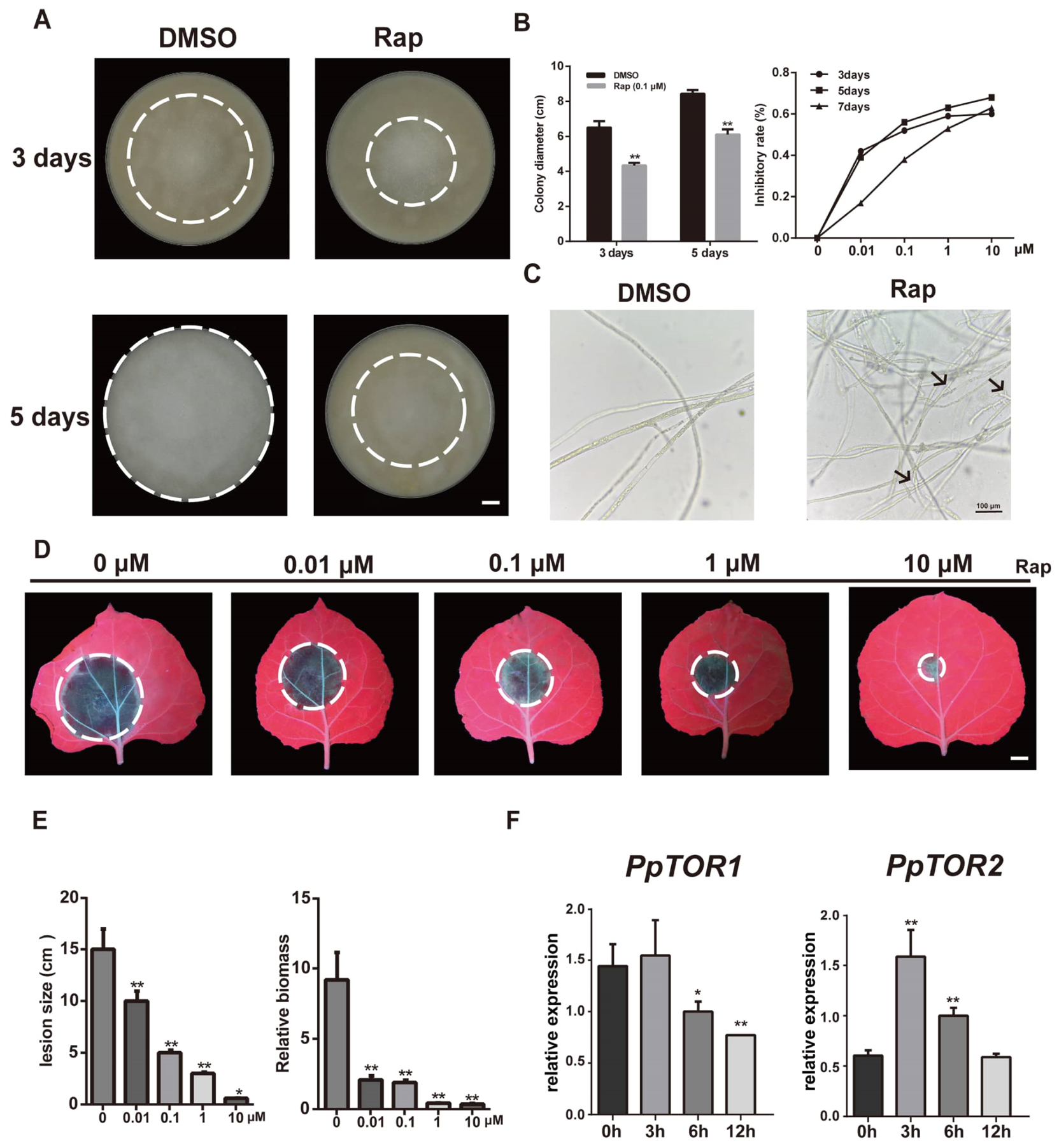
To characterize the biological function of PpTOR, the growth and development of P. parasitica under TOR inhibition were evaluated by using rapamycin treatment. The results showed that rapamycin significantly inhibited the vegetative growth of P. parasitica in a dose-dependent manner. Moreover, rapamycin-exposed hyphae displayed distinct morphological abnormalities, including excessive branching, hyphal distortion, and irregular cellular swelling (Figure 1A–C). Collectively, these results demonstrate that PpTOR plays a critical role in the hyphal growth of P. parasitica. We further investigated the role of PpTOR in regulating the pathogenicity of P. parasitica. The data revealed that both the lesion area and pathogen biomass significantly decreased with increasing rapamycin concentration, suggesting that PpTOR inhibition significantly attenuated P. parasitica infection in N. benthamiana (Figure 1D,E). Notably, PpTOR2 was significantly upregulated during early host infection, whereas the abundance of the PpTOR1 transcript remained relatively stable across all the sampled time points. This divergent expression pattern strongly implies functional specialization between the two PpTOR paralogs.
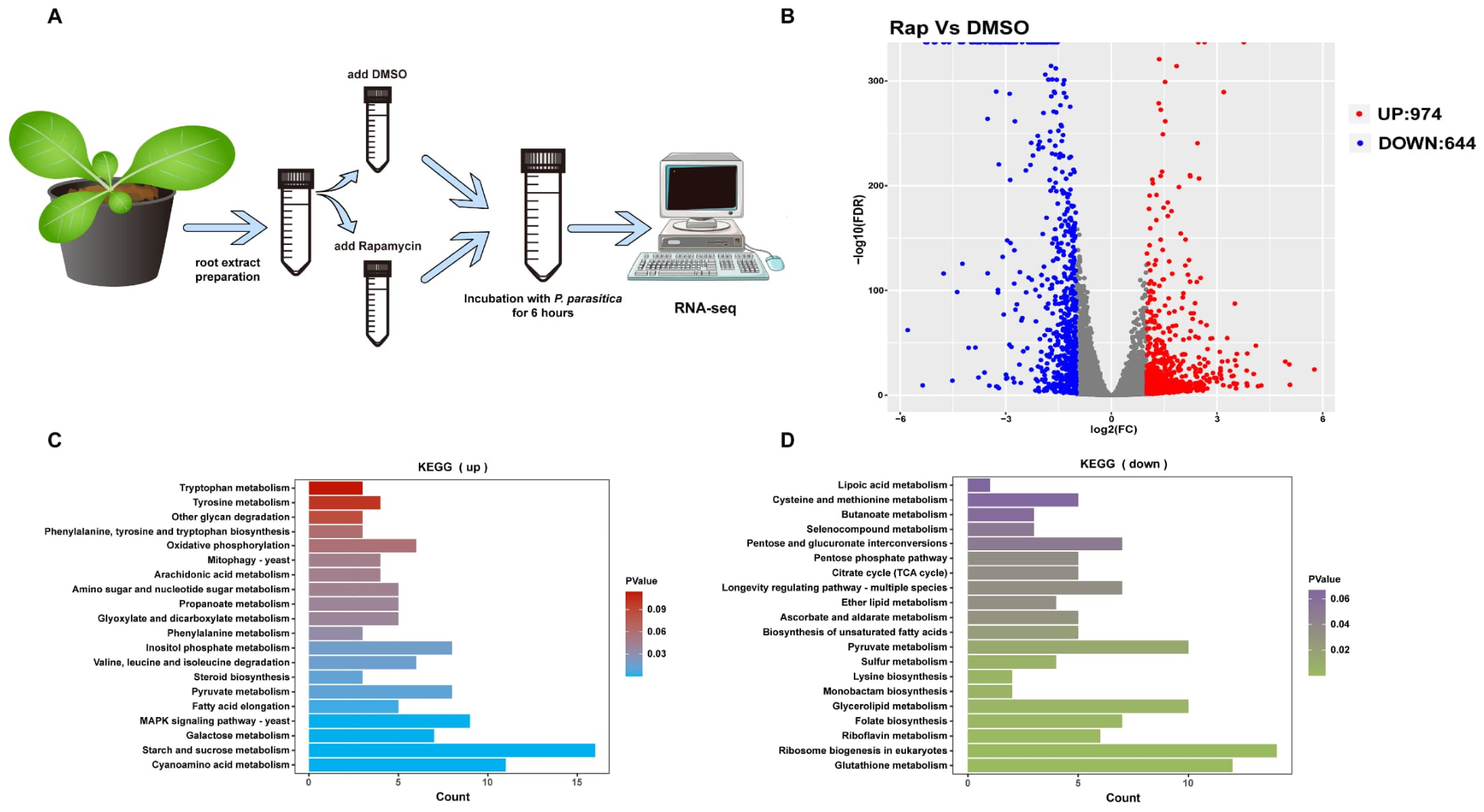
To further elucidate the function of PpTOR, transcriptome sequencing of P. parasitica mycelia treated with rapamycin under conditions simulating plant infection (using tobacco root extract) was performed (Figure 2A). Transcriptome data revealed that inhibition of PpTOR resulted in the differential expression of 1618 genes, comprising 974 upregulated genes and 644 downregulated genes (Figure 2B, Table S3). Gene Ontology (GO) enrichment analysis indicated that these differentially expressed genes (DEGs) were significantly enriched in various functional categories, including metabolic processes, cellular biosynthetic processes, the cell cycle, and cell development (Figure S3, Table S4). Kyoto Encyclopedia of Genes and Genomes (KEGG) pathway enrichment analysis further revealed that the upregulated and downregulated DEGs were annotated to 74 and 79 pathways, respectively (Table S5). Among these functions, TOR-related conserved functions such as ribosome biogenesis, amino acid metabolism, autophagy, and the MAPK signaling pathway were significantly enriched (Figure 2C,D; Table S5), suggesting that PpTOR performs evolutionarily conserved core regulatory functions in P. parasitica.
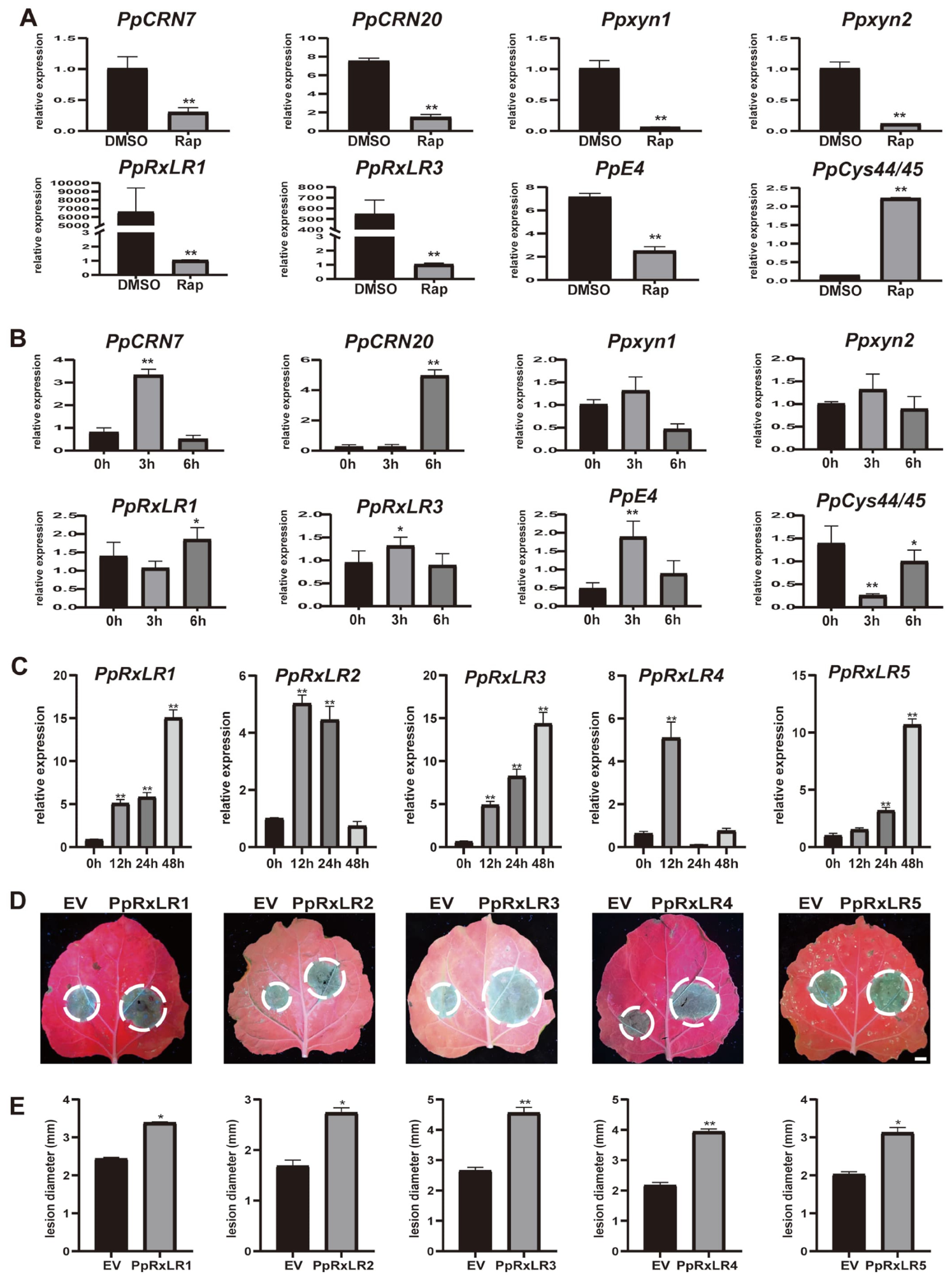
Oomycetes secrete a large array of effector proteins, including RxLR and CRN effectors, into host plants to facilitate infection [34]. Therefore, we focused on analyzing the changes in the expression of RxLR and CRN effector genes, as well as other previously reported virulence-related factors in P. parasitica (such as the cysteine protease PpCys44/45 and the hydrolase PpXYN1/2), under PpTOR inhibition [10,11,18,19]. The expression of numerous effector proteins and virulence-related genes was significantly altered upon rapamycin treatment (Figure S4). RT–qPCR assays further validated the transcriptional changes in key virulence genes. Most RxLR/CRN effector and hydrolase genes were repressed by rapamycin-mediated TOR inhibition, whereas cysteine protease genes were upregulated (Figure 3A). In addition, the expression of these virulence genes was rapidly activated in response to host root-derived signals, supporting their crucial roles during infection (Figure 3B).
Five differentially expressed genes, PpRxLR genes (named PpRxLR1-5), were selected to investigate whether the PpTOR-regulated PpRxLR effectors directly participate in pathogenicity. RT–qPCR assays revealed that the expression of these genes significantly increased during the early stages of P. parasitica infection (Figure 3C). Additionally, transient overexpression of these effectors led to larger lesion areas, suggesting that these effectors increased plant susceptibility to P. parasitica. Among them, PpRxLR3 exhibited the most pronounced virulence-promoting effect (Figure 3D,E). Collectively, these results indicate that PpTOR acts as an upstream regulator of P. parasitica pathogenicity by governing the expression of a suite of effector genes, with PpRxLR3 representing a key downstream virulence determinant.

3.3. PpRxLR3 Contributes to P. parasitica Infection by Attenuating JA Biosynthesis and Signaling in Plants

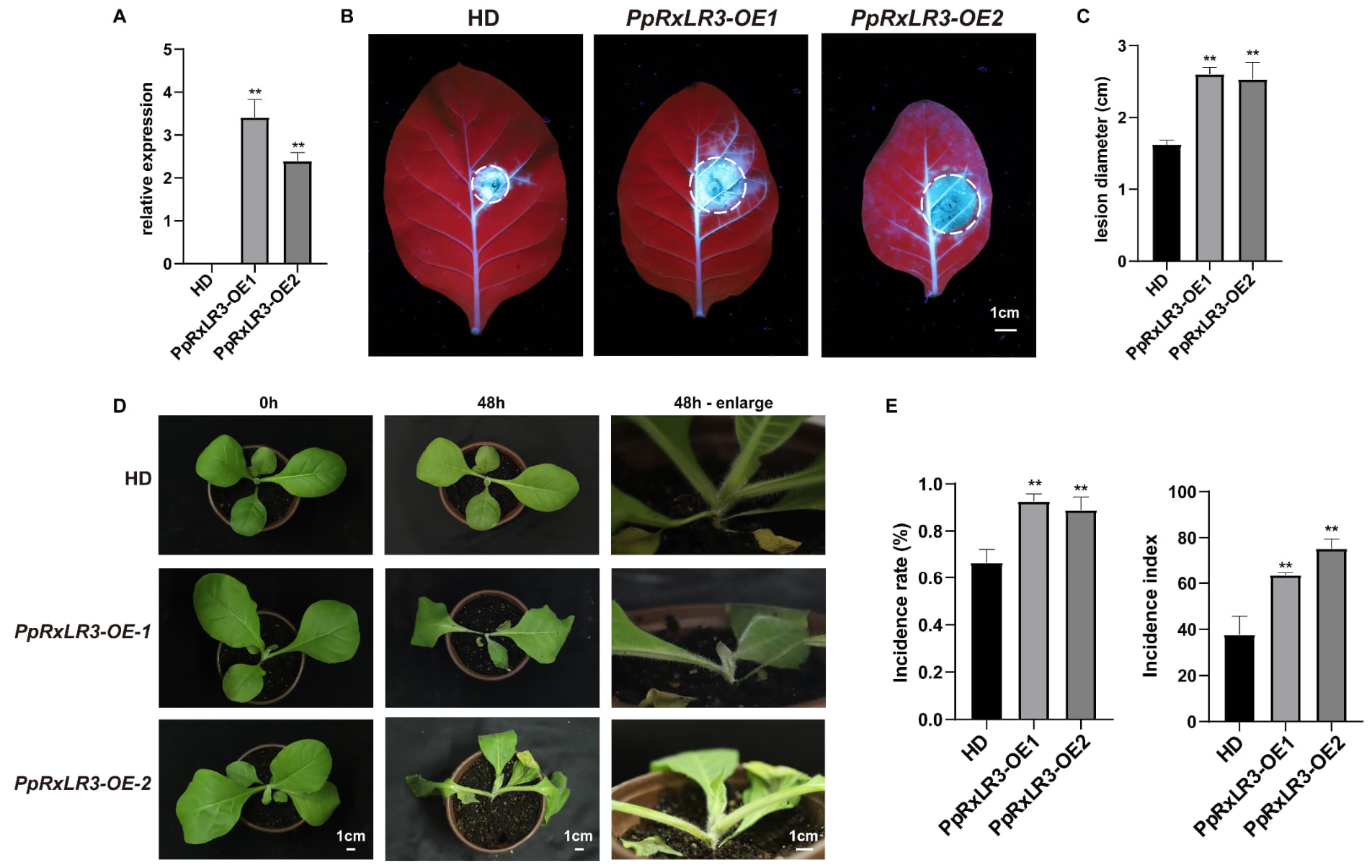
To characterize the biological function of PpRxLR3, PpRxLR3-overexpressing tobacco plants (PpRxLR3-OE1 and PpRxLR3-OE2) whose transgene expression levels were confirmed to be high by RT–qPCR were generated (Figure 4A). Compared with those on wild-type (HD) leaves, lesions on leaves from PpRxLR3-OE plants were significantly greater after inoculation with P. parasitica (Figure 4B,C). Consistent with these findings, whole-plant inoculation assays also revealed that compared with HD plants, PpRxLR3-OE plants developed more severe disease symptoms, including rapid leaf wilting and a black shank phenotype (Figure 4D). The disease incidence rate and index were consistently greater in the transgenic plants than in the WT plants (Figure 4E). These results demonstrate that PpRxLR3 functions as a virulence effector that increases plant susceptibility to P. parasitica.
To elucidate the molecular mechanisms underlying PpRxLR3-mediated susceptibility, we performed transcriptome sequencing on PpRxLR3-OE and HD plants. A total of 3644 differentially expressed genes (DEGs) were identified, with 2076 upregulated and 1568 downregulated genes in the transgenic plants (Figure S5, Table S6). Functional enrichment analysis indicated that these DEGs were significantly overrepresented in categories related to plant–pathogen interactions and immunity, including systemic acquired resistance (SAR), the MAPK signaling pathway, cell wall organization/biogenesis, photosynthesis, and multiple hormone signaling pathways (Figures S6–S8, Table S7). This global transcriptional reprogramming suggests that PpRxLR3 perturbs multiple defense and signaling networks in the host.
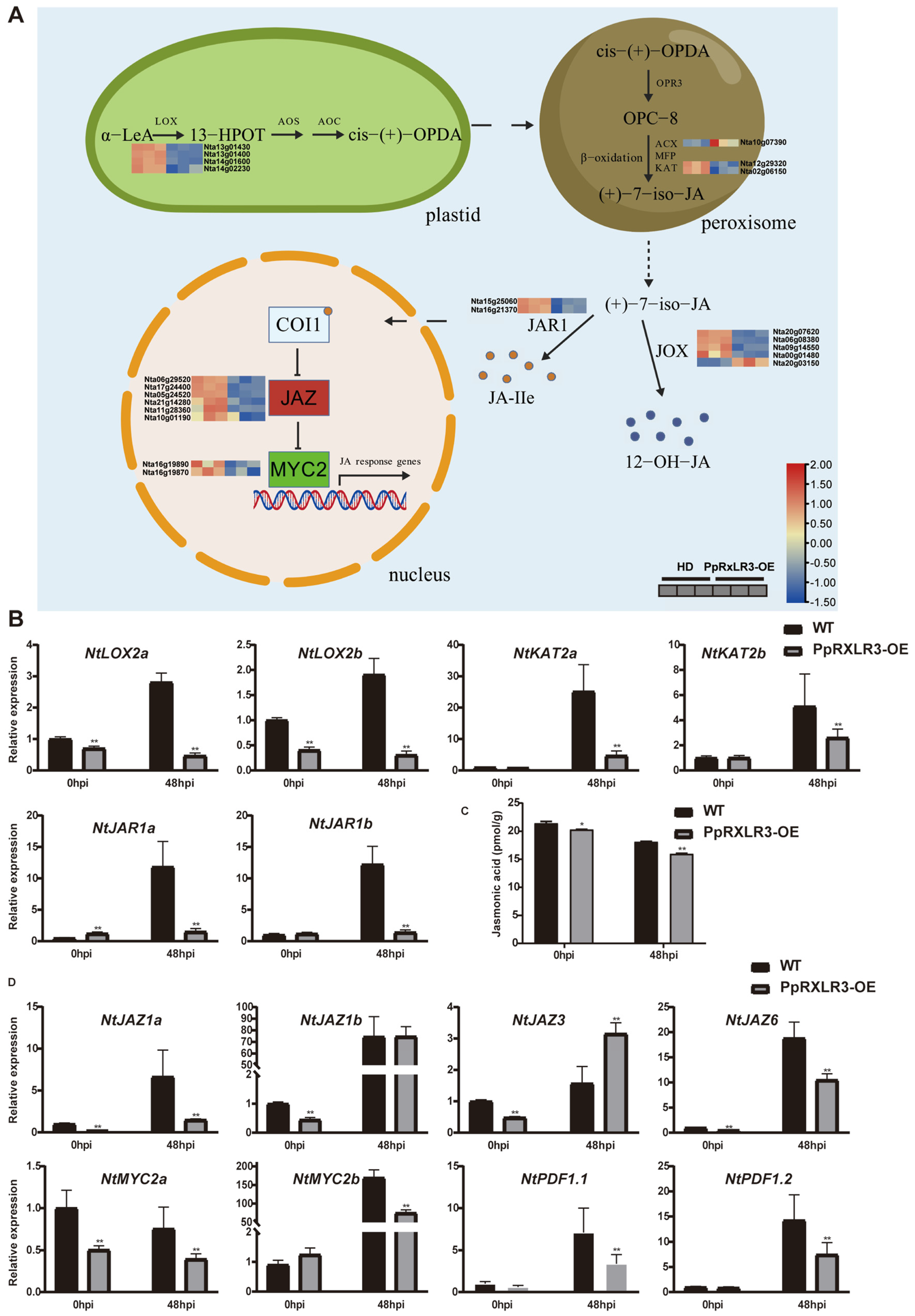
In plant–pathogen interactions, multiple phytohormones participate in the regulation of pathogen resistance in plants [35,36]. Among them, jasmonic acid (JA)-mediated signaling is important in plant immune responses against pathogens [37,38]. Our transcriptomic analysis revealed that numerous genes involved in JA biosynthesis (e.g., LOX, AOS, AOC, and OPR3) and signaling (e.g., COI1, JAZ, and MYC2) were differentially expressed in PpRxLR3-OE plants even before infection (Figure 5A, Table S8). More importantly, while P. parasitica infection promoted the expression of these JA-related genes in HD plants, this induction was markedly attenuated or reversed in PpRxLR3-OE plants (Figure 5B). To determine whether these transcriptional changes corresponded to physiological alterations, we quantified JA levels in PpRxLR3-OE plants. Consistent with the gene expression data, the JA content was significantly lower in the PpRxLR3-OE plants than in the HD plants (Figure 5C). Similarly, the expression of genes involved in JA signal transduction and response was also significantly suppressed in the OE plants after P. parasitica infection (Figure 5D). Collectively, these results indicate that PpRxLR3 overexpression is associated with the suppression of both JA biosynthesis and signaling.
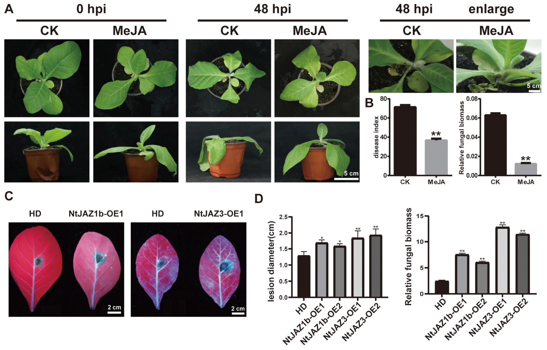
To functionally validate the importance of JA signaling in plant resistance to P. parasitica, we performed two complementary experiments. First, exogenous application of methyl jasmonate (MeJA), a bioactive JA derivative, significantly increased plant resistance to P. parasitica, reducing disease severity (Figure 6A,B). Second, since JAZ proteins are negative regulators of JA signaling, we generated JAZ-overexpressing plants (Figure S9). Conversely, overexpression of JAZ (which represses JA responses) significantly compromised plant resistance, leading to increased susceptibility (Figure 6C,D). These results suggest that JA signaling plays a crucial role in regulating plant resistance to P. parasitica.
In addition to jasmonic acid (JA) signaling, plant innate immunity (PTI) and salicylic acid (SA) signaling play central roles in plant defense against pathogen invasion [36]. Our transcriptomic analysis revealed differential expression of key genes in these pathways (Figures S6–S8). Further validation by quantitative PCR confirmed that these genes are strongly upregulated during infection (Figure S8C) [40,41,42,43,44]. In contrast, this induction was significantly suppressed in PpRxLR3-overexpressing (OE) plants (Figure S8C). In addition to disrupting immune signaling, PpRxLR3 also targets fundamental aspects of host physiology. Transcriptomic profiling revealed substantial dysregulation of numerous photosynthesis-related genes in PpRxLR3-OE plants (Figure S8, Table S9). Compared with the wild-type controls, these plants consistently exhibited growth retardation and a significant reduction in leaf photosynthetic capacity (Figure S10). In summary, PpTOR is essential for the pathogenicity of P. parasitica. It functions as a crucial regulator of P. parasitica virulence by modulating the expression of a suite of effector proteins, including PpRxLR3, which act in concert to dismantle multiple layers of host defense.

3.4. Silencing of PpTOR Promotes Plant Resistance to P. parasitica

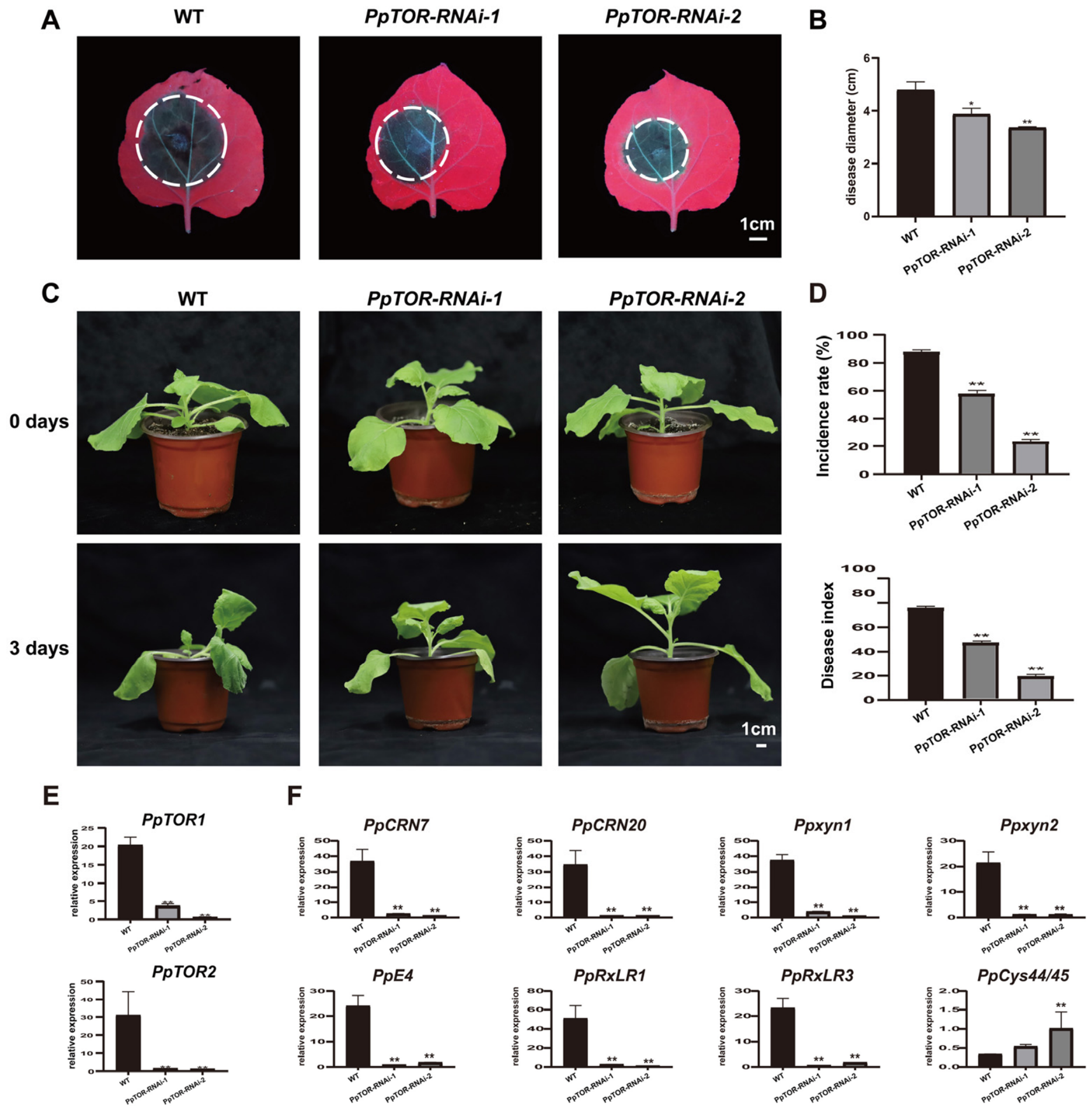
Having established PpTOR as a master regulator of growth and pathogenicity in P. parasitica, we explored its potential as a target for disease control using host-induced gene silencing (HIGS). HIGS is a promising strategy wherein the host plant expresses double-stranded RNA (dsRNA) homologous to essential pathogen genes, leading to their silencing and consequent reduction in disease [45,46,47]. We generated stable N. benthamiana lines expressing dsRNA targeting PpTOR (PpTOR-RNAi plants) (Figure S11). In detached leaf assays, compared with wild-type (WT) control plants, PpTOR-RNAi plants developed significantly smaller lesions after inoculation with P. parasitica (Figure 7A,B). Whole-plant inoculation at the stem base further confirmed enhanced resistance. When the WT plants exhibited severe wilting within 3 days of incubation, the PpTOR-RNAi plants presented minimal disease symptoms under the same conditions (Figure 7C). Consequently, both the disease incidence rate and the disease index were significantly lower in the PpTOR-RNAi plants (Figure 7D). In addition, we confirmed the efficacy of HIGS. Compared with those in P. parasitica-infected WT plants, the transcript levels of both PpTOR1 and PpTOR2 were drastically reduced in pathogen-colonizing PpTOR-RNAi plants, with a silencing efficiency of more than 90% (Figure 7E). Consistently, the expression of pathogenicity-related genes in P. parasitica was also markedly suppressed (Figure 7F).
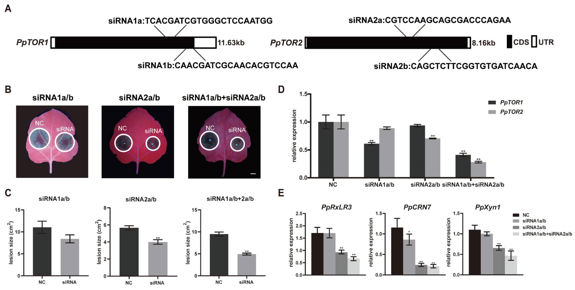
To verify the silencing signal, we performed sRNA sequencing (sRNA-Seq) on PpTOR-RNAi plants. The data revealed abundant production of PpTOR-specific small interfering RNAs (siRNAs) in the transgenic plants compared with the WT controls (Table S11). Furthermore, we investigated whether P. parasitica could take up sRNAs from the environment. Fluorescence labeling experiments revealed that P. parasitica mycelia efficiently internalized exogenous siRNAs, suggesting that direct application of synthetic siRNAs could serve as a nontransgenic strategy to silence PpTOR (Figure S12). Additionally, we artificially synthesized four siRNA sequences that can specifically target PpTOR1 and PpTOR2 and reported that treatment with PpTOR2-specific dsRNA significantly impaired pathogen virulence, whereas treatment with PpTOR1-specific siRNA had a more modest effect. The most pronounced reduction in disease was observed when a combination of PpTOR1- and PpTOR2-specific siRNAs was applied (Figure 8A–D). Corroborating these findings, the expression of virulence-associated effector genes in the treated pathogen was most strongly suppressed under combined siRNA treatment (Figure 8E).
In summary, our results demonstrate that silencing PpTOR through either HIGS or exogenous siRNA application effectively reduces the pathogenicity of P. parasitica by dampening the expression of its downstream virulence program. This finding establishes TOR not only as a critical virulence regulator but also as a promising target for developing RNA-based control strategies against oomycete diseases.

4. Discussion

P. parasitica is a devastating oomycete pathogen that causes severe root and crown rot diseases worldwide [1]. Effective control remains challenging because of its rapid infection cycle and limited management options [48]. In this study, the conserved kinase PpTOR was established as an essential regulator of P. parasitica growth and pathogenicity. We demonstrate that PpTOR coordinates the expression of a suite of virulence factors, including RxLR and CRN effectors, and that one key downstream effector, PpRxLR3, promotes infection by broadly suppressing plant resistance-related signaling. Furthermore, HIGS- and sRNA-mediated silencing of PpTOR proved to be effective strategies for enhancing plant resistance, highlighting its potential as a novel target for disease control.
TOR kinase is a well-conserved integrator of nutrient and environmental cues that modulates the growth and pathogenicity of pathogens [23,24,25,26,27,28,29]. In this study, our findings extend its pivotal role to pathogenic oomycetes. The inhibition of PpTOR by rapamycin severely impaired the mycelial growth and pathogenicity of P. parasitica, which is consistent with the findings of previous reports on other pathogens [23,24]. Transcriptomic profiling further revealed that PpTOR inhibition dysregulated a vast network of genes involved in core cellular processes such as ribosome biogenesis, amino acid metabolism, and autophagy. More significantly, we identified a pronounced suppression of genes encoding key virulence determinants, including RxLR and CRN effectors. This positions PpTOR not only as a growth regulator but also as a critical signaling nexus that integrates upstream cues to modulate downstream virulence programs. Under favorable conditions, active PpTOR signaling may promote the growth of P. parasitica and the production of the effector arsenal necessary for host invasion.
Recent studies in fungal pathogens have illuminated the expanded role of TOR beyond growth regulation. For instance, in Fusarium oxysporum, FoTOR not only promotes ribosome biogenesis via FoSIK1 but also directly regulates the expression of numerous cell wall-degrading enzymes (CWDEs), which are crucial for breaching the plant cell wall barrier during early infection [24,49]. These findings highlight the unique function of TOR in pathogenicity. Our findings in the oomycete Phytophthora parasitica align with this paradigm, as PpTOR inhibition led to the coordinated downregulation of a suite of CWDEs and key effector genes (e.g., PpRxLR3). Furthermore, evidence from F. oxysporum indicates that TOR can affect virulence through downstream transcription factors, such as the bZIP protein MeaB [26]. This convergence of evidence—from fungal systems to our oomycete model—highlights a paradigm in which TOR acts as a switch, synchronizing the pathogen’s biosynthetic capacity (e.g., ribosome biogenesis) with the timed expression of invasion machinery (e.g., CWDEs and effectors) through the modulation of key transcription factors.
Given that TOR inhibition broadly suppresses pathogen growth and metabolism, a potential confounding factor is that the observed repression of virulence genes might result from nonspecific secondary transcriptional remodeling triggered by growth arrest rather than direct PpTOR-dependent regulation. To rule out this possibility, we performed rigorous cross-validation using two independent gene-specific silencing systems, namely, transgenic host-induced gene silencing (HIGS) and exogenous spray-induced gene silencing (SIGS). Both RNAi approaches specifically reduced PpTOR transcript abundance without causing global metabolic repression or obvious growth inhibition during early pathogen colonization. Importantly, both genetic silencing systems recapitulated the specific repression of RxLR/CRN virulence effectors observed in rapamycin-treated mycelia. The high consistency between chemical inhibition and gene-specific silencing strongly indicates that the altered virulence transcriptome is a direct outcome of impaired PpTOR signaling rather than a general stress response secondary to growth suppression.
We further excluded growth-associated confounding effects through time-course expression analysis of virulence genes in response to host-derived signals. RxLR and CRN effector genes were rapidly and specifically induced within 3–6 h following exposure to host root cues, which occurred long before any detectable alteration in mycelial growth or biomass accumulation. These rapid induction kinetics, together with the independent expression pattern of effector genes relative to general growth-related transcripts, further demonstrate that TOR signaling directly modulates pathogen virulence programs in response to host signals. Therefore, PpTOR may govern infection-related effector expression in a growth-independent manner, confirming its specific and central role in coordinating oomycete pathogenicity. However, the specific identity of the downstream factors of PpTOR that orchestrate this virulence-related transcriptional reprogramming during P. parasitica infection remains a pivotal and unresolved question and represents a key frontier for future research.
Notably, unlike most fungi that harbor only a single TOR copy, the oomycete P. parasitica possesses two distinct TOR paralogs, designated PpTOR1 and PpTOR2. Our transcriptional and functional assays revealed clear functional divergence between these two isoforms. PpTOR2 expression sharply increased during the early stage of host infection, and compared with suppression of PpTOR1, siRNA-mediated silencing of PpTOR2 had a more potent inhibitory effect on pathogen virulence. These findings may indicate that PpTOR2 serves as an infection-specific TOR isoform that activates the full virulence repertoire of P. parasitica, including RxLR/CRLR effector expression and cell wall-degrading enzyme activity. In contrast, PpTOR1 maintains basal TOR signaling to support routine vegetative growth under saprophytic conditions and contributes minimally to pathogenicity. The retention of dual TOR paralogs in oomycetes may represent an adaptive evolutionary strategy enabling the independent regulation of saprophytic growth and parasitic infection. Further genetic knockout or knockdown assays in P. parasitica will help fully elucidate the distinct downstream regulatory networks of the two TOR isoforms.
In Phytophthora species, RxLR effector proteins contribute to infection by suppressing host plant resistance [34]. Our integrated approach identified PpRxLR3 as a key effector downstream of PpTOR. PpRxLR3 overexpression in plants increased susceptibility, and transcriptomic profiling of these plants revealed a systematic dampening of defense responses, particularly within the JA pathway—a well-established pillar of plant disease resistance [50,51,52]. Pathogens can increase their virulence by interfering with JA biosynthesis or JA signal transduction [53,54,55]. Within the Phytophthora genus, pathogens frequently employ effector proteins to suppress JA biosynthesis or disrupt JA-mediated signaling, thereby facilitating host colonization [56,57]. Specifically, our data demonstrate that PpRxLR3 suppresses the transcription of genes involved in JA biosynthesis and signaling. Functional validation confirmed that an active JA pathway enhances resistance to P. parasitica, whereas its inhibition (via JAZ overexpression) increases susceptibility. Taken together, these findings position JA signaling as a critical host target compromised by Phytophthora genus pathogens to facilitate infection and underscore its potential value as a target for breeding resistance against Phytophthora genus pathogens. Notably, the endogenous JA content in this study was quantified using an ELISA-based method. Although this approach is widely applicable for relative hormone quantification in plant immune studies, it has lower detection specificity and resolution than the gold-standard LC–MS/MS platform does. Future studies will adopt LC–MS/MS-based hormone profiling to achieve more accurate and high-confidence quantification of hormone accumulation (e.g., that of salicylic acid) and further validate the hormone-suppressive effect of PpRxLR3.
In addition to JA signaling, SA represents a central hormone in plant defense against pathogens [58]. PpRxLR3 significantly attenuated the infection-triggered induction of the SA receptor gene NPR1 and the marker gene PR1, effectively disabling this major defense branch. During the initial stages of plant infection, PTI is rapidly activated to protect plants from pathogenic invasion. In response, pathogens deliver a large number of effector proteins into plant cells to disrupt PTI and facilitate invasion and colonization [5]. PpRxLR3 also abolished the pathogen-induced expression of core PTI components, such as the coreceptor NtBAK1, its negative regulator NtBIR2 and SAR-related genes, suggesting that PpRxLR3 likely blocks the early activation of PTI signaling cascades, including the MAPK pathway and SAR. Furthermore, in addition to directly suppressing the defense response in plants, the manipulation of host physiology by PpRxLR3 likely involves fundamental processes such as photosynthesis. Overall, this multipronged attack on diverse defense-related pathways and host physiology highlights a sophisticated mechanism through which P. parasitica dismantles host defenses. By simultaneously targeting these interconnected networks, PpRxLR3 ensures the comprehensive suppression of defense signaling, preventing the host from mounting an effective counterresponse and facilitating successful colonization.
A limitation of the current functional characterization of PpRxLR3 is the absence of pathogen-intrinsic gene deletion data, which are restricted by the low genetic tractability of P. parasitica. Despite the lack of loss-of-function mutants, multiple layers of gain-of-function evidence sufficiently support the virulent role of PpRxLR3. This evidence includes transient agroinfiltration assays and stable overexpression in two independent tobacco varieties, together with its infection-induced and TOR-dependent expression profile during host colonization. Collectively, these results strongly demonstrate that PpRxLR3 acts as a core virulence effector of P. parasitica to inhibit host immune defenses. Future studies should perform gene knockout assays of PpRxLR3 to verify its indispensable function during infection with an authentic pathogen. Further investigation into the molecular mechanisms by which PpRxLR3 suppresses plant resistance and identification of its host-interacting targets will advance our mechanistic understanding of oomycete virulence strategies and provide novel insights for the development of precise disease control approaches.
Given the central role of PpTOR in P. parasitica, we evaluated PpTOR as a target for HIGS, a promising RNAi-based strategy for disease control [45,59]. Our PpTOR-RNAi plants exhibited strong resistance, accompanied by the silencing of both PpTOR1/2 and key downstream virulence genes such as PpCRN7, PpXYN1, and PpRxLR3. This successfully translates our mechanistic understanding into a proof-of-concept application. In a previous study, Zhang et al. reported that the production of dsRNA in Arabidopsis did not effectively silence the expression of target genes of P. parasitica [60]. However, HIGS has been used to suppress the occurrence of several other oomycete diseases. Govindarajulu et al. reported that the HIGS method could effectively control B. lactucae, a biotrophic oomycete that induces downy mildew disease in lettuce [61]. In Phytophthora, the HIGS method could also confer resistance to P. capsici and P. infestans in N. benthamiana and potato by targeting different target genes [32,62]. Notably, a recent study revealed that Phytophthora could inhibit the biogenesis of pentatricopeptide-repeat protein (PPR)-derived small interfering RNAs (siRNAs), which are located in extracellular vesicles and silence target genes in Phytophthora, through the effector protein PSR2 in Arabidopsis, suggesting that pathogens may regulate the production of siRNA in Arabidopsis through the secretion of effector proteins [63]. There may be complex and different mechanisms for dsRNA or siRNA exchange between different hosts and pathogens, and the mechanism by which pathogens take up dsRNA or siRNA molecules from plant cells remains unclear.
In addition to HIGS, spray-induced gene silencing (SIGS) has emerged as a rapid and effective method for disease prevention and control in plants [64,65]. SIGS can constrain pathogen infection through the use of double-stranded RNA (dsRNA) or single-stranded RNA (sRNA) to directly suppress the transcription levels of pathogen virulence-related genes [65,66]. Interestingly, a recent study revealed that Phytophthora capsici can effectively take up environmental small interfering RNAs (siRNAs) and that SIGS can be used to silence the expression of P. capsici virulence-related genes to restrict the infection of P. capsici in tobacco [67]. Our results also demonstrate that P. parasitica can take up exogenous dsRNA, making the SIGS method viable for controlling P. parasitica by silencing PpTOR. By designing specific siRNAs against PpTOR1/2 and employing efficient delivery systems, it may be possible to develop a nontransgenic, environmentally friendly control method. Furthermore, given the highly conserved functional role of TOR across plant pathogens, targeting TOR via SIGS represents a promising strategy to effectively control multiple oomycete diseases by directly impairing the pathogen at its core regulatory node, as demonstrated in this study. Even though some pathogens have difficulty taking up siRNA from the environment, Wang et al. reported that nanocarriers enable more efficient delivery of siRNA into P. infestans [68,69] for the management of potato late blight. These findings indicate that the SIGS method can be utilized to inhibit the expression of pathogen TOR genes, thereby effectively controlling diseases caused by various oomycete pathogens in a nontransgenic and environmentally friendly manner. Our synthetic siRNA assays establish a proof-of-concept RNA-based control strategy against P. parasitica. Quantitative comparison of TOR transcript knockdown efficiency further explains the differential disease-suppressive potency between single siRNA treatments and combined siRNA cocktails. Despite these promising results, the translational application of laboratory-based SIGS technology in commercial agricultural practices requires further evaluation of key environmental parameters. These key factors include siRNA persistence on leaf surfaces, resistance to rain wash-off, tolerance to UV degradation, and systemic absorption efficiency in mature crop tissues. Nanoparticle delivery systems have been experimentally validated to optimize SIGS performance in Phytophthora pathosystems and can substantially increase siRNA stability and pathogen uptake under field conditions. Therefore, nanoparticle-assisted delivery represents a promising strategy for future translational research to advance the field application of RNA-mediated disease control approaches.
Fungicide application has emerged as the primary method for preventing and controlling P. parasitica because of its high efficiency and convenience. However, issues related to environmental pollution and pathogen resistance to fungicides caused by long-term application cannot be ignored. In this study, we provide compelling evidence that PpTOR is indispensable for the pathogenicity of P. parasitica and demonstrate that TOR suppression can effectively mitigate P. parasitica-induced disease under laboratory conditions. Moreover, considering that P. parasitica shares numerous biological characteristics with most Phytophthora pathogens, the TOR gene not only plays a pivotal role in P. parasitica but is also highly conserved in other Phytophthora pathogens and even broader oomycete genera, suggesting that TOR may have similar functional mechanisms and regulatory pathways in these organisms. These findings provide a theoretical foundation for developing broad-spectrum and efficient control strategies against oomycete diseases by targeting TOR.

Supplementary Materials

The following supporting information can be downloaded at https://www.mdpi.com/article/10.3390/horticulturae12091072/s1, Figure S1: Identification and characterization of TOR homologs in P. parasitica; Figure S2: Phylogenetic analysis of TOR homologs within oomycetes; Figure S3: GO enrichment analysis of differentially expressed genes (DEGs) in P. parasitica under PpTOR inhibition; Figure S4: PpTOR inhibition downregulates the expression of virulence-associated genes in P. parasitica; Figure S5: Overview of transcriptomic changes in PpRxLR3-overexpressing plants; Figure S6: KEGG pathway enrichment analysis of DEGs in PpRxLR3-OE plants; Figure S7: GO enrichment and expression profiles of defense-related DEGs in PpRxLR3-OE tobacco plants; Figure S8: Alterations in photosynthesis- and defense-associated gene expression upon PpRxLR3 overexpression in tobacco; Figure S9: Validation of NtJAZ gene overexpression in transgenic plants; Figure S10: PpRxLR3 overexpression impaired plant growth and photosynthesis; Figure S11: Generation and molecular identification of PpTOR1/2-RNAi transgenic plants; Figure S12: P. parasitica could effectively take up environmental siRNAs. Table S1: Accession numbers of TOR genes from different Phytophthora species and Oomycete genera; Table S2: Putative homologous proteins of the core components of TORC1 (RAPTOR and LST8) and TORC2 (RAPTOR and RICTOR) in P. parasitica; Table S3: Transcriptomic profiles of P. parasitica under rapamycin treatment (PpTOR inhibition); Table S4: Complete GO enrichment results for DEGs in P. parasitica under PpTOR inhibition; Table S5: Complete KEGG pathway enrichment results for DEGs in P. parasitica under PpTOR inhibition; Table S6: Transcriptomic profiles of PpRxLR3-overexpressing tobacco plants; Table S7: Complete GO enrichment results for DEGs in PpRxLR3-overexpressing plants; Table S8: List of DEGs related to jasmonic acid biosynthesis and signaling in PpRxLR3-overexpressing plants; Table S9: Complete KEGG pathway enrichment results for DEGs in PpRxLR3-OE plants; Table S10: sRNAs identified by sRNA-Seq that target PpTOR; Table S11. Primers used in this study.

Author Contributions

Conceptualization, K.D. and J.Z.; investigation, B.W., Y.H., Z.L. and W.Y.; software, B.W. and X.D.; writing—original draft preparation, B.W., X.D. and K.D.; writing—review and editing, J.Z. and K.D.; funding acquisition, K.D. All authors have read and agreed to the published version of the manuscript.

Funding

This work was supported by the Fundamental Research Funds for the Central Universities (SWU120072).

Data Availability Statement

The original contributions presented in this study are included in the article/Supplementary Material. Further inquiries can be directed to the corresponding authors.

Acknowledgments

The authors are grateful to Kang Zhang and Fangjie Xiong for their critical revisions and helpful suggestions during manuscript preparation.

Conflicts of Interest

The authors declare no conflicts of interest.

References

  1. Kamoun, S.; Furzer, O.; Jones, J.D.; Judelson, H.S.; Ali, G.S.; Dalio, R.J.; Roy, S.G.; Schena, L.; Zambounis, A.; Panabières, F.; et al. The Top 10 oomycete pathogens in molecular plant pathology. Mol. Plant Pathol. 2015, 16, 413–434. [Google Scholar] [CrossRef] [Scilit] [PubMed]
  2. Sawake, M.M.; Moharil, M.P.; Ingle, Y.V.; Jadhav, P.V.; Ingle, A.P.; Khelurkar, V.C.; Paithankar, D.H.; Bathe, G.A.; Gade, A.K. Management of Phytophthora parasitica causing gummosis in citrus using biogenic copper oxide nanoparticles. J. Appl. Microbiol. 2022, 132, 3142–3154. [Google Scholar] [CrossRef] [Scilit] [PubMed]
  3. Santos, M.; Diánez, F.; Sánchez-Montesinos, B.; Huertas, V.; Moreno-Gavira, A.; Esteban García, B.; Garrido-Cárdenas, J.A.; Gea, F.J. Biocontrol of Diseases Caused by Phytophthora capsici and P. parasitica in Pepper Plants. J. Fungi 2023, 9, 360. [Google Scholar] [CrossRef] [Scilit] [PubMed]
  4. Yang, J.; Wang, Q.; Li, L.; Li, P.; Yin, M.; Xu, S.; Chen, Y.; Feng, X.; Wang, B. Chemical Composition and Antifungal Activity of Zanthoxylum armatum Fruit Essential Oil against Phytophthora capsici. Molecules 2022, 27, 8636. [Google Scholar] [CrossRef] [Scilit] [PubMed]
  5. Yuan, M.; Ngou, B.P.M.; Ding, P.; Xin, X.F. PTI-ETI crosstalk: An integrative view of plant immunity. Curr. Opin. Plant Biol. 2021, 62, 102030. [Google Scholar] [CrossRef] [Scilit] [PubMed]
  6. Lo Presti, L.; Lanver, D.; Schweizer, G.; Tanaka, S.; Liang, L.; Tollot, M.; Zuccaro, A.; Reissmann, S.; Kahmann, R. Fungal effectors and plant susceptibility. Annu. Rev. Plant Biol. 2015, 66, 513–545. [Google Scholar] [CrossRef] [Scilit] [PubMed]
  7. Mach, J. Phytophthora infestans RXLR effectors target vesicle trafficking. Plant Cell 2021, 33, 1401–1402. [Google Scholar] [CrossRef] [Scilit] [PubMed]
  8. Bos, J.I.; Armstrong, M.R.; Gilroy, E.M.; Boevink, P.C.; Hein, I.; Taylor, R.M.; Zhendong, T.; Engelhardt, S.; Vetukuri, R.R.; Harrower, B.; et al. Phytophthora infestans effector AVR3a is essential for virulence and manipulates plant immunity by stabilizing host E3 ligase CMPG1. Proc. Natl. Acad. Sci. USA 2010, 107, 9909–9914. [Google Scholar] [CrossRef] [Scilit] [PubMed]
  9. Liu, R.; Chen, T.; Yin, X.; Xiang, G.; Peng, J.; Fu, Q.; Li, M.; Shang, B.; Ma, H.; Liu, G.; et al. A Plasmopara viticola RXLR effector targets a chloroplast protein PsbP to inhibit ROS production in grapevine. Plant J. 2021, 106, 1557–1570. [Google Scholar] [CrossRef] [Scilit] [PubMed]
  10. Huang, G.; Liu, Z.; Gu, B.; Zhao, H.; Jia, J.; Fan, G.; Meng, Y.; Du, Y.; Shan, W. An RXLR effector secreted by Phytophthora parasitica is a virulence factor and triggers cell death in various plants. Mol. Plant Pathol. 2019, 20, 356–371. [Google Scholar] [CrossRef] [Scilit] [PubMed]
  11. Maximo, H.J.; Dalio, R.J.D.; Dias, R.O.; Litholdo, C.G., Jr.; Felizatti, H.L.; Machado, M.A. PpCRN7 and PpCRN20 of Phythophthora parasitica regulate plant cell death leading to enhancement of host susceptibility. BMC Plant Biol. 2019, 19, 544. [Google Scholar] [CrossRef] [Scilit] [PubMed]
  12. Kamoun, S. A Catalog of the Effector Secretome of Plant Pathogenic Oomycetes. Annu. Rev. Phytopathol. 2006, 44, 41–60. [Google Scholar] [CrossRef] [Scilit] [PubMed]
  13. Fu, L.; Liu, Y.; Qin, G.; Wu, P.; Zi, H.; Xu, Z.; Zhao, X.; Wang, Y.; Li, Y.; Yang, S.; et al. The TOR–EIN2 axis mediates nuclear signaling to modulate plant growth. Nature 2021, 591, 288–292. [Google Scholar] [CrossRef] [Scilit] [PubMed]
  14. Salguero-Linares, J.; Coll, N.S. Plant proteases in the control of the hypersensitive response. J. Exp. Bot. 2019, 70, 2087–2095. [Google Scholar] [CrossRef] [Scilit] [PubMed]
  15. Thomas, E.L.; van der Hoorn, R.A.L. Ten Prominent Host Proteases in Plant–Pathogen Interactions. Int. J. Mol. Sci. 2018, 19, 639. [Google Scholar] [CrossRef] [Scilit] [PubMed]
  16. Gong, Y.; Fu, Y.; Xie, J.; Li, B.; Chen, T.; Lin, Y.; Chen, W.; Jiang, D.; Cheng, J. Sclerotinia sclerotiorum SsCut1 Modulates Virulence and Cutinase Activity. J. Fungi 2022, 8, 526. [Google Scholar] [CrossRef] [Scilit] [PubMed]
  17. Gui, Y.J.; Zhang, W.Q.; Zhang, D.D.; Zhou, L.; Short, D.P.G.; Wang, J.; Ma, X.F.; Li, T.G.; Kong, Z.Q.; Wang, B.L.; et al. A Verticillium dahliae Extracellular Cutinase Modulates Plant Immune Responses. Mol. Plant–Microbe Interact. 2018, 31, 260–273. [Google Scholar] [CrossRef] [Scilit] [PubMed]
  18. Lai, M.W.; Liou, R.F. Two genes encoding GH10 xylanases are essential for the virulence of the oomycete plant pathogen Phytophthora parasitica. Curr. Genet. 2018, 64, 931–943. [Google Scholar] [CrossRef] [Scilit] [PubMed]
  19. Zhang, Q.; Li, W.; Yang, J.; Xu, J.; Meng, Y.; Shan, W. Two Phytophthora parasitica cysteine protease genes, PpCys44 and PpCys45, trigger cell death in various Nicotiana spp. and act as virulence factors. Mol. Plant Pathol. 2020, 21, 541–554. [Google Scholar] [CrossRef] [Scilit] [PubMed]
  20. Battaglioni, S.; Benjamin, D.; Wälchli, M.; Maier, T.; Hall, M.N. mTOR substrate phosphorylation in growth control. Cell 2022, 185, 1814–1836. [Google Scholar] [CrossRef] [Scilit] [PubMed]
  21. Yang, H.; Rudge, D.G.; Koos, J.D.; Vaidialingam, B.; Yang, H.J.; Pavletich, N.P. mTOR kinase structure, mechanism and regulation. Nature 2013, 497, 217–223. [Google Scholar] [CrossRef] [Scilit] [PubMed]
  22. Xiong, F.; Liu, M.; Zhuo, F.; Yin, H.; Deng, K.; Feng, S.; Liu, Y.; Luo, X.; Feng, L.; Zhang, S.; et al. Host-induced gene silencing of BcTOR in Botrytis cinerea enhances plant resistance to gray mold. Mol. Plant Pathol. 2019, 20, 1722–1739. [Google Scholar] [CrossRef] [Scilit] [PubMed]
  23. Li, L.; Zhu, T.; Song, Y.; Luo, X.; Feng, L.; Zhuo, F.; Li, F.; Ren, M. Functional Characterization of Target of Rapamycin Signaling in Verticillium dahliae. Front. Microbiol. 2019, 10, 501. [Google Scholar] [CrossRef] [Scilit] [PubMed]
  24. Li, L.; Zhu, T.; Song, Y.; Luo, X.; Datla, R.; Ren, M. Target of rapamycin controls hyphal growth and pathogenicity through FoTIP4 in Fusarium oxysporum. Mol. Plant Pathol. 2021, 22, 1239–1255. [Google Scholar] [CrossRef] [Scilit] [PubMed]
  25. Jiao, W.; Ding, W.; Rollins, J.A.; Liu, J.; Zhang, Y.; Zhang, X.; Pan, H. Cross-Talk and Multiple Control of Target of Rapamycin (TOR) in Sclerotinia sclerotiorum. Microbiol. Spectr. 2023, 11, e0001323. [Google Scholar] [CrossRef] [Scilit] [PubMed]
  26. López-Berges, M.S.; Rispail, N.; Prados-Rosales, R.C.; Di Pietro, A. A nitrogen response pathway regulates virulence functions in Fusarium oxysporum via the protein kinase TOR and the bZIP protein MeaB. Plant Cell 2010, 22, 2459–2475. [Google Scholar] [CrossRef] [Scilit] [PubMed]
  27. Meléndez, H.G.; Billon-Grand, G.; Fèvre, M.; Mey, G. Role of the Botrytis cinerea FKBP12 ortholog in pathogenic development and in sulfur regulation. Fungal Genet. Biol. 2009, 46, 308–320. [Google Scholar] [CrossRef] [Scilit] [PubMed]
  28. Teichert, S.; Wottawa, M.; Schönig, B.; Tudzynski, B. Role of the Fusarium fujikuroi TOR kinase in nitrogen regulation and secondary metabolism. Eukaryot. Cell 2006, 5, 1807–1819. [Google Scholar] [CrossRef] [Scilit] [PubMed]
  29. Yu, F.; Gu, Q.; Yun, Y.; Yin, Y.; Xu, J.R.; Shim, W.B.; Ma, Z. The TOR signaling pathway regulates vegetative development and virulence in Fusarium graminearum. New Phytol. 2014, 203, 219–232. [Google Scholar] [CrossRef] [Scilit] [PubMed]
  30. Kim, D.; Pertea, G.; Trapnell, C.; Pimentel, H.; Kelley, R.; Salzberg, S.L. TopHat2: Accurate alignment of transcriptomes in the presence of insertions, deletions and gene fusions. Genome Biol. 2013, 14, R36. [Google Scholar] [CrossRef] [Scilit] [PubMed]
  31. Robinson, M.D.; McCarthy, D.J.; Smyth, G.K. edgeR: A Bioconductor package for differential expression analysis of digital gene expression data. Bioinformatics 2010, 26, 139–140. [Google Scholar] [CrossRef] [Scilit] [PubMed]
  32. Wang, Z.; Gao, X.; Zhong, S.; Li, Y.; Shi, M.; Zhang, B.; Zhang, S.; Shen, H.; Liu, X. Host-induced gene silencing of PcCesA3 and PcOSBP1 confers resistance to Phytophthora capsici in Nicotiana benthamiana through NbDCL3 and NbDCL4 processed small interfering RNAs. Int. J. Biol. Macromol. 2022, 222, 1665–1675. [Google Scholar] [CrossRef] [Scilit] [PubMed]
  33. Jin, J.H.; Wang, M.; Zhang, H.X.; Khan, A.; Wei, A.M.; Luo, D.X.; Gong, Z.H. Genome-wide identification of the AP2/ERF transcription factor family in pepper (Capsicum annuum L.). Genome 2018, 61, 663–674. [Google Scholar] [CrossRef] [Scilit] [PubMed]
  34. Naveed, Z.A.; Wei, X.; Chen, J.; Mubeen, H.; Ali, G.S. The PTI to ETI Continuum in Phytophthora-Plant Interactions. Front. Plant Sci. 2020, 11, 593905. [Google Scholar] [CrossRef] [Scilit] [PubMed]
  35. Ding, L.-N.; Li, Y.-T.; Wu, Y.-Z.; Li, T.; Geng, R.; Cao, J.; Zhang, W.; Tan, X.-L. Plant Disease Resistance-Related Signaling Pathways: Recent Progress and Future Prospects. Int. J. Mol. Sci. 2022, 23, 16200. [Google Scholar] [CrossRef] [Scilit] [PubMed]
  36. Zhou, J.M.; Zhang, Y. Plant Immunity: Danger Perception and Signaling. Cell 2020, 181, 978–989. [Google Scholar] [CrossRef] [Scilit] [PubMed]
  37. Hou, S.; Tsuda, K. Salicylic acid and jasmonic acid crosstalk in plant immunity. Essays Biochem. 2022, 66, 647–656. [Google Scholar] [CrossRef] [Scilit] [PubMed]
  38. Zhang, M.; Li, W.; Zhang, T.; Liu, Y.; Liu, L. Botrytis cinerea-induced F-box protein 1 enhances disease resistance by inhibiting JAO/JOX-mediated jasmonic acid catabolism in Arabidopsis. Mol. Plant 2024, 17, 297–311. [Google Scholar] [CrossRef] [Scilit] [PubMed]
  39. Wang, Y.; Mostafa, S.; Zeng, W.; Jin, B. Function and Mechanism of Jasmonic Acid in Plant Responses to Abiotic and Biotic Stresses. Int. J. Mol. Sci. 2021, 22, 8568. [Google Scholar] [CrossRef] [Scilit] [PubMed]
  40. Kiba, A.; Nakano, M.; Ohnishi, K.; Hikichi, Y. The SEC14 phospholipid transfer protein regulates pathogen-associated molecular pattern-triggered immunity in Nicotiana benthamiana. Plant Physiol. Biochem. 2018, 125, 212–218. [Google Scholar] [CrossRef] [Scilit] [PubMed]
  41. Yi, S.Y.; Shirasu, K.; Moon, J.S.; Lee, S.G.; Kwon, S.Y. The activated SA and JA signaling pathways have an influence on flg22-triggered oxidative burst and callose deposition. PLoS ONE 2014, 9, e88951. [Google Scholar] [CrossRef] [Scilit] [PubMed]
  42. Blaum, B.S.; Mazzotta, S.; Nöldeke, E.R.; Halter, T.; Madlung, J.; Kemmerling, B.; Stehle, T. Structure of the pseudokinase domain of BIR2, a regulator of BAK1-mediated immune signaling in Arabidopsis. J. Struct. Biol. 2014, 186, 112–121. [Google Scholar] [CrossRef] [Scilit] [PubMed]
  43. Halter, T.; Imkampe, J.; Mazzotta, S.; Wierzba, M.; Postel, S.; Bücherl, C.; Kiefer, C.; Stahl, M.; Chinchilla, D.; Wang, X.; et al. The leucine-rich repeat receptor kinase BIR2 is a negative regulator of BAK1 in plant immunity. Curr. Biol. 2014, 24, 134–143. [Google Scholar] [CrossRef] [Scilit] [PubMed]
  44. Peng, Y.; Yang, J.; Li, X.; Zhang, Y. Salicylic Acid: Biosynthesis and Signaling. Annu. Rev. Plant Biol. 2021, 72, 761–791. [Google Scholar] [CrossRef] [Scilit] [PubMed]
  45. Koch, A.; Wassenegger, M. Host-induced gene silencing—Mechanisms and applications. New Phytol. 2021, 231, 54–59. [Google Scholar] [CrossRef] [Scilit] [PubMed]
  46. Niu, D.; Hamby, R.; Sanchez, J.N.; Cai, Q.; Yan, Q.; Jin, H. RNAs—A new frontier in crop protection. Curr. Opin. Biotechnol. 2021, 70, 204–212. [Google Scholar] [CrossRef] [Scilit] [PubMed]
  47. Tang, J.; Gu, X.; Liu, J.; He, Z. Roles of small RNAs in crop disease resistance. Stress Biol. 2021, 1, 6. [Google Scholar] [CrossRef] [Scilit] [PubMed]
  48. Meng, Y.; Zhang, Q.; Ding, W.; Shan, W. Phytophthora parasitica: A model oomycete plant pathogen. Mycology 2014, 5, 43–51. [Google Scholar] [CrossRef] [Scilit] [PubMed]
  49. Lorrai, R.; Ferrari, S. Host Cell Wall Damage during Pathogen Infection: Mechanisms of Perception and Role in Plant–Pathogen Interactions. Plants 2021, 10, 399. [Google Scholar] [CrossRef] [Scilit] [PubMed]
  50. Nakano, M.; Omae, N.; Tsuda, K. Interorganismal phytohormone networks in plant–microbe interactions. Curr. Opin. Plant Biol. 2022, 68, 102258. [Google Scholar] [CrossRef] [Scilit] [PubMed]
  51. Zhao, P.; Yang, H.; Sun, Y.; Zhang, J.; Gao, K.; Wu, J.; Zhu, C.; Yin, C.; Chen, X.; Liu, Q.; et al. Targeted MYC2 stabilization confers citrus Huanglongbing resistance. Science 2025, 388, 191–198. [Google Scholar] [CrossRef] [Scilit] [PubMed]
  52. Gupta, A.; Bhardwaj, M.; Tran, L.P. Jasmonic Acid at the Crossroads of Plant Immunity and Pseudomonas syringae Virulence. Int. J. Mol. Sci. 2020, 21, 7482. [Google Scholar] [CrossRef] [Scilit] [PubMed]
  53. Cui, H.; Wang, Y.; Xue, L.; Chu, J.; Yan, C.; Fu, J.; Chen, M.; Innes, R.W.; Zhou, J.M. Pseudomonas syringae effector protein AvrB perturbs Arabidopsis hormone signaling by activating MAP kinase 4. Cell Host Microbe 2010, 7, 164–175. [Google Scholar] [CrossRef] [Scilit] [PubMed]
  54. Zhou, Z.; Wu, Y.; Yang, Y.; Du, M.; Zhang, X.; Guo, Y.; Li, C.; Zhou, J.M. An Arabidopsis Plasma Membrane Proton ATPase Modulates JA Signaling and Is Exploited by the Pseudomonas syringae Effector Protein AvrB for Stomatal Invasion. Plant Cell 2015, 27, 2032–2041. [Google Scholar] [CrossRef] [Scilit] [PubMed]
  55. Jiang, S.; Yao, J.; Ma, K.W.; Zhou, H.; Song, J.; He, S.Y.; Ma, W. Bacterial effector activates jasmonate signaling by directly targeting JAZ transcriptional repressors. PLoS Pathog. 2013, 9, e1003715. [Google Scholar] [CrossRef] [Scilit] [PubMed]
  56. Wang, H.; Zhao, D.; Wei, J.; Xiong, Y.; Chen, S.; Liu, J.; Liu, Z.; Du, J.; Li, C. A Phytophthora infestans RXLR effector PiAVR3b suppresses plant immunity by perturbing jasmonic acid biosynthesis. Sci. Hortic. 2024, 331, 113122. [Google Scholar] [CrossRef] [Scilit]
  57. Zhao, Y.; Yang, B.; Xu, H.; Wu, J.; Xu, Z.; Wang, Y. The Phytophthora effector Avh94 manipulates host jasmonic acid signaling to promote infection. J. Integr. Plant Biol. 2022, 64, 2199–2210. [Google Scholar] [CrossRef] [Scilit] [PubMed]
  58. Spoel, S.H.; Dong, X. Salicylic acid in plant immunity and beyond. Plant Cell 2024, 36, 1451–1464. [Google Scholar] [CrossRef] [Scilit] [PubMed]
  59. Zand Karimi, H.; Innes, R.W. Molecular mechanisms underlying host-induced gene silencing. Plant Cell 2022, 34, 3183–3199. [Google Scholar] [CrossRef] [Scilit] [PubMed]
  60. Zhang, M.; Wang, Q.; Xu, K.; Meng, Y.; Quan, J.; Shan, W. Production of dsRNA sequences in the host plant is not sufficient to initiate gene silencing in the colonizing oomycete pathogen Phytophthora parasitica. PLoS ONE 2011, 6, e28114. [Google Scholar] [CrossRef] [Scilit] [PubMed]
  61. Govindarajulu, M.; Epstein, L.; Wroblewski, T.; Michelmore, R.W. Host-induced gene silencing inhibits the biotrophic pathogen causing downy mildew of lettuce. Plant Biotechnol. J. 2015, 13, 875–883. [Google Scholar] [CrossRef] [Scilit] [PubMed]
  62. Sanju, S.; Siddappa, S.; Thakur, A.; Shukla, P.K.; Srivastava, N.; Pattanayak, D.; Sharma, S.; Singh, B.P. Host-mediated gene silencing of a single effector gene from the potato pathogen Phytophthora infestans imparts partial resistance to late blight disease. Funct. Integr. Genom. 2015, 15, 697–706. [Google Scholar] [CrossRef] [Scilit] [PubMed]
  63. Hou, Y.; Zhai, Y.; Feng, L.; Karimi, H.Z.; Rutter, B.D.; Zeng, L.; Choi, D.S.; Zhang, B.; Gu, W.; Chen, X.; et al. A Phytophthora Effector Suppresses Trans-Kingdom RNAi to Promote Disease Susceptibility. Cell Host Microbe 2019, 25, 153–165.e155. [Google Scholar] [CrossRef] [Scilit] [PubMed]
  64. Hernández-Soto, A.; Chacón-Cerdas, R. RNAi Crop Protection Advances. Int. J. Mol. Sci. 2021, 22, 12148. [Google Scholar] [CrossRef] [Scilit] [PubMed]
  65. Koeppe, S.; Kawchuk, L.; Kalischuk, M. RNA Interference Past and Future Applications in Plants. Int. J. Mol. Sci. 2023, 24, 9755. [Google Scholar] [CrossRef] [Scilit] [PubMed]
  66. Ghosh, S.; Patra, S.; Ray, S. A Combinatorial Nanobased Spray-Induced Gene Silencing Technique for Crop Protection and Improvement. ACS Omega 2023, 8, 22345–22351. [Google Scholar] [CrossRef] [Scilit] [PubMed]
  67. Cheng, W.; Lin, M.; Chu, M.; Xiang, G.; Guo, J.; Jiang, Y.; Guan, D.; He, S. RNAi-Based Gene Silencing of RXLR Effectors Protects Plants Against the Oomycete Pathogen Phytophthora capsici. Mol. Plant–Microbe Interact. 2022, 35, 440–449. [Google Scholar] [CrossRef] [Scilit] [PubMed]
  68. Wang, Y.; Li, M.; Ying, J.; Shen, J.; Dou, D.; Yin, M.; Whisson, S.C.; Birch, P.R.J.; Yan, S.; Wang, X. High-efficiency green management of potato late blight by a self-assembled multicomponent nanobioprotectant. Nat. Commun. 2023, 14, 5622. [Google Scholar] [CrossRef] [Scilit] [PubMed]
  69. Qiao, L.; Lan, C.; Capriotti, L.; Ah-Fong, A.; Nino Sanchez, J.; Hamby, R.; Heller, J.; Zhao, H.; Glass, N.L.; Judelson, H.S.; et al. Spray-induced gene silencing for disease control is dependent on the efficiency of pathogen RNA uptake. Plant Biotechnol. J. 2021, 19, 1756–1768. [Google Scholar] [CrossRef] [Scilit] [PubMed]
Figure 1. Rapamycin inhibited the mycelial growth and pathogenicity of P. parasitica in a dose-dependent manner. (A,B) P. parasitica mycelial plug growth under gradient rapamycin treatment. Equal-sized P. parasitica mycelial plugs were cultured on oatmeal agar (OMA) supplemented with 0, 0.01, 0.1, 1, or 10 μM rapamycin, with DMSO as a solvent control. Colony diameters were measured at 3, 5 and 7 days post-inoculation (dpi). Five colonies were measured per biological replicate, with three independent biological replicates. Representative colony photographs are displayed in (A), and the quantitative inhibition rates are presented in (B). (C) Hyphal morphological observation. Mycelia cultured on OMA with or without 10 μM rapamycin for 3 d were collected from colony edges and imaged under an optical microscope. Black arrows indicate positions of excessive hyphal branching. Scale bar = 100 μm. (D,E) Detached leaf inoculation experiment. The abaxial side of N. benthamiana leaves was pre-sprayed with a series of rapamycin solutions (0, 0.01, 0.1, 1, or 10 μM) or a DMSO control, followed by P. parasitica inoculation. Lesion diameters were photographed (D) and quantified at 48 h post-inoculation (hpi), and the relative pathogen biomass in the leaf tissue was determined via RT–qPCR (E). Six N. benthamiana leaves from three independent plants were measured per biological replicate, with three independent biological replicates. (F) Temporal expression patterns of PpTOR1 and PpTOR2 during host infection. Infected N. benthamiana leaf tissues were harvested at 0, 3, 6, and 12 hpi for RNA extraction and RT–qPCR quantification. The error bars represent the standard deviation. Statistical comparisons between the DMSO and rapamycin groups were performed using one-way (E,F) or two-way (B) ANOVA followed by Dunnett’s multiple-comparison test. Asterisks denote significant differences relative to the control group (*, p < 0.05; **, p < 0.01).
Horticulturae 12 01072 g001
Figure 2. Transcriptome-wide transcriptional reprogramming of P. parasitica upon chemical inhibition of PpTOR by rapamycin. (A) Schematic workflow of RNA-sequencing sample preparation. P. parasitica mycelia were cultured in tobacco root extraction medium supplemented with 10 μM rapamycin or DMSO for 6 h to mimic host infection signals before RNA isolation. (B) Volcano plot displaying differentially expressed genes (DEGs) between rapamycin-treated and control mycelia. Red dots represent up-regulated genes, blue dots represent down-regulated genes, and gray dots indicate genes with no significant differential expression. (C,D) Top 20 enriched Kyoto Encyclopedia of Genes and Genomes (KEGG) pathways for upregulated (C) and downregulated (D) DEGs, ranked by adjusted p-value.
Horticulturae 12 01072 g002
Figure 3. Functional screening of PpTOR-affected PpRxLR effector proteins that increase host susceptibility to P. parasitica. (A) RT–qPCR validation of core virulence gene expression after 6 h of rapamycin treatment in tobacco root extract culture. P. parasitica mycelia were cultured in tobacco root extraction medium supplemented with 10 μM rapamycin or DMSO for 6 h to mimic host infection signals for RNA isolation and RT–qPCR. (B) Time-course expression profiles of virulence genes in P. parasitica incubated with tobacco root extract. P. parasitica mycelia were cultured in tobacco root extraction medium to mimic host infection signals, and samples were collected at 0 h, 3 h and 6 h for RNA isolation and RT–qPCR. (C) Dynamic transcript accumulation of five selected PpRxLR effectors during early infection of N. benthamiana. P. parasitica was incubated with leaves of N. benthamiana for 48 h, and P. parasitica samples were collected at 0 h, 12 h, 24 h and 48 h for RNA isolation and RT–qPCR. (D,E) Transient agroinfiltration virulence assay. Individual PpRxLR effectors or empty vector (EV) controls were transiently expressed in N. benthamiana leaves. Identical infiltration sites were challenged with P. parasitica at 36 h post-agroinfiltration. Disease lesions were photographed at 48 hpi (D), and lesion diameters were quantified (E). Six N. benthamiana leaves from two independent plants were measured per biological replicate, with three independent biological replicates. The error bars represent the standard deviation. Statistical analysis was performed using one-way ANOVA followed by Dunnett’s multiple-comparison test. Asterisks denote significant differences relative to the control group (*, p < 0.05; **, p < 0.01).
Horticulturae 12 01072 g003
Figure 4. Overexpression of PpRxLR3 in tobacco increases host susceptibility to P. parasitica. (A) RT–qPCR verification of PpRxLR3 transcript abundance in two independent PpRxLR3-overexpressing (PpRxLR3-OE) N. tabacum cv. Honghua Dajinyuan (HD) lines and wild-type (WT) HD plants. Seedlings grown in the greenhouse at the six-leaf stage were harvested for RNA isolation and RT–qPCR analysis. (B,C) Detached leaf infection assay. Six-leaf-stage leaves from WT and PpRxLR3-OE plants were inoculated with P. parasitica. Representative lesion phenotypes at 48 hpi are shown in (B), and lesion diameters were statistically quantified (C). Six leaves from two independent WT and PpRxLR3-OE plants were measured per biological replicate, with three independent biological replicates. (D,E) Whole-plant susceptibility test. Intact WT and PpRxLR3-OE seedlings were inoculated with P. parasitica at the stem bases, and disease symptoms were documented at 48 hpi (D). The corresponding disease incidence and disease index were calculated (E). Ten WT and PpRxLR3-OE plants were measured per biological replicate, with three independent biological replicates. The error bars represent the standard deviation. Statistical analysis was performed using one-way ANOVA followed by Dunnett’s multiple-comparison test. Asterisks denote significant differences relative to the control group (** p < 0.01).
Horticulturae 12 01072 g004
Figure 5. PpRxLR3 overexpression is associated with attenuated jasmonic acid (JA) biosynthesis and signal transduction in tobacco. (A) Schematic diagram of the canonical JA biosynthesis and signaling cascade. The schematic model diagram was generated using the BioGDP online tool (https://biogdp.com/). The color gradient corresponds to gene expression levels and is shown as log2-transformed FPKM values of JA pathway genes in PpRxLR3-OE plants relative to those in WT plants, with red indicating upregulation and blue indicating downregulation. Key catalytic enzymes (LOX, AOS, AOC, OPR3, KAT, and JAR1) and signaling components (COI1, JAZ repressors, and MYC2) are labeled according to published JA regulatory models [39]. (B) RT–qPCR quantification of JA biosynthetic genes in WT and PpRxLR3-OE plants. (C) Changes in JA content in WT and PpRxLR3-OE plants. (D) Transcript levels of JA signaling regulatory genes at 0 and 48 hpi. Seedlings of HD and PpRxLR3-OE plants were incubated with P. parasitica for 48 h, after which plant samples were collected for RNA isolation, RT–qPCR and JA content determination. The error bars represent the standard deviation. Statistical analysis was performed using two-way ANOVA followed by Dunnett’s multiple-comparison test. Only pre-planned pairwise comparisons are marked in the figure. Asterisks denote significant differences relative to the control group (* p < 0.05, ** p < 0.01).
Horticulturae 12 01072 g005
Figure 6. JA signaling positively mediates tobacco defense against P. parasitica. (A,B) Exogenous methyl jasmonate (MeJA) complementation assay. WT tobacco plants were pre-sprayed with 0.1 mM MeJA or 0.1 mM DMSO (mock solvent, CK) prior to pathogen inoculation. Whole-plant disease phenotypes were captured at 48 hpi (A), and the disease index plus relative pathogen biomass was quantified (B). Seedlings at the six-leaf stage after inoculation with P. parasitica for 48 h were harvested for DNA isolation and relative biomass analysis. Ten seedlings of MeJA-treated and CK plants were measured per biological replicate, with three independent biological replicates. (C,D) Detached leaf infection assay. Leaves from WT and NtJAZ-OE plants at the six-leaf stage were inoculated with P. parasitica. Representative lesion phenotypes at 48 hpi are shown in (C), and lesion diameter and fungal biomass data were statistically quantified (D). Six leaves from two independent WT and NtJAZ-OE plants were measured per biological replicate, with three independent biological replicates. The error bars represent the standard deviation. Statistical analysis was performed using one-way ANOVA followed by Dunnett’s multiple-comparison test. Asterisks denote significant differences relative to the control group (*, p < 0.05; **, p < 0.01).
Horticulturae 12 01072 g006
Figure 7. Host-induced gene silencing (HIGS) of PpTOR1/2 enhances tobacco resistance to P. parasitica. (A,B) Detached leaf infection assay of PpTOR-RNAi and wild-type N. benthamiana. Leaf tissues were inoculated with equal doses of P. parasitica; lesion photographs were taken at 48 hpi (A), and lesion sizes were measured (B). Six leaves from three independent WT and PpTOR-RNAi plants were measured per biological replicate, with three independent biological replicates. (C,D) Whole-plant susceptibility test. Intact WT and PpTOR-RNAi seedlings were inoculated with P. parasitica at the stem base, and disease symptoms were documented at 48 hpi (C). The corresponding disease incidence and disease index were calculated (D). Ten WT and PpTOR-RNAi plants were measured per biological replicate, with three independent biological replicates. (E,F) RT–qPCR quantification of PpTOR and virulence gene transcripts in P. parasitica colonizing WT or PpTOR-RNAi plant tissues. P. parasitica was incubated with leaves of N. benthamiana for 48 h, and P. parasitica samples were collected after 48 h for RNA isolation and RT–qPCR. The error bars represent the standard deviation. Statistical analysis was performed using one-way ANOVA followed by Dunnett’s multiple-comparison test. Asterisks denote significant differences relative to the control group (*, p < 0.05; **, p < 0.01).
Horticulturae 12 01072 g007
Figure 8. Exogenous application of PpTOR-targeted synthetic siRNAs attenuates P. parasitica virulence. (A) Gene structure schematic of PpTOR1 and PpTOR2, showing the targeting positions and sequences of four specific synthetic siRNAs (siRNA1a/b targeting PpTOR1; siRNA2a/b targeting PpTOR2). (B,C) Detached leaf inoculation after siRNA treatment. Disease lesions at 48 hpi are shown (B), and lesion area statistics are displayed (C). (D,E) RT–qPCR detection of PpTOR1/2 transcript abundance (D) and downstream virulence effector gene expression (E) in siRNA-treated mycelia. P. parasitica mycelia were mixed with different siRNA mixtures at a concentration of 40 ng/μL. The resulting mixtures were then inoculated onto leaves of N. benthamiana. Phenotypic observation, data statistics and quantitative PCR analysis were performed at 48 h post-inoculation. The error bars represent the standard deviation. Statistical analysis was performed using one- (C,E) or two (D) -way ANOVA followed by Dunnett’s multiple-comparison test. Asterisks denote significant differences relative to the control group (*, p < 0.05; **, p < 0.01).
Horticulturae 12 01072 g008
Disclaimer/Publisher’s Note: The statements, opinions and data contained in all publications are solely those of the individual author(s) and contributor(s) and not of MDPI and/or the editor(s). MDPI and/or the editor(s) disclaim responsibility for any injury to people or property resulting from any ideas, methods, instructions or products referred to in the content.