Article

Occurrence of Black Spot Caused by Alternaria alternata on Celosia cristata L. in Henan Province, China and In Vitro Screening of Control Agents


1
Henan Province Key Laboratory of Zhang Zhongjing Formulae and Herbs for Immunoregulation, Nanyang Institute of Technology, Nanyang 473306, China
2
State Key Laboratory for Quality Ensurance and Sustainable Use of Dao-di Herbs, Beijing 100700, China
*
Author to whom correspondence should be addressed.
Horticulturae 2026, 12(8), 1002; https://doi.org/10.3390/horticulturae12081002
Submission received: 17 July 2026 / Revised: 9 August 2026 / Accepted: 11 August 2026 / Published: 13 August 2026
(This article belongs to the Section Plant Pathology and Disease Management (PPDM))

Abstract

Celosia cristata is an important ornamental and medicinal plant widely cultivated in China. In July 2025, a severe black spot disease was observed on C. cristata in the Zhang Zhongjing Medicinal Plant Garden, Nanyang, Henan Province, China, resulting in substantial defoliation and yield loss. To identify the causal agent and provide a scientific basis for disease management, the pathogen was isolated and purified from diseased tissues using the tissue isolation method. The pathogen was identified based on morphological characteristics, multi-gene sequence analysis (ITS, RPB2, and TEF1-α), and pathogenicity tests conducted in accordance with Koch’s postulates. Seven isolates with uniform morphological features were obtained. Sequence analysis and multilocus phylogenetic reconstruction indicated that the isolates shared 99–100% identity with Alternaria alternata. Pathogenicity tests demonstrated that the isolates induced typical black spot symptoms on healthy plants. To our knowledge, this is the first report of A. alternata causing black spot disease on C. cristata in Henan Province. In vitro effective fungicides were screened, among which mancozeb demonstrated the strongest inhibitory activity against A. alternata, exhibiting an EC50 value of 97.72 mg/L. Additionally, the biocontrol agent Trichoderma harzianum achieved a mycelial growth inhibition rate of 69.95% against the pathogen. These findings provide a scientific foundation for the future prevention and control of black spot disease in C. cristata.

1. Introduction

Celosia cristata L. is an annual herbaceous plant belonging to the genus Celosia in the family Amaranthaceae, renowned for its distinctive flattened, fleshy, spicate inflorescences. Native to tropical regions of Africa and Asia, it is now widely cultivated throughout both northern and southern China and is predominantly distributed in tropical and subtropical areas [1]. In addition to its ornamental value in landscape enhancement and aesthetic appeal, it also exhibits significant medicinal potential. In China, C. cristata has been used as a medicinal plant for more than a thousand years, exhibiting pharmacological activities such as hemostatic, anti-inflammatory, antioxidant, antiviral, and immunomodulatory effects [2,3]. C. cristata prefers environments with abundant sunlight and warm, humid conditions; therefore, sufficient light is required throughout its entire growth period. The optimal growth temperature ranges from 15 to 30 °C. The species is not highly demanding with respect to soil type and can be cultivated in most soils; however, loose, fertile, and well-drained soils are most suitable [4,5]. Henan Province is located in a temperate monsoon climate zone, characterized by distinct seasons, hot and rainy summers, clear and dry autumns, and ample annual sunshine. Consequently, C. cristata is widely cultivated throughout the province [6]. However, particularly during the transition from spring to summer, these climatic conditions are highly conducive to the occurrence of various foliar diseases. Previous studies have confirmed that both Alternaria alternantherae [7] and begomoviruses [8] can cause foliar lesions in C. cristata. In July 2025, black spot symptoms were observed on the leaves of C. cristata in the Zhang Zhongjing Medicinal Plant Garden in Nanyang City. At the early stage of disease development, nearly circular, oval, or irregular lesions appeared on the leaves, exhibiting dark brown to brown coloration. As the disease progressed, the centers of the lesions became paler and concentric rings formed, accompanied by the growth of a light black mold layer on the lesion surface. Under severe infection, lesions coalesced, resulting in leaf abscission, with a disease incidence of 100% among plants examined (n = 30 plants surveyed). This disease severely impairs photosynthesis and normal growth of C. cristata, thereby reducing its ornamental value and medicinal properties.
Accurate identification of the pathogen responsible for C. cristata black spot disease is crucial for achieving precise disease management. Only by determining the specific pathogen species can we gain a comprehensive understanding of its biological characteristics, epidemiology, and transmission routes, which in turn facilitates the formulation of targeted, scientific, and effective control measures. However, no information is currently available regarding the etiology of black spot disease affecting C. cristata in Henan Province. Consequently, in this study, the pathogen was isolated from symptomatic tissues using the tissue isolation method, and its pathogenicity was verified according to Koch’s postulates. Subsequently, the pathogen was identified using morphological and molecular techniques, as species delimitation within the genus Alternaria often requires multilocus phylogenetic analyses due to overlapping morphological characters. Furthermore, potential fungicides and biological control strategies were screened in vitro for disease control. This research aims to provide a solid theoretical foundation for the integrated management of C. cristata black spot, thereby ensuring the healthy cultivation of C. cristata and supporting the sustainable development of its related industry.

2. Materials and Methods

2.1. Pathogen Isolation and Purification

In August 2025, ten C. cristata plants infected with black spot disease were randomly collected from five sampling sites within the Zhang Zhongjing Medicinal Plant Garden in Nanyang City (32°58′0″ N, 112°32′50″ E; altitude 116 m). The samples were immediately transported to the laboratory, where the pathogen causing black spot disease was isolated using the tissue isolation method. From each diseased leaf, tissues at the junction between healthy and diseased areas were selected and cut into approximately 5 mm × 5 mm pieces using sterilized scissors. The tissue pieces were sequentially immersed in 75% ethanol for 30 s and in 0.1% mercuric chloride solution (following institutional biosafety regulations) for 2 min for surface sterilization to eliminate epiphytic microorganisms. After sterilization, the tissues were rinsed 3–5 times with sterile distilled water to remove residual disinfectants. The treated tissue pieces were then placed onto potato dextrose agar (PDA) plates, with 3–4 pieces per plate evenly distributed. The plates were then incubated at 25 °C in a constant-temperature incubator and monitored daily for colony development. Once fungal colonies emerged, hyphal tips from the colony margins were transferred to fresh PDA plates for purification. This purification procedure was repeated three times until pure single-colony isolates were obtained. The purified isolates were finally transferred onto PDA slants and stored at 4 °C for subsequent use.

2.2. Morphological Identification

The purified isolates were put onto the PDA plates, with one mycelial plug of 5 mm in diameter placed on each plate. The inoculated plates were incubated in a constant-temperature incubator at 25 °C. Colony growth was monitored periodically, and macroscopic characteristics such as colony morphology, color, and texture were recorded. After 10 days of cultivation, the micro-morphological characteristics of the pathogen were examined under a light microscope (DM1000, Leica, Germany Wetzlar) using the slide preparation method. Specifically, a small amount of vigorously growing mycelia and conidia was collected and placed on a glass slide, to which a drop of distilled water was added. The conidia were evenly dispersed using a dissecting needle, and a coverslip was gently applied to prepare a temporary mount. Under the light microscope, the morphology, color, and mode of attachment of conidiophores, as well as the shape, size, color, and septation of conidia, were observed. Simultaneously, 50 conidia were randomly selected for size measurement. The observed morphological features were compared with existing morphological descriptions in the literature on Alternaria species to preliminarily determine the taxonomic identity of the pathogen.

2.3. DNA Extraction, Amplification and Sequencing

Five randomly selected isolates were subjected to DNA extraction and sequencing. The selected isolates were first cultured on PDA medium, and mycelia were harvested after 7 days of incubation. Genomic DNA was then extracted from the mycelia using a fungal DNA extraction kit (Beijing Tsingke Biotech Co., Ltd., Beijing, China) in accordance with the manufacturer’s instructions. DNA concentration and purity were assessed spectrophotometrically. Polymerase chain reaction (PCR) amplification was performed using the universal primer pairs ITS1/ITS4 [9] for the internal transcribed spacer (ITS) region of ribosomal DNA, rpb2-5F/rpb2-7CR [10] for the second largest subunit of RNA polymerase II (RPB2) region, and EF1-728F/EF1-986R [10] for the translation elongation factor 1-α (TEF1-α) region. All amplification reactions were conducted using a Bio-Rad T100 thermal cycler (Bio-RadCalifornia, Hercules, USA ). Each PCR was carried out in a total volume of 50 µL, containing 45 µL of 1× TSE102 PCR mix, 2 µL each of forward and reverse primers (10 pM), and 1 µL of template DNA. The primer sequences and PCR cycling conditions are listed in Table 1.
After verification by 1% agarose gel electrophoresis, qualified PCR products were submitted to Beijing Tsingke Biotechnology Co., Ltd. Beijing, China for bidirectional Sanger sequencing. The resulting sequences were deposited in the GenBank database (http://www.ncbi.nlm.nih.gov, accessed on 6 February 2026). Homology analyses were then performed using the nucleotide BLAST program (https://blast.ncbi.nlm.nih.gov/Blast.cgi, accessed on 6 February 2026). Sequences of closely related strains, together with those of type strains, were downloaded and imported into MEGA-X (version X) [11] for alignment using the Clustal W algorithm, and saved in MEGA format. Subsequently, a multigene concatenated phylogenetic tree (ITS–RPB2TEF1-α gene sequences) was constructed using the maximum likelihood method, with bootstrap analysis based on 1000 replicates. The taxonomic position of the isolated strains within fungal classification was determined by analyzing the topology of the phylogenetic tree.

2.4. Pathogenicity Assays

The pathogenicity of the isolates was evaluated according to the method described by Yang et al. (2022) [12], with minor modifications. Uniformly growing and healthy C. cristata seedlings were selected. Leaf surfaces were sterilized with 75% ethanol, rinsed thoroughly with sterile water, and air-dried. The purified isolates (JGHB-2 to JGHB-3) were situated onto PDA medium and incubated at 25 °C for 10 days. Conidia were then harvested to prepare a conidial suspension, and the concentration was adjusted to 1 × 10⁶ conidia m−1 using a hemocytometer. The conidial suspension was applied onto the leaf surfaces using a micropipette. Each leaf received two drops (10 µL drop−1) of the suspension, and three leaves per plant were inoculated. Control plants were mock-inoculated with sterile water using the same procedure. Five plants were used for each treatment. After inoculation, the plants were maintained in an artificial climate chamber at 25 °C, with relative humidity ≥90% and a 12 h light/12 h dark photoperiod. Disease symptoms were observed daily, and the onset of disease, lesion morphology, and lesion expansion were recorded. After symptom development, the pathogen was re-isolated from diseased tissues following the standard procedures for pathogen isolation and purification, and the isolates were cultured and identified. The pathogenicity of the isolate was confirmed when the re-isolated pathogen exhibited morphological characteristics identical to those of the originally inoculated strain.

2.5. Effects of Control Agents on Pathogens

The sensitivity of the C. cristata black spot pathogen to 80% mancozeb and 75% chlorothalonil (Syngenta [Suzhou] Crop Protection Co., Ltd. Suzhou, China) was determined using the mycelial growth rate method [13]. First, PDA plates containing different concentrations of the fungicides were prepared. Specifically, five concentration gradients were established for 80% mancozeb (15.63, 31.25, 62.5, 125, and 250 mg/L) and 75% chlorothalonil (312.5, 625, 1250, 2500, and 5000 mg/L) and set aside for use. The test strain was pre-cultured on sterile PDA plates at 25 °C under light for 7 days. Mycelial plugs (5 mm in diameter) were then cut from the margins of the colonies and placed individually onto the center of the fungicide-amended PDA plates. PDA plates without fungicides served as the control, and each treatment was performed in triplicate. After incubation at 25 °C under light for 7 days, colony diameters were measured using the cross-measurement method, and the mean value for each treatment was calculated. The inhibition rate of mycelial growth under different fungicide treatments was calculated using the following formula (where D1 represents the control colony diameter, D2 represents the treated colony diameter, and D0 represents the mycelial plug diameter):
Antifungal rate (%) = (D1 − D2)/(D1 − D0) × 100
The toxicity regression equations (where x represents the logarithm of the fungicide concentration and y represents the inhibition rate) and correlation coefficients for each fungicide were calculated using Microsoft Excel 2019. The median effective concentration (EC50) values of the fungicides and their 95% confidence intervals were determined using DPS software (version 9.01).
To evaluate the antifungal activity of Trichoderma harzianum T22 (Zhengzhou Zhongke Chemical Products Co., Ltd. Zhengzhou, China) against the black spot pathogen of C. cristata, a dual-culture assay was employed. Actively growing mycelial plugs (0.5 cm in diameter) of both T. harzianum (cultured for 3 days) and the pathogen strain (cultured for 5 days) were placed on PDA plates at a distance of 4.5 cm from each other. A control group, having solely the pathogen plug, was established. Each treatment was performed in triplicate. Experiments were conducted under a completely randomized design. The inoculated Petri dishes were incubated in a constant-temperature incubator at 25 °C. Once the control colony had nearly covered one side of the Petri dish, the radius of the control colony (Rc) and the radius of the pathogen colony growing toward the T. harzianum strain (Rp) were measured. The inhibition rate was calculated using the following formula, with three replicates performed for each strain:
Inhibition rate (%) = (Rc − Rp)/(Rc − 0.25) × 100

2.6. Statistical Analysis

The inhibition rate data were analyzed by one-way analysis of variance (ANOVA) and Tukey’s multiple range test (p < 0.05) using SPSS 18.0 software (SPSS Inc., Chicago, IL, USA).

3. Results

3.1. Description of Symptoms

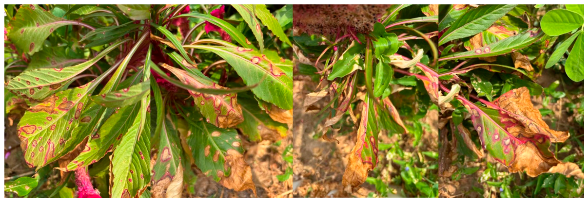
A survey conducted in the Zhang Zhongjing Medicinal Plant Garden of Nanyang Institute of Technology revealed that black spot disease of C. cristata primarily affects the leaves, with a disease incidence reaching 100% (n = 30). At the early stage of infection, nearly circular, elliptical, or irregular lesions appear on the leaf surface. The lesions are dark brown to brown in color and are clearly demarcated from healthy tissue. As the disease progresses, the lesions gradually enlarge; at later stages, the central areas become paler, turning grayish white or light brown, and conspicuous concentric rings develop (Figure 1). Under humid conditions, a light black mold layer forms on the lesion surface, consisting of the pathogen’s conidiophores and conidia. In severely affected areas, multiple lesions coalesce, resulting in extensive leaf chlorosis and necrosis, eventually leading to leaf abscission. This disease potentially impairs the growth and ornamental value of C. cristata.

3.2. Morphological Characterization of the Pathogen

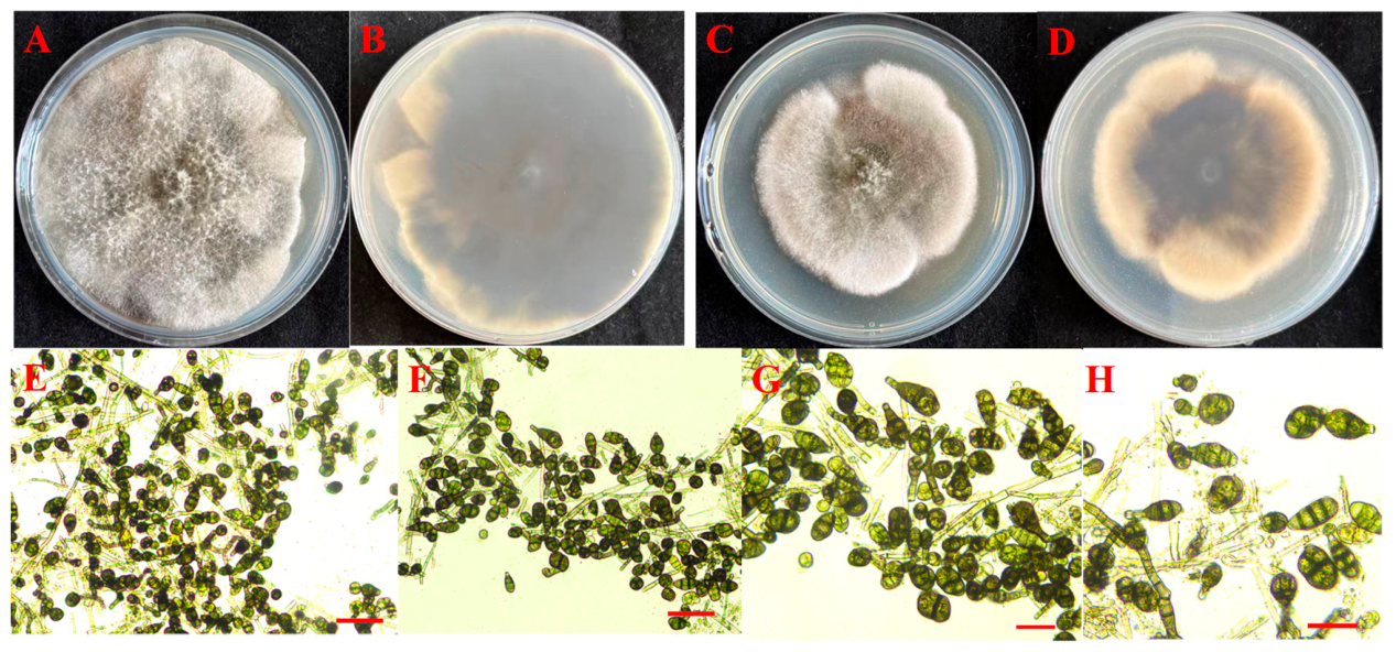
From the collected diseased leaf tissues, seven isolates with similar colony morphologies were successfully obtained through repeated isolation and purification. When cultured on PDA medium, colonies were initially white and floccose, gradually turning grayish black to black with prolonged incubation. The colony margins were entire and circular, with well-developed aerial mycelia and a loose texture (Figure 2A–D). Light microscopic observations revealed distinctive morphological characteristics of the conidiophores and conidia. Conidiophores were solitary or in clusters, dark brown in color, and bore conidia that were often arranged in chains. The conidia were morphologically diverse, predominantly clavate, but also ovoid or obpyriform, brown in color, and possessed multiple transverse, longitudinal, or oblique septa, with conspicuous constrictions at the transverse septa (Figure 2E–H). Conidia measured 19.11–43.11 (mean 29.12 ± 5.46) × 10.22–17.33 (mean 13.77 ± 1.63) µm (n = 50). Based on colony characteristics and conidial morphology, the isolates were preliminarily identified as belonging to the genus Alternaria [10,14].

3.3. Molecular Characterization and Phylogenetic Analysis

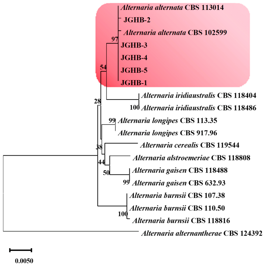
To further precisely determine the taxonomic identity of the isolates causing black spot disease of C. cristata, five representative isolates (JGHB-1 to JGHB-5) were randomly selected for additional gene sequencing and comparative analysis. The resulting sequences were deposited in GenBank under the following accession numbers: ITS (PX945268–PX945269, PZ203761–PZ203763), RPB2 (PX960074–PX960075, PZ225211–PZ225213), and TEF1-α (PX960072–PX960073, PZ225208–PZ225210). BLAST analyses were performed using the NCBI database. The results showed that all obtained sequences shared 99–100% identity with corresponding sequences of Alternaria alternata. Furthermore, the corresponding ITS, RPB2, and TEF1-α gene sequences were aligned with the homologous loci of 14 related type strains (Table S1) within the same genus. A phylogenetic tree based on ITS, RPB2, and TEF1-α sequences were constructed using the maximum likelihood method. As shown in Figure 3, isolates JGHB-1 to JGHB-5 clustered within the Alternaria alternata species complex with a bootstrap value of 97%, indicating strong phylogenetic support. Based on the combined results of molecular and morphological identification, the pathogenic isolates JGHB-1 to JGHB-5 were identified as A. alternata.

3.4. Pathogenicity Test

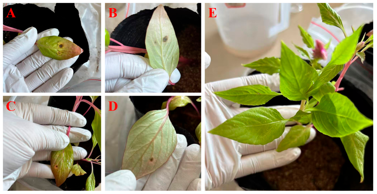
Two randomly selected isolates (JGHB-2 and JGHB-3) were evaluated for pathogenicity. A prepared spore suspension of the isolate was inoculated onto healthy C. cristata. Under moist incubation conditions, inoculated leaves began to exhibit symptoms after 3 days. At the early stage, pinpoint-sized brown spots appeared on the leaf surface; these lesions gradually expanded and developed into black spot symptoms consistent with those observed in the field. The lesions were nearly circular, elliptical, or irregular in shape and ranged in color from dark brown to brown (Figure 4A–D). In contrast, control leaves inoculated with sterile water showed no disease symptoms (Figure 4E). The pathogen was re-isolated from the diseased leaves, and subsequent culturing and morphological observations demonstrated that the re-isolated organism exhibited morphological characteristics identical to those of the original inoculated strain. Through these procedures, Koch’s postulates were fulfilled, confirming that this isolate is the causal pathogen of black spot disease in C. cristata.

3.5. Inhibitory Effects of Fungicides and T. harzianum on the Pathogen Causing Black Spot Disease on C. cristata

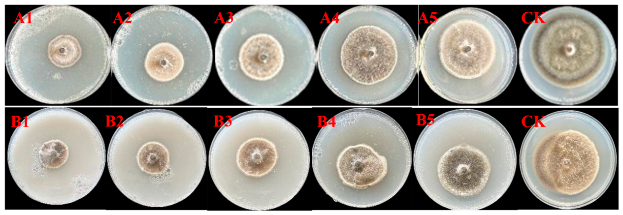
This study evaluated the toxicity of two fungicides, 80% mancozeb and 75% chlorothalonil, against the mycelial growth of A. alternata, the causal agent of black spot disease on C. cristata. The inhibitory effects of both fungicides on the mycelium exhibited a concentration-dependent relationship, with the inhibition rate progressively increasing as the fungicide concentration rose (Table 2 and Table 3, Figure 5). Based on the measured inhibition rates, toxicity regression equations for the two fungicides were established, and their EC50 values were calculated. The correlation coefficient (R2) of the toxicity regression equations for the two fungicides were 0.9824 and 0.9962, respectively (Table 4). Both values approached 1, indicating a high degree of linear fit between fungicide concentration and the inhibition rate, thereby confirming the reliability of the experimental data. A substantial difference was observed in the EC50 between the two fungicides: the EC50 of 80% mancozeb was only 97.72 mg/L, whereas that of 75% chlorothalonil reached 2469.64 mg/L. These results demonstrate that mancozeb exhibits stronger inhibitory activity against this black spot pathogen, with a toxicity significantly superior to that of chlorothalonil.
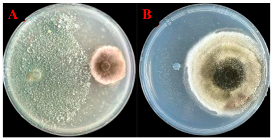
In this study, the dual culture method was employed to systematically evaluate the inhibitory effect of T. harzianum on the mycelial growth of the C. cristata black spot pathogen. The results of the dual culture assay demonstrated that T. harzianum exhibited a significant antagonistic effect against the pathogen, markedly suppressing its colony growth (Figure 6). The calculated average inhibition rate reached 69.95% ± 5.00 (n = 3), indicating that this biocontrol fungus has promising potential for the biological control of the fungus causing black spot disease on C. cristata.

4. Discussion

C. cristata is not only highly ornamental; in China, its dried inflorescences are also used as a traditional medicinal material with astringent and hemostatic properties and are included in the Chinese Pharmacopoeia [3,15]. In this study, C. cristata black spot disease occurring in the Zhang Zhongjing Medicinal Plant Garden in Nanyang City, Henan Province, was investigated. Seven morphologically similar fungal isolates were obtained using the tissue isolation method. Following purification, the pathogen was identified as A. alternata based on colony characteristics, conidial morphology, multi-gene combined sequencing, and phylogenetic analysis, and its pathogenicity was further confirmed by Koch’s postulates. This study represents the first report of black spot disease caused by A. alternata on C. cristata plants in Henan Province, providing a valuable reference for the precise prevention and control of this disease.
Alternaria spp. belong to the phylum Ascomycota, class Dothideomycetes, order Pleosporales, family Pleosporaceae, and genus Alternaria. Species of the genus Alternaria are ubiquitous plant pathogenic fungi with a broad host range and high species diversity; to date, more than 300 Alternaria species have been reported [16]. These pathogens can infect cereal crops, economic crops, and ornamental plants, causing various diseases such as leaf spot and black spot. However, the precise identification of Alternaria species remains challenging. The taxonomy of this genus has undergone extensive revision, and reliable species delimitation typically requires a combination of morphological characterization and multi-locus phylogenetic analysis [10]. As a representative species of the genus, A. alternata exhibits pathogenicity chiefly through the dissemination and infection of conidia. The conidia are dispersed to host surfaces via air currents and rain splash, where they germinate under favorable conditions to produce hyphae that penetrate the host epidermis, invade plant tissues, and secrete toxins that disrupt cellular structures, ultimately leading to lesion formation [17,18]. A. alternata has been reported to cause diseases in a wide range of crops, including tamarillo (Solanum betaceum) [19], honeysuckle (Lonicera japonica) [20], kiwiberry (Actinidia arguta) [21], Menispermum dauricum [22], kidney bean (Phaseolus vulgaris) [23], Yucca gloriosa [24], and Pinus bungeana [25]. These reports collectively demonstrate the remarkable ecological adaptability and host plasticity of A. alternata. In the present study, symptoms appeared on C. cristata leaves as early as 3 days after inoculation with A. alternata, indicating its strong infective capacity and virulence.
The climatic conditions of the Nanyang region provide a favorable environment for the growth, reproduction, and infection of A. alternata. This area is characterized by a temperate monsoon climate, with hot and rainy summers and abundant annual sunshine. The average temperature during July–August ranges from 25 to 30 °C, and relative humidity can exceed 80% [26], which closely matches the optimal temperature for A. alternata culture (25 °C) and the high-humidity conditions required for pathogenicity assays [27,28]. High temperature and humidity not only promote conidial germination and mycelial growth of A. alternata, but also prolong spore viability and enhance infection efficiency [29]. In addition, frequent summer rainfall facilitates spore dispersal, thereby accelerating disease spread and epidemic development [30,31]. In summary, the climatic characteristics and field cultivation conditions in the Nanyang region are highly consistent with the ecological adaptability of A. alternata, constituting a key environmental factor contributing to the occurrence of black spot disease on C. cristata. Accordingly, preventive fungicide applications should be implemented in advance during periods of high temperature and humidity, and rational planting density should be adopted to improve field ventilation, thereby reducing pathogen infection and dissemination.
Regarding the screening of chemical control agents, the results of this study demonstrated that 80% mancozeb exhibited significantly higher inhibitory activity against A. alternata than 75% chlorothalonil, with an EC50 value of only 97.72 mg/L, which is substantially lower than that of chlorothalonil (2469.64 mg/L). As a broad-spectrum protective fungicide, mancozeb acts through a multi-site mechanism that inhibits the activity of sulfhydryl-containing enzymes in fungi and disrupts their metabolic processes, thereby reducing the likelihood of pathogens developing resistance [32]. Mancozeb exhibits strong inhibitory activity against a broad spectrum of plant pathogens. Previous studies have reported that 43% mancozeb has an EC50 value of 202.1461 mg/L against Colletotrichum aenigma, the causal agent of leaf blight on Aucuba japonica [33], while 70% mancozeb exhibits an EC50 value of 447.760 mg/L against Fusarium solani, the pathogen responsible for white root rot in Bletilla striata [34]. These findings collectively demonstrate the high efficacy of mancozeb in suppressing plant pathogenic fungi. Although mancozeb and chlorothalonil have been banned by the European Union, they continue to be widely applied in numerous countries and regions due to their low toxicity and cost-effectiveness. In China, these two fungicides are commonly utilized to control foliar diseases in various crops. Nevertheless, given their potential risks to human health and environmental hazards, their practical application must strictly adhere to relevant regulatory standards to ensure compliant and safe usage. However, the long-term and exclusive application of chemical pesticides can lead to environmental pollution and pesticide residues, which demands particular caution, especially when applied to medicinal plants. Therefore, this study also evaluated the antagonistic efficacy of the biocontrol agent T. harzianum against A. alternata. Dual-culture assays showed a mean inhibition rate of 69.95%, indicating that T. harzianum has substantial antagonistic activity against the pathogen causing black spot in C. cristata, potentially mediated by competition, mycoparasitism, and antibiosis [35]. These findings clearly demonstrate that T. harzianum possesses outstanding potential for biocontrol development, making it a promising candidate strain for the eco-friendly management of C. cristata black spot disease. Future studies should conduct in vivo pot inoculation assays and field efficacy trials to further evaluate the strain’s control stability, colonization capacity, and environmental adaptability against Alternaria pathogens under natural conditions.
By leveraging the respective advantages of chemical and biological control, we recommend adopting an integrated management strategy that combines both approaches in practical cultivation. Although this study provides a scientific basis for understanding the disease cycle and implementing precise management of C. cristata black spot, further research is warranted. Future studies should focus on the differentiation of pathogen pathogenicity, variations in disease resistance among different C. cristata cultivars, and the evaluation of fungicide efficacy under field conditions to provide more comprehensive technical support for the sustainable, long-term control of this disease.

5. Conclusions

In this study, black spot disease affecting C. cristata in Nanyang, Henan Province, China, was systematically investigated. The causal pathogen was identified as A. alternata based on morphological characteristics, multi-gene (ITS, RPB2, and TEF1-α) phylogenetic analysis, and the fulfillment of Koch’s postulates. To our knowledge, this represents the first report of A. alternata causing black spot disease on C. cristata in Henan Province. Among the evaluated fungicides, 80% mancozeb exhibited the strongest inhibitory activity against A. alternata with an EC50 value of 97.72 mg/L. Additionally, the biocontrol agent T. harzianum inhibited mycelial growth by 69.95%, demonstrating substantial potential for biological control. These findings establish a scientific foundation for the integrated management of black spot disease on C. cristata.

Supplementary Materials

The following supporting information can be downloaded at https://www.mdpi.com/article/10.3390/horticulturae12081002/s1: Table S1: Descriptions and GenBank accession numbers for the Alternaria species included in the phylogenetic study.

Author Contributions

Conceptualization, K.Y. and C.Z.; methodology, M.L.; software, Q.J.; validation, K.Y., C.Z. and B.L.; formal analysis, C.L.; investigation, C.Z.; resources, M.L.; data curation, C.L. and B.L.; writing—original draft preparation, K.Y.; writing—review and editing, X.H.; visualization, M.L.; supervision, C.L.; project administration, K.Y. and X.H.; funding acquisition, K.Y. and X.H. All authors have read and agreed to the published version of the manuscript.

Funding

This research was funded by the National Modern Agricultural Industry Technology System (CARS-21), Doctoral Research Start-up Fund of Nanyang Institute of Technology (NGBJ-2024-06), Key project at central government level: The ability establishment of sustainable use for valuable Chinese medicine resources (2060302), Nanyang Municipal Science and Technology Research Project (25KJGG019), and the Key Science and Technology Program of Nanyang City (No. 25ZDZX002).

Data Availability Statement

The nucleotide sequences generated in this study were deposited in GenBank under the following accession numbers: ITS (PX945268–PX945269, PZ203761–PZ203763), RPB2 (PX960074–PX960075, PZ225211–PZ225213), and TEF1-α (PX960072–PX960073, PZ225208–PZ225210) (https://www.ncbi.nlm.nih.gov/genbank/, accessed on 6 February 2026).

Conflicts of Interest

The authors declare no conflicts of interest.

References

  1. Editorial Committee of Flora of China. Chinese Academy of Sciences Flora of China; Science Press: Beijing, China, 1979; 201p. [Google Scholar]
  2. Tripathi, N.; Khan, N. Phytochemical and pharmacological overview on Celosia cristata Linn. J. Adv. Res. 2021, 12, 46–51. [Google Scholar] [CrossRef] [Scilit]
  3. Zhao, X.; Ding, A.; Chen, P.; Zhang, L.; Qu, J.; Bao, B. Celosia cristata L.-an underutilized Chinese medicine: A review of the ethnic applications, phytochemistry, pharmacology, quality control and toxicity. J. Ethnopharmacol. 2024, 333, 118479. [Google Scholar] [CrossRef] [Scilit] [PubMed]
  4. Huang, W. Biological characteristics and cultivation management of Celosia cristata. Anhui Agric. Sci. Bull. 2012, 18, 111–112. [Google Scholar]
  5. Wang, Y.; Xing, Y.; Yu, Z. Cultivation and management of medicinal Celosia cristata. Spec. Econ. Anim. Plants 2018, 21, 37–38. [Google Scholar]
  6. Hou, P.; Wang, Y.; Qiu, S.; Wang, J.; Li, S.; Wang, H.; Yin, S.; Du, J. Impacts of climate change and human activities on vegetation NDVI changes in henan province from 2000 to 2020. Front. Environ. Sci. 2025, 13, 1682187. [Google Scholar] [CrossRef] [Scilit]
  7. Xiang, Y.X.; Zhou, J.; Li, Z.; Liu, H.F.; Deng, J.X. Identification and fungicide control of Alternaria alternantherae causing leaf spot on Celosia cristata and Alternanthera philoxeroides in China. Horticulturae 2026, 12, 750. [Google Scholar] [CrossRef] [Scilit]
  8. Lager, P.; Sharma, J.; Kumar, Y. First report of a begomovirus and associated betasatellite causing yellow vein mosaic disease of Celosia cristata. New Dis. Rep. 2023, 48, e12211. [Google Scholar] [CrossRef] [Scilit]
  9. Wang, T.Y.; Zhao, J.; Ma, G.P.; Bao, S.W.; Wu, X.H. Leaf blight of sunflower caused by Alternaria tenuissima and A. alternata in Beijing, China. Can. J. Plant Pathol. 2019, 41, 372–378. [Google Scholar] [CrossRef] [Scilit]
  10. Li, J.; Jiang, H.; Jeewon, R.; Hongsanan, S.; Bhat, D.; Tang, S.; Lumyong, S.; Mortimer, P.; Xu, J.; Camporesi, E.; et al. Alternaria: Update on species limits, evolution, multi-locus phylogeny, and classification. Stud. Fungi 2023, 8, 1–61. [Google Scholar] [CrossRef] [Scilit]
  11. Kumar, S.; Stecher, G.; Li, M.; Knyaz, C.; Tamura, K. MEGA X: Molecular evolutionary genetics analysis across computing platforms. Mol. Biol. Evol. 2018, 35, 1547–1549. [Google Scholar] [CrossRef] [Scilit] [PubMed]
  12. Yang, K.; Wang, H.L.; Ye, K.H.; Luo, C.; Wei, Z.X.; Huang, H.P.; Zhu, S.S.; Guo, L.W.; He, X.H. First report of anthracnose disease on Bletilla striata caused by Colletotrichum orchidophilum in Yunnan, China. Plant Dis. 2022, 106, 1070. [Google Scholar] [CrossRef] [Scilit] [PubMed]
  13. Xin, W.; Mao, Y.; Lu, F.; Li, T.; Wang, J.; Duan, Y.; Zhou, M. In vitro fungicidal activity and in planta control efficacy of coumoxystrobin against Magnaporthe oryzae. Pestic. Biochem. Physiol. 2020, 162, 78–85. [Google Scholar] [CrossRef] [Scilit] [PubMed]
  14. Gou, Y.N.; Aung, S.L.L.; Htun, A.A.; Huang, C.X.; Deng, J.X. Alternaria species in section Alternaria associated with Iris plants in China. Front. Microbiol. 2022, 13, 1036950. [Google Scholar] [CrossRef] [Scilit] [PubMed]
  15. National Pharmacopoeia Commission Chinese Pharmacopoeia. Chinese Pharmacopoeia; China Medical Science and Technology Press: Beijing, China, 2025; p. 210. [Google Scholar]
  16. Vinogradova, S.A.; Kiselev, K.V.; Suprun, A.R. An overview of the Alternaria genus: Ecology, pathogenicity and importance for Agriculture and Human Health. J. Fungi 2026, 12, 64. [Google Scholar] [CrossRef] [Scilit] [PubMed]
  17. Goldman, S.; Cacciola, S.O.; Ezra, D. Major pomegranate diseases, with a focus on the pathogenesis, epidemiology, and management challenges of Alternaria alternata diseases. Plant Dis. 2026, 110, 1505–1518. [Google Scholar] [CrossRef] [Scilit] [PubMed]
  18. Schmey, T.; Tominello-Ramirez, C.S.; Brune, C.; Stam, R. Alternaria diseases on potato and tomato. Mol. Plant Pathol. 2024, 25, e13435. [Google Scholar] [CrossRef] [Scilit] [PubMed]
  19. Silva, E.J.D.; Claus, A.; Hahn, M.H.; Koch, G.; Silva, M.C.C.; Calegario, R.F.; Duarte, H.D.S.S. Occurrence of Alternaria alternata causing leaf spot on tamarillo in Brazil. Crop Protect. 2024, 179, 106605. [Google Scholar] [CrossRef] [Scilit]
  20. He, P.; Luo, X.; Wu, X.; Cui, W. Leaf spot of Lonicera japonica Thunb. (honeysuckle) caused by Alternaria alternata newly reported in China. Crop Protect. 2023, 170, 106269. [Google Scholar] [CrossRef] [Scilit]
  21. Ruan, R.; Xi, D.; Zhang, C.; Huang, K.; Luo, H.; Liu, H. Alternaria alternata causing brown spot on kiwiberry (Actinidia arguta) in China. Crop Protect. 2022, 152, 105857. [Google Scholar] [CrossRef] [Scilit]
  22. Sun, H.F.; Wei, M.Y.; Li, N.; Yan, Y. First report of Alternaria alternata causing leaf spot on Menispermum dauricum in China. Plant Dis. 2022, 106, 1069. [Google Scholar] [CrossRef] [Scilit] [PubMed]
  23. Jin, Y.; Xiong, Y.; Xu, C.; Ren, J.; Guo, Y.; Zuo, Y.; Zhang, Y.; Geng, X. First report of Alternaria alternata causing leaf spot on Kidney Bean in China. Plant Dis. 2022, 106, 1531. [Google Scholar] [CrossRef] [Scilit] [PubMed]
  24. Zhang, Q.; Zhang, Y.; Shi, H.; Huo, Y. First report of leaf spot caused by Alternaria alternata on Yucca gloriosa in China. Plant Dis. 2022, 106, 1307. [Google Scholar] [CrossRef] [Scilit] [PubMed]
  25. Zhang, M.J.; Zheng, X.R.; Li, H.; Chen, F.M. Alternaria alternata, the causal agent of a new needle blight disease on Pinus bungeana. J. Fungi 2023, 9, 71. [Google Scholar] [CrossRef] [Scilit] [PubMed]
  26. Wang, B. Landscape architecture and plant landscape design based on climate types in Nanyang. J. Nanyang Norm. Univ. 2018, 17, 49–52. [Google Scholar]
  27. Sun, X.; Jiang, B.; Lv, Q.; Liu, G.; Bai, S.; Li, L. Biological characteristics of Alternaria alternata JM and its controlling effect on Descurainia sophia. Chin. J. Biol. Control 2025, 41, 1231–1240. [Google Scholar] [CrossRef]
  28. Geng, X.; Mvchir, H.; Liu, J.; Hua, K.; Miao, Q.; Shu, J. Molecular characterization and pathogenicity analysis of Alternaria alternata associated with leaf spot disease of Toona sinensis in China. Horticulturae 2025, 11, 279. [Google Scholar] [CrossRef] [Scilit]
  29. Fagodiya, R.K.; Trivedi, A.; Fagodia, B.L. Impact of weather parameters on Alternaria leaf spot of soybean incited by Alternaria alternata. Sci. Rep. 2022, 12, 6131. [Google Scholar] [CrossRef] [Scilit] [PubMed]
  30. Thomma, B.P.H.J. Alternaria spp.: From general saprophyte to specific parasite. Mol. Plant Pathol. 2003, 4, 225–236. [Google Scholar] [CrossRef] [Scilit] [PubMed]
  31. Gai, Y.; Niu, Q.; Kong, J.; Li, L.; Liang, X.; Cao, Y.; Zhou, X.; Sun, X.; Ma, H.; Wang, M.; et al. Genomic and transcriptomic characterization of Alternaria alternata during Infection. Agronomy 2023, 13, 809. [Google Scholar] [CrossRef] [Scilit]
  32. Yang, L.N.; He, M.H.; Ouyang, H.B.; Ouyang, H.B.; Zhu, W.; Pan, Z.C.; Sui, Q.J.; Shang, L.P.; Zhan, J. Cross-resistance of the pathogenic fungus Alternaria alternata to fungicides with different modes of action. BMC Microbiol. 2019, 19, 205. [Google Scholar] [CrossRef] [Scilit] [PubMed]
  33. Fan, R.; Liu, Y.; Bin, Y.; Huang, J.; Yi, B.; Tang, X.; Li, Y.; Cai, Y.; Yang, Z.; Yang, M.; et al. Identification of Colletotrichum aenigma as the new causal agent of leaf blight disease on Aucuba japonica Thunb., and screenings of effective fungicides for its sustainable management. Front. Microbiol. 2023, 14, 1222844. [Google Scholar] [CrossRef] [Scilit] [PubMed]
  34. Liang, F.; Jiang, X.; Liu, L.; Wang, F.; Liu, F.; Hu, S.; Tan, L.; Chen, X.; Xu, Y.; Xu, X.; et al. White root rot of Bletilla striata: The pathogen, biological characterization, and fungicide screening. Front. Microbiol. 2024, 15, 1374137. [Google Scholar] [CrossRef] [Scilit] [PubMed]
  35. Braun, H.; Woitsch, L.; Hetzer, B.; Geisen, R.; Zange, B.; Schmidt-Heydt, M. Trichoderma harzianum: Inhibition of mycotoxin producing fungi and toxin biosynthesis. Int. J. Food Microbiol. 2018, 280, 10–16. [Google Scholar] [CrossRef] [Scilit] [PubMed]
Figure 1. Field symptoms of black spot disease on C. cristata.
Horticulturae 12 01002 g001
Figure 2. Colony and conidial morphological characteristics of A. alternata. (AC). Frontal (A,C) and reverse (B,D) views of cultural features of A. alternata on PDA. (EH). Conidia. Scale bars = 50 μm in (E,F). Scale bars = 25 μm in (G,H).
Horticulturae 12 01002 g002
Figure 3. Phylogenetic tree generated based on the combined ITS, RPB2 and TEF1-α gene sequences.
Horticulturae 12 01002 g003
Figure 4. Symptoms on C. cristata leaves after inoculation of a conidial suspension. (AC). Frontal (A,C) and reverse (B,D) views of symptoms on C. cristata leaves. (E). C. cristata leaves remained healthy for seven days when being mock-inoculated with sterile water.
Horticulturae 12 01002 g004
Figure 5. Effects of different fungicides on mycelial growth. (A) Mancozeb. The concentrations corresponding to (A1A5) are 250, 125, 62.5, 31.25, and 15.63 mg/L, respectively. (B) Chlorothalonil. The concentrations corresponding to (B1B5) are 5000, 2500, 1250, 625, and 312.5 mg/L, respectively. CK: blank control.
Horticulturae 12 01002 g005
Figure 6. Inhibitory effect of T. harzianum on the mycelial growth of the pathogen causing black spot disease on C. cristata. (A) T. harzianum treatment. (B) Control.
Horticulturae 12 01002 g006
Table 1. Gene loci and primers used in this study.
GenePrimerSequence (5′–3′)PCR Conditions
ITSITS1 (F)
ITS4 (R)
5′-TCCGTAGGTGAACCTGCGG-3′
5′-TCCTCCGCTTATTGATATGC-3′
98 °C 2 min, (98 °C 10 s, 54 °C 10 s, 72 °C 10 s) 35 cycles, 72 °C 5 min
TEF1-αEF1-728 (F)
EF1-986R (R)
5′-CATCGAGAAGTTCGAGAAGG-3′
5′-TACTTGAAGGAACCCTTACC-3′
98 °C 2 min, (98 °C 10 s, 54 °C 10 s, 72 °C 10 s) 35 cycles, 72 °C 5 min
RPB2rpb2-5 (F)
rpb2-7C (R)
5′- GAYGAYMGWGATCAYTTYGG-3′
5′- CCCATRGCTTGYTTRCCCAT-3′
98 °C 2 min, (98 °C 10 s, 55 °C 10 s, 72 °C 10 s) 35 cycles, 72 °C 5 min
Table 2. Inhibitory rate of 80% mancozeb at different concentrations against the mycelial growth of A. alternata.
Fungicide Concentration (mg/L)Average Inhibition Rate (%)
15.6317.79 ± 3.78 d
31.2534.05 ± 8.10 cd
62.545.40 ± 2.30 bc
12552.76 ± 0.87 ab
25064.11 ± 5.96 a
Data for the average inhibition rate are presented as mean ± SD (n = 3). Letters to the right of the values indicate significant differences between treatments (p < 0.05).
Table 3. Inhibitory rate of 75% chlorothalonil at different concentrations against the mycelial growth of A. alternata.
Fungicide Concentration (mg/L)Average Inhibition Rate (%)
312.526.35 ± 4.05 d
62533.53 ± 2.76 cd
125041.35 ± 3.47 bc
250049.36 ± 4.84 ab
500059.29 ± 2.94 a
Data for the average inhibition rate are presented as mean ± SD (n = 3). Letters to the right of the values indicate significant differences between treatments (p < 0.05).
Table 4. Toxicity of different fungicides on the mycelial growth of the pathogen causing black spot disease on C. cristata.
Fungicide NameToxic Regression EquationCorrelation Coefficient (R2)EC50 (mg/L)95% CI
80% mancozeby = 0.3699x − 0.23610.982497.7279.02~124.88
75% chlorothalonily = 0.2715x − 0.42110.99622469.642341.53~2618.62
Disclaimer/Publisher’s Note: The statements, opinions and data contained in all publications are solely those of the individual author(s) and contributor(s) and not of MDPI and/or the editor(s). MDPI and/or the editor(s) disclaim responsibility for any injury to people or property resulting from any ideas, methods, instructions or products referred to in the content.