Article

Antagonistic and Plant Growth-Promoting Potential of Endophytic Trichoderma Against Fusarium Root Rot in Loquat (Eriobotrya japonica)


1
Key Laboratory of Agricultural Biosafety and Green Production of Upper Yangtze River (Ministry of Education), College of Horticulture and Landscape Architecture, Southwest University, Beibei, Chongqing 400715, China
2
Symcode (Chongqing) Biotechnology Co., Ltd., Beibei, Chongqing 400715, China
*
Author to whom correspondence should be addressed.
Horticulturae 2026, 12(7), 859; https://doi.org/10.3390/horticulturae12070859
Submission received: 9 June 2026 / Revised: 6 July 2026 / Accepted: 13 July 2026 / Published: 15 July 2026

Abstract

Loquat (Eriobotrya japonica) is highly susceptible to destructive soil-borne Fusarium pathogens, which cause severe vascular wilt and root rot. While Trichoderma species are widely applied as biocontrol agents, the potential of endophytic Trichoderma strains from loquat remains largely unexplored. This study aimed to isolate and evaluate loquat endophytic Trichoderma strains for their antifungal and plant growth-promoting capabilities. Ten isolates were identified through morphological and phylogenetic analyses as belonging to T. asperellum, T. virens and T. hamatum. Antagonistic activity was initially screened in vitro via dual-culture assays, followed by in vivo greenhouse pot experiments. In vitro results revealed that T. asperellum strains (B077R1 and B077B3) and T. virens GFR9 strongly inhibited the mycelial growth of both F. oxysporum and F. solani (inhibition rates > 50.65%), whereas T. hamatum isolates exhibited weaker antagonism (<35.18%). Moreover, T. asperellum B077R1 demonstrated the highest biocontrol efficacy in the greenhouse pot assay, significantly reducing disease severity caused by F. oxysporum and F. solani by 45.99% and 55.50%, respectively. This strain also significantly enhanced plant height, total biomass (fresh and dry weight) and photosynthetic pigment content (p < 0.05). These findings suggest that newly isolated endophytic Trichoderma strains from loquat exhibit robust host compatibility, offering a promising sustainable strategy for managing root rot in perennial fruit crops.

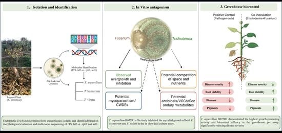
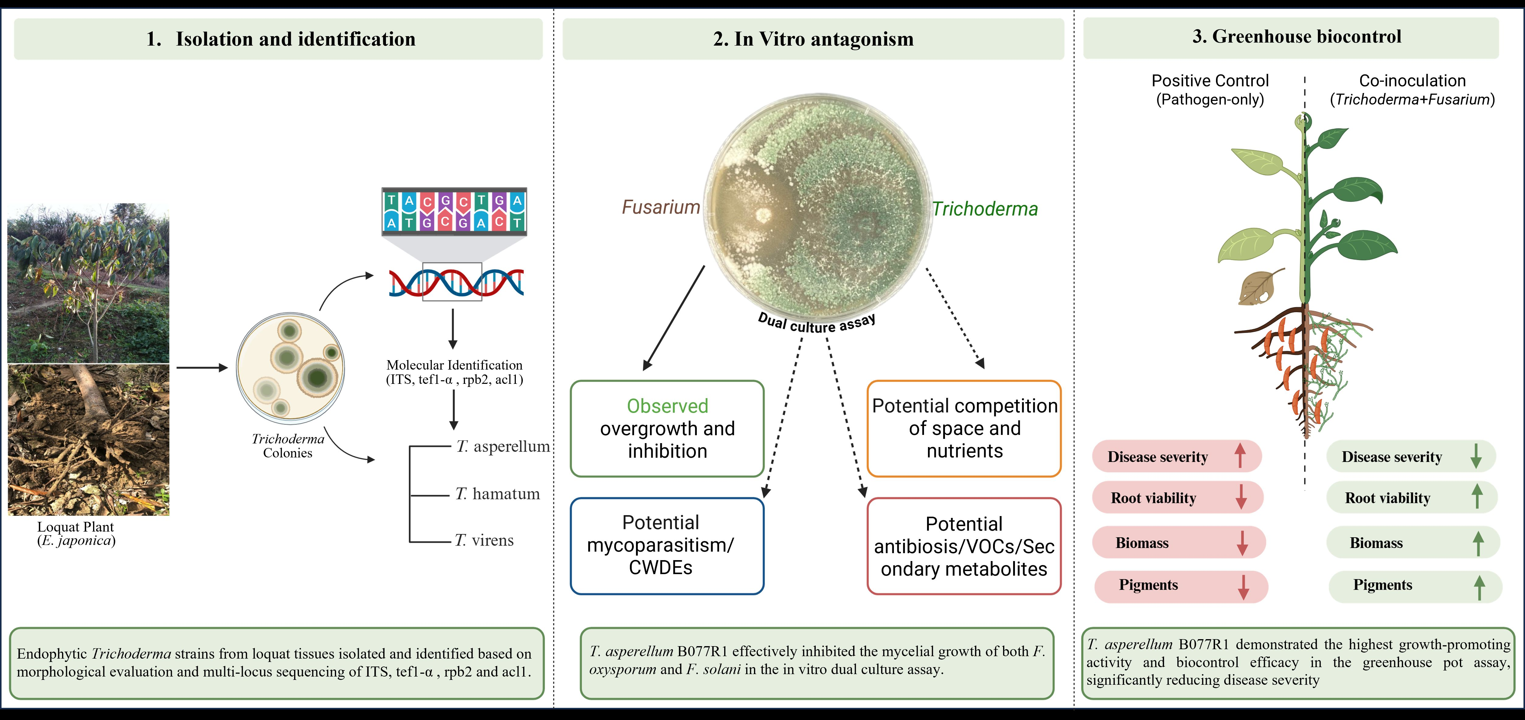
Graphical Abstract

1. Introduction

Loquat (Eriobotrya japonica), a member of the Rosaceae family, is a commercially vital perennial fruit-bearing tree indigenous to southern China. Unlike most stone fruit trees, loquat blooms in winter and its fruit matures in summer, filling a critical gap in the fresh fruit market and commanding high economic returns [1]. It has been extensively introduced to subtropical regions worldwide, including Japan, Spain, Turkey and Italy [1,2]. China is the predominant producer, with cultivation spanning over 170,000 hectares and an annual yield of around 400,000 tons [3]. Beyond its agronomic value, the loquat is increasingly recognized as a functional food. The leaves, flowers and fruit are rich in bioactive compounds such as ursolic acid, carotenoids, and phenolics, which contribute to its significant antioxidant and therapeutic properties in traditional medicine [4]. Consequently, the loquat industry plays a crucial role in increasing farmers’ income and rural revitalization.
Despite its economic potential, the sustainable development of the loquat industry is severely threatened by soil-borne diseases. The plant is highly susceptible to vascular wilt pathogens, particularly Fusarium species, which colonize the xylem vessels and obstruct water and nutrient transport [5]. Among these, Fusarium-induced root rot represents the most destructive constraint [6]. Fusarium solani and Fusarium oxysporum, ubiquitous soil inhabitants notorious for infecting a wide range of crops, have been identified as the primary etiological agents driving this decline in loquat orchards due to root and fruit rot [7,8]. In several Chinese production regions, such as Chongqing, Yunnan, and Fujian province, the F. solani species complex (FSSC) and the F. oxysporum species complex (FOSC) have infected approximately 5–10% of plantations. According to Wu [8], the symptoms of the disease include foliar chlorosis (yellowing), wilting, decay of the main root, and cracking of lateral roots, eventually causing the plant to defoliate and die. Given the severity of these symptoms and the rapid spread of the pathogens, effective management of Fusarium root rot has become an urgent priority for the loquat industry.
Conventionally, chemical fungicides are employed for disease management. However, their excessive application or misuse poses environmental risks and accelerates the selection pressure for pathogen resistance. Consequently, biological control has emerged as a sustainable alternative for managing Fusarium wilt, offering benefits such as environmental compatibility and cost-effectiveness [9]. Among biological control agents (BCAs), Trichoderma spp. are one of the most widely studied and commercially utilized species [10]. Substantial evidence underscores their efficacy against Fusarium-induced disease. For instance, in vitro assays have demonstrated the potent antifungal activities of T. viride and T. harzianum against F. solani [11]. In practical applications, T. asperellum (e.g., T-34 and TM11) has exhibited remarkable biocontrol potential against F. oxysporum and F. commune, significantly reducing the incidence of root rot disease in tomato and blueberry [12,13,14]. Similarly, T. gamsii 6085 has shown strong antagonism against rice pathogens [15], while field applications of T. harzianum SQR-T037 have been reported to enhance tomato biomass and optimize soil nutrient balance [16].
This biocontrol success relies on a multi-faceted defense strategy involving direct pathogen suppression through mycoparasitism and the secretion of hydrolytic enzymes (e.g., chitinases, glucanases, and proteases) [17]. Additionally, these fungi inhibit pathogen growth by synthesizing volatile antimicrobial compounds and prime the plant defense system by inducing systemic resistance [18]. Furthermore, Trichoderma spp. can enhance nutrient uptake and secrete phytohormones (e.g., auxins, gibberellins, salicylic acid and abscisic acid), thereby boosting plant vigor and biomass accumulation even under biotic or abiotic stress [19].
Despite this proven efficacy, the field application of commercial Trichoderma formulations often yields inconsistent results. Current perspectives emphasize that introduced strains frequently fail to bridge the gap between laboratory and field efficacy due to the inability to align with specific endophyte behaviors and ecological constraints [20,21,22]. In contrast, indigenous endophytic strains possess co-evolved advantages derived from long-term host interactions, granting them superior biotransformation abilities and environmental fitness [23,24]. Moreover, effective biocontrol is increasingly linked to host-specific transcriptional regulation, suggesting that intimate host-microbe compatibility is a prerequisite for optimal efficacy [25]. However, despite their known potential, the specific application of indigenous endophytic Trichoderma in loquat against Fusarium-induced vascular wilt remains largely unexplored.
We hypothesized that endophytic Trichoderma from loquat suppresses Fusarium spp. and promotes loquat growth under greenhouse conditions. Therefore, this study was designed to isolate and identify endophytic Trichoderma species from loquat. It aimed to assess the antagonistic ability of the isolated Trichoderma strains against Fusarium oxysporum and Fusarium solani, and to investigate their capability to promote loquat growth and enhance physiological resilience under greenhouse conditions.

2. Materials and Methods

2.1. Isolation and Culture Conditions

Root samples exhibiting root rot symptoms were collected from loquat orchards at Yangdu Base, Zhejiang Province. Endophytic fungi were isolated according to [26] with slight modifications. Explants were washed and cut into small segments approximately 0.5 cm wide and 1 cm long using sterilized blades on a clean bench. These segments were surface-sterilized using ethanol (75%) for 4 min and hydrogen peroxide (10%) for 8 min, followed by 3–4 rinses with sterile distilled water, and dried on sterilized filter paper [27]. Moreover, the potato dextrose agar (PDA; AOBOX BIO-TECH, Cat 02-023, Beijing, China) media were augmented with streptomycin sulfate (30 μg mL−1) and incubated at 26 °C with a 12 h light/dark cycle. Plates were regularly flipped and checked for possible contamination, while light exposure helped to promote fungal sporulation [28]. Emerging fungal hyphae were excised with a sterile scalpel and cultured on fresh PDA to obtain pure colonies. Isolates were deposited in the Loquat Germplasm Resources Laboratory, Southwest University, Chongqing, China, assigned unique identification codes (e.g., B077R1, B077R7, and GFR9), and preserved long-term in 30% glycerol at −80 °C.

2.2. Morphological Identification and Characterization

To conduct a comparative analysis of colony growth and morphological traits, plates from fresh monosporic Trichoderma cultures were inoculated onto 9 cm Petri dishes filled with 20 mL of PDA, cornmeal dextrose agar (CMA), or synthetic low-nutrient agar (SNA) and incubated at 28 ± 1 °C with 70 ± 5% relative humidity, under a 12:12 h light/dark photoperiod [29]. The examination of morphological characteristics included phialide structure, conidial morphology, and chlamydospore formation, using a Nikon Eclipse Ci microscope (Nikon Corporation, Shinagawa, Japan) paired with a Nikon DS-Ri2 camera. Measurements and image analyses were carried out by using NIS Elements software (v. 4.30.01, Nikon), focusing on 30 individual phialides and conidia for each isolate. The staining and examination of fungal specimens were performed in accordance with the protocols proposed by [30,31].

2.3. Molecular Identification and Phylogenetic Analysis

Trichoderma isolates were cultured on PDA for 3–4 days, and gDNA was isolated by using the CTAB method with minor modifications [32]. The nuclear ribosomal ITS1-5.8S rRNA-ITS2 region (ITS), translation elongation factor 1-α (tef1-α), RNA polymerase II second largest subunit (rpb2) and ATP citrate lyase large subunit (acl1) gene regions were amplified by PCR [33]. PCR was carried out in a 40 μL reaction mixture containing 1 μL gDNA, 20 μL of 2 × Taq PCR Master Mix, 2 μL of each primer (10 µM) and 15 μL of ddH2O. Each 50 μL reaction contained 1 ng of gDNA, 20 μL of 2× Taq PCR Master Mix, 1 μM of each primer, and 15 μL of ddH2O. Primer sequences are provided in Supplementary Table S1. The sequences were deposited in GenBank, and the corresponding accession numbers are listed in Supplementary Table S2.
Phylogenetic analyses were performed based on a combined dataset of ITS, tef1-α, rpb2 and acl1 to advance taxonomic resolution [33,34]. Maximum likelihood (ML) analysis was conducted using RAxML-HPC2 (v8.2.10) through the CIPRES Science Gateway (http://www.phylo.org/) under the GTRGAMMA model with 1000 bootstrap replicates. Maximum parsimony (MP) analysis was carried out in PAUP v4.0a169 with 1000 heuristic replicates. Bayesian inference (BI) was carried out in MrBayes v3.2.6 implemented in PhyloSuite v1.2.3. Phylogenetic trees were visualized using FigTree v1.4.2.

2.4. Antagonistic Activity of Trichoderma In Vitro

In vitro antagonistic activity of Trichoderma spp. against F. solani (FSSC) and F. oxysporum (FOSC) was evaluated using a direct confrontation assay on PDA [35]. The pathogenic FSSC used in this study was previously recovered from infected loquat roots [8]. While the FOSC was identified via multi-locus sequence analysis (ITS, tef1-α and rpb2) using BLASTn and Fusarium-ID (GenBank accession no. MT974325, MT992555, MW072791; unpublished). Mycelial plugs (5 mm3) from the actively growing margins of 7-day-old Fusarium cultures and 10 Trichoderma isolates derived from loquat explants were co-inoculated onto 9 cm PDA plates to assess interspecific interactions and inhibition [36]. Plugs were placed 3 cm apart and 3 cm from the plate edge. For each Fusarium-Trichoderma combination, three plates were used as biological replicates, with Fusarium monocultures serving as controls (three replicates each). Cultures were incubated in the dark at 28 ± 1 °C for 8 days. Antagonistic effects were expressed as percentage growth inhibition (PGI), calculated according to [37].
P G I ( % ) = [ ( C T ) / C ] × 100
where C = Radial growth of the pathogen (control), and T = Radial growth of the pathogen (treated).

2.5. Soil and Plant Material for Bioagents Efficacy Assessment

Commercial nutrient soil (Klasmann, Geeste, Germany) was mixed with perlite and vermiculite (7:2:1, v/v/v), moistened, and autoclaved at 121 °C for 15 min. Disinfected pots (12.7 cm × 11.4 cm) were filled to three-quarters capacity with the sterile substrate and adjusted to 60% field capacity before transplanting. The susceptible loquat cultivar ‘Jinhua No. 1’ was used for greenhouse assays. Loquat seeds were surface-sterilized with 50% carbendazim diluted at 1:500 for 1 h. The seedlings were then cultivated in sterile substrate for 6 months until they reached 5–6 true leaves. Uniform and healthy seedlings were selected for the experiment. The roots were washed gently, surface sterilized with 3% sodium hypochlorite for 1 min, washed three times with sterile distilled water and transplanted into the prepared pots.

2.6. Preparation of Spore Suspension and Pot Experiment

Fungal strains were sub-cultured on PDA, and conidia were harvested from 7-day-old cultures into sterile distilled water. Trichoderma and Fusarium were incubated under 12 h light/dark cycles to promote sporulation [38]. A hemocytometer was used to adjust the required concentration of the microconidia suspension (1 × 10⁶ spores/mL) to inoculate selected roots of loquat seedlings for the pot experiments to evaluate disease resistance and plant growth, following previously described inoculation methods [8,39]. Prior to inoculation, small wounds were made on the fibrous roots with a sterile scalpel, and the taproots were gently punctured using a sterile needle. This approach simulated natural root injuries under controlled greenhouse conditions and facilitated subsequent Fusarium infection. The positive controls (pathogen-only) and Trichoderma co-inoculated with Fusarium (treated groups) were included in the treatments. The following 4 treatments were developed: (1) FOSC + H2O, which received 20 mL of F. oxysporum suspension and 20 mL of sterile distilled water; (2) FSSC + H2O, which received 20 mL of F. solani and 20 mL of sterile distilled water; (3) co-inoculation with F. oxysporum (20 mL) and 20 mL of Trichoderma suspension (T. asperellum B077R1, T. hamatum B077R7, or T. virens GFR9); and (4) co-inoculation with F. solani (20 mL) and 20 mL of the corresponding Trichoderma spore suspension. The greenhouse experiments were conducted for 60 days, with a second identical inoculation performed at 30 days post-inoculation (dpi). The disease symptoms were monitored repeatedly on the same plants at different intervals, whereas final growth and physiological parameters were recorded at 60 dpi. To ensure successful root colonization, the Trichoderma and Fusarium suspension were applied directly to the root zone of each pot/plant as a soil drench using wide-orifice pipette tips (5 mL). The suspension was applied slowly around the base of the stem and root zone to allow infiltration into the rhizosphere and to avoid surface runoff. All inoculated plants were maintained in a controlled growth room at 25 ± 1 °C temperature, 80% relative humidity, and a 16/8 h light/dark photoperiod. The pot assay was arranged in a completely randomized design, and the pots were randomly positioned in the growth room to minimize positional effects. All treatments were conducted with three biological replicates.

2.7. Assessment of Plant Growth and Biomass Production

To assess the plant growth in response to the different treatment combinations, several morphological and physiological parameters were measured [40,41]. These parameters included plant height (cm), plant fresh weight (g), plant dry weight (g), root fresh weight (g), and root dry weight (g). The disease severity index (DSI) was evaluated at 60 dpi by assessing the extent of root rot and root discoloration using a 0–5 rating scale. The scale was defined as follows: 0 = roots well developed with no discoloration; 1 = slight browning or less than 25% root discoloration; 2 = 25–50% discoloration; 3 = 50–75% root discoloration; 4 = severe browning of the hypocotyl or >75% discoloration of root; and 5 = completely discolored roots, leading to plant death [42,43]. Since root discoloration affects both external and internal tissues, visual assessments of the proportion of the discolored root area (0–100%) were employed to standardize the disease ratings. The DSI was calculated using the following formula:
D S I ( % ) = ( D e g r e e   o f   i n f e c t i o n × N u m b e r   o f   p l a n t s   w i t h   s a m e   d e g r e e ) × 100 / ( T o t a l   n u m b e r   o f   p l a n t s × H i g h e s t   d e g r e e   o f   i n f e c t i o n )
Additionally, plant vigor was evaluated based on a wilting severity scale ranging from 0 to 4: 4 = completely healthy plant; 3 = thin stem and reduced height; 2 = presence of brown lesions on the stem and yellowing leaf tips; 1 = wilting of stems and leaves; and 0 = completely dead plant. Root vigor was quantified using the triphenyl tetrazolium chloride (TTC) reduction assay [44].

2.8. Photosynthetic Pigment Content Measurements

Total chlorophyll content of loquat leaves was determined using a UV/VIS spectrophotometer (AOELAB) following the method of [45]. Fresh leaves (0.1 g) were cut into fine threads and submerged for 72 h in 10 mL of an extraction solvent containing 95% ethanol and 80% acetone (1:2) in the dark at 4 °C till complete tissue decolorization. The absorbance of the resulting extract was measured at 470 nm, 645 nm and 663 nm [46]. Contents of chlorophyll a (Ca), chlorophyll b (Cb), total carotenoids (Cx+c) and the total chlorophyll content were converted to mg·g−1 fresh weight (FW). The concentrations (µg·mL−1) for chlorophyll a, chlorophyll b and the sum of leaf carotenoids were calculated using the following equations [47].
Ca = 12.25 A663.2 − 2.79 A646.8
Cb = 21.50 A646.8 − 5.10 A663.2
Cx+c = (1000 A470 − 1.82. Ca − 85.02.cb)/198
where A663.2, A646.8 and A470 represent the absorbance values at 663.2 nm, 646.8 nm and 470 nm, respectively.
The leaf photosynthetic pigment content per unit mass (LPC) and per unit leaf area (LPA) were calculated as follows:
L P C m g · g 1 = C   m g L 1 × V t   m L F W 1   g × 1000 × n
L P A m g · c m 2 = C   m g L 1 × V t   m L F W 0   g × 1000 × n
where C is the total leaf photosynthetic pigment content (Ca + Cb + Cx+c, converted to mg·L−1), FW1 is the fresh weight of the extracted leaf (g), FW0 is the area of the extracted leaf (cm2), Vt is the volume of extraction solution (Vt = 10 mL), and n is the dilution ratio (n = 1).

2.9. Statistical Analyses

Prior to the ANOVA, the data were tested for normality and homogeneity of variances using the Shapiro–Wilk and Levene’s tests, respectively. All data shown represent the mean ± standard error (S.E.) derived from three biological replicates. Statistical analyses were conducted using a one-way analysis of variance (ANOVA) in Statistix 10.0 (Analytical Software, Tallahassee, FL, USA). The significance of differences between treatments was assessed using Tukey’s honestly significant difference (HSD) test at p < 0.05. Further statistical details are provided in the results section and figure captions.

3. Results

3.1. Isolation and Morphological Characterization of Endophytic Trichoderma from Loquat

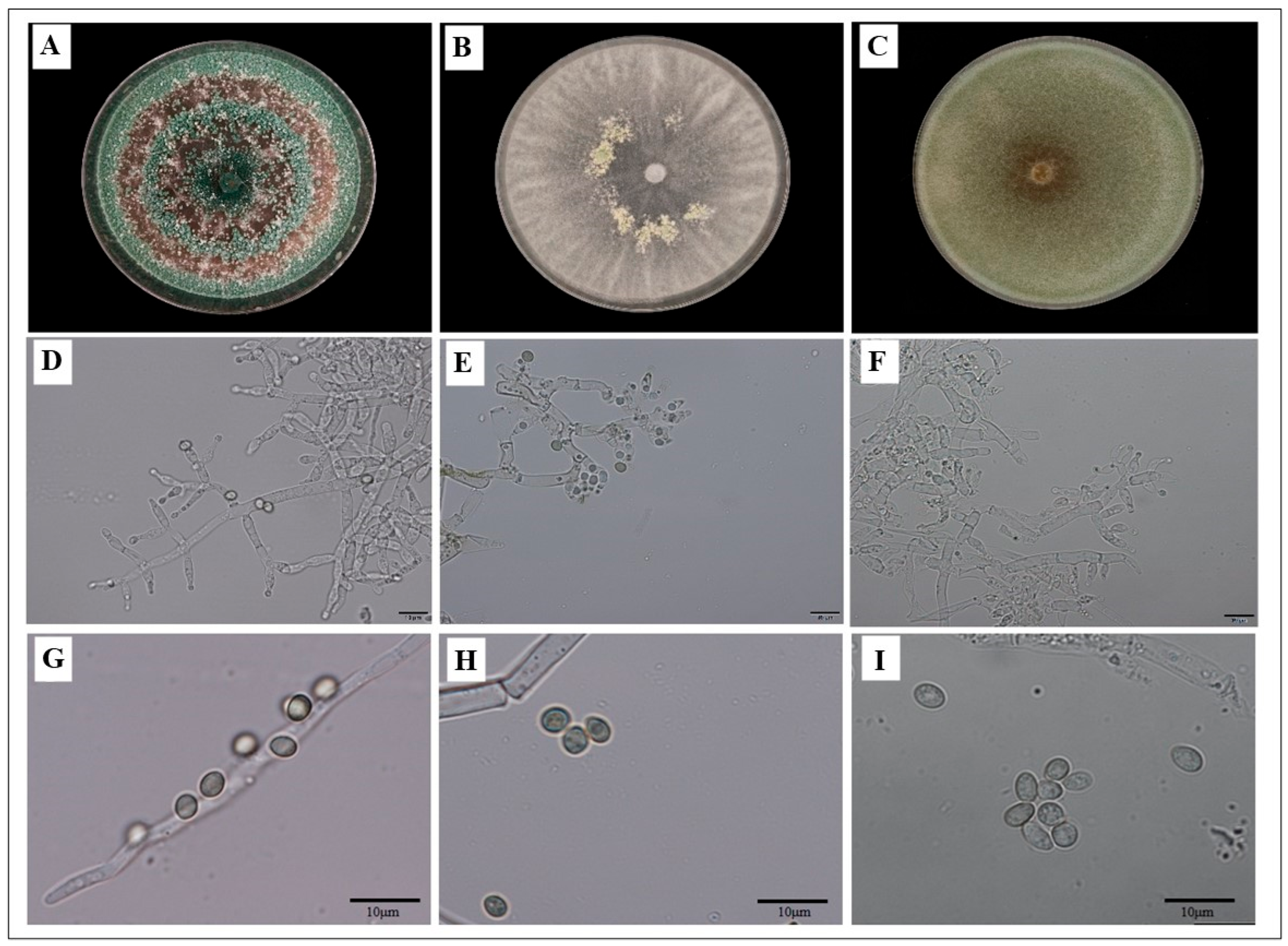
A total of 100 endophytic fungal isolates were recovered from loquat tissues, among which 39 isolates were obtained from roots. Molecular identification based on the ITS region revealed that Fusarium (35.90%) was the dominant genus, followed by Trichoderma (23.07%), Lasiodiplodia (12.82%), Meyerozyma (10.26%), Calonectria (5.12%), Phomopsis (2.56%), Xylaria (2.56%), and Macrophomina (2.56%). Although 23 isolates were initially identified as Trichoderma, 10 representative isolates were selected for detailed characterization based on preliminary screening for vigorous growth and distinct morphotypes. Morphological analysis of the selected Trichoderma isolates showed rapid mycelial proliferation, typically occupying an entire 9 cm Petri plate within 4 days. Conidiophore formation notably increased on the third day of hyphal growth. Sporulation proceeded via the emergence of clustered, spherical conidia at the branch tips, developing into dense spore masses after 7 days. Based on macro- and micro-morphological characteristics, these 10 Trichoderma isolates were classified into 3 different groups. Group 1 isolates (B077R1, B077R2, B077B3, B077R5, B077R6, and B077R9) exhibited rapid growth, completely filling the plates in 3–4 days. Their aerial hyphae developed compact, short villi closely appressed to the medium surface. Colonies initially appeared white, progressively turning green and ultimately forming dark green concentric rings during advanced sporulation. Spore clusters displayed an uneven distribution, and the conidiophores were primarily pyramidal with ampulliform phialides. The conidia were round to oval, measuring 3.53–3.81 × 2.90–3.00 μm. Conversely, Group 2 isolates (B077R7, B256R1, and B256R2) displayed restricted growth and an absence of sporulation on PDA. However, when cultivated on CMA, sporulation occurred after 4–5 days, yielding white, low-density, cotton-like colonies without a distinct odor. Hyphal branches typically showed an opposite arrangement with conical morphology. Generally, one or more ampule peduncles were connected to the apex of the conidiophores, and the conidia produced on CMA were oval-shaped with a smooth texture, measuring 3.81–4.10 × 3.22–3.35 μm. Isolate GFR9, categorized as Group 3, established rapid growth on PDA, covering the medium surface within 3–4 days. Colonies formed cotton-like masses or compact clusters with irregularly distributed conidiophores, lacking distinct conical or pyramidal structures. The conidia were spherical, pale green, and measured 4.00–4.51 × 3.33–3.48 μm. Numerous chlamydospores were observed, which were generally spherical and light brown (Figure 1).

3.2. Multi-Locus Phylogenetic Analysis of the Trichoderma Isolates

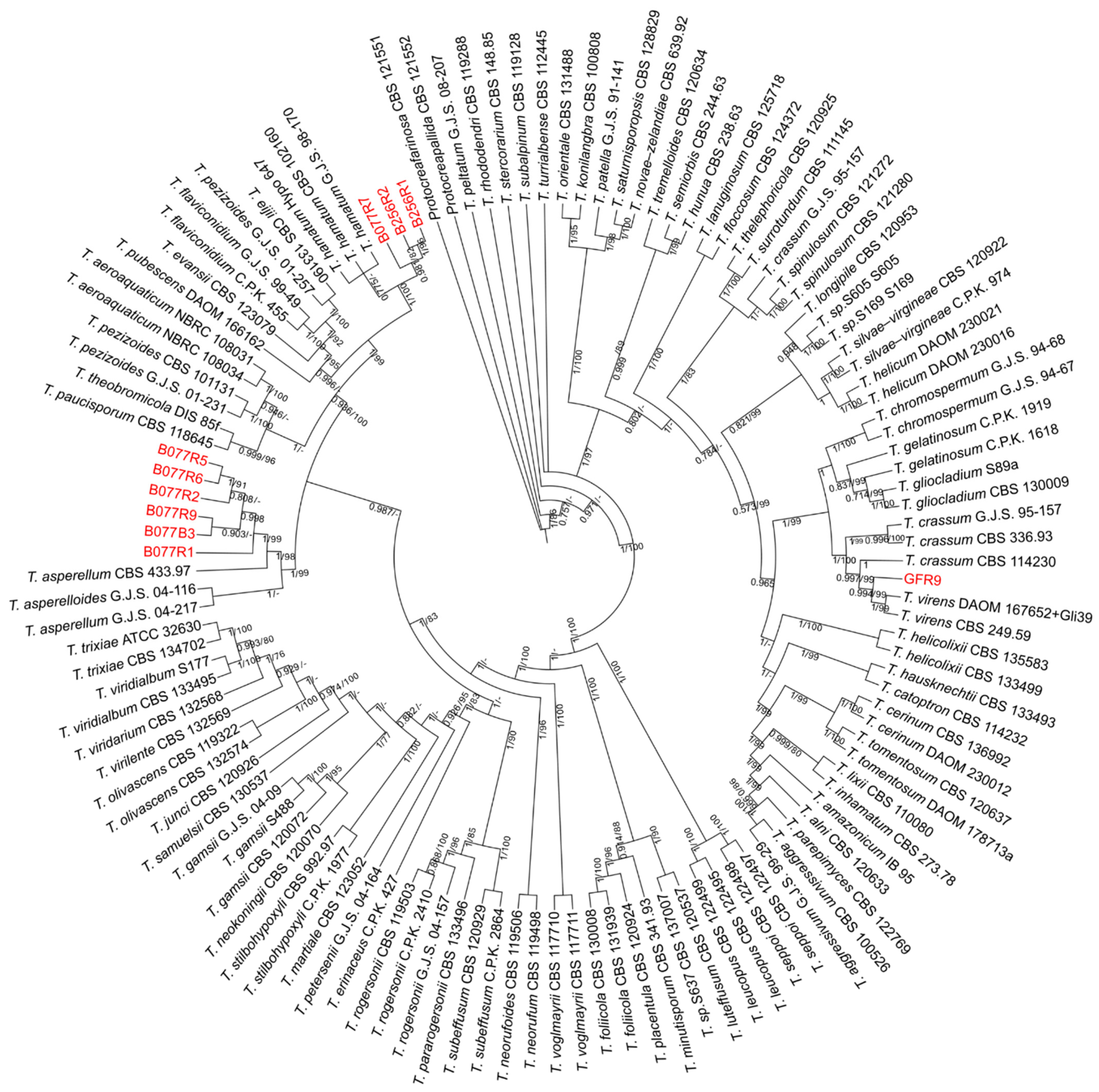
To elucidate the robust taxonomic affinities of the selected isolates, a multi-locus phylogeny was inferred from a concatenated sequence dataset of four genetic loci including ITS, tef1-α, rpb2 and acl1. The tree topology derived from the BI analysis was consistent with that obtained in an ML analysis. However, the resulting phylogenetic tree successfully resolved the ten isolates into three well-supported clades, aligning them with recognized Trichoderma species (Figure 2). Specifically, six isolates (B077R1, B077R2, B077B3, B077R5, B077R6, and B077R9) clustered tightly with T. asperellum reference isolates (CBS 433.97 and G.J.S. 04-217) and T. asperelloides (G.J.S. 04-116) with a high bootstrap support (BIBP/MLBP = 1/99). Three isolates (B077R7, B256R1 and B256R2) are grouped with T. hamatum reference isolates (G.J.S. 98-170, CBS 102160 and Hypo 647), supported by a bootstrap value (BIBP/MLBP = 1/100). Furthermore, isolate GFR9 formed a distinct monophyletic clade alongside T. virens CBS 249.59 (ex-type) and DAOM 167652, supported by a bootstrap value (BIBP/MLBP = 0.99/99). (Figure 2).

3.3. In Vitro Antifungal Activity of Trichoderma via Dual Culture Assay

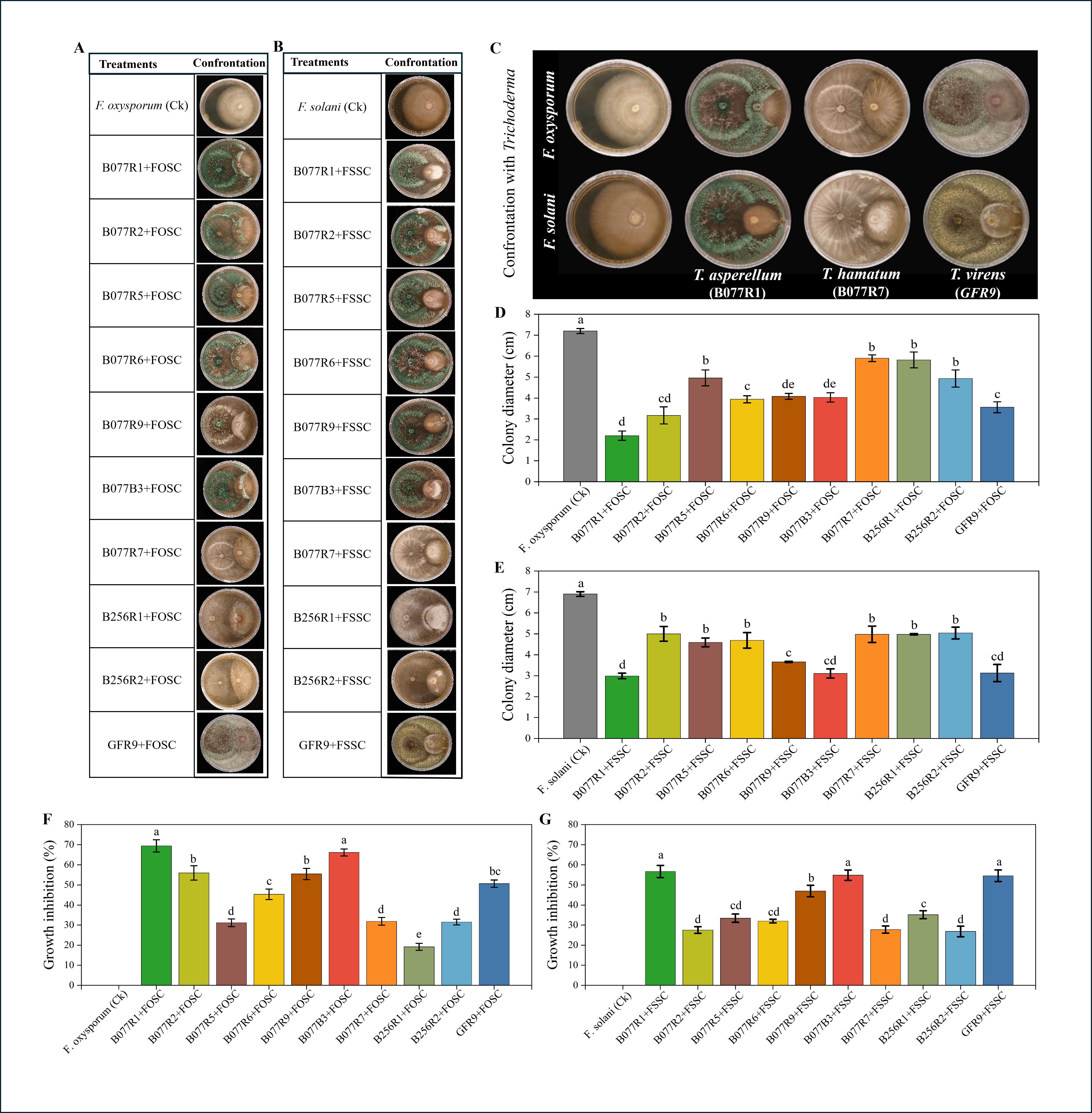
In vitro dual culture assays demonstrated that all Trichoderma isolates possessed antagonistic activity against both F. oxysporum and F. solani. Macroscopic observations (Figure 3C) indicated that the Trichoderma isolates rapidly colonized the agar surface, indicative of spatial and nutritional competition, and eventually overgrew the pathogen colonies. When confronted with F. oxysporum, T. asperellum B077R1 exhibited the highest inhibitory effect, with a growth suppression rate of 69.42%, followed by B077B3 (66.15%) and B077R2 (55.94%). In contrast, T. hamatum B256R1 showed the lowest inhibition at 19.20%. These suppression rates were consistent with the significant reductions observed in pathogen colony diameters (Figure 3D,F). A similar inhibitory trend was observed against F. solani. T. asperellum B077R1 yielded the maximum inhibition (56.67%), followed by B077B3 (54.85%) and T. virens GFR9 (54.56%), whereas B256R2 showed the lowest efficacy (26.88%) (Figure 3E,G). Overall, T. asperellum B077R1 and T. virens GFR9 consistently exhibited superior and broad-spectrum antagonistic potential against both Fusarium species.

3.4. In Vivo Biocontrol Efficacy of Trichoderma Isolates Against Fusarium Root Rot in Loquat

3.4.1. Effect of Trichoderma Isolates on the Disease Severity and Root Viability

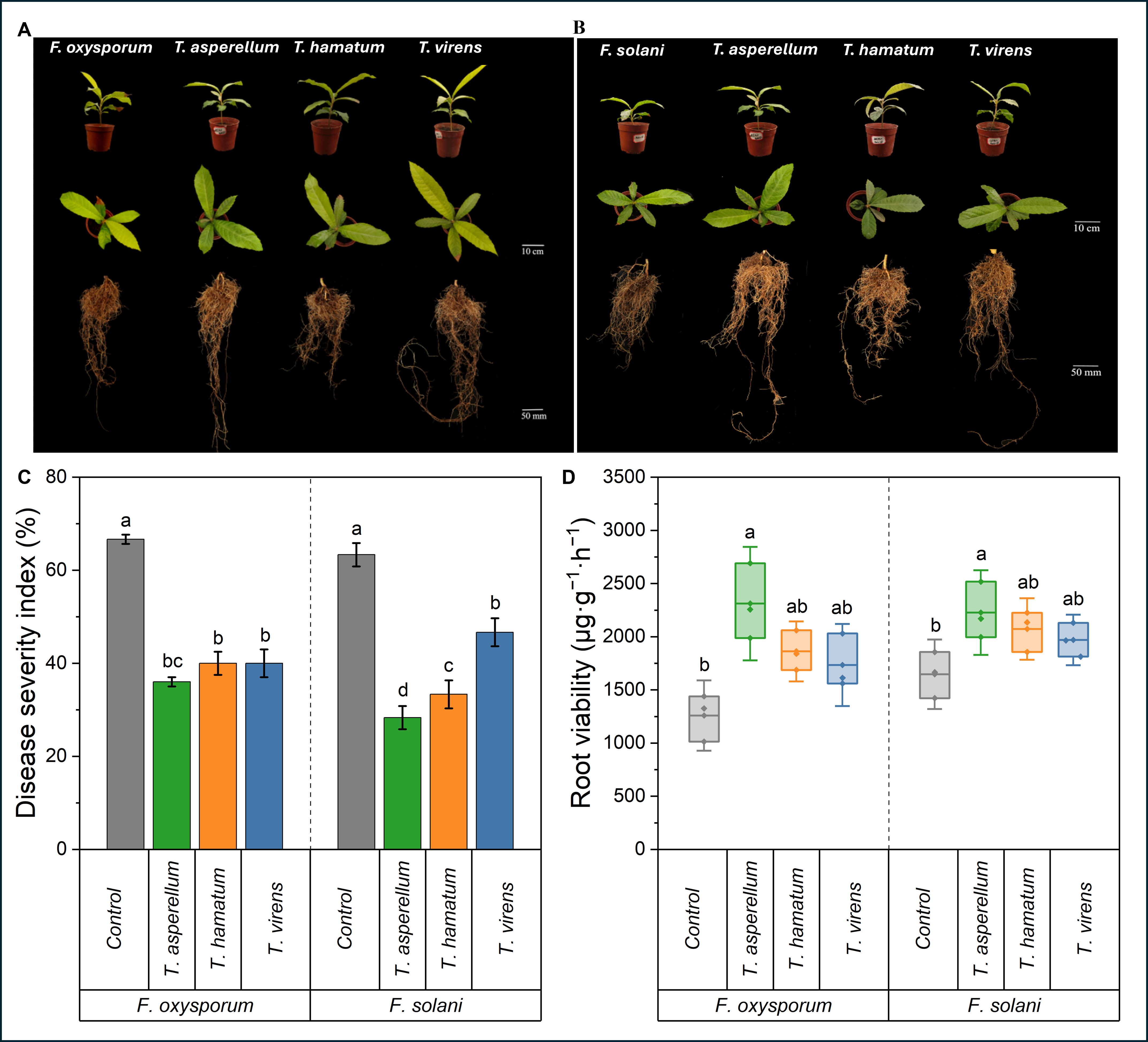
The application of different treatments resulted in varying degrees of disease severity and plant physiological responses. Loquat plants inoculated exclusively with either F. oxysporum or F. solani exhibited typical symptoms, including foliar chlorosis, root browning, and fine root decay (Figure 4A,B). These infected root systems displayed severe structural degradation, leading to a substantial reduction in total biomass compared to the Trichoderma co-inoculated treatments. In contrast, co-inoculation with Trichoderma isolates significantly mitigated these symptoms, with plants maintaining green foliage and developing robust root systems with fewer necrotic lesions. As shown in Figure 4C, the highest disease severity was recorded in the F. oxysporum-infected control, with a disease severity index (DSI) of 66.66%. However, co-inoculation with T. asperellum B077R1 significantly reduced the DSI to 45.99%. Similarly, T. hamatum B077R7 and T. virens GFR9 reduced the DSI to 39.99%. A similar trend was observed against F. solani, with the DSI reaching 63.33% in the pathogen-only control and significantly decreasing to 55.50% following T. asperellum B077R1 treatment.
Root viability, quantified by the TTC reduction assay, exhibited significant variation among treatments (Figure 4D). Under F. oxysporum stress, plants treated with T. asperellum B077R1 exhibited the highest root viability (2311.59 µg·g−1·h−1), followed by T. hamatum B077R7 (1862.31 µg·g−1·h−1) and T. virens GFR9 (1734.48 µg·g−1·h−1). All Trichoderma treatments resulted in significantly higher viability compared to the F. oxysporum-infected control (1259.42 µg·g−1·h−1). Under F. solani stress, T. asperellum B077R1 again provided the maximum protection (2227.54 µg·g−1·h−1), followed by T. hamatum B077R7 (2072.46 µg·g−1·h−1) and T. virens GFR9 (1969.57 µg·g−1·h−1). These values were notably higher than the 1647.82 µg·g−1·h−1 recorded in the F. solani—infected control.

3.4.2. Effect of Trichoderma Isolates on Plant Growth Parameters

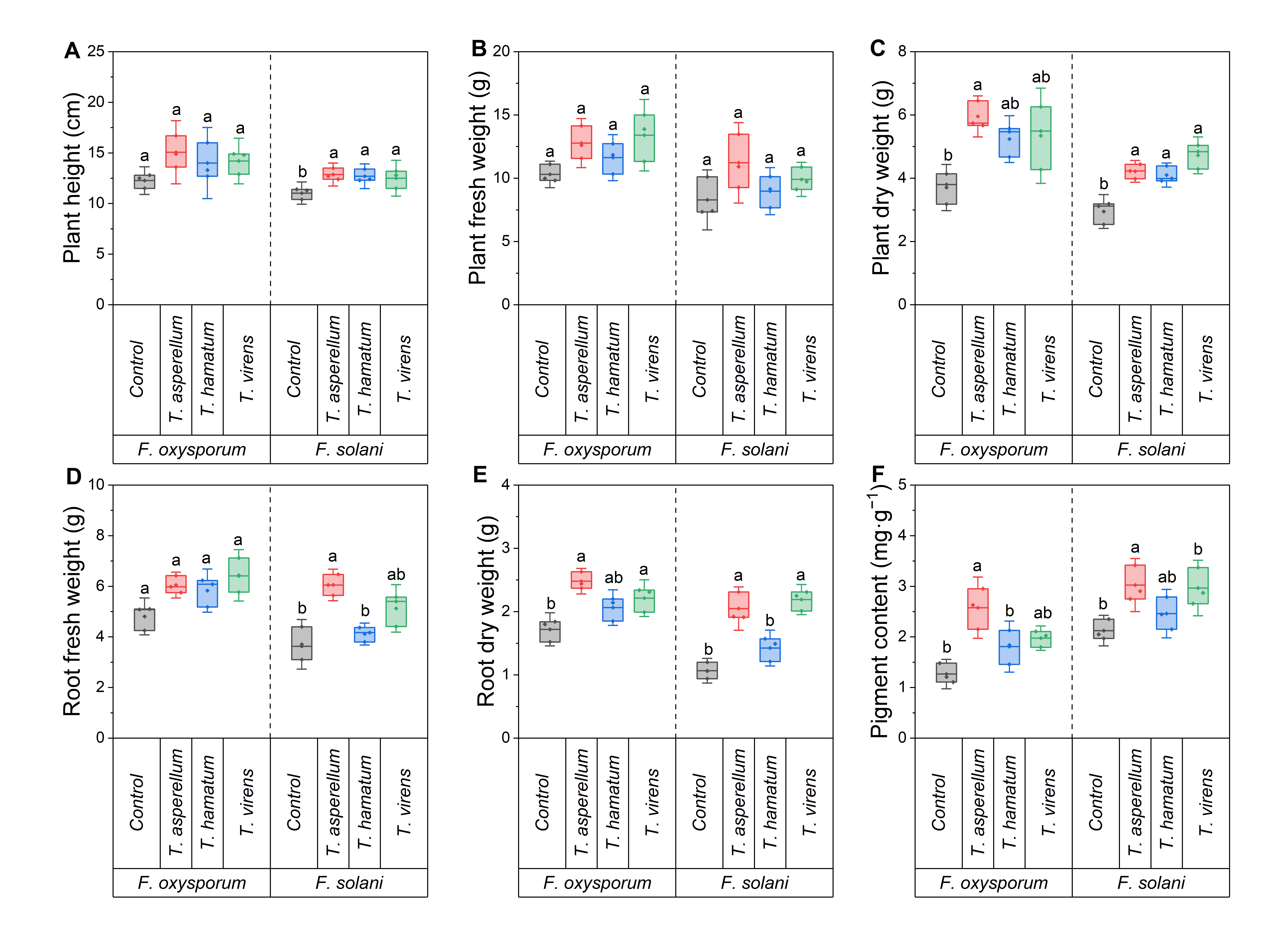
Consistent with the reduction in disease severity, Trichoderma inoculation effectively alleviated the growth inhibition induced by Fusarium stress, particularly in terms of biomass accumulation (Figure 5A–E). Under F. oxysporum stress, although plant height showed no statistical difference among the infected treatments (Figure 5A), the application of T. asperellum B077R1 significantly mitigated the loss of plant biomass compared to the stunted pathogen-only control. Specifically, B077R1 treatment maximized plant fresh weight (12.77 g) and dry weight (5.95 g), which were significantly higher than those of the infected control. Root development was correspondingly sustained, with fresh and dry weights reaching 6.05 g and 2.48 g, respectively. These growth parameters generally surpassed the values obtained for T. hamatum B077R7 and T. virens GFR9. A commensurate protective effect was observed against F. solani. While plant fresh weight did not differ significantly across treatments (Figure 5B), inoculation with B077R1 effectively counteracted the depletion of dry matter caused by the pathogen, yielding the highest plant dry weight (4.22 g) and root dry weight (2.05 g). In stark contrast, the F. solani-only control exhibited severely restricted growth. Collectively, these results indicate that the antagonistic activity of T. asperellum B077R1 effectively preserves plant vigor and dry matter accumulation under severe pathogen pressure.

3.4.3. Effect of Trichoderma Isolates on Leaf Photosynthetic Pigment Content

As foliar chlorosis is a primary symptom of Fusarium-induced root rot, the total content of photosynthetic pigments (chlorophyll a, b, and carotenoids) was quantified to evaluate the physiological health of the plants. Inoculation with F. oxysporum or F. solani alone imposed severe stress, resulting in the lowest pigment accumulation in the infected controls. However, co-inoculation with Trichoderma isolates alleviated this chlorosis to varying extents. Among the treatments, T. asperellum B077R1 exhibited the most consistent and strongest capability to preserve the photosynthetic machinery (Figure 5F). Under F. oxysporum stress, the pigment content in the infected control was severely suppressed to 1.26 mg·g−1. Treatment with B077R1 effectively counteracted this degradation, increasing the total pigment content to 2.6 mg·g−1 (a 105% increase), which was the only treatment statistically higher than the control. Conversely, T. hamatum B077R7 did not show a significant difference from the infected control. A parallel trend was observed under F. solani stress. While the infected control exhibited depleted pigment levels (2.11 mg·g−1), the B077R1 treatment maintained a significantly higher concentration at 3.10 mg·g−1 (an increase of 46.9%). These findings demonstrate that T. asperellum B077R1 effectively prevents pathogen-induced chlorosis, thereby sustaining the vital photosynthetic potential required for plant recovery.

4. Discussion

The effectiveness of biological control agents (BCAs) often depends on their ability to adapt to local rhizosphere conditions. In this study, we screened indigenous Trichoderma endophytes from loquat roots to identify potential antagonists against Fusarium-induced root rot. Although isolates of T. asperellum, T. hamatum, and T. virens were identified, their biocontrol activities varied significantly. This variability indicates that biocontrol efficacy is often strain-specific rather than conserved at the species level, presenting a challenge for consistent field application. For instance, while T. hamatum has been reported as effective against Fusarium root rot in Panax notoginseng [48], our isolate T. hamatum B077R7 showed limited activity compared to T. asperellum B077R1. This discrepancy suggests that host-genotype compatibility plays a pivotal role in determining BCA performance. Indeed, the effectiveness of Trichoderma as a biocontrol agent is primarily driven by its competence in the rhizosphere, persistence, and ability to establish a stable and beneficial relationship with roots that improve plant resistance and promote soil health [49,50]. Consequently, reliance on general commercial strains may yield inconsistent results in loquat orchards. Our findings strongly support the strategy of isolating and utilizing indigenous strains, such as T. asperellum B077R1, which are pre-adapted to the specific ecological niche of the target crop.
Loquat root rot is caused by a complex of pathogens, primarily F. solani and F. oxysporum. Our results demonstrated that T. asperellum B077R1 effectively inhibited both pathogens in vitro and in vivo. These results align with previous reports demonstrating the broad-spectrum efficacy of T. asperellum across diverse crops. For example, T. asperellum has successfully controlled Fusarium wilt in cucumber [51] and banana [52], as well as root rot in dry beans [53]. In dual culture assays, the isolate B077R1 exhibited rapid mycelial growth, physically overgrowing and spatially restricting the Fusarium colonies (Figure 3). This behavior points to direct mycoparasitism and competition for nutrients as primary modes of action. Direct mycoparasitism involves the secretion of cell-wall-degrading enzymes (e.g., chitinases) and antimicrobial secondary metabolites, which lyse pathogen hyphae [54,55]. For instance, T. virens from the roots of healthy apple trees directly suppressed F. proliferatum and released secondary metabolites exhibiting both antifungal and plant growth-promoting activities [56]. However, antagonistic activity on agar plates does not always translate to soil environments. In our pot experiments, the sustained protection, which was characterized by significantly reduced disease severity and high root viability, suggests that B077R1 also possesses strong rhizosphere competence. By successfully colonizing the root surface, B077R1 likely excludes pathogens from infection sites. This ecological exclusion mechanism parallels findings in watermelon, where T. asperellum application reduced pathogen abundance by modulating the rhizosphere microbiome and enriching beneficial taxa like Pseudomonas [57]. Therefore, based on the combined in vitro and in vivo data, it is likely that B077R1 suppresses loquat root rot through a synergistic mechanism of direct mycoparasitic attack and competitive exclusion in the rhizosphere.
Fusarium infection is known to cause chlorosis and pigment loss, often attributed to the production of toxins such as fusaric acid and the induction of oxidative stress [58]. Previous studies on apple seedlings indicated that such infection leads to increased membrane permeability and reduced leaf water content, resulting in cellular damage that directly contributes to photosynthetic pigment degradation [59]. In this study, infection with F. oxysporum and F. solani significantly reduced chlorophyll and carotenoid contents, but treatment with B077R1 effectively prevented this degradation. This maintenance of photosynthetic pigments indicates that Trichoderma alleviates physiological stress, possibly by scavenging reactive oxygen species (ROS) and maintaining metabolic homeostasis. Previous research in maize seedlings demonstrated that T. asperellum enhances antioxidant enzyme activities, thereby protecting cellular structures from oxidative damage [60]. By preserving chloroplast integrity [32], B077R1 ensures the maintenance of photosynthetic capacity, which provides the necessary energy for the biomass recovery observed in treated plants. This physiological defense mechanism complements direct pathogen suppression, ensuring plant survival even under pathogen stress.
In addition to disease suppression, treatment with B077R1 significantly increased plant biomass and root development. This growth promotion is likely mediated by robust root colonization and the subsequent reciprocal exchange of metabolites. Trichoderma species are known to solubilize soil nutrients [61] and produce phytohormones, such as auxins and gibberellins, which stimulate root proliferation [19]. Upon successful root colonization, Trichoderma develops a mutualistic relationship with the host plant, marked by a reciprocal exchange of metabolites and nutrients. Similar auxin-mediated effects have been reported in Arabidopsis, where T. virens enhanced lateral root formation [62], and in peppermint, where T. viride colonization promoted both vegetative growth and essential oil biosynthesis [63]. The growth improvements observed were primarily due to reduced growth suppression caused by pathogens, as healthy mock-inoculated and Trichoderma-alone controls were not included. These controls should be included in future studies to determine if the growth recovery is due to biocontrol or to direct growth promotion of the plants.
Furthermore, the significant reduction in disease symptoms suggests the potential activation of plant defense mechanisms. Trichoderma species are widely reported to induce systemic resistance (ISR) via salicylic acid and jasmonic acid signaling pathways [64,65]. While the specific molecular pathways in the loquat-Trichoderma interaction remain to be fully elucidated, the robust phenotypic recovery observed in our study strongly implies that T. asperellum B077R1 primes the host immune system. Collectively, our findings demonstrate that T. asperellum B077R1 provides protection against Fusarium root rot through direct pathogen suppression and the effective preservation of host physiological vigor and growth.
In this study, we screened and evaluated individual Trichoderma isolates. However, compared to single strains, microbial consortia typically exhibit superior environmental resilience and stronger antagonistic effects in natural ecosystems [66]. Therefore, developing and evaluating a consortium using these promising Trichoderma isolates will be a critical next step to further enhance their practical efficacy in the field. Although this study highlights the biocontrol and growth-promoting abilities of the isolated Trichoderma strains, it mainly relies on phenotypic and pathological data. We acknowledge that the lack of physiological and biochemical assays is a limitation, as these are needed to fully explain the plant-microbe interactions. Therefore, our future work will include biochemical and molecular analyses to better understand the specific mechanisms behind these effects.

5. Conclusions

This study demonstrates the significant biocontrol potential of indigenous endophytic Trichoderma isolates against Fusarium-induced root rot in loquat. Based on the strong in vitro antagonistic activity observed against both F. oxysporum and F. solani, in vivo assays further corroborated these findings by demonstrating that inoculation with all the tested isolates, most notably T. asperellum B077R1, effectively mitigated disease severity while preserving root viability, plant biomass, and photosynthetic capacity under severe pathogen stress. These findings highlight the ecological advantage of utilizing host-adapted endophytes for disease management. Ultimately, T. asperellum B077R1 represents a highly promising candidate for the development of sustainable biocontrol strategies to protect perennial fruit crops against soil-borne pathogens, warranting further investigation in field-scale applications.

Supplementary Materials

The following supporting information can be downloaded at: https://www.mdpi.com/article/10.3390/horticulturae12070859/s1, Table S1: Primers used in this study; Table S2: GenBank accession numbers of isolated Trichoderma strains.

Author Contributions

M.H.: Investigation, Writing—original draft, Writing—review and editing, Methodology, Validation. T.Y.: Writing—review and editing. P.L.: Investigation, Writing—review and editing. W.S.: Writing—review and editing. Y.Z.: Methodology, Investigation, Software. P.Y.: Writing—review and editing. X.G. and G.L.: Investigation, Writing—review and editing. Q.G. and D.W.: Conceptualization, Funding acquisition, Investigation, Resources, Writing—review and editing. All authors have read and agreed to the published version of the manuscript.

Funding

This work is supported by the Chongqing Central Forestry Reform and Development Fund—Science and Technology Promotion and Demonstration Project (Yulin Ketui [2024] No. 7), the Chongqing Science and Technology Commission (Grant No. CSTB2025NSCQ-GPX0548), and the University Innovation and Entrepreneurship Training Program (S202310635165, S202210635206).

Data Availability Statement

The original contributions presented in this study are included in the article/Supplementary Materials. Further inquiries can be directed to the corresponding author.

Acknowledgments

The authors acknowledge OLIVA AI LIMITED (No. 16967010) for supporting the article processing charges of this publication. The funder had no role in study design, data collection, analysis, interpretation, manuscript preparation, or the decision to publish.

Conflicts of Interest

Author Peng Yu was employed by Symcode (Chongqing) Biotechnology Co., Ltd. The remaining authors declare that the research was conducted in the absence of any commercial or financial relationships that could be construed as a potential conflict of interest.

References

  1. Badenes, M.L.; Janick, J.; Lin, S.; Zhang, Z.; Liang, G.L.; Wang, W. Breeding Loquat. In Plant Breeding Reviews; Wiley: Hoboken, NJ, USA, 2013; pp. 259–296. [Google Scholar]
  2. Janick, J. Genetic Alteration Associated with Fruit Domestication. Acta Hortic. 2007, 750, 27–36. [Google Scholar] [CrossRef] [Scilit]
  3. Liu, Y.; Zhang, W.; Xu, C.; Li, X. Biological Activities of Extracts from Loquat (Eriobotrya japonica Lindl.): A Review. Int. J. Mol. Sci. 2016, 17, 1983. [Google Scholar] [CrossRef] [Scilit] [PubMed]
  4. Khouya, T.; Ramchoun, M.; Elbouny, H.; Hmidani, A.; Bouhlali, E.d.T.; Alem, C. Loquat (Eriobotrya japonica (Thunb) Lindl.): Evaluation of Nutritional Value, Polyphenol Composition, Antidiabetic Effect, and Toxicity of Leaf Aqueous Extract. J. Ethnopharmacol. 2022, 296, 115473. [Google Scholar] [CrossRef] [Scilit] [PubMed]
  5. Munkvold, G.P. Fusarium Species and Their Associated Mycotoxins. In Methods in Molecular Biology; Humana Press: New York, NY, USA, 2017; pp. 51–106. [Google Scholar]
  6. Ma, L.-J.; Geiser, D.M.; Proctor, R.H.; Rooney, A.P.; O’Donnell, K.; Trail, F.; Gardiner, D.M.; Manners, J.M.; Kazan, K. Fusarium Pathogenomics. Annu. Rev. Microbiol. 2013, 67, 399–416. [Google Scholar] [CrossRef] [Scilit] [PubMed]
  7. Abbas, M.F.; Naz, F.; Rauf, C.A.; Mehmood, N.; Zhang, X.; Rosli, B.H.; Gleason, M.L. First Report of Fusarium solani Causing Fruit Rot of Loquat (Eriobotrya japonica) in Pakistan. Plant Dis. 2017, 101, 839. [Google Scholar] [CrossRef] [Scilit]
  8. Wu, D.; Zhang, D.H.; Wang, C.X.; Wei, Y.; Timko, M.P.; Liang, G.L. First Report of Fusarium solani Species Complex Causing Root Rot of Loquat (Eriobotrya japonica) in China. Plant Dis. 2021, 105, 1562. [Google Scholar] [CrossRef] [Scilit]
  9. Xu, W.; Yang, Q.; Yang, F.; Xie, X.; Goodwin, P.H.; Deng, X.; Tian, B.; Yang, L. Evaluation and Genome Analysis of Bacillus Subtilis YB-04 as a Potential Biocontrol Agent Against Fusarium Wilt and Growth Promotion Agent of Cucumber. Front. Microbiol. 2022, 13, 885430. [Google Scholar] [CrossRef] [Scilit] [PubMed]
  10. Mishra, K.K.; Kumar, V.; Wagh, A.K.; Panda, S.R. The Promise of Trichoderma Formulations: Insight from Bibliometric Analysis. Stud. Fungi 2024, 9, e014. [Google Scholar] [CrossRef] [Scilit]
  11. Shams, A.H.M.; Helaly, A.A.; Algeblawi, A.M.; Awad-Allah, E.F.A. Efficacy of Seed-Biopriming with Trichoderma spp. and Foliar Spraying of ZnO-Nanoparticles Induce Cherry Tomato Growth and Resistance to Fusarium Wilt Disease. Plants 2023, 12, 3117. [Google Scholar] [CrossRef] [Scilit] [PubMed]
  12. Cotxarrera, L.; Trillas-Gay, M.I.; Steinberg, C.; Alabouvette, C. Use of Sewage Sludge Compost and Trichoderma asperellum Isolates to Suppress Fusarium Wilt of Tomato. Soil Biol. Biochem. 2002, 34, 467–476. [Google Scholar] [CrossRef] [Scilit]
  13. Li, S.; Zhang, F.-M.; Shang, X.-J.; Hou, R. Control Effect and Mechanism of Trichoderma asperellum TM11 against Blueberry Root Rot. Pol. J. Microbiol. 2023, 72, 325–337. [Google Scholar] [CrossRef] [Scilit] [PubMed]
  14. Yang, X.; Chen, L.; Yong, X.; Shen, Q. Formulations Can Affect Rhizosphere Colonization and Biocontrol Efficiency of Trichoderma Harzianum SQR-T037 against Fusarium Wilt of Cucumbers. Biol. Fertil. Soils 2011, 47, 239–248. [Google Scholar] [CrossRef] [Scilit]
  15. Matarese, F.; Sarrocco, S.; Gruber, S.; Seidl-Seiboth, V.; Vannacci, G. Biocontrol of Fusarium Head Blight: Interactions between Trichoderma and Mycotoxigenic Fusarium. Microbiology 2012, 158, 98–106. [Google Scholar] [CrossRef] [Scilit] [PubMed]
  16. Cai, F.; Chen, W.; Wei, Z.; Pang, G.; Li, R.; Ran, W.; Shen, Q. Colonization of Trichoderma Harzianum Strain SQR-T037 on Tomato Roots and Its Relationship to Plant Growth, Nutrient Availability and Soil Microflora. Plant Soil 2015, 388, 337–350. [Google Scholar] [CrossRef] [Scilit]
  17. Thambugala, K.M.; Daranagama, D.A.; Phillips, A.J.L.; Kannangara, S.D.; Promputtha, I. Fungi vs. Fungi in Biocontrol: An Overview of Fungal Antagonists Applied Against Fungal Plant Pathogens. Front. Cell. Infect. Microbiol. 2020, 10, 604923. [Google Scholar] [CrossRef] [Scilit] [PubMed]
  18. Saldaña-Mendoza, S.A.; Pacios-Michelena, S.; Palacios-Ponce, A.S.; Chávez-González, M.L.; Aguilar, C.N. Trichoderma as a Biological Control Agent: Mechanisms of Action, Benefits for Crops and Development of Formulations. World J. Microbiol. Biotechnol. 2023, 39, 269. [Google Scholar] [CrossRef] [Scilit] [PubMed]
  19. Illescas, M.; Pedrero-Méndez, A.; Pitorini-Bovolini, M.; Hermosa, R.; Monte, E. Phytohormone Production Profiles in Trichoderma Species and Their Relationship to Wheat Plant Responses to Water Stress. Pathogens 2021, 10, 991. [Google Scholar] [CrossRef] [Scilit] [PubMed]
  20. Malcolm, G.M.; Kuldau, G.A.; Gugino, B.K.; Jiménez-Gasco, M.d.M. Hidden Host Plant Associations of Soilborne Fungal Pathogens: An Ecological Perspective. Phytopathology 2013, 103, 538–544. [Google Scholar] [CrossRef] [Scilit] [PubMed]
  21. Card, S.; Johnson, L.; Teasdale, S.; Caradus, J. Deciphering Endophyte Behaviour: The Link between Endophyte Biology and Efficacious Biological Control Agents. FEMS Microbiol. Ecol. 2016, 92, fiw114. [Google Scholar] [CrossRef] [Scilit] [PubMed]
  22. Collinge, D.B.; Jensen, D.F.; Rabiey, M.; Sarrocco, S.; Shaw, M.W.; Shaw, R.H. Biological Control of Plant Diseases—What Has Been Achieved and What Is the Direction? Plant Pathol. 2022, 71, 1024–1047. [Google Scholar] [CrossRef] [Scilit]
  23. Chen, H.; Mao, L.; Zhao, N.; Xia, C.; Liu, J.; Kubicek, C.P.; Wu, W.; Xu, S.; Zhang, C. Verification of TRI3 Acetylation of Trichodermol to Trichodermin in the Plant Endophyte Trichoderma Taxi. Front. Microbiol. 2021, 12, 731425. [Google Scholar] [CrossRef] [Scilit] [PubMed]
  24. Liu, X.; Zhou, Z.-Y.; Cui, J.-L.; Wang, M.-L.; Wang, J.-H. Biotransformation Ability of Endophytic Fungi: From Species Evolution to Industrial Applications. Appl. Microbiol. Biotechnol. 2021, 105, 7095–7113. [Google Scholar] [CrossRef] [Scilit] [PubMed]
  25. Aamir, M.; Shanmugam, V.; Dubey, M.K.; Husain, F.M.; Adil, M.; Ansari, W.A.; Rai, A.; Sah, P. Transcriptomic Characterization of Trichoderma Harzianum T34 Primed Tomato Plants: Assessment of Biocontrol Agent Induced Host Specific Gene Expression and Plant Growth Promotion. BMC Plant Biol. 2023, 23, 552. [Google Scholar] [CrossRef] [Scilit] [PubMed]
  26. Rajani, P.; Rajasekaran, C.; Vasanthakumari, M.M.; Olsson, S.B.; Ravikanth, G.; Uma Shaanker, R. Inhibition of Plant Pathogenic Fungi by Endophytic Trichoderma spp. through Mycoparasitism and Volatile Organic Compounds. Microbiol. Res. 2021, 242, 126595. [Google Scholar] [CrossRef] [Scilit] [PubMed]
  27. Pu, X.; Qu, X.; Chen, F.; Bao, J.; Zhang, G.; Luo, Y. Camptothecin-Producing Endophytic Fungus Trichoderma Atroviride LY357: Isolation, Identification, and Fermentation Conditions Optimization for Camptothecin Production. Appl. Microbiol. Biotechnol. 2013, 97, 9365–9375. [Google Scholar] [CrossRef] [Scilit] [PubMed]
  28. Steyaert, J.M.; Weld, R.J.; Stewart, A. Isolate-Specific Conidiation in Trichoderma in Response to Different Nitrogen Sources. Fungal Biol. 2010, 114, 179–188. [Google Scholar] [CrossRef] [Scilit] [PubMed]
  29. Inglis, P.W.; Mello, S.C.M.; Martins, I.; Silva, J.B.T.; Macêdo, K.; Sifuentes, D.N.; Valadares-Inglis, M.C. Trichoderma from Brazilian Garlic and Onion Crop Soils and Description of Two New Species: Trichoderma Azevedoi and Trichoderma Peberdyi. PLoS ONE 2020, 15, e0228485. [Google Scholar] [CrossRef] [Scilit] [PubMed]
  30. Bissett, J. A Revision of the Genus Trichoderma. I. Section Longibrachiatum Sect. Nov. Can. J. Bot. 1984, 62, 924–931. [Google Scholar] [CrossRef] [Scilit]
  31. Samuels, G.J.; Petrini, O.; Manguin, S. Morphological and Macromolecular Characterization of Hypocrea schweinitzii and Its Trichoderma Anamorph. Mycologia 1994, 86, 421–435. [Google Scholar] [CrossRef] [Scilit]
  32. Aris, A.; Mohd Zainudin, N.A.I.; Ibrahim, M.H. Growth and Photosynthetic Performance of Fusarium solani Infected Cucumis sativus L. Treated with Trichoderma asperellum. J. Taibah Univ. Sci. 2023, 17, 2161292. [Google Scholar] [CrossRef] [Scilit]
  33. Jaklitsch, W.M.; Voglmayr, H. Biodiversity of Trichoderma (Hypocreaceae) in Southern Europe and Macaronesia. Stud. Mycol. 2015, 80, 1. [Google Scholar] [CrossRef] [Scilit] [PubMed]
  34. Cai, F.; Druzhinina, I.S. In Honor of John Bissett: Authoritative Guidelines on Molecular Identification of Trichoderma. Fungal Divers. 2021, 107, 1–69. [Google Scholar] [CrossRef] [Scilit]
  35. Toghueo, R.M.K.; Eke, P.; Zabalgogeazcoa, Í.; de Aldana, B.R.V.; Nana, L.W.; Boyom, F.F. Biocontrol and Growth Enhancement Potential of Two Endophytic Trichoderma spp. from Terminalia catappa against the Causative Agent of Common Bean Root Rot (Fusarium solani). Biol. Control 2016, 96, 8–20. [Google Scholar] [CrossRef] [Scilit]
  36. Bunbury-Blanchette, A.L.; Walker, A.K. Trichoderma Species Show Biocontrol Potential in Dual Culture and Greenhouse Bioassays against Fusarium Basal Rot of Onion. Biol. Control 2019, 130, 127–135. [Google Scholar] [CrossRef] [Scilit]
  37. Baiyee, B.; Pornsuriya, C.; Ito, S.; Sunpapao, A. Trichoderma Spirale T76-1 Displays Biocontrol Activity against Leaf Spot on Lettuce (Lactuca sativa L.) Caused by Corynespora cassiicola or Curvularia aeria. Biol. Control 2019, 129, 195–200. [Google Scholar] [CrossRef] [Scilit]
  38. Snyder, W.C.; Hansen, H.N. The Effect of Light on Taxonomic Characters in Fusarium. Mycologia 1941, 33, 580–591. [Google Scholar] [CrossRef] [Scilit]
  39. Izquierdo-García, L.F.; González-Almario, A.; Cotes, A.M.; Moreno-Velandia, C.A. Trichoderma virens Gl006 and Bacillus Velezensis Bs006: A Compatible Interaction Controlling Fusarium Wilt of Cape Gooseberry. Sci. Rep. 2020, 10, 6857. [Google Scholar] [CrossRef] [Scilit] [PubMed]
  40. Yedidia, I.; Srivastva, A.K.; Kapulnik, Y.; Chet, I. Effect of Trichoderma Harzianum on Microelement Concentrations and Increased Growth of Cucumber Plants. Plant Soil 2001, 235, 235–242. [Google Scholar] [CrossRef] [Scilit]
  41. Inbar, J.; Abramsky, M.; Cohen, D.; Chet, I. Plant Growth Enhancement and Disease Control by Trichoderma harzianum in Vegetable Seedlings Grown under Commercial Conditions. Eur. J. Plant Pathol. 1994, 100, 337–346. [Google Scholar] [CrossRef] [Scilit]
  42. Arias, M.M.D.; Leandro, L.F.; Munkvold, G.P. Aggressiveness of Fusarium Species and Impact of Root Infection on Growth and Yield of Soybeans. Phytopathology 2013, 103, 822–832. [Google Scholar] [CrossRef] [Scilit] [PubMed]
  43. Filion, M.; St-Arnaud, M.; Jabaji-Hare, S.H. Quantification of Fusarium solani f. Sp. Phaseoli in Mycorrhizal Bean Plants and Surrounding Mycorrhizosphere Soil Using Real-Time Polymerase Chain Reaction and Direct Isolations on Selective Media. Phytopathology 2003, 93, 229–235. [Google Scholar] [CrossRef] [Scilit] [PubMed]
  44. Li, H.; Shi, J.; Wang, Z.; Zhang, W.; Yang, H. H2S Pretreatment Mitigates the Alkaline Salt Stress on Malus Hupehensis Roots by Regulating Na+/K+ Homeostasis and Oxidative Stress. Plant Physiol. Biochem. 2020, 156, 233–241. [Google Scholar] [CrossRef] [Scilit] [PubMed]
  45. Wellburn, A.R. The Spectral Determination of Chlorophylls a and b, as Well as Total Carotenoids, Using Various Solvents with Spectrophotometers of Different Resolution. J. Plant Physiol. 1994, 144, 307–313. [Google Scholar] [CrossRef] [Scilit]
  46. Lichtenthaler, H.K.; Buschmann, C. Extraction of Phtosynthetic Tissues: Chlorophylls and Carotenoids. Curr. Protoc. Food Anal. Chem. 2001, 1, F4.2.1–F4.2.6. [Google Scholar] [CrossRef] [Scilit]
  47. Lichtenthaler, H.K.; Buschmann, C. Chlorophylls and Carotenoids: Measurement and Characterization by UV-VIS Spectroscopy. Curr. Protoc. Food Anal. Chem. 2001, 1, F4.3.1–F4.3.8. [Google Scholar] [CrossRef] [Scilit]
  48. Feng, Y.; Shuai, X.; Chen, J.; Zhang, Q.; Jia, L.; Sun, L.; Su, Y.; Su, Y.; Dong, G.; Liu, T.; et al. Unveiling the Genomic Features and Biocontrol Potential of Trichoderma Hamatum Against Root Rot Pathogens. J. Fungi 2025, 11, 126. [Google Scholar] [CrossRef] [Scilit] [PubMed]
  49. Pradhan, D.A.; Bagagoni, P.; Makandar, R. Assessing Rhizosphere Trichoderma asperellum Strains for Root Colonizing and Antagonistic Competencies against Fusarium Wilt through Molecular and Biochemical Responses in Castor. Biol. Control 2023, 184, 105280. [Google Scholar] [CrossRef] [Scilit]
  50. Van Poucke, K.; França, S.C.; Haegeman, A.; Casanova, E.; Heungens, K. Strain-Specific and Sensitive Monitoring of the Biocontrol Agent Trichoderma asperellum T34 in Growing Medium via Real-Time PCR. Biocontrol Sci. Technol. 2024, 34, 355–374. [Google Scholar] [CrossRef] [Scilit]
  51. Wu, Q.; Sun, R.; Ni, M.; Yu, J.; Li, Y.; Yu, C.; Dou, K.; Ren, J.; Chen, J. Identification of a Novel Fungus, Trichoderma asperellum GDFS1009, and Comprehensive Evaluation of Its Biocontrol Efficacy. PLoS ONE 2017, 12, e0179957. [Google Scholar] [CrossRef] [Scilit] [PubMed]
  52. Long, W.; Chen, Y.; Wei, Y.; Feng, J.; Zhou, D.; Cai, B.; Qi, D.; Zhang, M.; Zhao, Y.; Li, K.; et al. A Newly Isolated Trichoderma Parareesei N4-3 Exhibiting a Biocontrol Potential for Banana Fusarium Wilt by Hyperparasitism. Front. Plant Sci. 2023, 14, 1289959. [Google Scholar] [CrossRef] [Scilit] [PubMed]
  53. Elshahawy, I.E.; Marrez, D.A. Antagonistic Activity of Trichoderma asperellum against Fusarium Species, Chemical Profile and Their Efficacy for Management of Fusarium-root Rot Disease in Dry Bean. Pest Manag. Sci. 2024, 80, 1153–1167. [Google Scholar] [CrossRef] [Scilit] [PubMed]
  54. Stracquadanio, C.; Quiles, J.M.; Meca, G.; Cacciola, S.O. Antifungal Activity of Bioactive Metabolites Produced by Trichoderma asperellum and Trichoderma Atroviride in Liquid Medium. J. Fungi 2020, 6, 263. [Google Scholar] [CrossRef] [Scilit] [PubMed]
  55. Vinale, F.; Sivasithamparam, K. Beneficial Effects of Trichoderma Secondary Metabolites on Crops. Phytother. Res. 2020, 34, 2835–2842. [Google Scholar] [CrossRef] [Scilit] [PubMed]
  56. Wang, H.; Tang, W.; Mao, Y.; Ma, S.; Chen, X.; Shen, X.; Yin, C.; Mao, Z. Isolation of Trichoderma virens 6PS-2 and Its Effects on Fusarium proliferatum f. Sp. Malus Domestica MR5 Related to Apple Replant Disease in China. Hortic. Plant J. 2024, 10, 1291–1308. [Google Scholar] [CrossRef] [Scilit]
  57. Zhang, Y.; Tian, C.; Xiao, J.; Wei, L.; Tian, Y.; Liang, Z. Soil Inoculation of Trichoderma asperellum M45a Regulates Rhizosphere Microbes and Triggers Watermelon Resistance to Fusarium Wilt. AMB Express 2020, 10, 189. [Google Scholar] [CrossRef] [Scilit] [PubMed]
  58. Sharma, A.; Salwan, R.; Kaur, R.; Sharma, R.; Sharma, V. Characterization and Evaluation of Bioformulation from Antagonistic and Flower Inducing Trichoderma asperellum Isolate UCRD5. Biocatal. Agric. Biotechnol. 2022, 43, 102437. [Google Scholar] [CrossRef] [Scilit]
  59. Yan, K.; Han, G.; Ren, C.; Zhao, S.; Wu, X.; Bian, T. Fusarium solani Infection Depressed Photosystem Performance by Inducing Foliage Wilting in Apple Seedlings. Front. Plant Sci. 2018, 9, 479. [Google Scholar] [CrossRef] [Scilit] [PubMed]
  60. Fu, J.; Liu, Z.; Li, Z.; Wang, Y.; Yang, K. Alleviation of the Effects of Saline-Alkaline Stress on Maize Seedlings by Regulation of Active Oxygen Metabolism by Trichoderma asperellum. PLoS ONE 2017, 12, e0179617. [Google Scholar] [CrossRef] [Scilit] [PubMed]
  61. Tandon, A.; Fatima, T.; Anshu; Shukla, D.; Tripathi, P.; Srivastava, S.; Singh, P.C. Phosphate Solubilization by Trichoderma koningiopsis (NBRI-PR5) under Abiotic Stress Conditions. J. King Saud Univ. Sci. 2020, 32, 791–798. [Google Scholar] [CrossRef] [Scilit]
  62. Contreras-Cornejo, H.A.; Macías-Rodríguez, L.; Cortés-Penagos, C.; Lόpez-Bucio, J. Trichoderma virens, a Plant Beneficial Fungus, Enhances Biomass Production and Promotes Lateral Root Growth through an Auxin-Dependent Mechanism in Arabidopsis. Plant Physiol. 2009, 149, 1579–1592. [Google Scholar] [CrossRef] [Scilit] [PubMed]
  63. Guo, K.; Sui, Y.; Li, Z.; Huang, Y.; Zhang, H.; Wang, W. Colonization of Trichoderma Viride Tv-1511 in Peppermint (Mentha × piperita L.) Roots Promotes Essential Oil Production by Triggering ROS-Mediated MAPK Activation. Plant Physiol. Biochem. 2020, 151, 705–718. [Google Scholar] [CrossRef] [Scilit] [PubMed]
  64. Herrera-Téllez, V.I.; Cruz-Olmedo, A.K.; Plasencia, J.; Gavilanes-Ruíz, M.; Arce-Cervantes, O.; Hernández-León, S.; Saucedo-García, M. The Protective Effect of Trichoderma asperellum on Tomato Plants against Fusarium oxysporum and Botrytis cinerea Diseases Involves Inhibition of Reactive Oxygen Species Production. Int. J. Mol. Sci. 2019, 20, 2007. [Google Scholar] [CrossRef] [Scilit] [PubMed]
  65. Sehim, A.E.; Hewedy, O.A.; Altammar, K.A.; Alhumaidi, M.S.; Abd Elghaffar, R.Y. Trichoderma asperellum Empowers Tomato Plants and Suppresses Fusarium oxysporum through Priming Responses. Front. Microbiol. 2023, 14, 1140378. [Google Scholar] [CrossRef] [Scilit] [PubMed]
  66. Ray, P.; Lakshmanan, V.; Labbé, J.L.; Craven, K.D. Microbe to Microbiome: A Paradigm Shift in the Application of Microorganisms for Sustainable Agriculture. Front. Microbiol. 2020, 11, 622926. [Google Scholar] [CrossRef] [Scilit] [PubMed]
Figure 1. Macro- and micro- morphological characteristics of representative endophytic Trichoderma isolates. (AC) Colony morphologies of T. asperellum on PDA (A), T. hamatum on CMA (B), and T. virens on PDA (C). (DF) Microscopic features showing conidiophores and phialides of T. asperellum (D), T. hamatum (E), and T. virens (F). (GI) Conidia and chlamydospores of T. asperellum (G), T. hamatum (H), and T. virens (I). Scale bars = 10 µm.
Horticulturae 12 00859 g001
Figure 2. Neighbor-joining phylogenetic tree of the representative endophytic Trichoderma isolates. The phylogeny was inferred from the concatenated sequences of four genetic loci (ITS, tef1-α, rpb2 and acl1). Maximum Likelihood Bootstrap values ≥ 70% (right) and Bayesian posterior probability values ≥ 0.9 (left) are indicated at nodes (BIBP/MLBP). Novel species are indicated in red font. Bootstrap support values are indicated at the nodes.
Horticulturae 12 00859 g002
Figure 3. In vitro antagonistic activity of endophytic Trichoderma isolates against Fusarium pathogens in dual culture assays. (A,B) Representative confrontation images of Trichoderma against F. oxysporum (A) and F. solani (B). (C) Macroscopic observations of spatial competition and overgrowth by representative T. asperellum (B077R1), T. hamatum (B077R7), and T. virens (GFR9) against both pathogens. (D,E) Colony diameters of F. oxysporum (D) and F. solani (E) co-cultured with different Trichoderma isolates. (F,G) Percent growth inhibition of F. oxysporum (F) and F. solani (G) co-cultured with different Trichoderma isolates. Data are presented as the mean ± standard error (SE) of three independent replicates (n = 3). Statistical significance was determined using one-way ANOVA, and different lowercase letters above the bars indicate statistically significant differences according to Tukey’s HSD test (p < 0.05).
Horticulturae 12 00859 g003
Figure 4. Biocontrol efficacy of Trichoderma against Fusarium root rot in loquat. (A,B) Morphological observations of plant shoots and roots under F. oxysporum (A) and F. solani (B) stress, either inoculated alone (Control) or co-inoculated with T. asperellum B077R1, T. hamatum B077R7, and T. virens GFR9. (C) Disease severity index (%). (D) Root viability (µg·g−1·h−1). The bars and box plots represent the means of three biological replicates with standard errors. Different lowercase letters above the bars indicate statistical significance at p < 0.05 according to Tukey’s test.
Horticulturae 12 00859 g004
Figure 5. Effect of Trichoderma isolates on physiological parameters of loquat under Fusarium stress. (A) Plant height, (B) Fresh weight, (C) Dry weight, (D) Root fresh weight, (E) Root dry weight, (F) Photosynthetic pigment content. The bars and box plots represent the means of three biological replicates with standard errors. Different lowercase letters above the bars indicate statistical significance at p < 0.05 according to Tukey’s test.
Horticulturae 12 00859 g005
Disclaimer/Publisher’s Note: The statements, opinions and data contained in all publications are solely those of the individual author(s) and contributor(s) and not of MDPI and/or the editor(s). MDPI and/or the editor(s) disclaim responsibility for any injury to people or property resulting from any ideas, methods, instructions or products referred to in the content.