Article

SlHDZ19 Promotes Tomato Thermotolerance via a PLA2-Dependent Lipid-Metabolic Transcriptional Program


1
Key Laboratory of Agricultural Biosecurity and Green Production of Upper Yangtze River (Ministry of Education), College of Horticulture and Landscape Architecture, Southwest University, Chongqing 400715, China
2
Agricultural Technology Service Center of Qianjiang, Chongqing 409000, China
3
Vegetable Research Institute, Mianyang Academy of Agricultural Sciences, Mianyang 621000, China
*
Author to whom correspondence should be addressed.
These authors contributed equally to this work.
Horticulturae 2026, 12(5), 639; https://doi.org/10.3390/horticulturae12050639
Submission received: 9 April 2026 / Revised: 13 May 2026 / Accepted: 19 May 2026 / Published: 21 May 2026
(This article belongs to the Section Biotic and Abiotic Stress)

Abstract

Heat stress (HS), increasingly intensified by climate change, severely restricts tomato growth and productivity. Although core heat shock factor-mediated transcriptional networks have been extensively characterized, how lipid metabolic reprogramming is transcriptionally coordinated during thermotolerance remains unclear. Using SlHDZ19 overexpression and mutant lines together with transcriptomic, biochemical, promoter-binding, and gene-silencing analyses, we show that the homeobox-leucine zipper transcription factor SlHDZ19 promotes tomato (Solanum lycopersicum) thermotolerance by activating a PLA2-dependent lipid-metabolic transcriptional program in leaves. SlHDZ19 overexpression generally improved heat-stress performance, while SlHDZ19 mutant lines exhibited heightened sensitivity, including more severe wilting, higher electrolyte leakage, and reduced proline accumulation and CAT activity under heat stress. Transcriptomic analysis revealed that SlHDZ19 is required for the full induction of canonical heat-responsive genes and that the linoleic acid metabolism pathway was repeatedly implicated in SlHDZ19-dependent transcriptional changes. SlHDZ19 binds to and activates the promoters of SlPLA2α, which encodes a phospholipase A2 involved in releasing linoleic acid from membrane lipids, and three lipoxygenase genes (SlLox7, SlLox8, and SlLoxC), accompanied by elevated overall PLA2 and LOX activities in SlHDZ19-overexpressing plants. Moreover, genetic silencing of SlPLA2α in both wild-type and SlHDZ19-overexpressing backgrounds supported its functional requirement downstream of SlHDZ19 in thermotolerance. Collectively, our findings support a thermotolerance module in which SlHDZ19 transcriptionally regulates PLA2- and LOX-associated steps of linoleic acid metabolism, potentially linking lipid-associated signaling and membrane remodeling with heat stress adaptation in tomato.

1. Introduction

Global warming is increasing the frequency and intensity of heat stress, posing a severe threat to crop productivity and global food security [1,2,3]. Therefore, elucidating the molecular mechanisms underlying plant thermotolerance is crucial for breeding resilient crops.
Plant responses to heat stress involve a well-characterized transcriptional program orchestrated by heat shock factors (HSFs) [4,5]. Upon heat-stress (HS) perception, HsfA1s activate downstream genes, including HsfA2, HsfA7a, HsfBs and DREB2A, which in turn induce heat shock proteins and other protective factors to establish thermotolerance [4,6,7,8]. Recent studies further indicate that heat-stress responses are initiated through multiple sensing and signaling layers, including heat-induced redox imbalance, macromolecular damage, and plasma membrane-associated signaling events [9,10]. For example, a BAM1–PBS1 plasma membrane receptor complex was recently shown to relay early heat signals to ROS production and nuclear transcriptional reprogramming in Arabidopsis [11]. However, beyond this canonical transcriptional network, how metabolic pathways are transcriptionally coordinated during heat adaptation remains less clear. The plasma membrane is one of the earliest cellular sites affected by heat stress, and heat-induced changes in membrane fluidity and integrity can trigger Ca2+, ROS, and lipid-derived signaling events [12,13]. Consistently, membrane stability under heat stress is increasingly recognized to depend on coordinated lipid remodeling, lipid-derived signaling, and protective defense mechanisms [9,10]. Among these lipid-related responses, linoleic acid metabolism is particularly relevant because it connects membrane lipid remodeling with the production of bioactive oxylipins. Phospholipase A2 (PLA2) releases linoleic and linolenic acids from membrane phospholipids, whereas lipoxygenases (LOXs) oxidize these fatty acids to generate oxylipin-related products involved in stress responses [14,15,16,17]. Thus, the coordinated regulation of PLA2 and LOX genes may represent an important mechanism linking membrane remodeling, lipid-derived signaling, and thermotolerance.
Despite this potential importance, the upstream transcriptional regulators that activate PLA2/LOX-associated linoleic acid metabolism during heat stress remain largely unknown [18]. Previous studies have established HSF-HSP-centered transcriptional networks and recently identified membrane-associated heat-sensing and signaling mechanisms; however, how heat-responsive transcription factors connect early heat perception with lipid metabolic reprogramming remains poorly understood [4,5,9,10,11]. We therefore hypothesized that specific heat-responsive transcription factors may enhance thermotolerance by directly activating PLA2- and LOX-associated transcriptional programs in the linoleic acid metabolic pathway.
Homeodomain-leucine zipper (HD-Zip) transcription factors are known integrators of developmental and stress signaling pathways [19,20]. Several HD-Zip proteins have been implicated in thermotolerance, including CaHDZ15 in pepper, LlHOX6/LlHB16 in lily, and MdHB7 in apple [21,22,23]. In parallel, emerging evidence suggests that some HD-Zip proteins can also interface with lipid metabolism. For example, Arabidopsis PDF2 regulates phospholipid catabolic genes, while tomato SlHDZIV8 affects glycerophospholipid and fatty acyl accumulation through interaction with SlnsLTP33 [24,25]. These findings raise the possibility that HD-Zip transcription factors may connect abiotic stress responses with lipid metabolic regulation. However, whether HD-Zip I members participate in lipid-associated thermotolerance, particularly through PLA2/LOX-related linoleic acid metabolism, remains unknown.
In tomato, SlHDZ19 (an HD-Zip I protein, Solyc04g005800) has been implicated in oxidative stress tolerance through the regulation of ascorbate biosynthesis [26]. However, its role in heat stress adaptation and lipid-metabolic transcriptional regulation has not been investigated. Based on the hypothesis outlined above, we examined whether SlHDZ19 promotes tomato thermotolerance by activating PLA2- and LOX-encoding genes in the linoleic acid metabolic pathway. To test this hypothesis, we combined genetic, transcriptomic, biochemical, promoter-binding, and VIGS-based approaches. Our results indicate that SlHDZ19 positively regulates tomato thermotolerance and activates SlPLA2α and several LOX genes, thereby supporting a regulatory link between HD-Zip-mediated transcriptional control and PLA2-associated lipid metabolic reprogramming under heat stress.

2. Materials and Methods

2.1. Plant Material and Growth Conditions

AC (Solanum lycopersicum cv. Ailsa Craig) was used as the wild-type background in this study. The plant materials used in this study included wild-type AC plants, SlHDZ19-overexpressing lines, CRISPR/Cas9-generated SlHDZ19 mutant lines, SlPLA2α-silenced VIGS plants, Nicotiana benthamiana plants for transient expression assays, and Arabidopsis plants/protoplasts for dual-luciferase assays. AC and SlHDZ19 transgenic plants were grown in experimental fields at Southwest University, Chongqing, China. For controlled experiments, tomato, N. benthamiana, and Arabidopsis plants were grown in a growth room under a 14 h light/10 h dark photoperiod at a constant temperature of 25 °C, with 65–85% relative humidity and a light intensity of approximately 7000 lux.
After approximately 40 days of growth, plants with uniform vigor were selected and subjected to various stress treatments. For heat stress treatment, plants were exposed to 42 °C under the same photoperiod and normal-light conditions, a temperature selected based on preliminary experiments and previous tomato thermotolerance studies using similar high-temperature treatments [27,28,29]. The 24 h treatment was used to evaluate heat sensitivity in SlHDZ19 mutant and VIGS plants, whereas the 48 h treatment was used to assess the enhanced heat-stress performance of SlHDZ19-overexpressing lines. In each experiment, AC plants grown and treated in parallel under the same conditions were used as the corresponding wild-type controls. The abbreviations of genotypes and treatments used throughout the study are summarized in Table S4. The applied conditions included temperature stress (4 °C, 25 °C and 42 °C), salt stress (0 mM, 100 mM, 300 mM and 500 mM NaCl), natural drought and rehydration post-drought. For each treatment, leaf samples were collected from at least three independent plants as biological replicates. Sampling was performed uniformly from the third to fifth fully expanded leaves from the bottom to minimize variation caused by leaf age and developmental stage. The collected materials were immediately flash-frozen in liquid nitrogen and stored at −80 °C for further analysis.

2.2. Vector Construction and Tomato Transformation

To generate the overexpression and CRISPR/Cas9 mutagenesis constructs, the SlHDZ19 coding sequence was amplified from AC and cloned into the pVCT2024 vector with BamHI and SacI, under the control of the CaMV 35S promoter. Likewise, for CRISPR/Cas9-mediated mutagenesis, single-guide RNAs (sgRNAs) targeting the exon of SlHDZ19 were designed using the CRISPR-P 2.0 web tool. The CRISPR-Cas9 target cassette of the SlHDZ19 was cloned into the BsaI-digested pKSE401 vector by T4 ligation. All primers used are listed in Table S1. All constructs were verified by sequencing and introduced into Agrobacterium strain LBA4404 by electroporation.
Tomato transformation was performed as previously described by [30], with minor modifications. Cotyledons excised from 7-day-old AC seedlings were precultured for 2 days on MS medium containing 1.75 mg·L−1 zeatin and 1 mg·L−1 IAA, then immersed in Agrobacterium suspension (OD600 = 0.6) for 10 min. After 48 h of dark co-cultivation, explants were transferred to selection medium with 75 mg·L−1 kanamycin and 200 mg·L−1 timentin. Regenerated shoots were rooted on antibiotic-containing MS medium. Homozygous T2/T3 lines were identified by segregation analysis and PCR, and three independent lines per construct were used for phenotyping. Primers are listed in Table S1.

2.3. Determination of Physiological Indexes

Relative conductivity was measured using the method previously reported [31]. A total of 0.1 g fresh weight of leaf samples was collected in 10 mL deionized water and vacuumed for 30 min, and then stored at room temperature for 1 h. The initial electrolyte leakage (R1) was measured with a conductivity detector (DDSJ-308A, Leici, Shanghai, China) at 25 °C. The samples were then boiled for 20 min and cooled to room temperature. The final electrolyte leakage (R2) was then measured at 25 °C. REL was calculated as R1/R2.
Chlorophyll content. Leaf samples were extracted with 20 mL of ethanol and placed in the dark for 24 h. Absorbance was measured at OD665 and OD649 using a Lambda 900 scanning spectrophotometer (PerkinElmer, Waltham, MA, USA).
For MDA content, 0.3 g of leaf sample was ground and placed in 3 mL of 10% (m/v) trichloroacetic acid (TCA). After centrifugation at 10,000 rpm for 15 min, 2 mL of the supernatant was mixed with 2 mL of 0.6% thiobarbituric acid. The mixture was heated in boiling water for 20 min and then cooled to room temperature. After centrifugation at 10,000 rpm for 15 min, the absorbance of the supernatant was measured at OD450, OD532, and OD600 nm. The MDA content (μM) was calculated as 6.45 × (OD532 − OD600) − 0.56 × OD450.
Proline content. Proline was determined by the sulfosalicylic acid method [31]. In brief, around 0.2 g leaf samples were ground in 5 mL of 3% (w/v) sulfosalicylic acid, placed in boiling water for 10 min, cooled to room temperature, and the supernatant was obtained by centrifugation at 4000 rpm for 10 min. The supernatant (2 mL) was transferred to a new tube, mixed with 2 mL glacial acetic acid and 2 mL 2.5% (w/v) acid ninhydrin, and boiled for 30 min. After cooling, 4 mL toluene was added to measure the proline content at OD520.

2.4. RNA-Seq Analysis and RT-qPCR Analysis

Leaves of the AC, overexpression, and SlHDZ19 mutant lines before and after HS were collected for RNA extraction. For RNA-seq, OE-36 and CR-1 were selected as representative SlHDZ19-overexpressing and mutant lines, respectively. Three independent plants were sampled for each genotype and treatment as biological replicates. Total RNA was isolated using RNAiso Plus (TaKaRa, Dalian, China) as described in the manufacturer’s instructions. RNA was reverse transcribed by using the PrimeScriptTM RT Reagent Kit (TaKaRa, Dalian, China).
The samples without HS treatment were referred to as CK, while those with HS treatment were referred to as HT according to the genotype and treatment abbreviations summarized in Table S4. The libraries were sent to Luoning Biotech for transcriptome sequencing analysis. RNA quality was assessed using an Agilent 2100 Bioanalyzer (Agilent Technologies, Santa Clara, CA, USA). cDNA libraries were constructed after mRNA enrichment and sequenced by Luoning Biotech on an Illumina platform to generate 150 bp reads. After quality filtering, clean reads were aligned to the tomato reference genome Solanum lycopersicum-Ensembl-51 using HISAT2. Gene-level read counts were obtained using featureCounts, and gene expression levels were normalized as FPKM values. DEGs were identified using DESeq2 with thresholds of |log2 (fold change)| ≥ 1 and adjusted p-value (padj) ≤ 0.05. KEGG pathway enrichment analysis was performed using clusterProfiler4.0.
Quantitative real-time PCR (RT-qPCR) was performed on a CFX96 Real-Time PCR System (Bio-Rad Laboratories, Hercules, CA, USA) with Eva Green SMA (Bio-Rad Laboratories, Hercules, CA, USA). The SlELF-α gene was used as an internal control for normalization. Relative expressions of the detected genes were calculated using the 2−∆∆Ct method.

2.5. Subcellular Localization of SlHDZ19

The coding sequence (CDS) of SlHDZ19 was cloned into the pCAMBIA1300-GFP vector via BamHI and SacI. The resulting Pro35S:SlHDZ19-GFP plasmid was introduced into tomato protoplasts isolated from suspension culture cells using PEG4000-mediated transformation, following the protocol described previously [32]. After incubation in the dark for 16 h, protoplasts were stained with DAPI and observed under a confocal microscope (LSM780, Carl Zeiss AG, Oberkochen, Germany).
The transient expression of the Pro35S:SlHDZ19-GFP in Nicotiana benthamiana leaves was achieved via Agrobacterium-mediated transformation. In brief, the Agrobacterium tumefaciens strain GV3101 containing the target plasmid was grown in YEB medium supplemented with antibiotics until the optical density reached OD600 = 0.5–0.8. The cells were then suspended to an OD600 = 0.4 in MES buffer (10 mM MgCl2, 10 mM MES; pH 5.7) and kept in the dark for 3 h before inoculation. Leaves were stained with DAPI solution 2 days after transformation and incubated for 2 h before imaging. Pro35S:GFP was used as a control. The images were taken using a confocal microscope (LSM780, Carl Zeiss, Germany).

2.6. Double Luciferase Reporter Assay

For the transient dual-luciferase reporter assay (DLR) in Arabidopsis protoplasts, we followed the previously described protocol [32]. Pro35S: Renilla-LUC activity was used as an internal control to normalize firefly LUC activity measured using a luminometer (GloMax; Promega, Madison, WI, USA).

2.7. Enzyme-Linked Immunosorbent Assays (ELISA)

Total PLA2 and LOX enzyme activities were measured in AC and SlHDZ19 transgenic lines before and after HS treatment, following the manufacturer’s instructions. Briefly, approximately 0.1 g of leaves was ground in liquid nitrogen. The frozen powder was then homogenized in 900 μL PBS (pH 7.4) with 1% (w/v) polyvinylpolypyrrolidone (PVPP) and 1 mM phenylmethylsulfonyl fluoride (PMSF). After centrifugation at 10,000× g for 20 min, the supernatant was collected. ELISA kits JN80355 and JN80550 (JiNing Company, Shanghai, China) were used to measure PLA2 and LOX enzyme activities using 10 μL supernatant, respectively.

2.8. Electrophoretic Mobility Shift Assays (EMSA)

The CDS sequence was cloned into the pET32a vector digested with EcoRI and HindIII using a seamless cloning kit (M2026L, US EVERBRIGHT, Suzhou, China), and was expressed in E. coli BL21(DE3) strain. The His-SlHDZ19 protein expression was induced with IPTG and purified following the manufacturer’s protocol (P2226, Beyotime). All forward probes used for EMSA assays were labelled with biotin at the 5′ end by the TSINGKE company (Hangzhou, China). Competition experiments using unlabeled identical oligonucleotides were performed to confirm specific binding. The EMSA assay was conducted according to the instructions of the EMSA kit GS002 from Beyotime company (Beyotime Biotechnology, Shanghai, China). The sequences of probes and oligonucleotides used are listed in Table S1.

2.9. Virus-Induced Gene Silencing of SlPLA

For VIGS analysis, a SlPLA2α-specific fragment was amplified and cloned into the pTRV2 vector to generate pTRV2-SlPLA. The SlPLA2α fragment used for VIGS was selected using the Sol Genomics Network VIGS design tool and further checked by BLAST against the tomato genome/transcriptome using the Sol Genomics Network BLAST tool. No highly similar off-target sequences were detected, indicating that the selected fragment was suitable for specific silencing of SlPLA2α.
Agrobacterium tumefaciens strains carrying pTRV1, pTRV2 empty vector, or pTRV2-SlPLA were grown in YEB medium supplemented with appropriate antibiotics until OD600 reached 0.5–0.8. Cells were collected and resuspended to OD600 = 0.4 in MES buffer (10 mM MgCl2, 10 mM MES; pH 5.7), as described above for Agrobacterium-mediated transient expression. The pTRV1 suspension was mixed with either pTRV2 or pTRV2-SlPLA2α suspension at a 1:1 ratio and kept in the dark for 3 h before infiltration. The mixed Agrobacterium suspension was infiltrated into tomato leaves using a needleless syringe. The pTRV2 empty vector was used as the negative control, and the silencing efficiency of SlPLA2α was confirmed by RT-qPCR before heat-stress treatment.

3. Results

3.1. SlHDZ19 Is a Homeobox-Leucine Zipper Containing Protein in Tomato

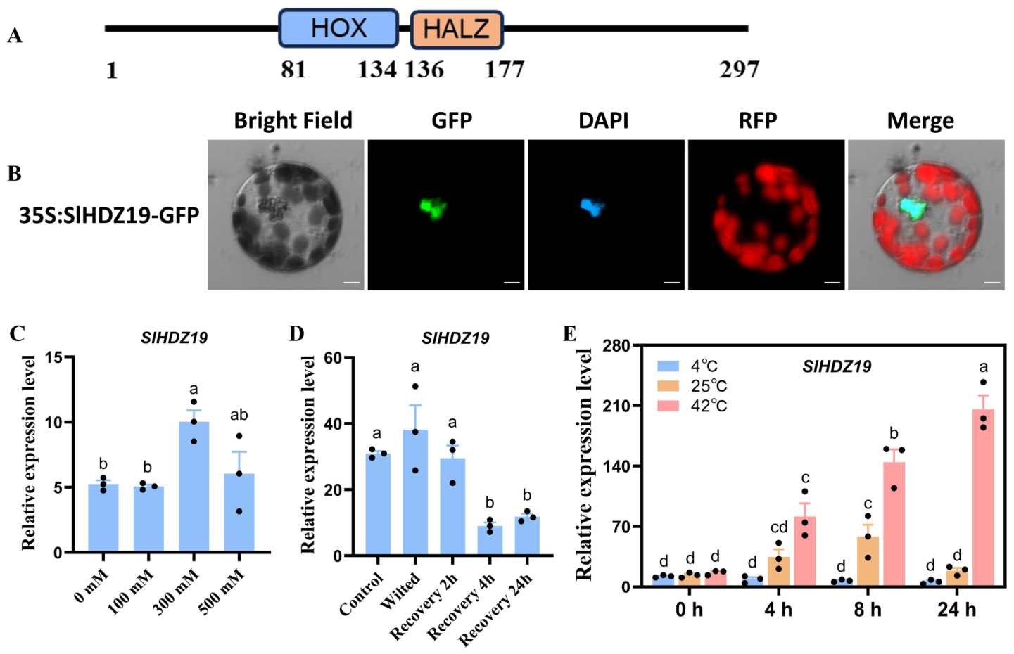
The conserved domains of SlHDZ19 were analyzed using the Conserved Domain Database (CDD) on the NCBI website. Domain analysis revealed that SlHDZ19 contains two conserved domains: a homeobox domain (HOX, amino acids 81–134) and a homeobox-associated leucine zipper domain (HALZ, amino acids 136–177) (Figure 1A). HALZ proteins are transcription factors that may function in plant development regulation and/or stress adaptation. Therefore, we investigated the subcellular location of SlHDZ19 in tomato protoplasts and N. benthamiana leaves via transient expression. Fluorescence imaging showed that the SlHDZ19-GFP fusion protein was localized exclusively to the nucleus and co-localized with DAPI staining, whereas the signal of free GFP protein was observed in both the nucleus and cytoplasm (Figure 1B).
To explore the biological function of SlHDZ19, we treated plants with salt, drought, low-temperature and high-temperature stresses and examined its transcript levels under these abiotic stresses. The expression of SlHDZ19 was significantly induced under 300 mM NaCl treatment, whereas no significant change was observed at 100 mM or 500 mM NaCl (Figure 1C). SlHDZ19 expression did not change significantly under drought or low-temperature (4 °C) stress (Figure 1D,E). In contrast, under heat stress conditions (42 °C), the expression of SlHDZ19 was highly enhanced at all time points examined (Figure 1E). Taken together, we inferred that SlHDZ19 may be involved in the heat stress (HS) response in tomato.

3.2. Phenotype and Physiological Analysis of Plants After HS Treatment

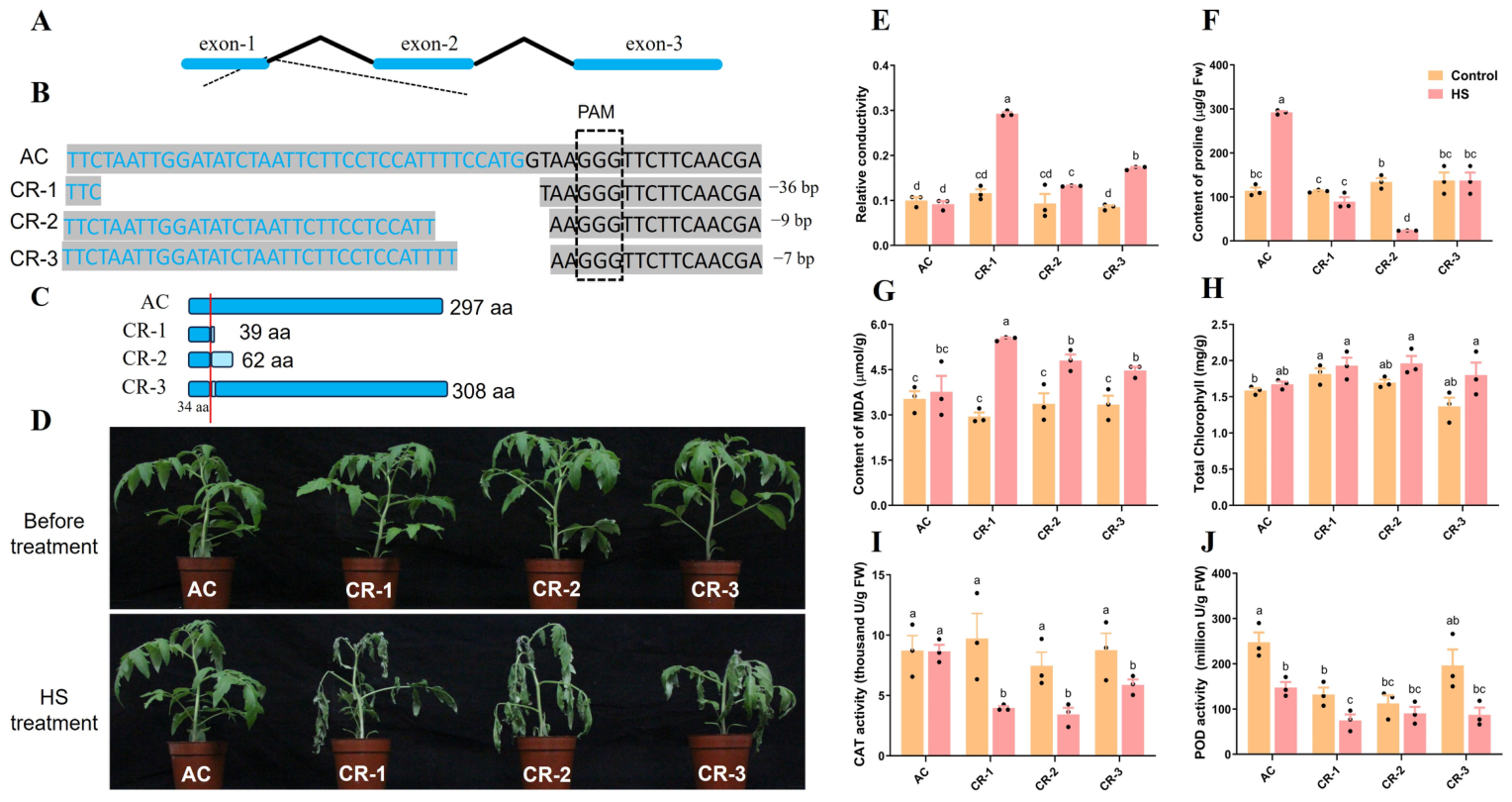
To investigate the function of SlHDZ19, we generated and utilized Cas9-mediated mutant (CR) and overexpression (OE) lines. We obtained three independent SlHDZ19 CR lines. DNA sequencing revealed deletions of 27 bp, 7 bp and 9 bp in the first exon and first intron of lines CR-1, CR-2 and CR-3, respectively. These mutations all resulted in the loss of the initial “GT” dinucleotide in the first intron. Given the “GT-AG” rule for intron splicing, these deletions are predicted to cause RNA splicing defects (Figure 2A,B). To determine whether the splicing errors altered the protein sequence, we cloned and sequenced the coding sequences (CDS) from all CR lines. Sequencing results indicated that the mutated SlHDZ19 alleles encoded truncated proteins of 39 and 62 amino acids (aa) in the CR-1 and CR-2 lines, respectively. In contrast, the CR-3 line was predicted to encode a near-full-length altered protein of 308 aa containing an additional 11-amino-acid sequence (Figure 2C). The sequencing results of the mutated sites and the putative coding protein alignment are shown in Figure S1A,B. Therefore, CR-1 and CR-2 were considered strong loss-of-function alleles, whereas CR-3 was treated as a CRISPR/Cas9-generated mutant allele with potentially impaired SlHDZ19 function.
Before HS treatment, no obvious difference in plant growth or development was observed between the wild type (AC) and CR lines. However, after 24 h treatment at 42 °C, while AC plants showed no visible wilting, all three CR lines showed increased heat sensitivity, with CR-1 and CR-2 displaying stronger wilting phenotypes than CR-3 (Figure 2D). Several physiological indices were measured to evaluate the physiological responses of plants under HS. Under normal conditions, no significant differences were detected between AC and the CR lines in the levels of malondialdehyde (MDA), proline, total chlorophyll, or relative electrolyte leakage, nor in the activities of catalase (CAT) and peroxidase (POD) (Figure 2E–J). Under HS conditions, however, the CR lines showed significantly higher relative electrolyte leakage than AC. In contrast, proline content was significantly reduced in all CR lines compared to AC. Conversely, CAT activity was significantly lower in the CR lines than in AC under HS. For POD activity, only CR-1 exhibited a significant decrease relative to AC, while the reductions in CR-2 and CR-3 were not statistically significant. No significant difference in total chlorophyll content was observed between genotypes under HS (Figure 2E–J).
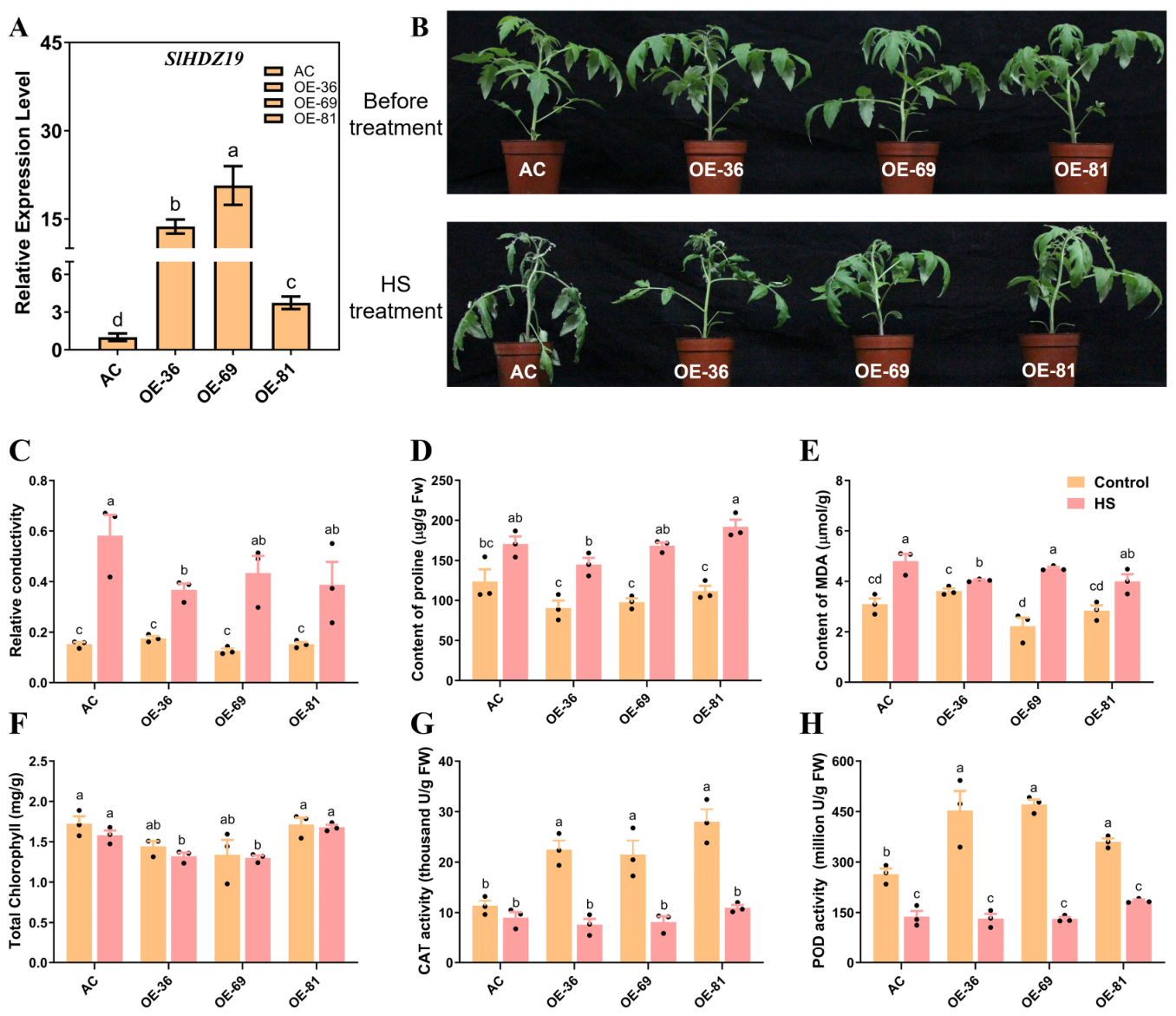
In addition, we employed three OE lines of SlHDZ19 for heat treatment. All three lines, OE-36, OE-69 and OE-81 showed elevated expression of SlHDZ19 (Figure 3A). Similar to the CR lines, the OE lines showed no growth or developmental defects under normal conditions. After 2 days of 42 °C HS treatment, all leaves of the wild type plants were desiccated and withered, while only mild wilting was observed in a few leaves of OE lines (Figure 3B). Thus, all three SlHDZ19-overexpressing lines displayed visibly improved heat-stress performance compared with AC. Physiological measurements under normal conditions revealed no significant differences between AC and OE lines in relative electrolyte leakage, proline, MDA, or total chlorophyll content (Figure 3C–F). After 48 h of heat stress (42 °C), OE-36 showed significantly lower relative electrolyte leakage and MDA content than AC, while OE-69 and OE-81 exhibited similar decreasing trends that did not reach statistical significance (Figure 3C,E). Proline content did not show consistent significant differences between AC and OE lines under heat stress (Figure 3D). Total chlorophyll content showed line-dependent differences after heat stress, with significant changes observed in OE-36 and OE-69 but not in OE-81 (Figure 3A,F). Under normal conditions, CAT and POD activities were significantly higher in all three OE lines than in AC (Figure 3G,H). Following heat stress, however, these activities declined in all genotypes, and no significant differences were observed between OE lines and AC. These results indicate that SlHDZ19 overexpression confers a clear improvement in visible heat-stress performance, while individual physiological indices show line- and parameter-dependent variation among OE lines. Together with the mutant-line results, especially the strong loss-of-function evidence from CR-1 and CR-2 and the consistent but weaker phenotype of CR-3, these findings support SlHDZ19 as a positive regulator of heat stress tolerance in tomato.

3.3. RNA-Seq Analysis of Tomato Plants Under Heat Stress Conditions

Having established that SlHDZ19 positively regulates thermotolerance at the physiological level, we next performed RNA sequencing to identify genes and pathways potentially regulated by this transcription factor. For RNA-seq, OE-36 and CR-1 were selected as representative SlHDZ19-overexpressing and mutant lines, respectively. A total of 24 cDNA libraries were constructed from OE-36, CR-1, and their corresponding AC controls under control (CK) and heat treatment (HT, 42 °C) conditions, with three biological replicates for each genotype and treatment. For clarity, the genotype and treatment abbreviations used in the RNA-seq analysis are summarized in Table S4, including AC, OE, CR, CK and HT. Each library produced approximately 39.4 to 47.3 million raw reads, and an average of 96.7% of clean reads was successfully mapped to the tomato reference genome (Tables S2 and S3), confirming the high quality and reliability of the sequencing data.
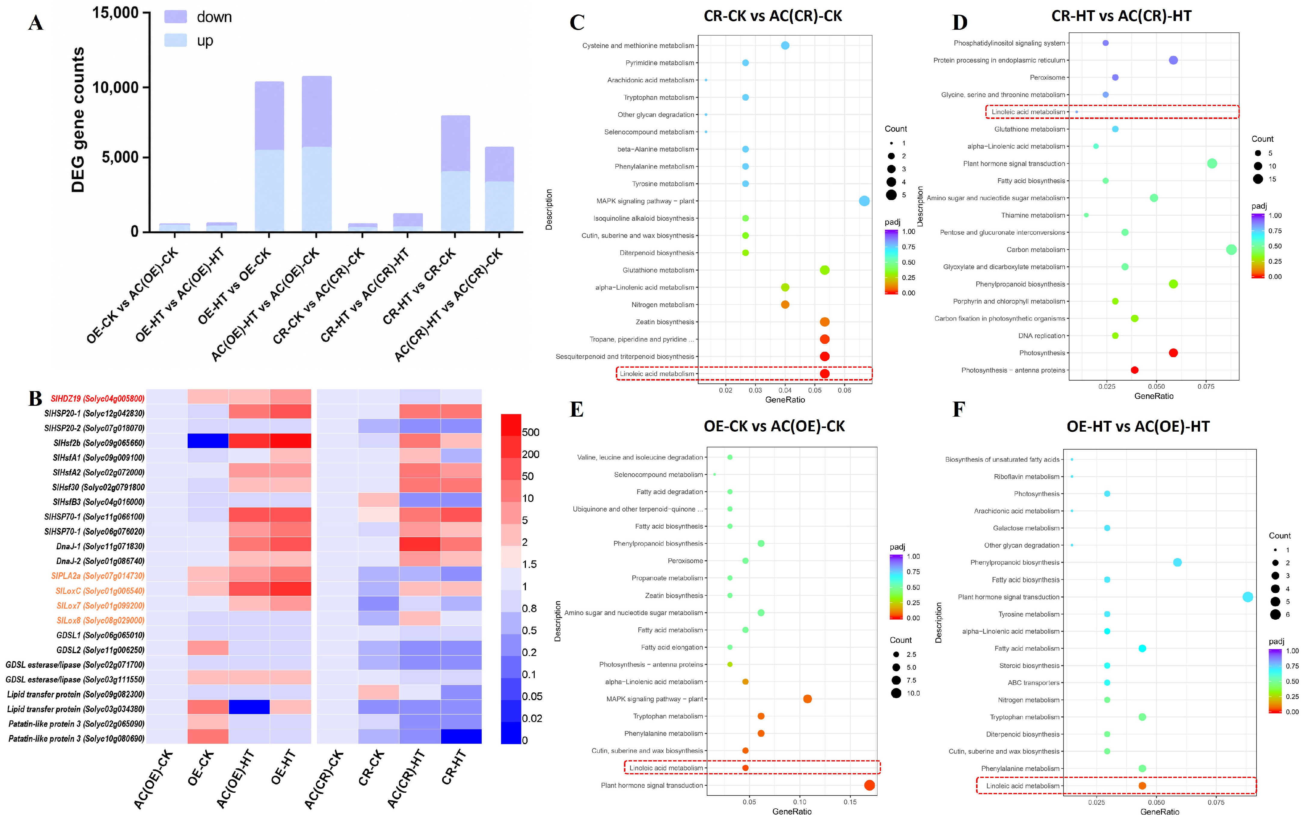
Differentially expressed genes (DEGs) were identified using the criteria of |log2(FoldChange)| ≥ 1 and an adjusted p-value (padj) ≤ 0.05. As expected, the number of DEGs increased substantially under heat stress across all genotypes, indicating a global transcriptional reprogramming in response to HS (Figure 4A). Notably, key heat-responsive genes were differentially regulated in the transgenic lines. In CR lines under HS, several HSFs (Solyc09g082670 and Solyc12g007070), HSP70s (Solyc03g117600, Solyc07g043560, Solyc09g010630), HSP90 (Solyc07g065840) and DnaJ (Solyc05g006820) were downregulated (Figure 4B). Conversely, in OE lines under HS, other heat-responsive genes, including an HSF (Solyc09g065660), HSP70 (Solyc03g117600), HSP20 (Solyc12g042830) and a heat shock protein-related gene (Solyc07g018070) were all up-regulated (Figure 4B). These results further support the role of SlHDZ19 as a positive regulator of the transcriptional heat stress response.
To further investigate the biological pathways affected by SlHDZ19, we performed KEGG pathway enrichment using the clusterProfiler package. Detailed KEGG enrichment statistics, including KEGG ID, GeneRatio, BgRatio, gene counts, p-values, adjusted p-values, and up-/down-regulated gene IDs, are provided in Table S5. In this table, the linoleic acid metabolism pathway (sly00591) is highlighted in the KEGG enrichment sheet. Under control conditions, linoleic acid metabolism was among the top enriched pathways in both OE-CK vs. AC(OE)-CK and CR-CK vs. AC(CR)-CK (Figure 4C,E; Table S5). Under heat stress treatment, linoleic acid metabolism also appeared among the top enriched pathways in OE-HT vs. AC(OE)-HT, whereas photosynthesis-related pathways were most affected in CR-HT vs. AC(CR)-HT (Figure 4D,F; Table S5). Collectively, these pathway-level analyses indicate that linoleic acid metabolism is repeatedly associated with SlHDZ19-dependent transcriptional changes, although the enrichment strength varies among genotype and treatment comparisons.
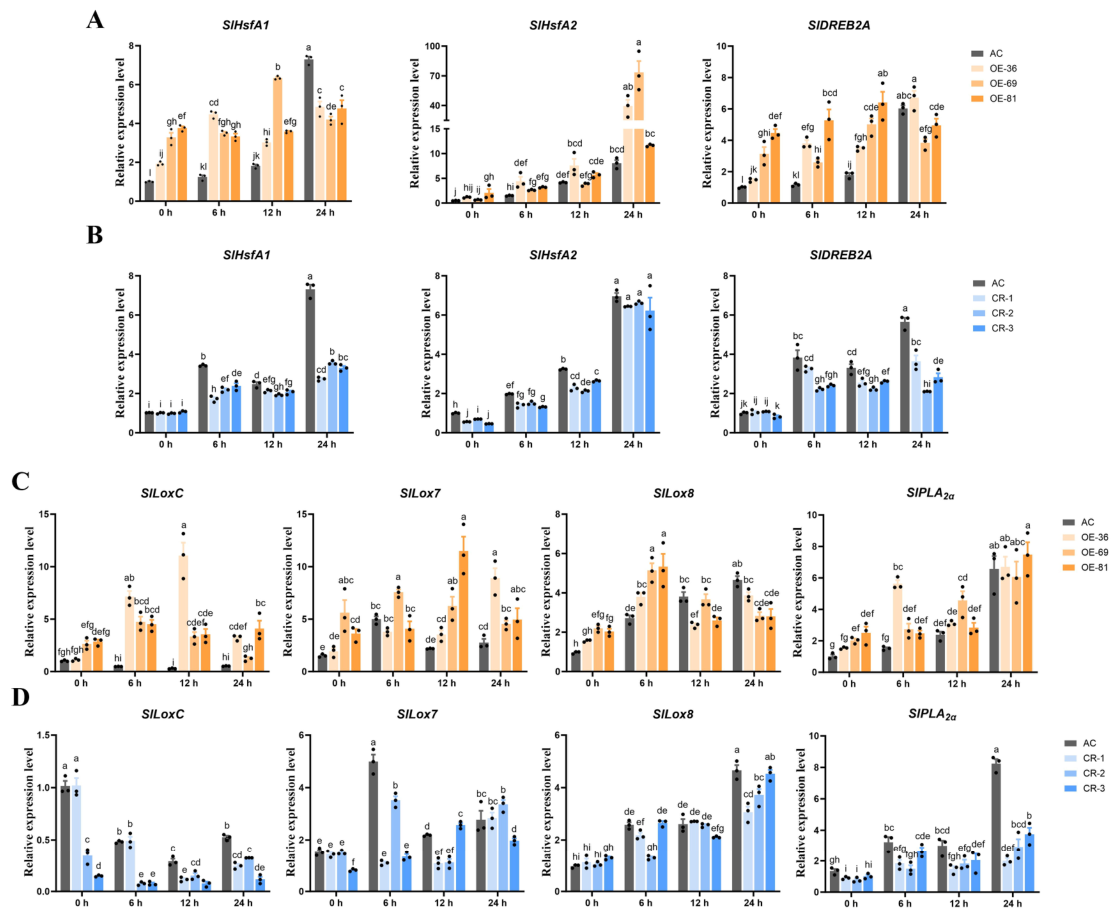
To verify the results, we analyzed the expression of three known thermotolerance-related genes SlHsfA2, SlHsfA1 and SlDREB2A by qRT-PCR in the AC, OE, and CR lines with and without HS treatment (Figure 5A,B). Consistent with RNA-seq data, the transcript levels of SlHsfA2, SlHsfA1 and SlDREB2A were down-regulated in CR lines under HS treatment compared to AC. Conversely, in the OE lines, their expression was up-regulated even under normal growing conditions (Figure 5A,B). Therefore, SlHDZ19 is required for the full activation of core heat stress response genes.
For further analysis, we focused on the KEGG-enriched linoleic acid metabolism pathway and annotation-supported PLA2/LOX-associated lipid-related genes. Because KEGG pathway assignment may not include all tomato LOX family members, lipid/LOX-related DEGs identified based on functional or conserved-domain annotations were also highlighted in the corresponding DEG sheets of Table S5. Among these genes, SlPLA2α (phospholipase A2-alpha, Solyc07g014730) and SlLoxC (lipoxygenase C, Solyc01g006540) were both consistently up-regulated in OE lines, while downregulated in CR lines under both control and heat stress conditions (Figure 5C,D). Two genes encoding 9S-lipoxygenase (9S-LOX), SlLox7 (Solyc01g099200) and SlLox8 (Solyc08g029000), were up-regulated in OE lines and down-regulated in CR lines under normal conditions, although their differential expression under heat stress was less pronounced (Figure 5C,D).
In summary, our transcriptomic and RT-qPCR evidence support that SlHDZ19 contributes to the activation of core heat stress-responsive genes. Furthermore, the recurrent enrichment of linoleic acid metabolism and the differential expression of PLA2/LOX-associated lipid-related genes support a role for SlHDZ19 in regulating a PLA2/LOX-associated lipid-metabolic transcriptional program.

3.4. SlHDZ19 Is Involved in Linoleic Acid Metabolism in Tomato

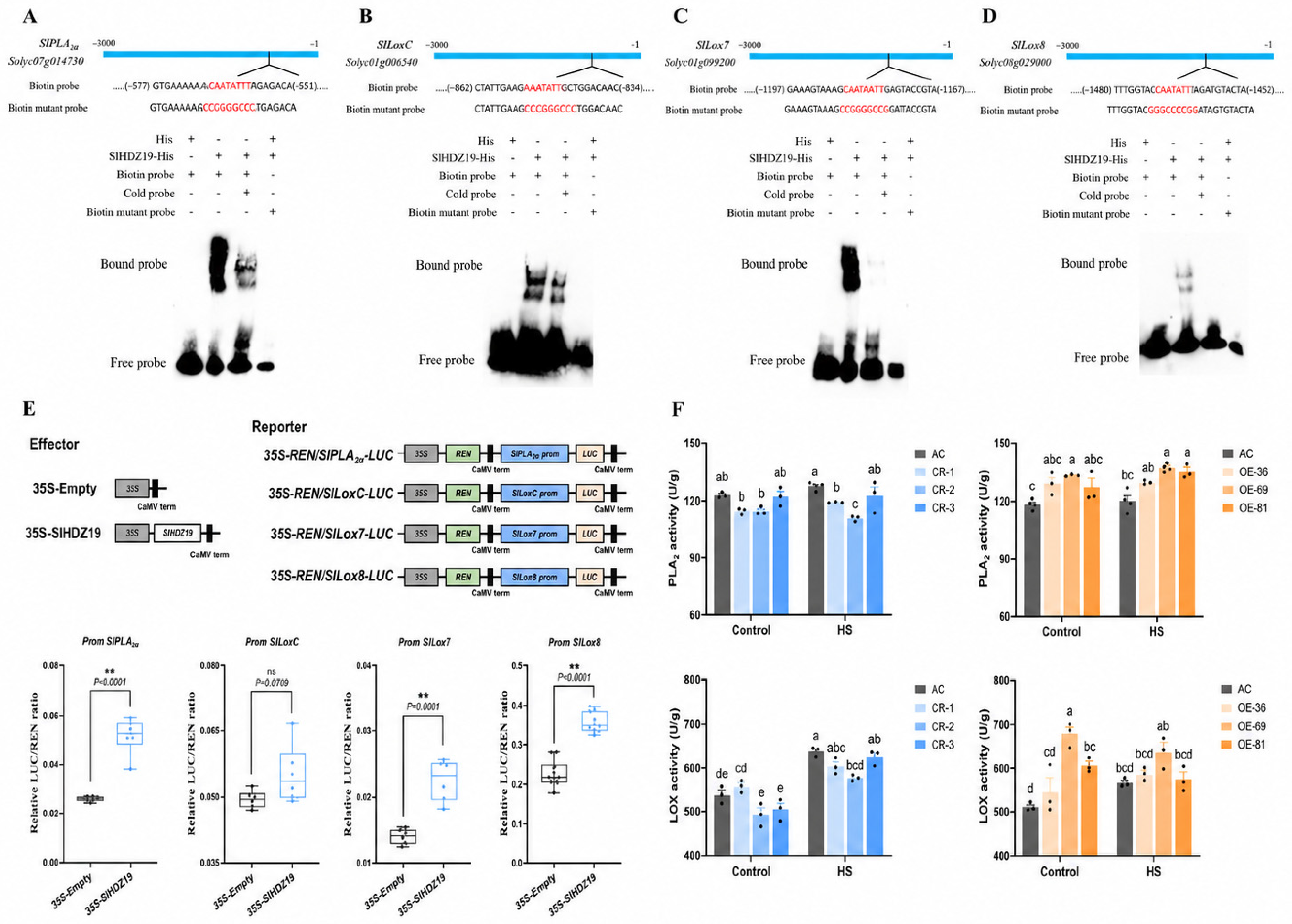
To verify our hypothesis, the 3000 bp promoters of the PLA enzyme-encoding gene SlPLA2α, three putative LOX enzyme-encoding genes SlLoxC, SlLox7 and SlLox8 were analyzed. Based on published data [26], a bioinformatic analysis identified a putative SlHDZ19-binding motif in these promoter regions (Figure 6A–D). To determine whether SlHDZ19 binds directly to these motifs, we performed electrophoretic mobility shift assays (EMSAs). A recombinant His-SlHDZ19 fusion protein was purified (Figure S2) and incubated with biotin-labeled DNA probes containing the predicted binding sites. The EMSA assay confirmed that SlHDZ19 bound to the promoter of SlPLA2α and three Lox genes SlLoxC, SlLox7 and SlLox8 in vitro (Figure 6A–D).
At the same time, we conducted dual-luciferase reporter assays in Arabidopsis mesophyll protoplasts to assess the transcriptional regulatory activity of SlHDZ19. The promoter sequences containing the putative binding motifs of target genes were fused to the firefly luciferase reporter gene. These reporter constructs were co-transfected with either a SlHDZ19 effector plasmid or an empty vector control. The results showed that SlHDZ19 significantly enhanced the transcriptional activity of the SlPLA2α, SlLox7 and SlLox8 promoters. A weaker but reproducible activation was also observed for the SlLoxC promoter (Figure 6E). Together, the EMSA and dual-LUC results support a direct transcriptional regulatory relationship between SlHDZ19 and these PLA2/LOX-associated genes, although in vivo promoter occupancy remains to be confirmed.
To further link SlHDZ19-mediated transcription to downstream metabolic activity, we measured the enzymatic activities of PLA2 and LOX. PLA2 activity was significantly lower in the SlHDZ19 mutant (CR) lines compared to AC, under both control and HS conditions. While only a slight difference was observed between AC and the overexpression (OE) lines under normal conditions, PLA2 activity became significantly higher in OE lines after HS (Figure 6F). In contrast, LOX activity was significantly elevated in OE lines compared to AC under normal conditions. Following HS treatment, LOX activity remained elevated, but the further increase relative to pre-stress level was not statistically significant. In CR lines, LOX activity exhibited a consistent but non-significant decreasing trend under both normal and HS conditions, with no statistically significant differences compared to AC (Figure 6F). This attenuated LOX response in the CR background may be due to functional redundancy among multiple LOX gene family members in tomato, and total LOX activity may not fully capture isoform-specific contributions.

3.5. Silencing of SlPLA Compromises Tomato Thermotolerance

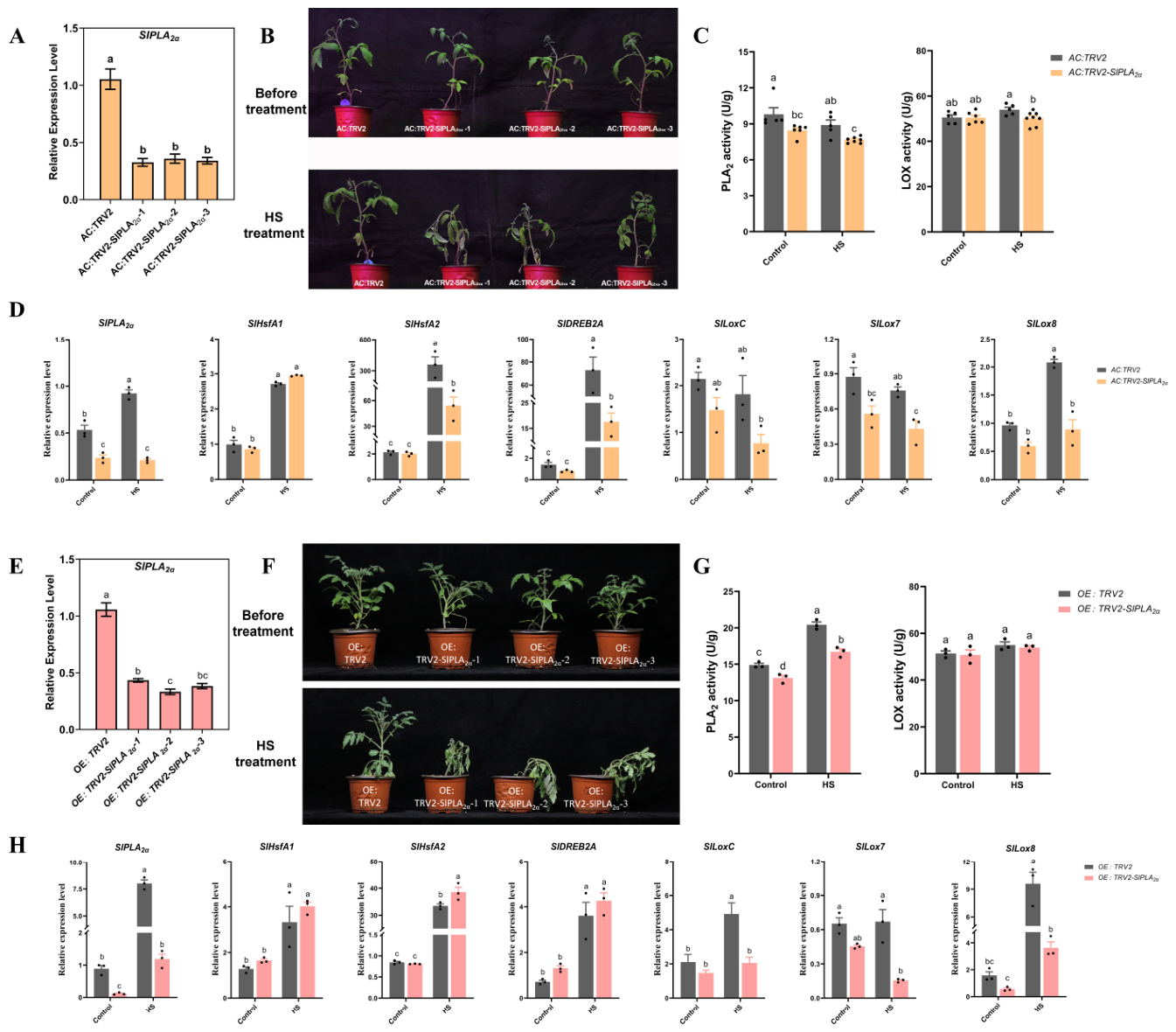
To further validate the functional importance of SlPLA2α within the SlHDZ19-mediated thermotolerance pathway, we employed virus-induced gene silencing (VIGS) to specifically downregulate its expression. We targeted SlPLA2α because it acts upstream in the proposed linoleic acid metabolism-related pathway, and its silencing allowed us to test the functional contribution of this step to thermotolerance. qRT-PCR analysis confirmed efficient silencing, with transcript levels reduced to 30–50% of those in wild-type plants (Figure 7A). Following 24 h of heat stress treatment, the SlPLA2α-silenced plants showed obvious wilting compared to wild-type controls (Figure 7B). Consistent with the transcript reduction, PLA2 enzyme activity was significantly compromised in silenced plants, to a degree comparable to that observed in the SlHDZ19-CR lines (Figure 7C). Meanwhile, LOX enzyme activity showed a slight decrease (Figure 7C). The heat-induction of SlHSFA2 and SlDREB2A was reduced in the SlPLA2α-silenced plants compared to the wild type (Figure 7D). Three LOX genes also showed a slight reduction, especially SlLox7 and SlLox8. These transcriptional changes are likely secondary effects resulting from the disruption of the linoleic acid metabolic pathway. To further determine whether the enhanced thermotolerance of SlHDZ19-overexpressing plants is mediated by SlPLA2α, we silenced SlPLA in the OE-69 background using VIGS. After silencing, these plants exhibited significantly reduced heat tolerance compared to empty-vector controls (Figure 7F), accompanied by a marked decrease in PLA2 enzyme activity (Figure 7G). These results indicate that up-regulation of SlPLA is functionally required for SlHDZ19-conferred thermotolerance (Figure 7E–H).
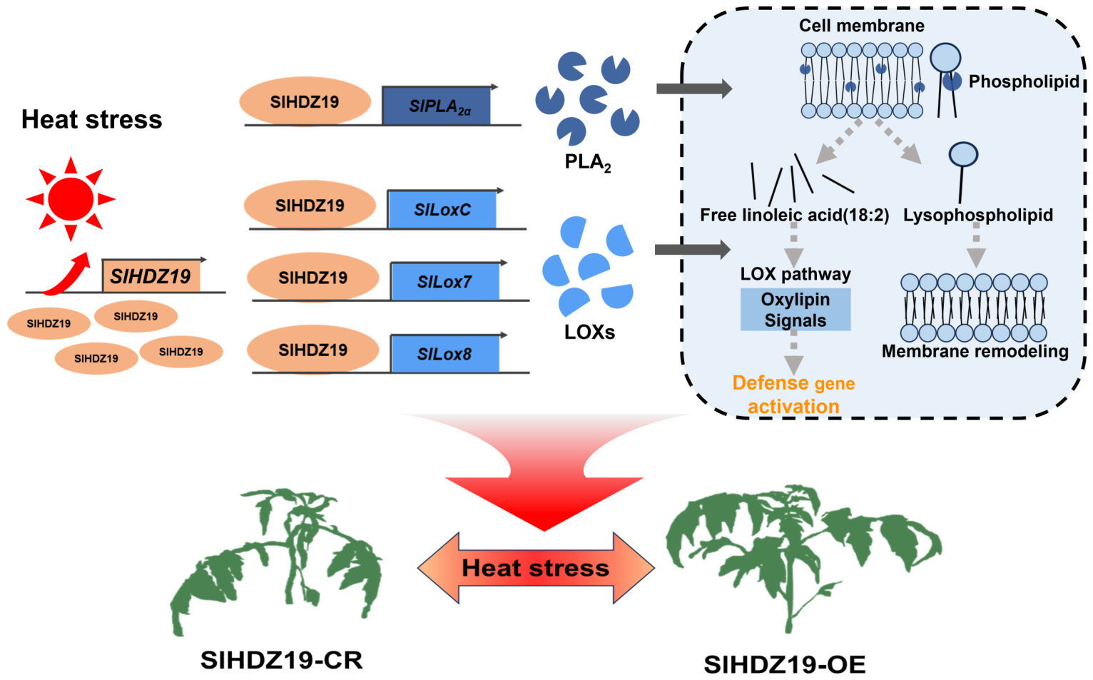
Collectively, these results provide direct genetic evidence that SlPLA2α is a key functional component downstream of SlHDZ19. The concomitant decline in thermotolerance, PLA2 activity, and the expression of key downstream genes upon SlPLA2α silencing further supports the important role of the SlHDZ19-SlPLA regulatory axis in mediating heat stress adaptation via linoleic acid metabolism (Figure 8).

4. Discussion

4.1. SlHDZ19 Positively Regulates Heat Tolerance in Tomato

The development of heat-tolerant crops is a critical objective for future breeding in the context of global warming [33]. In this study, we demonstrated that SlHDZ19 is a nuclear-localized transcription factor whose expression can be induced by HS treatment. Genetic analysis showed that SlHDZ19-overexpressing lines displayed visibly improved heat-stress performance compared with wild-type (AC) after 2 days of HS, although the statistical significance of individual physiological indices varied among independent OE lines and measured parameters. In contrast, SlHDZ19 mutant plants, particularly the strong loss-of-function alleles CR-1 and CR-2, exhibited increased heat sensitivity after 1 day under the same HS condition, while CR-3 showed a consistent but relatively weaker mutant phenotype. Physiological measurements, including levels of free proline, MDA and relative conductivity, further supported the role of SlHDZ19 in heat-stress responses, although individual indices showed genotype- and parameter-dependent variation. In addition, the enzyme activities of CAT and POD were significantly reduced in CR lines under HS but were elevated in OE lines even under normal conditions. After prolonged heat stress, however, CAT and POD activities declined in all genotypes and no significant differences were detected between OE lines and AC, suggesting that the elevated basal antioxidant capacity of OE lines may contribute to the early phase of heat-stress response rather than fully explain the later heat-stress phenotype. Given that CAT and POD activities reflect the capacity for reactive oxygen species (ROS) scavenging [34], these results indicate that SlHDZ19-mediated thermotolerance may be associated, at least in part, with modulation of antioxidant defenses.
This finding is consistent with a previous report demonstrating that SlHDZ19 may directly bind to the promoter of the ascorbate biosynthesis gene SlGMP3 to mediate oxidative stress [26]. Although the present physiological analyses support the role of SlHDZ19 in heat-stress responses, future studies incorporating survival rate, biomass recovery, chlorophyll fluorescence, direct ROS measurements, and broader membrane-stability assays would provide a more comprehensive evaluation of whole-plant thermotolerance. However, while oxidative stress is a common component of abiotic stress responses, the precise biological functions and downstream molecular mechanisms of SlHDZ19 in tomato require further elucidation. In this study, we identified that SlHDZ19 transcriptionally regulates genes involved in linoleic acid metabolism, providing a mechanistic link between transcriptional control and lipid-mediated stress adaptation.
Evidence from several plant species indicates that HD-Zip transcription factors can modulate thermotolerance through distinct downstream pathways. For example, CaHDZ15 promotes pepper basal thermotolerance by activating the CaHSFA6a–CaHSFA2 cascade [21], whereas LlHOX6 negatively regulates lily basal thermotolerance by antagonizing LlHB16-mediated activation of LlHSFA2 and LlMBF1c [22]. In addition, PtHDZ6 and MdHB7 enhance heat tolerance mainly through ROS-scavenging-, NAC-, or lignin-associated pathways [23,35]. Compared with these reported HSF/HSP-, ROS-, NAC-, and cell-wall-related mechanisms, our study suggests that SlHDZ19 contributes to tomato thermotolerance through a distinct PLA2/LOX-associated lipid-metabolic transcriptional program. This comparison indicates that HD-Zip-mediated thermotolerance may be achieved through multiple downstream routes, and places lipid-metabolic reprogramming as an additional mechanism within this regulatory framework.

4.2. SlHDZ19 Mediates the Heat Stress Response by Regulating Linoleic Acid Metabolism in Tomato

Building on the phenotypic and transcriptomic evidence described above, our results point to a PLA2/LOX-associated lipid-metabolic branch downstream of SlHDZ19. At the membrane level, this interpretation is consistent with a membrane-centered view of plant heat sensing and adaptation. Biological membranes are not only early targets of heat-induced damage but also active signaling platforms that connect changes in membrane fluidity, ROS production, calcium signaling, lipid remodeling, and nuclear transcriptional reprogramming [9,10,36,37]. For example, a plasma membrane BAM1–PBS1 receptor complex was recently shown to relay early heat signals to ROS production and heat-responsive transcriptomic reprogramming, while membrane stability under heat stress has been increasingly linked to coordinated lipid remodeling and lipid-derived signaling [11]. Linoleic acid, the most abundant fatty acid at the sn-2 position of membrane phospholipids in tomato leaves, serves as both a structural component and a precursor for bioactive oxylipins [38,39]. We demonstrated that SlHDZ19 directly binds in vitro to the promoters of SlPLA2α and three LOX genes SlLoxC, SlLox7 and SlLox8, and activates these promoters in transient assays. PLA2 plays a pivotal role in membrane regeneration and signaling by hydrolyzing phospholipids to release free linoleic acid and lysophospholipids [40,41]. This activity is essential for maintaining membrane integrity under stress and for generating lipid-derived signals.
To genetically validate that SlPLA2α mediates the thermotolerance conferred by SlHDZ19, we performed VIGS to silence SlPLA2α. Silencing in the wild-type background phenocopied the heat-sensitive phenotype of SlHDZ19-CR lines, accompanied by reduced PLA2 activity (Figure 7A–C). Moreover, silencing SlPLA2α in the SlHDZ19 overexpression background significantly compromised thermotolerance (Figure 7E–H), demonstrating that upregulation of this gene is functionally required for SlHDZ19-conferred heat resilience. These results provide strong genetic evidence that SlPLA2α is a key downstream component required for SlHDZ19-mediated thermotolerance. This aligns with the known role of PLA2 in facilitating membrane repair and remodeling, as observed in Arabidopsis pollen development [42]. Furthermore, the specific up-regulation of the secretory isoform PLA, which can translocate to the apoplast [43], implies an additional layer of regulation. The potential extracellular localization of PLA, a key site for environmental sensing and intercellular communication [5,12,13], suggests that SlHDZ19 may coordinate broader stress signaling or reinforce the cell wall-plasma membrane interface under heat stress.
In the proposed pathway, linoleic acid released by PLA2 serves as a substrate for LOXs. We found that SlHDZ19 also transcriptionally activates several LOX genes, thereby supporting a potential transcriptional route for channeling linoleic acid toward the oxylipin biosynthesis pathway. The 9S-LOXs encoded by SlLox7 and SlLox8 catalyze the committed step, producing hydroperoxides such as 9-HPODE, which are precursors to a diverse array of oxylipins [44]. By contrast, the weaker LOX-activity changes observed in CR lines may be due to functional redundancy among the multiple LOX gene family members in tomato and to the use of total LOX activity measurements, which may not fully resolve isoform-specific contributions. Oxylipins such as 9-/13-HODE and 9-/13-OxoODE are potent signaling molecules that enhance stress resilience [16,45,46]. By potentially channeling linoleic acid into the enzymatic LOX pathway, SlHDZ19 may help mitigate uncontrolled non-enzymatic lipid peroxidation caused by stress-induced ROS while simultaneously promoting the formation of beneficial signaling molecules [47,48]. Thus, the coordinated transcriptional activation of PLA2 and LOX genes by SlHDZ19 supports a working model of a PLA2/LOX-associated lipid-metabolic regulatory circuit. This circuit may serve a dual protective function by supporting membrane integrity through PLA2-mediated remodeling and promoting the formation of oxylipin-related signals via LOXs, which may then amplify the heat stress response [18].
Collectively, our results support a SlHDZ19-mediated transcriptional regulatory module associated with lipid metabolism in response to heat stress. We propose that this SlHDZ19-PLA2-LOX module may integrate into the broader network of heat signal transduction (Figure 8). Notably, a recent study demonstrated that the lipid second messenger phosphatidic acid (PA) activates a nuclear cAMP degradation pathway to drive thermotolerance [13]. In this context, our finding that SlHDZ19 transcriptionally activates SlPLA2α suggests a potential link: PLA2-mediated membrane hydrolysis may contribute to the precursor pool for PA synthesis, thereby potentially modulating this core PA-cAMP signaling axis. Thus, SlHDZ19 may act as an upstream transcriptional regulator linking PLA2-associated transcriptional regulation to lipid-mediated thermosensing pathways. Furthermore, potential oxylipin-related signals generated through the LOX branch could constitute a parallel or complementary signaling stream. Future work should explore potential crosstalk by analyzing PA dynamics and cAMP homeostasis in SlHDZ19 mutants. We acknowledge that direct measurements of lipid metabolites, such as linoleic acid, linoleic acid-derived oxylipins or PA levels, would provide more definitive evidence for the proposed lipid signaling cascade. Because lipid profiling by GC-MS or LC-MS was not performed in the present study, our conclusion is restricted to the transcriptional and enzymatic regulation of linoleic acid metabolism-related genes rather than direct metabolite accumulation. Therefore, the current model should be regarded as a working hypothesis based on consistent transcriptional, biochemical, and genetic evidence, rather than a definitive claim.
In addition, heat stress in natural environments often occurs together with changes in light intensity, especially high irradiance. Because the present study was conducted under controlled normal-light conditions to focus on the temperature response of SlHDZ19, whether the SlHDZ19–SlPLA regulatory module also contributes to combined heat-high-light stress tolerance remains to be investigated. Future studies incorporating lipidomic profiling, in vivo promoter-binding assays such as ChIP-qPCR, and combined heat–light stress treatments will be necessary to validate and refine this model. Unraveling such network-level interactions will be crucial for designing multi-target strategies to enhance crop thermotolerance.

5. Conclusions

In summary, our study identifies SlHDZ19 as a positive regulator of tomato thermotolerance and links this HD-Zip I transcription factor to a PLA2-dependent lipid-metabolic transcriptional program. SlHDZ19 activates SlPLA2α and several LOX genes, and SlPLA2α is functionally required for SlHDZ19-associated heat tolerance. These findings support a working model in which SlHDZ19 promotes heat adaptation, at least in part, through PLA2/LOX-associated transcriptional regulation. Future lipidomic and in vivo promoter-binding analyses will be needed to fully resolve the downstream lipid metabolites and signaling events.

Supplementary Materials

The following supporting information can be downloaded at: https://www.mdpi.com/article/10.3390/horticulturae12050639/s1, Table S1: Primers used in this study. Table S2: Summary of RNA-Seq sample sequencing data quality. Table S3: Statistics of RNA-Seq sample and reference genome comparison. Table S4: Abbreviations of genotypes and treatments used in this study. Table S5: KEGG enrichment statistics and DEG information from RNA-seq comparisons. Figure S1: (A): The DNA sequences of mutated sites; (B): Alignment of predicted SlHDZ19 protein sequences encoded by mutated SlHDZ19 alleles in CR lines. Figure S2: Immunodetection of purified His-SlHDZ19 fusion protein from DE3 cells. Figure S3: Subcellular localization of SlHDZ19 in Nicotiana benthamiana leaves.

Author Contributions

Conceptualization, J.L. and Y.P.; methodology, D.D. and C.S.; software, J.W.; validation, X.H., and D.D.; investigation, X.H.; resources, Y.S. and Y.C.; data analysis, L.W. and K.M.; data collection, X.H., K.M. and J.D.; writing—original draft preparation, X.H. and D.D.; writing—review and editing, X.H. and K.M.; visualization, J.D. and J.W.; funding acquisition, Y.P., X.H. and H.H. All authors have read and agreed to the published version of the manuscript.

Funding

This work was supported by grants from National Natural Science Foundation of China (No. 32172597), Sichuan and Chongqing Science and Technology Co-operation Projects of Beibei district, Chongqing (cykjhz2024-24), the Chongqing Science Foundation (CSTB2024NSCQ-MSX1283), China Agriculture Research System (CARS-23-B08), Chongqing Graduate Research Innovation Project (CYB240119) and National-level College Students’ Innovation and Entrepreneurship Training Program (202450631077).

Data Availability Statement

The data supporting the findings of this study are available within the article and its Supplementary Materials. RNA-seq data are available from the corresponding author upon reasonable request.

Acknowledgments

We are grateful to Bi-Yue Ding from Southwest University for her invaluable technical support with confocal microscope imaging.

Conflicts of Interest

The funders had no role in the design of the study; in the collection, analyses, or interpretation of data; in the writing of the manuscript; or in the decision to publish the results.

References

  1. Zhao, C.; Liu, B.; Piao, S.L.; Wang, X.H.; Lobell, D.B.; Huang, Y.; Huang, M.T.; Yao, Y.T.; Bassu, S.; Ciais, P.; et al. Temperature increase reduces global yields of major crops in four independent estimates. Proc. Natl. Acad. Sci. USA 2017, 114, 9326–9331. [Google Scholar] [CrossRef]
  2. Wang, X.H.; Zhao, C.; Müller, C.; Wang, C.Z.; Ciais, P.; Janssens, I.; Peñuelas, J.; Asseng, S.; Li, T.; Elliott, J.; et al. Emergent constraint on crop yield response to warmer temperature from field experiments. Nat. Sustain. 2020, 3, 908–916. [Google Scholar] [CrossRef]
  3. Hultgren, A.; Carleton, T.; Delgado, M.; Gergel, D.R.; Greenstone, M.; Houser, T.; Hsiang, S.; Jina, A.M.; Kopp, R.E.; Malevich, S.B.; et al. Impacts of climate change on global agriculture accounting for adaptation. Nature 2025, 642, 644–652. [Google Scholar] [CrossRef]
  4. Ohama, N.; Sato, H.; Shinozaki, K.; Yamaguchi-Shinozaki, K. Transcriptional regulatory network of plant heat stress response. Trends Plant Sci. 2017, 22, 53–65. [Google Scholar] [CrossRef]
  5. Kan, Y.; Mu, X.R.; Gao, J.; Lin, H.X.; Lin, Y.S. The molecular basis of heat stress responses in plants. Mol. Plant 2023, 16, 1612–1634. [Google Scholar] [CrossRef] [PubMed]
  6. Yen, M.J.; Lin, K.H.; Thalimaraw, L.; Yang, H.R.; Boonyaves, K.; Cheng, C.Y.; Wu, T.Y. Conserved HSFA1-dependent chromatin dynamics drive heat stress responses in plants. Cell Rep. 2025, 44, 116714. [Google Scholar] [CrossRef] [PubMed]
  7. Raturi, V.; Zinta, G. HSFA1 heat shock factors integrate warm temperature and heat signals in plants. Trends Plant Sci. 2024, 29, 1165–1167. [Google Scholar] [CrossRef]
  8. Kevei, Z.; Ferreira, S.D.S.; Casenave, C.M.P.; Kurowski, T.; Mohareb, F.; Rickett, D.; Stain, C.; Thompson, A.J. Missense mutation of a class B heat shock factor is responsible for the tomato bushy root-2 phenotype. Mol. Hortic. 2022, 2, 4. [Google Scholar] [CrossRef]
  9. Chen, B.; Wang, Y.; Zhu, Y.; Li, J.; Wan, L.; Zhu, C.; Huang, Q.; Abd Elhamid, M.A.; Soaud, S.A.; Yan, K.; et al. Membrane stability under heat stress: Molecular signaling, lipid remodeling, and defense mechanisms in plants. Plant Cell Rep. 2026, 45, 32. [Google Scholar] [CrossRef]
  10. Calixto, C.P.G. Molecular aspects of heat stress sensing in land plants. Plant J. 2025, 121, e70069. [Google Scholar] [CrossRef] [PubMed]
  11. Li, J.; Wu, S.; Wang, K.; Xu, Y.; Zhang, X.; Li, X.; Song, W.; Zhou, J.-M.; Gong, Z.; Yang, S.; et al. A plasma membrane receptor complex mediates early heat-responsive signaling to trigger nuclear transcriptomic reprogramming in Arabidopsis. Mol. Plant 2025, 18, 2018–2034. [Google Scholar] [CrossRef]
  12. Hayes, S.; Schachtschabel, J.; Mishkind, M.; Munnik, T.; Arisz, S.A. Hot topic: Thermosensing in plants. Plant Cell Environ. 2021, 44, 2018–2033. [Google Scholar] [CrossRef]
  13. Kan, Y.; Mu, X.R.; Qu, F.F.; Dai, Z.A.; Gao, J.; Liu, N.J.; Li, S.Q.; Shan, J.X.; Ye, W.W.; Dong, N.Q.; et al. A stepwise decoding mechanism for heat sensing in plants connects lipid remodeling to a nuclear signaling cascade. Cell 2025, 188, 7378–7396.e23. [Google Scholar] [CrossRef] [PubMed]
  14. Lee, H.Y.; Bahn, S.C.; Shin, J.S.; Hwang, I.; Back, K.; Doelling, J.H.; Ryu, S.B. Multiple forms of secretory phospholipase A2 in plants. Prog. Lipid Res. 2005, 44, 52–67. [Google Scholar] [CrossRef]
  15. Marmey, P.; Jalloul, A.; Alhamdia, M.; Assigbetse, K.; Cacas, J.L.; Voloudakis, A.E.; Champion, A.; Clerivet, A.; Montillet, J.L.; Nicole, M. The 9-lipoxygenase GhLOX1 gene is associated with the hypersensitive reaction of cotton Gossypium hirsutum to Xanthomonas campestris pv malvacearum. Plant Physiol. Biochem. 2007, 45, 596–606. [Google Scholar] [CrossRef]
  16. Badiali, C.; Beccaccioli, M.; Sciubba, F.; Chronopoulou, L.; Petruccelli, V.; Palocci, C.; Reverberi, M.; Miccheli, A.; Pasqua, G.; Brasili, E. Pterostilbene-loaded PLGA nanoparticles alter phenylpropanoid and oxylipin metabolism in Solanum lycopersicum L. leaves. Sci. Rep. 2024, 14, 21941. [Google Scholar] [CrossRef]
  17. Pretorius, C.J.; Zeiss, D.R.; Dubery, I.A. The presence of oxygenated lipids in plant defense in response to biotic stress: A metabolomics appraisal. Plant Signal. Behav. 2021, 16, 1989215. [Google Scholar] [CrossRef]
  18. Pandey, M.; Ganotra, J.; Singh, A.; Parchuri, P.; Giri, J. Lipid-mediated responses to nutrient and other stresses: Roles in plant adaptation and signaling. J. Exp. Bot. 2025, eraf482. [Google Scholar] [CrossRef]
  19. Li, Y.X.; Yang, Z.R.; Zhang, Y.Y.; Guo, J.J.; Liu, L.L.; Wang, C.F.; Wang, B.S.; Han, G.L. The roles of HD-ZIP proteins in plant abiotic stress tolerance. Front. Plant Sci. 2022, 13, 1027071. [Google Scholar] [CrossRef] [PubMed]
  20. Liu, S.; Zhao, L.Y.; Cheng, M.Z.; Sun, J.F.; Ji, X.M.; Ullah, A.; Xie, G.S. Calmodulins and calmodulin-like proteins-mediated plant organellar calcium signaling networks under abiotic stress. Crop J. 2024, 12, 1321–1332. [Google Scholar] [CrossRef]
  21. Mou, S.; He, W.; Jiang, H.; Meng, Q.; Zhang, T.; Liu, Z.; Qiu, A.; He, S. Transcription factor CaHDZ15 promotes pepper basal thermotolerance by activating HEAT SHOCK FACTORA6a. Plant Physiol. 2024, 195, 812–831. [Google Scholar] [CrossRef] [PubMed]
  22. Wu, Z.; Li, T.; Zhang, Y.; Zhang, D.; Teng, N. HD-Zip I protein LlHOX6 antagonizes homeobox protein LlHB16 to attenuate basal thermotolerance in lily. Plant Physiol. 2024, 194, 1870–1888. [Google Scholar] [CrossRef]
  23. Qiu, L.; Qi, Z.; Zhang, W.; Li, N.; Mao, K.; Yang, J.; Ma, F. MdHB7 enhances apple basal thermotolerance by activating ROS scavenging and lignin accumulation. Plant J. 2026, 125, e70767. [Google Scholar] [CrossRef]
  24. Wojciechowska, I.; Mukherjee, T.; Knox-Brown, P.; Hu, X.; Khosla, A.; Subedi, B.; Ahmad, B.; Mathews, G.L.; Panagakis, A.A.; Thompson, K.A.; et al. Arabidopsis PROTODERMAL FACTOR2 binds lysophosphatidylcholines and transcriptionally regulates phospholipid metabolism. New Phytol. 2024, 244, 1498–1518. [Google Scholar] [CrossRef]
  25. Gao, Y.; Ruan, J.; Wang, G.; Zhang, Y.; Wang, Y.; Fan, B.; Chen, B.; Xue, D.; Zhang, S.; Hu, Z.; et al. SlHDZIV8-SlnsLTP33 module controls trichome density and lipid metabolism in tomato. Plant J. 2025, 123, e70398. [Google Scholar] [CrossRef]
  26. Hu, T.X.; Ye, J.; Tao, P.W.; Li, H.X.; Zhang, J.H.; Zhang, Y.Y.; Ye, Z.B. The tomato HD-Zip I transcription factor SIHZ24 modulates ascorbate accumulation through positive regulation of the D-mannose/L-galactose pathway. Plant J. 2016, 85, 16–29. [Google Scholar] [CrossRef]
  27. He, L.; Xu, J.; Li, C.; Shen, C.; He, Q.; Lei, X.; Zhang, H.; Guo, L.; Lin, T.; Guo, Y.D.; et al. SlHSFB2b-mediated inhibition of jasmonic acid catabolism enhances tomato tolerance to combined high light and heat stress. Plant Physiol. 2025, 199, kiaf547. [Google Scholar] [CrossRef]
  28. He, M.; Wu, J.; Yang, Y.; Cui, Q.; Fan, N.; Chi, C.; Yan, H.; Liu, Y.; Pan, Z.; Zhang, K.; et al. The HsfA1a-BAG5b module mediates thermotolerance through activating autophagy in tomato. Plant Physiol. 2025, 199, kiaf526. [Google Scholar] [CrossRef]
  29. Meng, X.; Kang, Z.; Chen, G.; Feng, Y.; Zhang, Y.; Li, G.; Hu, S.; Zhu, C.; Li, T.; Hu, X. The SlMYC2-SlNAC72-SlMIEL1 module contributes to high-temperature tolerance in tomato by regulating jasmonic acid biosynthesis. Plant Physiol. 2026, 200, kiag028. [Google Scholar] [CrossRef] [PubMed]
  30. Sun, X.C.; Gao, Y.F.; Li, H.R.; Yang, S.Z.; Liu, Y.S. Over-expression of SlWRKY39 leads to enhanced resistance to multiple stress factors in tomato. J. Plant Biol. 2015, 58, 52–60. [Google Scholar] [CrossRef]
  31. Cao, X.; Jiang, F.L.; Wang, X.; Zang, Y.W.; Wu, Z. Comprehensive evaluation and screening for chilling-tolerance in tomato lines at the seedling stage. Euphytica 2015, 205, 569–584. [Google Scholar] [CrossRef]
  32. Yoo, S.D.; Cho, Y.H.; Sheen, J. Arabidopsis mesophyll protoplasts: A versatile cell system for transient gene expression analysis. Nat. Protoc. 2007, 2, 1565–1572. [Google Scholar] [CrossRef]
  33. Itoh, H.; Yamashita, H.; Wada, K.C.; Yonemaru, J. Real-time emulation of future global warming reveals realistic impacts on the phenological response and quality deterioration in rice. Proc. Natl. Acad. Sci. USA 2024, 121, e2316497121. [Google Scholar] [CrossRef] [PubMed]
  34. Li, J.H.; Ai, G.; Wang, Y.L.; Ding, Y.; Hu, X.M.; Liang, Y.; Yan, Q.X.; Wu, K.; Huang, R.; Chen, C.R.; et al. A truncated B-box zinc finger transcription factor confers drought sensitivity in modern cultivated tomatoes. Nat. Commun. 2024, 15, 8013. [Google Scholar] [CrossRef] [PubMed]
  35. Ji, M.; Liu, M.; Zheng, X.; Xiao, C.; Ji, Y.; Duan, Y.; Zhu, Y.; Xue, J.; Bo, C.; Xue, T. Pinellia ternata HD-Zip6 gene positively regulates heat stress tolerance in transgenic Arabidopsis by increasing ROS scavenging and NAC019 expression. Plant Stress 2025, 16, 100806. [Google Scholar] [CrossRef]
  36. Sharma, P.; Lakra, N.; Goyal, A.; Ahlawat, Y.K.; Zaid, A.; Siddique, K.H.M. Drought and heat stress mediated activation of lipid signaling in plants: A critical review. Front. Plant Sci. 2023, 14, 1216835. [Google Scholar] [CrossRef]
  37. Shajar, F.; Nabi, A.U.; Manzoor, A.; Saleem, S.; Mushtaq, N.U.; Manzoor, S.; Rehman, R.U. Understanding lipid-mediated defense mechanisms in plant responses to abiotic stress. Nucleus 2025, 1–14. [Google Scholar] [CrossRef]
  38. Browse, J.; Warwick, N.; Somerville, C.R.; Slack, C.R. Fluxes through the prokaryotic and eukaryotic pathways of lipid synthesis in the ‘16:3’ plant Arabidopsis thaliana. Biochem. J. 1986, 235, 25–31. [Google Scholar] [CrossRef]
  39. Wolter, F.P.; Schmidt, R.; Heinz, E. Chilling sensitivity of Arabidopsis thaliana with genetically engineered membrane lipids. EMBO J. 1992, 11, 4685–4692. [Google Scholar] [CrossRef]
  40. Brown, W.J.; Chambers, K.; Doody, A. Phospholipase A2 (PLA2) enzymes in membrane trafficking: Mediators of membrane shape and function. Traffic 2003, 4, 214–221. [Google Scholar] [CrossRef]
  41. Staneva, G.; Angelova, M.I.; Koumanov, K. Phospholipase A2 promotes raft budding and fission from giant liposomes. Chem. Phys. Lipids 2004, 129, 53–62. [Google Scholar] [CrossRef]
  42. Kim, H.J.; Ok, S.H.; Bahn, S.C.; Jang, J.; Oh, S.A.; Park, S.K.; Twell, D.; Ryu, S.B.; Shin, J.S. Endoplasmic reticulum- and Golgi-localized phospholipase A plays critical roles in Arabidopsis pollen development and germination. Plant Cell 2011, 23, 94–110. [Google Scholar] [CrossRef]
  43. Jung, J.; Kumar, K.; Lee, H.Y.; Park, Y.I.; Cho, H.T.; Ryu, S.B. Translocation of phospholipase A to apoplasts is modulated by developmental stages and bacterial infection in Arabidopsis. Front. Plant Sci. 2012, 3, 126. [Google Scholar] [CrossRef]
  44. Andreou, A.Z.; Hornung, E.; Kunze, S.; Rosahl, S.; Feussner, I. On the substrate binding of linoleate 9-lipoxygenases. Lipids 2009, 44, 207–215. [Google Scholar] [CrossRef] [PubMed]
  45. Mariutto, M.; Fauconnier, M.L.; Ongena, M.; Laloux, M.; Wathelet, J.P.; du Jardin, P.; Thonart, P.; Dommes, J. Reprogramming of fatty acid and oxylipin synthesis in rhizobacteria-induced systemic resistance in tomato. Plant Mol. Biol. 2014, 84, 455–467. [Google Scholar] [CrossRef] [PubMed]
  46. Wasternack, C.; Feussner, I. The oxylipin pathways: Biochemistry and function. Annu. Rev. Plant Biol. 2018, 69, 363–386. [Google Scholar] [CrossRef]
  47. Cohen-Hoch, D.; Chen, T.M.; Sharabi, L.; Dezorella, N.; Itkin, M.; Feiguelman, G.; Malitsky, S.; Fluhr, R. Osmotic stress in roots drives lipoxygenase-dependent plastid remodeling through singlet oxygen production. Plant Physiol. 2024, 197, kiae589. [Google Scholar] [CrossRef] [PubMed]
  48. Farmer, E.E.; Mueller, M.J. ROS-mediated lipid peroxidation and RES-activated signaling. Annu. Rev. Plant Biol. 2013, 64, 429–450. [Google Scholar] [CrossRef]
Figure 1. Characterization of SlHDZ19 in tomato. (A): Schematic diagram of SlHDZ19 protein structure. (B): Subcellular localization of SlHDZ19-GFP fusion protein in tomato protoplasts. The green represents GFP signal, the blue signal of DAPI indicates the position of the nucleus, red represents chlorophyll autofluorescence. Bar = 10 μm. (C,D): Relative expression levels of SlHDZ19 under salt stress (C) and drought stress (D). The NaCl concentrations used are 0, 100, 300 and 500 mM. (E): Relative expression levels of SlHDZ19 in leaf samples under cold (4 °C) and heat (42 °C) conditions at 0, 4, 8, and 24 h after treatments. Values represent means ± SE of three biological replicates. Different lowercase letters indicate a significant difference (p < 0.05) as determined by one-way ANOVA and Duncan’s multiple comparisons.
Horticulturae 12 00639 g001
Figure 2. SlHDZ19 mutation increases thermal sensitivity in tomato. (A): Schematic diagram of SlHDZ19 gene. (B,C): PAM site and the DNA base deletions in CR-1, CR-2 and CR-3 SlHDZ19 mutant lines and schematic diagram of the coding protein sequences in the three CR lines. (D): Representative phenotypes of AC and SlHDZ19 mutant lines before and after 24 h of heat treatment at 42 °C. CR lines, especially CR-1 and CR-2, showed more severe wilting than AC after heat stress. (EJ): Relative electrolyte conductivity (E), and the content of proline (F), MDA (G), and total chlorophyll (H) in AC and CR lines (CR-1, CR-2 and CR-3) after one day of HS treatment. CAT (I) and POD (J) activities in AC and CR lines before and after one day of HS treatment. Values represent means ± SE (n = 3). Different lowercase letters indicate significant differences (p < 0.05) as determined by one-way ANOVA and Duncan’s multiple comparisons.
Horticulturae 12 00639 g002
Figure 3. SlHDZ19-overexpressing lines enhance thermotolerance under heat stress. (A): Expression levels of SlHDZ19 in leaf samples of three overexpression lines (OE-36, OE-69, and OE-81) measured by RT-qPCR. Values represent means ± SD of three biological replicates. Different lowercase letters indicate significant differences (p < 0.05) as determined by one-way ANOVA and Duncan’s multiple comparisons. (B): Representative phenotypes of AC and SlHDZ19-overexpressing lines before and after 48 h of heat treatment at 42 °C. OE lines showed less severe wilting than AC after heat stress. (CF): Relative electrolyte leakage (C), proline content (D), MDA content (E), and total chlorophyll content (F) in AC and OE lines after two days of HS treatment. (G,H): CAT (G) and POD (H) activities of AC and OE lines before and after two days of HS treatment. Values represent means ± SE of three biological replicates. Different lowercase letters indicate significant differences (p < 0.05) as determined by one-way ANOVA and Duncan’s multiple comparisons.
Horticulturae 12 00639 g003
Figure 4. The RNA-Seq of leaf samples before and after heat stress treatment. (A): The number of differentially expressed genes (DEGs) in each comparison. CK and HT indicate control and heat treatment conditions, respectively. AC(OE) and AC(CR) indicate wild-type AC plants grown and treated in parallel with OE and CR lines, respectively. The genotype and treatment abbreviations used in this figure are summarized in Table S4. (B): Heat map showing expression of heat stress-responsive genes, including HSPs, HSFs, DnaJ, and genes involved in linoleic acid metabolism, in AC, SlHDZ19-overexpressing (OE), and CR lines. (C,D): Top 20 enriched KEGG pathways for CR-CK vs. AC(CR)-CK (C) and CR-HT vs. AC(CR)-HT (D), respectively. Red boxes highlight the specific KEGG pathways of interest. (E,F): Top 20 enriched KEGG pathways for OE-CK vs. AC(OE)-CK (E) and OE-HT vs. AC(OE)-HT (F), respectively. Red boxes highlight the specific KEGG pathways of interest.
Horticulturae 12 00639 g004
Figure 5. RT-qPCR analysis of related genes in OE and CR lines under normal and heat stress conditions. (A,B): Relative expression of SlHsfA2, SlHsfA1 and SlDREB2A in leaf samples of three OE lines and three CR lines at 0, 6, 12, and 24 h after HS treatment (42 °C). (C,D): Expression levels of three LOX genes SlLoxC (Solyc01g006540), SlLox7 (Solyc01g099200), SlLox8 (Solyc08g029000) and SlPLA2α (Solyc07g014730) in leaf samples of OE and CR lines after HS treatment. Values represent means ± SE of three biological replicates. Different lowercase letters indicate significant differences (p < 0.05) as determined by one-way ANOVA and Duncan’s multiple comparisons.
Horticulturae 12 00639 g005
Figure 6. SlHDZ19 binds to and activates the promoters of SlPLA2α and LOX genes. (AD): Distribution of potential binding sites for SlHDZ19 on promoters of SlPLA2α (Solyc07g014730) and three putative LOX genes SlLoxC (Solyc01g006540), SlLox7 (Solyc01g099200) and SlLox8 (Solyc08g029000); EMSA analysis showing the binding of recombinant SlHDZ19-His protein to the promoter element regions of SlPLA2α and LOX genes, respectively. (E): Transactivation assay in Arabidopsis protoplasts. LUC activity was measured 12 h after transformation with Pro35S:SlHDZ19 and PromSlPLA2α:LUC or PromSlLox:LUC constructs; values represent means ± SE of independent transient expression assays. The n-values were n = 6 for PromSlPLA2α, PromSlLoxC and PromSlLox7; and n = 9 for PromSlLox8. ‘**’ indicates a significant difference at p < 0.01; ‘ns’ indicates no significant difference (p ≥ 0.05) as determined by Student’s t-test. (F): Total enzyme activities of PLA2 and LOX in AC, OE and CR lines measured by ELISA assays; values represent means ± SE of three biological replicates. Different lowercase letters indicate a significant difference (p < 0.05) as determined by one-way ANOVA and Duncan’s multiple comparisons.
Horticulturae 12 00639 g006
Figure 7. Silencing of SlPLA2α reduces heat tolerance in tomato. (A): The relative expression of SlPLA2α in AC:TRV2-SlPLA2α plants measured by RT-qPCR. Values represent means ± SE of three biological replicates. Different lowercase letters indicate significant differences (p < 0.05) as determined by one-way ANOVA and Duncan’s multiple comparisons. (B): Photographs of AC:TRV2-SlPLA2α plant lines and AC:TRV2 (empty vector) before and after heat stress (42 °C) treatment for 24 h. (C): Enzyme activities of PLA2 and LOX in AC:TRV2-SlPLA2α and AC:TRV2 lines as measured by ELISA. Values represent means ± SE of three biological replicates. Different lowercase letters indicate a significant difference (p < 0.05) as determined by one-way ANOVA and Duncan’s multiple comparisons. (D): Relative expression levels of SlPLA2α, SlLoxC, SlLox7, SlLox8, SlHsfA1, SlHsfA2 and SlDREB2A in leaf samples of AC:TRV2-SlPLA2α after HS treatment. Values represent means ± SE of three biological replicates. Different lowercase letters indicate a significant difference (p < 0.05) as determined by one-way ANOVA and Duncan’s multiple comparisons. (E): The relative expression of SlPLA in OE:TRV2-SlPLA2α plants by RT-qPCR; values represent means ± SE of three biological replicates. Different lowercase letters indicate a significant difference (p < 0.05) as determined by one-way ANOVA and Duncan’s multiple comparisons. (F): Photographs of OE:TRV2-SlPLA2α plant lines and OE:TRV2 before and after heat stress (42 °C) treatment for 48 h. (G): Enzyme activities of total PLA2 and LOX in OE:TRV2-SlPLA2α and OE:TRV2 (empty vector) lines as measured by ELISA assays; values represent means ± SE of three biological replicates. Different lowercase letters indicate a significant difference (p < 0.05) as determined by one-way ANOVA and Duncan’s multiple comparisons. (H): The relative expression level of SlPLA2α, SlLoxC, SlLox7, SlLox8, SlHsfA1, SlHsfA2 and SlDREB2A in leaf samples of OE:TRV2-SlPLA2α after HS treatment. Values represent means ± SE of three biological replicates; different lowercase letters indicate a significant difference (p < 0.05) as determined by one-way ANOVA and Duncan’s multiple comparisons.
Horticulturae 12 00639 g007
Figure 8. A proposed model of SlHDZ19-mediated thermotolerance via lipid metabolism. Under heat stress, the transcription factor SlHDZ19 is induced and is proposed to activate SlPLA2α and three LOX genes (SlLox7, SlLox8, SlLoxC) through promoter binding and transcriptional regulation. In this model, increased PLA2 activity may promote membrane remodeling and the release of linoleic acid (18:2), which can serve as a substrate for the LOX pathway. The area within the dashed box outlines the hypothesized downstream signaling and metabolic network, based on our transcriptional data and supported by published literature. In this model, LOX-derived oxylipins may activate defense responses, while the phospholipid metabolic flux potentially contributes to phosphatidic acid (PA) synthesis and membrane repair processes, thereby contributing to enhanced thermotolerance.
Horticulturae 12 00639 g008
Disclaimer/Publisher’s Note: The statements, opinions and data contained in all publications are solely those of the individual author(s) and contributor(s) and not of MDPI and/or the editor(s). MDPI and/or the editor(s) disclaim responsibility for any injury to people or property resulting from any ideas, methods, instructions or products referred to in the content.