Article

Temporal Dynamics of Auxins and Strigolactones in the Root Emergence Zone of Chestnut Cuttings During Adventitious Root Formation


Biotechnical Faculty, Department of Agronomy, University of Ljubljana, 1000 Ljubljana, Slovenia
*
Author to whom correspondence should be addressed.
Horticulturae 2026, 12(5), 575; https://doi.org/10.3390/horticulturae12050575
Submission received: 1 April 2026 / Revised: 29 April 2026 / Accepted: 5 May 2026 / Published: 8 May 2026

Abstract

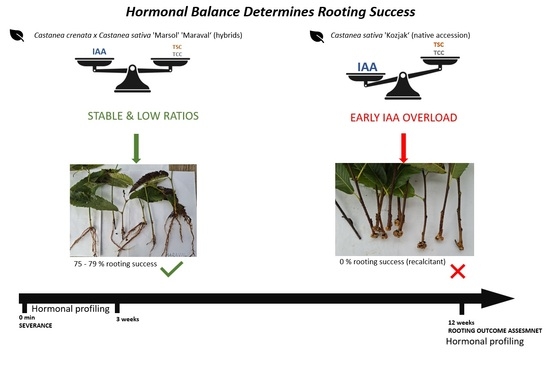
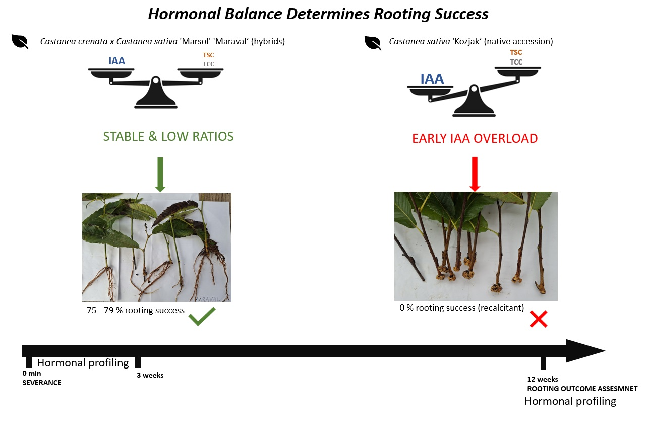
Adventitious root (AR) formation is essential for the vegetative propagation of woody plants, yet recalcitrant species within Castanea spp. exhibit strong genotype-dependent differences in rooting ability. Strigolactones (SLs) and auxins are key phytohormones regulating AR development, but their dynamic interplay remains poorly understood. We analysed three Castanea genotypes with contrasting rooting capacities—two hybrids, Castanea crenata × Castanea sativa (‘Maraval’ and ‘Marsol’), and one native accession, Castanea sativa (‘Kozjak’)—to investigate temporal changes in auxin and SL content following severance. Cuttings were sampled at multiple time points from 0 min to 3 weeks prior to visible root formation, and again at 12 weeks, when rooting outcomes were assessed. Individual tissues were analysed for active auxins, conjugated and oxidised auxin metabolites, and strigolactones. From these data, total auxin content (TAC), total auxin-related compounds (TCC), total strigolactone content (TSC), and the ratios TAC:TCC and TAC:TSC were calculated. Rooting success differed markedly among genotypes: ‘Maraval’ and ‘Marsol’ rooted successfully (75–79%), whereas ‘Kozjak’ failed to produce roots and instead formed extensive callus. Hormonal profiling indicated that rooting competence depends not only on auxin levels but also on the dynamic balance between active auxins, inactive metabolites, and strigolactones. ‘Kozjak’ showed an early predominance of free IAA and elevated SL levels, correlating with inhibited AR initiation, whereas the hybrid genotypes maintained more stable hormonal ratios, which supported successful rooting. These results highlight the importance of coordinated hormonal balance rather than absolute hormone concentrations in regulating AR formation. Our study provides new insights into hormone dynamics underlying rooting recalcitrance in chestnut and suggests potential strategies to improve clonal propagation of woody species.

Graphical Abstract

1. Introduction

Strigolactones (SLs) are a group of terpenoid compounds with important developmental and physiological roles in plants [1,2]. They are novel carotenoid-derived molecules that are produced in very low amounts and are biologically active at pico- to nanomolar concentrations [3]. In plants, SLs influence numerous developmental processes, including adventitious root (AR) formation [4,5]. AR formation from stem cuttings is essential for efficient and successful clonal propagation of woody plants [5]. The regulation of AR formation by SLs can be either positive or negative, depending largely on the plant species and experimental conditions [6]. The species-specific effects of SLs on adventitious rooting raise important questions that remain to be clarified. Previous studies have demonstrated that the plant hormone strigolactone suppresses AR formation in arabidopsis (Arabidopsis thaliana), pea (Pisum sativum), and maize (Zea mays) [7,8]. Consistently, in tomato (Solanum lycopersicum) and poplar (Populus alba), the knockdown of genes involved in SL biosynthesis (CCD8) or metabolism (MAX4) resulted in increased AR formation, further supporting the inhibitory role of SLs in adventitious rooting [5]. In contrast, Sun et al. [9] reported that rice mutants defective in strigolactone pathways developed significantly fewer ARs during the seedling stage and exhibited a reduced number of ARs. Application of the synthetic SL analog GR24 restored AR formation in the SL biosynthesis mutant (d10), whereas no such recovery was observed in the SL signaling mutant (d3), indicating that functional SL signaling is required for this response [9].
Several plant hormones have been reported to play critical roles in the regulation of AR formation. Among them, the auxin gradient is considered the primary regulatory signal, acting as a master controller of AR initiation and development [5,10,11]. Indole-3-acetic acid (IAA) is the predominant biologically active form of auxin in plants and plays a central role in the induction of both ARs and lateral roots [6]. Free IAA levels are regulated not only by de novo biosynthesis but also by irreversible oxidative catabolism and the reversible conjugation of IAA with sugars and amino acids [12]. To date, only conjugates of oxIAA with aspartate and glutamate have been identified in plants. In addition to conjugation, IAA can be inactivated through oxidation into 2-oxindole-3-acetic acid (oxIAA). Thus far, only oxIAA-Asp and oxIAA-Glu have been identified as endogenous metabolites within the genus Arabidopsis [12]. Accumulating evidence indicates that SLs interact synergistically with other plant hormones, particularly auxin, to regulate root development [13,14]. In addition, auxin has been shown to influence SL biosynthesis, thereby contributing to the modulation of apical dominance [15]. Overall, the interaction between SLs and auxin in the regulation of AR formation appears to be complex and is not yet fully understood [6]. Previous research [13] supports a positive and synergistic role of SLs and auxin in AR formation in melon, mediated through coordinated regulation of hormone biosynthesis, signaling pathways, and endogenous hormone levels. Rasmussen et al. [7] report that auxin and SL pathways are largely independent. Auxin can promote AR formation even in strigolactone-deficient mutants (max3), indicating that SLs are not required for auxin’s action. Conversely, SLs can suppress AR formation even when auxin levels are high, showing that their inhibitory effect is not simply due to reduced auxin synthesis. Evidence also suggests a potential cross-talk via auxin transport, as SLs may inhibit AR formation by decreasing basipetal auxin flow to the rooting zone [7,16].
Previous studies have largely focused on auxin-related processes or single time-point measurements. In contrast, this study provides a time-resolved, multi-hormonal analysis of both auxin metabolism and strigolactone dynamics across genotypes with contrasting rooting capacities. Despite extensive studies on auxin-mediated adventitious rooting, the temporal interaction between auxins and strigolactones in Castanea spp. remains poorly understood, particularly in genotypes with contrasting rooting abilities. Understanding these hormonal dynamics is crucial for improving clonal propagation efficiency in this economically important genus.

2. Material and Methods

2.1. Plant Material and Preparation of the Cuttings

The study was conducted on the two commercial cultivars ‘Marsol’ and ‘Maraval’ (Castanea crenata Siebold & Zucc. × Castanea sativa Mill.) and the native cultivar ‘Kozjak’ (Castanea sativa Mill.), which were collected in June from an Experimental Field for Nut Crops in Maribor, Slovenia (46°34′01″ N; 15°37′51″ E; 280 m a.s.l.). Cuttings were collected from 29-year-old stock plants that are regularly maintained and subjected to annual heavy pruning, which promotes the maintenance of physiological juvenility of the donor material. The shoots of the current year of each genotype were used as plant material for the experiment. The cuttings were shortened to a length of 20 cm, and the lower three leaves were removed.

2.2. Growing Conditions

The experiment was conducted in a greenhouse at the Biotechnical Faculty, University of Ljubljana (46°3′4″ N, 14°30′18″ E) with an automated high-pressure fogging system (Plantog, Fischamend, Austria) maintaining 98–100% relative humidity to minimize transpiration and prevent wilting. The propagation substrate was a 1:1 (v/v) peat–sand mixture supplemented with Osmocote Exact fertilizer (16% N, 9% P, 12% K, 2% MgO, trace elements; ICL Group Ltd., Tel Aviv, Israel).
Fogging ran daily from 08:00 to 20:00, with 40 s on/1 min off cycles on sunny days and 40 s on/3 min off on overcast or rainy days; it was inactive at night. Lighting conditions reflected the natural photoperiod of Ljubljana from June to September, corresponding to the seasonal daylight pattern of the Northern Hemisphere.

2.3. Measurement of Climate Parameters

Substrate temperature, leaf lamina temperature, and air temperature were continuously recorded at hourly intervals using T-Soil and Lat-B3 sensors (Ecomatik, Dachau, Germany), positioned at leaf level and 3 cm above the leaf lamina of the upper leaves on the cuttings. Continuous monitoring was performed to ensure accurate characterization of microclimatic conditions during the rooting experiment.

3. Experimental Design

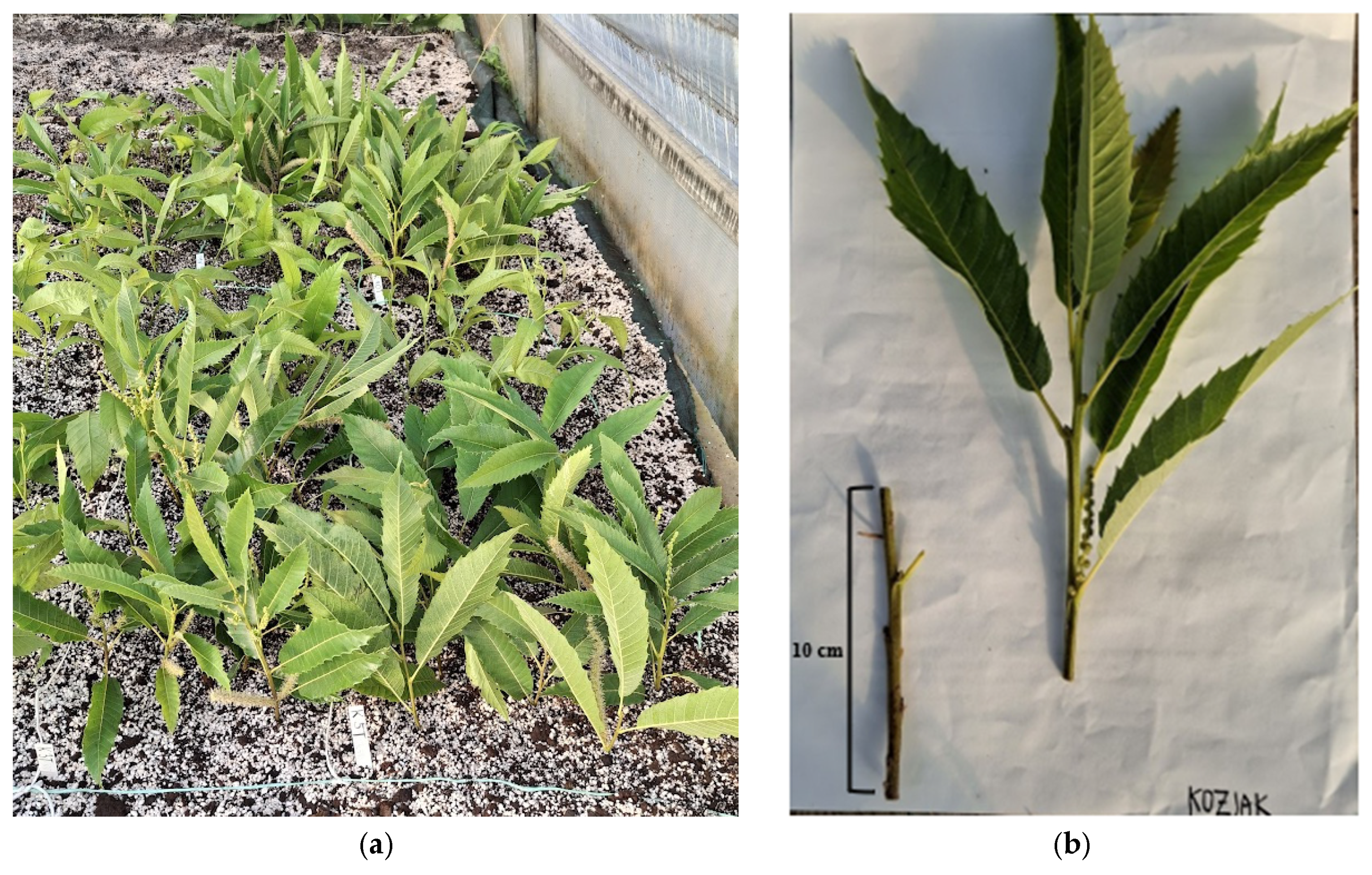
Pre-prepared cuttings were placed in the propagation bed within the fogging system according to randomly selected, marked plots (Figure 1a). To investigate temporal changes in endogenous hormone content following severance, samples were collected at eight time points spanning the period before visible root formation: immediately after severance (0 min) and after 30 min, 1 h, 2 h, 4 h, 24 h, 1 week, and 3 weeks post-severance. At each sampling time, the basal 10 cm of each cutting was excised (Figure 1b) and used for hormone analysis.
The experiment followed a two-factorial design with genotype (‘Maraval’, ‘Marsol’, ‘Kozjak’) and time after severance as fixed factors. Cuttings were randomly assigned to sampling time points and experimental units to ensure unbiased representation across treatments. For each treatment, four biological replicates were prepared, each consisting of three leafy cuttings. Cuttings belonging to the same replicate were immediately placed into labelled bags, frozen in liquid nitrogen, lyophilized, and ground to a fine powder prior to hormone extraction. The samples were stored in vacuum-sealed bags to prevent moisture absorption and potential hormone degradation.
In addition to the samples used for hormonal profiling before visible root formation, three treatment groups were established to evaluate the rooting performance of each genotype. Each group included four replicates of six cuttings maintained under standard rooting conditions for 12 weeks, after which rooting success and quality were assessed. Phytohormone content in different tissues was also analyzed based on rooting outcome: rooted cuttings, cuttings with callus, and unrooted cuttings.

3.1. Phytohormone Extraction and Analyses

We quantified a broad range of auxin-related compounds, including indole-3-acetic acid (IAA), indole-3-butyric acid (IBA), 4-chloro-indole-3-acetic acid (4-Cl-IAA), indole-3-acetyl-aspartic acid (IAA-Asp), indole-3-acetyl-glutamic acid (IAA-Glu), oxindole-3-acetic acid (ox-IAA), and 2-oxindole-3-acetyl-aspartic acid (ox-IAA-Asp), as well as strigolactones, such as strigol, strigon, 5-deoxystrigol, and orobanchol. In addition, three composite hormone pools were calculated. Total active auxins (TAC) comprised the sum of IAA, IBA, and 4-Cl-IAA. Auxin-related inactive forms included the conjugated and oxidized auxins IAA-Asp, IAA-Glu, ox-IAA, and ox-IAA-Asp (TCC). Total strigolactone content (TSC) was defined as the combined levels of strigol, strigon, orobanchol, and 5-deoxystrigol.
Hormone extraction was initiated four days after lyophilization. Auxin-related compounds were extracted following the protocol of Kunc et al. (2026) [10], using the same equipment as reported therein. Briefly, 100 mg of powdered tissue was extracted with 1 mL of isopropanol by shaking on ice for 30 min using a Unimax 1010 orbital shaker (Heidolph Instruments GmbH & Co. KG, Schwabach, Germany). This was followed by centrifugation at 12,000× g for 10 min at 4 °C using an Eppendorf 5810 R centrifuge (Eppendorf SE, Hamburg, Germany). The supernatant was transferred to a clean tube, evaporated to dryness using a SpeedVac ((Eppendorf SE, Hamburg, Germany) and reconstituted in 200 µL of methanol. After vortexing and a second centrifugation under the same conditions, the final supernatant was transferred to autosampler vials and stored at −80 °C until analysis. Strigolactones were extracted from approximately 250 mg of plant material using 1.5 mL of cold methanol:acetonitrile (1:1, v/v). Samples were vortexed and incubated for 1 h at 4 °C, followed by centrifugation at 1400 rpm for 10 min at 4 °C. The supernatant was transferred to a new tube, and liquid–liquid partitioning was performed by adding 1.5 mL of ethyl acetate:water (1:1, v/v). After mixing and centrifugation under the same conditions, the upper ethyl acetate phase was collected. This extraction step was repeated twice, and the ethyl acetate fractions were combined. The combined extract was dried in a SpeedVac at a temperature below 30 °C and reconstituted in 200 µL of methanol:water (30:70, v/v) and transferred to autosampler vials and stored at −80 °C until analysis.
Phytohormones were quantified using an ultra-high-performance liquid chromatography (UHPLC) system (Dionex UltiMate 3000, Thermo Scientific, Waltham, MA, USA) coupled to a tandem mass spectrometer (LTQ XL, Thermo Scientific, Waltham, MA, USA), following the procedure of Kunc et al. (2024) [11]. Detection was performed in selected reaction monitoring (SRM) mode, targeting specific precursor-to-product ion transitions for each phytohormone and the corresponding external standards (Table 1). Contents were determined from calibration curves of external standards and corrected for recovery efficiency during extraction. (See Supplementary Material Table S1).

3.2. Rooting Assessment

Cuttings were qualitatively classified into four categories: rooted, non-rooted, decayed/rotted, and those exhibiting callus formation. For rooted cuttings, the number of main roots was counted, and their position of origin (basal or acrobasal) was recorded; rooting success was defined by the presence or absence of roots.
For phytohormone analysis of rooting outcome, tissues were sampled according to rooting outcome for each genotype: the root system in rooted cuttings (labeled as roots), callus tissue in callus-forming cuttings (labeled as callus), and the basal 10 cm segment in unrooted cuttings (labeled as none), as in the first part of the experiment prior to root formation.

3.3. Chemicals

For the extraction and subsequent analytical procedures, the following reagents were utilized: isopropanol and methanol (Merck KGaA, Darmstadt, Germany), formic acid (Kemika d.d., Zagreb, Croatia), acetonitrile and ethyl acetate (Sigma-Aldrich Chemie GmbH, Steinheim, Germany). The following standards were employed for the identification and quantification of phytohormones: indole-3-acetic acid and indole-3-butyric acid (Sigma-Aldrich Chemie GmbH, Steinheim, Germany), indole-3-acetyl-L-aspartic acid (Biosynth Ltd., Compton, UK), 4-chloroindole-3-acetic acid (Biosynth Ltd., Staad, Switzerland), indole-3-acetyl-glutamic acid, oxindole-3-acetic acid, and 2-oxindole-3-acetyl-aspartic acid (Biosynth Ltd., Compton, UK), strigol (CymitQuímica S.L., Barcelona, Spain), 5-deoxystrigol (Wuhan Chemfaces Biochemical Co., Ltd., Wuhan, China), strigol (Chiralix B.V., Nijmegen, The Netherlands), and orobanchol (Chiralix B.V., Nijmegen, The Netherlands).

3.4. Statistical Analyses

All statistical analyses were performed in R (R Foundation for Statistical Computing, Vienna, Austria) using the R Commander graphical user interface (version 4.3.0). Differences among multiple groups were evaluated using one-way analysis of variance (ANOVA). When significant effects were detected, Tukey’s multiple range test was applied for post-hoc pairwise comparisons. Results are presented as means ± standard error (SE), and statistical significance was determined at the 95% confidence level (p < 0.05).
Data processing, visualization, and all statistical analyses were performed in R (R Foundation for Statistical Computing, Vienna, Austria). These tasks were conducted using the R Commander interface (version 4.3.0). Hormone concentration values were converted to numeric format and mean values were calculated for each genotype and time point using base R functions. Prior to visualization, the data were standardized by column using Z-score transformation to allow comparison of relative differences among hormones. Heatmaps were generated using the pheatmap package with row and column clustering. A custom warm color palette was created using the colorRampPalette function from the grDevices package. Differences in rooting pattern distribution (acrobasal vs. basal) between genotypes were assessed using Pearson’s chi-squared test. Differences in the number of main roots between genotypes were evaluated using an independent two-sample t-test.

4. Results

4.1. Measurement of Climate Parameters

Air, leaf, and soil temperatures exhibited pronounced diurnal patterns across all months. Minimum temperatures were consistently recorded in the early morning hours (05:00–07:00), reaching approximately 17.0–22.0 °C for air, 17.5–22.6 °C for leaf, and 19.0–25.9 °C for soil temperatures. Maximum air and leaf temperatures occurred around midday to early afternoon (12:00–15:00), with peak values of 42.3 °C (air) and 42.5 °C (leaf) recorded in June. In contrast, soil temperature showed a delayed response, with maximum values observed in the late afternoon (16:00–17:00), reaching up to 39.5 °C in June. Overall, the highest temperatures for all three variables were observed in June, followed by a gradual decline toward autumn (See Supplementary Material Figures S1–S3).

4.2. Phytohormone Content Prior to Visible Root Formation

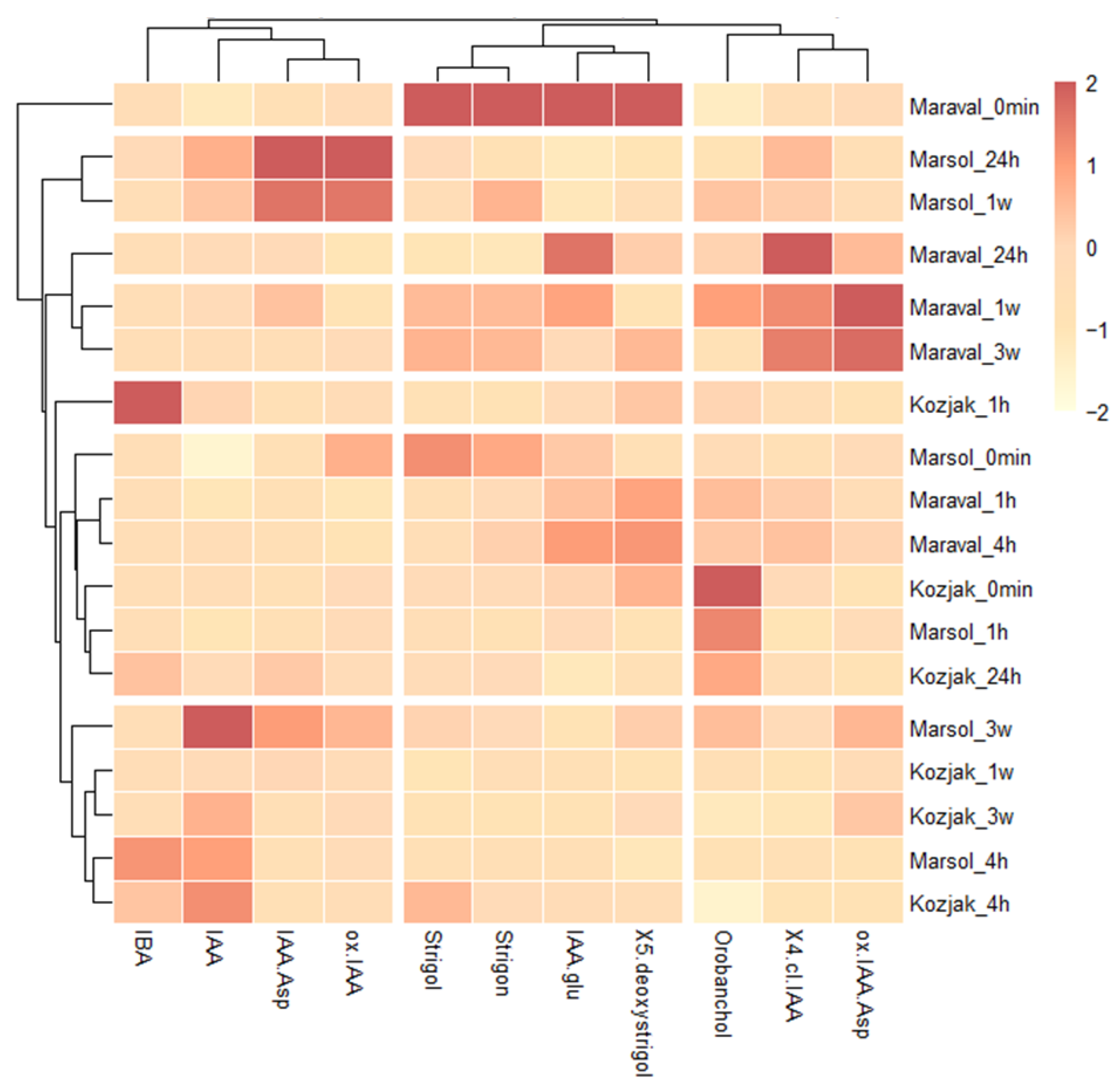
The heatmap (Z-score scaled per hormone) highlights relative increases and decreases across genotypes and time points (Figure 2). The colour scale represents the Z-score (standard deviations from the mean), where red indicates higher relative concentrations and yellow/white indicates lower concentrations. The dendrograms tell us how the variables are related: the vertical dendrogram groups the samples, and the horizontal dendrogram groups the hormones.
Early phase (0 min) of the ‘Maraval’ cultivar starts with a high concentration of strigol and strigon, which then drops significantly in subsequent Maraval samples. As time progresses, Maraval shows a distinct shift toward higher concentrations of 4-Cl-IAA and ox-IAA-Asp. Marsol shows a very specific metabolic window at 24 h and 1 week. During this time, there is a significant accumulation of IAA-Asp and ox-IAA. The horizontal dendrogram (top) separates the hormones into clusters. There is a clear distinction in how the plants use different strigolactones. Strigol and Strigon are primarily dominated by the early stage of the ‘Maraval’ genotype, while Orobanchol shows a completely different pattern, peaking in ‘Kozjak’ (0 min and 24 h) and ‘Marsol’ (1 h). The highest IBA signal was observed in the ‘Kozjak’ genotype at 1 h, followed by ‘Marsol’ and ‘Kozjak’ at 4 h. A strong IAA signal was detected in ‘Marsol’ at 4 h and at later time points, reaching its peak at 3 weeks. In the ‘Kozjak’ genotype, the IAA signal was prominent at 4 h and 3 weeks (Figure 2).
During the early stages (0 min–4 h), ‘Kozjak’ displays the highest IAA:TCC ratios (1.55–2.47) (See supplementary Material, Table S4), which decline at later time points (24 h–3 weeks). In contrast, the IAA:TSC ratio shows the opposite trend: it is lower at early stages (0 min–24 h; 0.31–0.44) and increases at later stages (1.04–1.14). In ‘Marsol’, the IAA:TCC ratio reaches its peak at 4 h and then decreases. A similar trend is observed for the IAA:TSC ratio: it increases up to 4 h, declines thereafter, but remains relatively high at 24 h. After one week, it decreases further and then rises again at 3 weeks. Throughout the entire period (0 min–3 weeks), both the IAA:TCC and IAA:TSC ratios remained relatively low in the ‘Maraval’ genotype (Figure 2, Tables S2 and S4).

4.3. Rooting Assessment

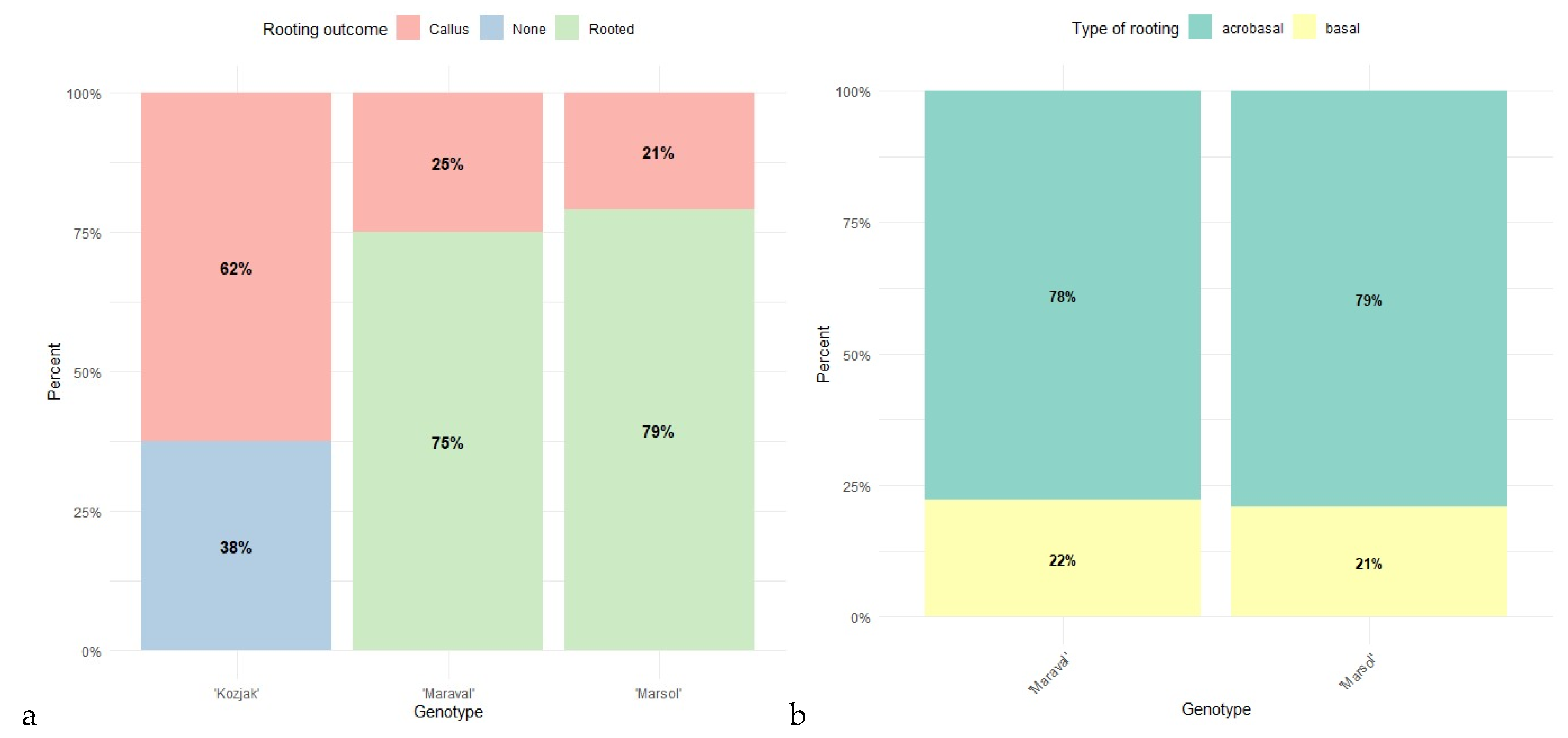
The frequency of rooting responses differed significantly among genotypes (Pearson’s χ2 test: χ2 = 43.54, df = 4, p < 0.001). ‘Kozjak’ cuttings predominantly formed callus (62.5%) or showed no visible response (37.5%), while none developed roots. In contrast, rooting was the dominant response in ‘Maraval’ (75.0%) and ‘Marsol’ (79.2%), with no cuttings remaining without visible response. The largest contributions to the chi-square statistic originated from deviations in rooting and non-response frequencies in ‘Kozjak’, indicating a markedly different regeneration pattern compared with the other genotypes (Figure 3a). The distribution of acrobasal and basal rooting did not differ between genotypes (Pearson’s χ2 test: χ2 = 0.007, df = 1, p = 0.93).
In both ‘Maraval’ and ‘Marsol’, acrobasal rooting was more frequent than basal rooting, accounting for 48.3–51.7% and 50.0% of the responses, respectively. The observed frequencies closely matched the expected counts, indicating a comparable rooting pattern between the two genotypes (Figure 3b).
The mean number of main roots did not differ significantly between genotypes (two-sample t-test: t = 0.56, df = 35, p = 0.58). ‘Maraval’ cuttings developed on average 4.44 ± 0.66 main roots (mean ± SE, n = 18), while ‘Marsol’ developed 3.95 ± 0.60 main roots (n = 19). The 95% confidence interval for the difference in means ranged from −1.31 to 2.31, indicating substantial overlap between genotypes (Table 2).

4.4. Phytohormone Content in Cuttings with Different Rooting Outcomes

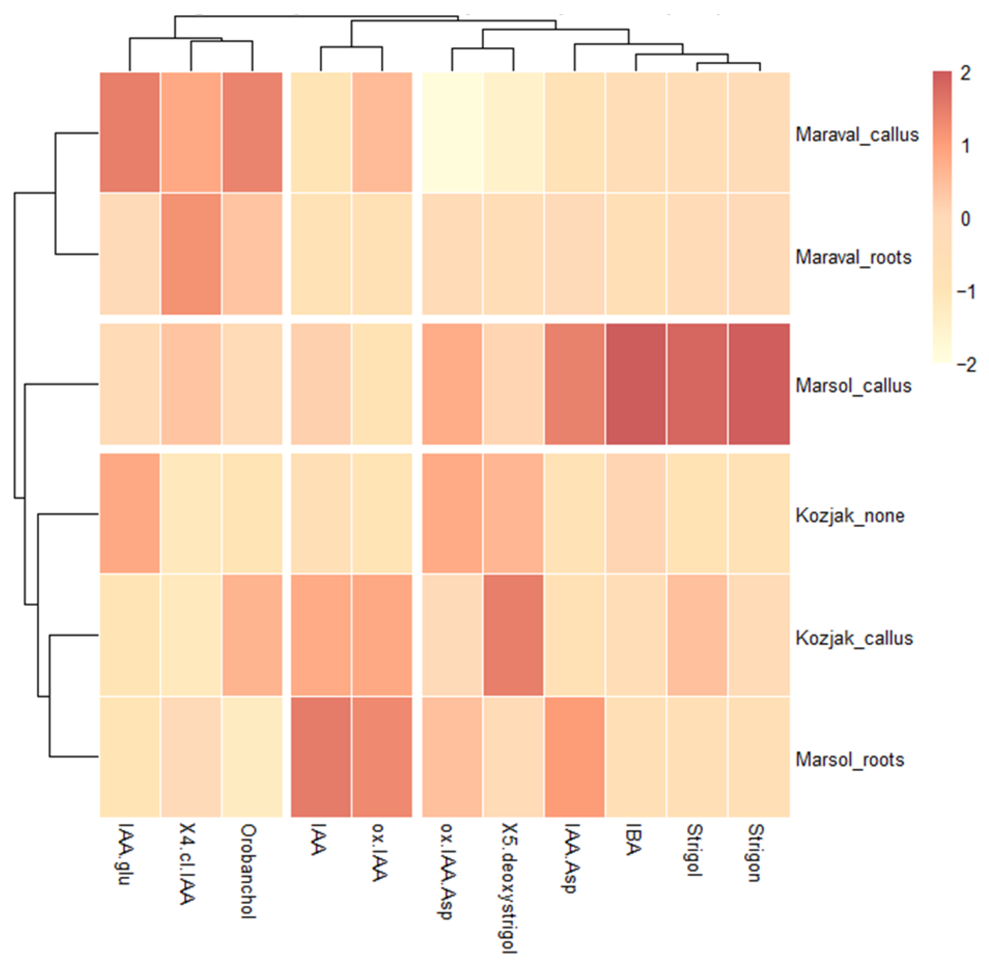
Hierarchical clustering by tissue type (Figure 4) revealed that the ‘Marsol’ callus represents a unique metabolic state characterized by a robust accumulation of auxin IBA, conjugated auxin IAA-Asp and strigolactones (strigol and strigon). In contrast to the callus, the roots show a sharp increase in IAA and ox-IAA. The ‘Maraval’ genotype maintains a highly consistent hormonal profile across both callus and root tissues. Both ‘Maraval’ tissue types cluster together at the top, dominated by IAA.glu, 4-Cl-IAA, and orobanchol. The ‘Kozjak’ cuttings in which neither root nor callus formation was observed (labelled as ‘Kozjak’_none) are characterized by a lack of significant hormonal activity; the highest signal was observed in IAA-Glu, ox-IAA-Asp and 5-deoxystrigol. The ‘Kozjak’ callus is characterized by a specific accumulation of 5-deoxystrigol, a precursor in the strigolactone biosynthetic pathway. The values of orobanchol, IAA, and ox-IAA show a moderate positive deviation from the mean (Figure 4).
Significant differences in IAA content were observed between the roots of ‘Maraval’ (21.08 ± 2.95 ng g−1 DW) and ‘Marsol’ (122.81 ± 35.47 ng g−1 DW). Significant differences were also detected in 5-deoxystrigol content between the callus of ‘Kozjak’ (78.15 ± 16.91 ng g−1 DW) and ‘Maraval’ (0.00 ± 0.00 ng g−1 DW). No significant differences were observed in the content of other phytohormones (Table S3).
In ‘Marsol’, pronounced differences between callus and roots were observed. Root tissue had the highest IAA content among all samples (122.81 ng g−1 DW) and the highest IAA:TCC (0.73) and IAA:TSC (1.10) ratios. In contrast, the ‘Marsol’ callus showed lower IAA:TSC (0.32) and IAA:TCC (0.35) ratios. The ‘Kozjak’ callus exhibited higher IAA:TCC (0.65) and IAA:TSC (0.50) ratios compared to non-callus tissues (0.17 and 0.24, respectively). In ‘Maraval’, the IAA:TCC ratio was higher in callus tissue (0.40) than in roots (0.16), whereas the IAA:TSC ratios in the two tissues were very similar (0.16 and 0.17) (Figure 4, Tables S3 and S5).

5. Discussion

Adventitious rooting is the formation of roots from non-root tissues in response to stress, occurring in three stages: induction, initiation, and expression. It is controlled by a regulatory network in which auxin is central, while jasmonic acid, plant age, and environmental or technical factors influence success. Wounding triggers hormonal changes and auxin redistribution, creating a local maximum that reprograms cells [17,18]. Several plant hormones play important roles in regulating AR formation, with the auxin gradient acting as the primary master regulator [5]. A transient auxin peak drives the transition from induction to initiation, followed by further changes leading to root development and emergence [17,18]. The ability of plants to form adventitious roots on the basal part of stem cuttings varies widely among different species and cultivars [19,20,21]. Chestnut is a woody species known for its difficulty in both sexual and vegetative propagation [17,22,23,24]. Numerous studies [17,19,25] have reported the inherent challenges of inducing root formation in C. sativa under both in vivo and in vitro conditions. Hybrids, such as C. sativa × C. crenata, are increasingly favoured due to their superior rooting ability [19]. Our results demonstrated successful rooting in the hybrids ′Maraval′ and ‘Marsol’, which is consistent with previous findings [19]. In the present experiment, no differences were observed between the cultivars ′Maraval′ and ‘Marsol’, although the rooting percentage was lower (75.0% and 79.2%) than that reported in earlier studies (100.0% and 90.48%) [19]. In this experiment, ‘Kozjak’ failed to develop AR altogether and formed only a thick callus, which is commonly observed in species that are more difficult to root [26]. These results indicate that year-to-year variation in rooting success can be considerable. The failure of ‘Kozjak’ to root may also be associated with the timing of AR development assessment in this cultivar. In the first experiment, rooting was evaluated 120 days after severance [19], whereas in the present study, it was assessed after 12 weeks (84 days). Although adventitious roots typically begin to develop 3–5 weeks after the beginning of propagation [27], in ‘Kozjak’, the formation of a thick callus may delay or hinder root emergence through the callus tissue. Consequently, the 12-week evaluation period may not have been sufficient to capture delayed rooting in this genotype, potentially leading to an underestimation of its rooting capacity. The occurrence of callus formation is consistent with our previous results, where callus formation was significantly lower in the hybrids compared to C. sativa [19]. The number of main roots and the pattern of basal/acrobasal rooting also did not differ between the two hybrid cultivars in the present experiment, which is in agreement with earlier findings [19]. In some plant species, such as Eucalyptus grandis L. and Pisum sativum L., lower endogenous IAA levels have been reported in difficult-to-root mature cuttings compared with easy-to-root juvenile ones [18,28,29]. In our previous study [19], in which we investigated cuttings of different genotypes of Castanea, we similarly confirmed genotype-dependent differences. However, in the present experiment, the highest endogenous free IAA levels at the earliest sampling times after severance (0 min, 1 h) were detected in the genotype ‘Kozjak’, which did not form any roots. Later, in the induction phase (4–24 h), no significant differences in free IAA levels were observed among the genotypes.
We aimed to determine whether the hormonal profile underlies the poor rooting performance of the genotype ‘Kozjak’. Excessively high IAA content may exert an inhibitory effect on AR formation [8], which could partly explain the poor rooting results observed in this genotype. The role of SLs in AR formation remains insufficiently understood: some studies report a positive effect, whereas others suggest a negative regulatory role [6]. In our case, high SL levels were detected in the genotype ‘Maraval’, which generally roots well compared to recalcitrant Castanea species. The observed lack of rooting in ‘Kozjak’ may be related to interannual variation in rooting success and differences in evaluation time points compared to previous studies. However, these explanations remain speculative, as they were not directly investigated within the scope of the present study. Callus formation was observed in all genotypes; however, it was substantially more pronounced in ‘Kozjak’ than in ′Maraval′ and ‘Marsol’. Despite the high IAA levels in ‘Kozjak’, which would typically be expected to promote AR formation, the extensive callus tissue may represent a physical barrier preventing root emergence or may delay root protrusion. In ‘Marsol’ and ′Maraval′, roots developed predominantly acrobasally, and not exclusively at the base of the cutting. This pattern allows roots to form even in the presence of callus tissue at the base of the cutting because the callus is less extensive and roots can more readily penetrate it. Previous results indicate [19] that in ‘Kozjak’, root development is restricted to the basal region, which may further explain its poorer rooting performance, as basal roots may take longer to penetrate the callus or may fail to do so altogether.
The relationship between SLs and auxin during AR formation appears to be complex [6]. Furthermore, differences between easy-to-root and difficult-to-root genotypes have been attributed to variations in the content of inactive auxin conjugates [30]. During the induction phase, the IAA:TCC ratio suggests genotype-specific differences in auxin homeostasis. The predominance of free IAA in ‘Kozjak’ at 0 min and 1 h may indicate a reduced capacity for auxin conjugation or oxidation immediately after severance, potentially leading to transient auxin overaccumulation. In contrast, the shift toward conjugated and oxidized forms in ′Maraval′ and ‘Marsol’ at the same time points could reflect a tighter regulation of active auxin levels, enabling a more controlled hormonal response during the early stages of AR induction. A similar complexity is evident in the IAA:TSC ratio. The initial predominance of SLs in all three genotypes (at 0 min and 1 h) may point to a general role of SLs in the early wound or stress response. However, the divergence observed after 4 h—where the IAA:TCC ratio favours free IAA in ‘Kozjak’ and ‘Marsol’, but not in ‘Maraval’—raises questions about how distinct hormonal dynamics can lead to comparable rooting performance, as observed for ‘Maraval’ and ‘Marsol’. This suggests that successful AR formation may not depend on a single hormonal pattern, but rather on the coordination and timing of multiple regulatory pathways. The contrasting IAA:TSC ratios between ‘Kozjak’ and ‘Marsol’ at 4 h further may support this view. In ‘Kozjak’, the continued predominance of SLs relative to free IAA may contribute to the inhibition of root initiation. These observations imply that the balance between auxin and SLs, rather than their absolute levels, may be critical in determining rooting competence. Interestingly, ‘Maraval’ displays a comparatively stable IAA:TCC and IAA:TSC ratio over time. After 12 weeks, the contrast between callus and root tissue within the same genotype indicates that the developmental outcome may depend on whether active auxin becomes sufficiently dominant over conjugated forms and SLs at the critical stage of root initiation. These results reinforce the concept that the relative balance between active auxin, its inactive metabolites, and SLs is a key determinant of developmental fate in chestnut cuttings. High IAA levels can be associated with either rooting (‘Marsol’) or excessive callusing without root formation (‘Kozjak’), depending on the accompanying hormonal context. Therefore, it appears that coordinated hormonal ratios—rather than absolute content—define whether tissues proceed toward organized adventitious root formation or remain in an undifferentiated callus state.

6. Conclusions

In summary, our results confirm pronounced genotype-dependent differences in adventitious root formation in Castanea, evident not only in rooting performance but also in distinct hormonal dynamics during the early phases after severance. While the hybrids ‘Maraval’ and ‘Marsol’ exhibited comparable and relatively high rooting capacity, the native genotype ‘Kozjak’ failed to form adventitious roots and instead developed extensive callus tissue. The hormonal profiling suggests that rooting competence is determined not solely by the absolute content of free IAA, but by the dynamic balance between active auxins, their inactive conjugated or oxidised forms, and strigolactones. In ‘Kozjak’, the early predominance of free IAA and distinct IAA:TCC and IAA:TSC ratios may reflect altered auxin homeostasis, potentially leading to hormonal imbalance and impaired root initiation. In contrast, the more stable hormonal ratios observed in ‘Maraval’ and the coordinated shifts in ‘Marsol’ indicate that successful adventitious root formation may depend on tightly regulated hormonal crosstalk rather than elevated auxin levels. For future studies, the inclusion of jasmonic acid—recognised as one of the earliest signalling molecules triggered by wounding—would help clarify the initial stress response following severance and its interaction with auxin and strigolactone pathways. Such an integrative approach, combining hormonal profiling with molecular analyses, could substantially improve our understanding of the regulatory framework underlying adventitious root formation in chestnut. In addition, integrating gene expression analyses of key regulators involved in auxin biosynthesis, transport, conjugation, and signalling, as well as strigolactone-related pathways, would provide deeper mechanistic insight into genotype-specific differences.

Supplementary Materials

The following supporting information can be downloaded at: https://www.mdpi.com/article/10.3390/horticulturae12050575/s1, Table S1 Percentage of recovery during the extraction of phytohormones. Table S2 Contents of analyzed phytohormones over time, before root emergence (0 min—3 weeks) (ng g−1 DW). Table S3 Contents of analyzed phytohormones in different tissues after 12 weeks (ng g−1 DW). Table S4 Ratios between free IAA and total auxin related compunds content (TCC) and total strigolactones content (TSC) before root emergence (0min- 3 weeks). Table S5 Ratios between IAA and total auxin related compounds content (TCC) and total strigolactones content (TSC) in different tissues (after 12 weeks). Figure S1 Average hourly air temperature (°C) measured 3 cm above the leaf lamina during the experimental period. Figure S2 Average hourly leaf lamina temperature (°C) measured during the experimental period. Figure S3 Average hourly soil temperature (°C) measured during the experimental period.

Author Contributions

P.K. and G.O.; Data curation, P.K. and G.O.; Formal analysis, P.K. and A.M.; Funding acquisition, G.O. and R.V.; Investigation, P.K.; Methodology, P.K. and M.C.G.; Project administration, G.O. and R.V.; Resources, G.O. and R.V.; Visualization, P.K.; Writing—original draft, P.K.; Writing—review and editing, G.O., A.M., M.C.G. and R.V. All authors have read and agreed to the published version of the manuscript.

Funding

This work is part of the program Horticulture P4-0013-0481 supported by the Slovenian Research and Innovation Agency (ARIS).

Data Availability Statement

The raw data supporting the conclusions of this article will be made available by the authors on request.

Acknowledgments

The authors acknowledge the financial support of the Slovenian Research Agency (ARRS) within the infrastructural centre IC RRG-AG (IO-0022-0481-001).

Conflicts of Interest

The authors declare that they have no known competing financial interests or personal relationships that could have appeared to influence the work reported in this paper.

References

  1. Koltai, H.; Dor, E.; Hershenhorn, J.; Joel, D.M.; Weininger, S.; Lekalla, S.; Shealtiel, H.; Bhattacharya, C.; Eliahu, E.; Resnick, N.; et al. Strigolactones’ effect on root growth and root-hair elongation may be mediated by auxin-efflux carriers. J. Plant Growth Regul. 2010, 29, 129–136. [Google Scholar] [CrossRef] [Scilit]
  2. Kapulnik, Y.; Delaux, P.M.; Resnick, N.; Mayzlish-Gati, E.; Wininger, S.; Bhattacharya, C.; Séjalon-Delmas, N.; Combier, J.P.; Bécard, G.; Belausov, E.; et al. Strigolactones affect lateral root formation and root-hair elongation in Arabidopsis. Planta 2011, 233, 209–216. [Google Scholar] [CrossRef] [Scilit] [PubMed]
  3. Ur Rehman, N.; Li, X.; Zeng, P.; Guo, S.; Jan, S.; Liu, Y.; Huang, Y.; Xie, Q. Harmony but not uniformity: Role of strigolactone in plants. Biomolecules 2021, 11, 1616. [Google Scholar] [CrossRef] [Scilit] [PubMed]
  4. Kapoor, R.T.; Alam, P.; Chen, Y.; Ahmad, P. Strigolactones in plants: From development to abiotic stress management. J. Plant Growth Regul. 2024, 43, 903–919. [Google Scholar] [CrossRef] [Scilit]
  5. Fan, X.; Li, Y.; Deng, C.H.; Wang, S.; Wang, Z.; Wang, Y.; Qiu, C.; Xu, X.; Han, Z.; Li, W. Strigolactone regulates adventitious root formation via the MdSMXL7-MdWRKY6-MdBRC1 signaling cascade in apple. Plant J. 2023, 113, 772–786. [Google Scholar] [CrossRef] [Scilit]
  6. Sun, H.; Tao, J.; Gu, P.; Xu, G.; Zhang, Y. The role of strigolactones in root development. Plant Signal. Behav. 2016, 11, e1110662. [Google Scholar] [CrossRef] [Scilit]
  7. Rasmussen, A.; Mason, M.G.; de Cuyper, C.; Brewer, P.B.; Herold, S.; Agusti, J.; Geelen, D.; Greb, T.; Goormachtig, S.; Beeckman, T.; et al. Strigolactones suppress adventitious rooting in Arabidopsis and pea. Plant Physiol. 2012, 158, 1976–1987. [Google Scholar] [CrossRef] [Scilit]
  8. Zhang, Q.; Gong, M.; Xu, X.; Li, H.; Deng, W. Roles of auxin in the growth, development, and stress tolerance of horticultural plants. Cells 2022, 11, 2761. [Google Scholar] [CrossRef] [Scilit]
  9. Sun, H.; Tao, J.; Hou, M.; Huang, S.; Chen, S.; Liang, Z.; Xie, T.; Wei, Y.; Xie, X.; Yoneyama, K.; et al. A strigolactone signal is required for adventitious root formation in rice. Ann. Bot. 2015, 115, 1155–1162. [Google Scholar] [CrossRef] [Scilit]
  10. Kunc, P.; Medic, A.; Osterc, G. Wound-induced dynamics of selected auxins and jasmonates suggest interhormonal crosstalk during the induction phase of adventitious root formation in Prunus subhirtella ‘Autumnalis’. Acta Physiol. Plant. 2026, 48, 4. [Google Scholar] [CrossRef] [Scilit]
  11. Kunc, P.; Medič, A.; Hudina, M.; Veberič, R.; Osterc, G. Physiological age of stock plants determines phytohormonal changes in leafy cuttings of Prunus subhirtella ‘Autumnalis’. J. Plant Growth Regul. 2024, 44, 721–730. [Google Scholar] [CrossRef] [Scilit]
  12. Hladík, P.; Petřík, I.; Sabelová, V.; Novák, O.; Strnad, M.; Pěnčík, A. Metabolic profiles of 2-oxindole-3-acetyl-amino acid conjugates differ in various plant species. Plants 2023, 12, 2551. [Google Scholar] [CrossRef] [Scilit]
  13. Li, J.; Fan, M.; Zhang, Q.; Lü, G.; Wu, X.; Gong, B.; Wang, Y.; Zhang, Y.; Gao, H. Transcriptome analysis reveals that auxin promotes strigolactone-induced adventitious root growth in the hypocotyl of melon seedlings. Front. Plant Sci. 2023, 14, 1192340. [Google Scholar] [CrossRef] [Scilit] [PubMed]
  14. Kapulnik, Y.; Resnick, N.; Mayzlish-Gati, E.; Kaplan, Y.; Wininger, S.; Hershenhorn, J.; Koltai, H. Strigolactones interact with ethylene and auxin in regulating root-hair elongation in Arabidopsis. J. Exp. Bot. 2011, 62, 2915–2924. [Google Scholar] [CrossRef] [Scilit] [PubMed]
  15. Ferguson, B.J.; Beveridge, C.A. Roles for auxin, cytokinin, and strigolactone in regulating shoot branching. Plant Physiol. 2009, 149, 1929–1944. [Google Scholar] [CrossRef] [Scilit] [PubMed]
  16. Liu, S.; Li, X.; Xu, L.; Zhang, G. Hormone Functions in Adventitious Root Formation during Cutting Propagation of Woody Plants. J. Plant Res. 2025, 138, 907–914. [Google Scholar] [CrossRef] [Scilit]
  17. Vielba, J.M.; Vidal, N.; Carmen San José, M.; Rico, S.; Sánchez, C. Recent Advances in Adventitious Root Formation in Chestnut. Plants 2020, 9, 1543. [Google Scholar] [CrossRef] [Scilit] [PubMed]
  18. Liu, P.; Zhang, S.; Wang, X.; Du, Y.; He, Q.; Zhang, Y.; Shen, L.; Hu, H.; Zhang, G.; Li, X. Adventitious Root Formation in Cuttings: Insights from Arabidopsis and Prospects for Woody Plants. Biomolecules 2025, 15, 1089. [Google Scholar] [CrossRef] [Scilit]
  19. Kunc, P.; Medic, A.; Osterc, G. Involvement of auxin, flavonoids and strigolactones in the different rooting ability of European chestnut (Castanea sativa) and hybrids (Castanea crenata × Castanea sativa). Plants 2024, 13, 2088. [Google Scholar] [CrossRef] [Scilit]
  20. Wang, X.; Li, Y.; Li, Z.; Gu, X.; Wang, Z.; Qin, X.; Li, Q. Investigating the mechanisms of adventitious root formation in semi-tender cuttings of Prunus mume: Phenotypic, phytohormone, and transcriptomic insights. Int. J. Mol. Sci. 2025, 26, 62416. [Google Scholar] [CrossRef] [Scilit]
  21. Maghradze, D.; Rehman, S.; Chutlashvili, A.; Kikilashvili, S.; Kikvadze, M.; Shamugia, A.; Charkviani, S.; McGovern, P.; Failla, O.; Gotsiridze, O.; et al. Differences in rooting ability between wild and cultivated Vitis vinifera. Oeno One 2025, 59, 9371. [Google Scholar] [CrossRef] [Scilit]
  22. Osterc, G.; Štefančič, M.; Solar, A.; Štampar, F. Potential involvement of flavonoids in the rooting response of chestnut hybrid (Castanea crenata × Castanea sativa) clones. Aust. J. Exp. Agric. 2007, 47, 96–102. [Google Scholar] [CrossRef] [Scilit]
  23. Song, G.; Chen, Q.; Callow, P.; Mandujano, M.; Han, X.; Cuenca, B.; Bonito, G.; Medina-Mora, C.; Fulbright, D.W.; Guyer, D.E. Efficient micropropagation of chestnut hybrids (Castanea spp.) using modified woody plant medium and zeatin riboside. Hortic. Plant J. 2021, 7, 174–180. [Google Scholar] [CrossRef] [Scilit]
  24. Lu, X.; Cuarto, M.; Liang, H. Histology of adventitious root formation and phytohormone analysis of American chestnut cuttings. J. Environ. Hortic. 2023, 41, 80–86. [Google Scholar] [CrossRef] [Scilit]
  25. Feijó, J.A.; Salomé, M.S.; Pais, M.S.S. Rejuvenation of adult specimens of Castanea sativa Mill. through in vitro micropropagation. In Plant Aging: Basic and Applied Approaches; Rodríguez, R., Tamés, R.S., Durzan, D.J., Eds.; Plenum Press: New York, NY, USA, 1990; pp. 353–359. [Google Scholar]
  26. Kunc, P.; Medic, A.; Veberič, R.; Osterc, G. Does the physiological age of stock plant material affect the uptake of indole-3-butyric acid (IBA) in leafy cuttings of Prunus subhirtella ‘Autumnalis’? Horticulturae 2024, 10, 296. [Google Scholar] [CrossRef] [Scilit]
  27. St Hilaire, R.; Fierro Berwart, C.A.; Pérez-Muñoz, C.A. Adventitious root formation and development in cuttings of Mussaenda erythrophylla L. Schum. & Thonn. HortScience 1996, 31, 1023–1025. [Google Scholar] [CrossRef] [Scilit]
  28. Roth, O.; Yechezkel, S.; Serero, O.; Eliyahu, A.; Vints, I.; Tzeela, P.; Carignano, A.; Janacek, D.P.; Peters, V.; Kessel, A.; et al. Slow release of a synthetic auxin induces formation of adventitious roots in recalcitrant woody plants. Nat. Biotechnol. 2023, 42, 1705–1716. [Google Scholar] [CrossRef] [Scilit]
  29. da Costa, C.T.; de Almeida, M.R.; Ruedell, C.M.; Schwambach, J.; Maraschin, F.S.; Fett-Neto, A.G. When stress and development go hand in hand: Main hormonal controls of adventitious rooting in cuttings. Front. Plant Sci. 2013, 4, 133. [Google Scholar] [CrossRef] [Scilit]
  30. Gonin, M.; Bergougnoux, V.; Nguyen, T.D.; Gantet, P.; Champion, A. What makes adventitious roots? Plants 2019, 8, 240. [Google Scholar] [CrossRef] [Scilit]
Figure 1. Representative experimental setup and sampling procedure. (a) Cuttings placed in the propagation bed; (b) basal 10 cm of cuttings sampled for hormone analysis.
Horticulturae 12 00575 g001
Figure 2. Hierarchical clustered heatmap showing the concentrations of various plant hormones across different chestnut cultivars (‘Maraval’, ‘Marsol’, and ‘Kozjak’) at various time points prior to visible root formation (IAA: Indole-3-acetic acid; IBA: Indole-3-butyric acid, X4-.cl.IAA: 4-chloroindole—3-acetic acid; IAA.Asp: Indole-3-acetyl-aspartic acid; IAA.glu: Indole-3-acetyl-glutamic acid; ox.IAA: Oxindole-3-acetic acid; ox.IAA.Asp: Asp 2-Oxindole-3-acetyl-aspartic acid; X5.deoxystrigol: 5-deoxystrigol.
Horticulturae 12 00575 g002
Figure 3. Rooting assessment 12 weeks after severance. (a) Percentage (%) of cuttings that formed roots, developed callus, or showed no visible response (neither rooting nor callus formation, labelled as ‘none’); (b) percent of acrobasal and basal rooted cuttings.
Horticulturae 12 00575 g003
Figure 4. Phytohormone content in cuttings with different rooting outcomes (IAA: Indole-3-acetic acid; IBA: Indole-3-butyric acid, X4-cl.IAA: 4-chloroindole—3-acetic acid; IAA.Asp: Indole-3-acetyl-aspartic acid; IAA.glu: Indole-3-acetyl-glutamic acid; ox.IAA: Oxindole-3-acetic acid; ox.IAA.Asp: Asp 2-Oxindole-3-acetyl-aspartic acid; X5.deoxystrigol: 5-deoxystrigol.
Horticulturae 12 00575 g004
Table 1. SRM transitions for phytohormone quantifications.
PhytohormoneRetention TimePseudo Molecular Ions (m/z) Fragmentation Pattern
(min) [ M H ] [ M + H ] + (Relative Peak Intensity %)
IAA4.57174 130 (100)
IBA6.23202 158 (100)
4-Cl-IAA5.33208 164 (100)
IAA-Asp3.90289 132 (100), 174 (90),
IAA-Glu3.93303 128 (100), 285 (92), 146 (25), 174 (15)
ox-IAA3.60190 146 (100)
ox-IAA-Asp3.30305 132 (100), 190 (65), 287 (45), 261 (40)
Strigol15.53 347233, 250, 329
Strigon15.39 345230, 328
5-deoxystrigol16.59 331299, 313,
Orobanchol15.42 347233, 328
IAA: Indole-3-acetic acid; IBA: indole-3-butyric acid; 4-Cl-IAA: 4-chloroindole-3-acetic acid; IAA-Asp: Indole-3-acety-aspartic acid; IAA-Glu: Indole-3-acetyl-glutamic acid; ox-IAA: Oxindole-3-acetic acid; ox-IAA-Asp: Asp 2-Oxindole-3-acetyl-aspartic acid.
Table 2. Mean number of main roots in ‘Maraval’ and ‘Marsol’ cuttings.
‘Maraval’‘Marsol’p-Value
Number of main roots4.44 ± 0.663.95 ± 0.600.5809
Disclaimer/Publisher’s Note: The statements, opinions and data contained in all publications are solely those of the individual author(s) and contributor(s) and not of MDPI and/or the editor(s). MDPI and/or the editor(s) disclaim responsibility for any injury to people or property resulting from any ideas, methods, instructions or products referred to in the content.