Article

CRISPR/Cas9-Mediated Knockout of BnaFAH Enhanced Brassica napus Resistance to Plutella xylostella Under a 2-Day Short-Day Photoperiod


1
Jiangxi Key Laboratory of Crop Growth and Development Regulation, College of Life Sciences and Environmental Resources, Yichun University, Yichun 336000, China
2
Institute of Horticulture, Jiangxi Academy of Agricultural Sciences, Nanchang 330200, China
*
Author to whom correspondence should be addressed.
Horticulturae 2026, 12(4), 403; https://doi.org/10.3390/horticulturae12040403
Submission received: 2 February 2026 / Revised: 20 March 2026 / Accepted: 23 March 2026 / Published: 24 March 2026
(This article belongs to the Special Issue Genetics and Molecular Breeding of Brassica Crops)

Abstract

The diamondback moth (Plutella xylostella) severely threatens global oilseed rape (Brassica napus L.) production. This study demonstrates that CRISPR/Cas9-mediated knockout of two homologous BnaFAH, involved in tyrosine degradation, confers enhanced Brassica napus resistance to Plutella xylostella under a 2-day short-day (SD2) photoperiod. Multi-omics analyses revealed that this resistance is associated with a coordinated response: BnaFAH deficiency triggers reactive oxygen species (ROS) accumulation, which is closely associated with activating the jasmonic acid (JA) biosynthetic and signaling pathways. This led to significant upregulation of key JA biosynthetic genes and accumulation of JA, its precursors (OPDA, OPC-4, and OPC-6), and bioactive conjugates (JA-Ile and JA-Phe). Pharmacological analyses support the central role of JA, as exogenous application of methyl jasmonate (MeJA) enhanced insect resistance, whereas the JA biosynthesis inhibitor DIECA suppressed resistance. Scavenging ROS with sodium selenite prevented both JA pathway upregulation and insect resistance, suggesting that ROS may act upstream to activate the JA biosynthetic and signaling pathways. These findings support a previously unrecognized “photoperiod-dependent ROS-JA” defense module, revealing how metabolic perturbation under specific environmental cues can be co-opted to enhance plant immunity, offering new targets for breeding resistant rapeseed varieties.

1. Introduction

Oilseed rape (Brassica napus L.; Brassicaceae) is a major oilseed crop throughout the world [1]. The oil produced from rapeseed has undergone a steady increase over the past 30 years and is considered the world’s third most important source of vegetable (triglyceride) oil [2]. This plant is subject to infestation by various insect pests in the field, often necessitating the implementation of control measures by growers to safeguard the crop [3]. Among these, the diamondback moth (Plutella xylostella) is a major cosmopolitan pest of Brassica and other cruciferous crops worldwide [4,5]. The larvae feed on leaves of host plants, causing substantial crop losses [6]. Globally, it causes approximately 4–5 billion RMB of economic losses each year [7], with annual losses in China reaching as high as 7.7 billion RMB [8]. Chemical control methods are still widely used but face dual challenges: widespread resistance has been detected in Plutella xylostella to most insecticides, as well as serious risks to the environment and human health [9,10,11]. Therefore, exploring safe and effective alternative management strategies is crucial [12]. To avoid such undesirable consequences, many scientists have shifted their focus to less harmful practices for crop protection. One of the most environmentally friendly and economically viable insect control methods is the use of resistant varieties and the enhancement of plant defense systems. It is important to note, however, that such constitutive or inducible plant defenses are not entirely specific and may also affect non-target beneficial insects, including pollinators and natural predators. This is a key ecological consideration in developing resistance-based management strategies.
Plants deploy sophisticated inducible defense systems against herbivores, largely orchestrated by hormone signaling networks [13,14]. The jasmonic acid (JA) pathway is a cornerstone of defense against chewing insects like Plutella xylostella [15,16]. The biosynthesis of jasmonic acid (JA) and its active conjugate JA-Ile initiates in the chloroplasts and completes its core steps in the peroxisomes. The pathway begins with the release and oxidation of α-linolenic acid, proceeds through the key intermediate 12-oxo-phytodienoic acid (OPDA), and involves subsequent reduction and multiple rounds of β-oxidation in the peroxisomes to ultimately yield JA. JA can be further conjugated with amino acids (e.g., isoleucine to form JA-Ile) or hydroxylated [17,18,19,20,21,22,23,24,25] (Figure S1). The role of JA in mediating insect resistance has been extensively investigated across various plant species, highlighting its central position in plant defense mechanisms [15,16]. JA is a pivotal plant hormone that orchestrates defense responses against herbivorous insects, including the diamondback moth (Plutella xylostella), which is a global pest of cruciferous crops. Recent studies highlight JA’s role in priming anti-herbivore traits through modulating defense metabolites, signaling pathways, and crosstalk with other hormonal systems. JA biosynthesis is initiated via the octadecanoid pathway, where linolenic acid is oxidized by lipoxygenases (LOXs) to form JA precursors. Upon herbivore attack, JA binds to its receptor COI1, triggering the degradation of JAZ repressors and activation of transcription factors like MYC2, which regulate defense-related genes [26]. For example, JA-induced MYC2 activation in Arabidopsis enhances the production of glucosinolates and protease inhibitors, which deter Plutella xylostella from feeding on leaves [27]. Exogenous JA application mimics herbivore-induced defenses. Treating cabbage with JA or its analog BTH (1,2,3-benzothiadiazole-7-carbothioate) elevates indole glucosinolates and polyphenol oxidase, significantly reducing Plutella xylostella larval growth and fecundity [28]. Field trials provide evidence that JA-elicited plants delay pest colonization by 10–15 days, ultimately reducing crop damage by 30–50%. Genetic manipulation of JA metabolism improves resistance. Silencing JA hydroxylases (e.g., NaJOX genes) in Nicotiana attenuata increases JA-Ile levels, enhancing nicotine and diterpenoid glycosides production, which suppresses Spodoptera litura feeding [29]. Similarly, overexpression of JAR1 (JA-Ile synthase) in Brassica species amplifies defense priming against Plutella xylostella.
ROS and JA signaling pathways intricately regulate plant development and stress responses across various tiers [30]. JA signaling pathways serve as pivotal regulators of ROS production during plant resistance to pathogens and pests [31,32]. Recent findings indicate that plant extracellular self-DNA induces ROS production and immunity through the JA signaling pathway [33]. On the other hand, ROS induces the biosynthesis of JA. After 8 h dark/light conversion of light-grown flu plants, 1O2-mediated lipid peroxidation was observed as a mainly enzymatic reaction, producing 13-HPOT, and then synthesizing OPDA and JA [34]. 1O2 stimulates biosynthesis of SA and JA, activating SA/JA signaling and the expression of responsive genes in PCD8 RNAi (pcd8) mutants [35]. Intracellular H2O2 in cat2-induced oxidative stress can be identified by transcriptomic analysis of genes involved in the synthesis and modification of JA [36]. CATALASE2-N interacts with and promotes ACX2/3 implicated in JA biosynthesis and enhances plant resistance to Botrytis cinerea [37].
Beyond direct defense pathways, primary metabolism also interfaces with immunity [38]. Tyrosine catabolism, mediated by enzymes including fumarylacetoacetate hydrolase (FAH), has been implicated in maintaining redox homeostasis and influencing stress responses [39]. We previously identified two BnFAH homologous genes, BnaA06G0083400WE (BnaA06FAH) and BnaC05G0101700WE (BnaC05FAH) in westar, and designed sgRNAs to edit BnaA06FAH and BnaC05FAH using the CRISPR/Cas9 system. Simultaneous knockout of both BnaFAH alleles (BnaC05FAH and BnaA06FAH) leads to the accumulation of ROS under a short-day photoperiod (SD) [40]. However, it remains unknown whether this ROS accumulation translates into enhanced insect resistance, and if so, what signaling pathways mediate this effect. In this study, we found that the bnafah mutant exhibited enhanced resistance to Plutella xylostella under an SD for 2 days (SD2). Comparative transcriptome and LC-ESI-MS/MS analysis suggested that increased resistance in bnafah may be attributed to the accumulation of JA. Exogenous application confirmed that JA controls the resistance of bnafah to Plutella xylostella. Moreover, the application of ROS antioxidant selenium dramatically reduces the expression of JA biosynthesis genes; meanwhile, the resistance of bnafah to Plutella xylostella is also reduced. Taken together, our findings confirm that the loss of BnaFAH under SD2 results in JA accumulation and insect resistance in Brassica napus, suggesting that ROS signaling molecules induce the formation of phytohormones such as JA.

2. Materials and Methods

2.1. Plant Materials and Growth

The Brassica napus L. cultivar “Westar” used in this study was obtained from our laboratory. For insect feeding trials, seeds of wild-type and bnafah plants were surface-sterilized and plated on the Murashige and Skoog (MS) medium and then germinated under long-day (LD) conditions (16 h light/8 h dark) at 25 °C for 5 days. Seedlings were subsequently transplanted into pots containing a standardized soil mixture of peat moss:vermiculite (3:1, v/v) to ensure consistent nutrient availability and drainage. Plants were grown in growth chambers under controlled environmental conditions: a temperature of 25 °C, relative humidity of 70% ± 5%, and a light intensity of 200 μmol m−2 s−1. After 3 weeks of LD growth, plants were transferred to SD conditions (8 h light/16 h dark) for 0, 1, or 2 days prior to insect infestation, with all environmental parameters maintained as described above.

2.2. BnaFAH Phylogenetic Tree Construction and Multiple Sequence Alignment

The genome data and protein sequence files used in this study were sourced from the Brassica napus Multi-Omics Information Resource Database (https://yanglab.hzau.edu.cn/BnIR) (accessed on 18 December 2023) and the NCBI database (https://www.ncbi.nlm.nih.gov) (accessed on 18 December 2023). Protein sequences of homologous genes to BnaFAH from Arabidopsis thaliana, Oryza sativa (rice), Zea mays (maize), Raphanus sativus (radish), and Sorghum bicolor (broomcorn) were downloaded from NCBI. These sequences were merged with the rapeseed protein sequences for phylogenetic analysis. Multiple sequence alignment of all protein sequences was performed using the Clustal W module in MEGA 11.0. A phylogenetic tree was constructed via maximum likelihood estimation (MLE) with the “Poisson model” selected as the Model/Method parameter. To handle missing data, the “Partial deletion” option was applied. To ensure stability, 1000 bootstrap replicates were performed. Finally, the phylogenetic tree was visualized using the iTOL online tool.
Transcript information for homologous BnaFAH in the aforementioned crops was extracted from gff annotation files in the respective databases. Motif analysis was performed on protein sequences of the crops using the MEME software (version 5.5.9) in the BioLinux system. The following parameters were set: nmotifs = 10, minw = 6, and maxw = 50. The output included conserved protein motifs across all species. Protein domain analysis was conducted using the NCBI CD search online tool to identify conserved domains in the BnaFAH homologous protein sequences. The transcript information, motif data, and domain annotations were visually integrated using the Gene Structure View module in TB tools.

2.3. Protein Subcellular Localization

Arabidopsis protoplasts were extracted from a 4-week-old leaf of seedlings. The fusion construct pCAMBIA1301S-35S:BnaA06/C05FAH-GFP was co-transformed into Arabidopsis protoplasts. The constructs were introduced into protoplasts through polyethylene glycol (PEG)-calcium-mediated transformation56. After incubation in the dark for 18–24 h, the subcellular localization of the fluorescent proteins was determined by confocal laser scanning microscopy (Nikon, Nikon C2-ER, Tokyo, Japan). Fluorescing cells were imaged using a filter set with excitation wavelengths of 488 nm, as well as emission filters at 510 nm for GFP fusion, respectively.

2.4. RT-qPCR Assays

RNA isolation and relative quantification of mRNA expression were performed as described in our previous article [40]. Total RNA was extracted using TRIzol reagent (Thermo Fisher Scientific, Carlsbad, CA, USA). After treatment with RNase-free DNase I (37 °C for 30 min, 65 °C for 10 min) to eliminate genomic DNA, RNA concentration and purity were assessed via OD260/280 and OD260/230 ratios (NanoDrop ND-1000, Wilmington, NC, USA). cDNA synthesis employed oligo-dT and random primers with the ReverTraAce qPCR RT kit. RT-qPCR was performed on a Bio-Rad CFX Connect system (Hercules, CA, USA) using SYBR qPCR mix (Roche, Mannheim, Germany) in 96-well plates, with cycling conditions: 95 °C for 10 min (initial denaturation), followed by 40 cycles of 95 °C for 15 s and 60 °C for 60 s. BnaACTIN7 served as the internal control. The primers for the genes tested via RT-qPCR are listed in Supplementary Table S1. Gene expression was calculated from three technical replicates using the 2−ΔΔCt method, with three independent biological replicates per group. Group differences were analyzed via a two-tailed Student’s t-test.

2.5. Mapping Reads and DEG Analysis

Raw data were analyzed following prior protocols [40]. Quality (base/sequence) was assessed, then data filtered via Trim Galore to remove adapters, low-quality/N-containing reads, and short fragments (<20 bp; trimming if either read in a pair was short). Qualified reads (avg Q20 ≥ 85%, Q30 ≥ 80%) were mapped to the Brassica napus L. genome (GCF_020379485.1) using HISAT2, retaining only uniquely mapped reads. Gene expression was quantified via FPKM normalization. Differentially Expressed Genes (DEGs) were identified with DESeq2 (Q ≤ 0.05, |log2FC| ≥ 1). Heatmaps were generated using R packages (version 1.30.1) and TBtools (version 1.113) based on log2-transformed FPKM values.

2.6. Gene Ontology (GO) Functional and KEGG Pathway Enrichment Analysis of DEGs

To elucidate phenotypic changes, we conducted GO (Gene Ontology) and KEGG (Kyoto Encyclopedia of Genes and Genomes) enrichment analyses on differentially expressed genes (DEGs) using hypergeometric tests (Phyper). Significance thresholds were determined via Bonferroni-corrected Q-values (Q ≤ 0.05), integrating molecular function/pathway associations from both databases.

2.7. Expression Profiles of Plant JA-Related Marker Genes

By combining previous studies, 35 genes related to JA biosynthesis were finally selected. The FPKM expression values of these genes were then row-normalized and used to generate heatmaps.

2.8. Quantification of JA and Derivatives by LC-ESI-MS/MS

Endogenous levels of JA, its biosynthetic precursors [12-oxo-phytodienoic acid (OPDA), OPC-6, OPC-4], hydroxylated derivative (12-OH-JA), and bioactive amino acid conjugates (JA-Ile, JA-Val, JA-Phe) were quantified as previously described with modifications [41]. Approximately 50 mg of fresh plant tissue was harvested, flash frozen in liquid nitrogen, and ground to a fine powder. The metabolites were extracted with 1 mL of cold methanol/water/formic acid (15:4:1, v/v/v) containing 100 ng of dihydrojasmonic acid (DHJA, TCI Chemicals, Tokyo, Japan) as an internal standard for 30 min at 4 °C on a rotary shaker. Following centrifugation (12,000× g, 15 min, 4 °C), the supernatant was collected. The pellet was re-extracted with 0.5 mL of the same extraction solvent. The combined supernatants were evaporated to dryness under a gentle stream of nitrogen gas. The residue was reconstituted in 100 µL of 80% (v/v) methanol, sonicated for 5 min, and filtered through a 0.22 µm PTFE membrane prior to LC-MS/MS analysis.
Analysis was performed on an LC-ESI-MS/MS system consisting of a Shimadzu Nevera UHPLC (Shimadzu, Kyoto, Japan) coupled to an AB Sciex 6500+ QTRAP triple quadrupole mass spectrometer (SCIEX, Toronto, ON, Canada). Chromatographic separation was achieved on a Waters Acquity UPLC HSS T3 column (2.1 × 100 mm, 1.8 µm, Waters, Milford, CT, USA) maintained at 40 °C. The mobile phase consisted of (A) 0.04% acetic acid in water and (B) acetonitrile. A gradient elution program was used at a flow rate of 0.35 mL/min: 0–1 min, 10% B; 1–8 min, 10–60% B; 8–9 min, 60–95% B; 9–10.5 min, 95% B; 10.5–10.6 min, 95–10% B; followed by a 3.4 min re-equilibration at 10% B. The injection volume was 2 µL.
The mass spectrometer was operated in negative electrospray ionization (ESI) mode. Source parameters were as follows: ion spray voltage, −4500 V; source temperature, 550 °C; curtain gas (CUR), 35 psi; and nebulizer gas (GS1) and heater gas (GS2), 50 psi. Data were acquired in multiple reaction monitoring (MRM) mode.
Quantification was performed using the internal standard method with an isotope-labeled internal standard (dihydrojasmonic acid, DHJA) for correction of extraction and ionization efficiency. Calibration curves were constructed for each analyte using authentic standards over a linear range of 0.1–500 ng/mL. Analyst Software (version 1.7, AB Sciex) was used for data acquisition and processing. Metabolite levels were normalized to both the internal standard and the fresh weight of the tissue sample.

2.9. Molecular Characterization of Different Mutant Lines

Molecular characterization of mutant lines was conducted as previously described [40]. Genomic DNA was extracted from leaf tissues using the DNA Quick Plant System (TIANGEN BIOTECH, Beijing, China). PCR amplification (with Pfu DNA Polymerase, TIANGEN BIOTECH, Beijing, China) utilized genome-specific primers to amplify gRNA1-3 for BnaC05FAH (BnFAH-C1/-C2/-C3 sets) and BnaA06FAH (BnFAH-A1/-A2/-A3 sets), alongside Cas9-F/Cas9-R for Cas9 detection. Primer details are provided in Supplementary Table S1.

2.10. Insect Feeding Trials

Plutella xylostella larvae were used as insect materials. Hatched larvae were reared on a standard artificial diet (consisting of wheat germ, yeast powder, casein, sucrose, agar, and vitamin mixture; pH 6.0–6.5) [42] at 25 ± 1 °C, 70% ± 5% relative humidity, and a 16 h light/8 h dark photoperiod. Prior to the feeding trial, larvae were fasted for 8 h under the same environmental conditions to standardize feeding motivation. Uniform second-instar larvae were randomly selected from a large, synchronously developed larval population and further screened for consistent body size, active movement, and no visible physical damage; larvae were then individually weighed, and only those with a body weight in the range of 0.4–0.6 mg (to ensure minimal initial weight variation) were grouped with 25–30 larvae per group. The average initial body weight of larvae across all treatment groups was 0.50 ± 0.03 mg (mean ± SE, n = 3 biological replicates, 25–30 larvae per replicate). A one-way ANOVA was performed to confirm that there were no significant differences in initial larval weight between treatment groups (p = 0.72 > 0.05), ensuring baseline weight uniformity. For the feeding trial, detached leaves and individual larvae were placed in containers, with the trial conducted under controlled conditions: 25 ± 1 °C, 70% ± 5% RH, and a 16 h light/8 h dark photoperiod (consistent with plant growth conditions). Larval-consumed plant material was replaced with fresh leaves daily to ensure continuous food availability. Larvae were removed, and their weight and mortality rates were recorded daily throughout the experimental period; individuals that died before the pupation stage were excluded from larval weight and pupation rate calculations. The number of successfully pupating individuals was recorded at the pupation stage, and the pupation rate was calculated as the percentage of surviving larvae that successfully pupated relative to the initial number of larvae in each group. The experiment was performed with three biological replicates, and the average larval weight per replicate was used for statistical analysis. Shapiro–Wilk tests were used to verify the normality of residuals, and Levene’s tests to confirm homogeneity of variances. A two-tailed Student t-test was performed to assess whether larval weights differed depending on the plant line they fed on.

2.11. DAB Staining

DAB staining was performed as previously described [40] with minor modifications. DAB solution was prepared at a concentration of 1 mg·mL−1 by dissolving 500 mg DAB (Sigma-Aldrich, St. Louis, MO, USA) in 450 mL of distilled water, and the pH was adjusted to 3.0 with 0.2 M HCl. Rosette leaves from 3-week-old plants grown under long-day conditions were transferred to SD conditions for 3 days, and then immersed in 500 mL DAB solution. To enhance dye uptake, leaf samples were subjected to vacuum infiltration for 10 min at 0.08 MPa, which was repeated twice. The containers were covered with aluminum foil to protect DAB from light degradation, and leaves were incubated in DAB solution for 8 h in the dark. After incubation, the DAB solution was discarded, and chlorophyll was removed by soaking leaves in 95% ethanol for 8 h with gentle shaking until the tissues were completely bleached.

2.12. Sodium Selenite Treatment

For the sodium selenite treatments, a 2 mM solution of sodium selenite (Sigma-Aldrich, St. Louis, MO, USA) in water was used. Three-week-old bnafah plants were sprayed uniformly to runoff using a hand-held spray bottle. Plants were sprayed three times per day for three consecutive days under long-day conditions (16 h light/8 h dark, light intensity: 200 μmol m−2 s−1), then transferred to SD conditions (8 h light/16 h dark, same light intensity), and the treatment regimen continued for an additional three days. Water was used as a mock treatment. The phenotypes of bnafah were observed and photographed.

2.13. MeJA and DIECA Treatment

For MeJA and DIECA treatments, 5 mM MeJA (Sigma-Aldrich, St. Louis, MO, USA) and 1.5 mM DIECA (Sigma-Aldrich, St. Louis, MO, USA) solutions in water (with 0.1% Tween-20 as a surfactant) were used. Three-week-old wild-type and bnafah plants were uniformly sprayed to runoff using a hand-held spray bottle. Plants were sprayed once per day for three consecutive days under long-day conditions (as described in Section 2.12), then transferred to short-day conditions (as described in Section 2.12), and the treatment continued for two additional days. Water containing 0.1% Tween-20 was used as a mock treatment.

2.14. CRISPR/Cas9 Plasmid Construction and Transformation

CRISPR-P (http://cbi.hzau.edu.cn/crispr/) (accessed on 7 July 2020) was used to select three sgRNAs that targeted BnaC05FAH and BnaA06FAH (with detailed sequences given in Figure S2 and [40]). The three sgRNAs were subsequently cloned and inserted into the modified pHSbdcas9i binary plasmid via the Golden Gate ligation method (BsaI-mediated) and transformed into Agrobacterium tumefaciens GV3101; the recombinant Agrobacterium was used for Agrobacterium-mediated transformation of Westar hypocotyl explants (hygromycin selection, 50 mg L−1). Transgenic T0 plants were generated via callus induction and plant regeneration, and bnafah double-null mutants were identified using PCR amplification and Sanger sequencing of target regions. The primer sets are listed in Table S1. Transgene-free homozygous mutants were screened from the T2 generation (Cas9 transgene PCR detection) and validated by sequencing for all subsequent experiments (detailed genotyping is presented in Tables S2 and S3 and [40]).

2.15. RNA Sequencing and Transcriptome Analysis

For transcriptomic profiling, total RNA was extracted from leaf tissues of 4-week-old bnafah and wild-type (Westar) plants subjected to long-day (LD) or short-day (SD) conditions for 0, 1, and 2 days (yielding six groups: MLD, MSD1, MSD2, WLD, WSD1, and WSD2) using TRIzol reagent according to the manufacturer’s instructions. RNA concentration and purity were measured spectrophotometrically (NanoDrop ND-1000), and integrity was assessed. For each of the six groups, three biologically independent RNA samples were used for library construction.
Strand-specific cDNA libraries were prepared and sequenced on an Illumina platform in paired-end mode (PE150) by a commercial service provider. The raw sequencing reads were first processed for quality control: adapter sequences and low-quality or N-containing bases were removed using Trim Galore, and read pairs were discarded if either read was shorter than 20 bp. Sequencing quality was considered qualified when the average Q20 was ≥85%, and the average Q30 was ≥80%. The resulting high-quality clean reads were then uniquely mapped to the Brassica napus reference genome (assembly GCF_020379485.1) using HISAT2 (v2.0.4).
Read counts for each gene were calculated based on uniquely mapped reads. Gene expression levels were normalized and represented as Fragments Per Kilobase of transcript per Million mapped reads (FPKM). Differential expression analysis between sample groups was performed using the DESeq2 R package. Genes with an adjusted p-value (Q value) ≤ 0.05 and an absolute log2 fold change (|log2FC|) ≥ 1 were identified as significantly differentially expressed genes (DEGs). The Venn diagrams of DEGs were generated using the R package “VennDiagram”.
Gene Ontology (GO) functional enrichment and Kyoto Encyclopedia of Genes and Genomes (KEGG) pathway enrichment analyses for the DEG sets were conducted using the clusterProfiler software (version 4.16.0). The enrichment analyses were based on the hypergeometric distribution principle, using all genes annotated in the respective databases as the background. Terms with a corrected p-value (padj) < 0.05 were considered significantly enriched. Heatmaps were generated based on log2-transformed FPKM values using relevant R packages.

3. Results

3.1. Bioinformatics Analysis and Localization of BnaFAH

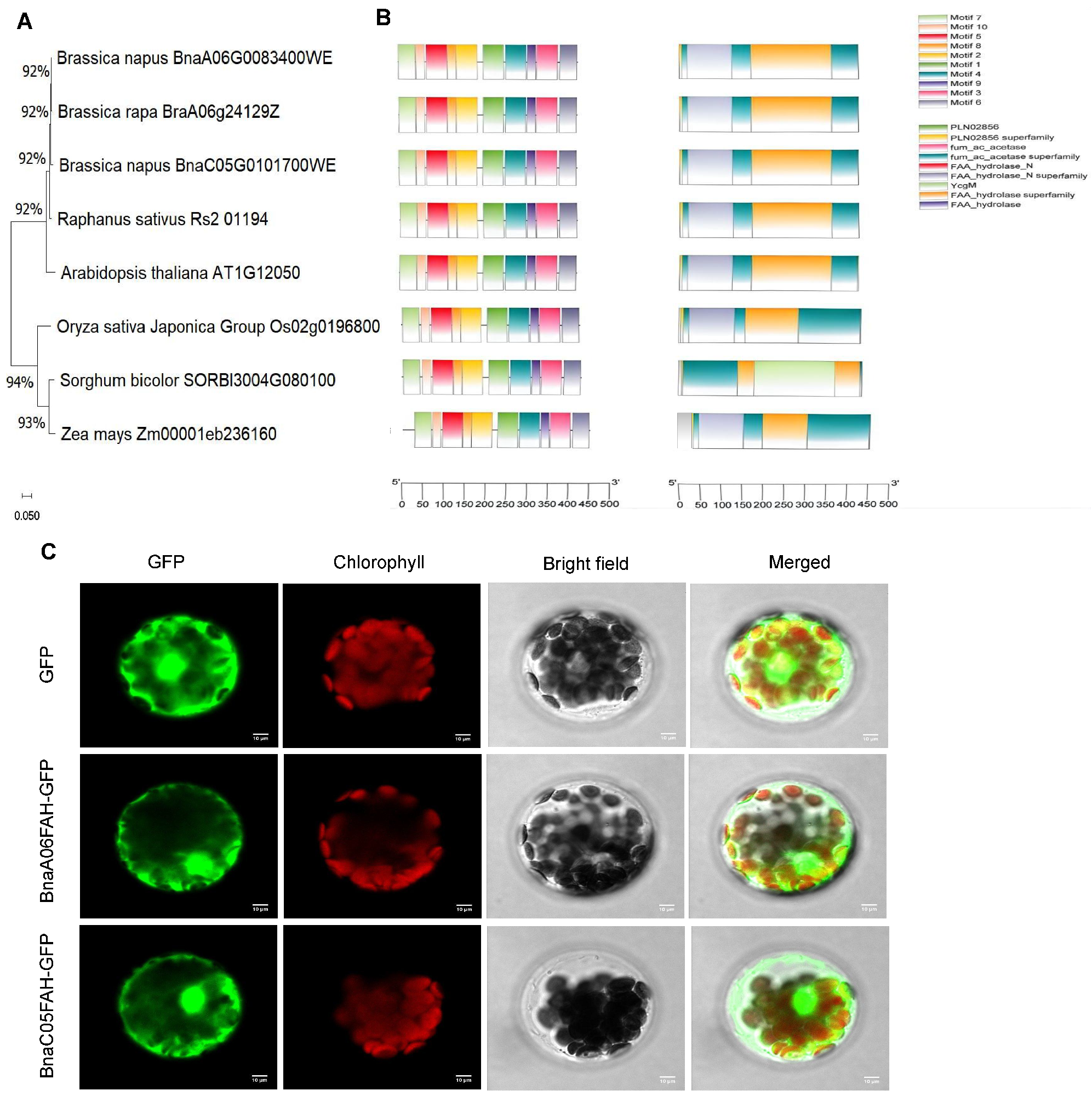
Two rapeseed FAHs [BnaA06G0083400WE (BnaA06FAH) and BnaC05G0101700WE (BnaC05FAH)] were classified through blast searches through comparisons to the sequence of AtFAH [40]. According to the phylogenetic tree, the BnaFAH gene is most closely related to the sequence from Brassica rapa, followed by Camelina sativa and Arabidopsis thaliana (Figure 1A). Multiple sequence alignments and conserved motif/domain analysis of BnaFAH proteins with homologs from Arabidopsis thaliana, Oryza sativa, Zea mays, Raphanus sativus, and Sorghum bicolor revealed highly conserved core functional motifs (Motif 1–10) with nearly identical arrangement and composition across all examined species (Figure 1B, left panel). In contrast, the unique domain architecture of the Sorghum bicolor FAH homolog, featuring a YcgM insertion within its core hydrolase domain, exemplifies how domain fusion drives functional diversity and innovation in the FAH superfamily during evolution (Figure 1B, right panel). This structural pattern directly supports the evolutionary conservation of the FAH core enzymatic function across angiosperms, and subtle divergences in the domain reflect species-specific adaptive evolution during speciation without compromising core catalytic activity. To examine the subcellular localization of the BnaFAH proteins, we transiently expressed a fusion of BnaA06FAH and BnaC05FAH to the green fluorescent protein (GFP). When transiently expressed in Arabidopsis protoplasts, BnaA06FAH-GFP and BnaC05FAH-GFP localized in the cytoplasm, indicating that both BnaA06FAH and BnaC05FAH are cytoplasm-localized proteins (Figure 1C).

3.2. The Double Mutant of BnaFAH Enhances Rapeseed Resistance Against Plutella xylostella (L.) Under SD2

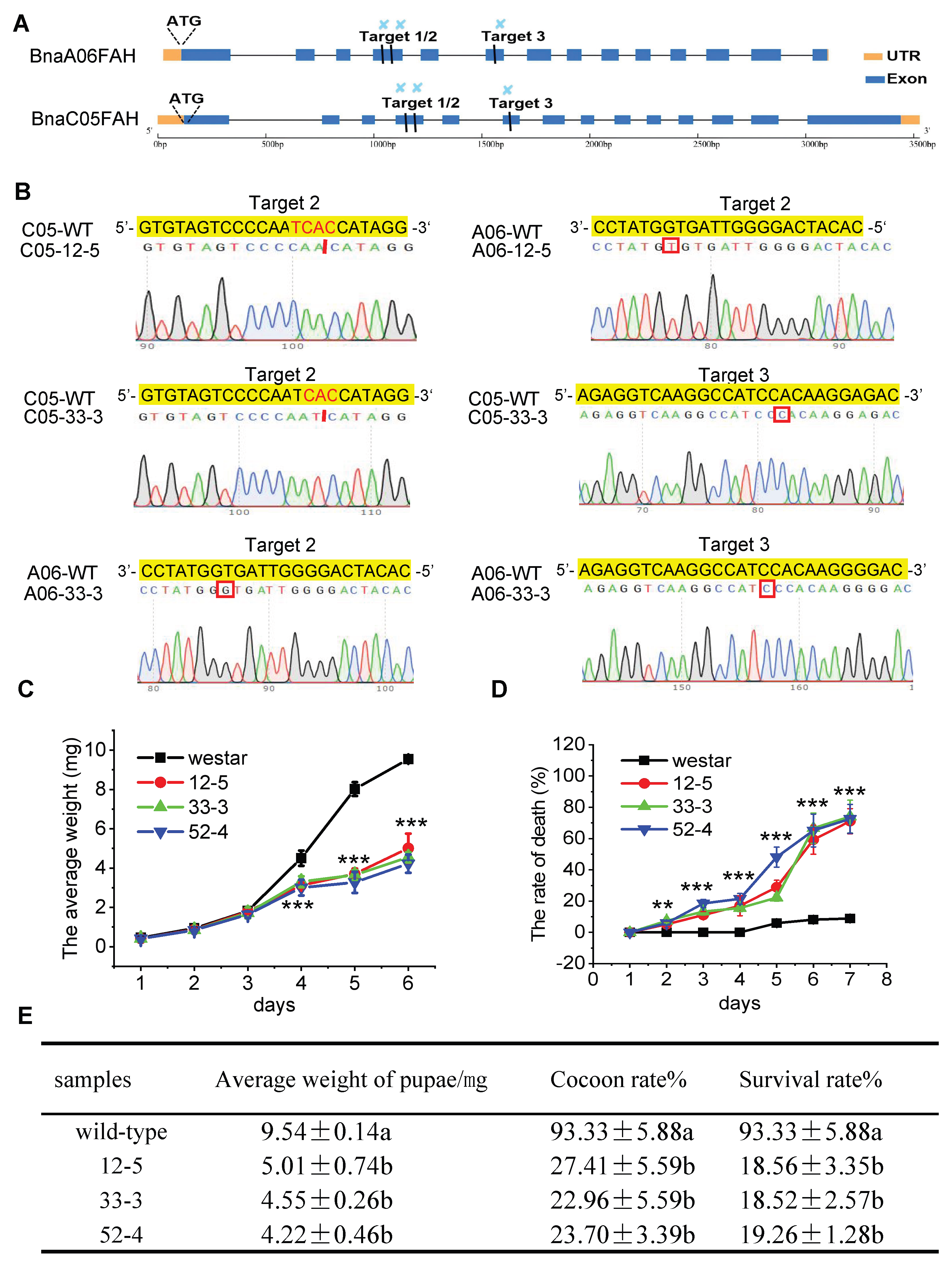
Previously, we used CRISPR/Cas9 to generate double bnafah mutants. Three gRNAs targeting the conserved sequences were designed in the fourth and sixth exons of BnaA06FAH and BnaC05FAH (Figure 2A); then, stable rapeseed transformation was performed, and 25 independent T0 transgenic lines were obtained with mutations of BnaC05FAH or BnaA06FAH (Table S2). Sequencing of T0 plants identified lines, such as T0-12 and T0-33, that were heterozygous double mutants for BnaFAH (Table S2). Through self-pollination and screening of the T1 progeny, we obtained two independent, transgene-free (lacking the Cas9gene), homozygous double mutant lines, designated bnafah 12-5 and bnafah 33-3 (Figure S3). Sequencing confirmed that both lines harbored biallelic frame shift mutations in both BnaA06FAH and BnaC05FAH, resulting in predicted loss-of-function alleles (Figure 2B; for detailed allele sequences and mutation descriptions, see Table S3). These two lines were used for all subsequent experiments.
To investigate the effect of the BnaFAH mutation on insect development under different photoperiods, 4-week-old Westar (WT) and bnafah plants grown under SD conditions for 0 (LD), 1 (SD1), and 2 days (SD2) were exposed to Plutella xylostella larvae. The insect bioassay was conducted with three independent biological replicates. After a feeding trial, the weight and the death rate of the larvae were measured to assess the resistance of plants. Larval weight data and mortality rates were compared using a two-tailed Student’s t-test. Notably, statistical analyses confirmed significant differences between mutant and wild-type plants despite the observed variability in mortality rates, which validates the ANOVA assumptions (Shapiro–Wilk test, p > 0.05 for normality). Our results demonstrate that bnafah enhanced resistance to Plutella xylostella larvae under SD2 (Figure 2C,D and Figure S5). The average weight of the larvae that fed on bnafah 12-5, 33-3, and 52-4 plants grown under SD2 was only 52.51%, 47.65%, and 44.27%, respectively, compared to that of larvae that fed on WT plants (Figure 2C). The death rate of Plutella xylostella that fed on bnafah mutant plants grown under SD2 was statistically significantly higher than that of insects that fed on wild-type plants (Figure 2D). Moreover, Plutella xylostella that fed on bnafah under SD2 showed statistically significant reductions in the cocoon number and survival rate in comparison to insects that fed on control plants (Figure 2E). Together, the results demonstrate that loss of BnaFAH induces Brassica napus defense against Plutella xylostella under SD2.

3.3. Transcriptome Analysis Indicates Alpha-Linolenic Acid Metabolism Participates in Plutella xylostella (L.) Resistance in Bnafah

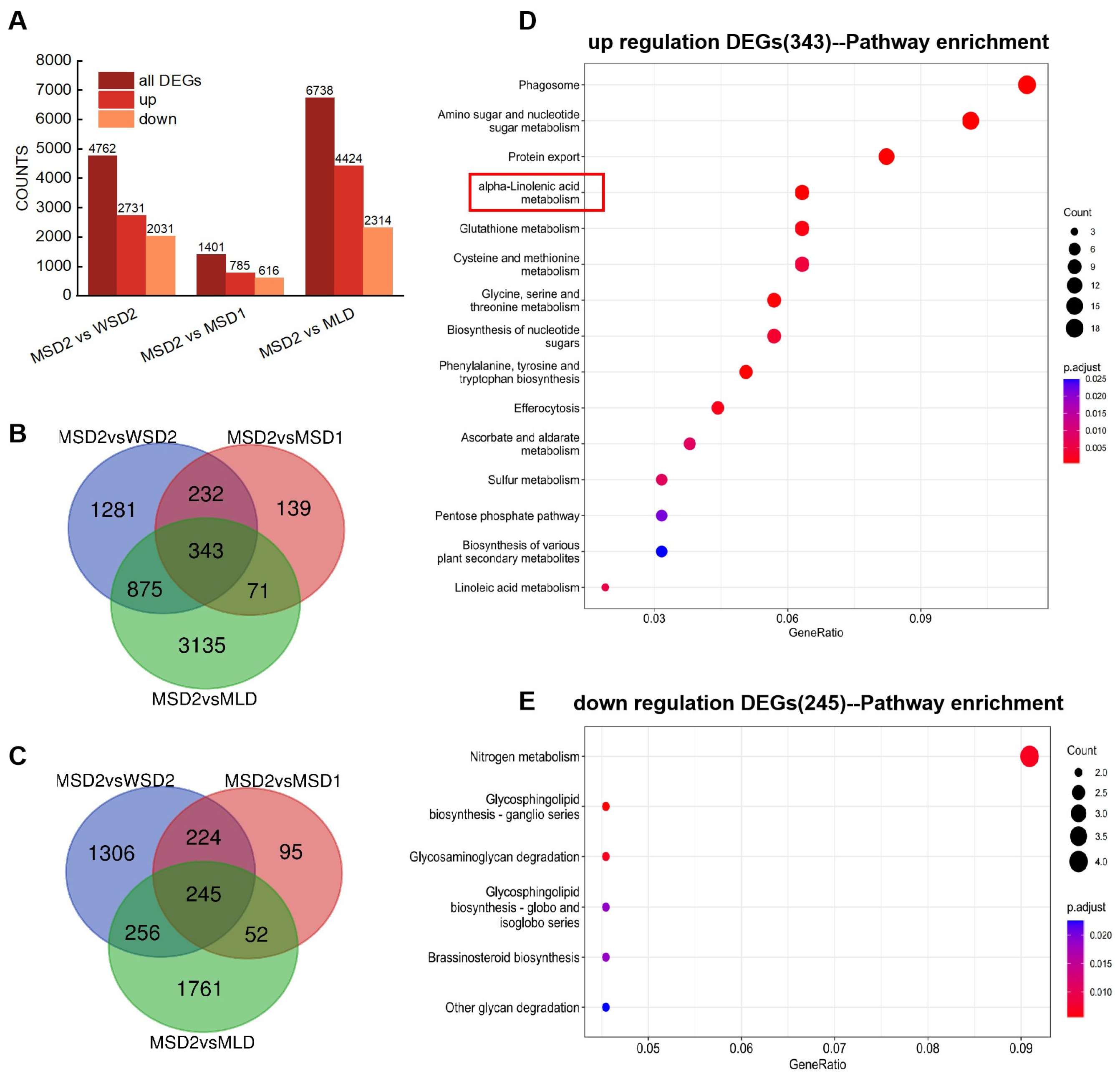
To explore how BnaA06FAH and BnaC05FAH mutations enhance rapeseed resistance against Plutella xylostella (L.), six cDNA libraries were constructed: bnafah under LD/SD1/SD2 (MLD/MSD1/MSD2) and wild type under LD/SD1/SD2 (WLD/WSD1/WSD2). RNA-seq profiling was performed between Westar (wild-type) and the double-mutant bnafah. The reads per kilobase per million mapped reads (RPKM) values of three biological replicates for each sample were highly correlated, indicating that the RNA-seq data were reliable (Figure S6A). Using |log 2 (foldchange)| > 1 and padj < 0.05 as the threshold criterion for DEGs screening, the volcano plot results showed that the number of DEGs in bnafah under SD2 compared to the control is more than that under LD and SD1 (Figure S6B). KEGG pathway enrichment analysis of DEGs between bnafah and wild type under different photoperiod showed that the enriched pathway in bnafah under SD2 is different from that under SD1 and LD (Figure S7A–C). Since bnafah induces Brassica napus defense against Plutella xylostella under SD2, to investigate the cause of insect resistance enhancement in bnafah, three groups were constructed: MSD2 vs. WSD2, MSD2 vs. MSD1, and MSD2 vs. MLD. These comparisons allow us to pinpoint genes whose upregulation is specifically associated with the bnafah-SD2 interaction that confers insect resistance. A total of 2731 upregulated and 2031 downregulated DEGs were detected in MSD2 vs. WSD2, 785 upregulated and 616 downregulated DEGs were detected in MSD2 vs. MSD1, and 4424 upregulated and 2314 downregulated DEGs were detected in MSD2 vs. MLD (Figure 3A). KEGG enrichment analysis of DEGs of each group demonstrates the common pathway is alpha-linolenic acid metabolism (Figure S7C–E). To identify which DEGs had the same expression patterns among the three groups, a total of 343 upregulated genes (Table S4) and 245 downregulated genes (Table S5) were identified in the three sets of comparisons (Figure 3B,C). KEGG enrichment analysis on the co-upregulated and downregulated genes further illustrates that up-DEGs were significantly enriched in alpha-linolenic acid metabolism and other defense-associated metabolic pathways (Figure 3D,E), including phenylpropanoid biosynthesis, which contributes to the production of antimicrobial flavonoids and the strengthening of cell walls via lignin deposition, and glutathione metabolism, which is central to cellular redox homeostasis and detoxification. The concurrent upregulation of these pathways suggests that the bnafah-SD2 condition triggers a broad-spectrum defense reprogramming. Since alpha-linolenic acid metabolism is the top-ranking pathway, it strongly suggests that alpha-linolenic acid metabolism may participate in regulating insect resistance in bnafah.

3.4. Loss of Both BnaA06FAH and BnaC05FAH Under SD2 Promotes JA Biosynthesis

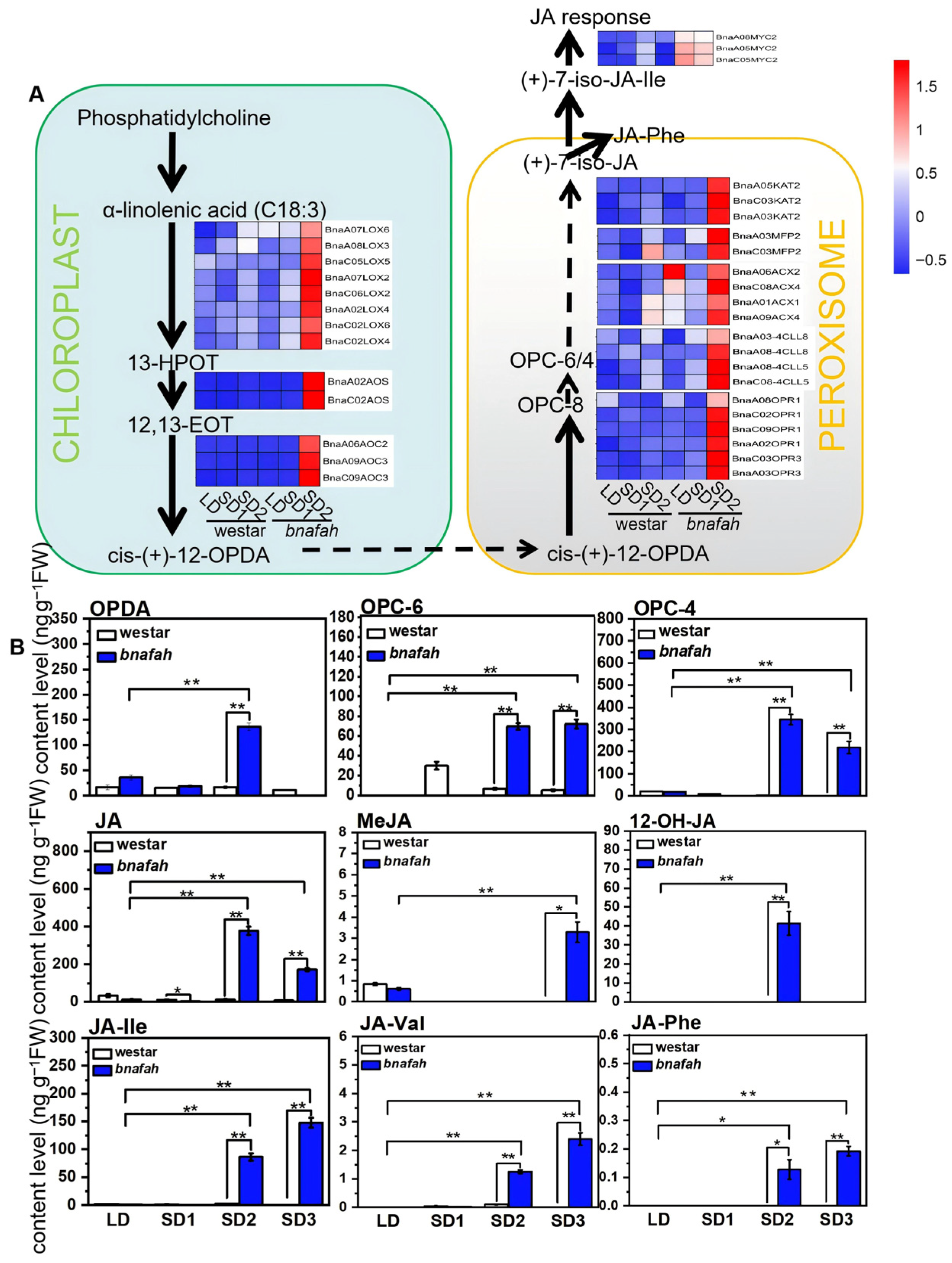
Given that the KEGG pathway analysis of transcriptomic data suggested that the JA pathway plays a potential role in the mutant’s insect resistance, our subsequent analysis focused on the expression of genes involved in JA biosynthesis in the alpha-linolenic acid metabolism pathway. We focused on the JA pathway due to its important roles in wound response and selected genes related to synthesis, signaling, and the response of JA from RNA-seq data, which were visualized in a heatmap. As shown in Figure 4A, the expression levels of the JA synthesis genes, including BnaLOX2/3/4/6, BnaAOS, BnaAOC2/3, BnaOPR1/3, BnaACX1/2/4, and BnaKAT2, as well as the JA signaling gene BnaMYC2, were markedly upregulated in bnafah under SD2. The results suggest that loss of BnaFAH under SD2 upregulates genes involved in JA biosynthesis and signaling cascades.
To further confirm how the JA biosynthetic pathway is affected in double bnafah mutants under SDs, we measured the levels of JA precursors and derivatives in bnafah for 0, 1, 2, and 3 short days. Consistent with transcriptomic profiling, LC-MS quantification revealed the level of JA and JA biosynthesis precursors such as OPDA, OPC-6, and OPC-4, the hydroxylated derivative 12-OH-JA, and JA conjugates including JA-Phe, JA-Val, and JA-Ile in bnafah leaves for 2 short days were significantly greater than that for 0 days (Figure 4B). Notably, accumulation persisted in SD3 for both upstream precursors (OPC-6 and OPC-4) and JA conjugates, including JA-Phe, JA-Val, and JA-Ile (p < 0.05, Student’s t-test) (Figure 4B). Taken together, these results suggest that the genetic disruption of BnaFAH triggers significant upregulation of JA biosynthetic genes under SD2, and this transcriptional reprogramming drives multilevel metabolic reprogramming, culminating in the robust accumulation of JA, JA synthesis precursors, and even JA derivatives and conjugates.

3.5. The Effect of the MeJA and JA Biosynthesis Inhibitor DIECA on the Insect Resistance of the Bnafah Mutant

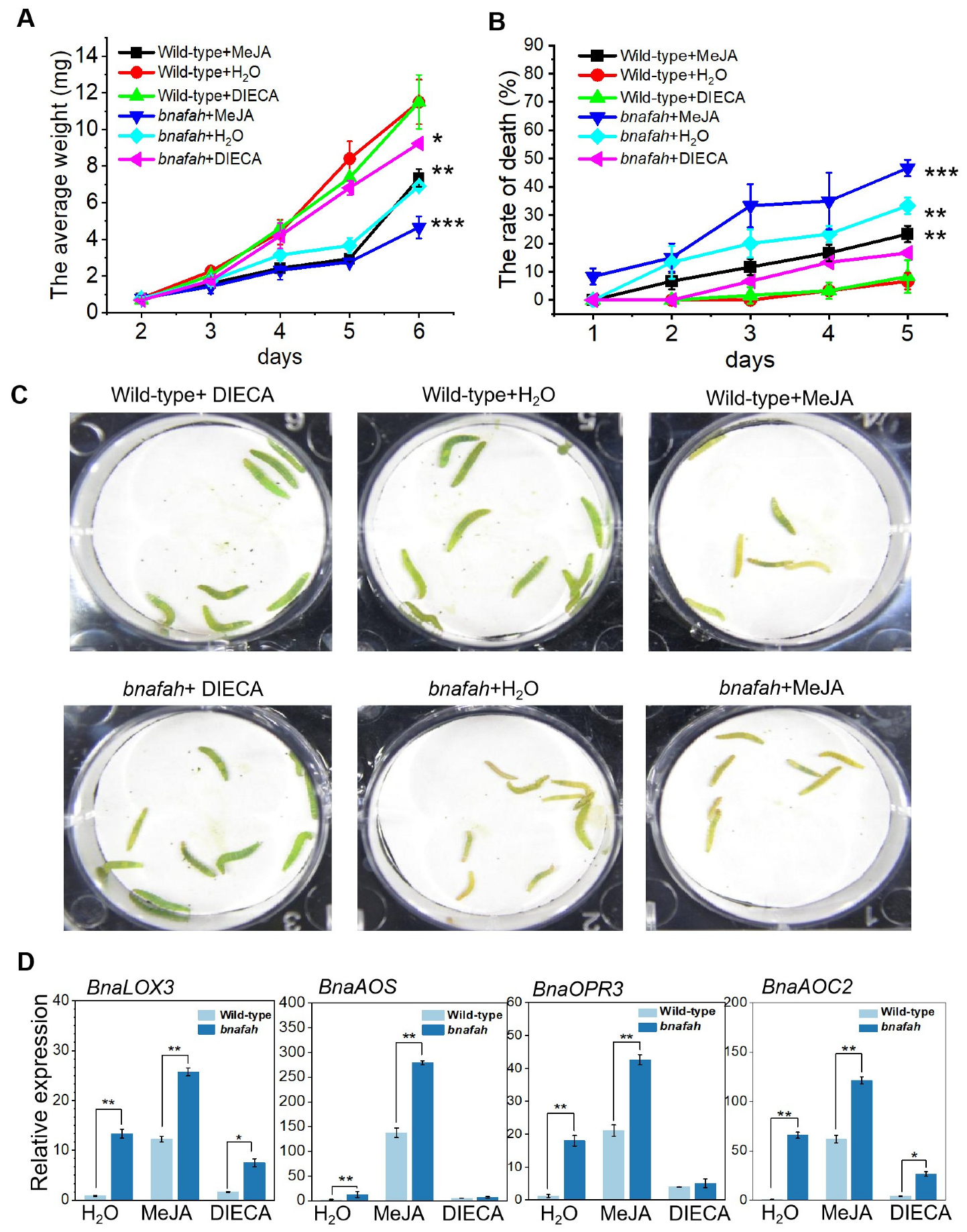
To confirm the role of JA in the enhanced resistance of bnafah mutants to Plutella xylostella under SD2, we applied exogenous methyl jasmonate (MeJA) and the JA biosynthesis inhibitor diethyldithiocarbamic acid (DIECA) to wild-type and mutant plants grown under SD2; we then measured larval weight and mortality. In wild-type plants, MeJA treatment enhanced resistance, while DIECA partially compromised it. In bnafah mutants, larval growth was already suppressed under control conditions (bnafah + H2O) compared to wild-type plants (Figure 5A). Exogenous MeJA further amplified this resistance, reducing larval weight and increasing mortality relative to the bnafah control (Figure 5A–C). Conversely, DIECA treatment abolished the mutant’s resistance, resulting in the highest larval weights and lowest mortality (Figure 5A–C). Moreover, the expression of JA biosynthetic genes in wild-type and bnafah plants under different treatments (H2O, MeJA, and DIECA) was analyzed. Both wild-type and bnafah showed significantly increased expressions of BnaLOX3, BnaAOS, BnaAOC2, and BnaOPR3 upon MeJA treatment compared to H2O, while the expression of all four genes was repressed by DIECA (Figure 5D). In summary, the data demonstrate that the enhanced insect resistance in the bnafah mutant is critically dependent on the JA signaling pathway. This conclusion is supported by the concurrent suppression of both resistance and JA-biosynthetic gene expression following pharmacological inhibition. These findings underscore JA signaling as a key and necessary component of the effective defense response in the bnafah mutant.

3.6. Removal of ROS Decreases Bnafah Resistance to Insects

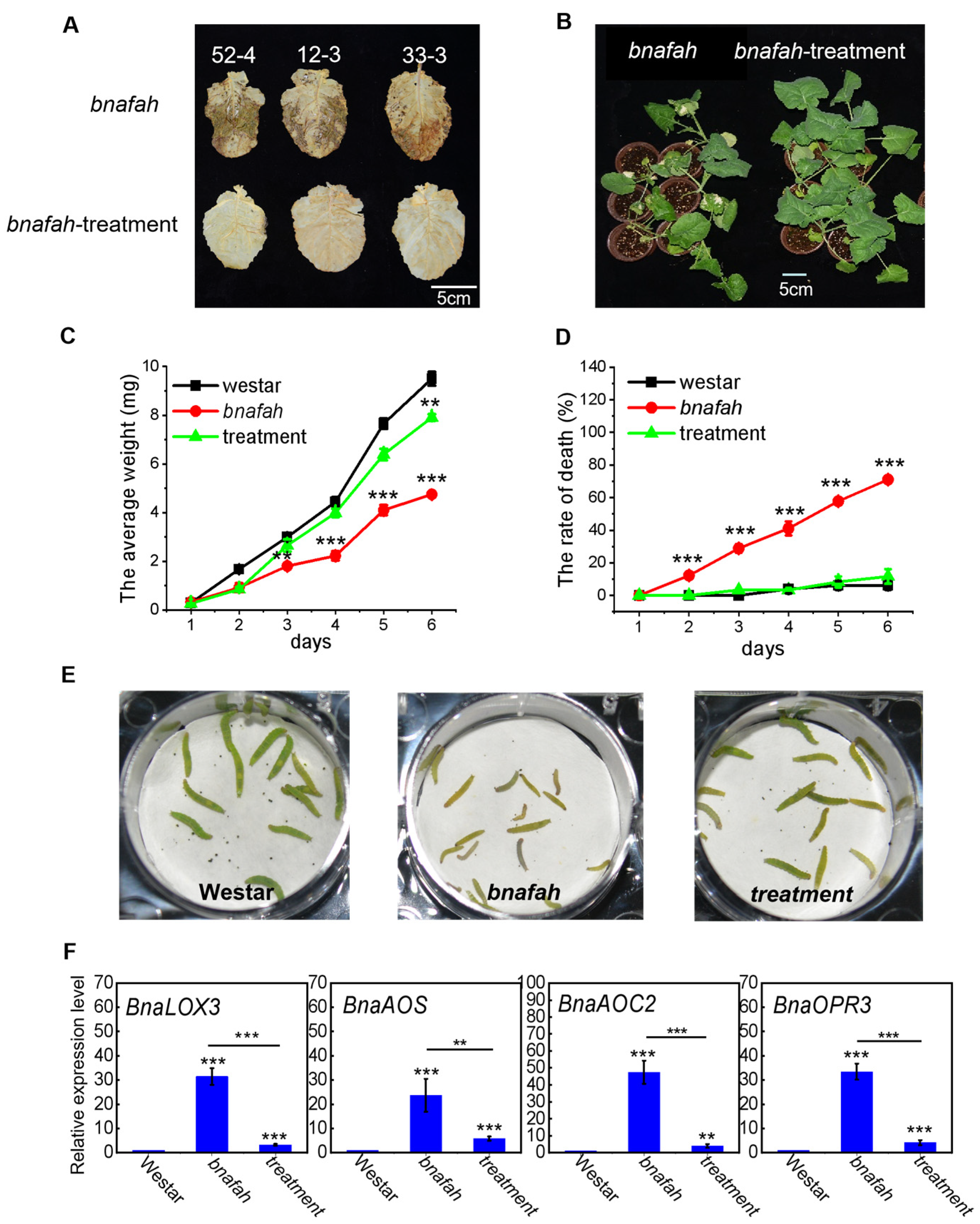
Double mutant bnafah accumulates H2O2 under SD. To confirm that the JA level is related to H2O2 content, bnafah were treated with 2 mM sodium selenite under SD. The results showed that the content of H2O2 in bnafah was dramatically decreased after sodium selenite treatment (Figure 6A), resulting in the decline of cell death (Figure 6B). To confirm the effect of sodium selenite treatment on the resistance of bnafah to Plutella xylostella, we inoculated WT, bnafah− (without), and bnafah+ (with sodium selenite) Brassica napus plants under SD with Plutella xylostella larvae, and the WT group was the control group. We measured the weight and death rate of Plutella xylostella larvae from 1 to 6 days. The weight was lower, but the death rate was higher in bnafah− (without) than in control plants (Figure 6C,D). However, after treatment with sodium selenite, the decline in weight and increase in death rate in bnafah disappeared, and both closely followed the trends of the control. Additionally, the size of larvae that ate bnafah leaves was significantly smaller than that of larvae that ate WT plants (Figure 6E). On the contrary, after treatment with sodium selenite, the size of the larvae was almost the same as that of wild-type plants. Because JA plays an important role in plant defense, we tested the expression of JA biosynthesis genes in plants with WT, bnafah− (without), and bnafah+ (with sodium selenite) under SD. The results showed that the expression of JA biosynthesis genes BnaLOX3, BnaAOS, BnaAOC2, and BnaOPR3 was significantly higher in bnafah plants than in WT plants (Figure 6F); however, the expression of these genes all declined back to the WT level once treated with sodium selenite. These results demonstrate that JA plays a critical role in the defense of Brassica napus against Plutella xylostella, and that the accumulation of H2O2 is required for the activation of this JA-mediated defense pathway.

4. Discussion

Fumarylacetoacetate hydrolase (FAH) catalyzes the final step of the tyrosine degradation pathway. Studies on Arabidopsis have shown that loss of FAH function leads to the accumulation of a toxic metabolite under SD conditions [43], which, in turn, induces the accumulation of ROS [44]. This SD-dependent burst in ROS suggests a potential link between FAH disruption and stress responses. To explore whether this mechanism operates in an important crop species and to assess its potential agronomic implications, we turned to oilseed rape (Brassica napus L.), a globally vital source of vegetable oil, animal feed, and industrial raw materials. The genome of the Brassica napus is complex, with most genes presented as multiple homologous copies that may have redundant or divergent functions. We identified two BnaFAH orthologs in the cultivar “Westar”: BnaA06FAH and BnaC05FAH. Phylogenetic analysis confirmed their close relationship to AtFAH (Figure 1A), and sequence alignment revealed conserved protein motifs across the species (Figure 1B). Subcellular localization predicted these to be cytoplasmic proteins (Figure 1C). To investigate their functional roles, we used CRISPR/Cas9-mediated mutagenesis to generate a series of transgene-free mutant lines. The double-null mutant (bnafah) indeed exhibited significant ROS accumulation under SD conditions [40], mirroring the phenotype observed in Arabidopsis and establishing a basis for further functional analysis.
In this research, we demonstrate that the loss of BnaFAH in Brassica napus confers enhanced resistance to the diamondback moth, Plutella xylostella, specifically under SD2 (Figure 2C–E). This resistance is mechanistically driven by a coordinated signaling cascade, wherein disruption of the tyrosine degradation pathway triggers a rapid accumulation of ROS, which in turn acts as an upstream signal to activate JA biosynthesis and signaling. Based on these observations, we propose a “photoperiod-ROS-JA” defense module, a mechanistic model that reveals a previously underappreciated link between primary metabolism, light signaling, and induced immunity in an important crop species, while the functional connections between ROS and JA are supported by our pharmacological and genetic data, the precise link between photoperiod perception and the initiation of the ROS burst remains hypothetical. A central finding of this research is the role of ROS as a critical initiator of the defense response in bnafah resistance to Plutella xylostella. ROS are pivotal signaling molecules in plant immunity, orchestrating a complex defense network against insects. The induction of ROS plays a pivotal role in plant defense mechanisms against herbivorous pests such as Plutella xylostella [45,46,47,48,49]. We observed that bnafah mutants accumulated significant levels of H2O2 under SD2 [40] (Figure 6A), and the application of the ROS scavenger sodium selenite not only reduced H2O2 content but also effectively abolished the enhanced insect resistance and upregulation of key JA biosynthetic genes (BnaLOX3, BnaAOS, BnaAOC2, BnaOPR3) (Figure 6). This positions ROS accumulation as an early, indispensable event upstream of JA pathway activation in this specific genetic and environmental context of the plant. This sequence appears to invert the canonical signaling hierarchy, in which JA is typically considered the primary inducer of ROS production during herbivory [30,31]. However, the induction of JA by ROS has also been researched. Exogenous ascorbic acid (AsA, H2O2 scavenger) treatment can inhibit the accumulation of H2O2, while reducing JA levels and the expression of sesquiterpene synthase genes [50]. H2O2 promotes JA synthesis by activating the activity of the ACX enzyme. In Arabidopsis thaliana, the N-terminal domain of CAT2 (CAT2-N) directly binds to and activates ACX2/ACX3, promoting the conversion of substrate OPC4-CoA to JA precursor [51]. Transgenic plants overexpressing CAT2-N have increased ACX activity, increased JA accumulation, and enhanced resistance to Botrytis cinerea; however, JA synthesis in acx2acx3 double mutants was blocked, and the disease sensitivity increased [37]. Consistent with these reports, our data show that ROS scavenging abolishes both JA biosynthesis gene expression and insect resistance. To fully close the signaling loop, future studies directly quantifying the levels of JA and its active forms (e.g., JA-Ile) in plants following ROS scavenging will provide definitive metabolite-level evidence for the role of ROS as an upstream driver of JA biosynthesis. However, the exact mechanism—whether direct (e.g., via redox activation of JA biosynthetic enzymes like ACX) or indirect (e.g., via ROS-induced transcriptional reprogramming)—remains an open question that requires further biochemical and genetic validation. In addition, oxidative stress induces the expression of genes related to the JA synthesis pathway, leading to the accumulation of JA-modified forms [36]. These results suggest that ROS act upstream or in conjunction with JA to facilitate defense gene activation following stress stimuli. ROS not only serve as early signaling molecules following stress or pathogen attack but also interact with JA signaling to orchestrate effective immune responses. Our findings align with emerging evidence that metabolic perturbations can reconfigure signaling networks [34,35]. The accumulation of toxic intermediates from the blocked tyrosine pathway in bnafah likely induces cellular stress, leading to a pre-emptive ROS burst that primes the JA-dependent defense machinery. This suggests that ROS serves as a metabolic damage signal that orchestrates downstream immune responses under conditions of metabolic compromise.
In plants, JA is the major defense hormone in activating defense reactions against herbivorous insects [52]. Exogenous application of JA induces resistance to economically important insect pests in winter wheat, highlighting its potential as a defense elicitor in crop protection [53]. Similarly, JA application in groundnut enhances resistance to Helicoverpa armigera, with mid-gut digestive and detoxifying enzymes serving as indicators of the induced resistance, suggesting that JA influences insect physiology and survival [54]. Derivatives of JA that come into existence in the cytosol, such as methyl ester of JA (MeJA), cis-jasmone, 12-OH-JA, or JA-Ile, also play a vital role in regulating plant defensive responses against insects. For example, JA-Ile is involved in the host-plant resistance mechanism against the soybean aphid [55]; moreover, the JA-amino acid conjugates JA-Val and JA-Leu are also involved in rice resistance to herbivores [56]. In recent years, the diamondback moth, Plutella xylostella, has become the most destructive insect pest of cruciferous plants, including the oilseed rape [57]. Application of JA significantly reduced the population growth parameters as well as the survival rate of immature Plutella xylostella [58]. The subsequent activation of the JA pathway in bnafah mutants was robust and multi-layered. Transcriptomic and metabolomic analyses revealed concurrent upregulation of JA biosynthetic genes and significant accumulation of JA, its precursors (OPDA, OPC-4, and OPC-6), and bioactive conjugates (JA-Ile and JA-Phe) (Figure 4). The functional significance of this JA pathway activation was unequivocally demonstrated through chemical interventions: exogenous MeJA enhanced resistance, whereas the JA biosynthesis inhibitor DIECA suppressed it (Figure 5). Future studies incorporating direct quantification of JA pathway metabolites in inhibitor-treated plants and functional validation of the transcriptomically identified candidate genes will provide complementary biochemical and mechanistic depth to the defense model. The magnitude of JA-induced gene expression and the resultant anti-herbivore phenotype were substantially greater in the bnafah than in wild-type, indicating a heightened sensitivity or responsiveness to JA signaling. This synergistic effect between the bnafah and JA application underscores that the mutation creates a physiological state that amplifies the efficacy of the JA defense program, possibly through epigenetic or post-translational modifications that lower the threshold for defense gene activation [59,60].
In summary, this study shows that BnaFAH knockout disrupts tyrosine degradation, triggering a photoperiod-dependent “photoperiod-ROS-JA” signaling cascade that enhances resistance to Plutella xylostella. This photoperiodic sensitivity suggests that circadian or light-quality perception interacts with the metabolic state resulting from BnaFAH deficiency, potentially by modulating ROS-scavenging enzymes, entraining the circadian clock, or altering specific light receptor activity. BnaFAH, as a negative regulator of insect resistance, is a promising target for genome-editing strategies. Given the strict photoperiod dependency of the resistant phenotype, breeding efforts must consider genotype-by-environment interactions, particularly day-length conditions in target planting regions. From an applied perspective, this knowledge could inform agricultural management strategies aimed at enhancing innate plant resistance. For instance, for varieties carrying this trait, crop scheduling could be optimized so that the most pest-susceptible growth stages (e.g., seedling or early flowering) coincide with natural short-day seasons. In protected cultivation systems, targeted light management (e.g., using blackout cloths to shorten day length) during vulnerable periods could be deployed to deliberately activate this defense pathway, potentially reducing pesticide reliance. Our findings are highly significant for sustainable pest management in rapeseed production, providing an environmentally friendly alternative to synthetic insecticides. Future studies should investigate the molecular links between light signaling, circadian clock components, and BnaFAH-mediated metabolic changes, and validate the stability and durability of resistance under field conditions to translate lab findings into practical, insecticide-sparing solutions for resilient agricultural systems.

5. Conclusions

This study establishes a mechanistic link between a lesion in primary metabolism and enhanced insect resistance in Brassica napus, mediated by a finely tuned “photoperiod-ROS-JA” signaling module. In summary, our work reveals that targeted genetic modification can reconfigure signaling networks to enhance plant defense, wherein ROS serves as a pivotal metabolic damage signal activating JA-mediated immunity. These findings not only advance our understanding of the interplay between primary metabolism, redox signaling, and phytohormone-regulated defenses but also identify BnaFAH and its downstream components as promising candidate targets for developing novel, sustainable crop protection strategies in rapeseed. Further field validation is required to confirm their utility for practical breeding applications. Future research should focus on validating the efficacy of this resistance under field conditions and elucidating the precise molecular sensors linking the SD2 signal to the metabolic perturbation.

Supplementary Materials

The following supporting information can be downloaded at: https://www.mdpi.com/article/10.3390/horticulturae12040403/s1. Figure S1. Synthesis of oxylipins and their intracellular transport; Figure S2: CRISPR/Cas9 induced targeted mutagenesis of the BnaFAH gene in Brassica napus. L; Figure S3: Isolation of transgene-free T1 plants from T12, T33 mutant lines; Figure S4: Multiple alignment of the amino acid sequences of the mutant; Figure S5: The average weight of Plutella xylostella larva exposed to 0-day short-day (LD), 1-day short-day (SD1) and 2-day short-day (SD2) wild-type and bnafah under long-day conditions for 2–5 days; Figure S6: Spearman correlation heat map between TPM expression (A) and difference analysis volcano plot (B) in wild-type (AACC) and bnafah (aacc) grown under short day for 0 day (LD), 1 day (SD1), and 2 days (SD2); Figure S7: Transcriptome analysis of the bnafah mutant under different photoperiod compared with different control; Table S1: The primer sets used in this study; Table S2: Genotypes of different CRISPR/Cas9-meditated targeted T0 mutant lines; Table S3: Detailed genotyping of CRISPR/Cas9-edited BnaFAH mutant lines; Table S4: Expression and annotation analysis of 343 co-upregulated genes; Table S5: Expression and annotation analysis of 245 co-downregulated genes.

Author Contributions

Conceptualization, T.Z. and Z.Z.; methodology, T.Z.; validation, T.Z., Z.Z., C.S. and M.X.; formal analysis, C.L.; investigation, T.Z. and Z.Z.; resources, M.X.; data curation, G.C.; writing—original draft preparation, T.Z.; writing—review and editing, T.Z. and Z.Z.; project administration, M.X. and G.C.; funding acquisition, T.Z., Z.Z. and G.C. All authors have read and agreed to the published version of the manuscript.

Funding

This research was funded by the Jiangxi Natural Science Foundation of China (20232BAB215031), the Key Research and Development Program Project of Yichun (2024ZDYFJH04), the Science and Technology Project of Jiangxi Provincial Department of Education (GJJ2401607), and the National Natural Science Foundation of China (32260088).

Institutional Review Board Statement

Not applicable.

Informed Consent Statement

Not applicable.

Data Availability Statement

The original data presented in the study are openly available in the Gene Expression Omnibus (GEO) repository, and the GEO accession number is GSE272643.

Acknowledgments

The KEGG project is partially supported by the National Bioscience Database Center of the Japan Science and Technology Agency. Computational resources were provided by the Bioinformatics Center, Institute for Chemical Research, Kyoto University.

Conflicts of Interest

The authors declare no conflicts of interest.

References

  1. Liu, D.; Yu, L.; Wei, L.; Yu, P.; Wang, J.; Zhao, H.; Zhang, Y.; Zhang, S.; Yang, Z.; Chen, G.; et al. BnTIR: An online transcriptome platform for exploring RNA-seq libraries for oil crop Brassica napus. Plant Biotechnol. J. 2021, 19, 1895–1897. [Google Scholar] [CrossRef]
  2. So, K.K.Y.; Duncan, R.W. Breeding Canola (Brassica napus L.) for Protein in Feed and Food. Plants 2021, 10, 2220. [Google Scholar] [CrossRef] [PubMed]
  3. Chakrabarty, S.; Shashank, P.R. Economically Important Insects on Cruciferous Crops (Brassicaceae): An Updated Global Inventory. Zool. Stud. 2025, 64, e30. [Google Scholar] [CrossRef] [PubMed]
  4. Qian, C.; Li, J.; Wu, S.; Yang, Y.; Wu, Y.; Wang, X. Cross-resistance and genetics of field-evolved resistance to chlorfenapyr in Plutella xylostella. Insect. Sci. 2024, 31, 533–541. [Google Scholar] [CrossRef] [PubMed]
  5. Zhu, L.; Li, Z.; Zhang, S.; Xu, B.; Zhang, Y.; Zalucki, M.P.; Wu, Q.; Yin, X. Population dynamics of the diamondback moth, Plutella xylostella (L.), in northern China: The effects of migration, cropping patterns and climate. Pest Manag. Sci. 2018, 74, 1845–1853. [Google Scholar] [CrossRef]
  6. Jafary-Jahed, M.; Razmjou, J.; Nouri-Ganbalani, G.; Naseri, B.; Hassanpour, M.; Leppla, N.C. Life Table Parameters and Oviposition Preference of Plutella xylostella (Lepidoptera: Plutellidae) on Six Brassicaceous Crop Plants. J. Econ. Entomol. 2019, 112, 932–938. [Google Scholar] [CrossRef]
  7. Furlong, M.J.; Wright, D.J.; Dosdall, L.M. Diamondback moth ecology and management: Problems, progress, and prospects. Annu. Rev. Entomol. 2013, 58, 517–541. [Google Scholar] [CrossRef]
  8. Li, Z.; Feng, X.; Liu, S.S.; You, M.; Furlong, M.J. Biology, Ecology, and Management of the Diamondback Moth in China. Annu. Rev. Entomol. 2016, 61, 277–296. [Google Scholar] [CrossRef]
  9. Guo, Z.; Ma, H.; Tang, J.; Wu, M.; He, S.; Wan, H.; Li, J.; Ma, K. Chlorantraniliprole Resistance in Spodoptera frugiperda: Resistance Monitoring, Resistance Risk, and Resistance Mechanisms. J. Agric. Food Chem. 2024, 72, 16651–16660. [Google Scholar] [CrossRef]
  10. Zhang, S.; Zhang, X.; Shen, J.; Li, D.; Wan, H.; You, H.; Li, J. Cross-resistance and biochemical mechanisms of resistance to indoxacarb in the diamondback moth, Plutella xylostella. Pestic. Biochem. Phys. 2017, 140, 85–89. [Google Scholar] [CrossRef]
  11. Kaur, R.; Choudhary, D.; Bali, S.; Bandral, S.S.; Singh, V.; Ahmad, M.A.; Rani, N.; Singh, T.G.; Chandrasekaran, B. Pesticides: An alarming detrimental to health and environment. Sci. Total Environ. 2024, 915, 170113. [Google Scholar] [CrossRef]
  12. Ma, C.; Zhang, W.; Peng, Y.; Zhao, F.; Chang, X.Q.; Xing, K.; Zhu, L.; Ma, G.; Yang, H.P.; Rudolf, V.H.W. Climate warming promotes pesticide resistance through expanding overwintering range of a global pest. Nat. Commun. 2021, 12, 5351. [Google Scholar] [CrossRef] [PubMed]
  13. Erb, M. Plant Defenses against Herbivory: Closing the Fitness Gap. Trends Plant. Sci. 2018, 23, 187–194. [Google Scholar] [CrossRef] [PubMed]
  14. Erb, M.; Reymond, P. Molecular Interactions Between Plants and Insect Herbivores. Annu. Rev. Plant Biol. 2019, 70, 527–557. [Google Scholar] [CrossRef]
  15. Bruinsma, M.; van Broekhoven, S.; Poelman, E.H.; Posthumus, M.A.; Müller, M.J.; van Loon, J.J.; Dicke, M. Inhibition of lipoxygenase affects induction of both direct and indirect plant defences against herbivorous insects. Oecologia 2010, 162, 393–404. [Google Scholar] [CrossRef] [PubMed]
  16. He, F.; Yang, P.; Zhang, Y.; Li, H.; Wei, S.Y.; Deng, S.L.; Liu, J.H.; Wang, T.; Li, B.; Lin, T. The PtrRZFP4-PtrJAZ2 module regulates the jasmonic acid signaling pathway involved in the interaction between poplar and herbivorous insects. Plant J. 2025, 124, e70502. [Google Scholar] [CrossRef]
  17. Vick, B.A.; Zimmerman, D.C. Biosynthesis of jasmonic Acid by several plant species. Plant Physiol. 1984, 75, 458–461. [Google Scholar] [CrossRef]
  18. Feussner, I.; Wasternack, C. The lipoxygenase pathway. Annu. Rev. Plant Biol. 2002, 53, 275–297. [Google Scholar] [CrossRef]
  19. Weber, H.; Vick, B.A.; Farmer, E.E. Dinor-oxo-phytodienoic acid: A new hexadecanoid signal in the jasmonate family. Proc. Natl. Acad. Sci. USA 1997, 94, 10473–10478. [Google Scholar] [CrossRef]
  20. Schaller, F.; Biesgen, C.; Mussig, C.; Altmann, T.; Weiler, E.W. 12-Oxophytodienoate reductase 3 (OPR3) is the isoenzyme involved in jasmonate biosynthesis. Planta 2000, 210, 979–984. [Google Scholar] [CrossRef]
  21. Schilmiller, A.L.; Howe, G.A. Systemic signaling in the wound response. Curr. Opin. Plant Biol. 2005, 8, 369–377. [Google Scholar] [CrossRef]
  22. Staswick, P.E.; Tiryaki, I. The oxylipin signal jasmonic acid is activated by an enzyme that conjugates it to isoleucine in Arabidopsis. Plant Cell 2004, 16, 2117–2127. [Google Scholar] [CrossRef] [PubMed]
  23. Ghorbel, M.; Brini, F.; Sharma, A.; Landi, M. Role of jasmonic acid in plants: The molecular point of view. Plant Cell Rep. 2021, 40, 1471–1494. [Google Scholar] [CrossRef]
  24. Zhang, X.; Wang, D.; Elberse, J.; Qi, L.; Shi, W.; Peng, Y.L.; Schuurink, R.C.; Van den Ackerveken, G.; Liu, J. Structure-guided analysis of Arabidopsis JASMONATE-INDUCED OXYGENASE (JOX) 2 reveals key residues for recognition of jasmonic acid substrate by plant JOXs. Mol. Plant 2021, 14, 820–828. [Google Scholar] [CrossRef] [PubMed]
  25. Fonseca, S.; Chini, A.; Hamberg, M.; Adie, B.; Porzel, A.; Kramell, R.; Miersch, O.; Wasternack, C.; Solano, R. (+)-7-iso-Jasmonoyl-L-isoleucine is the endogenous bioactive jasmonate. Nat. Chem. Biol. 2009, 5, 344–350. [Google Scholar] [CrossRef] [PubMed]
  26. Vos, I.A.; Verhage, A.; Schuurink, R.C.; Watt, L.G.; Pieterse, C.M.; Van Wees, S.C. Onset of herbivore-induced resistance in systemic tissue primed for jasmonate-dependent defenses is activated by abscisic acid. Front. Plant Sci. 2013, 4, 539. [Google Scholar] [CrossRef]
  27. Pangesti, N.; Reichelt, M.; van de Mortel, J.E.; Kapsomenou, E.; Gershenzon, J.; van Loon, J.J.; Dicke, M.; Pineda, A. Jasmonic Acid and Ethylene Signaling Pathways Regulate Glucosinolate Levels in Plants During Rhizobacteria-Induced Systemic Resistance Against a Leaf-Chewing Herbivore. J. Chem. Ecol. 2016, 42, 1212–1225. [Google Scholar] [CrossRef]
  28. Scott, I.M.; Samara, R.; Renaud, J.B.; Sumarah, M.W. Plant growth regulator-mediated anti-herbivore responses of cabbage (Brassica oleracea) against cabbage looper Trichoplusia ni Hübner (Lepidoptera: Noctuidae). Pestic. Biochem. Phys. 2017, 141, 9–17. [Google Scholar] [CrossRef]
  29. Tang, J.; Yang, D.; Wu, J.; Chen, S.; Wang, L. Silencing JA hydroxylases in Nicotiana attenuata enhances jasmonic acid-isoleucine-mediated defenses against Spodoptera litura. Plant Divers. 2020, 42, 111–119. [Google Scholar] [CrossRef]
  30. Devireddy, A.R.; Zandalinas, S.I.; Fichman, Y.; Mittler, R. Integration of reactive oxygen species and hormone signaling during abiotic stress. Plant J. 2021, 105, 459–476. [Google Scholar] [CrossRef]
  31. Zhou, K.; Han, T.; Pan, B.; Hu, X.; Chen, X.; Liu, X.; Fei, S.; Yang, Y.; Li, W.; Du, M. Robustness in jasmonate signaling: Mechanisms of concerted regulation and implications for crop improvement. Abiotech 2025, 6, 618–637. [Google Scholar] [CrossRef] [PubMed]
  32. Du, J.; Yang, Z.; Li, L.; Li, Y.; Zhang, H.; Chen, J.; Sun, Z. A rice DELLA protein OsSLR1 positively regulates rice resistance to southern rice black-streaked dwarf virus infection. BMC Plant Biol. 2025, 25, 379. [Google Scholar] [CrossRef] [PubMed]
  33. Zhou, X.; Gao, H.; Zhang, X.; Khashi, U.; Rahman, M.; Mazzoleni, S.; Du, M.; Wu, F. Plant extracellular self-DNA inhibits growth and induces immunity via the jasmonate signaling pathway. Plant Physiol. 2023, 192, 2475–2491. [Google Scholar] [CrossRef] [PubMed]
  34. Przybyla, D.; Gobel, C.; Imboden, A.; Hamberg, M.; Feussner, I.; Apel, K. Enzymatic, but not non-enzymatic, 1O2-mediated peroxidation of polyunsaturated fatty acids forms part of the EXECUTER1-dependent stress response program in the flu mutant of Arabidopsis thaliana. Plant J. 2008, 54, 236–248. [Google Scholar] [CrossRef]
  35. Geng, R.; Li, X.; Huang, J.; Zhou, W. The chloroplast singlet oxygen-triggered biosynthesis of salicylic acid and jasmonic acid is mediated by EX1 and GUN1 in Arabidopsis. Plant Cell Environ. 2024, 47, 2852–2864. [Google Scholar] [CrossRef]
  36. Lelarge-Trouverie, C.; Cohen, M.; Tremulot, L.; Van Breusegem, F.; Mhamdi, A.; Noctor, G. Metabolite modification in oxidative stress responses: A case study of two defense hormones. Free Radical Bio. Med. 2023, 196, 145–155. [Google Scholar] [CrossRef]
  37. Zhang, Y.; Song, R.F.; Yuan, H.M.; Li, T.T.; Wang, L.F.; Lu, K.K.; Guo, J.X.; Liu, W.C. Overexpressing the N-terminus of CATALASE2 enhances plant jasmonic acid biosynthesis and resistance to necrotrophic pathogen. Botrytis cinerea B05.10. Mol. Plant Pathol. 2021, 22, 1226–1238. [Google Scholar] [CrossRef]
  38. Vasantha-Srinivasan, P.; Noh, M.Y.; Park, K.B.; Kim, T.Y.; Jung, W.J.; Senthil-Nathan, S.; Han, Y.S. Plant immunity to insect herbivores: Mechanisms, interactions, and innovations for sustainable pest management. Front. Plant Sci. 2025, 16, 1599450. [Google Scholar] [CrossRef]
  39. Wari, D.; Aboshi, T.; Shinya, T.; Galis, I. Integrated view of plant metabolic defense with particular focus on chewing herbivores. J. Integr. Plant Biol. 2022, 64, 449–475. [Google Scholar] [CrossRef]
  40. Zhou, Z.; Zhi, T.; Zou, J.; Chen, G. Transcriptome analysis to identify genes related to programmed cell death resulted from manipulating of BnaFAH ortholog by CRISPR/Cas9 in Brassica napus. Sci. Rep. 2024, 14, 26389. [Google Scholar] [CrossRef]
  41. Zhang, X.; Wang, G.; Zhang, S.; Chen, S.; Wang, Y.; Wen, P.; Ma, X.; Shi, Y.; Qi, R.; Yang, Y.; et al. Genomes of the Banyan Tree and Pollinator Wasp Provide Insights into Fig-Wasp Coevolution. Cell 2020, 183, 875–889. [Google Scholar] [CrossRef] [PubMed]
  42. Biever, K.D.; Boldt, P.E. Continuous Laboratory Rearing of the Diamondback Moth and Related Biological Data. Ann. Entomol. Soc. Am. 1971, 64, 651–655. [Google Scholar] [CrossRef]
  43. Zhou, L.; Yang, Z.; Zhi, T.; Zhou, Z.; Wang, X.; Ren, C.; Qiu, B. A GC/MS method for determination of succinylacetone in Arabidopsis thaliana. Anal. Bioanal. Chem. 2016, 408, 4661–4667. [Google Scholar] [CrossRef] [PubMed]
  44. Zhi, T.; Zhou, Z.; Qiu, B.; Zhu, Q.; Xiong, X.; Ren, C. Loss of fumarylacetoacetate hydrolase causes light-dependent increases in protochlorophyllide and cell death in Arabidopsis. Plant J. 2019, 98, 622–638. [Google Scholar] [CrossRef]
  45. Mahanta, D.K.; Komal, J.; Samal, I.; Bhoi, T.K.; Kumar, P.V.D.; Mohapatra, S.; Athulya, R.; Majhi, P.K.; Mastinu, A. Plant Defense Responses to Insect Herbivores Through Molecular Signaling, Secondary Metabolites, and Associated Epigenetic Regulation. Plant Environ. Interact. 2025, 6, e70035. [Google Scholar] [CrossRef]
  46. Kroes, A.; Stam, J.M.; David, A.; Boland, W.; van Loon, J.J.; Dicke, M.; Poelman, E.H. Plant-mediated interactions between two herbivores differentially affect a subsequently arriving third herbivore in populations of wild cabbage. Plant Biol. 2016, 18, 981–991. [Google Scholar] [CrossRef]
  47. Chen, H.; Chen, C.; Huang, S.; Zhao, M.; Wang, T.; Jiang, T.; Wang, C.; Tao, Z.; Zhang, Y.; Wang, Y.; et al. Inactivation of RPX1 in Arabidopsis confers resistance to Plutella xylostella through the accumulation of the homoterpene DMNT. Plant Cell Environ. 2023, 46, 946–961. [Google Scholar] [CrossRef]
  48. Hao, X.; Wang, S.; Fu, Y.; Liu, Y.; Shen, H.; Jiang, L.; McLamore, E.S.; Shen, Y. The WRKY46-MYC2 module plays a critical role in E-2-hexenal-induced anti-herbivore responses by promoting flavonoid accumulation. Plant Commun. 2024, 5, 100734. [Google Scholar] [CrossRef]
  49. Wen, X.; Feng, K.; Wei, P.; Li, J.; Li, M.; Dou, W.; Guo, Z.; Zhang, Y.; He, L. The ROS-FOXO pathway mediates broad-spectrum detoxification of acaricides in Tetranychus cinnabarinus. Commun. Biol. 2025, 8, 1274. [Google Scholar] [CrossRef]
  50. Lv, F.; Li, S.; Feng, J.; Liu, P.; Gao, Z.; Yang, Y.; Xu, Y.; Wei, J. Hydrogen peroxide burst triggers accumulation of jasmonates and salicylic acid inducing sesquiterpene biosynthesis in wounded Aquilaria sinesis. J. Plant Physiol. 2019, 234–235, 167–175. [Google Scholar] [CrossRef]
  51. Yuan, H.M.; Liu, W.C.; Lu, Y.T. CATALASE2 Coordinates SA-Mediated Repression of Both Auxin Accumulation and JA Biosynthesis in Plant Defenses. Cell Host Microbe. 2017, 21, 143–155. [Google Scholar] [CrossRef] [PubMed]
  52. Mao, Y.B.; Liu, Y.Q.; Chen, D.Y.; Chen, F.Y.; Fang, X.; Hong, G.J.; Wang, L.J.; Wang, J.W.; Chen, X.Y. Jasmonate response decay and defense metabolite accumulation contributes to age-regulated dynamics of plant insect resistance. Nat. Commun. 2017, 8, 13925. [Google Scholar] [CrossRef] [PubMed]
  53. El-Wakeil, N.E.; Volkmar, C.; Sallam, A.A. Jasmonic acid induces resistance to economically important insect pests in winter wheat. Pest Manag. Sci. 2010, 66, 549–554. [Google Scholar] [CrossRef] [PubMed]
  54. War, A.R.; Paulraj, M.G.; Ignacimuthu, S.; Sharma, H.C. Induced resistance to Helicoverpa armigera through exogenous application of jasmonic acid and salicylic acid in groundnut, Arachis hypogaea. Pest Manag. Sci. 2015, 71, 72–82. [Google Scholar] [CrossRef]
  55. Yates-Stewart, A.D.; Pekarcik, A.; Michel, A.; Blakeslee, J.J. Jasmonic Acid-Isoleucine (JA-Ile) Is Involved in the Host-Plant Resistance Mechanism Against the Soybean Aphid (Hemiptera: Aphididae). J. Econ. Entomol. 2020, 113, 2972–2978. [Google Scholar] [CrossRef]
  56. Fu, W.; Jin, G.; Jimenez-Aleman, G.H.; Wang, X.; Song, J.; Li, S.; Lou, Y.; Li, R. The jasmonic acid-amino acid conjugates JA-Val and JA-Leu are involved in rice resistance to herbivores. Plant Cell Environ. 2022, 45, 262–272. [Google Scholar] [CrossRef]
  57. Askri, S.; Fu, W.; Abd, E.W.; Adil, M.F.; Sehar, S.; Ali, A.; Ullah, N.; Munawar, A.; Zhou, W.; Jiang, L.; et al. Comparative metabolomics elucidates the early defense response mechanisms to Plutella xylostella infestation in Brassica napus. Plant Physiol. Biochem. 2025, 221, 109678. [Google Scholar] [CrossRef]
  58. Nouri-Ganbalani, G.; Borzoui, E.; Shahnavazi, M.; Nouri, A. Induction of Resistance Against Plutella xylostella (L.) (Lep.: Plutellidae) by Jasmonic Acid and Mealy Cabbage Aphid Feeding in Brassica napus L. Front. Physiol. 2018, 9, 859. [Google Scholar] [CrossRef]
  59. Ahmar, S.; Gill, R.A.; Jung, K.; Faheem, A.; Qasim, M.U.; Mubeen, M.; Zhou, W. Conventional and Molecular Techniques from Simple Breeding to Speed Breeding in Crop Plants: Recent Advances and Future Outlook. Int. J. Mol. Sci. 2020, 21, 2590. [Google Scholar] [CrossRef]
  60. Bohra, A.; Chand Jha, U.; Godwin, I.D.; Kumar Varshney, R. Genomic interventions for sustainable agriculture. Plant Biotechnol. J. 2020, 18, 2388–2405. [Google Scholar] [CrossRef]
Figure 1. Localization and characterization of BnaFAH. (A) Phylogenetic tree of BnaFAH and its homologs in plants. The GenBank accession number and transcript assembly number for each sequence used in the tree are listed below: BnaA06G0083400WE, BnaC05G0101700WE, BrFAH (BraA06g24129Z, XP_009148616.1), RsFAH (Rs2_01194, XP_018483238.1), AtFAH (AT1G12050, NP_172669.2), OsFAH (Os02g0196800, XP_015622682.1), ZmFAH (Zm00001eb236160, NP_001131569.1), and SbFAH (SORBI3004G080100, XP_021314627.1). Sequences of the FAH proteins were aligned with Clustal W, and then submitted to MEGAX to construct the phylogenetic trees based on the maximum-likelihood method. The numbers at the nodes represent bootstrap support values based on 1000 replicates. The BnaFAH proteins cluster with the Brassica rapa FAH homolog, forming a distinct Brassicaceae clade, which is closely related to Camelina sativa and Arabidopsis thaliana—a pattern consistent with cruciferous plant taxonomy, demonstrating conservative evolution of FAH within the Brassicaceae family. (B) Conserved motif (left) and domain (right) architecture of FAH proteins. Core functional motifs (Motif 1–10) are highly conserved across all species, while minor variations exist in non-core regions of conserved domains, supporting conserved core enzymatic function with species-specific adaptive divergence. (C) Subcellular localization of BnaA06FAH and BnaC05FAH Analysis of subcellular localization of BnaFAH-GFP (green fluorescent protein) protein in Arabidopsis protoplasts (scale bars = 10 μm).
Horticulturae 12 00403 g001
Figure 2. Loss of BnaFAH enhances plant defense against Plutella xylostella. (A) The gene structure of BnaFAH. Exon regions are shown as blue boxes. The position of Targets 1, 2, and 3 was marked. (B) Gene editing analysis of bnafah 12-5 and 33-3 mutants. Red shapes in DNA sequences represent the edited site and type of editing. Lines represent deletion; squares represent insertion. (C) The average weight of Plutella xylostella fed on 2-short day wild-type and bnafah mutants grown under long day for 6 days. (D) The death rate of Plutella xylostella fed on 2-short day wild-type and bnafah mutants grown under long day for 7 days. Data presented are mean values of three replicates with SD (n = 3). Shapiro–Wilk tests were used to verify the normality of residuals, and Levene’s tests to confirm homogeneity of variances. The data were compared using the two-tailed Student’s t-test. Significant differences between mutants and the wild-type control at each time point are indicated by asterisks (** p < 0.01, *** p < 0.001). (E) Growth indices of immature stage of Plutella xylostella fed on 2-short day wild-type and bnafah mutants grown under long day for 6 days. Different letters indicate significant differences at p < 0.05. Data are presented as mean values ± SD (n = 3).
Horticulturae 12 00403 g002
Figure 3. Transcriptome analysis in the bnafah mutant under 2-day short day conditions (SD2) compared with different CKs. (A) The statistical analysis of total upregulated and downregulated DEGs in different comparison groups. (B) Venn diagram of up- = regulated DEGs obtained from MSD2 vs. WSD2, MSD2 vs. MSD1, and MSD2 vs. MLD. (C) Venn diagram of down-regulated DEGs obtained from MSD2 vs. WSD2, MSD2 vs. MSD1, and MSD2 vs. MLD. (D) KEGG enrichment analysis of 343 co-upregulated DEGs. (E) KEGG enrichment analysis of 245 co-downregulated DEGs. Each data point (red shape) represents a significantly enriched pathway. The size of the point corresponds to the number of DEGs mapped to that pathway (Count), and the color intensity (red gradient) represents the significance level of enrichment (−log10(p.adjust)), as indicated in the separate legends. The red box highlights the “alpha-Linolenic acid metabolism” pathway.
Horticulturae 12 00403 g003
Figure 4. Loss of BnaFAH induces expression of JA biosynthesis and signaling cascade genes under short-day conditions for 2 days. (A) Synthesis of oxylipins and their intracellular transport. Comparison of expression levels of genes involved in JA biosynthesis, signaling, and response in bnafah and WT by RNA-seq under short-day conditions for 0 (LD), 1 (SD1), and 2 (SD2) days. (B) The content of JA synthesis metabolites in bnafah and WT under short-day conditions for 0 (LD), 1 (SD1), 2 (SD2), and 3 (SD3) days. Data presented are mean values of three replicates with SD (n = 3). The data were compared using the two-tailed Student’s t-test; * significant at p < 0.05, ** significant at p < 0.01. 12,13-EOT, 12,13(S)-epoxy-9(Z),11,15(Z)-octadecatrienoic acid; 13-HPOT, (13S)-hydroperoxy-octadecatrienoic acid.
Horticulturae 12 00403 g004
Figure 5. The effect of MeJA and DIECA on bnafah defense against Plutella xylostella. (A,B) The average weight (A) and the death rate (B) of Plutella xylostella fed on 2-days short-day wild-type (westar) and bnafah treated without (H2O) or with 5 mM MeJA or with 1.5 mM DIECA grown under long-day conditions for 6 days. Data are pooled from three independent biological replicates (total n > 75 larvae per genotype/treatment group) and are presented as mean ± SD. Significant differences were determined by Student’s t tests (* p < 0.05; ** p < 0.01; *** p < 0.001). (C) Larval phenotype of Plutella xylostella was fed on 2-day short-day wild-type (westar) and bnafah treated without (H2O) or with 5 mM MeJA or with 1.5 mM DIECA grown under long-day conditions for 6 days. (D) Expression of BnaLOX3, BnaAOS, BnaAOC2, and BnaOPR3 in the wild-type (westar), bnafah, and bnafah treated with MeJA and DIECA under short-day conditions for 2 days. Relative expression levels are normalized to those of BnACTIN7. Data represent means ± SD using independent samples (n > 3). Significant differences were determined by Student’s t tests (* p < 0.05; ** p < 0.01).
Horticulturae 12 00403 g005
Figure 6. Declining H2O2 content in bnafah reduces mutant defense against Plutella xylostella. (A) DAB staining of H2O2 in bnafah treated without (up) or with (down) 2 mM sodium selenite grown under short day conditions for 3 days. Bar = 5 cm. (B) Phenotype of bnafah treated without (left) or with (right) 2 mM sodium selenite under short-day conditions for 3 days, Bar = 5 cm. (C,D) The average weight (C) and the death rate (D) of Plutella xylostella fed on 2-day short-day wild-type (westar) and bnafah treated without (bnafah) or with 2 mM sodium selenite (treatment) grown under long day for 1–6 days. Data represent means ± SD (n > 20 larvae per genotype). Significant differences were determined by Student’s t tests (** p < 0.01; *** p < 0.001). (E) Larval phenotype of Plutella xylostella fed on 2-day short-day wild-type (westar), bnafah treated with or without 2 mM sodium selenite, grown under long-day conditions for 6 days. (F) Expression of BnaLOX3, BnaAOS, BnaAOC2, and BnaOPR3 in the wild-type (westar); bnafah and bnafah treated with sodium selenite under short-day conditions for 2 days. Data represent means ± SD using independent samples (n > 3). Significant differences were determined by Student’s t tests (** p < 0.01; *** p < 0.001).
Horticulturae 12 00403 g006
Disclaimer/Publisher’s Note: The statements, opinions and data contained in all publications are solely those of the individual author(s) and contributor(s) and not of MDPI and/or the editor(s). MDPI and/or the editor(s) disclaim responsibility for any injury to people or property resulting from any ideas, methods, instructions or products referred to in the content.