Article

PbeVAMP724 Alleviates Cell Death and Enhances Resistance to Valsa Canker in Pyrus betulifolia

1
College of Horticulture, Gansu Agricultural University, Lanzhou 730070, China
2
Key Laboratory of Crop Science in Arid Environment of Gansu Province, Lanzhou 730070, China
3
Institute of Fruit and Floriculture of Gansu Academy of Agricultural Sciences, Lanzhou 730070, China
*
Authors to whom correspondence should be addressed.
Horticulturae 2026, 12(2), 245; https://doi.org/10.3390/horticulturae12020245
Submission received: 7 January 2026 / Revised: 10 February 2026 / Accepted: 13 February 2026 / Published: 18 February 2026
(This article belongs to the Section Plant Pathology and Disease Management (PPDM))

Abstract

Valsa canker, caused by Valsa pyri (Vp), severely threatens global pear production. The VAMP72 family modulates plant immunity, but its role in Valsa canker resistance remains unclear. In this study, it was found that PbeVAMP724, encoding a membrane-localized SNARE protein, was significantly induced by Vp infection and Abscisic acid (ABA), salicylic acid (SA), and jasmonic acid (JA) in the resistant pear rootstock Pyrus betulaefolia. Transient overexpression of PbeVAMP724 in ‘Huangguan’ fruits reduced Vp-induced lesions, and the lesion diameter was reduced by 23.1% at 48 h and 20.0% at 72 h compared to the empty vector control (pFGC-5941), whereas VIGS silencing compromised the resistance. Stable overexpression in suspension cells ‘Duli-G03’ (P. betulifolia) enhanced tolerance to Vp metabolites (VpM), alleviated cell death, and induced ROS bursts and defense responses. Weighted gene co-expression network analysis (WGCNA) revealed that phloem/xylem histogenesis-related genes (GWHGAAYT028948, GWHGAAYT039435) are co-expressed with PbeVAMP724. In conclusion, we demonstrate that PbeVAMP724 integrates hormone signaling, triggers ROS bursts, and activates defense responses to positively regulate resistance to Valsa canker in pear, representing a promising candidate for breeding Valsa canker-resistant pear varieties.

1. Introduction

Valsa canker, caused by necrotrophic pathogen from Valsa species, has been becoming a destructive fungal disease, which was discovered from various wooding plants [1,2]. As economically staple fruit crops, apple (Malus × domestica) and pear (Pyrus spp.) also suffered from serious threatening from Valsa mali (Vm) and V. pyri (Vp), respectively [3,4], causing annual losses of up to 60% in major pear-producing regions. The mycelia of the pathogen penetrate to phloem tissue on trunks or branches via wounds, resulting from pruning and insects [5]. The pathogen invades the cortical tissue of pear branches, causing cortical rot and phloem necrosis, damaging the plant’s vascular tissues, leading to weakened tree vigor and branch withering and even the death of the entire plant in severe cases. For mature, infected trees, the sprouting rate and fruit set rate decrease sharply, and the fruiting life of the tree is significantly shortened [6]. Traditional control strategies, integrating agricultural practices and chemical interventions, not only entail substantial environmental risks but also exert selective pressure on pathogens, driving them to secrete detoxification-related proteins and ultimately develop fungicide resistance [7], with increasing cases of fungicide resistance reported in Asia and Europe. The cultivation of resistant varieties has been regarded as the most economical and effective approach for disease management. However, its practical application and advancement have been severely constrained by the inherently prolonged breeding cycle [8]. Currently, it is urgent to systematically elucidate the resistance mechanisms and screen major genes contributing to disease resistance.
The plant immune response system is a multi-level defense mechanism for plants to resist the invasion of pathogens (such as bacteria, fungi, viruses, and oomycetes) [9]. Different from the adaptive immunity of animals, plants rely on the innate immune system to trigger immune signaling pathways which has evolved two strategies to detect pathogens, involving cell surface and intracellular immune receptors [10,11]. The first strategy is pattern-triggered immunity (PTI), triggered by the recognition of pathogen-associated molecular patterns (PAMPs) via membrane-localized pattern-recognition receptors (PRRs) [12,13]. PTI is typically effective in repelling a wide range of pathogens and providing broad-spectrum disease resistance [14]. However, successfully colonized pathogens inhibit plant PTI by secreting effector proteins that target host receptors or defense signal components, thereby triggering the second strategy of plants: effector-triggered immunity (ETI) [15]. Correspondingly, pathogens will evolve new effector molecules to block the ETI response. In general, plants are constantly equipping themselves to resist the attack of pathogens, and pathogens are constantly equipping themselves to attack plants [16,17]. In the plant immune network, vesicle transport-related genes play a key role in mediating the localization of disease resistance-related proteins, the transmission of pathogen recognition signals, and the transport of cell wall repair substances to participate in the host response to stress [18,19].
SNARE proteins are evolutionarily conserved core regulators of intracellular membrane fusion and vesicular trafficking in all eukaryotes, a canonical feature that underpins their universal role in mediating diverse secretory and membrane remodeling processes across species [20]. As an important member of the SNARE (soluble N-ethylmaleimide-sensitive factor attachment protein receptor) protein superfamily, the VAMP72 (vesicle-associated membrane protein 72) family is mainly responsible for regulating the fusion process of plant cell intima vesicles and target membranes [21]. Studies have shown that this family of genes plays an important role in plant antifungal and bacterial diseases [22,23,24]. For example, Arabidopsis AtVAMP721/722 enhances resistance to pathogens by mediating the secretion of disease resistance-related proteins, and rice OsVAMP724 is involved in regulating programmed cell death to resist disease infection [24]. However, in horticultural plants, whether VAMP72 family members are involved in the regulation of rot resistance and its specific mechanism of action are not clear, and there are still obvious gaps in related research.
Herein, we focus on PbeVAMP724 from the resistant pear rootstock P. betulifolia. Transcriptome data previously revealed differential expression of this gene following Vp infection, implying its potential involvement in regulating Valsa canker resistance. The transcriptome dataset used in this study was obtained from the NCBI Sequence Read Archive (SRA) under the accession number PRJNA829646. In the experimental design, each group contained n = 3 independent biological replicates: one group consisted of suspension cells treated with VpM, and the other comprised untreated control cells. Samples were collected at 0, 1, 3, and 6 h post-treatment (hpt). Using the DESeq2 package, genes with |log2(fold change)| ≥ 2 and FDR-adjusted p-value ≤ 0.05 were defined as differentially expressed genes (DEGs). This study aims to (i) functionally characterize PbeVAMP724, (ii) elucidate its involvement in hormone signaling and ROS dynamics, and (iii) identify co-expressed genes through WGCNA, providing a candidate gene for resistance breeding. The findings will not only enrich our understanding of the genetic network governing disease resistance in horticultural plants but also provide a valuable candidate gene and theoretical basis for the genetic engineering-based breeding of Valsa canker-resistant pear varieties.

2. Materials and Methods

2.1. Plant Material and Growth Conditions

‘Huangguan’ (Pyrus bretschneideri) fruits were purchased from a supermarket in Gansu Agricultural University and selected for uniform commercial maturity, including consistent size, color, and absence of physical damage or decay. Fruit surfaces were sterilized with 75% ethanol for 15 s, followed by rinsing with sterile distilled water three times. A uniform wound with a diameter of 5 mm was created on each fruit using a sterile punch. Each treatment group consisted of 3 independent fruits, and all operations were performed under aseptic conditions. The suspension cells of ‘Duli-G03’ were induced by young leaves in our laboratory. After subculture, the loose and well-conditioned parts of the tissue were selected and suspended in a constant temperature shaker at 25 °C and 120 rpm. Valsa pyri (Vp) strain Vp-P-007 was isolated and identified by our laboratory, which was a typical pear Valsa canker pathogen [25]. For strain identification, genomic DNA was extracted from fresh mycelium, ligated into a T-vector, and subjected to Sanger sequencing. The obtained sequences were used for homology alignment and taxonomic identification. The strains were inoculated on potato dextrose agar (PDA) medium and cultured at 25 °C for 3–4 d to activate the strains, which could be used to inoculate the experimental materials. Valsa pyri metabolites (VpM) were prepared via the following procedure: mycelial plugs (5 mm in diameter) were punched from activated Valsa pyri colonies and then inoculated into 100 mL of liquid potato dextrose broth (PDB) medium for fermentation. The cells were placed in a dark environment for static culture, and the culture bottle was shaken every 12 h for 30 s. After 72 h of cultivation, the bacteria were removed with centrifuge at 8000 rpm. The supernatant was collected after centrifugation, and the final clarified filtrate was the metabolite of Valsa pyri. The pre-cultured cells were treated with 10% and 20% VpM (diluted using liquid MS medium), and the samples were collected at 0, 1, 3, and 6 h, respectively. The samples were immediately placed in liquid nitrogen and stored in 80 °C refrigerator for total RNA extraction. Three biological replicates were set for each treatment.

2.2. Bioinformatics Analysis of PbeVAMP724

The CDS and protein sequences and homologous sequences of PbeVAMP724 were downloaded from The Arabidopsis Information Resource (TAIR, https://www.arabidopsis.org/) and the Genome Database for Rosaceae (GDR, https://www.rosaceae.org/) and the National Center for Biotechnology Information (NCBI, https://www.ncbi.nlm.nih.gov/). The domains and genes of PbeVAMP724 were analyzed using the online websites SMART (http://smart.emblheidelberg.de/) and GSDS (https://gsds.gao-lab.org/). DNAMAN was used for multiple sequence alignment, and the phylogenetic tree was constructed using the neighbor-joining (NJ) method and ME-GA 5.0 software (https://www.megasoftware.net/). PlantCARE (http://bioinformatics.psb.ugent.be/webtools/plantcare/html/ accessed on 20 July 2025) was used to predict cis-acting elements.

2.3. Exogenous and Hormone Treatment

For hormone cell treatments, the following exogenous hormone concentrations were used: abscisic acid (ABA) at 100 μmol/L, SA at 100 μmol/L, and JA at 50 μmol/L (we observed that a concentration of 100 μmol/L caused cell death during the experiment). P. betulifolia-G03 suspension cells were prepared as described above. Hormone treatment durations were selected as 3 h, 6 h, 12 h, 24 h, and 48 h, with untreated wild-type cells serving as the CK control group.

2.4. Subcellular Localization of PbeVAMP724

The PbeVAMP724-GFP recombinant vector and empty pCAMBIA1300-GFP were co-transformed into Agrobacterium GV3101. The full-length coding sequence of the target gene was amplified using gene-specific primers designed to remove the native stop codon, thereby ensuring in-frame fusion of the target gene with the GFP reporter gene in pCAMBIA1300-GFP.These primers contained SacI and SalI restriction sites to facilitate directional cloning, with sequences as follows: forward primer (F): 5′-gagaacacgggggacggagctcATGACTCAGGAATCGTTCATCTA-3′ (Sac I site underlined); reverse primer (R): 5′-cttgctcaccatggtgtcgacGTTCGTGCAGTCAAATCCCTGGC-3′ (SaII site underlined). The PCR product was purified, digested with the restriction endonucleases SacI and SalSaI, and then directly ligated with the plant expression vector pCAMBIA1300-GFP that had been linearized with the same restriction enzymes. A total of 3 pots of N. benthamiana plants was selected for the experiment in this study, and healthy, young tobacco leaves were sampled for observation under a laser confocal microscope. Sterile syringes were used to inject the left and right half of the leaf disc (control/experimental group). After injection, the conventional photoperiod was restored after dark culture for 24 h. After 48 h, the laser confocal microscope was observed, and the excitation/emission wavelength was 488/507 nm.

2.5. The Carrier Construction of PbeVAMP724

The full-length cDNA sequence of PbeVAMP724 was cloned into pFGC5941 plant expression vector to construct the pFGC5941-PbeVAMP724 overexpression vector. The 180 bp specific fragment of PbeVAMP724 gene was selected and cloned into TRV2 virus vector. The TRV2-PbeVAMP724 gene silencing vector was constructed, and the Agrobacterium solution containing pFGC5941-PbeVAMP724 and TRV2-PbeVAMP724 recombinant plasmids was finally obtained for subsequent functional verification and plant transformation experiments.

2.6. Transient Expression of PbeVAMP724 in Fruit

The Agrobacterium GV3101 positive colony and empty vector carrying pFGC5941-PbeVAMP724 and TRV2-PbeVAMP724 were cultured in 15 mL LB liquid medium containing 100 mg·mL−1 kanamycin and 50 mg·mL−1 rifampicin at 28 °C and 180 r·min−1 oscillation until the OD600 value was 1.0, and centrifuged at 5000 r·min−1 for 8 min. After being resuspended in an MES buffer containing 100 μmol·L−1 acetosyringone (As) and pH 5.2 and placed at 4 °C for 3–4 h, the fruit of ‘Huangguan’ pear was transiently expressed using the Agrobacterium infiltration method, and the fruit was injected with Agrobacterium containing an empty vector as control. Each fruit was injected at four sites, with 200 μL per site. The incubation humidity was set to 60%. All the infiltration and observation experiments were performed with three biological replicates. After 3 d, the activated pathogen Vp-P-007 was inoculated on the surface of the fruit injected with the bacterial solution, and the dark culture was continued in the incubator at 25 °C. The photos were taken and the lesion size was counted at 48 and 72 h after inoculation. Lesion diameter was measured using a vernier caliper, and the lesion diameter was recorded per inoculation site.

2.7. Stable Overexpression of PbeVAMP724

The suspension cells of ‘Duli-G03’ were pre-cultured at 25 °C for 3 d in a constant temperature oscillation incubator. The Agrobacterium GV3101 containing PbeVAMP724 was resuspended in an MS liquid medium containing 100 μM AS. The OD600 was 0.6–0.8, and the cells were slowly shaken at room temperature for 1 h. The 2 mL resuspension was mixed with 20 mL pre-cultured cells, and then the cells were shaken for 15 min and transferred to an MS solid medium for a dark culture at 25 °C for 3 d. The cells were washed three times with an MS liquid medium containing 600 μmol·L−1 Cefotaxime sodium salt. After removing Agrobacterium, the cells were transferred to an MS medium supplemented with 50 mg·L −1 glufosinate ammonium, and cultured in the dark at 25 °C for 20 days. The overexpressed cell lines were screened using PCR and qRT-PCR. The wild-type cells of ‘Duli-G03’and PbeVAMP724 overexpressing cells were used as materials, and treated with 10% and 20% (volume ratio) of VpM for 1, 3, and 6 h, respectively, with untreated cells as a blank control. The large cell masses in the overexpressed cells and the wild-type cells of ‘Duli-G03’were removed using 40 mesh cell sieve filtration and cultured in the dark, at 25 °C and 120 r·min−1 for 3 days. The same amount of cells was taken and spread on the MS solid medium plate. After 24 h, the mycelial plug width, a lesion diameter of 5 mm, was inoculated on the surface of the cells, and the cells were cultured in a mold incubator at 25 °C. The lesion diameter was measured to evaluate fungal growth. The lesion size was counted every 12 h, and the cell activity was measured using MTT staining at 72 h.

2.8. Determination of ROS Production

Wild-type ‘Duli-G03’ suspension cells and PbeVAMP724 overexpression cells were treated with 10% and 20% VpM for 0, 1, 3, and 6 h, respectively. The samples were collected and incubated with ROS-specific fluorescent dye H2DCFDA (The final concentration used was 10 μM) at 37 °C for 10 min in the dark, and then the labeled cells were quantified using a Spark microplate reader (Tecan, Switzerland). The excitation wavelength of this fluorescent dye is 488 nm, and the emission wavelength is 525 nm.

2.9. Weighted Gene Co-Expression Network Analysis (WGCNA)

We used the method described by Mao et al. [26] to pre-culture the suspension cells and collect the metabolites of Valsa pyri (VpM). The wild-type ‘Duli-G03’ suspension cells were treated with a 20% VpM solution, and the samples were collected at 0 h, 1 h, 3 h, and 6 h, respectively. Total RNA extraction was performed as described above. The first strand of cDNA was synthesized using random hexameric primers; the second strand of cDNA was synthesized using RNase H and DNA polymerase I. T4 DNA polymerase was used to modify the cDNA ends, and Klenow 3′→5′ polymerase was used to add a single adenine (A) nucleotide to the 3′ end. T4 ligase was employed to join sequencing adapters to cDNA fragments. After fragment enrichment, cDNA was amplified using PCR on an ABI StepOnePlus instrument. The resulting cDNA library was then subjected to high-throughput sequencing. The experiment was repeated three times. RNA-seq data were generated using the Illumina HiSeq 2000 system of China Suzhou Biomedical Technology Co., Ltd. (Suzhou, China) and the measured sequence data were mapped to the Pbe genome using the STAR algorithm, and then gene expression analysis was performed [27]. Then we used the WGCNA R package (https://CRAN.Rproject.org/package=WGCNA (accessed on 20 July 2025)) in the R language to identify gene modules with highly similar expression patterns. According to the transcriptome data, we used Spearman correlation coefficient to calculate the pairwise correlation between PbeVAMP724 and all other detected genes. Genes with an absolute correlation coefficient |r| greater than 0.35 with PbeVAMP724 were identified as having a significant co-expression trend, and these genes were used to construct a co-expression network. All the analysis work is completed in the R programming environment, and the network visualization is realized by Cytoscape software (version 3.10.4).

2.10. GO Enrichment Analysis of Co-Expressed Genes

Functional enrichment analysis was performed using the background set of all annotated protein-coding genes in the reference genome. Enrichment significance was evaluated using the hypergeometric distribution test. The resulting p-values were adjusted for multiple testing using the Benjamini–Hochberg false discovery rate (FDR) correction. Terms with an adjusted p-value < 0.05 were considered significantly enriched. GO annotation information of proteins with a significant co-expression relationship with PbeVAMP724 was retrieved from the publicly available eggnog (2.1.9) database [28]. The method to achieve this goal was, first, to upload the complete sequence of these proteins, then obtain the corresponding gene annotation information, and then use the R language package clusterProfiler (4.8.3) for enrichment analysis [29]. In addition, the expression matrix was constructed using RNA sequencing data previously generated by the laboratory, and a heat map was generated using the R language package pheatmap (https://CRAN.Rproject.org/package=pheatmap accessed on 20 July 2025) to visually observe the expression patterns of these proteins. The purpose of these analyses is to identify genes that are co-expressed with PbeVAMP724.

2.11. qRT-PCR Analysis

Total RNA was extracted using the RNAout kit (160906-50, Tiandz, Beijing, China), and primers were designed using the online software Primer 3.0 (https://www.primer3plus.com—Pick Primers accessed on 20 July 2025). After quality assessment using agarose gel electrophoresis, cDNA synthesis and qRT-PCR amplification were performed using PrimeScript RT reagent Kit with gDNA Eraser (RR047, TaKaRa, Dalian, China) and SYBR Green Pro Taq HS premixed qPCR kit (AG11718, TaKaRa, Dalian, China), respectively. Quantitative real-time PCR (qRT-PCR) amplification was performed according to the method of Zuo et al. [30]. After the reaction, the fluorescence value change curve and melting curve were analyzed, and the relative gene expression was calculated by 2−ΔΔCT method. The primers used for qRT-PCR are listed in Supplementary Table S1.

2.12. Statistical Analysis

Differences between means were determined in Microsoft Excel 2016 using Student’s t-test, which was used for the significance analysis of all bar charts in the manuscript. * p < 0.05, ** p < 0.01 indicates significant difference between treatments relative to the control in the same period.

3. Results

3.1. PbeVAMP724 Encodes a Typical SNARE Protein

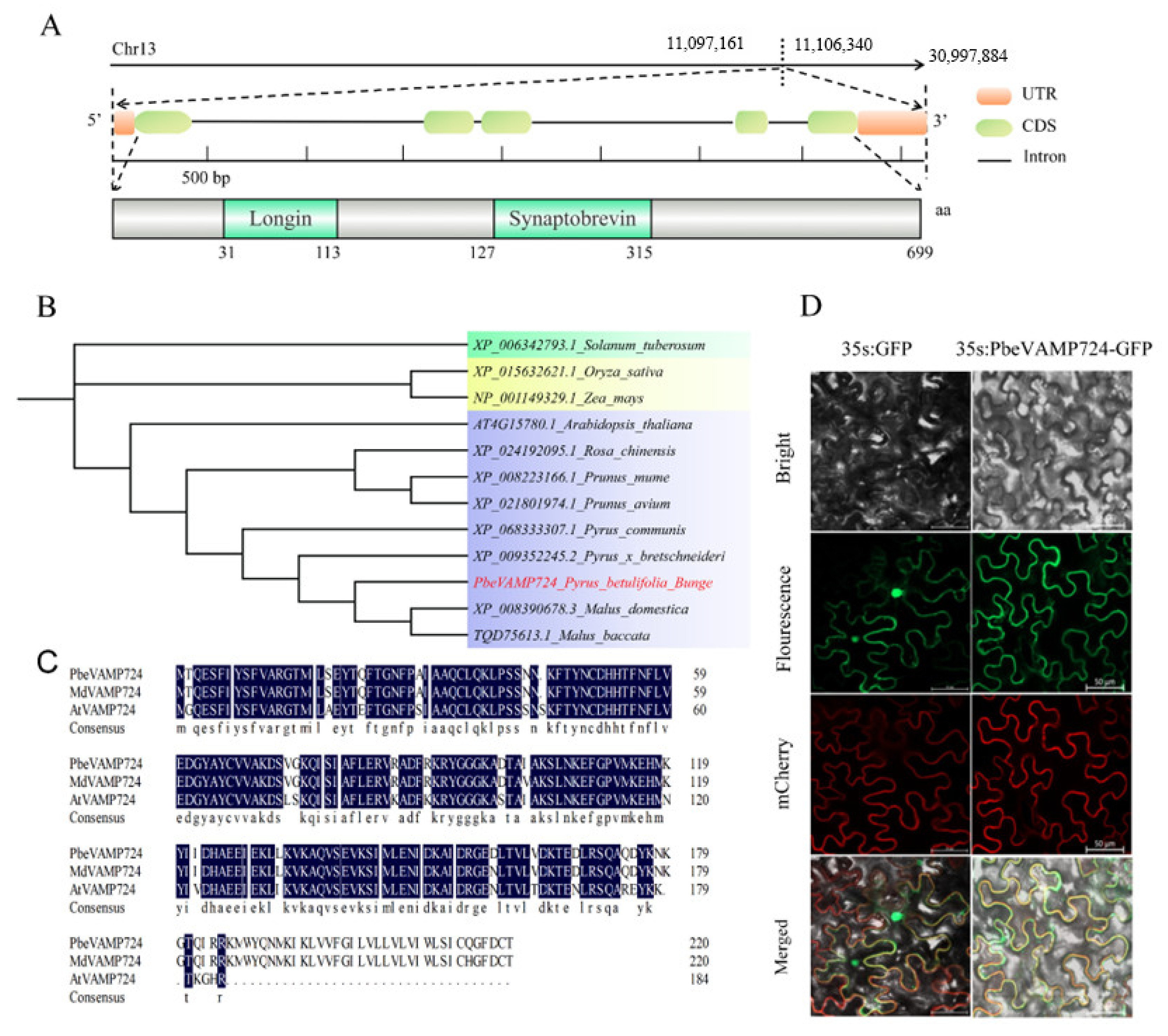
In order to analyze the potential function of Chr13.g22919 protein, we first analyzed its gene structure and domain composition. The results showed that the gene was located on chromosome 13 and contained UTR, CDS, and introns (Figure 1A). Domain analysis found that the protein did not contain signal peptide and a transmembrane region, but possess an N-terminal “Longin” domain, which is regulated by the regulated-SNARE-like domain and a C-terminal “Synaptobrevin” domain (Figure 1A). Subsequent BLAST searches against the NCBI database showed that the target sequence shared high homology with vesicle-associated membrane protein 724 (VAMP724) from diverse plant species and thus designated as PbeVAMP724. The phylogenetic tree displays that PbeVAMP724 has high homology with other species of Rosaceae (Figure 1B). Multiple sequence alignment analysis demonstrated that PbeVAMP724 shares 72.5% and 98.64% sequence identity with its homologous proteins from Arabidopsis thaliana and Malus × domestica, respectively (Figure 1C). Subcellular localization analysis revealed that this protein is localized to the cell membrane (Figure 1D). The above results indicate that PbeVAMP724 encodes a typical SNARE protein.

3.2. PbeVAMP724 Responds to Hormone Signals

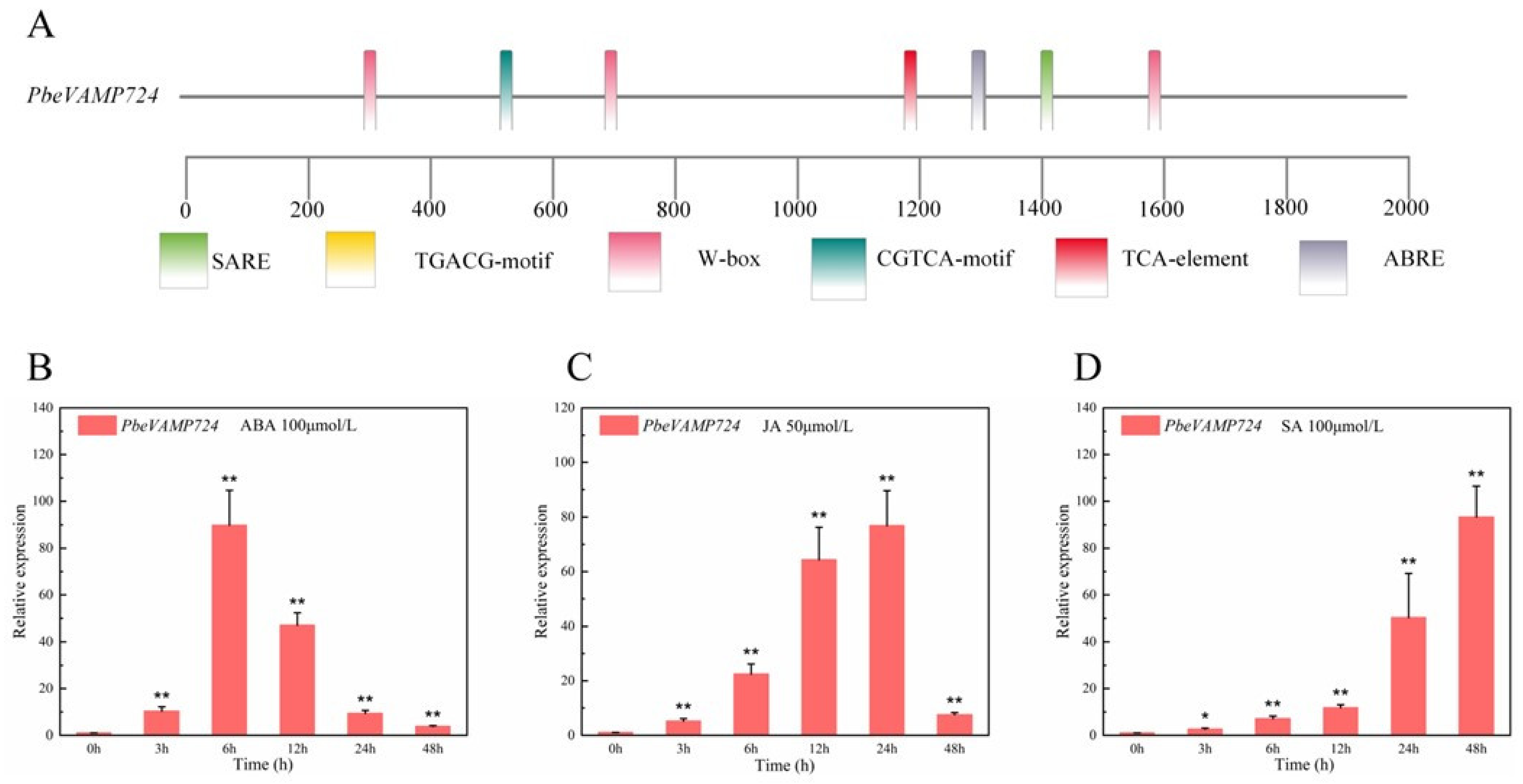
To clarify the potential signals involved in the responsiveness of PbeVAMP724, we first investigated cis-acting element prediction on its 2000 bp promoter region (Figure 2A). We determined 6 cis-elements, such as SARE, TCA-element, CGTCA-motif, W-box, and ABRE. Among these, TCA-element, ABRE and CGTCA-motif or TGACG-motif are well studied, which respond to SA, ABA, and JA signals, respectively. To verify this, we treated ‘Duli-G03’ suspension cells with ABA (100 μmol/L), JA (50 μmol/L), and SA (100 μmol/L), then detected PbeVAMP724 expression via qRT-PCR (Figure 2B–D). The results showed that PbeVAMP724 expression was significantly upregulated under treatments with each of the three distinct hormones (ABA, JA and SA). Under ABA treatment, relative expression levels peaked at 6 h, reaching 90-fold that of the control, and then gradually declined, though remaining elevated relative to the control (Figure 2B). By contrast, JA treatment induced a continuous increase in gene expression, with maximum accumulation detected at 24 h, reaching 80-fold that of the control (Figure 2C). For SA treatment, expression rose steadily over time and reached its peak at 48 h, reaching 90-fold that of the control, (Figure 2D). Collective findings suggest PbeVAMP724 may participate in hormone-mediated stress responses in plants.

3.3. PbeVAMP724 Was Differentially Expressed in Response to Vp Infection

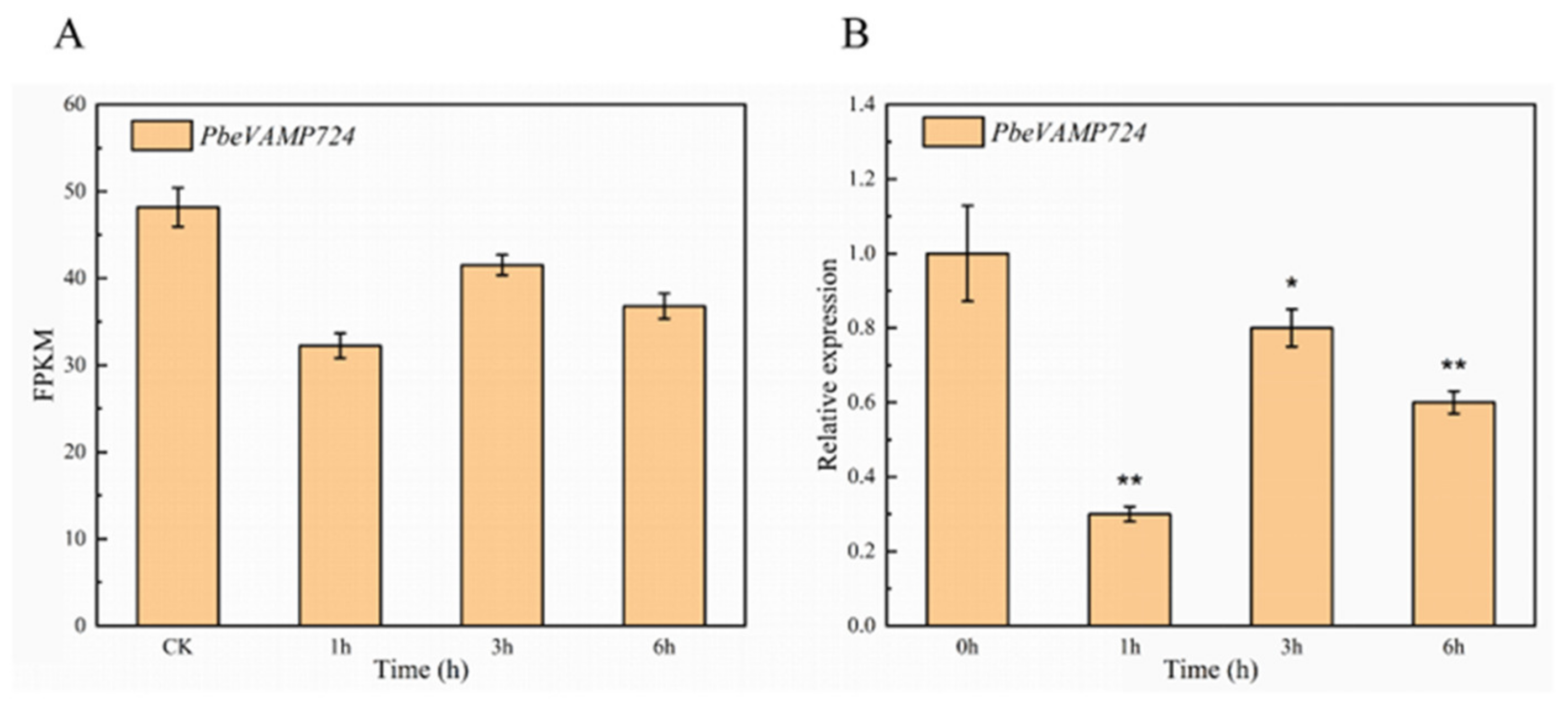
To characterize how PbeVAMP724 responds to Valsa canker infection, we first analyzed its expression pattern using previous RNA-seq data in ‘Duli-G03’ suspension cells treated with Valsa pyri metabolites (VpM) [25]. In RNA-seq data (Figure 3A), the FPKM values of PbeVAMP724 were lower in Vp-infected samples (1 h, 3 h, 6 h) than in the mock-inoculated control (CK), indicating downregulated expression during pathogen infection. qRT-PCR validation further confirmed this trend: compared with the 0 h control, PbeVAMP724 expression was significantly reduced at 1 h (Figure 3B) and showed differential changes (with a transient increase at 3 h followed by a decrease at 6 h) during Vp infection. These results collectively demonstrate that PbeVAMP724 is differentially expressed (predominantly downregulated) in response to Valsa canker infection.

3.4. PbeVAMP724 Positively Regulated Vp Resistance of Pear Fruit

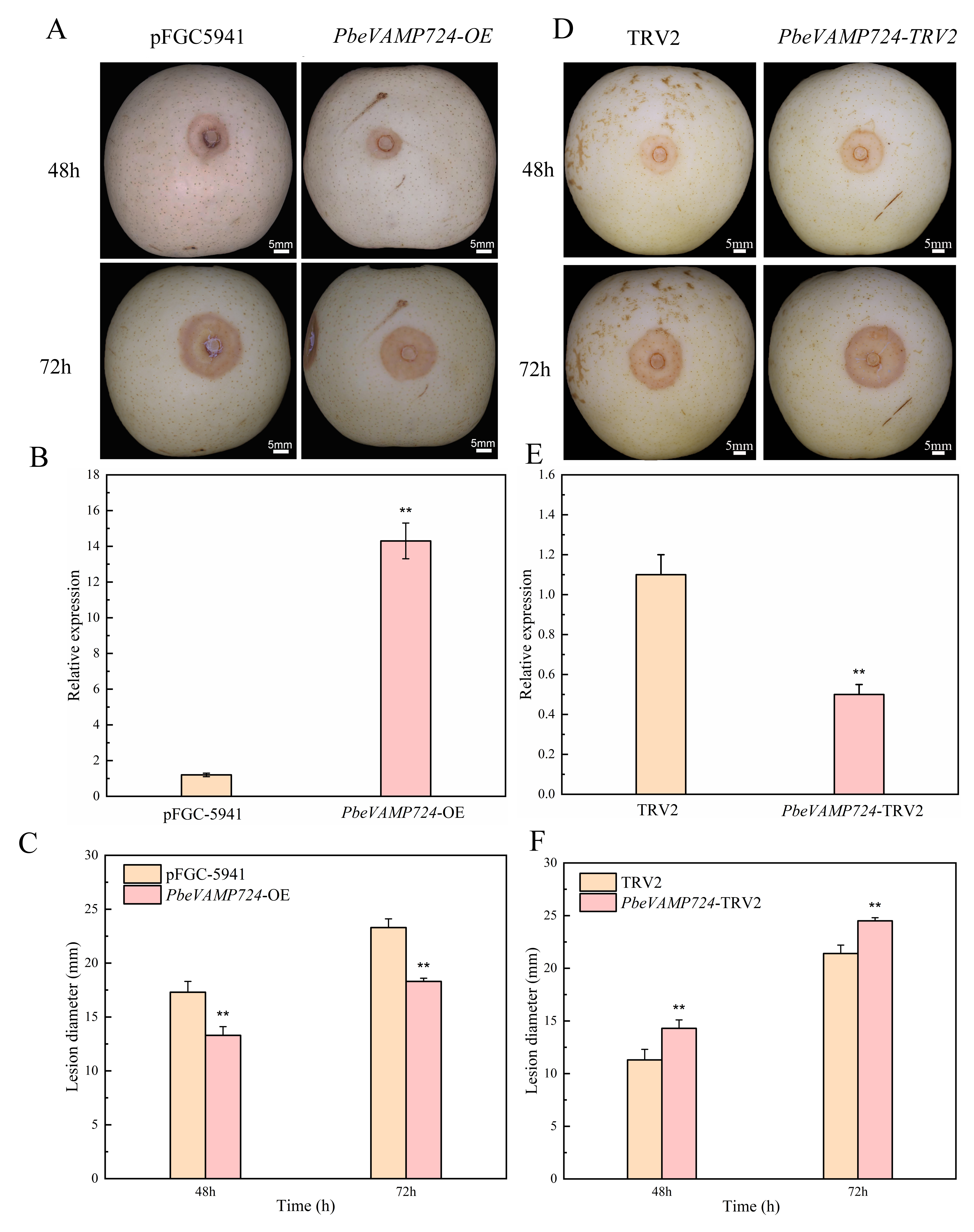
To assess the role of PbeVAMP724 in plant resistance to Vp, we generated PbeVAMP724-overexpressing (OE) lines and virus-induced gene silencing (VIGS, PbeVAMP724-TRV2) lines, followed by Vp inoculation. At 48 h and 72 h post-inoculation, PbeVAMP724-OE fruits exhibited milder disease symptoms than the empty vector control (pFGC5941; Figure 4A), and the lesion diameter was reduced by 23.1% at 48 h and 20.0% at 72 h compared to the empty vector control (pFGC-5941). qRT-PCR confirmed that PbeVAMP724 expression was significantly upregulated in OE lines (Figure 4B). Correspondingly, lesion diameters of PbeVAMP724-OE fruits were markedly smaller than those of controls at both 48 h and 72 h (Figure 4C). In contrast, PbeVAMP724-TRV2 fruits showed more severe disease symptoms compared to the TRV2 control (Figure 4D). qRT-PCR verified that PbeVAMP724 expression was significantly reduced in silenced lines (Figure 4E). Consistently, lesion diameters of PbeVAMP724-TRV2 fruits were markedly larger than those of controls at 72 h (Figure 4F), and the lesion diameter was increased by 36.4% at 48 h and 19.05% at 72 h compared to the empty vector control (TRV2). These results indicate that PbeVAMP724 positively regulates pear resistance to Vp infection.

3.5. Overexpression of PbeVAMP724 Enhanced Vp Resistanceof ‘Duli-G03’ Suspension Cells

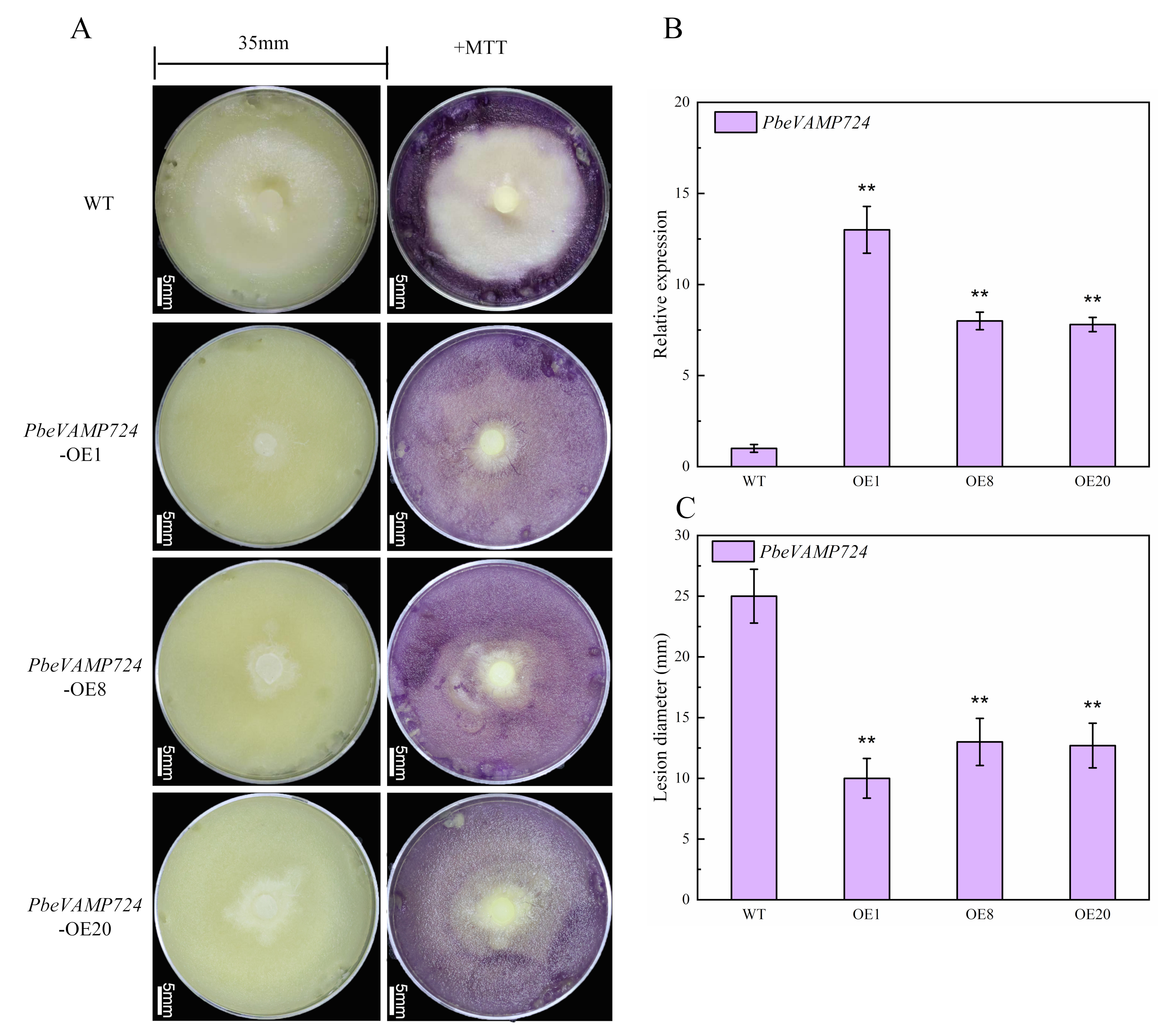
We next transformed PbeVAMP724 into wild type (WT) ‘Duli-G03’ suspension cells and obtained three overexpression lines, named PbeVAMP724-OE1, PbeVAMP724-OE8, and PbeVAMP724-OE20, respectively. After inoculation with pathogens, the growth of pathogens on PbeVAMP724 overexpressing cells was inhibited compared with the control, and MTT staining showed that the transgenic cell lines showed a higher survival rate than WT (Figure 5A). Compared with the control, the lesion areas of OE1, OE8, and OE20 were reduced by 60%, 47%, and 50%. The overexpression of PbeVAMP724 in transgenic lines was verified using qRT-PCR (Figure 5B). After 48 h of inoculation, the colony diameter on the WT cells was about 20 mm, while the colony diameter on the overexpression cell line was only 10 to 13 mm (Figure 5C). The PbeVAMP724-OE1 strain had the strongest resistance, with a colony diameter of 11 mm. These findings establish that overexpression of PbeVAMP724 drastically improved the resistance of pear ‘Duli-G03’ cells to Valsa canker.

3.6. PbeVAMP724 Boosts the Activity of ‘Duli-G03’ Suspension Cells and Promotes the Burst of Reactive Oxygen Species

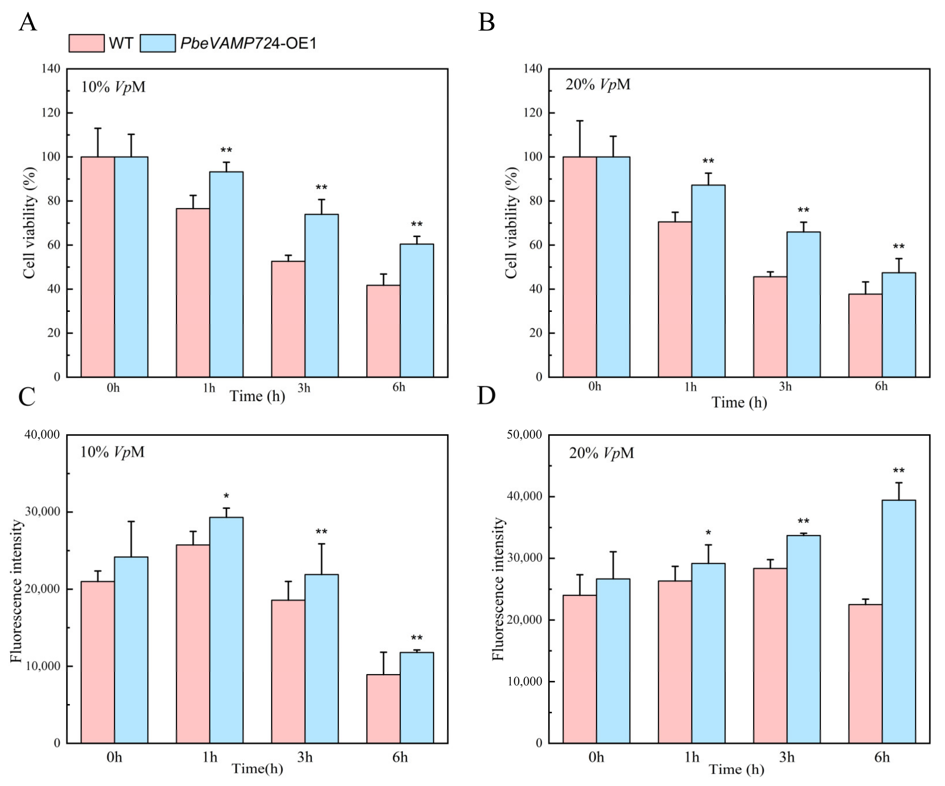
The suspension cells of wild type (WT) and PbeVAMP724 overexpression cell lines were treated with Valsa pyri metabolites (VpM). We found that under both 10% and 20% VpM treatments, cell viability exhibited a time-dependent decline. Specifically, after 6 h of 10% VpM treatment, WT cell viability decreased by 56%, while the OE1 line showed only 39% reduction. Similarly, at 6 h of 20% VpM treatment, WT viability dropped by 60%, whereas OE1 viability decreased by 55%. (Figure 6A,B). Correspondingly, ROS fluorescence intensity displayed a dynamic response: at 1 h of 10% VpM treatment, ROS accumulation in OE1 cells was 10.2% higher than in WT, and this difference became more pronounced at 6 h of 20% VpM treatment, whereas OE1 showed an 86.4% increase in ROS levels compared to WT (Figure 6C,D). These findings collectively indicate that PbeVAMP724 overexpression enhances cell viability retention and modulates ROS accumulation under VpM stress.

3.7. PbeVAMP724 Regulates Downstream Immune Signals

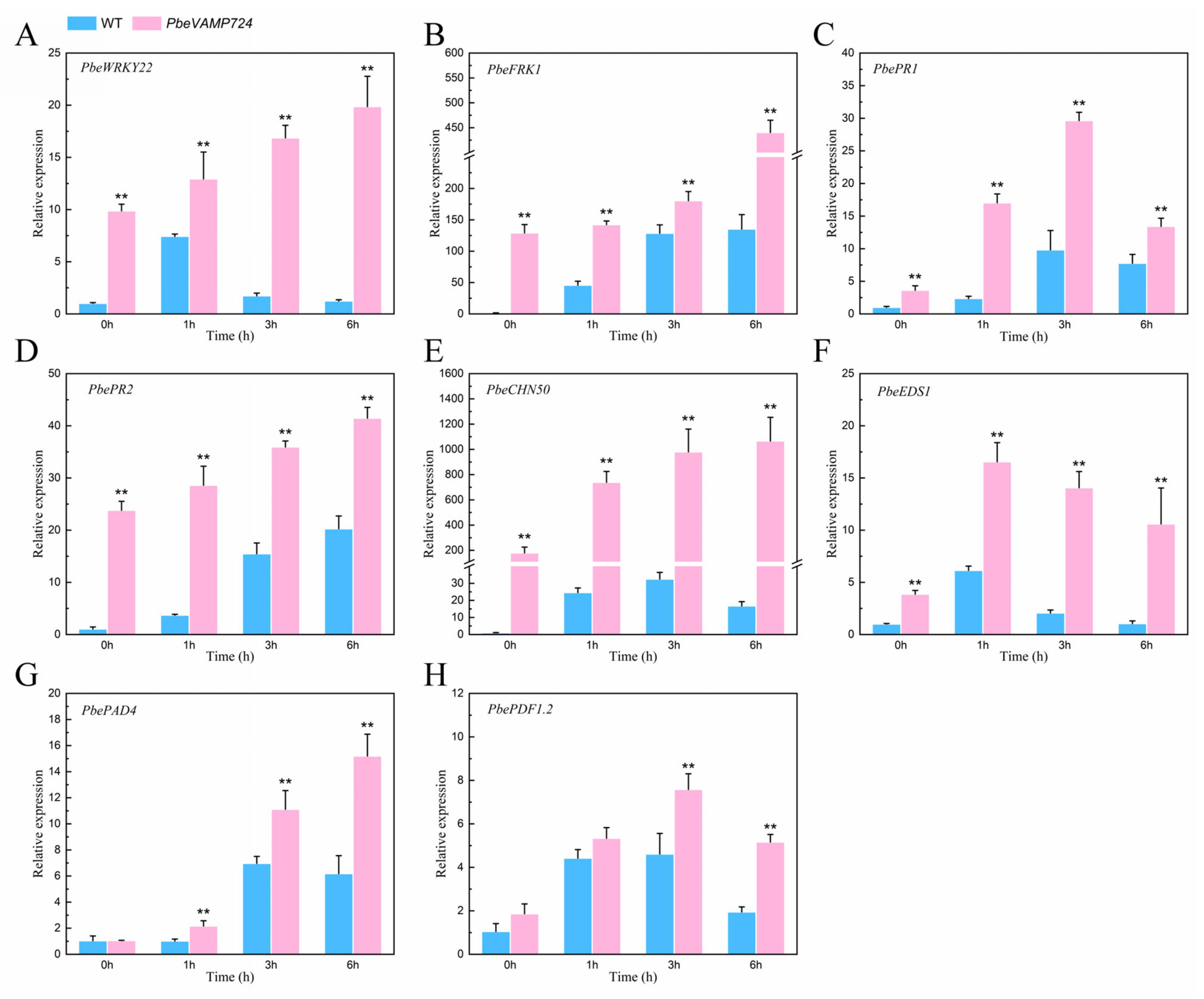
According to the functional detection results of PbeVAMP724, we used qRT-PCR to detect the expression levels of immune response-related genes in ‘Duli-G03’ wild type and PbeVAMP724 cell lines. We found that multiple signaling pathways were activated during VpM treatment of suspension cells (Figure 7). PTI-related genes PbeWRKY22 and PbeFRK1 were not significantly increased in wild type, but showed different degrees of upregulation in PbeVAMP724 (Figure 7A,B). SA-related genes PbePR1, Pbe CHN50, and PbePR2 also showed the same trend as PTI-related genes. Among them, the upward trend of PbeCHN50 in PbeVAMP724 was the most obvious, even after 6 h of VpM treatment, it reached 500 times that of the wild type (Figure 7C–E). Compared to the WT cells, overexpression of PbeVAMP724 resulted in greater expression of the R-gene-related genes PbeEDS1 and PbePAD4 at 1 h and 6 h after VpM treatment (Figure 7F,G). In addition, jasmonic acid-related genes were also up-regulated at different time points. By and large, overexpression of PbeVAMP724 significantly activated the expression of downstream disease resistance-related genes, indicating that PbeVAMP724 enhances the disease resistance of plants by regulating the transcription of defense genes.

3.8. Co-Expression Network, GO Enrichment, and Expression Level Validation of PbeVAMP724 in P. betulifolia

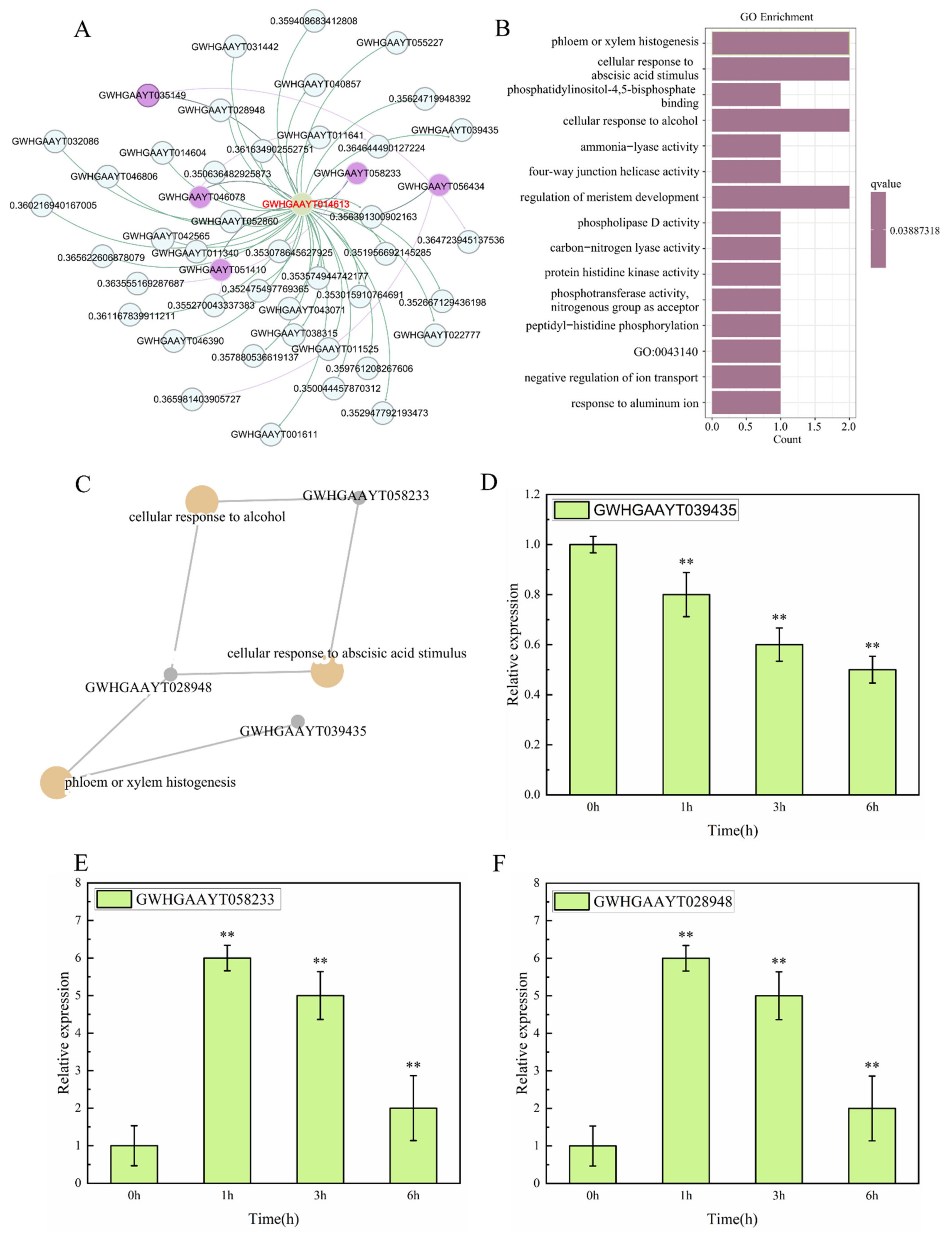
To elucidate the potential mechanism by which PbeVAMP724 regulates pear resistance to Valsa canker, we systematically analyzed transcriptome data and identified a gene module significantly co-expressed with PbeVAMP724 (|r| > 0.35). We found that 23 genes were associated with PbeVAMP724 (Figure 8A), where PbeVAMP724 served as the core node. Using GO enrichment analysis on these 23 genes, the results showed that functional terms such as “phloem or xylem histogenesis”, “cellular response”, “cellular response to alcohol”, and “regulation of meristem development” were significantly enriched (Figure 8B). Among these, “phloem or xylem histogenesis” exhibited the highest enrichment level, suggesting that PbeVAMP724 may be involved in regulating vascular tissue-related processes in pear. Further analysis of gene intersections across enriched pathways revealed that GWHGAAYT058233, GWHGAAYT028948, and GWHGAAYT039435 are simultaneously involved in the pathways of “phloem or xylem histogenesis”, “cellular response to alcohol”, and “cellular response to abscisic acid stimulus” (Figure 8C). For instance, GWHGAAYT058233 and GWHGAAYT028948 overlap in the “cellular response to alcohol” pathway; GWHGAAYT028948 and GWHGAAYT039435 are associated with the “phloem or xylem histogenesis” pathway. To validate the above co-expression relationships, we detected the expression patterns of these three genes after treatment with VpM using qRT-PCR. The expression level of GWHGAAYT039435 showed no significant change after treatment (Figure 8D; in contrast, the expression levels of GWHGAAYT058233 (Figure 8E) and GWHGAAYT028948 (Figure 8F) increased significantly with prolonged treatment time, consistent with the expression trend of PbeVAMP724. In summary, we demonstrated that PbeVAMP724 participates in pear resistance to Valsa canker by regulating the expression of genes related to vascular tissue development and cellular stress responses, among which GWHGAAYT058233 and GWHGAAYT028948 are key downstream co-regulatory genes.

4. Discussion

Vesicle transport-related genes play crucial roles in mediating plant immune responses by regulating protein localization, signal transmission, and cell wall repair [18,31]. As key members of the SNARE protein superfamily, VAMP72 genes have been reported to participate in plant disease resistance. For instance, AtVAMP721/722 enhancing pathogen resistance through mediating defense-related protein secretion [32]. Zhu et al. [33] demonstrated that tomato VAMP727 enhances plant resistance to necrotrophic fungi by mediating the vesicle secretion of pathogenesis-related (PR) proteins, such as PR1 and chitinase, suggesting that VAMP72 family proteins may participate in plant defense responses via distinct pathways. In this study, PbeVAMP724 was isolated from resistant rootstock P. betulifolia and bioinformatics analysis revealed it encodes a typical SNARE protein with conserved Longin and Synaptobrevin domains, localized exclusively at the cell membrane. This structural feature is consistent with the characteristics of VAMP72 family members in other plants, which are known to regulate vesicle fusion and participate in immune signaling [34]. Phylogenetic analysis showed PbeVAMP724 has high homology with Rosaceae species, especially 98.64% identity with Malus × domestica homologous protein, suggesting evolutionary conservation and potential functional similarity in disease resistance.
Plant hormone signaling pathways (ABA, SA, JA) are closely involved in regulating immune responses against pathogens [35]. In this study, Cis-acting element prediction identified SARE, TCA-element, and ABRE in the PbeVAMP724 promoter, which are responsive to SA, ABA, and JA signals. qRT-PCR verification showed PbeVAMP724 expression was significantly induced by these three hormones, with distinct temporal expression patterns. This indicates PbeVAMP724 may integrate multiple hormone signals to participate in pear stress responses. Additionally, PbeVAMP724 expression was differentially regulated under Vp infection, further supporting its involvement in pathogen-triggered defense signaling. It should be noted that this study was conducted under controlled conditions; field trials are required to validate resistance.
Functional verification through transient overexpression and VIGS in ‘Huangguan’ pear fruits demonstrated that PbeVAMP724 overexpression reduced lesion expansion, while gene silencing enhanced disease susceptibility. Stable overexpression in ‘Duli’ suspension cells further confirmed its positive regulatory role in resistance to Vp. Interestingly, PbeVAMP724-OE cells maintained higher viability and promoted ROS burst under VpM treatment. ROS accumulation is an early defense response in plants, and an appropriate ROS burst can enhance pathogen resistance by regulating cell wall reinforcement and defense gene expression [36,37]. The dynamic regulation of ROS by PbeVAMP724 may be a key mechanism underlying its resistance function. It should be noted that excessive ROS accumulation carries potential risks to cellular integrity, as high levels of ROS can trigger oxidative damage to lipids, proteins, and nucleic acids, leading to metabolic disorders or even programmed cell death [38,39]. Therefore, maintaining a dynamic redox balance between ROS production and scavenging is essential for normal cellular function and sustainable defense responses. Plant defense responses against necrotrophic pathogens rely on the coordinated activation of multi-layered signaling networks, with defense-related genes acting as core executors of resistance [40]. For instance, PTI marker genes like WRKY22 and FRK1 are rapidly induced upon pathogen recognition, initiating early immune signaling events such as protein phosphorylation and ROS burst [41,42], while salicylic acid (SA)-pathway genes including PR1, PR2, and CHN50 reinforce systemic acquired resistance (SAR) and inhibit pathogen colonization [41,42]. R-gene-related genes such as EDS1 and PAD4 further amplify defense responses by mediating effector-triggered immunity (ETI) and hypersensitive response (HR) [43,44], and JA pathway genes like PDF1.2 contribute to resistance against necrotrophic pathogens by regulating secondary metabolite synthesis and cell wall reinforcement [45,46]. In this study, qRT-PCR analysis showed PbeVAMP724 overexpression significantly upregulated the expression of PbeWRKY22, PbeFRK1 (PTI-related), PbePR1, PbePR2, PbeCHN50 (SApathway), PbeEDS1, PbePAD4 (R-gene-related), and PbePDF1.2 (JA pathway), suggesting PbeVAMP724 activating PTI, SA, R-gene-related, and JA pathway gene expression. Among these, PbeCHN50 showed the most dramatic upregulation (500-fold higher than WT at 6 h post-VpM treatment), indicating it may be a key downstream target of PbeVAMP724 in regulating pear resistance to Valsa canker.
Phloem and xylem are important tissues for pathogen colonization and nutrient transport [47,48], and genes involved in their development may enhance resistance by strengthening tissue structure or inhibiting pathogen spread. WGCNA identified 23 genes co-expressed with PbeVAMP724 under VpM stimulation, and GO enrichment analysis revealed enrichment in terms related to “phloem and xylem histogenesis”, “cellular response to alcohol”, and “regulation of meristem”. This indicates PbeVAMP724 may participate in resistance through regulating vascular tissue development and stress response-related biological processes, forming a complex regulatory network with co-expressed genes.
Collectively, this study demonstrates that PbeVAMP724 positively regulates Valsa canker resistance in pyrus species. ABA, SA, JA, and ROS signals, as well as the vascular development process, are involved in the PbeVAMP724-mediated immune response. Our results provide important insights into the molecular mechanisms of regulation of resistance to Valsa canker and have implications for future breeding for resistance, so future studies should evaluate interaction with microbiome and response to multiple pathogens.

Supplementary Materials

The following supporting information can be downloaded at: https://www.mdpi.com/article/10.3390/horticulturae12020245/s1, Table S1, Genes and primer information associated with qRT-PCR.

Author Contributions

C.Z. conceived, designed, and coordinated this study. H.H. and W.W. performed the experiments. M.C., Y.L. and E.S. performed, collected, analyzed, and deposited the data. C.Z. and Q.C. proofread the final draft. All authors have read and agreed to the published version of the manuscript.

Funding

This research was funded by the Industrial Support Project of Higher Education Institutions in Gansu Province (2025CYZC-035); the National Natural Science Foundation of China (32260741); Longyuan Talent Project of Gansu Province (LYYC-2024-04) and Fuxi Talent Project of Gansu Agricultural University (Gaufx-03Y12).

Data Availability Statement

The original contributions presented in this study are included in the article and supplementary material. Further inquiries can be directed to the corresponding authors.

Acknowledgments

The authors would like to thank China National Knowledge Infrastructure (CNKI) for providing access to e-resources.

Conflicts of Interest

The authors declare no conflicts of interest.

References

  1. Li, L.M.; Zheng, T.; Chen, Y.; Sui, Y.; Ding, R.; Hou, L.; Zheng, F.; Zhu, C. The antagonistic mechanisms of Streptomyces sioyaensis on the growth and metabolism of poplar canker pathogen Valsa sordida. Biol. Control 2020, 151, 104392. [Google Scholar] [CrossRef] [Scilit]
  2. Fan, X.L.; Du, Z.; Liang, Y.M.; Tian, C.M. Melanconis (Melanconidaceae) associated with Betula spp. in China. Mycol. Prog. 2016, 15, 40. [Google Scholar] [CrossRef] [Scilit]
  3. Feng, H.; Wang, C.L.; He, Y.T.; Tang, L.; Han, P.L.; Liang, J.H.; Huang, L.L. Apple Valsa canker: Insights into pathogenesis and disease control. Phytopathol. Res. 2023, 5, 45. [Google Scholar] [CrossRef] [Scilit]
  4. Wang, X.L.; Shi, C.M.; Gleason, M.L.; Huang, L.L. Fungal species associated with apple Valsa canker in East Asia. Phytopathol. Res. 2020, 2, 35. [Google Scholar] [CrossRef] [Scilit]
  5. Abe, K.; Kotoda, N.; Kato, H.; Soejima, J.I. Genetic studies on resistance to Valsa canker in apple: Genetic variance and breeding values estimated from intra- and inter-specific hybrid progeny populations. Tree Genet. Genomes 2011, 7, 363–372. [Google Scholar] [CrossRef] [Scilit]
  6. Holland, L.A.; Travadon, R.; Lawrence, D.P.; Nouri, M.T.; Trouillas, F.P. Evaluation of pruning wound protection products for the management of almond canker diseases in California. Plant Dis. 2021, 105, 3368–3375. [Google Scholar] [CrossRef] [Scilit] [PubMed]
  7. Wang, X.L.; Zang, R.; Yin, Z.Y.; Kang, Z.S.; Huang, L.L. Delimiting cryptic pathogen species causing apple Valsa canker with multilocus data. Ecol. Evol. 2014, 4, 1369–1380. [Google Scholar] [CrossRef] [Scilit] [PubMed]
  8. Liu, S.S.; Tao, C.Y.; Xu, X.; Shen, Z.Z.; Kuramae, E.E.; Wang, Z.; Li, C.Y.; Li, R.; Shen, Q.R.; Kowalchuk, G.A. Harnessing soil feedback from resistant bananas to suppress pathogens in susceptible varieties. J. Appl. Ecol. 2025, 63, 1365–2664. [Google Scholar] [CrossRef] [Scilit]
  9. Dodds, P.N.; Rathjen, J.P. Plant immunity: Towards an integrated view of plant–pathogen interactions. Nat. Rev. Genet. 2010, 11, 539–548. [Google Scholar] [CrossRef] [Scilit] [PubMed]
  10. Shan, L.B.; He, P.; Li, J.M.; Heese, A.; Peck, S.C.; Nürnberger, T.; Martin, G.B.; Sheen, J. Bacterial effectors target the common signaling partner BAK1 to disrupt multiple MAMP receptor-signaling complexes and impede plant immunity. Cell Host Microbe 2008, 4, 17–27. [Google Scholar] [CrossRef] [Scilit] [PubMed]
  11. Bjornson, M.; Zipfel, C. Plant immunity: Crosstalk between plant immune receptors. Curr. Biol. 2021, 31, R796–R798. [Google Scholar] [CrossRef] [Scilit] [PubMed]
  12. Boller, T.; Felix, G. A Renaissance of Elicitors: Perception of Microbe-Associated Molecular Patterns and Danger Signals by Pattern-Recognition Receptors. Annu. Rev. Plant Biol. 2009, 60, 379–406. [Google Scholar] [CrossRef] [Scilit] [PubMed]
  13. Wu, C.H.; Derevnina, L.; Kamoun, S. Receptor networks underpin plant immunity. Science 2018, 360, 1300–1301. [Google Scholar] [CrossRef] [Scilit] [PubMed]
  14. Ngou, B.P.M.; Jones, J.D.G.; Ding, P.T. Plant immune networks. Trends Plant Sci. 2022, 27, 255–273. [Google Scholar] [CrossRef] [Scilit] [PubMed]
  15. Remick, B.C.; Gaidt, M.M.; Vance, R.E. Effector-Triggered Immunity. Annu. Rev. Immunol. 2023, 41, 453–481. [Google Scholar] [CrossRef] [Scilit] [PubMed]
  16. Yuan, M.H.; Ngou, B.P.M.; Ding, P.T.; Xin, X.F. PTI-ETI crosstalk: An integrative view of plant immunity. Plant Cell Rep. 2021, 62, 102030. [Google Scholar] [CrossRef] [Scilit] [PubMed]
  17. Yu, X.Q.; Niu, H.Q.; Liu, C.; Wang, H.L.; Yin, W.L.; Xia, X.L. PTI-ETI synergistic signal mechanisms in plant immunity. Plant Biotechnol. J. 2024, 22, 2113–2128. [Google Scholar] [CrossRef] [Scilit] [PubMed]
  18. Yun, H.S.; Kwon, C. Vesicle trafficking in plant immunity. Curr. Opin. Plant Biol. 2017, 40, 34–42. [Google Scholar] [CrossRef] [Scilit] [PubMed]
  19. Heller, J.; Clavé, C.; Gladieux, P.; Saupe, S.J.; Glass, N.L. NLR surveillance of essential SEC-9 SNARE proteins induces programmed cell death upon allorecognition in filamentous fungi. Proc. Natl. Acad. Sci. USA 2018, 115, E2292–E2301. [Google Scholar] [CrossRef] [Scilit] [PubMed]
  20. Fujimoto, M.; Ebine, K.; Nishimura, K.; Tsutsumi, N.; Ueda, T. Longin R-SNARE is retrieved from the plasma membrane by ANTH domain-containing proteins in Arabidopsis. Proc. Natl. Acad. Sci. USA 2020, 117, 25150–25158. [Google Scholar] [CrossRef] [Scilit] [PubMed]
  21. Sollner, T.; Bennett, M.K.; Whiteheart, S.W.; Scheller, R.H.; Rothman, J.E. A protein assembly-disassembly pathway in vitro that may correspond to sequential steps of synaptic vesicle docking, activation, and fusion. Cell 1993, 75, 409–418. [Google Scholar] [CrossRef] [Scilit] [PubMed]
  22. Gao, L.Y.; Pei, Y.K.; Wang, P.; Cen, Y.H.; Yan, X.; Hou, Y.X. Cotton SNARE complex component GhSYP121 regulates salicylic acid signaling during defense against Verticillium dahliae. J. Cell. Physiol. 2024, 239, e31329. [Google Scholar] [CrossRef] [Scilit] [PubMed]
  23. Lin, L.L.; Wu, Q.Q.; Wang, S.; Gong, Q.; Huang, X.W.; Abubakar, Y.S.; Liu, Y.; Cao, J.Y.; Hu, J.X.; Wang, Z.H.; et al. Recycling of Trans-Golgi SNAREs Promotes Apoplastic Effector Secretion for Effective Host Invasion in Magnaporthe oryzae. Plant Cell Environ. 2025, 48, 6047–6065. [Google Scholar] [CrossRef] [Scilit] [PubMed]
  24. Gu, Y.N.; Zavaliev, R.; Dong, X.N. Membrane Trafficking in Plant Immunity. Mol. Plant. 2017, 10, 1026–1034. [Google Scholar] [CrossRef] [Scilit] [PubMed]
  25. Zhao, D.; Tian, Y.Z.; Yu, H.Q.; Mao, X.; Wang, C.; Duo, H.; Sun, E.; Zuo, C.W. Establishment of the “Valsa pyri metabolites (VpM)-suspension cell”-based system to study the response of pears to VpM. Physiol. Mol. Plant Pathol. 2022, 120, 101850. [Google Scholar] [CrossRef] [Scilit]
  26. Mao, X.; Wang, C.; Lv, Q.Q.; Tian, Y.Z.; Wang, D.D.; Chen, B.H.; Mao, J.; Li, W.F.; Chu, M.Y.; Zuo, C.W. Cyclic nucleotide gated channel genes (CNGCs) in Rosaceae: Genome-wide annotation, evolution and the roles on Valsa canker resistance. Plant Cell Rep. 2021, 40, 2369–2382. [Google Scholar] [CrossRef] [Scilit] [PubMed]
  27. Dobin, A.; Davis, C.A.; Schlesinger, F.; Drenkow, J.; Zaleski, C.; Jha, S.; Batut, P.; Chaisson, M.; Gingeras, T.R. STAR: Ultrafast universal RNA-seq aligner. Bioinformatics 2013, 29, 15–21. [Google Scholar] [CrossRef] [Scilit] [PubMed]
  28. Cantalapiedra, C.P.; Hernández-Plaza, A.; Letunic, I.; Bork, P.; Huerta-Cepas, J. eggNOG-mapper v2: Functional Annotation, Orthology Assignments, and Domain Prediction at the Metagenomic Scale. Mol. Biol. Evol. 2021, 38, 5825–5829. [Google Scholar] [CrossRef] [Scilit] [PubMed]
  29. Yu, G.; Wang, L.G.; Han, Y.; He, Q.Y. clusterProfiler: An R package for comparing biological themes among gene clusters. OMICS 2012, 16, 284–287. [Google Scholar] [CrossRef] [Scilit] [PubMed]
  30. Zuo, C.W.; Mao, J.; Chen, Z.; Chu, M.Y.; Duo, H.; Chen, B.H. RNA sequencing analysis provides new insights into dynamic molecular responses to Valsa mali pathogenicity in apple ‘Changfu No. 2’. Tree Genet. Genomes 2018, 14, 75. [Google Scholar] [CrossRef] [Scilit]
  31. Liu, P.F.; Li, M.M.; Liu, R.R.; Zhao, Y.P. Diverse Role of SNARE Protein GS28 in Vesicle Trafficking and Diseases. Curr. Protein Pept. Sci. 2023, 24, 288–295. [Google Scholar] [CrossRef] [Scilit] [PubMed]
  32. Wang, Y.; Cordewener, J.H.G.; America, A.H.P.; Shan, W.X.; Bouwmeester, K.; Govers, F. Arabidopsis Lectin Receptor Kinases LecRK-IX.1 and LecRK-IX.2 Are Functional Analogs in Regulating Phytophthora Resistance and Plant Cell Death. Mol. Plant-Microbe Interact. 2015, 28, 1032–1048. [Google Scholar] [CrossRef] [Scilit] [PubMed]
  33. Zhu, X.F.; Liu, Y.; Gai, X.T.; Zhou, Y.; Xia, Z.Y.; Chen, L.J.; Duan, Y.X.; Xuan, Y.H. SNARE proteins SYP22 and VAMP727 negatively regulate plant defense. Plant Signal. Behavior 2019, 14, 1610300. [Google Scholar] [CrossRef] [Scilit] [PubMed]
  34. Wang, Y.; Bouwmeester, K. L-type lectin receptor kinases: New forces in plant immunity. PLoS Pathog. 2017, 13, e1006433. [Google Scholar] [CrossRef] [Scilit] [PubMed]
  35. Ngou, B.P.M.; Ding, P.T.; Jones, J.D.G. Thirty years of resistance: Zig-zag through the plant immune system. Plant Cell 2022, 34, 1447–1478. [Google Scholar] [CrossRef] [Scilit] [PubMed]
  36. Luo, X.M.; Xu, N.; Huang, J.K.; Gao, F.; Zou, H.S.; Boudsocq, M.; Coaker, G.; Liu, J. A Lectin Receptor-Like Kinase Mediates Pattern-Triggered Salicylic Acid Signaling. Plant Physiol. 2017, 174, 2501–2514. [Google Scholar] [CrossRef] [Scilit] [PubMed]
  37. Mittler, R. ROS are good. Trends Plant Sci. 2017, 22, 11–19. [Google Scholar] [CrossRef] [Scilit] [PubMed]
  38. Bolwell, G.P.; Daudi, A. Reactive Oxygen Species in Plant–Pathogen Interactions; Signaling and Communication in Plants; Springer: Berlin/Heidelberg, Germany, 2009. [Google Scholar] [CrossRef] [Scilit]
  39. Jones, J.D.G.; Dangl, J.L. The plant immune system. Nature 2006, 444, 323–329. [Google Scholar] [CrossRef] [Scilit] [PubMed]
  40. Hsu, F.C.; Chou, M.Y.; Chou, S.J. Submergence confers immunity mediated by the WRKY22 transcription factor in Arabidopsis. Plant Cell. 2013, 25, 2699–2713. [Google Scholar] [CrossRef] [Scilit] [PubMed]
  41. An, C.; Mou, Z. Salicylic acid and its function in plant immunity. J. Integr. Plant Biol. 2011, 53, 412–428. [Google Scholar] [CrossRef] [Scilit] [PubMed]
  42. Lee, H.I.; León, J.; Raskin, I. Biosynthesis and metabolism of salicylic acid. Proc. Natl. Acad. Sci. USA 1995, 92, 4076–4079. [Google Scholar] [CrossRef] [Scilit] [PubMed]
  43. Tailor, A.; Bhatla, S.C. R gene-mediated resistance in the management of plant diseases. J. Plant Biochem. Biotechnol. 2024, 33, 5–23. [Google Scholar] [CrossRef] [Scilit]
  44. Malik, N.; George, J.K. Resistance genes in Piper colubrinum: In silico survey from leaf transcriptome and expression studies upon challenge inoculation with Phytophthora capsici. Appl. Biochem. Biotechnol. 2018, 184, 987–1008. [Google Scholar] [CrossRef] [Scilit] [PubMed]
  45. Turner, J.G.; Ellis, C.; Devoto, A. The jasmonate signal pathway. Plant Cell 2002, 14, S153–S164. [Google Scholar] [CrossRef] [Scilit] [PubMed]
  46. Ruan, J.; Zhou, Y.; Zhou, M. Jasmonic acid signaling pathway in plants. Int. J. Mol. Sci. 2019, 20, 2479. [Google Scholar] [CrossRef] [Scilit] [PubMed]
  47. Bové, J.M.; Garnier, M. Phloem-and xylem-restricted plant pathogenic bacteria. Plant Sci. 2003, 164, 423–438. [Google Scholar] [CrossRef] [Scilit]
  48. Lacointe, A.; Minchin, P.E.H. Modelling phloem and xylem transport within a complex architecture. Funct. Plant Biol. 2008, 35, 772–780. [Google Scholar] [CrossRef] [Scilit] [PubMed]
Figure 1. Bioinformatics analysis and subcellular localization of Pyrus betulifolia vesicle-associated membrane protein 724 (PbeVAMP724). (A) The structural domain of PbeVAMP724, consisting of an N-terminal “Longin” domain and a C-terminal “Synaptobrevin” domain. (B) Phylogenetic tree constructed from the full-length peptide sequences of PbeVAMP724. (C) Multiple sequence alignment of PbeVAMP724. (D) Subcellular localization of the PbeVAMP724 protein in Nicotiana benthamiana leaves. PbeVAMP724-GFP is localized in the cell membrane. Green fluorescence indicates the PbeVAMP724–GFP fusion protein, red fluorescence indicates the membrane marker, and yellow fluorescence in the merged panel represents the co-localization between the target protein and the plasma membrane.
Horticulturae 12 00245 g001
Figure 2. Cis-acting elements in the PbeVAMP724 promoter and its expression patterns under hormone treatments. (A) Schematic diagram of cis-regulatory elements in the PbeVAMP724 promoter region. Different colors represent distinct elements: SARE (green), TGACG-motif (yellow), W-box (pink), CGTCA-motif (teal), TCA-element (dark red), and ABRE (gray). (BD) Relative expression levels of PbeVAMP724 (detected by qRT-PCR) at different time points under hormone treatments. (B) Treatment with 100 μM ABA; (C) treatment with 50 μM JA; (D) treatment with 100 μM SA. The y-axis indicates relative expression levels, normalized to the control group (the expression value in the 0 h was set to 1). Data are presented as mean ± standard deviation (n = 3 biological replicates). Statistical significance relative to the 0 h control: * p < 0.05 and ** p < 0.01. Vertical bars represent ± SD.
Horticulturae 12 00245 g002
Figure 3. The expression pattern of PbeVAMP724 in response to the pathogen signaling. (A) The expression pattern of PbeVAMP724 from RNAseq data with ‘Duli-G03’ suspension cells treated by Valsa pyri (Vp) for 1, 3, and 6 h. FPKM: fragments per kilobase of exon model per million mapped fragments. (B) Expression pattern of PbeVAMP724 by qRT-PCR. For bars in each group, the asterisk means significantly different at * p < 0.05 and ** p < 0.01. Vertical bars represent ± SD.
Horticulturae 12 00245 g003
Figure 4. Expression of PbeVAMP724 enhances the resistance of fruit of the susceptible pear variety Pyrus bretschneideri ‘Huangguan’ by Vp and. Overexpression was achieved using the in pFGC-5941 vector, and virus-induced gene-silencing was achieved using the TRV2 vector. The empty vectors were used as the controls. Infiltrated fruits were incubated in darkness at 25 °C for 3 d, after which they were inoculated with the Valsa canker pathogens. (A) Lesions at the infiltration sites of the overexpressing fruits at 72 h after infection with Valsa. (B) The lesion diameters on the empty-vector controls and overexpression fruits following infection with Valsa. (C) Gene expression level of overexpressing. (D) Lesions at the infiltration sites of the TVR2-silenced fruits at 72 h after infection with Valsa. (E) The lesion diameters on the empty-vector controls and silenced and TVR2-silenced fruits following infection with Valsa. (F) Gene expression level of TVR2-silenced fruits. Data are means (±SD), n = 3. Significant differences compared with the empty-vector controls were determined using Student’s t-test: * p < 0.05, ** p < 0.01.
Horticulturae 12 00245 g004
Figure 5. Expression of PbeVAMP724 positively regulates the resistance of ‘Duli-G03’ suspension cells to Vp. (A) Lesions on potato dextrose agar plates of wild-type (WT) cells and cells of three PbeVAMP724-overexpressing lines 2 d after inoculation with Vp. Plates were stained with MTT to distinguish living cells (purple). (B) The relative expression level of PbeVAMP724 in the transgenic lines was determined using qRT-PCR, using Actin as the reference. Expression is relative to the WT, the value of which was set as 1. (C) Lesion (size of pathogen mycelial growth) diameters on plates of the WT and transgenic lines 48 h after inoculation with Vp. All data are means (±SD), n = 3. Significant differences compared with the WT were determined using Student’s t-test: ** p < 0.01.
Horticulturae 12 00245 g005
Figure 6. Cell viability and reactive oxygen species (ROS) under treatment with 10% and 20% VpM. (A,B) Cell viability under treatment with 10% and 20% VpM at 0 h, 1 h, 3 h, and 6 h. (C,D) Reactive oxygen species (ROS) under treatment with 10% and 20% VpM at 0 h, 1 h, 3 h, and 6 h. All data are means (±SD), n = 3. Significant differences compared with the WT were determined using Student’s t-test: * p < 0.05,** p < 0.01.
Horticulturae 12 00245 g006
Figure 7. Expression levels of defense marker genes in suspension cells of the Pyrus betulifolia ‘Duli-G03’ wild-type (WT), cells overexpressing PbeVAMP724 following treatment with a 20% solution of VpM. (AG) Relative expression of (A) PbeWRKY22, (B) PbeFRK1, (C) PbePR1, (D) PbePR2, (E) PbeCHN50, (F) PbeEDS1, (G) PbePAD4 and (H) PbePDF1.2, as determined by qRT-PCR. Actin was used as the reference and expression is relative to that in the WT (0 h), the value of which was set as 1. All data are means (±SD), n = 3. Significant differences compared with the WT were determined using Student’s t-test: ** p < 0.01.
Horticulturae 12 00245 g007
Figure 8. Co-expression network, GO enrichment, and gene expression validation of PbeVAMP724 (A) Co-expression network analysis of PbeVAMP724. Nodes represent genes; the values labeled on the edges between nodes indicate the correlation coefficients between the corresponding gene and PbeVAMP724 (|r| > 0.35), and the purple node denotes the core gene PbeVAMP724; (B) GO functional enrichment analysis of co-expressed genes. The length of the bar represents the number of enriched genes, and the color corresponds to enrichment significance (p-value, 0.03887318). The labeled terms are significantly enriched functional categories; (C) Pathway intersection network of key genes. Nodes represent genes or GO functional terms, and edges indicate that the gene is involved in the corresponding pathway; (DF) qRT-PCR validation results of key genes after Valsa pyri metabolite (VpM) treatment. (D) Expression pattern of GWHGAAYT039435; (E) Expression pattern of GWHGAAYT058233; (F) Expression pattern of GWHGAAYT028948. Data are presented as mean ± standard deviation, ** p < 0.01.
Horticulturae 12 00245 g008
Disclaimer/Publisher’s Note: The statements, opinions and data contained in all publications are solely those of the individual author(s) and contributor(s) and not of MDPI and/or the editor(s). MDPI and/or the editor(s) disclaim responsibility for any injury to people or property resulting from any ideas, methods, instructions or products referred to in the content.