Article

Genomic Characterization of Glyoxalase I Genes in Amaranthus palmeri Reveals Their Roles in Methylglyoxal Detoxification and Stress Adaptation

1
MARA Key Laboratory of Sustainable Crop Production in the Middle Reaches of the Yangtze River (Co-Construction by Ministry and Province)/Hubei Key Laboratory of Waterlogging Disaster and Agricultural Use of Wetland, College of Agriculture, Yangtze University, Jingzhou 434025, China
2
Hubei Key Laboratory of Quality Control of Characteristic Fruits and Vegetables, Hubei Engineering University, Xiaogan 432000, China
*
Authors to whom correspondence should be addressed.
These authors contributed equally to this work.
Horticulturae 2026, 12(2), 190; https://doi.org/10.3390/horticulturae12020190
Submission received: 23 December 2025 / Revised: 26 January 2026 / Accepted: 31 January 2026 / Published: 3 February 2026
(This article belongs to the Special Issue Conventional and Organic Weed Management in Horticultural Production)

Abstract

Glyoxalase I (GLYI) is the key regulatory enzyme in the glyoxalase pathway. This pathway enables plants to neutralize methylglyoxal (MG) using glutathione (GSH), a mechanism significant for their acclimation to environmental stress. While functionally significant, the specific functions of GLYI genes in Amaranthus palmeri remain unexplored. In this study, integrated bioinformatics and expression analysis was used to identify five GLYI genes in A. palmeri. The results indicate that ApGLYI proteins are hydrophilic and slightly acidic, localized to scaffolds 1, 11, 13, and 16 of the A. palmeri genome. Phylogenetic analysis grouped ApGLYIs with other plant GLYI proteins into three distinct clades, each exhibiting conserved motif patterns. Expression analyses demonstrate that ApGLYI genes participate in both early and late regulatory phases of MG detoxification and signaling, responding to diverse stimuli including high temperature, NaCl, osmotic stress, exogenous methylglyoxal, abscisic acid (ABA), and methyl jasmonate (MeJA). Conversely, glufosinate ammonium treatment appears to compromise this cellular detoxification system. These results offer the evolutionary trajectory and functional significance of the ApGLYI gene. They establish a foundation for subsequent studies toward managing A. palmeri infestation and using these genes to improve stress resilience in cultivated crops through breeding strategies.

1. Introduction

Methylglyoxal (MG) is a reactive α-dicarbonyl compound generated as a metabolic byproduct in plants, predominantly from pathways like sugar metabolism. Its major source is the non-enzymatic degradation of glycolysis intermediates, specifically dihydroxyacetone phosphate and glyceraldehyde-3-phosphate [1,2,3]. Minor sources include photosynthetic dark reactions, fatty acid β-oxidation, and protein degradation [4,5,6]. Under normal physiological conditions, potent detoxification systems tightly regulate MG to maintain low basal levels. At these concentrations, MG serves as a significant signaling molecule, modulating diverse functions such as seed germination and dormancy release [7], the regulation of stomatal aperture for drought adaptation [8], and the induction of pathogen-responsive defense genes [9]. This metabolic equilibrium is disrupted under stress conditions, where the accelerated non-enzymatic formation of MG typically outpaces the capacity of the cellular detoxification system, resulting in a rapid, damaging accumulation of MG.
Excessive levels of MG cause cellular damage through several key mechanisms. It facilitates protein glycation by creating advanced glycation end products via its reaction with arginine and lysine residues on proteins. This process compromises cellular integrity by destabilizing structural proteins and inhibiting enzymatic activity [10,11]. Second, MG damages nucleic acids by forming adducts with DNA bases, potentially leading to mutations, strand breaks, and disrupted gene expression [12]. Finally, MG compromises the antioxidant system by depleting glutathione (GSH) and inhibiting major antioxidant enzymes, including superoxide dismutase (SOD) and catalase (CAT) [13]. The resulting accumulation of reactive oxygen species (ROS) causes oxidative stress and inhibits metabolic functions, a cascade that can progress to cell death. Plants counteract this damage primarily via the detoxification enzyme glyoxalase I (GLYI), which is GSH-dependent. This conserved enzyme is paramount for initiating MG clearance, catalyzing the conjugation of MG with GSH to form S-D-lactoylglutathione [14].
The glyoxalase system, with GLYI as a central component, is widely recognized as a biomarker for plant stress resistance [4]. Its involvement has been documented in diverse stress contexts, including zinc exposure in Brassica oleracea [15], low temperature in rice roots [16], and salt, heavy metal, and light stress in pumpkin seedlings [17]. Functional genetic analyses consistently highlight its significant role. For instance, overexpressing Brassica juncea GLYI (BjGLYI) show improved tolerance to elevated methylglyoxal and salt stress [18,19]. Conversely, knocking out rice stress-inducible GLYI gene increases susceptibility to stress [20]. The glyoxalase system’s functions extend beyond abiotic stress. The heterologous expression of Arabidopsis GLYI has proven effective in boosting stress resistance and diminishing MG content across different organisms [21]. Correspondingly, in tomato, the combined overexpression of GLYI and GLYII enhanced salinity tolerance through mechanisms that reduce oxidative injury and sustain chlorophyll content [22]. GLYI expression is induced in Brassica following infection by Sclerotinia sclerotiorum [23]. The expression of several BraGLY genes was activated in Brassica rapa upon infection by Plasmodiophora brassicae, a response observed in plants with both resistant and susceptible phenotypes. Notably, BraGLYII13 exhibited a stronger induction in the resistant line at 12 h (h) post-inoculation, suggesting a potential link to defense mechanisms [24]. In rice, both GLYI and GLYII are implicated in regulating seed vigor [25].
Amaranthus palmeri (Palmer amaranth) poses a major challenge to agricultural productivity worldwide [26,27]. Its capacity for interspecific hybridization facilitates the movement of adaptive traits into susceptible crop species [28]. Rapid growth and high competitiveness severely limit water and nutrient availability, reducing yields in vegetable and fruit production systems [29]. Phytotoxic compounds released by A. palmeri can impair seedling establishment and stress tolerance in horticultural crops [30]. The weed is also responsible for substantial yield losses in soybean, maize, and sweet potato [31,32,33,34]. Its rapid evolution of multiple-herbicide resistance and tolerance to diverse abiotic stresses further complicates management [35,36].
Although GLYI genes have been comprehensively studied across many species [37], their function in A. palmeri remained unstudied. Therefore, investigating the GLY gene family in A. palmeri will advance our fundamental understanding of its stress adaptation mechanisms and reveal specific metabolic liabilities that could provide a foundation for developing novel and targeted management approaches. This study aims to perform a systematic identification and characterization of GLYI genes in A. palmeri through bioinformatics and expression analyses. Gene expression was assessed under multiple treatments, including high temperature, abscisic acid (ABA), methyl jasmonate (MeJA), exogenous methylglyoxal, NaCl, glufosinate-ammonium, and PEG6000-induced osmotic stress. The results provide a foundational resource for future research into ApGLYI genes, which may inform novel strategies for controlling this invasive weed and could be leveraged to improve stress resilience in cultivated crops through breeding.

2. Materials and Methods

2.1. Identification and Sequence Analysis of ApGLYIs

Genome assembly and annotation files for A. palmeri were retrieved from the Comparative Genomics (CoGe) platform (https://genomevolution.org/coge/, accessed on 11 April 2025). An initial screen for potential ApGLYI proteins was performed using the Hidden Markov Model (HMM) search function in TBtool v2.226 [38], which utilized the PF00903 HMM profile for glyoxalase I from Pfam. Additionally, known GLYI protein sequences from A. thaliana and maize [20], were retrieved from the TAIR10 (http://www.arabidopsis.org/, accessed on 11 April 2025) and MaizeGDB (https://www.maizegdb.org/, accessed on 11 April 2025) databases, respectively. These sequences were employed to perform a BLASTp search (e-value threshold < 10−10) against the A. palmeri proteome. Candidate sequences from both search strategies were merged, and subsequent refinement involved removing redundant or incomplete entries. All candidate sequences were subsequently validated for the presence of the core glyoxalase functional domain using the InterProScan, NCBI Conserved Domain Database (CDD), and SMART databases. The resulting protein sequences containing the glyoxalase domain were defined as the ApGLYI genes.

2.2. Phylogenetic Analysis of GLYI Proteins

Phylogenetic relationships among the identified ApGLYI proteins were investigated using a comparative evolutionary analysis. A dataset comprising 50 GLYI protein sequences was compiled, including orthologs from Amaranthus hypochondriacus (AhpGLYIs), A. hybridus (AhbGLYIs), Portulaca amilis (FUGLYIs), Solanum lycopersicum (SlGLYIs), Arabidopsis thaliana (AtGLYIs), and Zea mays (ZmGLYIs) alongside the five ApGLYIs. Protein sequences were aligned using ClustalW2 [39]. A neighbor-joining phylogenetic tree was then constructed, with branch robustness assessed through 1000 bootstraps using the Jones–Taylor–Thornton (JTT) model, uniform rates, and pairwise deletion. Final tree visualization and annotation were completed using the Interactive Tree of Life (iTOL) platform [40].

2.3. Chromosomal Localization and Collinearity Analysis of ApGLYIs

To determine their genomic locations, the ApGLYI genes were mapped onto A. palmeri genome scaffolds. This mapping was performed using the corresponding General Feature Format (GFF3) annotation file and visualized with the Gene Location Visualizer function in TBtools v2.225. For a comparative genomic assessment, the annotated A. thaliana genome was acquired from the EnsemblPlants (https://plants.ensembl.org/, accessed on 23 July 2025) database. Putative orthologs between the ApGLYI genes and their counterparts in Arabidopsis were identified using the One-Step MCScanX tool with default settings. The syntenic relationships results were graphically represented using the Dual Synteny Plot feature in TBtools v2.225.

2.4. Structure, Motifs and Cis-Acting Elements Analyses of ApGLYI Genes

The structural organization of the ApGLYI genes, including their exon–intron arrangements, was visualized with the BioSequence Structure Illustrator in TBtools v2.225. Conserved motifs were analyzed using the MEME Suite web server (https://meme-suite.org/, accessed on 26 April 2025) under parameters set to detect a maximum of 10 motifs [41]. Both gene structures and motif distributions were subsequently visualized using TBtools. To identify putative cis-regulatory elements, the 2 kb promoter sequences upstream of each ApGLYI gene were analyzed using the PlantCARE database [42]. The resulting elements were then compiled and displayed as a heatmap using the pheatmap function in R [43].

2.5. Protein Characterization and 3D Homology Modeling of ApGLYIs

The ExPASy ProtParam server v10 (https://prosite.expasy.org/PS50011/, accessed on 17 April 2025) was employed to calculate physicochemical parameters for the ApGLYI proteins, including amino acid length (aa), molecular weight (MW), isoelectric point (pI), and grand average of hydropathicity (GRAVY). SignalP 6.0 (https://services.healthtech.dtu.dk/services/SignalP-6.0/, accessed on 17 April 2025) was used to predict the presence of signal peptides. Structural modeling was performed using SWISS-MODEL (https://www.swissmodel.expasy.org/, accessed on 17 April 2025) to generate three-dimensional (3D) structural models of the ApGLYI proteins. The stereochemical quality and reliability of these models were evaluated using PROSA (https://prosa.services.came.sbg.ac.at/prosa.php/, accessed on 18 April 2025) servers and SAVES v6.0 (https://saves.mbi.ucla.edu/, accessed on 18 April 2025).

2.6. Protein–Protein Interaction and MicroRNA-ApGLYIs Network Analyses

The STRING database [44] was used to predict a protein–protein interaction (PPI) network for the ApGLYI proteins, employing the A. thaliana genome as a reference model. A confidence threshold of 0.700 was applied for interactions, and the resulting network was visualized with Cytoscape v3.10.2. For post-transcriptional regulation analysis, mature to A. palmeri microRNA (miRNA) sequences were retrieved from the AGRDB database (http://www.nbpgr.ernet.in:8080/AmaranthGRD/downloads.aspx/, accessed on 4 May 2025). The psRNATarget web server [45] was employed to predict potential miRNA interactions with the ApGLYIs coding sequences (CDS). The predicted regulatory network between miRNAs and mRNAs was visualized in Cytoscape.

2.7. Seedling Growth and Stress Treatment of A. palmeri

A population of A. palmeri displaying herbicide resistance was obtained from agricultural fields in Jingzhou, Hubei Province. This population had persisted despite repeated in-field applications of different herbicides. Seeds of mature plants that survived these treatments were subsequently collected. The seeds were surface sterilized with a 1% hypochlorite solution, rinsed thoroughly with distilled water, and germinated on filter paper within Petri dishes. Germination occurred in a growth room with 55% average relative humidity, under a 16 h light/8 h dark photoperiod and a constant 28 °C temperature. Following germination, seedlings were transferred to a hydroponics system containing half-strength Hoagland nutrient solution. At the 4–6 leaf stage, plants were subjected to individual stress treatments: high temperature (40 °C), 100 µM abscisic acid (ABA), 100 µM methyl jasmonate (MeJA), 10 mM exogenous methylglyoxal, 200 mM NaCl, 2.25 g/L glufosinate-ammonium (GA), or 20% PEG6000. Each treatment was performed with three independent biological replicates. Leaf tissue samples were collected at designated time points (0, 1, 2, 3, 4, 6, 12, 24, 36, 48, and 72 h post-treatment), with three technical replicates per time point. Samples were immediately frozen in liquid nitrogen and stored at −80 °C for subsequent analysis.

2.8. RNA Extraction and Real-Time Quantitative PCR Analysis

RNA extraction was performed using Trizol reagent (Invitrogen, Gaithersburg, MD, USA). RNA integrity and concentration were evaluated using electrophoresis and NanoDrop 2000 spectrophotometry. First-strand complementary DNA was generated from the RNA template with HiScript Reverse Transcriptase following the supplied guidelines. Quantitative real-time PCR was performed on a CFX96 Real-Time PCR System employing ChamQ SYBR qPCR Master Mix; the UBQ gene was used for normalization (Supplementary File S1). Reaction volumes of 20 μL contained 10 μL of 2× SYBR Master Mix, 0.4 μL each of forward and reverse primers, 1 μL cDNA, and 8.2 μL nuclease-free water. The thermal profile involved an initial 95 °C step for 3 min, then 40 cycles of 95 °C for 10 s and 55 °C for 30 s, with three technical replicates per sample. Gene expression was analyzed using the 2−ΔΔCT method [46]. A two-way ANOVA in GraphPad Prism v10.2.0 was used to compare expression between conditions, considering p < 0.05 statistically significant.

2.9. Subcellular Localization Analysis of ApGLYI Proteins

Prediction of ApGLYI protein subcellular localization was performed via the Plant-mPLoc online tool (http://www.csbio.sjtu.edu.cn/bioinf/plant-multi/, accessed on 29 April 2025). To confirm prediction for ApGLYI-1 and ApGLYI-5, the CDS was amplified using specific primers (Supplementary File S1) and directionally ligated into the pCAMBIA1302-GFP vector to generate a C-terminal GFP fusion. The resulting ApGLYI-1-GFP and ApGLYI-5-GFP plasmid and a vector-only control were transformed into Agrobacterium tumefaciens strain GV3101. A single positive colony was shaken overnight, and the culture was used for infiltration into the abaxial side of Nicotiana benthamiana leaves. Following a 24 h dark incubation, plants were returned to standard light conditions. After 48 h of further growth, GFP fluorescence in infiltrated leaf tissues was observed using a laser scanning confocal microscope (Model BX53F2C, Yijingtong Optical Technology Co., Ltd., Shanghai, China).

3. Results

3.1. Identification, Chromosome Mapping, and Evolutionary Analyses of ApGLYI Proteins

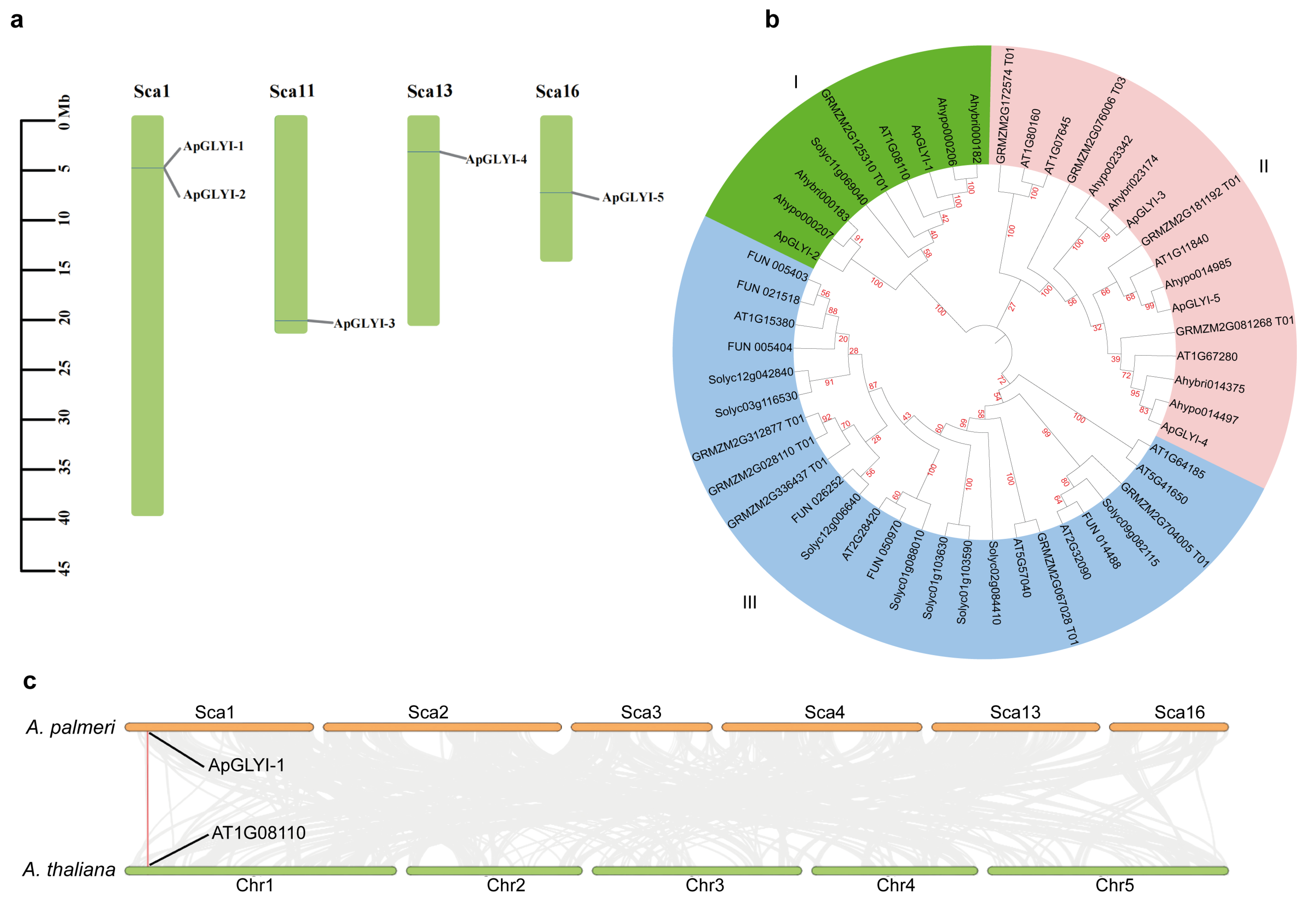
The candidate ApGLYI proteins identified via dual BLAST searches were validated by verifying the presence of functional glyoxalase I domains using the NCBI CDD, SMART, and InterPro databases. A total of five ApGLYI proteins met the specified criteria. Chromosomal mapping, based on the A. palmeri genome annotation, revealed that these five ApGLYI genes are distributed across four scaffolds: 1, 11, 13, and 16 (Figure 1a). Two genes (ApGLYI-1 and ApGLYI-2) were located on scaffold 1, while scaffolds 11, 13, and 16 each contained a single gene.
To assess evolutionary relationships, a phylogenetic analysis of 50 GLYI proteins was conducted. The resulting tree resolved the proteins into three subgroups (Figure 1b). The ApGLYI proteins were exclusively clustered within subgroups I and II. Subgroup I comprised 9 GLYIs, subgroup II contained 16, and subgroup III was the largest with 25 members. All GLYIs from Amaranthus species were confined to subgroups I and II, whereas subgroup III exclusively contained proteins from SlGLYIs, AtGLYIs, FUGLYIs, and ZmGLYIs. Analysis of conserved motifs revealed subgroup-specific patterns; motifs 8, 9, and 10 were conserved in subgroup I, while motifs 3, 4, and 7 were characteristic of subgroup II. Synteny analysis was conducted to assess ApGLYIs relationships with orthologous genes in Arabidopsis. The analysis identified one homologous pair between ApGLYIs and Arabidopsis (Figure 1c).

3.2. Physicochemical Characteristics and Structural Modeling of ApGLYI Proteins

The predicted ApGLYI proteins exhibited considerable variation in size, ranging from 184 to 358 amino acids (average 271.2 aa). Their calculated molecular weights varied from 20.74 to 39.74 kDa (mean 30.27 kDa), while theoretical isoelectric points range between 5.14 and 6.56 pI, with an average of 5.56 pI (Supplementary File S2). The aliphatic index varied from 63.64 to 87.13, indicating a moderate thermostability range, while GRAVY values confirmed all ApGLYIs as hydrophilic (GRAVY < 0), with ApGLYI-1 exhibiting the strong hydrophilicity (GRAVY < −0.5). Based on instability indices, ApGLYI-3, -4, and -5 were classified as stable proteins.
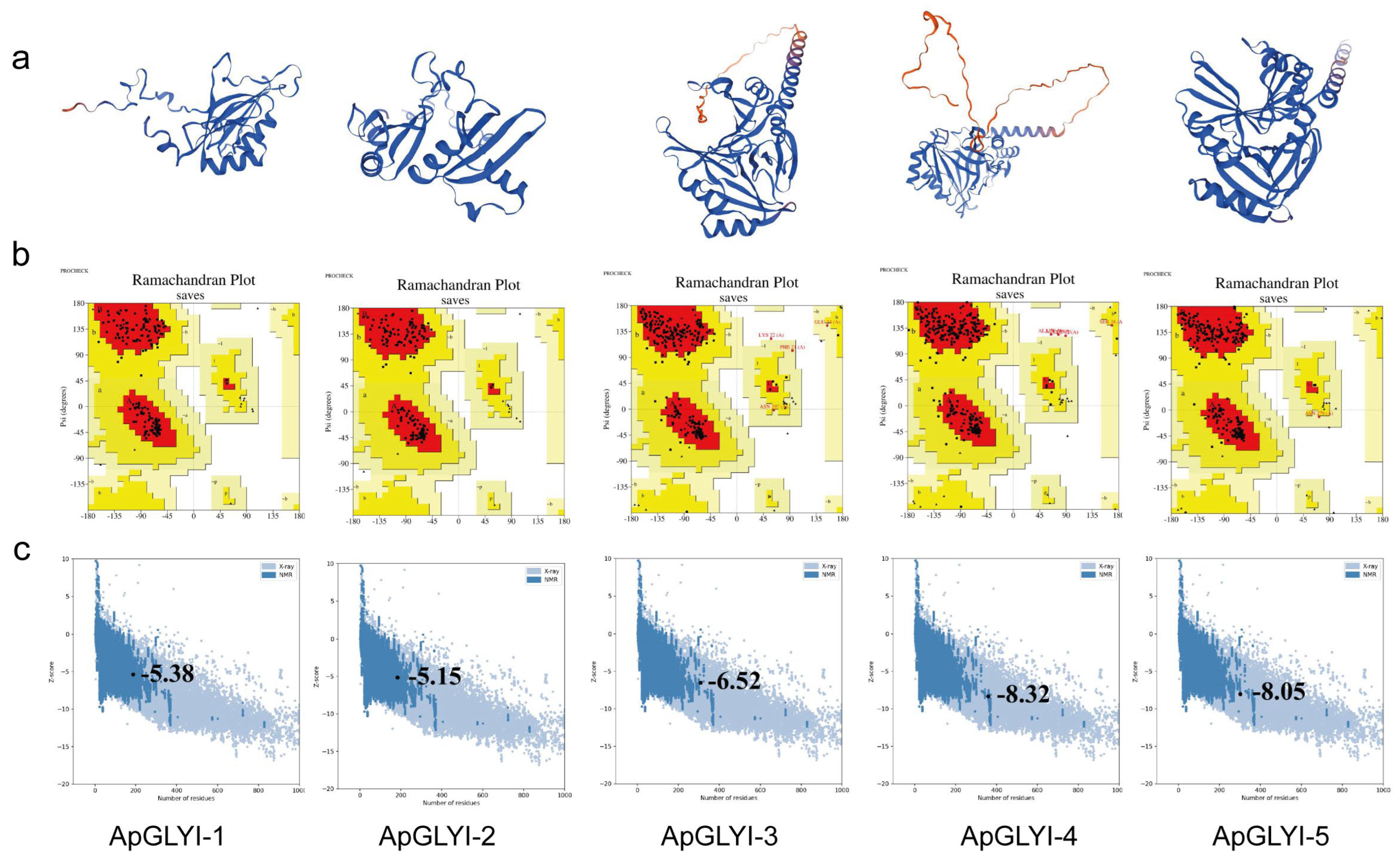
Secondary structure prediction indicated that the ApGLYI proteins comprise four structural elements: alpha helices (22.46–32.09%), β-turns (8.15–11.45%), random coils (31.01–41.30%), and extended strands (23.37–27.65%) (Supplementary File S3). Homology modeling generated three-dimensional structures suggesting a relatively low degree of structural complexity (Figure 2a). Ramachandran plot analysis validated the predicted models. Over 90% of the residues were located in the most favored region, with an additional 8–9% located in the additionally allowed regions. Less than 1% of residues were found in disallowed regions. This result confirms the structural plausibility of the backbone conformations (Figure 2b). Further validation using PROSA yielded Z-scores of −5.38, −5.15, −6.52, −8.32, and −8.05 for the respective ApGLYI models, supporting their overall quality (Figure 2c).

3.3. Motifs, Domain, Structure Dand Cis-Acting Elementsi ApGLYI Genes

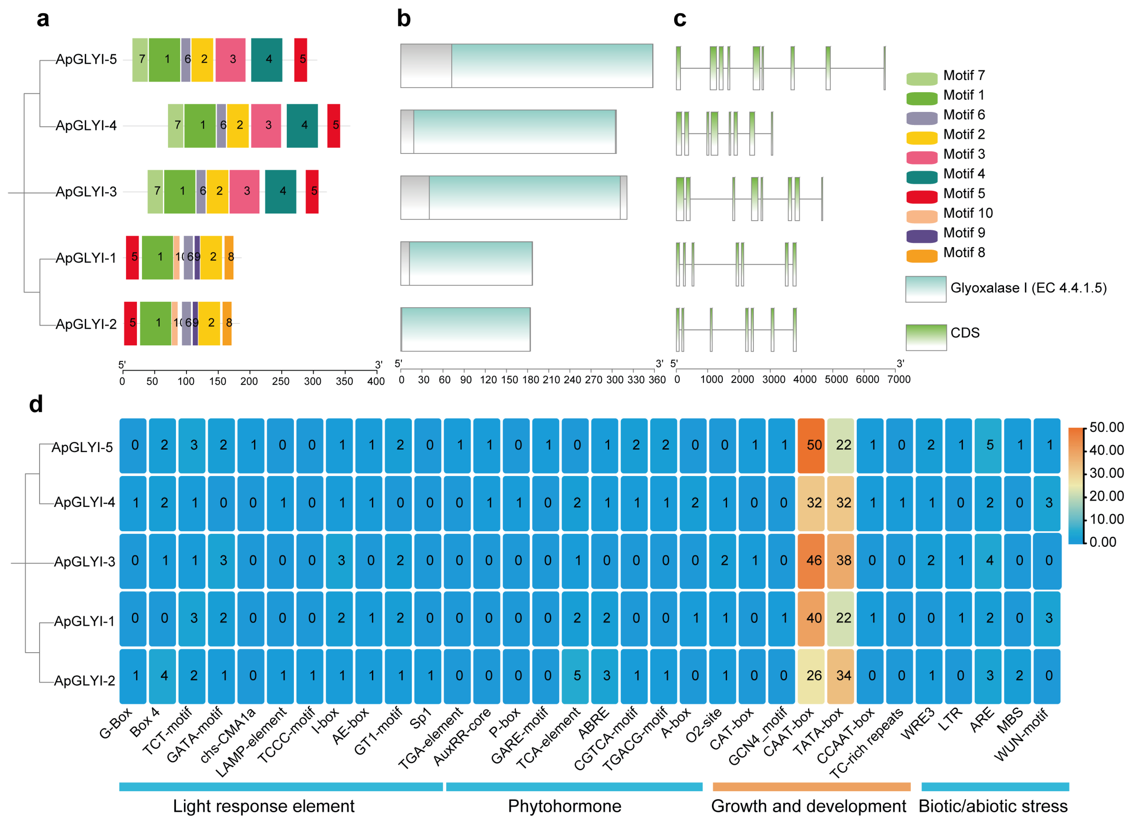
Conserved motif analysis of the ApGLYI proteins identified ten distinct motifs, with lengths ranging from 9 to 50 aa. Each motif appeared once per sequence, and all ApGLYI proteins contained a total of seven motifs (Figure 3a, Supplementary File S4). Of these, motifs 1, 2, 5, and 6 were conserved across all ApGLYI proteins. The remaining motifs exhibited subgroup-specific conservation: motifs 3, 4, and 7 were found exclusively in ApGLYI-3, ApGLYI-4, and ApGLYI-5, whereas motifs 8, 9, and 10 were unique to ApGLYI-1 and ApGLYI-2. Conserved domain analysis confirmed that all ApGLYI proteins harbor the Glyoxalase/Bleomycin resistance protein/Dioxygenase superfamily domain (PF00903), which is associated with lactoylglutathione lyase activity (Figure 3b). Analysis of gene structure revealed that ApGLYI genes lack untranslated regions (UTRs) and contain 7 to 9 introns, with a corresponding number of 7 to 9 exons (Figure 3c).
Analysis of the 2.0 kb promoter regions upstream of the ApGLYI genes identified numerous cis-acting elements. These included motifs such as TATA-box, CAAT-box, I-box, GT1-motif, GARE/P-box, AuxRR-core/TGA, ABRE, TCA-element, TGACG/CGTCA, CAT-box, GCN4-motif, ARE, LTR, MBS, and WUN-motif. These elements were assigned to four functional groups related to light, phytohormones, stress, and growth/development (Figure 3d).

3.4. Protein Interaction and MicroRNA-ApGLYI Network

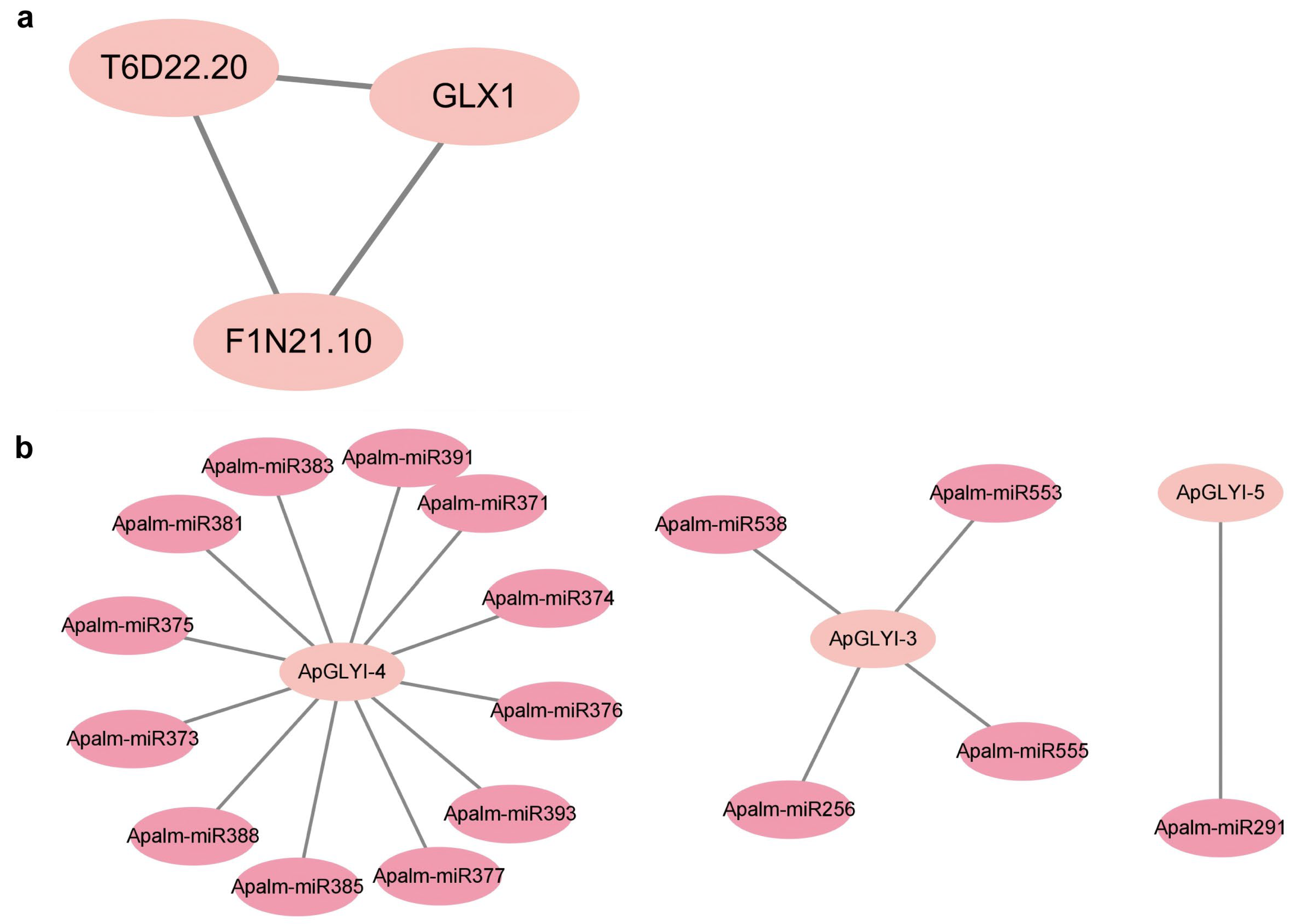
To explore the functional role of ApGLYI genes in the stress response of A. palmeri, a PPI network was generated using the Arabidopsis reference genome within the STRING platform. This network included three Arabidopsis lactoylglutathione lyase proteins: T6D22.20, F1N21.10, and GLX1 (Figure 4a). Gene Ontology (GO) enrichment analysis of the PPI network revealed significant terms related to the biological process ‘methylglyoxal catabolism to D-lactate via S-lactoyl-glutathione’ (GO:0019243) and the molecular function ‘lactoylglutathione lyase activity’ (GO:0004462). KEGG pathway analysis of the PPI network further showed enrichment in general metabolic processes, such as pyruvate metabolism.
To investigate possible post-transcriptional regulation, potential interactions between ApGLYI genes and miRNAs were predicted. This in silico analysis revealed 17 miRNAs that are predicted to target three of the ApGLYI genes (Figure 4b, Supplementary File S5). The targeting was unevenly distributed: ApGLYI-4 was predicted to be targeted by 12 miRNAs, ApGLYI-3 by four miRNAs, and ApGLYI-5 by Apalm-miR291. These results suggest that miRNA-mediated regulation may significantly influence ApGLYI gene expression.

3.5. Subcellular Localization Analysis of ApGLYIs

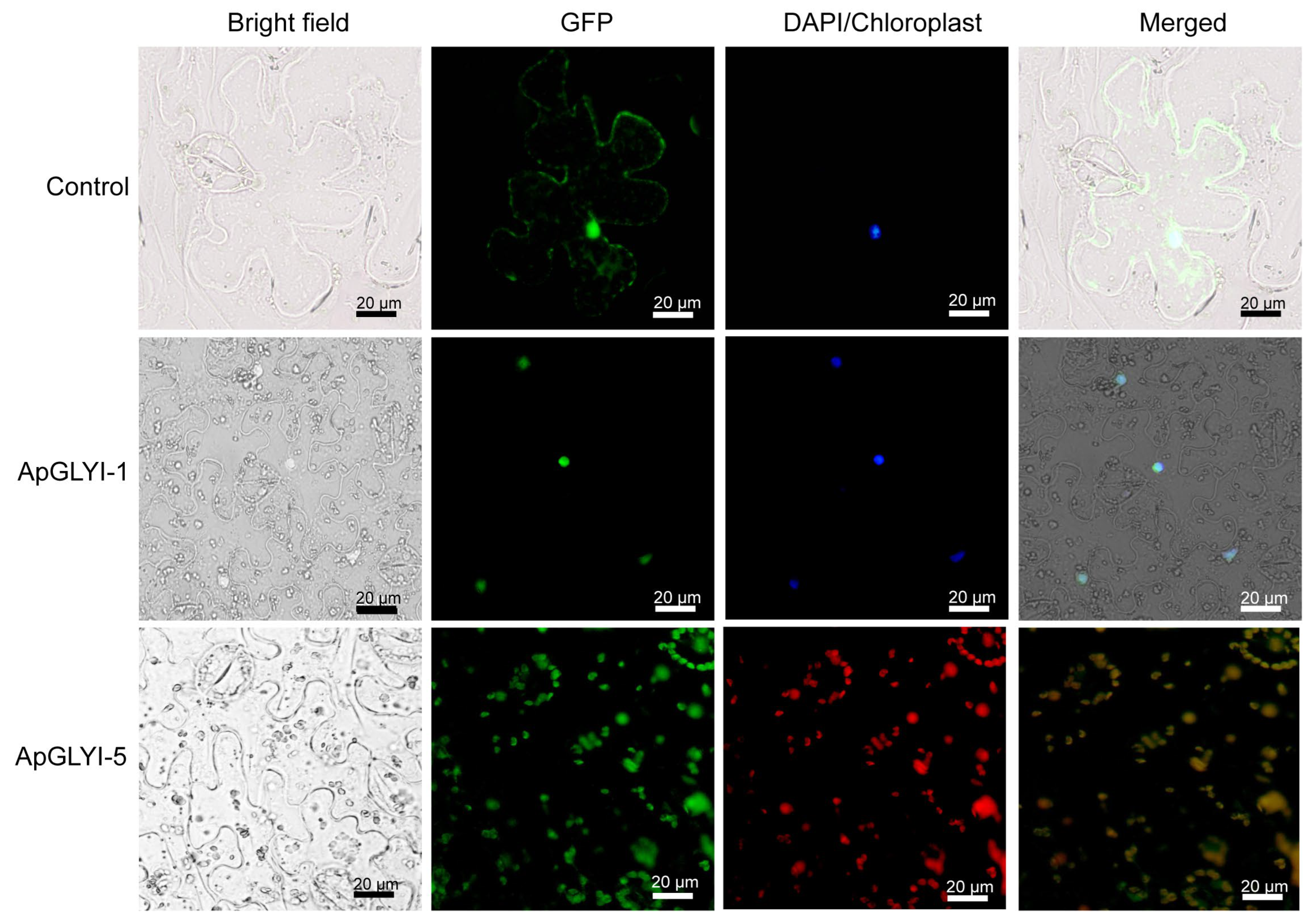
Computational predictions assigned the ApGLYI proteins to three separate cellular compartments: ApGLYI-1 to the nucleus, ApGLYI-2 and ApGLYI-3 to the cytoplasm, and ApGLYI-4 and ApGLYI-5 to the chloroplast (Supplementary File S2). To validate these predictions experimentally, a transient expression assay in N. benthamiana was conducted. The ApGLYI-1 and ApGLYI-5 CDS were fused to GFP under the control of the CaMV 35S promoter. Fluorescence imaging showed that the ApGLYI-1-GFP and ApGLYI-5-GFP fusion protein were specifically targeted to the nucleus and chloroplast, respectively, whereas the GFP-only control was distributed throughout the cell (Figure 5). This result aligns with the in silico prediction for ApGLYI-1 and ApGLYI-5, indicating their potential function in nuclear and photosynthetic activities.

3.6. Expression Analysis of ApGLYIs Under Diverse Stress Conditions

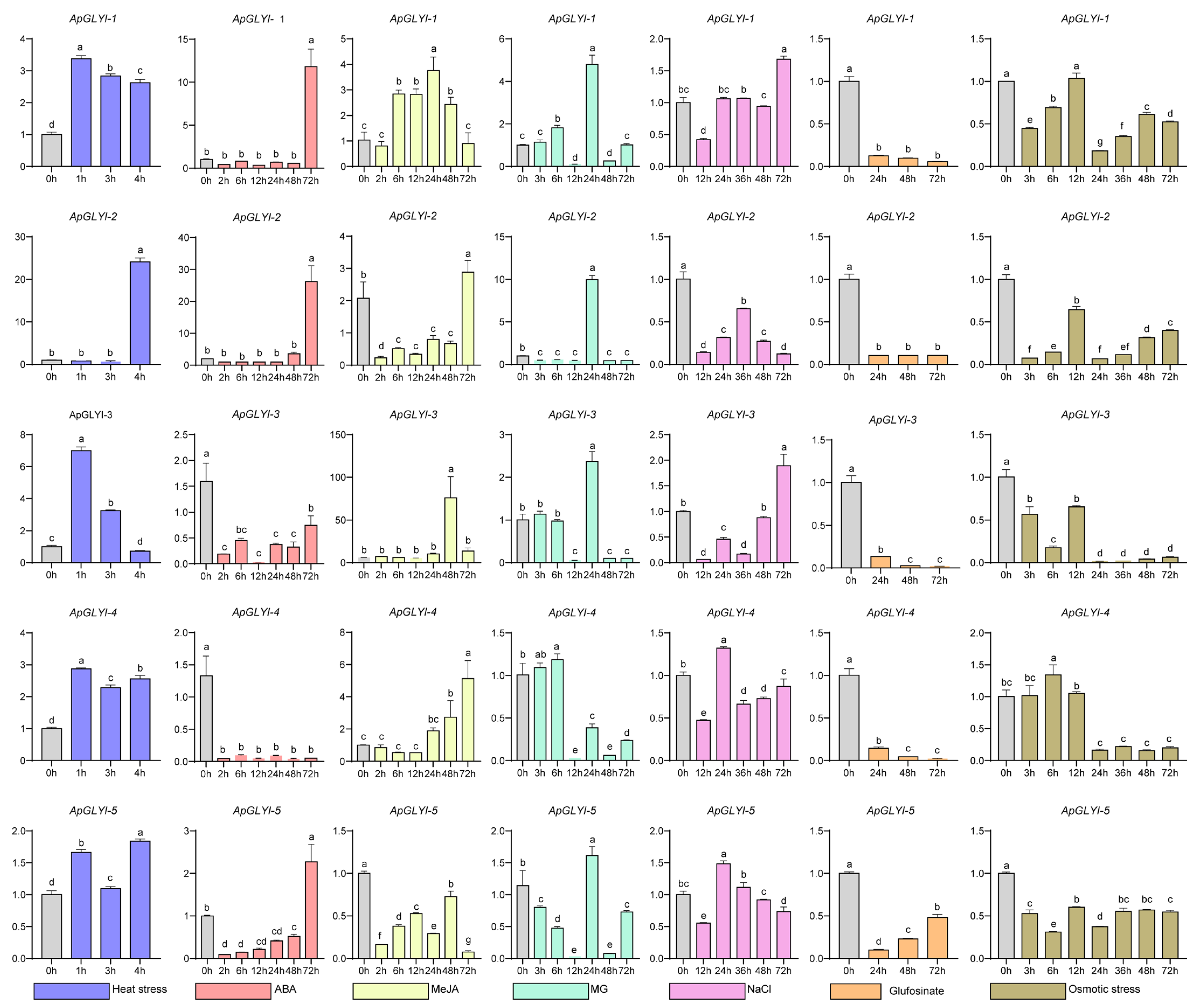
To assess the transcriptional response of ApGLYI genes to diverse stress and hormonal stimuli, their expression profiles were analyzed using RT-qPCR following treatments with high temperature, ABA, MeJA, exogenous MG, NaCl, glufosinate-ammonium, and PEG6000-induced osmotic stress.
Under heat stress, the ApGLYI genes displayed distinct time-dependent expression profiles. ApGLYI-1 and ApGLYI-3 transcripts accumulated rapidly, peaking at 1 h post-treatment and subsequently decreasing. A biphasic pattern was observed for ApGLYI-4 and ApGLYI-5, with an early peak at 1 h, a decline at 3 h, followed by a second rise at 4 h. Conversely, ApGLYI-2 expression was initially downregulated but showed significant induction by the 4 h time point (Figure 6).
Under ABA treatment, all ApGLYI genes except ApGLYI-3 and ApGLYI-4 showed initial suppression, followed by a rebound to levels exceeding the control by 72 h. MeJA application induced a gradual increase in ApGLYI-2 and ApGLYI-4 transcripts, peaking at 72 h. ApGLYI-1 expression rose at 6 h, peaked at 24 h, then declined sharply by 72 h. ApGLYI-3, initially expressed at low levels, was induced and reached a maximum at 48 h. Conversely, ApGLYI-5 expression was downregulated throughout the MeJA treatment period (Figure 6).
Exogenous MG, which directly induces glyoxalative stress, triggered a peak in expression for most ApGLYI genes at 24 h. ApGLYI-4 was an exception, displaying an earlier peak at 6 h before subsequent downregulation. During salt stress, ApGLYI expression was generally suppressed at 12 h. However, by 24 h, ApGLYI-4 and ApGLYI-5 levels increased and peaked before declining again. At the 72 h time point, ApGLYI-1 and ApGLYI-3 were upregulated, whereas ApGLYI-2 remained downregulated for the duration of the treatment (Figure 6).
Glufosinate-ammonium treatment resulted in the sustained downregulation of all ApGLYI genes. Under osmotic stress induced by PEG, ApGLYI-2, ApGLYI-3, and ApGLYI-5 were consistently suppressed. In contrast, ApGLYI-1 and ApGLYI-4 showed transient upregulation, peaking at 12 h and 6 h, respectively, prior to a return to downregulated expression levels (Figure 6).

4. Discussion

Methylglyoxal mediates key physiological responses in plants, including development, intracellular signaling, and heavy metal stress tolerance [47]. While GLYI genes have been extensively characterized across diverse plant species [4], their specific regulatory roles in the stress physiology of A. palmeri have yet to be investigated. Genomic studies have previously documented GLYI families in species such as rice [21], sorghum [48], soybean, and Arabidopsis [49]. In this study, five ApGLYI proteins were identified and their functions investigated in response to high temperature, ABA, MeJA, methylglyoxal, NaCl, glufosinate, and osmotic stress. The findings suggest that ApGLYIs are involved in regulating various stress conditions in A. palmeri.
The ApGLYI proteins exhibit physicochemical attributes that align with those of catalytically active GLYI enzymes, supporting their solubility and functional competence in cytosolic or organellar environments [20,50]. Aliphatic and instability indices further suggest structural stability for most isoforms, a feature consistent with sustained activity under stress conditions where proteostatic control is significant [4]. Secondary and tertiary structures, validated through Ramachandran plot and PROSA Z-score evaluations, verified the presence of the conserved glyoxalase structural fold, which consists of alternating α-helices and β-strands. This structural arrangement facilitates the formation of metal- and substrate-binding sites essential for catalyzing the isomerization of hemithioacetal to S-D-lactoylglutathione [14,21].
Analysis of conserved motifs found a central, conserved region present across all ApGLYI proteins, aligning with the recognized catalytic framework and glutathione-binding interfaces associated with the PF00903 family. Detection of the glyoxalase/bleomycin resistance protein/dioxygenase superfamily domain (PF00903) provides direct confirmation of lactoylglutathione lyase activity, validating function at the domain level [41,51]. Examination of gene structure revealed that ApGLYI genes possess a high intron content and were annotated as lacking UTRs. Such intron-rich architecture is frequently observed in plant GLYI gene families and has been linked to intricate transcriptional control under stress and developmental conditions [24,52]. While miRNA recognition sites are conventionally located within mRNA 3′UTRs, they can also reside in 5′UTRs or CDS [53]. Recent evidence indicates that genes lacking UTRs can still be subject to miRNA regulation [54,55]. The current study found that three ApGLYI genes are predicted targets for 17 distinct miRNAs, suggesting that alternative miRNA–mRNA interaction mechanisms may facilitate post-transcriptional regulation and stress-responsive modulation in A. palmeri, independent of conventional UTR-mediated interactions. Furthermore, the promoter regions of ApGLYI genes contain several responsive cis-acting elements, indicating their involvement in signaling pathways responsive to ABA, JA, drought, cold, wounding, and light stimuli [8].
Plant GLYIs are categorized within the vicinal oxygen chelate superfamily, a broad and functionally versatile group whose roles extend beyond the conventional detoxification of MG [56]. Recent study in Phyllostachys pubescens found several stress-inducible GLYI members, highlighting the evolutionary diversification of this gene family in monocots [57]. Parallel investigations in date palm have demonstrated that specific GLYI genes enhance resilience to salinity and drought, illustrating lineage-specific functional adaptations [58]. In the present study, the phylogenetic grouping of ApGLYI proteins into subgroups I and II, together with orthologs from other Amaranthus species, reflects shared evolutionary paths within the Amaranthaceae family, a pattern consistent with the tendency of GLYI genes to cluster taxonomically [24]. Furthermore, conserved motifs were found to be subgroup-specific, mirroring observations in rice and Arabidopsis, where distinct GLYI clades correlate with either Ni2+- or Zn2+-dependent enzymatic activity and contribute differentially to stress adaptation [20,21]. The absence of ApGLYIs from subgroup III indicates that A. palmeri may have lost certain isoforms present in other species. This streamlined glyoxalase system could represent a functional specialization aligned with the rapid stress response requirements of its invasive life history.
MG is primarily generated in the cytoplasm as a metabolic byproduct of glycolysis, with its production often heightened under stress conditions that perturb normal metabolic flow [3]. A secondary significant source is the photosynthetic apparatus, showing the significant role of chloroplast-localized GLYI enzymes in preserving photosynthetic function. Research on pumpkin seedlings demonstrated that chloroplastic GLYIs reduce MG accumulation during salt and light stress, thereby maintaining chlorophyll integrity and photosynthetic performance [17]. Additionally, MG can form adducts with nuclear DNA and proteins, which may lead to mutagenesis and interfere with transcriptional regulation under stress conditions [11]. In the present study, in silico predictions indicated that ApGLYI proteins are targeted to the cytoplasm, chloroplast, and nucleus. This implies a compartmentalized detoxification strategy, enabling immediate MG neutralization at its sites of production to limit oxidative and genotoxic damage. Characterization using transient expression in N. benthamiana confirmed the nuclear localization of ApGLYI-1. This finding aligns with reports on Arabidopsis GLYI4, which also localizes to the nucleus and contributes to jasmonate signaling and nuclear MG detoxification [8]. The nuclear localization of ApGLYI-1 implies a protective role in maintaining genome stability and a possible involvement in regulating stress-responsive gene expression, which may collectively support the adaptive capacity of A. palmeri.
Plant stressor frequently elevates MG levels as a result of increased glycolytic activity and metabolic disruption, making its removal via the glyoxalase system key for plant viability [59]. The current results demonstrate variable ApGLYI expression patterns, indicative of regulated mechanism that connects environmental sensing, hormone signaling, and localized detoxification. During heat stress, early transcriptional induction of ApGLYI genes was observed, in agreement with findings by Garai et al. [60] that show rapid GLYI upregulation under high temperatures to mitigate MG accumulation from heightened glycolysis. This immediate response points to a protective role for ApGLYIs in limiting protein glycation and oxidative injury. Under salt stress, ApGLYI expression was initially repressed, followed by the later induction of ApGLYI-4 and ApGLYI-5. In contrast, osmotic stress resulted in the sustained suppression of most ApGLYI genes, apart from a brief upregulation of ApGLYI-1 and ApGLYI-4. Such gene-specific expression dynamics are in agreement with studies in cereal crops, where variable GLYI regulation supports targeted MG detoxification and metabolic realignment during stress [20,61].
Transcriptional responses to hormonal treatments revealed that ABA initially repressed most ApGLYI genes, with induction occurring only at later time points, suggesting a temporally delayed detoxification response. In contrast, MeJA led to a progressive increase in ApGLYI-2 and ApGLYI-4 expression, aligning with established jasmonate-mediated defense signaling. This regulatory pattern parallels observations in Arabidopsis, where the nuclear-localized GLYI4 enzyme coordinates MG detoxification with jasmonate signaling, integrating carbonyl metabolism into hormone-activated stress responses [8]. These findings position ApGLYIs within broader phytohormone networks, where they may help balance MG clearance with other adaptive processes.
Treatment with exogenous MG induced ApGLYI expression, affirming their central role in detoxifying this reactive compound. Comparable upregulation has been documented in date palm, where PdGLX1 genes are activated by MG and enhance resilience to salinity and drought stress [58], further validating the conserved enzymatic function of ApGLYIs in protecting cellular metabolism. Conversely, exposure to glufosinate ammonium resulted in the sustained downregulation of all ApGLYI genes, implying a suppression of MG detoxification capacity. Although direct interactions between glyoxalase activity and herbicide toxicity are not fully characterized, diminished GLYI function could intensify MG accumulation and associated oxidative damage. Given the documented herbicide resistance of A. palmeri, alternative detoxification pathways may compensate; however, the observed repression of ApGLYIs points to a potential metabolic vulnerability that could be targeted for developing novel control strategies.
The stress-responsive characteristics of ApGLYI genes highlight their potential value for enhancing resilience in edible Amaranthus and other species. Leafy and grain amaranths, including A. cruentus, A. hypochondriacus, and A. tricolor are closely related to A. palmeri, and interspecific hybridization within the genus is well established [28,62]. The robust protein stability and strong induction of ApGLYIs under heat, salinity, and methylglyoxal stress indicate that A. palmeri alleles may serve as valuable genetic resources. Incorporating ApGLYI variants into edible amaranths through introgression or biotechnological gene transfer could enhance methylglyoxal detoxification capacity, consistent with improvements observed when GLYI genes were overexpressed in other crops [18,22]. Because edible amaranths frequently experience yield reductions under drought, salinity, and oxidative stress [63], using ApGLYI diversity in breeding programs may strengthen stress tolerance and promote more stable production in horticultural systems.

5. Conclusions

The present study provides a comprehensive genomic characterization of the GLYI gene family in A. palmeri, and investigates its contribution to MG detoxification under abiotic stress. A total of five ApGLYI genes were identified, all of which lack UTRs and are dispersed across four genomic scaffolds. Phylogenetic classification grouped the GLYI proteins into three distinct clades, with motif conservation patterns suggesting subgroup-specific functional specialization. Subcellular localization predictions and transient expression of ApGLYI-1 and ApGLYI-5 in N. benthamiana support a compartmentalized model for MG detoxification where specific isoforms are targeted to distinct organelles to mitigate site-specific oxidative and metabolic damage during stress. Expression profiling under diverse treatments revealed that ApGLYI genes are regulated in response to heat, salt, and osmotic stress, and are integrated into ABA and MeJA signaling networks to maintain cellular homeostasis. Notably, Glufosinate ammonium potentially suppressed ApGLYI expression, indicating a potential disruption of MG detoxification that could be leveraged in weed management strategies. Further molecular studies are required to fully explore the transcriptional and post-transcriptional regulation of ApGLYI genes under stress. The findings of the present study establish a foundational understanding of ApGLYIs, offering both novel targets for controlling A. palmeri infestations and promising genetic resources for improving stress resilience in cultivated crops through breeding approaches.

Supplementary Materials

The following supporting information can be downloaded at: https://www.mdpi.com/article/10.3390/horticulturae12020190/s1, Supplementary File S1: Primers used in this study; Supplementary File S2: Physicochemical properties of the ApGLYI proteins; Supplementary File S3: Protein secondary structure of ApGLYIs; Supplementary File S4: Conserved motifs of ApGLYI proteins; Supplementary File S5: Predicted miRNAs targeting ApGLYI genes.

Author Contributions

Conceptualization, methodology, validation, formal analysis, investigation, data curation, writing—original draft preparation, writing—review and editing, Z.W., Y.W. and D.B.; conceptualization, methodology, resources, writing—review and editing, supervision, funding acquisition, B.L., W.C., Y.L., F.W., T.F. and D.M. All authors have read and agreed to the published version of the manuscript.

Funding

This research was funded by National Key R&D Program of China (2024YFC2607600) and Humanities and Social Sciences Research Program funded by the Ministry of Education of the People’s Republic of China (25YJAZH180) and the Natural Science Funds of Hubei Province of China (2024AFB1015).

Data Availability Statement

The data presented in this study are available on request from the corresponding author due to privacy restrictions.

Conflicts of Interest

The authors declare no conflicts of interest.

References

  1. Hasanuzzaman, M.; Nahar, K.; Hossain, M.S.; Mahmud, J.A.; Rahman, A.; Inafuku, M.; Fujita, M. Coordinated actions of glyoxalase and antioxidant defense systems in conferring abiotic stress tolerance in plants. Int. J. Mol. Sci. 2017, 18, 200. [Google Scholar] [CrossRef] [Scilit]
  2. Sankaranarayanan, S.; Jamshed, M.; Kumar, A.; Skori, L.; Scandola, S.; Wang, T.; Samuel, M.A. Glyoxalase goes green: The expanding roles of glyoxalase in plants. Int. J. Mol. Sci. 2017, 18, 898. [Google Scholar] [CrossRef] [Scilit]
  3. Mostofa, M.G.; Ghosh, A.; Li, Z.G.; Siddiqui, M.N.; Fujita, M.; Tran, L.P. Methylglyoxal—A signaling molecule in plant abiotic stress responses. Free Radic. Biol. Med. 2018, 122, 96–109. [Google Scholar] [CrossRef] [Scilit]
  4. Kaur, C.; Singla-Pareek, S.L.; Sopory, S.K. Glyoxalase and methylglyoxal as biomarkers for plant stress tolerance. Crit. Rev. Plant Sci. 2014, 33, 429–456. [Google Scholar] [CrossRef] [Scilit]
  5. Thornalley, P.J.; Langborg, A.; Minhas, H.S. Formation of glyoxal, methylglyoxal and 3-deoxyglucosone in the glycation of proteins by glucose. Biochem. J. 1996, 344, 109–116. [Google Scholar] [CrossRef]
  6. Baynes, J.W. The role of AGEs in aging: Causation or correlation. Exp. Gerontol. 2001, 36, 1527–1537. [Google Scholar] [CrossRef] [Scilit]
  7. Li, Z.G. Methylglyoxal and glyoxalase system in plants: Old players, new concepts. Bot. Rev. 2016, 82, 183–203. [Google Scholar] [CrossRef] [Scilit]
  8. Proietti, S.; Falconieri, G.S.; Bertini, L.; Baccelli, I.; Paccosi, E.; Belardo, A.; Timperio, A.M.; Caruso, C. GLYI4 Plays A Role in Methylglyoxal Detoxification and Jasmonate-Mediated Stress Responses in Arabidopsis thaliana. Biomolecules 2019, 9, 635. [Google Scholar] [CrossRef] [Scilit]
  9. Fu, Z.W.; Li, J.H.; Gao, X.; Wang, S.J.; Yuan, T.T.; Lu, Y.T. Pathogen-induced methylglyoxal negatively regulates rice bacterial blight resistance by inhibiting OsCDR1 protease activity. Mol. Plant 2024, 17, 325–341. [Google Scholar] [CrossRef] [Scilit]
  10. Schmitz, J.; Dittmar, I.C.; Brockmann, J.D.; Schmidt, M.; Hüdig, M.; Rossoni, A.W.; Maurino, V.G. Defense against reactive carbonyl species involves at least three subcellular compartments where individual components of the system respond to cellular sugar status. Plant Cell 2017, 29, 3234–3254. [Google Scholar] [CrossRef] [Scilit]
  11. Kalapos, M.P. Methylglyoxal in living organisms: Chemistry, biochemistry, toxicology and biological implications. Toxicol. Lett. 1999, 110, 145–175. [Google Scholar] [CrossRef] [Scilit]
  12. Thornalley, P.J. Protein and nucleotide damage by glyoxal and methylglyoxal in physiological systems-role in ageing and disease. Drug Metab. Drug Interact. 2008, 23, 125. [Google Scholar] [CrossRef] [Scilit]
  13. Kaur, C.; Sharma, S.; Singla-Pareek, S.L.; Sopory, S.K. Methylglyoxal detoxification in plants: Role of glyoxalase pathway. Indian J. Plant Physiol. 2016, 21, 377–390. [Google Scholar] [CrossRef] [Scilit]
  14. Thornalley, P.J. The glyoxalase system: New developments towards functional characterization of a metabolic pathway fundamental to biological life. Biochem. J. 1990, 269, 1. [Google Scholar] [CrossRef] [Scilit]
  15. Saxena, M.; Bisht, R.; Roy, S.D.; Sopory, S.K.; Bhalla-Sarin, N. Cloning and characterization of a mitochondrial glyoxalase II from Brassica juncea that is upregulated by NaCl, Zn, and ABA. Biochem. Biophys. Res. Commun. 2005, 336, 813–819. [Google Scholar] [CrossRef] [Scilit]
  16. Lee, D.G.; Ahsan, N.; Lee, S.H.; Lee, J.J.; Bahk, J.D.; Kang, K.Y.; Lee, B.H. Chilling stress-induced proteomic changes in rice roots. J. Plant Physiol. 2009, 166, 1–11. [Google Scholar] [CrossRef] [Scilit]
  17. Hossain, M.A.; Fujita, M. Purification of glyoxalase I from onion bulbs and molecular cloning of its cDNA. Biosci. Biotechnol. Biochem. 2009, 73, 2007–2013. [Google Scholar] [CrossRef] [Scilit]
  18. Singla-Pareek, S.L.; Reddy, M.K.; Sopory, S.K. Genetic engineering of the glyoxalase pathway in tobacco leads to enhanced salinity tolerance. Proc. Natl. Acad. Sci. USA 2003, 100, 14672–14677. [Google Scholar] [CrossRef] [Scilit]
  19. Yadav, S.K.; Singla-Pareek, S.L.; Ray, M.; Reddy, M.K.; Sopory, S.K. Methylglyoxal levels in plants under salinity stress are dependent on glyoxalase I and glutathione. Biochem. Biophys. Res. Commun. 2005, 337, 61–67. [Google Scholar] [CrossRef] [Scilit]
  20. Mustafiz, A.; Singh, A.K.; Pareek, A.; Sopory, S.K.; Singla-Pareek, S.L. Genome-wide analysis of rice and Arabidopsis identifies two glyoxalase genes that are highly expressed in abiotic stresses. Funct. Integr. Genom. 2011, 11, 93–305. [Google Scholar] [CrossRef] [Scilit]
  21. Jain, M.; Batth, R.; Kumari, S.; Mustafiz, A. Arabidopsis thaliana contains both Ni2+ and Zn2+ dependent glyoxalase I enzymes and ectopic expression of the latter contributes more towards abiotic stress tolerance in E. coli. PLoS ONE 2016, 11, e0159348. [Google Scholar] [CrossRef] [Scilit]
  22. Álvarez Viveros, M.F.; Inostroza-Blancheteau, C.; Timmermann, T.; González, M.; Arce Johnson, P. Overexpression of GlyI and GlyII genes in transgenic tomato (Solanum lycopersicum Mill.) plants confers salt tolerance by decreasing oxidative stress. Mol. Biol. Rep. 2013, 40, 3281–3290. [Google Scholar] [CrossRef] [Scilit]
  23. Liang, Y.; Srivastava, S.; Rahman, M.H.; Strelkov, S.E.; Kav, N.N. Proteome changes in leaves of Brassica napus L. as a result of Sclerotinia sclerotiorum challenge. J. Agric. Food Chem. 2008, 56, 1963–1976. [Google Scholar] [CrossRef] [Scilit]
  24. Yan, G.; Xiao, X.; Wang, N.; Zhang, F.; Gao, G.; Xu, K.; Wu, X. Genome-wide analysis and expression profiles of glyoxalase gene families in Chinese cabbage (Brassica rapa L). PLoS ONE 2018, 13, e0191159. [Google Scholar] [CrossRef] [Scilit]
  25. Liu, S.; Liu, W.; Lai, J.; Liu, Q.; Zhang, W.; Chen, Z.; Gao, J.; Song, S.; Liu, J.; Xiao, Y. OsGLYI3, a glyoxalase gene expressed in rice seed, contributes to seed longevity and salt stress tolerance. Plant Physiol. Biochem. 2022, 183, 85–95. [Google Scholar] [CrossRef] [Scilit]
  26. Erbas, F. Successional Allelopathic Interactions of Amaranthus palmeri S. Wats. and Cereals. Sustainability 2025, 17, 3871. [Google Scholar] [CrossRef] [Scilit]
  27. Ward, S.M.; Webster, T.M.; Steckel, L.E. Palmer amaranth (Amaranthus palmeri): A review. Weed Technol. 2013, 27, 12–27. [Google Scholar] [CrossRef] [Scilit]
  28. Andini, R.; Sulaiman, M.I.; Kadapi, M.; Ohsawa, R. Polyploidy and Hybridization for Amaranth Crop Improvement. In The Amaranth Genome; Adhikary, D., Deyholos, M.K., Délano-Frier, J.P., Eds.; Compendium of Plant Genomes; Springer: Cham, Switzerland, 2021. [Google Scholar]
  29. Hung, Y.H. Palmer amaranth’s arsenal: Rearrangement of eccDNA provides dual herbicide resistance in Amaranthus palmeri. Plant Cell 2025, 37, koaf077. [Google Scholar] [CrossRef] [Scilit]
  30. Matzrafi, M.; Mennan, H.; Scarabel, L.; Torra, J.; Travlos, I.; Ulber, L. Mitigating agricultural impacts of Amaranthus palmeri: A call for comprehensive management strategies to limit further spread across Europe and the Mediterranean region. Weed Res. 2025, 65, e12675. [Google Scholar] [CrossRef] [Scilit]
  31. Bensch, C.N.; Horak, M.J.; Peterson, D. Interference of redroot pigweed (Amaranthus retroflexus), Palmer amaranth (A. palmeri), and common waterhemp (A. rudis) in soybean. Weed Sci. 2003, 51, 37–43. [Google Scholar] [CrossRef] [Scilit]
  32. Klingaman, T.E.; Oliver, L.R. Palmer amaranth (Amaranthus palmeri) interference in soybeans (Glycine max). Weed Sci. 1994, 42, 523–527. [Google Scholar] [CrossRef] [Scilit]
  33. Massinga, R.A.; Currie, R.S.; Horak, M.J.; Boyer, J., Jr. Interference of Palmer amaranth in corn. Weed Sci. 2001, 49, 202–208. [Google Scholar] [CrossRef] [Scilit]
  34. Meyers, S.L.; Jennings, K.M.; Schultheis, J.R.; Monks, D.W. Interference of Palmer amaranth (Amaranthus palmeri) in sweetpotato. Weed Sci. 2010, 58, 199–203. [Google Scholar] [CrossRef] [Scilit]
  35. Ulaş, F.; Torun, H.; Beffa, R.; Mennan, H. Global trends and research ınsights on herbicide resistance: A bibliometric analysis. Phytoparasitica 2025, 53, 77. [Google Scholar] [CrossRef] [Scilit]
  36. Ehleringer, J. Ecophysiology of Amaranthus palmeri, a Sonoran Desert summer annual. Oecologia 1983, 57, 107–112. [Google Scholar] [CrossRef] [Scilit]
  37. Sun, M.; Sun, S.; Jia, Z.; Zhang, H.; Ou, C.; Ma, W.; Wang, J.; Li, M.; Mao, P. Genome-wide analysis and expression profiling of glyoxalase gene families in oat (Avena sativa) indicate their responses to abiotic stress during seed germination. Front. Plant Sci. 2023, 14, 1215084. [Google Scholar] [CrossRef] [Scilit]
  38. Chen, C.; Chen, H.; Zhang, Y.; Thomas, H.R.; Frank, M.H.; He, Y.; Xia, R. TBtools: An integrative toolkit developed for interactive analyses of big biological data. Mol. Plant 2020, 13, 1194–1202. [Google Scholar] [CrossRef] [Scilit]
  39. Larkin, M.A.; Blackshields, G.; Brown, N.P.; Chenna, R.; McGettigan, P.A.; McWilliam, H.; Higgins, D.G. Clustal W and Clustal X version 2.0. Bioinformatics 2007, 23, 2947–2948. [Google Scholar] [CrossRef] [Scilit]
  40. Letunic, I.; Bork, P. Interactive Tree Of Life (iTOL) v5: An online tool for phylogenetic tree display and annotation. Nucleic Acids Res. 2021, 49, W293–W296. [Google Scholar] [CrossRef] [Scilit]
  41. Bailey, T.L.; Boden, M.; Buske, F.A.; Frith, M.; Grant, C.E.; Clementi, L.; Noble, W.S. MEME SUITE: Tools for motif discovery and searching. Nucleic Acids Res. 2009, 37, W202–W208. [Google Scholar] [CrossRef] [Scilit]
  42. Lescot, M.; Déhais, P.; Thijs, G.; Marchal, K.; Moreau, Y.; Van de Peer, Y.; Rombauts, S. PlantCARE, a database of plant cis-acting regulatory elements and a portal to tools for in silico analysis of promoter sequences. Nucleic Acids Res. 2002, 30, 325–327. [Google Scholar] [CrossRef] [Scilit]
  43. Huang, W.; He, Y.; Yang, L.; Lu, C.; Zhu, Y.; Sun, C.; Yin, J. Genome-wide analysis of growth-regulating factors (GRFs) in Triticum aestivum. PeerJ 2021, 9, e10701. [Google Scholar] [CrossRef] [Scilit]
  44. Szklarczyk, D.; Franceschini, A.; Wyder, S.; Forslund, K.; Heller, D.; Huerta-Cepas, J.; Von Mering, C. STRING v10: Protein–protein interaction networks, integrated over the tree of life. Nucleic Acids Res. 2015, 43, D447–D452. [Google Scholar] [CrossRef] [Scilit]
  45. Dai, X.; Zhuang, Z.; Zhao, P.X. psRNATarget: A plant small RNA target analysis server (2017 release). Nucleic Acids Res. 2018, 46, W49–W54. [Google Scholar] [CrossRef] [Scilit] [PubMed]
  46. Livak, K.J.; Schmittgen, T.D. Analysis of relative gene expression data using real-time quantitative PCR and the 2−ΔΔCT method. Methods 2001, 25, 402–408. [Google Scholar] [CrossRef] [Scilit] [PubMed]
  47. Zheng, Q.; Xin, J.; Zhao, C.; Tian, R. Role of methylglyoxal and glyoxalase in the regulation of plant response to heavy metal stress. Plant Cell Rep. 2024, 43, 103. [Google Scholar] [CrossRef] [Scilit]
  48. Bhowal, B.; Singla-Pareek, S.L.; Sopory, S.K.; Kaur, C. From methylglyoxal to pyruvate: A genome-wide study for the identification of glyoxalases and D-lactate dehydrogenases in Sorghum bicolor. BMC Genom. 2020, 21, 145. [Google Scholar] [CrossRef] [Scilit]
  49. Ghosh, A. Genome-wide identification of glyoxalase genes in Medicago truncatula and their expression profiling in response to various developmental and environmental stimuli. Front. Plant Sci. 2017, 8, 836. [Google Scholar] [CrossRef] [Scilit] [PubMed]
  50. Espartero, J.; Pintor-Toro, J.A.; Pardo, J.M. Differential accumulation of S-adenosylmethionine synthetase transcripts in response to salt stress. Plant Mol. Biol. 1994, 25, 217–227. [Google Scholar] [CrossRef] [Scilit]
  51. Blum, M.; Chang, H.Y.; Chuguransky, S.; Grego, T.; Kandasaamy, S.; Mitchell, A.; Finn, R.D. The InterPro protein families and domains database: 20 years on. Nucleic Acids Res. 2021, 49, D344–D354. [Google Scholar] [CrossRef] [Scilit]
  52. Li, T.; Cheng, X.; Wang, Y.; Yin, X.; Li, Z.; Liu, R.; Xu, Y. Genome-wide analysis of glyoxalase-like gene families in grape (Vitis vinifera L.) and their expression profiling in response to downy mildew infection. BMC Genom. 2019, 20, 362. [Google Scholar] [CrossRef] [Scilit]
  53. Diener, C.; Keller, A.; Meese, E. The miRNA-target interactions: An underestimated intricacy. Nucleic Acids Res. 2024, 52, 1544–1557. [Google Scholar] [PubMed]
  54. Liu, Q.; Zhao, Y.; Wang, Y.; Wei, Q.; Zhang, Y.; Li, S. Comprehensive genome-wide analysis of bHLH family genes in Nymphaea colorata and molecular characterization of NcTT8. BMC Plant Biol. 2025, 25, 878. [Google Scholar] [CrossRef] [Scilit] [PubMed]
  55. Wang, S.; Liu, X.; Bimpong, D.; Wang, Y.; Wang, F.; Chen, W.; Du, L.; Ma, D. Functional Characterization of Glutathione Peroxidase Genes Reveals Their Contribution to the Rapid Range Expansion of Amaranthus palmeri Under Stress Conditions. Agronomy 2025, 15, 2386. [Google Scholar] [CrossRef] [Scilit]
  56. Schmitz, J.; Rossoni, A.W.; Maurino, V.G. Dissecting the Physiological Function of Plant Glyoxalase I and Glyoxalase I-Like Proteins. Front. Plant Sci. 2018, 9, 1618. [Google Scholar] [CrossRef] [Scilit]
  57. Kumari, A.; Bhati, U.; Shankar, R.; Yadav, S.K.; Sopory, S.K.; Joshi, R. Genome-wide identification, characterization, and expression pattern analysis of the glyoxalase gene family in Phyllostachys pubescens during abiotic stresses. BMC Plant Biol. 2025, 25, 1364. [Google Scholar] [CrossRef] [Scilit]
  58. Jana, G.A.; Yaish, M.W. Functional characterization of the Glyoxalase-I (PdGLX1) gene family in date palm under abiotic stresses. Plant Signal Behav. 2020, 15, 1811527. [Google Scholar] [CrossRef] [Scilit]
  59. Hoque, T.S.; Hossain, M.A.; Mostofa, M.G.; Burritt, D.J.; Fujita, M.; Tran, L.-S.P. Methylglyoxal: An Emerging Signaling Molecule in Plant Abiotic Stress Responses and Tolerance. Front. Plant Sci. 2016, 7, 1341. [Google Scholar] [CrossRef] [Scilit]
  60. Garai, S.; Bhowal, B.; Kaur, C.; Singla-Pareek, S.L.; Sopory, S.K. What signals the glyoxalase pathway in plants? Physiol. Mol. Biol. Plants 2021, 27, 2407–2420. [Google Scholar] [CrossRef] [Scilit]
  61. Soccio, M.; Marangi, M.; Laus, M.N. Genome-Wide Expression Analysis of Glyoxalase I Genes Under Hyperosmotic Stress and Existence of a Stress-Responsive Mitochondrial Glyoxalase I Activity in Durum Wheat (Triticum durum Desf.). Front. Plant Sci. 2022, 13, 934523. [Google Scholar] [CrossRef] [Scilit]
  62. Joshi, D.C.; Sood, S.; Hosahatti, R.; Kant, L.; Pattanayak, A.; Kumar, A.; Yadav, D.; Stetter, M.G. From zero to hero: The past, present and future of grain amaranth breeding. Theor. Appl. Genet. 2018, 131, 1807–1823. [Google Scholar] [CrossRef] [Scilit]
  63. Mng’omba, S.A. Grain amaranth, a potential and resilient food crop amenable to processing for diverse food and other products. Front. Sustain. Food Syst. 2025, 9, 1656596. [Google Scholar] [CrossRef] [Scilit]
Figure 1. Chromosome mapping and evolutionary relationship of ApGLYI proteins. Chromosome mapping of ApGLYIs across the A. palmeri genome scaffolds (a). Evolutionary tree of GLYI proteins from A. palmeri, A. hypochondriacus, A. hybridus, Portulaca amilis, Solanum lycopersicum, A. thaliana, and Zea mays. The nodes in the tree were evaluated using bootstrap analysis with 1000 replicates, with the Jones–Taylor–Thornton (JTT) model, uniform rates, and pairwise deletion: a higher bootstrap value indicates strong support for the grouping at that node, while a lower value indicates less confidence in the grouping (b). Synteny relationship between ApGLYIs and homologous genes from Arabidopsis (c).
Horticulturae 12 00190 g001
Figure 2. Structural characteristics of ApGLYI proteins. 3D structural models of ApGLYIs proteins (a). Ramachandran plots of ApGLYIs protein structures. (b). Z-Scores of ApGLYIs proteins structures (c).
Horticulturae 12 00190 g002
Figure 3. Motifs, domain, and structural features, and cis-acting elements of ApGLYIs. Conserved motifs of the ApGPX proteins (a). Conserved domain of the ApGPX proteins (b). Structural architecture of the ApGPX genes (c). Cis-acting regulatory elements of the ApGPX genes located 2 kb upstream of the promoter region (d).
Horticulturae 12 00190 g003
Figure 4. Protein interaction and miRNA target ApGLYIs network. Protein–protein interaction network of the ApGLYI proteins based on A. thaliana genome in the STRING database (a). Interaction network of miRNA targets ApGLYI genes (b).
Horticulturae 12 00190 g004
Figure 5. Subcellular location of ApGLYI-1-GFP and ApGLYI-5-GFP fusion protein and empty PCAMBIA1302 vector using transient expression in N. benthamiana leaves.
Horticulturae 12 00190 g005
Figure 6. ApGLYI gene expression profile in response to heat, ABA, MeJA, exogenous methylglyoxal, NaCl, glufosinate ammonium, and osmotic stress. Statistical significance was determined using two-way ANOVA with Tukey’s post hoc test (p < 0.05). Different lowercase letters indicate significant differences between treatment groups within each time point. Data are presented as mean ± SE of three biological replicates.
Horticulturae 12 00190 g006
Disclaimer/Publisher’s Note: The statements, opinions and data contained in all publications are solely those of the individual author(s) and contributor(s) and not of MDPI and/or the editor(s). MDPI and/or the editor(s) disclaim responsibility for any injury to people or property resulting from any ideas, methods, instructions or products referred to in the content.