Article

Knockout of MDHAR Paralogs Suggests Broader Regulatory Roles Beyond Ascorbic Acid Recycling in Lettuce


1
Department of Agriculture, Food and Environment, University of Pisa, I-56124 Pisa, Italy
2
Department of Agricultural and Food Sciences and Technologies (DISTAL), Alma Mater Studiorum-Università di Bologna, I-40127 Bologna, Italy
3
The Genome Center and Department of Plant Sciences, University of California, Davis, CA 95616, USA
*
Authors to whom correspondence should be addressed.
These authors contributed equally to this work.
Horticulturae 2026, 12(1), 122; https://doi.org/10.3390/horticulturae12010122
Submission received: 29 December 2025 / Revised: 19 January 2026 / Accepted: 20 January 2026 / Published: 21 January 2026
(This article belongs to the Section Genetics, Genomics, Breeding, and Biotechnology (G2B2))

Abstract

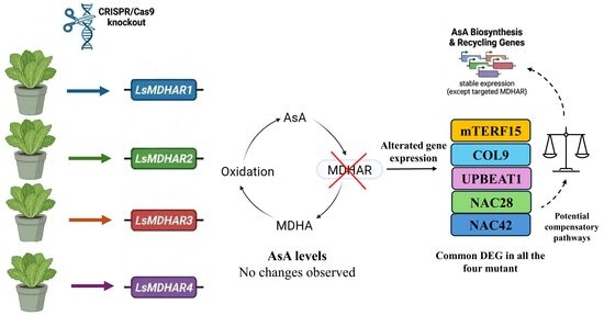
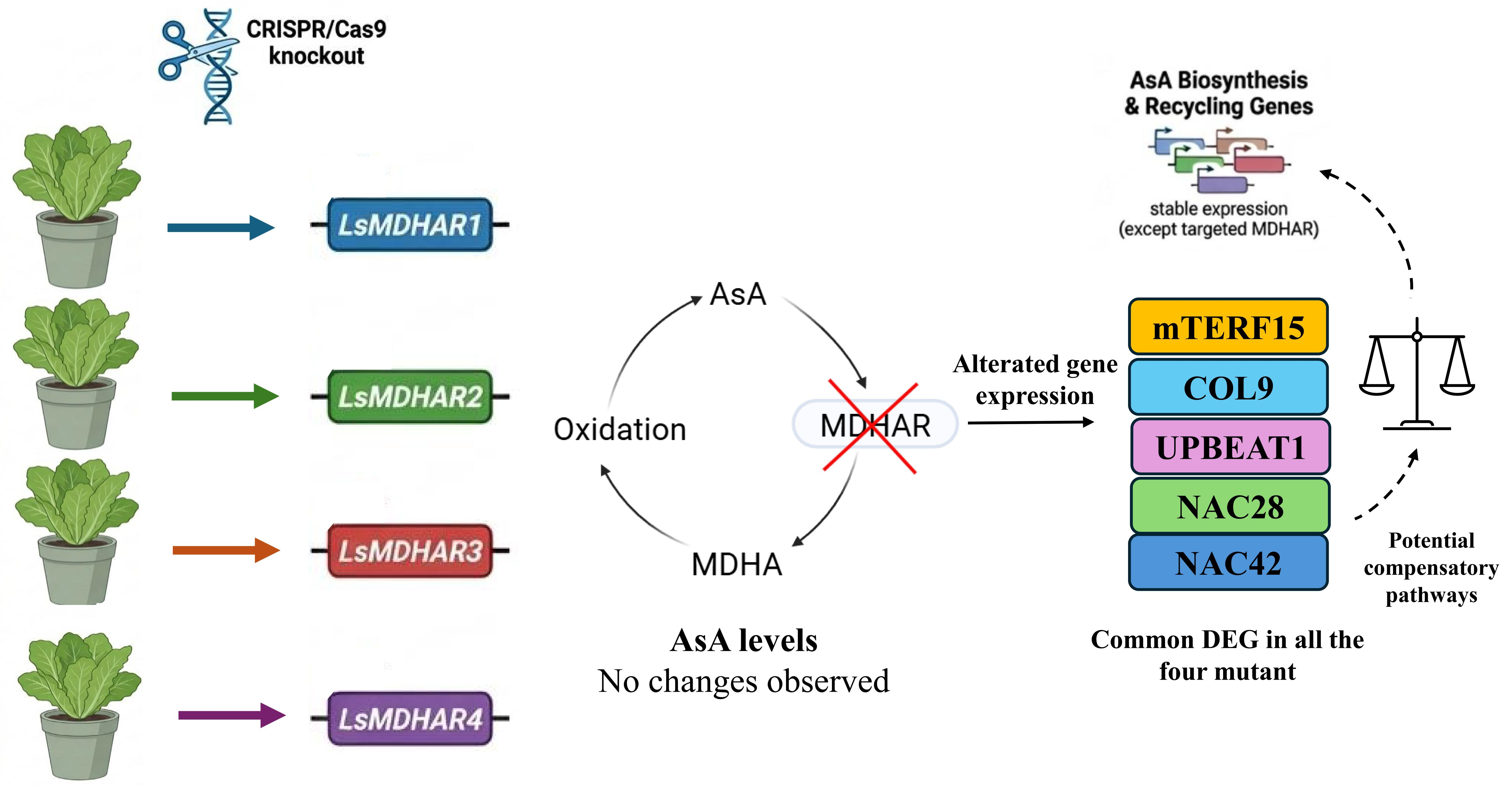
Ascorbic acid (AsA) is a key antioxidant and nutrient in plants, regulating reactive oxygen species (ROS) levels and maintaining cellular redox homeostasis. The AsA recycling pathway sustains AsA pools by restoring its oxidized forms, ensuring intracellular balance. Among the enzymes involved, monodehydroascorbate reductase (MDHAR) is important for the regeneration of AsA from monodehydroascorbate. In this study, we analyzed the four MDHAR paralogs in Lactuca sativa using CRISPR/Cas9 to determine whether disruption of individual MDHAR genes could alter AsA levels in lettuce leaves. Unexpectedly, none of the knockouts caused long-term changes in leaf AsA content. Transcriptomic analyses at 14 and 28 days showed minimal effects on AsA recycling or biosynthesis genes, except MDHAR genes. However, several other genes indirectly implicated in AsA regulation displayed differential expression in all mutants compared to the wild type, suggesting the presence of a complex regulatory network. In particular, genes encoding transcription factors (TFs), such as mTERF15, COL9, UPBEAT1, NAC28, and NAC42, were differentially regulated in all MDHAR mutants compared to the wild type at 28 days. These findings indicate that, although AsA content remains unchanged, MDHAR single knockouts alter expression of other genes through which the plants may indirectly compensate to maintain redox homeostasis.

Graphical Abstract

1. Introduction

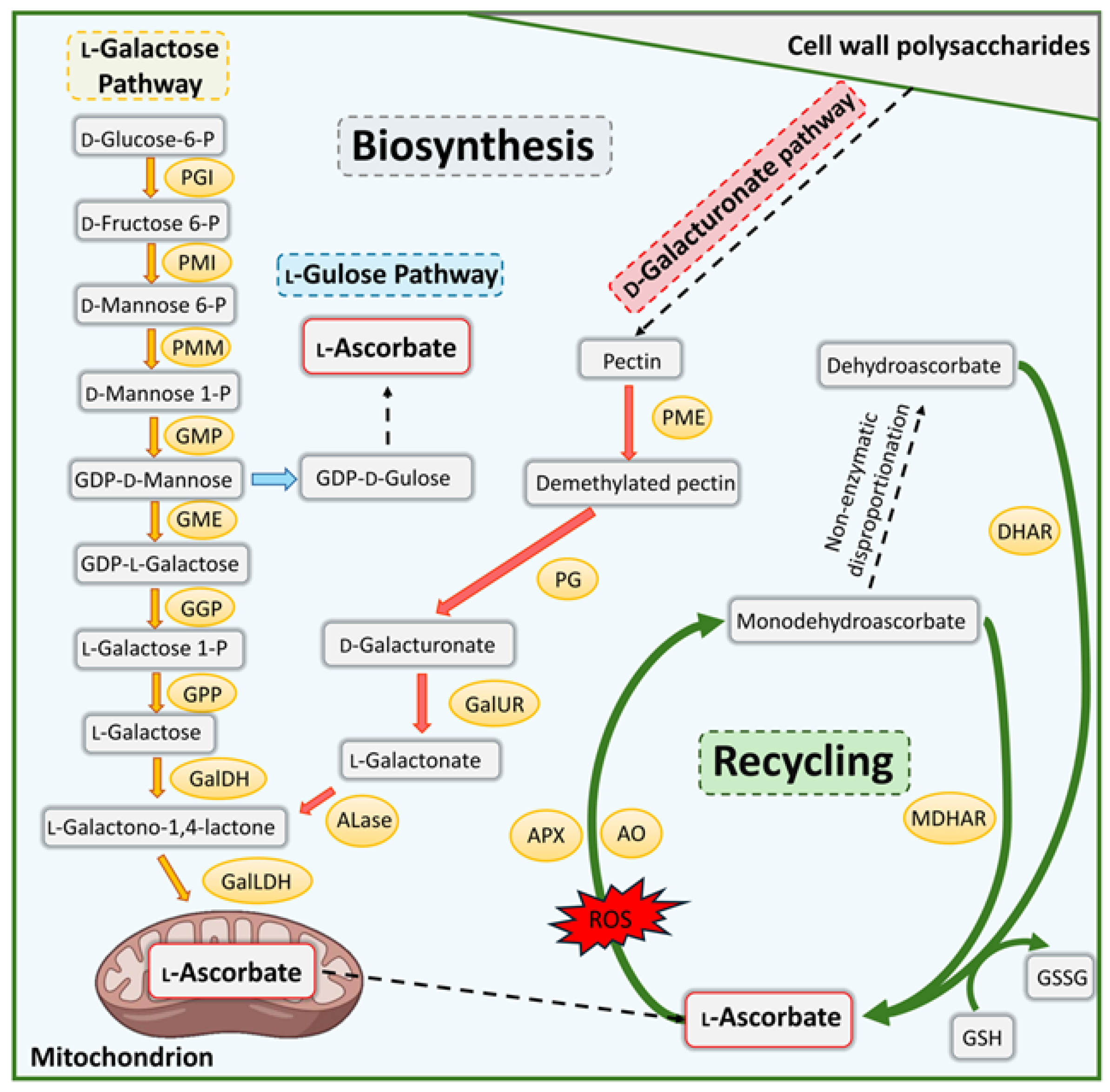
L-Ascorbate, widely known as vitamin C or ascorbic acid (AsA), represents the predominant water-soluble antioxidant in plants, where it is essential for scavenging reactive oxygen species (ROS) generated during photosynthesis, development, and abiotic and biotic stress responses [1]. AsA is not only a key non-enzymatic antioxidant but plays a crucial role in numerous cell processes, including regulating cell division and expansion, and photosynthesis. AsA also acts as a cofactor in many enzymatic activities and interacts with several phytohormones to modulate plant defence responses [2,3]. Consequently, AsA levels are tightly regulated through biosynthesis and recycling pathways. In plants, AsA is synthesized de novo through D-mannose/L-galactose (D-Man/L-Gal), L-gulose, myo-inositol, and D-galacturonate (D-GalUA) pathways. The D-Man/L-Gal, also called “Smirnoff–Wheeler” pathway has been identified as the primary route for plant ascorbate accumulation (Figure 1). This pathway takes place in the cytosol, except for the final step, which occurs in the mitochondrial intermembrane space (IMS). All genes present in this pathway have been characterized [4].
The enzyme GDP-D-mannose 3,5-epimerase (GME), part of the Smirnoff–Wheeler pathway, can also catalyze the 5-epimerization of GDP-D-mannose, leading to the production of GDP-L-gulose, thereby initiating an alternative route known as the L-gulose pathway (Figure 1). The D–GalUA is the alternative important route in plants, which uses pectin from cell wall degradation using the D-galacturonate reductase (GalUR) enzyme to complete AsA production [5] (Figure 1). The myo-inositol pathway has also been identified as involved in AsA biosynthesis; however, there is controversy regarding its effects on AsA accumulation [6].
Regeneration of AsA by the ascorbate–glutathione pathway (Foyer–Halliwell–Asada) [7] plays a central role in maintaining the redox balance and protecting cells from a wide range of radicals. AsA is oxidized to monodehydroascorbate (MDHA) by the ascorbate peroxidases (APXs) and apoplastic ascorbate oxidase (AO) in all cellular compartments. However, MDHA can then spontaneously convert into dehydroascorbate (DHA). Both MDHA and DHA are recycled back to AsA through the action of monodehydroascorbate reductase (MDHAR) and dehydroascorbate reductase (DHAR) enzymes, respectively, utilizing NAD(P)H and glutathione (GSH) as electron donors (Figure 1).
MDHAR (EC 1.6.5.4), a thiol-group-containing flavin adenine dinucleotide (FAD) monomeric enzyme, is ubiquitous in plant species, localized in chloroplast, mitochondria, peroxisomes, and cytosol cellular compartments [1]. During the last few years, efforts have been made to identify the function and interactions of MDHARs. In tobacco, lines over-expressing AtMDHAR1 exhibited 2.2-fold higher levels of reduced AsA than non-transformed wild-type (WT) plants. Conversely, studies using transgenic tomato lines over-expressing SlMDHAR3 showed a negative impact on the ascorbate pool in fruits [8]. A similar decrease in AsA content was also observed when MDHAR3 of Actinidia eriantha was over-expressed in tomato transgenic lines [9]. These conflicting results on the effects of MDHAR over-expression on AsA levels cannot be explained solely by changes in gene expression and enzymatic activities involved in AsA biosynthesis, recycling, or degradation, suggesting the existence of mechanisms regulating the AsA content that have yet to be characterized [10].
MDHAR is also involved in various and multifaceted oxidative reactions. In Arabidopsis, the loss of functional AtMDHAR4 leads to a seedling-lethal phenotype due to loss of detoxification of H2O2 during seed germination [11], while Atmdhar5 mutant lines show tolerance to the environmental pollutant 2,4,6-trinitrotoluene (TNT) [12]. Studies have also been conducted on the role of the MDHAR family of genes in stress responses. For example, in rice, silencing OsMDHAR3 increased salt sensitivity [13]. Over-expression of AtMDHAR1 in tobacco led to enhanced tolerance to salt, ozone, and polyethylene glycol stresses [14]. In tomato, over-expression of LeMDHAR enhanced tolerance to temperature and methylviologen-mediated oxidative stresses [15]. In contrast, over-expression of OsMDHAR4 promoted H2O2 scavenging and reduced H2O2-induced stomatal closure, increasing heat sensitivity [16]. Similarly, in Triticum aestivum, the knockdown of TaMDHAR4 and TaMDHAR6 genes enhanced the resistance to Puccinia striiformis f. sp. tritici (Pst), suggesting a negative role for two MDHAR isoforms in stripe rust resistance [17]. Furthermore, 15 TaMDHARs were found to interact with numerous transcription factors (TFs) involved in growth and development and responses to light, phytohormones, and stresses [18]. Altogether, these studies imply the direct or indirect involvement of MDHAR proteins in regulating AsA content, plant development, and stress responses. Considering the different roles of MDHAR genes observed in model species, it is important to characterize them in other crops as well.
Lettuce (Lactuca sativa L.) is an autogamous diploid species (2n = 2× = 18) belonging to the Asteraceae (a.k.a. Compositae) family. The lettuce genome has been sequenced and assembled, providing a high-resolution template for the application of genome editing protocols [19]. As a leafy vegetable consumed fresh, lettuce represents an agronomically and nutritionally relevant crop. It is widely cultivated and appreciated for its nutritional value and rapid production cycle [20]. It is also an important source of antioxidants, which contributes to its health-promoting properties, resistance to abiotic and biotic stresses, and as a browning-preventing agent [21]. Although lettuce does not rank among the vegetables with the highest vitamin C content, AsA plays a fundamental physiological role, and investigating the molecular mechanisms regulating its metabolism is therefore of considerable interest.
To determine whether a knockout of one of the four MDHAR genes in L. sativa could increase AsA levels, single-edited mutants (called M1, M2, M3, and M4) were generated using CRISPR/Cas9 technology. The resultant knockouts also provided the opportunity to investigate the impact of MDHAR genes in AsA levels. Moreover, RNA-seq analysis was performed to investigate shared DEGs across mutants, thus providing a first overview of possible effects of MDHAR knockouts on the expression of other genes.

2. Methods

2.1. Gene Identification and Analysis

The LsMDHAR genes were amplified and sequenced from L. sativa cv. Cobham Green using the reference sequence from GeneBank (L. sativa cv. Salinas, https://www.ncbi.nlm.nih.gov/datasets/genome/GCF_002870075.4/, accessed on 6 February 2023 [21]): LSAT_6X85001 for LsMDHAR1 gene; LSAT_2X91720 for LsMDHAR2 gene; LSAT_0X28220 for LsMDHAR3 gene; and LSAT_6X64280 for LsMDHAR4 gene. The primers for gene amplification are reported in Table S1.
To investigate the putative subcellular localization of each MDHAR protein in L. sativa, we performed a phylogenetic analysis using 41 amino acid MDHAR sequences of 21 species whose subcellular localization has been described. The MDHARs’ amino acid sequences (Table S2) from other species were used to perform an alignment completed by the ‘Clustal Omega’ tool [22]. The phylogenetic trees based on the maximum-likelihood method were built by IQ-TREE (https://www.hiv.lanl.gov/content/sequence/IQTREE/iqtree.html, [23] accessed on 3 July 2023) with a bootstrap value of 1000, to predict a putative cell localization of the four LsMDHARs.
To further confirm the putative cellular localization of MDHARs highlighted by the phylogenetic trees, the recently developed MULocDeep software (v1) was used (https://www.mu-loc.org/, accessed on 14 August 2023) [24].

2.2. Plasmid Construction and Transformation

To design the gRNAs for the CRISPR/Cas9 vector, we selected four regions for each gene using Geneious Prime software v2019.2.3 (Table S1). To minimize potential off-target effects, all gRNAs were selected such that any predicted off-target sites in the genome contained at least three mismatches using Cas-OFFinder [25] (Table S3).
Plasmids carrying all the CRISPR/Cas9 reagents were generated using the Golden Gate assembly [26]. Briefly, PCR products of template plasmids were used to have different blocks containing the LjUBIprom, Csy4SiteCut, gRNA1-4, Cas9Scaffold, and 35Ster. The primers were designed to contain the BsaI cut site (Table S1). After digestion, the blocks were introduced into the pL1-R3 backbone vector EC47822 to obtain the L1 plasmid pL1V-R3-LjUBIprom:[Csy4SiteCut:gRNA:Cas9Scaffold:Csy4SiteCut × 4]:35Ster. The other L1 plasmids (pL1M-R4-PcUBIprom:dsREDCDS:NOSter, pL1M-R2-AtUBIprom:Csy4CDS:P2Asite:Cas9CDS:HSPterm, pL1M-R1-NOSprom:NPTIICDS-NOSterm, and pL1M-ELE-L4E) were available from the laboratory of Prof. Richard Michelmore (Genome Center and Department of Plant Sciences, University of California, Davis, CA, USA). All the L1 plasmids and the L2 backbone vector EC50507 were digested with BbsI and ligated to obtain the final pL2M_MDHAR1-4 plasmids (Figures S1 and S2).
Transgenic lettuce plants were generated as described by Bertier et al. [27].

2.3. Plant Growth

Seeds were germinated on moistened filter paper in Petri dishes and distilled water, in a controlled environment chamber at 25 °C ± 1 with a 12 h light/12 h dark photoperiod. Seedlings were transferred in pots with soil to a growth chamber under 350 ± 25 μmol of photons/m−2 s−1, with a photoperiod of 16 h light/8 h dark, 22 °C ± 3 of temperature, and 70% ± 5 relative humidity. Plants were irrigated with Hoagland solution, and leaf material samples were collected from 14- and 28-day-old plants, 6 h into the light period. The fifth and sixth fully developed leaves were collected from the center of the rosette, selecting those with optimal light exposure, for ascorbic acid AsA and gene expression analysis.

2.4. Lettuce Genotyping

The DNA was isolated from leaves using a NucleoSpin® Plant II kit (Macherey-Nagel GmBh & Co, Düren, Germany), according to the manufacturer’s protocol. Positively transformed plants were detected through Cas9 and NPTII positive PCR products, using HOT FIREPol® DNA Polymerase (SolisBiodyne, Tartu, Estonia). The positive plants were further analyzed by PCR in the target gRNA regions using PhusionTM High-Fidelity DNA polymerase (Thermo Fisher Scientific Baltics UAB, Vilnius, Lithuania) and subjected to Sanger sequencing. The resulting nucleotide sequences were analyzed with Geneious Prime software v2019.2.3.

2.5. Ascorbic Acid Content

Total ascorbic acid (T-AsA) and reduced ascorbic acid (AsA) were quantified from five biological replicates collected from the four edited genotypes at 14 and 28 days after sowing, following the methodology outlined by Gillespie and Ainsworth, 2007 [28].

2.6. RNA Extraction and Gene Expression Analysis

Total RNA was isolated from the leaves of three biological replicates collected from independent wild-type (WT) plants (as control) and four edited genotypes at 14 and 28 days after sowing using Qiagen RNeasy Plant Mini-Kit (QIAGEN GmbH, Hilden, Germany), according to the manufacturer’s protocol.
The total RNA previously extracted from lettuce leaves was subjected to Illumina HiSeq 2500 platform sequencing. The 30 cDNA libraries were obtained using the TruSeq RNASeq Sample Prep kit, according to the manufacturer’s protocol (Illumina Inc., San Diego, CA, USA). All libraries consisted of 150 bp paired-end reads. The reads’ quality was assessed using FastQC v0.11.9 and improved by Trimmomatic v0.3.9 [29] as follows: SLIDINGWINDOW: 4:15; CROP: 100; HEADCROP: 15; MINLEN: 100. The rRNA traces were removed from the 100 bp paired-end read libraries, following Vangelisti et al. [20].
The high-quality reads were aligned on the reference transcriptome of L. sativa (https://phytozome-next.jgi.doe.gov/info/Lsativa_V8, accessed on 9 December 2024) using CLC Genomic Workbench v9.5.3 (CLC-BIO, Aarhus, Denmark) with the following parameters: mismatch cost  =  2, insertion/deletion cost  =  3, length fraction  =  0.9, similarity fraction  =  0.9.
Differentially expressed genes (DEGs) between WT and homozygous MDHAR14 mutants were detected using edgeR v3.34.1 [30] with a likelihood test. Only genes showing an RPKM > 1 (kilobase per million reads mapped) in at least one library were used for the analysis. A pairwise comparison was conducted between the WT and edited plants (M1, M2, M3, and M4) at 14 and 28 days from sowing, respectively. Genes were considered DEG for a |log2 fold change (FC)|  ≥  1 and false discovery rate (FDR; [31]) corrected p-value ≤ 0.05.
KEGG ortholog (KO) codes of corresponding DEGs were submitted to KEGG for a pathway network analysis (Kyoto Encyclopaedia of Genes and Genomes) [32].
A total of 0.5 μg RNA was used for reverse transcription. cDNA was synthesized using a Maxima First Strand cDNA Synthesis Kit (Thermo Fischer Scientific Baltics UAB, Vilnius, Lithuania). qRT–PCR was performed using Fast SYBRTM Green Master Mix (Applied Biosystems, Thermo Fischer Scientific) with the StepOneTM Real-Time PCR System (Applied Biosystems, Thermo Fischer Scientific, Waltham, MA USA). The LsGAPDH1 gene was used as a housekeeping gene to eliminate the variations caused by sample handling. The primer list is available in Table S1.

3. Results

3.1. Phylogenetic Classification Suggests Distinct Subcellular Localization of LsMDHAR Paralogs

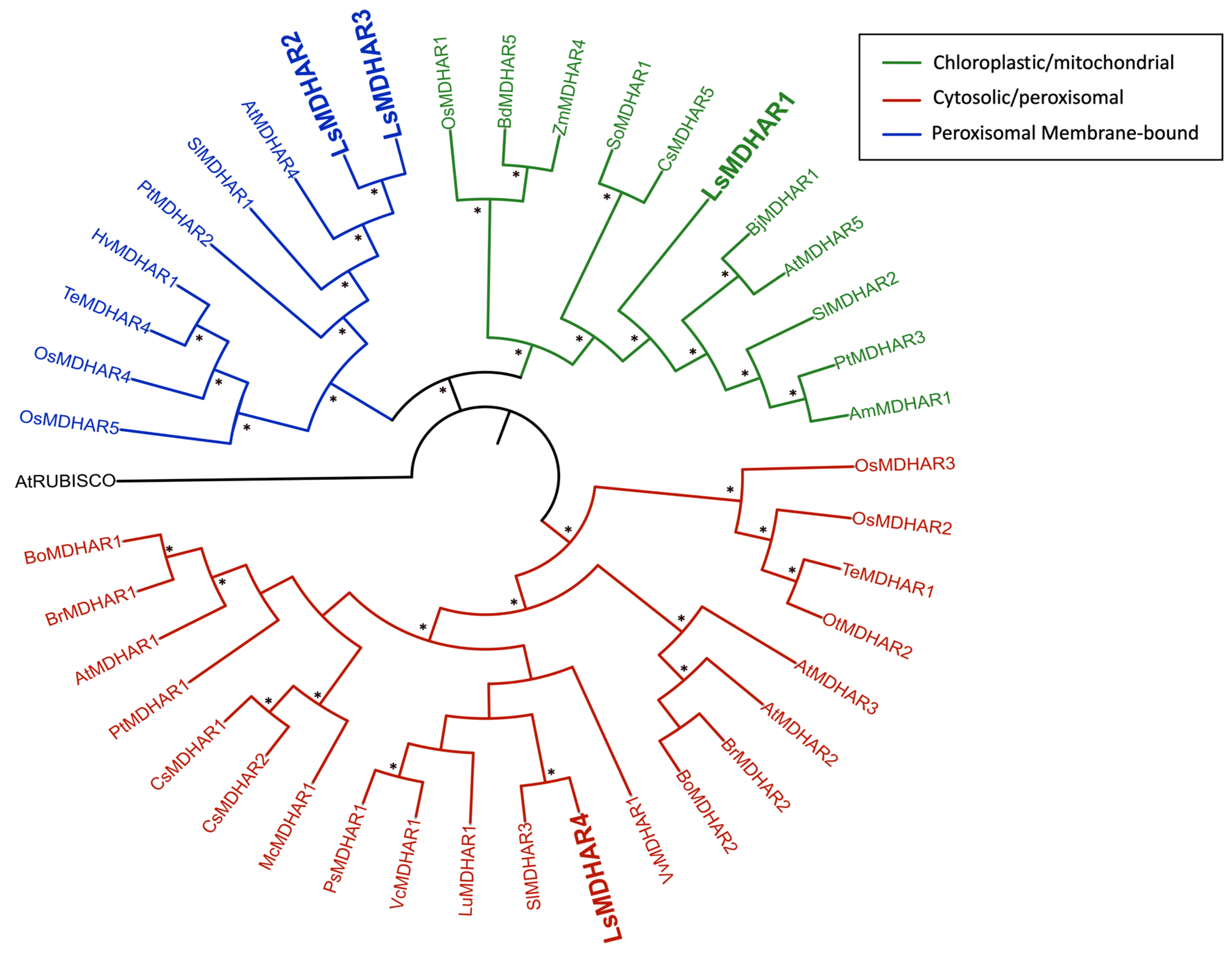
The phylogenetic analysis of the four MDHARs indicated that the four isoenzymes belong to three classes and their likely subcellular localization: LsMDHAR1 clustered with Class I (localized in the chloroplast and mitochondria), characterized by an N-terminal extension, included; LsMDHAR2 and LsMDHAR3 clustered with Class II (found in the peroxisome membrane), in some cases marked by a (-R(K/R)RRRW) terminal motif; and LsMDHAR4 clustered with Class III (cytosolic and peroxisomal), distinguished by a PTS1-like tripeptide (Figure 2) [33]. An analysis using MULocDeep [24] confirmed the putative localization. LsMDHAR1 was predicted to localize in the mitochondrion (p = 0.48) and chloroplast (p = 0.35), LsMDHAR2 and LsMDHAR3 in the peroxisome membrane (p = 0.92 and p = 0.46, respectively), and LsMDHAR4 in the peroxisome (p = 0.64) and cytosol (p = 0.59).

3.2. Knockout of LsMDHAR1–4 by CRISPR/Cas9 Technology Generated Stably Inherited Mutations

To investigate the role of individual MDHAR genes in determining the AsA level in the leaf of L. sativa, we generated independent mutant lines, M1, M2, M3, and M4, targeting LsMDHAR1–4, respectively, using the CRISPR/Cas9 system.
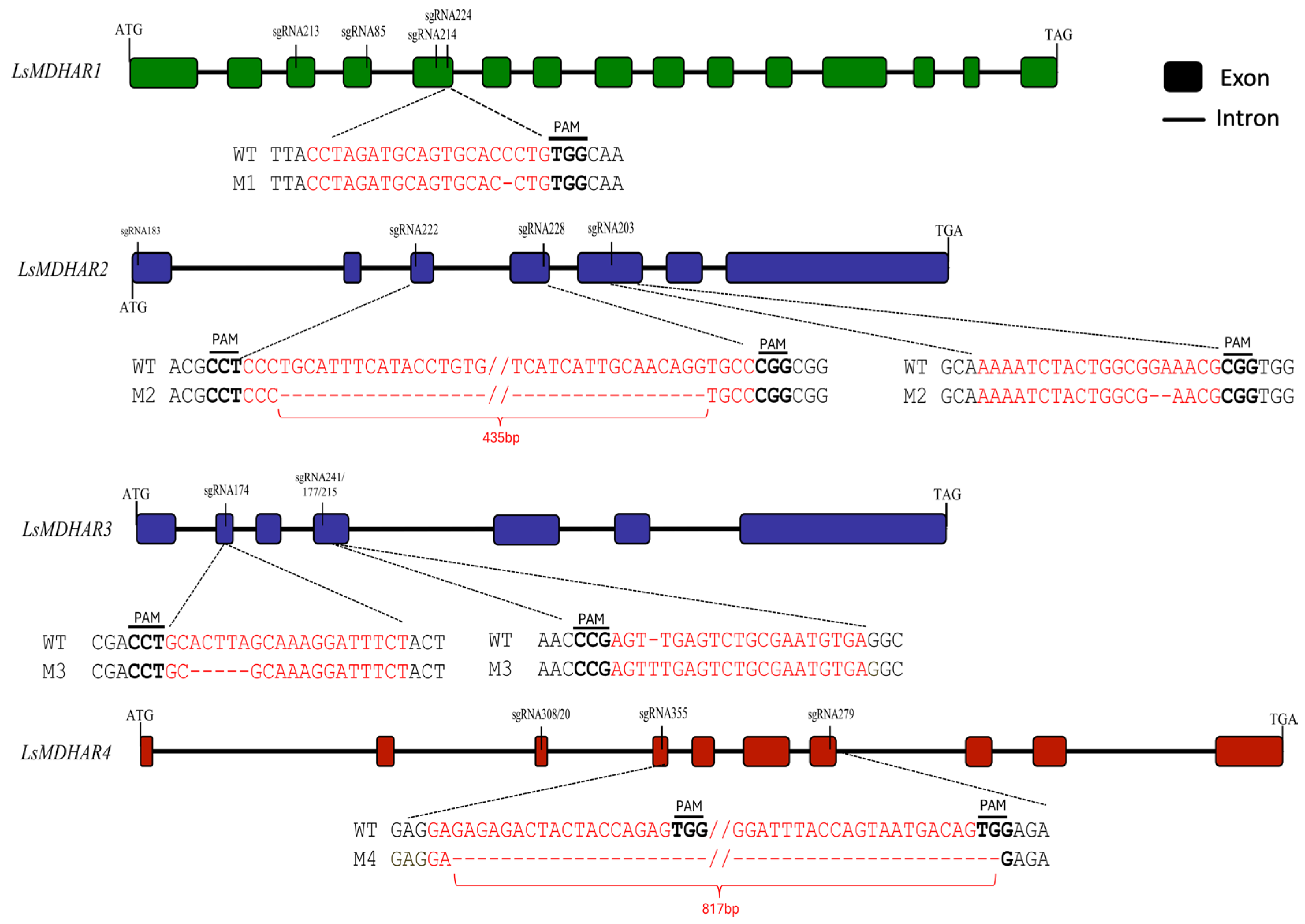
The T1 mutants (i.e., regenerated plants after the CRISPR/Cas9 transformation event) were self-fertilized. Of the resulting T2 plants, only homozygous plants were further self-pollinated to produce T3 families (Figure 3). For each edited gene, one representative homozygous T3 line was chosen for downstream analyses: the M1 line, carrying a −1 bp deletion induced by sgRNA224; the M2 line, harboring a −435 bp deletion between sgRNA222 and sgRNA228 in addition to a −2 bp deletion at sgRNA203; the M3 line, with a −5 bp deletion at sgRNA174 and a +1 bp insertion at sgRNA241; and the M4 line, containing a −817 bp deletion spanning the sgRNA355–sgRNA279 region (Figure 3). In all M1-4 lines, the introduced edits resulted in premature stop codons in the first part of the coding region, and the mutations were stably inherited across subsequent generations (Figure 3 and Figure S3).
To reduce potential off-target mutations in paralogs, all the sgRNAs were designed with at least three mismatches, except for sg174 (Table S3). This guide targeted the LsMDHAR3 gene, but it also had a potential off-target site in the LsMDHAR2 gene with two mismatches. To assess off-target editing, the LsMDHAR2 gene was amplified and sequenced in the M3 mutant line, confirming no off-target mutations in LsMDHAR2 (Figure S4).

3.3. Largely Unchanged AsA Content in Leaves of 14- and 28-Day-Old M1-4 Plants

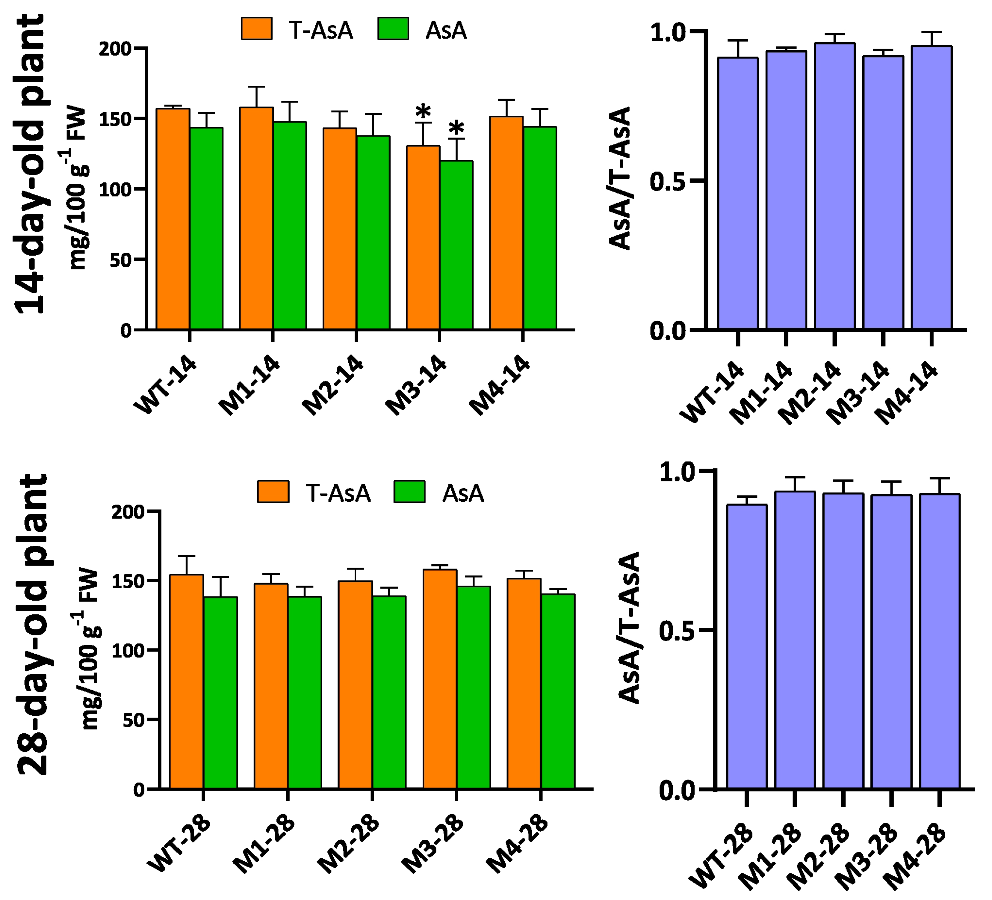
No visible phenotypic differences between mutants and wild-type plants were observed under the described growth conditions. Concerning AsA quantification, in leaves of 14-day-old plants no significant changes in total ascorbic acid (T-AsA), which included both the reduced (AsA) and oxidized (DHA) forms, were observed in all mutant lines except M3. M3 plants showed a small but statistically significant reduction of T-AsA and AsA content (Figure 4). No significant differences in T-AsA and AsA content were observed in leaves of 28-day-old plants in all M14 lines. The redox equilibrium (AsA/T-AsA) remained unaltered in leaves at both time-points (Figure 4) in the four mutant lines.

3.4. Transcriptional Changes in AsA Metabolism-Related Genes in M1-4 Mutants

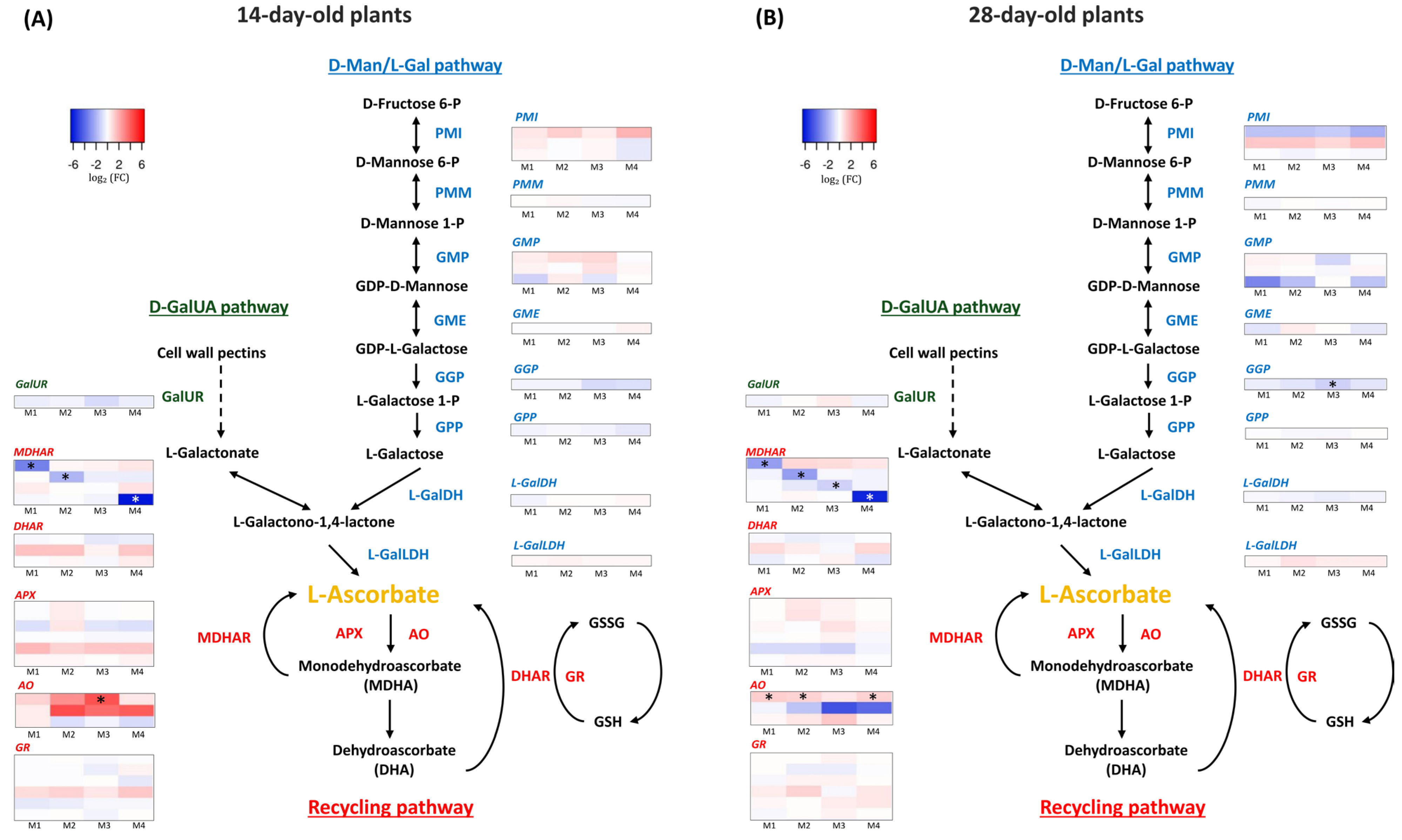
Gene expression in leaves of 14- and 28-day-old M14 plants was evaluated by RNA-seq analysis. Differentially expressed genes (DEGs) were identified through pairwise comparisons between the WT and each of the four mutant lines at 14 and 28 days (Table S4). We specifically searched for DEGs associated with the biosynthesis and recycling of AsA (Figure 5). Figure 5 presents a global overview of gene expression patterns associated with AsA biosynthesis and recycling at 14 and 28 days, showing no consistent changes of expression of genes of these pathways at both time-points, with a few exceptions. For example, at 14 days, a paralog of Ascorbate Oxidase (AO), involved in AsA recycling, was exclusively over-expressed in the M3 genotype. No other DEGs, except the targeted MDHAR1, MDHAR2, and MDHAR4 genes were found. At 28 days, the same AO paralog was over-expressed in M1, M2, and M4 genotypes. Interestingly, the gene GGP, encoding the GDP-L-galactose phosphorylase (GGP), was under-expressed in the genotype M3 (Figure 5). No further AsA-related genes were differentially expressed, except for the four MDHAR in the related edited genotypes. To assess the quality and reliability of the RNA-seq results, qRT-PCRs were conducted on the four LsMDHAR genes in each of the M14 genotypes (Figure S5). To assess the quality and reliability of the RNA-seq results, qRT-PCRs were conducted on the four LsMDHAR genes in each of the M14 genotypes (Figure S5). The results are consistent, in most cases, with RNA-seq results.

3.5. A Core of Differentially Expressed Genes Is Shared Across the LsMDHAR1–4 Edited Lines

To identify transcriptional responses consistently associated with the disruption of individual LsMDHAR genes, we focused on DEGs shared across all four edited lines.
Shared DEGs were analyzed separately at 14 and 28 days. We focused on shared DEGs among the mutants because these represented candidate genes potentially linked to the function of MDHAR proteins. Functional studies including KEGG analysis showed the genes in common were associated with a wide range of biological pathways (Table S5).
Notably, 10 over-expressed (OE) and two under-expressed (UE) genes were common across mutants at 14 days, while at 28 days, the shared core consisted of 49 OE and 7 UE genes (Table 1 and Table S5). At 14 days, the 10 OE shared genes included IQ-DOMAIN 17 (IQD17) and Cation/calcium exchanger 4 (CCX4), whereas at 28 days, common OE genes included DGD1 SUPPRESSOR 1 (DGS1) along with three genes encoding the TFs, NAC42, Mitochondrial transcription termination factor 15 (mTERF15), and CONSTANS-LIKE 9 (COL9). Among the UE genes, SH3 domain-containing protein (SH3P1) was identified at 14 days, while Serine/threonine-protein kinase CTR1 (CTR1) and two genes encoding the TFs, UPB1, and NAC28, were identified at 28 days (Table 1 and Table S5).

4. Discussion

4.1. Functional Redundancy of LsMDHARs and Stability of AsA Levels in Single Knockout Lines

Ascorbic acid (AsA) is an important component of the human diet as well as a key antioxidant and nutrient in plants, regulating reactive oxygen species (ROS) levels and maintaining cellular redox homeostasis. Although the AsA’s biosynthetic and recycling pathways have been studied for over a decade [1], the regulation of these processes is largely unknown. New biotechnological tools, such as CRISPR/Cas9, allow the generation of mutants that can be used to study gene function within complex pathways [34].
In this study, we used CRISPR/Cas9 technology to edit each of the four MDHAR1-4 paralogs in the genome of L. sativa. The primary objective was to test whether loss of individual MDHAR genes could modify AsA concentration in the leaf under non-stress conditions, which would be a useful dietary attribute. In addition, the resulting mutants provided an opportunity to explore DEGs shared among mutants that might point to potential compensatory or broader regulatory pathways.
In all M1–4 lines, mutations resulted in premature stop codons in the first part of coding region of MDHAR genes leading to loss of function (gene knockouts). Off-target analysis of paralogs confirmed the specificity of the edits, and no unwanted mutations were detected. No significant long-term changes (after 28 days) in AsA accumulation were observed in each mutant line. A small change in AsA content was measured in the M3 mutant at 14 days, while no variation was observed in the other edited lines.
These results are similar to those reported for Arabidopsis, in which even the generation of multiple MDHAR and DHAR mutants resulted in an unchanged AsA content and redox state [33]. Furthermore, the Arabidopsis mutant mdhar4 has normal leaf concentrations of AsA, despite having a growth defect in the absence of sucrose due to oxidative stress during germination [33]. Similarly, a quadruple mutant of Arabidopsis lacking all myo-inositol oxygenases (MIOX) (enzymes of the alternative glucuronic acid pathway) presented unchanged AsA levels compared to the wild type [6,35]. In addition, within the main biosynthesis pathway, Arabidopsis has two homologous GDP-L-galactose phosphorylase genes (VTC2 and VTC5). Here, the knockout of VTC5 alone causes a minimal reduction in AsA (approximately 90% compared to the wild-type level), thanks to compensation by the VTC2 enzyme [36].
The unaltered AsA levels could be the consequence of functional redundancy of the paralogs and/or compensatory mechanisms by other ascorbate recycling or biosynthetic pathways. Alternatively, the expression level of these genes may not critically influence AsA levels under the non-stress growth conditions used in our experiments. In such conditions, even if the corresponding transcripts are present, the proteins may not be synthesized or may remain inactive. Their function is mainly required under oxidative stress, when reduced AsA is depleted and recycling pathways are activated. Consequently, in the absence of oxidative stimuli, the impact of the mutations on AsA level might not be detectable. Additionally, we mutated one gene at a time, so functional redundancy among paralogs could prevent phenotypic consequences. Generating higher-order mutants (double, triple, or quadruple) and possibly combining them with oxidative stress treatments would help clarify whether MDHAR genes contribute to AsA homeostasis in lettuce. Although transcriptional compensation by paralogs can be ruled out based on our expression data, compensation through increased enzymatic activity of other isoforms cannot be excluded, as the small decrease in AsA content observed in M3 at 14 days may suggest.

4.2. Shared Transcriptional Responses After Single LsMDHARs Disruption

The RNA-seq analysis was conducted with the aim of elucidating possible compensatory mechanisms at the transcriptional level leading to the maintenance of AsA level in mutant plants, as well as on a possible broader role of MDHAR genes in lettuce, specifically focusing on the shared DEGs identified across all MDHAR mutant lines. Editing each MDHAR gene resulted in a frameshift mutation that led to the formation of premature stop codons and loss of functional mRNAs. The reduction in mRNA abundance may be due to nonsense-mediated mRNA decay (NMD) [37], which is activated when a premature termination codon (PTC) is generated. This phenomenon could occur also for the MDHAR3 gene in 14-day-old plants. At 14 days, transcript levels show only a downward trend and do not reach statistical significance in either RNA-seq or qRT-PCR analyses, likely due to the low baseline expression of the gene. Due to the very low baseline expression of MDHAR3 in lettuce leaves, transcript levels are expected to show high relative variability, making it difficult to detect statistically robust differences between wild-type and M3 mutant plants.
Although single LsMDHAR knockouts did not significantly affect AsA content, the presence of a shared set of DEGs across all mutants suggests that the loss of MDHAR activity triggers transcriptional responses that are not directly associated with AsA biosynthesis or recycling pathways. The analysis of DEGs common to the four mutants revealed genes not directly involved in AsA metabolism but consistently associated with MDHAR disruption, indicating a broader role of MDHARs across multiple KEGG pathways. These include RNA degradation, transcription machinery, transporters, plant hormone signal transduction, ribosome biogenesis, AMPK signaling pathway, mitochondrial biogenesis, ribosome biogenesis/proteasome, amino sugar and nucleotide sugar metabolism, translation/elongation factors, lignin/cell wall biosynthesis–laccase, valine, leucine and isoleucine degradation, phenylpropanoid biosynthesis, environmental adaptation, and glycosyltransferases. This result is consistent with previous findings showing that MDHAR, in addition to its role in AsA recycling, is linked to pathways involved in oxidative stress [11,12,13,14,15,16,17,18].
The number of DEGs shared among mutants was smaller at 14 days than at 28 days, highlighting temporal differences in gene regulation. At 28 days, regulation of genes possibly affecting AsA metabolism emerged. Interestingly, some of shared DEGs encode transcription factors. Among these TFs, CTR1 is a notable one. It encodes a negative regulator of ethylene signaling. In Arabidopsis, ctr1-1 mutants exhibited constitutive ethylene signaling and reduced AsA content due to impaired AsA synthesis and recycling [38]. The relationship between AsA and ethylene is made even more evident through the degradation of cell wall pectin, triggered by ethylene, enhancing the availability of intermediates in the main biosynthetic pathway, increasing the flux toward AsA–ethylene synthesis. In all four mutants, CTR1 was under-expressed at 28 days, a pattern that would be expected to enhance ethylene signaling and potentially reduce AsA levels. However, no significant differences in AsA content were detected in the plants, suggesting the presence of compensatory mechanisms that maintain AsA homeostasis despite transcriptional alterations in the ethylene pathway.
Another shared DEG belongs to the NAC family, one of the largest plant-specific TF families, regulating growth, development, metabolism, biotic, and abiotic stress response. NAC TFs are also involved in ethylene-mediated signaling regulation, acting upstream of ethylene response factors (ERFs) [39]. The genes encoding NAC42 and NAC28 were overexpressed and under-expressed, respectively, across all mutants, emphasizing the role of these TFs in AsA regulation. NAC42 binds to the promoter of D-Galacturonate reductase (GalUR), a key gene in the D-galacturonate pathway of AsA biosynthesis [40], suggesting a possible compensatory role to maintain AsA level in mutants as in WT. NAC28 expression was consistently reduced in all MDHAR knockout mutants at 28 days, despite the absence of stress treatments. This suggests a functional link between MDHAR activity and NAC28 regulation. The downregulation of a negative regulator of stress responses under non-stress conditions may reflect a compensatory mechanism aimed at maintaining redox homeostasis in the mutants. Alterations in AsA-related gene expression could influence redox signaling, leading to transcriptional adjustments that modulate NAC28 levels to stabilize the cellular redox state.
Furthermore, additional stress-related TFs, such as mTERF15 [41], COL9 [42], and UPBEAT1 [43], were shared among the mutants at 28 days, making them candidates for regulating AsA recycling also in normal conditions of growth. mTERF15 is a nuclear factor involved in the splicing of intron 3 of nad2 and in the activity of complex I, namely the NADH:ubiquinone oxidoreductase of the mitochondrial respiratory chain, which is required for proton movement from the mitochondrial matrix to the intermembrane space. In mterf15 mutants, the function of complex I is compromised, resulting in severe growth delay. In particular, mTERF15 has been proposed as a requirement for the assembly of complex I in Arabidopsis [41]. Interestingly, the enzyme GalLDH, which is involved in the final step of the AsA pathway, associates with complex I assembly intermediates [44]. Thus, mTERF15 could indirectly influence the activity of GalLDH and, consequently, regulate AsA. COL9 exerts a dual role, acting both as a flowering repressor and as a factor contributing to abiotic stress tolerance [42]. The heterologous expression of two mango MiCOL9 genes in Arabidopsis enhances salt and drought resistance, associated with improved ROS scavenging [45]. Finally, UPBEAT1 is a bHLH TF that directly modulates the expression of class III peroxidases, influencing the cellular redox state and governing the H2O2 gradient [43]. Collectively, our results indicate that these stress-linked TFs with roles in oxidative-stress responses are linked to MDHAR expression and can have a potential impact on AsA homeostasis also in not stressful conditions. The consistent modulation of these genes in all mutants points to shared transcriptional adjustments associated with the editing of individual MDHAR paralogs. While their direct involvement in AsA regulation remains to be clarified, their recurrent occurrence in different mutants suggests that they may act as regulatory nodes, representing promising candidates for future functional studies.

5. Conclusions

Using CRISPR/Cas9-mediated genome editing, this study provided the opportunity to study the role of individual LsMDHAR1-4 genes in determining AsA level in leaves of L. sativa. Although mutating individual MDHAR genes did not result in changes in AsA concentration in lettuce leaves, it provided insights into these complex biosynthesis and recycling pathways and their regulation. Transcriptomic analysis revealed no substantial expression effect in genes belonging to the biosynthesis and recycling pathway, which appears to remain remarkably stable in each individual mutant. However, differential expression of genes encoding several TFs in common to all mutants point to a higher level of regulatory responses, suggesting new candidate regulators. To better understand these complex interactions, ongoing work involves generating double, triple, and quadruple mutants to investigate possible compensatory and synergistic mechanisms.

Supplementary Materials

The following supporting information can be downloaded at https://www.mdpi.com/article/10.3390/horticulturae12010122/s1, Figure S1: Full sequence of the L2 level plasmid, Figure S2: Schematic representation of pL2M_MDHAR1-4 plasmids, Figure S3: Amino acid sequence of the LsMDHAR1-4 and mutant, Figure S4: Sanger sequencing chromatograms of LsMDHAR2 in M3 genotype, Figure S5: Relative mRNA abundance of LsMDHAR1, LsMDHAR2, LsMDHAR3, and LsMDHAR4 in the respectively edited genotype, Table S1: Primer sequence, Table S2: Species, accession number, and gene name used for phylogenetic analysis of MDHAR from plants, Table S3: Number and position of potential off-targets, Table S4: Differentially expressed genes, Table S5: Common differentially expressed genes in lettuces subjected to genome editing.

Author Contributions

U.R., S.S., A.V. (Ambra Viviani), C.P. and M.F.: investigation; U.R., S.S. and A.V. (Alberto Vangelisti): data curation; C.P., M.F., A.V. (Alberto Vangelisti), L.N., A.C., R.M. and T.G.: methodology; C.P., R.M., A.C. and T.G.: project design and supervision; U.R. and S.S.: writing—original draft. All authors have read and agreed to the published version of the manuscript.

Funding

This research received no external funding.

Data Availability Statement

The RNA-seq libraries have been deposited in the SRA (https://www.ncbi.nlm.nih.gov/sra, accessed on 7 March 2025) under the BioProject accession number PRJNA1232975.

Conflicts of Interest

The authors declare no conflicts of interest.

References

  1. Smirnoff, N.; Wheeler, G.L. The ascorbate biosynthesis pathway in plants is known, but there is a way to go with understanding control and functions. J. Exp. Bot. 2024, 75, 2604–2630. [Google Scholar] [CrossRef] [PubMed]
  2. Baldet, P.; Mori, K.; Decros, G.; Beauvoit, B.; Colombié, S.; Prigent, S.; Pétriacq, P.; Gibon, Y. Multi-regulated GDP-L-galactose phosphorylase calls the tune in ascorbate biosynthesis. J. Exp. Bot. 2024, 75, 2631–2643. [Google Scholar] [CrossRef] [PubMed]
  3. Rogo, U.; Viviani, A.; Pugliesi, C.; Fambrini, M.; Usai, G.; Castellacci, M.; Simoni, S. Improving crop tolerance to abiotic stress for sustainable agriculture: Progress in manipulating ascorbic acid metabolism via genome editing. Sustainability 2025, 17, 719. [Google Scholar] [CrossRef]
  4. Muñoz, P.; Castillejo, C.; Gómez, J.A.; Miranda, L.; Lesemann, S.; Olbricht, K.; Petit, A.; Chartier, P.; Haugeneder, A.; Trinkl, J.; et al. QTL analysis for ascorbic acid content in strawberry fruit reveals a complex genetic architecture and association with GDP-L-galactose phosphorylase. Hortic. Res. 2023, 10, uhad006. [Google Scholar] [CrossRef]
  5. Quiñones, C.O.; Gesto-Borroto, R.; Wilson, R.V.; Hernández-Madrigal, S.V.; Lorence, A. Alternative pathways leading to ascorbate biosynthesis in plants: Lessons from the last 25 years. J. Exp. Bot. 2024, 75, 2644–2663. [Google Scholar] [CrossRef]
  6. Ivanov, K.E.; Blöchl, C.; Tenhaken, R. The Myo-inositol pathway does not contribute to ascorbic acid synthesis. Plant Biol. 2019, 21, 95–102. [Google Scholar] [CrossRef]
  7. Conklin, P.L.; Foyer, C.H.; Hancock, R.D.; Ishikawa, T.; Smirnoff, N. Ascorbic acid metabolism and functions. J. Exp. Bot. 2024, 75, 2599–2603. [Google Scholar] [CrossRef]
  8. Gest, N.; Garchery, C.; Gautier, H.; Jiménez, A.; Stevens, R. Light-dependent regulation of ascorbate in tomato by a monodehydroascorbate reductase localized in peroxisomes and the cytosol. Plant Biotechnol. J. 2013, 11, 344–354. [Google Scholar] [CrossRef]
  9. Jia, D.; Gao, H.; He, Y.; Liao, G.; Lin, L.; Huang, C.; Xu, X. Kiwifruit monodehydroascorbate reductase 3 gene negatively regulates the accumulation of ascorbic acid in fruit of transgenic tomato plants. Int. J. Mol. Sci. 2023, 24, 17182. [Google Scholar] [CrossRef]
  10. Gest, N.; Gautier, H.; Stevens, R. Ascorbate as seen through plant evolution: The rise of a successful molecule? J. Exp. Bot. 2013, 64, 33–53. [Google Scholar] [CrossRef]
  11. Eastmond, P.J. MONODEHYROASCORBATE REDUCTASE4 is required for seed storage oil hydrolysis and postgerminative growth in Arabidopsis. Plant Cell 2007, 19, 1376–1387. [Google Scholar] [CrossRef] [PubMed]
  12. Johnston, E.J.; Rylott, E.L.; Beynon, E.; Lorenz, A.; Chechik, V.; Bruce, N.C. Monodehydroascorbate reductase mediates TNT toxicity in plants. Science 2015, 349, 1072–1075. [Google Scholar] [CrossRef] [PubMed]
  13. Kim, J.J.; Kim, Y.S.; Park, S.I.; Mok, J.E.; Kim, Y.H.; Park, H.M.; Kim, I.S.; Yoon, H.S. Cytosolic monodehydroascorbate reductase gene affects stress adaptation and grain yield under paddy field conditions in Oryza sativa L. japonica. Mol. Breed. 2017, 37, 118. [Google Scholar] [CrossRef]
  14. Eltayeb, A.E.; Kawano, N.; Badawi, G.H.; Kaminaka, H.; Sanekata, T.; Shibahara, T.; Inanaga, S.; Tanaka, K. Overexpression of monodehydroascorbate reductase in transgenic tobacco confers enhanced tolerance to ozone, salt and polyethylene glycol stresses. Planta 2007, 225, 1255–1264. [Google Scholar] [CrossRef]
  15. Li, F.; Wu, Q.Y.; Sun, Y.L.; Wang, L.Y.; Yang, X.H.; Meng, Q.W. Overexpression of chloroplastic monodehydroascorbate reductase enhanced tolerance to temperature and methyl viologen-mediated oxidative stresses. Physiol. Plant 2010, 139, 421–434. [Google Scholar] [CrossRef]
  16. Liu, J.; Sun, X.; Xu, F.; Zhang, Y.; Zhang, Q.; Miao, R.; Zhang, J.; Liang, J.; Xu, W. Suppression of OsMDHAR4 enhances heat tolerance by mediating H2O2-induced stomatal closure in rice plants. Rice 2018, 11, 38. [Google Scholar] [CrossRef]
  17. Feng, H.; Wang, X.; Zhang, Q.; Fu, Y.; Feng, C.; Wang, B.; Huang, L.; Kang, Z. Monodehydroascorbate reductase gene, regulated by the wheat PN-2013 miRNA, contributes to adult wheat plant resistance to stripe rust through ROS metabolism. Biochim. Biophys. Acta 2014, 1839, 1–12. [Google Scholar] [CrossRef]
  18. Madhu; Sharma, A.; Kaur, A.; Singh, K.; Upadhyay, S.K. Modulation in gene expression and enzyme activity suggested the roles of monodehydroascorbate reductase in development and stress response in bread wheat. Plant Sci. 2024, 338, 111902. [Google Scholar] [CrossRef]
  19. Reyes-Chin-Wo, S.; Wang, Z.; Yang, X.; Kozik, A.; Arikit, S.; Song, C.; Xia, L.; Froenicke, L.; Lavelle, D.O.; Truco, M.J. Genome assembly with in vitro proximity ligation data and whole-genome triplication in lettuce. Nat. Commun. 2017, 8, 14953. [Google Scholar] [CrossRef]
  20. Vangelisti, A.; Cela, F.; Simoni, S.; Avio, L.; Turrini, A.; Sbrana, C.; Incrocci, L.; Pardossi, A.; Rogo, U.; Natali, L.; et al. Arbuscular mycorrhizal symbiosis allows to overcome phosphorus deficiency in soilless culture of Lactuca sativa: A phenotypic and transcriptomic analysis. Sci. Hortic. 2024, 334, 113324. [Google Scholar] [CrossRef]
  21. Landi, M.; Fambrini, M.; Basile, A.; Salvini, M.; Guidi, L.; Pugliesi, C. Overexpression of L-galactono-1,4-lactone dehydrogenase (L-GalLDH) gene correlates with increased ascorbate concentration and reduced browning in leaves of Lactuca sativa L. after cutting. Plant Cell Tiss. Organ. Cult. 2015, 123, 109–120. [Google Scholar] [CrossRef]
  22. Sievers, F.; Higgins, D.G. Clustal omega for making accurate alignments of many protein sequences. Protein Sci. 2018, 27, 135–145. [Google Scholar] [CrossRef] [PubMed]
  23. Trifinopoulos, J.; Nguyen, L.T.; von Haeseler, A.; Minh, B.Q. W-IQ-TREE: A fast online phylogenetic tool for maximum likelihood analysis. Nucleic Acids Res. 2016, 44, W232–W235. [Google Scholar] [CrossRef] [PubMed]
  24. Jiang, Y.; Jiang, L.; Akhil, C.S.; Wang, D.; Zhang, Z.; Zhang, W.; Xu, D. MULocDeep web service for protein localization prediction and visualization at subcellular and suborganellar levels. Nucleic Acids Res. 2023, 51, W343–W349. [Google Scholar] [CrossRef]
  25. Bae, S.; Park, J.; Kim, J.-S. Cas-OFFinder: A fast and versatile algorithm that searches for potential off-target sites of Cas9 RNA-guided endonucleases. Bioinformatics 2014, 30, 1473–1475. [Google Scholar] [CrossRef]
  26. Engler, C.; Kandzia, R.; Marillonnet, S.A. One pot, one step, precision cloning method with high throughput capability. PLoS ONE 2008, 3, e3647. [Google Scholar] [CrossRef]
  27. Bertier, L.D.; Ron, M.; Huo, H.; Bradford, K.J.; Britt, A.B.; Michelmore, R.W. High-resolution analysis of the efficiency, heritability, and editing outcomes of CRISPR/CAS9-induced modifications of NCED4 in lettuce (Lactuca sativa). G3 2018, 8, 1513–1521. [Google Scholar] [CrossRef]
  28. Gillespie, K.M.; Ainsworth, E.A. Measurement of reduced, oxidized and total ascorbate content in plants. Nat. Protoc. 2007, 2, 871–874. [Google Scholar] [CrossRef]
  29. Bolger, A.M.; Lohse, M.; Usadel, B. Trimmomatic: A flexible trimmer for Illumina sequence data. Bioinformatics 2014, 30, 2114–2120. [Google Scholar] [CrossRef]
  30. Robinson, M.D.; McCarthy, D.J.; Smyth, G.K. edgeR: A Bioconductor package for differential expression analysis of digital gene expression data. Bioinformatics 2010, 26, 139–140. [Google Scholar] [CrossRef]
  31. Benjamini, Y.; Hochberg, Y. Controlling the false discovery rate: A practical and powerful approach to multiple testing. J. R. Stat. Soc. Ser. B Stat. Methodol. 1995, 57, 289–300. [Google Scholar] [CrossRef]
  32. Kanehisa, M.; Goto, S. KEGG: Kyoto encyclopedia of genes and genomes. Nucleic Acids Res. 2000, 28, 27–30. [Google Scholar] [CrossRef] [PubMed]
  33. Tanaka, M.; Takahashi, R.; Hamada, A.; Terai, Y.; Ogawa, T.; Sawa, Y.; Ishikawa, T.; Maruta, T. Distribution and functions of monodehydroascorbate reductases in plants: Comprehensive reverse genetic analysis of Arabidopsis thaliana enzymes. Antioxidants 2021, 10, 1726. [Google Scholar] [CrossRef] [PubMed]
  34. Rogo, U.; Simoni, S.; Fambrini, M.; Giordani, T.; Pugliesi, C.; Mascagni, F. Future-proofing agriculture: De novo domestication for sustainable and resilient crops. Int. J. Mol. Sci. 2024, 25, 2374. [Google Scholar] [CrossRef] [PubMed]
  35. Endres, S.; Tenhaken, R. Down-regulation of the myo-inositol oxygenase gene family has no effect on cell wall composition in Arabidopsis. Planta 2011, 234, 157–169. [Google Scholar] [CrossRef]
  36. Dowdle, J.; Ishikawa, T.; Gatzek, S.; Rolinski, S.; Smirnoff, N. Two genes in Arabidopsis thaliana encoding GDP-L-galactose phosphorylase are required for ascorbate biosynthesis and seedling viability. Plant J. 2007, 52, 673–689. [Google Scholar] [CrossRef]
  37. Shaul, O. Unique aspects of plant nonsense-mediated mRNA decay. Trends Plant Sci. 2015, 20, 767–779. [Google Scholar] [CrossRef]
  38. Gergoff, G.; Chaves, A.; Bartoli, C.G. Ethylene regulates ascorbic acid content during dark-induced leaf senescence. Plant Sci. 2010, 178, 207–212. [Google Scholar] [CrossRef]
  39. Han, K.; Zhao, Y.; Sun, Y.; Li, Y. NACs, generalist in plant life. Plant Biotechnol. J. 2023, 21, 2433–2457. [Google Scholar] [CrossRef]
  40. Su, L.; Zhang, T.; (Max) Cheng, Z.M. Identification of transcription factors contributing to vitamin C synthesis during Rosa roxburghii fruit development by integrating transcriptomics and metabolomics. Hortic. Plant J. 2024, 11, 123–132. [Google Scholar] [CrossRef]
  41. Hsu, Y.W.; Wang, H.J.; Hsieh, M.H.; Hsieh, H.L.; Jauh, G.Y. Arabidopsis mTERF15 is required for mitochondrial nad2 intron 3 splicing and functional complex I activity. PLoS ONE 2014, 9, e112360. [Google Scholar] [CrossRef]
  42. Cheng, X.-F.; Wang, Z.-Y. Overexpression of COL9, a CONSTANS-LIKE gene, delays flowering by reducing expression of CO and FT in Arabidopsis thaliana. Plant J. 2005, 43, 758–768. [Google Scholar] [CrossRef]
  43. Tsukagoshi, H.; Busch, W.; Benfey, P.N. Transcriptional regulation of ROS controls transition from proliferation to differentiation in the root. Cell 2010, 143, 606–616. [Google Scholar] [CrossRef]
  44. Schertl, P.; Sunderhaus, S.; Klodmann, J.; Grozeff, G.E.G.; Bartoli, C.G.; Braun, H.P. L-galactono-1, 4-lactone dehydrogenase (GLDH) forms part of three subcomplexes of mitochondrial complex I in Arabidopsis thaliana. J. Biol. Chem. 2012, 287, 14412–14419. [Google Scholar] [CrossRef]
  45. Liu, Y.; Luo, C.; Liang, R.; Lan, M.; Yu, H.; Guo, Y.; Chen, S.; Lu, T.; Mo, X.; He, X. Genome-wide identification of the mango CONSTANS (CO) family and functional analysis of two MiCOL9 genes in transgenic Arabidopsis. Front. Plant Sci. 2022, 13, 1028987. [Google Scholar] [CrossRef]
Figure 1. Ascorbic acid biosynthesis and recycling pathways in plants. L-Galactose pathway: PGI, phosphoglucose isomerase; PMI, phosphomannose isomerase; PMM, phosphomannose mutase; GMP, GDP-D-mannose pyrophosphorylase; GME, GDP-D-mannose 3,5 epimerase; GGP, GDP-L-galactose-phosphorylase, GPP, L-galactose-1-phosphate phosphatase; GalDH, L-Galactose dehydrogenase; GalLDH, L-galactono-1,4-lactone dehydrogenase. D-Galacturonate pathway: PME, pectin methylesterase; PG, polygalacturonase; GalUR, D-galacturonate reductase; ALase, aldonolactonase. Recycling pathway: AO, ascorbate oxidase; APX, ascorbate peroxidase; MDHAR, monodehydroascorbate reductase; DHAR, dehydroascorbate reductase, GSH, glutathione; GSSG, oxidized glutathione. Adapted from Muñoz et al., 2023 [4].
Horticulturae 12 00122 g001
Figure 2. Phylogenetic tree of MDHARs including LsMDHAR1–4 (highlighted in bold). Asterisks indicate bootstrap values above 60%.
Horticulturae 12 00122 g002
Figure 3. LsMDHAR14 gene knockouts mediated by the CRISPR/Cas9 system. Schematic map of the sgRNA region (highlighted in red) and sequencing results of the LsMDHAR14 homozygous mutant genotype (M14). PAMs (protospacer adjacent motifs; -NGG) are shown in bold.
Horticulturae 12 00122 g003
Figure 4. Total ascorbic acid (T-AsA), reduced ascorbic acid (AsA), and AsA/T-AsA redox state of the M1-M2-M3-M4 genotypes compared to WT at both 14 and 28 days. Data are means of five biological replicates ± standard deviation. Asterisks indicate statistically significant differences (* p-value  <  0.05) relative to WT, using unpaired t-tests.
Horticulturae 12 00122 g004
Figure 5. Heatmap of gene expression for the ascorbic acid (AsA) biosynthesis and recycling pathway. The heatmap shows log2 fold change (log2 FC) values (including isoforms) for mutants M1, M2, M3, and M4 at 14 (A) and 28 (B) days, calculated in comparison to the wild type (WT). Each column represents one mutant, and log2(RPKM) values were calculated from the average of three biological replicates per mutant. Genes encode the following enzymes: AO, ascorbate oxidase; DHAR, dehydroascorbate reductase; MDHAR, monodehydroascorbate reductase; GR, glutathione reductase; APX, ascorbate peroxidase; PMI, phosphomannose isomerase; PMM, phosphomannomutase; GMP, GDP-D-mannose pyrophosphorylase; GME, GDP-D-mannose 3,5-epimerase; GGP, GDP-L-galactose phosphorylase; L-GalDH, L-galactose dehydrogenase; L-GalLDH, L-galactono-1,4-lactone dehydrogenase; GalUR, D-galacturonate reductase. Significant differentially expressed genes (DEGs) are indicated by the asterisk.
Horticulturae 12 00122 g005
Table 1. The most relevant shared DEGs among the mutants. FC = fold change; FDR = false discovery rate corrected p-value; OE = over-expressed; UE = under-expressed.
Time- PointPhytozome Transcript IDGentypeGene CodeTranscript Descriptionlog2FCFDRExpression
14 daysLsat_1_v5_gn_2_136881.3M1IQD17IQ-DOMAIN 175.950.0012OE
M26.932.39 × 10−5OE
M37.232.19 × 10−6OE
M47.708.45 × 10−7OE
Lsat_1_v5_gn_5_140220.1M1CCX4Cation/calcium exchanger 48.280.0164OE
M27.770.0331OE
M39.190.0014OE
M48.140.0146OE
Lsat_1_v5_gn_8_128681.1M1SH3P1SH3 domain-containing protein−4.080.0304UE
M2−3.920.0429UE
M3−3.770.0182UE
M4−4.130.0191UE
28 daysLsat_1_v5_gn_2_36161.1M1DGS1DGD1 SUPPRESSOR 11.280.0043OE
M21.230.0024OE
M31.476.36 × 10−5OE
M41.230.0024OE
Lsat_1_v5_gn_7_112441.3M1NAC42NAC domain-containing protein 4210.710.0093OE
M29.390.0086OE
M310.210.0002OE
M411.344.86 × 10−5OE
Lsat_1_v5_gn_5_22980.1M1COL9Zinc finger protein CONSTANS-LIKE 98.060.0107OE
M27.150.0105OE
M38.050.0004OE
M49.094.66 × 10−5OE
Lsat_1_v5_gn_5_122401.1M1mTERF15B-box type zinc finger protein with CCT domain1.270.0467OE
M22.763.74 × 10−11OE
M32.933.47 × 10−21OE
M41.965.72 × 10−9OE
Lsat_1_v5_gn_2_71740.1M1NAC28NAC domain-containing protein 28−3.840.0002UE
M2−3.200.0007UE
M3−2.380.0019UE
M4−2.320.0026UE
Lsat_1_v5_gn_1_50121.4M1CTR1Serine/threonine-protein kinase CTR1−1.090.0221UE
M2−1.942.36 × 10−8UE
M3−2.173.98 × 10−11UE
M4−1.290.0007UE
Lsat_1_v5_gn_9_4661.1M1UPB1Transcription factor UPBEAT1−1.050.0163UE
M2−1.150.0017UE
M3−2.564.49 × 10−17UE
M4−1.691.69 × 10−8UE
Disclaimer/Publisher’s Note: The statements, opinions and data contained in all publications are solely those of the individual author(s) and contributor(s) and not of MDPI and/or the editor(s). MDPI and/or the editor(s) disclaim responsibility for any injury to people or property resulting from any ideas, methods, instructions or products referred to in the content.