Article

Micropropagation and Acclimatization of Hypericum aucheri

1
Department of Biology, School of Graduate Studies, Çanakkale Onsekiz Mart University, Çanakkale TR-17100, Türkiye
2
Department of Biology, Faculty of Sciences, Çanakkale Onsekiz Mart University, Çanakkale TR-17100, Türkiye
*
Author to whom correspondence should be addressed.
Horticulturae 2025, 11(9), 1069; https://doi.org/10.3390/horticulturae11091069
Submission received: 18 July 2025 / Revised: 15 August 2025 / Accepted: 28 August 2025 / Published: 5 September 2025
(This article belongs to the Section Genetics, Genomics, Breeding, and Biotechnology (G2B2))

Abstract

Hypericum aucheri is a Hypericum species distributed in Türkiye as well as Greece and Bulgaria with notable pharmacological potential. Its medicinal importance underlines the need for effective propagation strategies. This study developed an in vitro micropropagation protocol using nodal explants cultured on Murashige and Skoog (MS) medium supplemented with different concentrations of cytokinins—6-benzylaminopurine (BAP) (0, 1, 2 mg L−1) or kinetin (KIN) (0, 1, 2 mg L−1)—combined with 2,4-dichlorophenoxyacetic acid (2,4-D) (0, 0.5, 1 mg L−1) for shoot induction. The highest shoot induction and multiplication (100% and multiplication coefficient of 10.0) were achieved with KIN (2 mg L−1). Rooting was most successful on MS medium supplemented with α-naphthaleneacetic acid (NAA) (1 mg L−1) and activated charcoal (AC) (1 mg L−1), resulting in 100% rooting. Acclimatized plantlets showed an 80% survival rate under ex vitro conditions. This study presents the first efficient micropropagation system for H. aucheri, contributing to its ex situ conservation and providing a foundation for future pharmacological and biotechnological research.

1. Introduction

The genus Hypericum Tourn. ex L., belonging to the family Hypericaceae, encompasses over 500 perennial herbaceous or shrubby plant species [1,2]. Hypericum species are naturally distributed in almost all regions except polar or desert zones and low-altitude tropical areas [3]. In the flora of Türkiye, the genus Hypericum is represented by 107 species across 20 sections, of which 49 are endemic to the country [4]. In Turkish folk medicine, Hypericum species are commonly referred to by various vernacular names, including ‘kantaron’, ‘binbirdelik otu’, ‘kan otu’, ‘kılıç otu’, ‘yara otu’, and ‘kuzukıran’ [5]. This genus is rich in secondary metabolites such as naphthodianthrones (hypericin and pseudohypericin), phloroglucinols (hyperforin), flavonoids (rutin, hyperoside, isoquercitrin, quercitrin, quercetin, and amentoflavone), and phenylpropanoids (chlorogenic acid) [6,7]. These bioactive compounds are known for their therapeutic potential and have been traditionally used for their sedative, antiseptic, anti-inflammatory, antiasthmatic, antispasmodic, wound-healing, anti-ulcer, and antidiabetic effects [8,9,10,11]. Furthermore, they exhibit a wide range of pharmacological activities, including anti-aging, antimutagenic, antiviral, antiretroviral, antibacterial, photodynamic, and antitumor properties [6]. In modern medicine, extracts of Hypericum species have been applied in the treatment of depression [12], anxiety [13], and post-traumatic stress disorder [14].
Hypericum aucheri Jaub. & Spach (Sect. Crossophyllum) is a perennial plant species distributed in Northeastern Greece, Central and Southeastern Bulgaria, Europe, and the Northwestern Anatolian region of Türkiye [15]. This species can grow at elevations between 1500 to 1600 m in stony or sandy open habitats, on calcareous substrates, and even on siliceous soils [16]. The distribution area in Mount Ida (Kazdağı) consists of rocky habitats [17]. According to Efe et al. [18], Mount Ida generally exhibits the characteristics of a subalpine zone, characterized by a cold and humid mountain climate.
Many Hypericum species are known to exhibit low seed germination due to prolonged dormancy [19]. Vegetative propagation is also limited, primarily due to low rooting success, which is influenced by the timing of cuttings and various physiological factors [20]. Furthermore, harsh high-altitude, environmental conditions and habitat disturbances restrict natural regeneration [21]. In this context, plant tissue culture offers an effective alternative, as it enables year-round, climate-independent, and soil-free propagation under controlled conditions [22,23]. Micropropagation, a plant tissue culture technique, allows for the clonal propagation of plants under in vitro conditions using explants such as stems, leaves, or nodes. This method leverages the totipotency of plant cells to regenerate whole plants from a single cell [24,25]. It enables the rapid production of uniform, disease-free plants regardless of climatic conditions and contributes to germplasm conservation [26,27,28]. In vitro micropropagation studies on Hypericum species have predominantly focused on H. perforatum, employing leaf explants [29], leaf disks and stem segments [30], petals [31], hypocotyls [32], and nodal explants [33]. Numerous studies have also been conducted on shoot regeneration in different Hypericum species [34,35,36,37,38,39,40,41]. To date, there have been no published reports on the use of plant tissue culture methods for H. aucheri, despite its broad distribution range. Although not classified as threatened, the existing literature on H. aucheri is limited to a few studies, including the determination of secondary metabolites [42,43,44,45,46,47] and investigations into the antidepressant effects [48] and monoamine oxidase activity [49] of secondary metabolites. The lack of in vitro research on this species represents a significant gap, particularly considering its potential pharmacological value. Therefore, developing an effective in vitro micropropagation protocol for this species is essential for propagation under controlled conditions and for enabling future biotechnological and pharmaceutical applications. This study aimed, for the first time, to explore the micropropagation potential of H. aucheri. Nodal explants were used to evaluate the effects of different concentrations and combinations of plant growth regulators (PGRs) on in vitro shoot induction, multiplication, rooting, and acclimatization of H. aucheri.

2. Materials and Methods

2.1. Plant Material and Surface Sterilization

Plants of H. aucheri at the late flowering stage were collected from their natural habitat on Mount Ida (Türkiye) with permission from the relevant authority in August 2024. The field-collected plants were washed with tap water for 1 h. Under Laminar Air Flow, the plant samples were surface-sterilized with 70% ethanol for 30 s and rinsed with sterile distilled water. Subsequently, plant samples were immersed in 5% sodium hypochlorite solution with 1–2 drops of Tween-20 for 20 min. The surface sterilization was completed by washing the samples 5 times with sterile distilled water.

2.2. Shoot Induction and Multiplication

Nodal explants (approximately 1 cm in length) of H. aucheri were excised and transferred onto MS basal medium [50] supplemented with 30 g L−1 sucrose, 8 g L−1 phyto agar, 1 mg L−1 Polyvinylpyrrolidone (PVP, Sigma Aldrich, Missouri, USA) and different concentrations and combinations of PGRs. For shoot induction, eleven different MS media were prepared by supplementing with various concentrations of cytokinins—BAP or KIN (0, 1, 2 mg L−1)—combined with 2,4-D (0, 0.5, 1 mg L−1) (Table 1). The pH of the MS media was adjusted to 5.75 and they were autoclaved for 15 min at 121 °C. Each treatment consisted of five replications, with five nodal explants per replicate. All cultures were incubated in a plant growth room at 25 ± 2 °C, with a light intensity of 72 μmol m−2 s−1, a 16/8 h (light/dark) photoperiod, and 55–60% humidity. In addition, PGRs, sucrose, phyto agar, and MS medium were procured from Duchefa Biochemie (Haarlem, The Netherlands).
The shoot induction rate, multiplication coefficient, and explant necrosis rate were recorded after 4 weeks of culture. MS medium supplemented with 2 mg L−1 KIN was selected and used for shoot multiplication medium. To multiply the shoots, the well-developed shoots (ca. 1 cm in length) were excised and transferred to MS medium supplemented with 2 mg L−1 KIN, 30 g L−1 sucrose, 8 g L−1 phyto agar, and 1 mg L−1 AC. At the end of 12 weeks (comprising four weeks of induction and eight weeks of multiplication), the number of shoots per explant, shoot length, and number of nodes, leaves, and lateral shoots were calculated. The formulas used to calculate the shoot induction and multiplication parameters are presented below:
Shoot Induction Rate (%) = (Number of Shoot Induced Explants/Total Number of Explants) × 100
Explant Necrosis Rate (%) = (Number of Necrotic Explants/Total Number of Explants) × 100
Multiplication coefficients = Total Number of Shoots/Number of Shoot Induced Explants
Number of Shoots per Explant = Total Number of Shoots/Total Number of Explants)

2.3. Root Induction

The developed shoots (2–4 cm in length) were individually transferred to MS medium containing 30 g L−1 sucrose, 8 g L−1 phyto agar and supplemented with one of the following treatments for rooting: (a) 1 mg L−1 Indole-3-acetic acid (IAA), (b) 1 mg L−1 NAA and 1 mg L−1 PVP or (c) 1 mg L−1 NAA and 1 mg L−1 AC. After three weeks of culture, the number of rooted shoots was recorded, and root length and root morphology were measured. Data related to rooting were obtained using the formulas below:
Root Induction Rate (%) = (Number of Rooting Shoots/Total Number of Shoots) × 100
Number of Roots per Shoot = Total Number of Roots/Total Number of Shoots)

2.4. Acclimatization

Well-rooted plantlets were removed from the solid medium, gently rinsed under running water, and promptly transplanted into pots (5.5 cm in diameter × 7 cm in height) containing a peat and perlite mixture (2:1, v/v). The pots were covered with transparent plastic covers and maintained under plant growth room conditions (16/8 h light/dark photoperiod at 25 ± 2 °C, 55–60% relative humidity, and a light intensity of 72 μmol m−2 s−1). To maintain humidity, the covers were gradually perforated over a three-week period and completely removed by the end of the third week. The plantlets were then maintained under the same conditions for an additional week before being transferred to the greenhouse. During the first three weeks in the growth room, the plantlets were irrigated with 25% Hoagland nutrient solution, followed by 50% Hoagland solution in the fourth week. Prior to transfer, the plantlets were transplanted into larger pots (7 cm diameter × 10 cm height) and kept under the same conditions. They were then moved to a plastic-covered greenhouse with natural light, where daytime temperatures were maintained at 25 ± 2 °C, nighttime temperatures at 18 ± 2 °C, and relative humidity between 50–70%. In the greenhouse, the plantlets were irrigated with tap water every three days for eight weeks. The survival rate was assessed at the 12th week of acclimatization using the formula below:
Survival Rate (%) = (Number of Surviving Plantlets/Number of Acclimatized Plantlets) × 100

2.5. Statistical Analysis

The data are expressed as the mean ± standard error (SE), and differences among groups were analyzed using one-way analysis of variance (ANOVA) and Duncan’s multiple range test in IBM SPSS Statistics 27. Statistical significance was evaluated at p ≤ 0.05.

3. Results

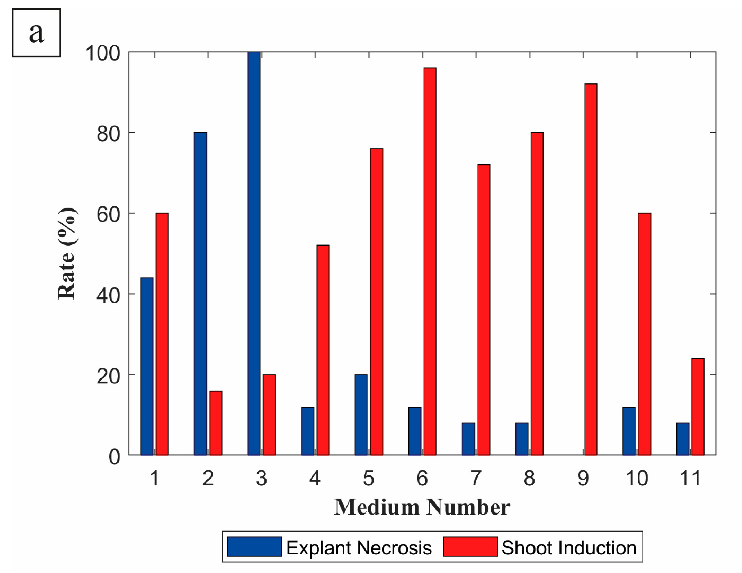
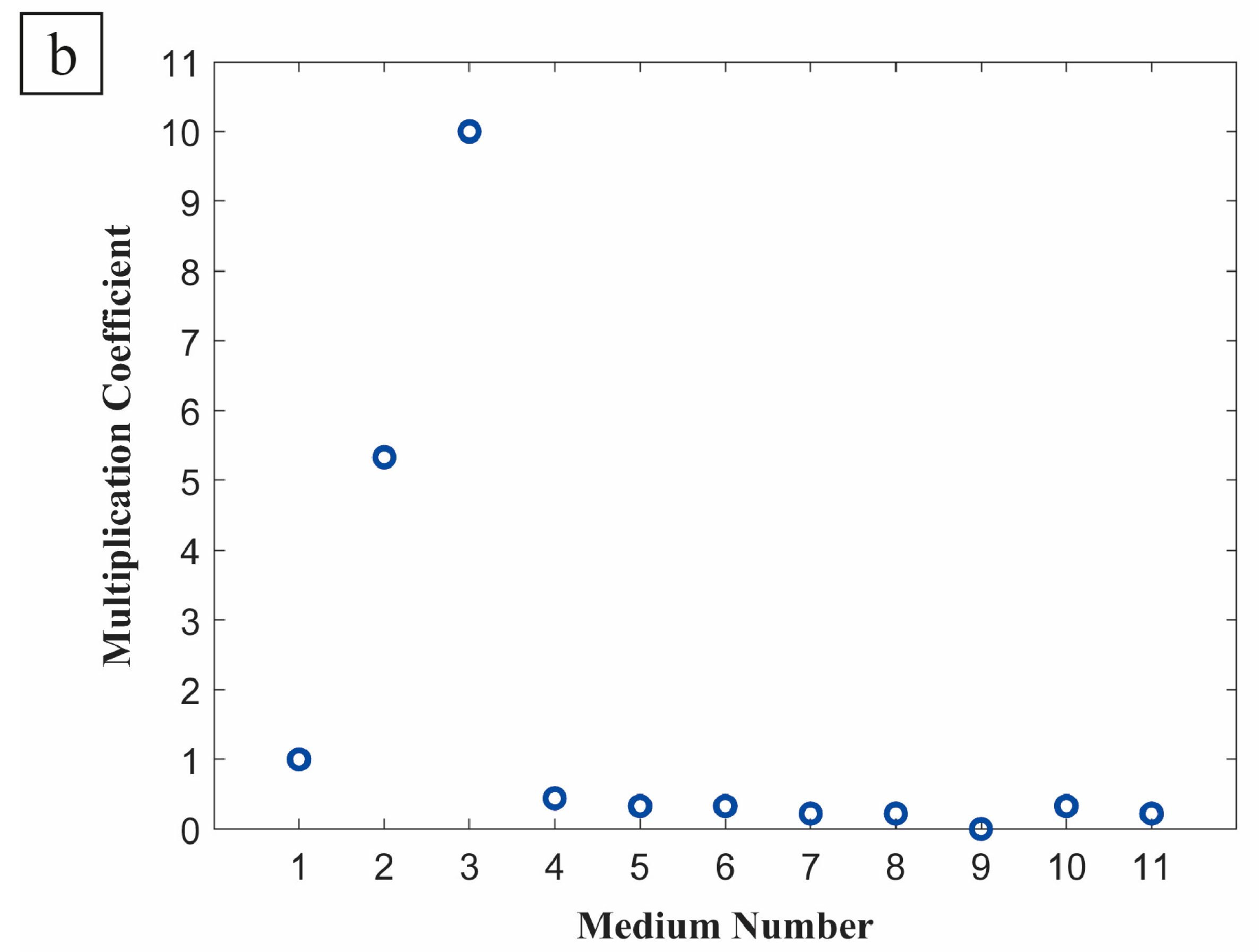
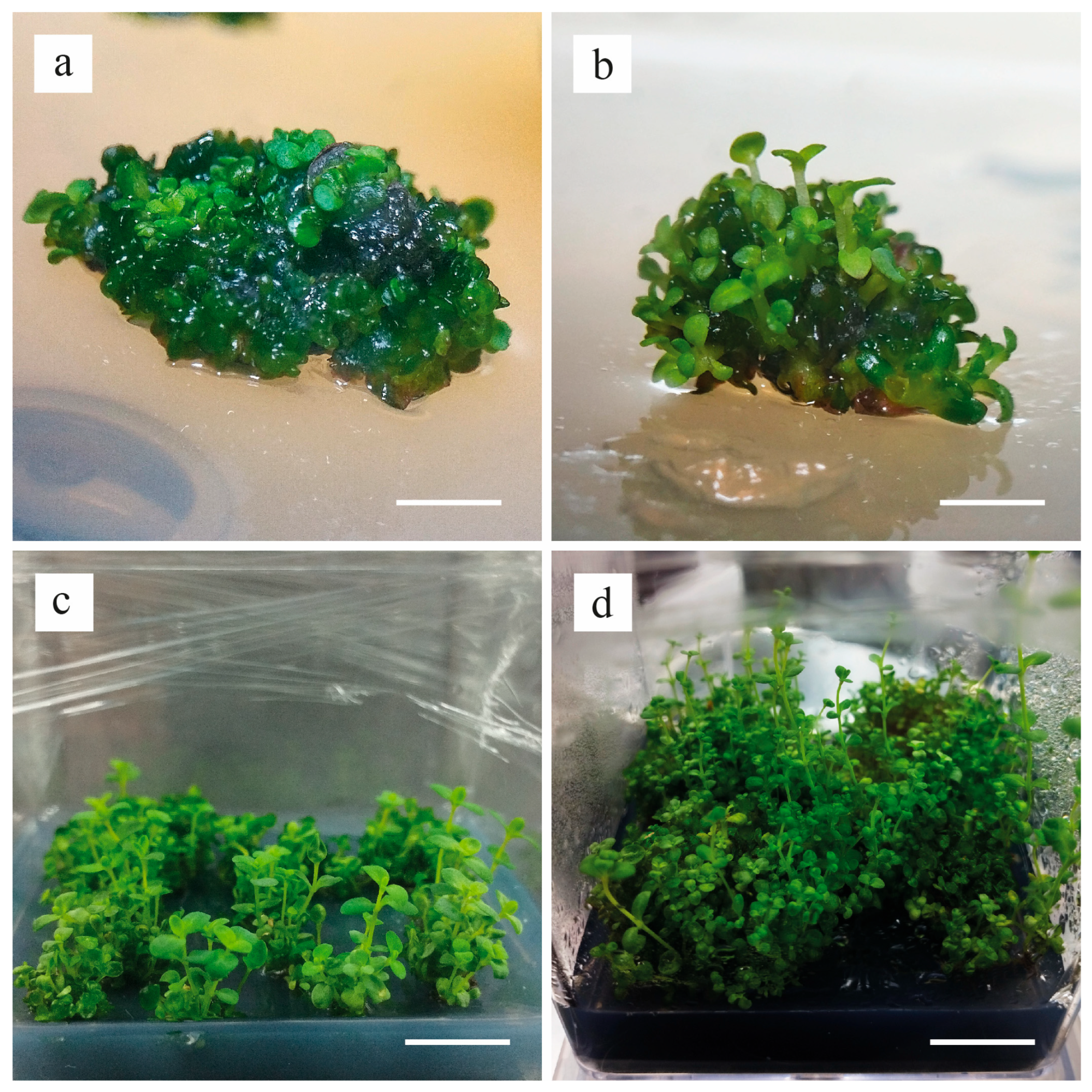
Nodal explants of H. aucheri were cultured in vitro using eleven different media (Figure 1 and Figure 2). On day 28, a limited number of direct shoots were developed in the PGRs-free medium (Figure 1b). The multiplication coefficient remained low (1.0), while the explant necrosis rate reached 60% in this medium.
In our study, only a few shoots were observed in MS media supplemented with 1 mg L−1 KIN and 0.5 mg L−1 2,4-D, 1 mg L−1 KIN and 1 mg L−1 2,4-D, 2 mg L−1 KIN and 0.5 mg L−1 2,4-D, 2 mg L−1 KIN and 1 mg L−1 2,4-D, 1 mg L−1 BAP and 0.5 mg L−1 2,4-D, 1 mg L−1 BAP and 1 mg L−1 2,4-D, 2 mg L−1 BAP and 0.5 mg L−1 2,4-D, 2 mg L−1 BAP and 1 mg L−1 2,4-D. These media primarily induced callus formation rather than shoot induction. Additionally, a relatively high rate of explant necrosis (24–96%) was also recorded in these treatments (Figure 2a).
Shoot induction was relatively high (80%), explant necrosis was low (16%), and the multiplication coefficient reached 5.33 in MS medium supplemented with 2 mg L−1 BAP alone (Figure 2b). However, chlorosis of the leaves and stems was observed in shoots developed in this medium (Figure 1c).
In the medium supplemented with 2 mg L−1 KIN, the highest shoot induction rate (100%) was recorded, and explant necrosis remained relatively low (20%). Callus formation was also observed; however, unlike other treatments, shoot organogenesis from the callus was more pronounced (Figure 1d). Both the callus and the regenerated shoots exhibited a healthy green coloration, indicative of active differentiation. Furthermore, this medium yielded the highest multiplication coefficient (10.0). Overall, among the tested treatments, statistical analysis confirmed that the MS medium supplemented with 2 mg L−1 KIN was significantly superior in terms of shoot induction and multiplication coefficient (p ≤ 0.05), supporting both observational and quantitative findings. Therefore, MS medium supplemented with 2 mg L−1 KIN was selected as shoot multiplication medium.
In the multiplication phase, the shoots of H. aucheri were subcultured twice over eight weeks in medium supplemented with 2 mg L−1 KIN (Table 2 and Figure 3). At the beginning of the second subculture, 1 mg L−1 AC was added to the MS medium to prevent medium browning caused by phenolic exudation. At the end of this period, an average of 89.93 shoots per explant was obtained, demonstrating the strong proliferative effect of this medium. The regenerated shoots had an average length of 1.38 cm, along with 2.27 nodes, 2.80 leaves, and 0.33 lateral shoots.
Shoots were cultured on three different rooting media, and data on rooting rate, number of roots per shoot, root length, and root morphology were recorded at the end of the three-week root induction period (Table 3 and Figure 4). According to the results, no root development was observed in the shoots cultured on MS medium supplemented with 1 mg L−1 IAA (Figure 4a). In contrast, although the medium supplemented with 1 mg L−1 NAA and 1 mg L−1 PVP resulted in a relatively high rooting rate (76%), the shoots developed hairy roots along with aqueous and brownish callus (Figure 4b). In addition, medium browning was also observed in this treatment. In the MS medium supplemented with 1 mg L−1 NAA and 1 mg L−1 AC, root development occurred in all shoots, resulting in the highest rooting rate (100%) with no hairy root or callus formation (Figure 4c).
Consistent with these observations, statistical analysis confirmed that the MS medium supplemented with 1 mg L−1 NAA and 1 mg L−1 AC was significantly superior (p ≤ 0.05) in terms of rooting rate, number of roots per shoot, and root length compared to the other media. Following root induction, plantlets developed on the medium supplemented with 1 mg L−1 NAA and 1 mg L−1 AC were transferred to soil under plant growth room conditions for acclimatization over a three-week period (Figure 4d). The transparent plastic covers placed over the plants were gradually removed, and by the third week, they had been completely removed, which maintained humidity levels and increased the plantlets’ survival rate. The plantlets were observed to continue growing and developing at this stage. Throughout the eight-week greenhouse acclimatization period, most plantlets showed continuous leaf growth and maintained turgidity, while a small proportion exhibited initial wilting during the first week, which stabilized after the second week. After a total of 12 weeks of observation, plantlets successfully acclimatized to ex vitro conditions, resulting in a survival rate of 80% (Figure 4e–h).

4. Discussion

Plant tissue culture plays a vital role in the propagation and conservation of plant species with economic and medicinal importance under in vitro conditions [51]. For this purpose, micropropagation techniques are widely employed. Compared to conventional propagation methods, micropropagation offers several advantages, including the production of disease-free plants, genetically uniform plants, reduced production time, and higher multiplication rates. This method typically involves several stages, shoot induction, multiplication, rooting, and acclimatization to environmental conditions [52,53,54].
The current study evaluated the influence of MS media containing or not containing PGRs on the micropropagation efficiency of H. aucheri. According to our results, nodal explants exhibited different shoot induction responses across eleven different media. While shoot induction was found to be moderate in the PGRs-free medium, the multiplication coefficient was determined to be low. Similarly, Önlü et al. [55] reported that no shoot development was observed from axillary bud explants of H. heterophyllum cultured on a PGRs-free medium. In a different plant species, Kaempferia koratensis, shoot production was also reported to remain at a low level on a PGRs-free medium [56]. These findings from the literature, along with the results of our study, suggest that the endogenous levels of PGRs may not be sufficient to initiate or support shoot induction. Therefore, the external supplementation of auxins or cytokinins to medium appears to be necessary.
In MS media where auxins and cytokinins were combined at various concentrations (BAP and 2,4-D or KIN and 2,4-D), a significant decrease in the shoot induction rate and multiplication coefficient was observed in H. aucheri, predominantly accompanied by the formation of undifferentiated callus tissue. These findings are consistent with previous studies conducted on both common and endemic Hypericum taxa. For instance, BAP and 2,4-D [29,57] or KIN and 2,4-D [58] combinations have been reported to induce callus formation under in vitro culture conditions in H. perforatum. Similarly, Yamaner and Erdağ [40] observed extensive callus development in H. adenotrichum cultured on MS medium containing BAP and NAA combinations. Moreover, a study conducted on different Hypericum species reported that while BAP and 2,4-D combinations induced callus formation in H. perforatum (100%), H. humifusum (90%) and H. leptophyllum (90%), callus production in H. athoum (90%) and H. heterophyllum (77.73%) was promoted by KIN and 2,4-D combinations [59].
In our study, the shoot induction rate of nodal explants was relatively higher on the MS medium supplemented with 2 mg L−1 BAP. However, the regenerated shoots exhibited chlorosis and were frequently associated with the formation of soft and brown-red callus at the base. Despite these issues, this treatment yielded more favorable results than either PGRs-free medium or those containing both cytokinin and 2,4-D. A similar callus formation was reported in a study on H. retusum, where the use of 2.0 or 2.5 mg L−1 BAP induced callus development [60]. In another study, 2 mg L−1 BAP was also reported to induce intensive shoot proliferation in H. gaitii [61]. Additionally, Akbas et al. [38] observed direct shoot organogenesis from H. spectabile on BAP-containing media. In contrast, in H. bupleuroides, BAP and 2,4-D combinations induced excessive callus formation, which later produced numerous shoots when transferred to the medium supplemented with 2 mg L−1 BAP [62]. These species-dependent responses suggest that while BAP is generally effective in promoting shoot induction across Hypericum species [55,63], the capacity for sustained organogenesis may vary significantly among taxa. Although our findings align with those of previous studies, 2 mg L−1 BAP was insufficient to promote sustained organogenesis development in H. aucheri, particularly from callus tissue, despite its ability to stimulate initial shoot induction.
Among all the media used for shoot induction, the medium supplemented with 2 mg L−1 KIN produced the most favorable and effective response in terms of shoot induction rate, multiplication coefficient, and other evaluated parameters. Furthermore, the calli formed in this medium exhibited higher organogenic potential and a greener appearance compared to those formed in other media. Similar findings have been reported in previous studies on various Hypericum species. For instance, in a study on H. bilgehan-bilgilii, the highest number of shoots per explant (4.67) was obtained on the medium supplemented with 2 mg L−1 KIN [40]. In a study conducted on the critically endangered species H. hookerianum, the highest number of shoots per explant (3.66) was obtained on the medium supplemented with 2.32 μM KIN [64]. Similarly, the Himalayan endemic species H. patulum has also been reported to produce comparable results [34]. Yamaner and Erdağ [40] reported that the use of KIN alone, applied at various concentrations (0.5, 1, 2, 3, 4, and 5 mg L−1) increased the number of shoots per explant in H. adenotrichum, with the highest shoot numbers obtained from media supplemented with 1 and 2 mg L−1 KIN (7.8 and 5.8, respectively). A study on H. retusum reported that increasing KIN concentrations (0.10–2.5 mg L−1) led to a higher number of shoots (11.25–35.68), although shoot length decreased with higher concentrations. The most optimal result was obtained with 1.5 mg L−1 KIN, which promoted multiple shoot formation while maintaining shoot morphology. Additionally, the same study indicated that KIN was more effective than BAP in promoting shoot elongation [60]. Different Hypericum species, whether endemic or not, have exhibited diverse responses in terms of callus formation and shoot regeneration under varying types and concentrations of PGRs. These differences are thought to be species-specific, reflecting inherent physiological and developmental variations among taxa [36,39,40,59,65,66].
Shoot induction occurred on nodal explants cultured on MS medium supplemented with 2 mg L−1 KIN via indirect organogenesis. Although somaclonal variation is a well-known drawback of indirect regeneration [67], our findings revealed that direct shoot induction was limited and exhibited low regeneration frequencies on MS medium without PGRs. To minimize the risk of somaclonal variation, nodal segments were selected as explants, and the number of subcultures was kept to a minimum. In addition, early-stage morphogenic calli were prioritized for regeneration.
During the shoot multiplication, a significant increase in the number of shoots per explant (mean 89.93) was observed on the medium supplemented with 2 mg L−1 KIN after 12 weeks, indicating a high proliferation in H. aucheri. Similar results have also been reported in the literature. In a study, the number of shoots per explant in H. perforatum was reported to range from 3.2 to 71.3 when micropropagated using different liquid culture systems [68]. In H. retusum, the highest shoot number per explant (35.68) was achieved using 2.5 mg L−1 KIN [60]. Likewise, in H. hookerianum, 2.32 μM KIN was reported to induce the highest average shoot number per explant (5.50) during the shoot multiplication phase [64].
Three different media were evaluated for root induction. In the MS medium supplemented with 1 mg L−1 IAA, root development was not observed in the multiplied shoots. Although several studies on Hypericum species have reported positive results using various concentrations of IAA for root induction [69,70], 1 mg/L IAA was not effective in our study. Similarly, several studies have indicated that IAA tends to produce more favorable rooting responses compared to IAA or NAA [64,71]. In contrast, the MS medium supplemented with 1 mg L−1 NAA and 1 mg L−1 PVP induced a higher rate of root development than the IAA-containing medium; however, it also resulted in the formation of abnormally hairy roots and aqueous callus tissue. The combination of 1 mg L−1 NAA and 1 mg L−1 PVP may have disrupted the hormonal balance and redox conditions, leading to stress-induced morphogenetic abnormalities such as hyperhydricity, hairy roots, aqueous brownish callus and medium browning due to phenolic oxidation. Similar root structures have previously been reported in H. perforatum under appropriate in vitro conditions on solid media [72,73]. However, this root morphology has been shown to be insufficient for the production of secondary metabolites such as hypericin and hyperforin [72]. Moreover, as observed in other species such as Arabidopsis thaliana, excessive root hair formation has been demonstrated to reduce root permeability and oxygen transfer, leading to physiological stress and potential limitations in culture performance [74]. Therefore, this abnormal root morphology may hinder the successful acclimatization of plantlets under ex vitro conditions. Nevertheless, further in vitro investigation in H. aucheri is recommended to better elucidate the relationship between hairy root formation, culture efficiency, and secondary metabolite production. In contrast, in the third rooting medium, which contained 1 mg L−1 NAA and 1 mg L−1 AC, all cultured shoots successfully developed roots. Cristea et al. [75] reported effective root induction in H. perforatum using a medium containing only 0.6 mg L−1 NAA. Similarly, a study on H. gaitii showed that increasing concentrations of NAA (1.0, 1.5, and 2 mg L−1) resulted in root induction rates ranging from 35.6 to 62.8% [61]. These results suggest that the variation in auxin response among different species may be attributed to species-specific physiological differences. Additionally, during the rooting stage, activated charcoal has been reported to enhance root development in micropropagated plants by creating dark, soil-like conditions and reducing tissue oxidation in vitro cultures [76,77,78]. In Lythrum salicaria, the combination of 0.5 mg L−1 NAA and 0.2 g L−1 AC yielded the most effective rooting response [79].
The acclimatization phase plays a critical role in the successful adaptation of rooted plantlets to soil and external environmental conditions, and it must be carried out gradually [80]. In the present study, the plantlets were successfully transferred to soil, and a high survival rate of 80% was recorded after 12 weeks. Covering the plants with transparent plastic covers and their gradual removal helped regulate humidity and facilitated the plantlets’ adaptation to ex vitro conditions. This result demonstrates the efficiency and reliability of the micropropagation protocol developed for H. aucheri, ensuring successful transition to ex vitro conditions. A similar acclimatization rate (80%) was previously reported for H. bilgehan-bilgilii [41]. In contrast, lower survival rates have been observed in other Hypericum species, such as 50% in H. gaitii [61] and 40% in H. heterophyllum [30]. Furthermore, while Onlu [81] reported successful acclimatization in H. pruinatum, the process was unsuccessful for H. heterophyllum.

5. Conclusions

This study demonstrated, for the first time, the micropropagation potential of H. aucheri. For the shoot induction, eleven different MS media supplemented with various combinations of BAP or KIN and 2,4-D were tested. Among these, the MS medium supplemented with 2 mg L−1 KIN was the most effective for shoot induction and multiplication. The highest rooting efficiency was achieved in the medium supplemented with 1 mg L−1 NAA and 1 mg L−1 AC. Following successful acclimatization, 80% of the regenerated plantlets survived under ex vitro conditions. These findings establish an efficient in vitro propagation system for H. aucheri, highlighting its importance for potential use in future pharmacological and biotechnological applications, and they may also contribute to the species’ ex situ conservation.

Author Contributions

Conceptualization, E.C. and N.Ç.; methodology, E.C. and N.Ç.; software, E.C.; validation, E.C. and N.Ç.; formal analysis, E.C. and N.Ç.; investigation, E.C.; resources, E.C.; data curation, E.C.; writing—original draft preparation, E.C.; writing—review and editing, E.C. and N.Ç.; visualization, E.C.; supervision, N.Ç. All authors have read and agreed to the published version of the manuscript.

Funding

This research received no external funding.

Data Availability Statement

Data are contained within the article.

Conflicts of Interest

The authors declare no conflicts of interest.

References

  1. Babacan, E.Y.; Bagci, E. Essential oil composition of Hypericum uniglandulosum Hausskn. ex Bornm. and Hypericum lydium Boiss. from Turkey. Int. J. Nat. Life Sci. 2017, 1, 12–16. [Google Scholar]
  2. POWO (Plants of the World Online). Hypericum L. Available online: https://powo.science.kew.org/taxon/urn:lsid:ipni.org:names:30002180-2 (accessed on 2 December 2024).
  3. Nürk, N.M.; Crockett, S.L. Morphological and phytochemical diversity among Hypericum species of the Mediterranean basin. Med. Aromat. Plant Sci. Biotechnol. 2011, 5, 14–28. [Google Scholar]
  4. Ersoy, E.; Özkan, E.E.; Boğa, M.; Mat, A. Evaluation of in vitro biological activities of three Hypericum species from Turkey. S. Afr. J. Bot. 2020, 130, 141–147. [Google Scholar] [CrossRef]
  5. Baytop, T. Therapy with Medicinal Plants in Turkey; Istanbul University Press: Istanbul, Turkey, 1999; pp. 66–167. [Google Scholar]
  6. Cirak, C.; Radusiene, J.; Jakstas, V.; Ivanauskas, L.; Seyis, F.; Yayla, F. Secondary metabolites of seven Hypericum species growing in Turkey. Pharm. Biol. 2016, 54, 2244–2253. [Google Scholar] [CrossRef] [PubMed]
  7. Marrelli, M.; Statti, G.; Conforti, F.; Menichini, F. New potential pharmaceutical applications of Hypericum species. Mini-Rev. Med. Chem. 2016, 16, 710–720. [Google Scholar] [CrossRef]
  8. Avato, P. A survey of the Hypericum genus: Secondary metabolites and bioactivity. Stud. Nat. Prod. Chem. 2005, 30, 603–634. [Google Scholar] [CrossRef]
  9. Bingol, U.; Cosge, B.; Gurbuz, B. Hypericum species in flora of Turkey. Med. Aromat. Plant Sci. Biotechnol. 2011, 5, 86–90. [Google Scholar]
  10. Senturk, H.; Kabay, S.; Ozden, H.; Bayramoglu, G.; Ustuner, M.C.; Ozturk, N.; Guven, G.; Kutlu, A.; Bilgi, G.; Ustuner, D.; et al. protective effect of Hypericum origanifolium in experimental renal ischemia/reperfusion injury in rats. Afr. J. Pharm. Pharmacol. 2013, 7, 2306. [Google Scholar] [CrossRef]
  11. Özkan, E.E.; Mat, A. An overview on Hypericum species of Turkey. J. Pharmacogn. Phytother. 2013, 5, 38–46. [Google Scholar] [CrossRef]
  12. Warnick, S.J., Jr.; Mehdi, L.; Kowalkowski, J. Wait—There’s evidence for that? Integrative medicine treatments for major depressive disorder. Int. J. Psychiatry Med. 2021, 56, 334–343. [Google Scholar] [CrossRef]
  13. Sevastre-Berghian, A.C.; Toma, V.A.; Sevastre, B.; Hanganu, D.; Vlase, L.; Benedec, D.; Oniga, I.; Baldea, I.; Olteanu, D.; Moldovan, R.; et al. Characterization and biological effects of Hypericum extracts on experimentally-induced anxiety, oxidative stress and inflammation in rats. J. Physiol. Pharmacol. 2018, 69, 789–800. [Google Scholar] [CrossRef]
  14. McFadden, S.L.; Hooker, B.L. Comparing perika St. John’s wort and sertraline for treatment of posttraumatic stress disorder in mice. J. Diet. Suppl. 2020, 17, 300–308. [Google Scholar] [CrossRef]
  15. Robson, N.K.B. Studies in the Genus Hypericum L. (Hypericaceae) 5(1). Sections 10. Olympia to 15/16. Crossophyllum. Phytotaxa 2010, 4, 5–126. [Google Scholar] [CrossRef]
  16. Robson, N.K.B. Studies in the Genus Hypericum L. (Hypericaceae) 9. Addenda, corrigenda, keys, lists and general discussion. Phytotaxa 2012, 72, 1–111. [Google Scholar] [CrossRef]
  17. Özbek, M.U.; Koç, M.; Hamzaoğlu, E. Contributions to the Hypericum L. section Oligostema (Boiss.) Stef. (Hypericaceae), and Hypericum turcicum sp. nov. as a New Species from Turkey. Turk. J. Bot. 2019, 43, 694–702. [Google Scholar] [CrossRef]
  18. Efe, R.; Sönmez, S.; Cürebal, İ.; Soykan, A. Ecological Conditions and Vegetation of Subalpine Zone of Kaz Mountain (Mount Ida, NW Turkey). In Climate Change Impacts on High-Altitude Ecosystems; Öztürk, M., Hakeem, K., Faridah-Hanum, I., Efe, R., Eds.; Springer: Cham, Switzerland, 2015; pp. 591–608. [Google Scholar] [CrossRef]
  19. Carta, A.; Probert, R.; Puglia, G.; Peruzzi, L.; Bedini, G. Local climate explains degree of seed dormancy in Hypericum elodes L. (Hypericaceae). Plant Biol. 2016, 18, 76–82. [Google Scholar] [CrossRef]
  20. Klein, J.D.; Cohen, S.; Hebbe, Y. Seasonal variation in rooting ability of myrtle (Myrtus communis L.) cuttings. Sci. Hortic. 2000, 83, 71–76. [Google Scholar] [CrossRef]
  21. Heywood, V.H.; Iriondo, J.M. Plant conservation: Old problems, new perspectives. Biol. Conserv. 2003, 113, 321–335. [Google Scholar] [CrossRef]
  22. George, E.F.; Hall, M.A.; De Klerk, G.J. Plant Propagation by Tissue Culture: Volume 1. The Background, 3rd ed.; Springer: Dordrecht, The Netherlands, 2008. [Google Scholar]
  23. Pence, V.C. The possibilities and challenges of in vitro methods for plant conservation. Kew Bull. 2010, 65, 539–547. [Google Scholar] [CrossRef]
  24. Gürel, A.; Hayta, Ş.; Nartop, P.; Bayraktar, M.; Fedakar, S.O. Bitki Hücre, Doku ve Organ Kültürü Uygulamaları; Ege Üniversitesi Basımevi: İzmir, Turkey, 2013; pp. 47–66. [Google Scholar]
  25. Su, Y.H.; Tang, L.P.; Zhao, X.Y.; Zhang, X.S. Plant cell totipotency: Insights into cellular reprogramming. J. Integr. Plant Biol. 2021, 63, 228–243. [Google Scholar] [CrossRef] [PubMed]
  26. Malik, C. Applications of biotechnology innovations in pharmaceutics and nutraceutics in multitherapeutic medicinal and special plants. In Pharmaceutics and Nutraceutics; Singh, K., Jahdon, M.L., Singh, D., Eds.; Aavishkar Publishers: Jaipur, India, 2007; Volume II, pp. 243–265. [Google Scholar]
  27. Jamsheed, S.; Rasool, S.; Koul, S.; Azooz, M.M.; Ahmad, P. Crop Improvement through Plant Tissue Culture. In Crop Improvement: New Approaches and Modern Techniques; Hakeem, K.R., Ahmad, P., Ozturk, M., Eds.; Springer: Dordrecht, The Netherlands, 2013; pp. 123–148. [Google Scholar] [CrossRef]
  28. Khan, I.; Khan, M.A.; Shehzad, M.A.; Ali, A.; Mohammad, S.; Ali, H.; Alyemeni, M.N.; Ahmad, P. Micropropagation and production of health-promoting lignans in Linum usitatissimum. Plants 2020, 9, 728. [Google Scholar] [CrossRef]
  29. Pretto, F.R.; Santarém, E.R. Callus formation and plant regeneration from Hypericum perforatum leaves. Plant Cell Tissue Organ Cult. 2000, 62, 107–113. [Google Scholar] [CrossRef]
  30. Ayan, A.K.; Cirak, C. In vitro multiplication of Hypericum heterophyllum, an endemic Turkish species. Am. J. Plant Physiol. 2006, 1, 76–81. [Google Scholar]
  31. Palmer, C.D.; Keller, W.A. Plant regeneration from petal explants of Hypericum perforatum L. Plant Cell Tissue Organ Cult. 2011, 105, 129–134. [Google Scholar] [CrossRef]
  32. Banerjee, A.; Bandyopadhyay, S.; Raychaudhuri, S.S. In vitro regeneration of Hypericum perforatum L. using thidiazuron and analysis of genetic stability of regenerants. Indian J. Biotechnol. 2012, 11, 92–98. [Google Scholar]
  33. Mir, M.Y.; Kamili, A.N.; Hassan, Q.P.; Rafi, S.; Parray, J.A.; Jan, S. In vitro regeneration and free radical scavenging assay of Hypericum perforatum L. Natl. Acad. Sci. Lett. 2019, 42, 161–167. [Google Scholar] [CrossRef]
  34. Baruah, A.; Sarma, D.; Saud, J.; Singh, R.S. In vitro regeneration of Hypericum patulum Thunb.—A medicinal plant. Indian J. Exp. Biol. 2001, 39, 947–949. [Google Scholar] [PubMed]
  35. Karakas, O.; Toker, Z.; Tilkat, E.; Ozen, H.C.; Onay, A. Effects of different concentrations of benzylaminopurine on shoot regeneration and hypericin content in Hypericum triquetrifolium Turra. Nat. Prod. Res. 2009, 23, 1459–1465. [Google Scholar] [CrossRef]
  36. Shilpashree, H.P.; Rai, R. In vitro plant regeneration and accumulation of flavonoids in Hypericum mysorense. Int. J. Integr. Biol. 2009, 8, 43–49. [Google Scholar]
  37. Oluk, E.; Orhan, S. Thidiazuron induced micropropagation of Hypericum triquetrifolium Turra. Afr. J. Biotechnol. 2009, 8, 3506–3510. [Google Scholar]
  38. Akbas, F.; Isikalan, C.; Namli, S.; Karakus, P.; Basaran, D. Direct plant regeneration from in vitro-derived leaf explants of Hypericum spectabile, a medicinal plant. J. Med. Plants Res. 2011, 5, 2175–2181. [Google Scholar]
  39. Cirak, C.; Radušienė, J.; Kurtarc, E.S.; Marksa, M.; Ivanauskas, L. In vitro plant regeneration and jasmonic acid induced bioactive chemical accumulations in two Hypericum species from Turkey. S. Afr. J. Bot. 2020, 128, 312–318. [Google Scholar] [CrossRef]
  40. Yamaner, O.; Erdağ, B. Callus induction and adventitious shoot regeneration of Hypericum adenotrichum Spach. Eskişehir Tek. Üniversitesi Bilim Ve Teknol. Derg.-C Yaşam Bilim. Ve Biyoteknoloji 2020, 9, 98–108. [Google Scholar] [CrossRef]
  41. Turker, H.; Unal, B.T. Development of a micropropagation protocol for endangered Hypericum bilgehan-bilgilii Başkose & Savran (Hypericaceae), local endemic to Turkey. Pak. J. Bot. 2022, 54, 1089–1095. [Google Scholar] [CrossRef]
  42. Kitanov, G.M.; Blinova, K.F.; Akhtardzhiev, K.; Rumenin, V. Flavonoids of Hypericum aucheri. Chem. Nat. Compd. 1979, 15, 760–761. [Google Scholar] [CrossRef]
  43. Kitanov, G.M. Biflavone, Flavonol, and Xanthone Glycosides from Hypericum aucheri. Chem. Nat. Compd. 1988, 24, 390–391. [Google Scholar] [CrossRef]
  44. Zheleva-Dimitrova, D.; Nedialkov, P.; Kitanov, G. Radical Scavenging and Antioxidant Activities of Methanolic Extracts from Hypericum Species Growing in Bulgaria. Pharmacogn. Mag. 2010, 6, 74. [Google Scholar] [CrossRef]
  45. Krasteva, I.; Nedelcheva, A.; Pavlova, D.; Zdraveva, P.; Nikolov, S.; Mitov, K. Influence of Serpentine Soils on the Flavonoid Content of Hypericum Populations Growing in Bulgaria. Afr. J. Pharm. Pharmacol. 2013, 7, 1762–1765. [Google Scholar] [CrossRef]
  46. Nedialkov, P.T.; Ilieva, Y.; Zheleva-Dimitrova, D.; Kokanova-Nedialkova, Z.; Momekov, G. Three New Prenyloxy Chromanones from Aerial Parts of Hypericum aucheri. Fitoterapia 2019, 139, 104421. [Google Scholar] [CrossRef] [PubMed]
  47. Marinov, T.; Kokanova-Nedialkova, Z.; Nedialkov, P. UHPLC-HRMS-Based Profiling and Simultaneous Quantification of the Hydrophilic Phenolic Compounds from the Aerial Parts of Hypericum aucheri Jaub. & Spach (Hypericaceae). Pharmacia 2024, 71, 1–11. [Google Scholar] [CrossRef]
  48. Dimitrov, M.; Nikolova, I.; Benbasat, N.; Kitanov, G.; Danchev, N. Acute Toxicity, Antidepressive and MAO Inhibitory Activity of Mangiferin Isolated from Hypericum aucheri. Biotechnol. Biotechnol. Equip. 2011, 25, 2668–2671. [Google Scholar] [CrossRef]
  49. Marinov, T.; Kondeva-Burdina, M.; Kokanova-Nedialkova, Z.; Nedialkov, P.T. Phenolic Constituents from Hypericum aucheri Jaub. & Spach—Isolation, Identification, and Preliminary Evaluation for hMAO-A/B and Neuroprotective Activity. Chemistry 2024, 6, 1535–1551. [Google Scholar] [CrossRef]
  50. Murashige, T.; Skoog, F. A revised medium for rapid growth and bioassays with tobacco tissue cultures. Physiol. Plant. 1962, 15, 473–497. [Google Scholar] [CrossRef]
  51. Pence, V.C. Evaluating costs for the in vitro propagation and preservation of endangered plants. Vitr. Cell. Dev. Biol.-Plant 2011, 47, 176–187. [Google Scholar] [CrossRef]
  52. Paek, K.Y.; Yu, K.J.; Park, S.I.; Sung, N.; Park, C. Micropropagation of Rehmannia glutinosa as medicinal plant by shoot tip and root segment culture. Acta Hortic. 1995, 390, 113–120. [Google Scholar] [CrossRef]
  53. Borthakur, M.; Singh, R.S. Direct plantlet regeneration from male inflorescence of medicinal yam (Dioscorea floribunda Mart. & Gal.). Vitr. Cell. Dev. Biol.-Plant 2002, 38, 183–185. [Google Scholar] [CrossRef]
  54. Uchendu, E.E.; Shukla, M.R.; Reed, B.M.; Brown, D.C.; Saxena, P.K.; Moo-Young, M. Improvement of ginseng by in vitro culture: Challenges and opportunities. In Comprehensive Biotechnology, 2nd ed.; Moo-Young, M., Ed.; Academic Press: Burlington, MA, USA, 2011; Volume 2, pp. 317–329. [Google Scholar]
  55. Önlü, Ş.; Yaman, C.; Kurtul, E.; Önlü, H.; Bahadir-Acikara, Ö.; Tusevski, O.; Simic, S.G.; Özcan, S. Production of elicitor-induced phytochemicals in callus and shoot cultures of Hypericum heterophyllum. S. Afr. J. Bot. 2025, 177, 295–304. [Google Scholar] [CrossRef]
  56. Saliwan, S.; Saensouk, S.; Saensouk, P. In vitro propagation of Kaempferia koratensis Picheans., an endemic plant of Thailand. Koch Cha Sarn J. Sci. 2022, 44, 1–13. [Google Scholar]
  57. Walker, T.S.; Bais, H.P.; Vivanco, J.M. Jasmonic acid-induced hypericin production in cell suspension cultures of Hypericum perforatum L. (St. John’s wort). Phytochemistry 2002, 60, 289–293. [Google Scholar] [CrossRef]
  58. Ayan, A.K.; Çirak, C.; Kevseroğlu, K.; Sökmen, A. Effects of explant types and different concentrations of sucrose and phytohormones on plant regeneration and hypericin content in Hypericum perforatum L. Turk. J. Agric. For. 2005, 29, 197–204. [Google Scholar]
  59. Ahmed, H.A.A.; Uranbey, S.; Salaj, T.; Mistrikova, V. Cell Suspension Cultures and High Frequency Shoot Regeneration of Some Hypericum species. J. Agri. Sci. 2025, 31, 319–331. [Google Scholar] [CrossRef]
  60. Namli, S.; Akbas, F.; Isikalan, C.; Tilkat, E.A.; Basaran, D. The Effect of Different Plant Hormones (PGRs) on Multiple Shoots f’Hypericum retusum Aucher. Plant Omics 2010, 3, 12–17. [Google Scholar]
  61. Swain, D.; Lenka, S.; Hota, T.; Rout, G.R. Micro-propagation of Hypericum gaitii Haines, an endangered medicinal plant: Assessment of genetic fidelity. Nucleus 2016, 59, 7–13. [Google Scholar] [CrossRef]
  62. Cirak, C.; Ayan, A.K.; Kevseroğlu, K. Direct and indirect regeneration of plants from internodal and leaf explants of Hypericum bupleuroides Gris. J. Plant Biol. 2007, 50, 24–28. [Google Scholar] [CrossRef]
  63. Asan, H.S.; Ozen, H.C.; Onay, A.; Nurettin, A. Effect of different BAP concentrations on proliferation and hypericin content of Hypericum retusum Aucher. Biol. Sci. Res. J. 2014, 7, 10–14. [Google Scholar]
  64. Varghese, R.J.; Bayyapureddy, A.; Pushparaj, S.P.; Seeni, S. A high-efficiency in vitro regeneration protocol and clonal uniformity analysis in Hypericum hookerianum Wight & Arn. Braz. J. Bot. 2016, 39, 377–386. [Google Scholar] [CrossRef]
  65. Bernardi, A.P.M.; Maurmann, N.; Rech, S.B.; Von Poser, G. Benzopyrans in Hypericum polyanthemum Klotzsch ex Reichardt cultured in vitro. Acta Physiol. Plant. 2007, 29, 165–170. [Google Scholar] [CrossRef]
  66. Coste, A.; Halmagyi, A.; Butiuc-Keul, A.L.; Deliu, C.; Coldea, G.; Hurdu, B. In vitro propagation and cryopreservation of Romanian endemic and rare Hypericum species. Plant Cell Tissue Organ Cult. 2012, 110, 213–226. [Google Scholar] [CrossRef]
  67. Bairu, M.W.; Aremu, A.O.; Van Staden, J. Somaclonal variation in plants: Causes and detection methods. Plant Growth Regul. 2011, 63, 147–173. [Google Scholar] [CrossRef]
  68. Savio, L.E.B.; Astarita, L.V.; Santarém, E.R. Secondary metabolism in micropropagated Hypericum perforatum L. grown in non-aerated liquid medium. Plant Cell Tissue Organ Cult. 2012, 108, 465–472. [Google Scholar] [CrossRef]
  69. Oluk, E.A.; Orhan, S.; Çakir, A.; Gönüz, A. High efficiency indirect shoot regeneration and hypericin content in embryogenic callus of Hypericum triquetrifolium Turra. Afr. J. Biotechnol. 2010, 9, 2229–2233. [Google Scholar]
  70. Shilpashree, H.P.; Rai, V.R. Effect of plant growth regulators on callus formation, plant regeneration and hypericin production in Hypericum mysorense Hyne. Int. J. Plant Dev. Biol. 2010, 4, 31–36. [Google Scholar]
  71. Abdollahpoor, M.; Kalantari, S.; Azizi, M.; Saadat, Y.A. In vitro shoot proliferation of Hypericum perforatum L. through indirect and direct plant regeneration. J. Med. Plants By-Prod. 2017, 6, 81–89. [Google Scholar] [CrossRef]
  72. Cui, X.H.; Chakrabarty, D.; Lee, E.J.; Paek, K.Y. Production of adventitious roots and secondary metabolites by Hypericum perforatum L. in a bioreactor. Bioresour. Technol. 2010, 101, 4708–4716. [Google Scholar] [CrossRef]
  73. Murthy, H.N.; Kim, Y.S.; Park, S.Y.; Paek, K.Y. Hypericins: Biotechnological production from cell and organ cultures. Appl. Microbiol. Biotechnol. 2014, 98, 9187–9198. [Google Scholar] [CrossRef] [PubMed]
  74. Shiao, T.L.; Doran, P.M. Root hairiness: Effect on fluid flow and oxygen transfer in hairy root cultures. J. Biotechnol. 2000, 83, 199–210. [Google Scholar] [CrossRef]
  75. Cristea, T.O.; Falticeanu, M.; Prisecaru, M. Studies regarding the morphogenetic reaction of Hypericum perforatum L. explants to in vitro medium conditions. Studii şi Comunicări Compl. Muz. Şt. Nat. „Ion Borcea” Bacău. 2006, 21, 164–167. [Google Scholar]
  76. Peng, L.C.; Qu, S.P.; Su, Y.; Zhang, Y.P.; Wang, L.H. Study on rapid propagation of Hylocereus undatus by two steps. Southwest China J. Agric. Sci. 2014, 27, 2529–2533. [Google Scholar]
  77. Máximo, W.P.F.; Santos, B.R.; Martins, J.P.R.; Beijo, L.A.; Barbosa, S. Multiplication and in vitro rooting of Handroanthus impetiginosus (Mart. Ex DC.) Mattos. Cienc. Florest. 2020, 30, 658–668. [Google Scholar] [CrossRef]
  78. Lee, Y.C.; Chang, J.C. Development of an improved micropropagation protocol for red-fleshed pitaya ‘Da Hong’ with and without activated charcoal and plant growth regulator combinations. Horticulturae 2022, 8, 104. [Google Scholar] [CrossRef]
  79. Ebrahimi, T.; Piri, K.; Abdoli, A.; Tohidfar, M. Effect of activated charcoal on in vitro propagation of Lythrum salicaria L. J. Med. Plants By-Prod. 2024, 13, 293–302. [Google Scholar]
  80. Hazarika, B.N.; Teixeira da Silva, J.A.; Talukdar, A. Effective acclimatization of in vitro cultured plants: Methods, physiology and genetics. In Floriculture, Ornamental and Plant Biotechnology; Teixeira da Silva, J.A., Ed.; Global Science Books: Isleworth, UK, 2006; Volume 2, pp. 427–438. [Google Scholar]
  81. Onlu, S. In Vitro Plant Regeneration and Secondary Metabolite Production in Hypericum Species. Ph.D. Thesis, Department of Field Crops, University of Ankara, Ankara, Turkey, 2019; 50p. [Google Scholar]
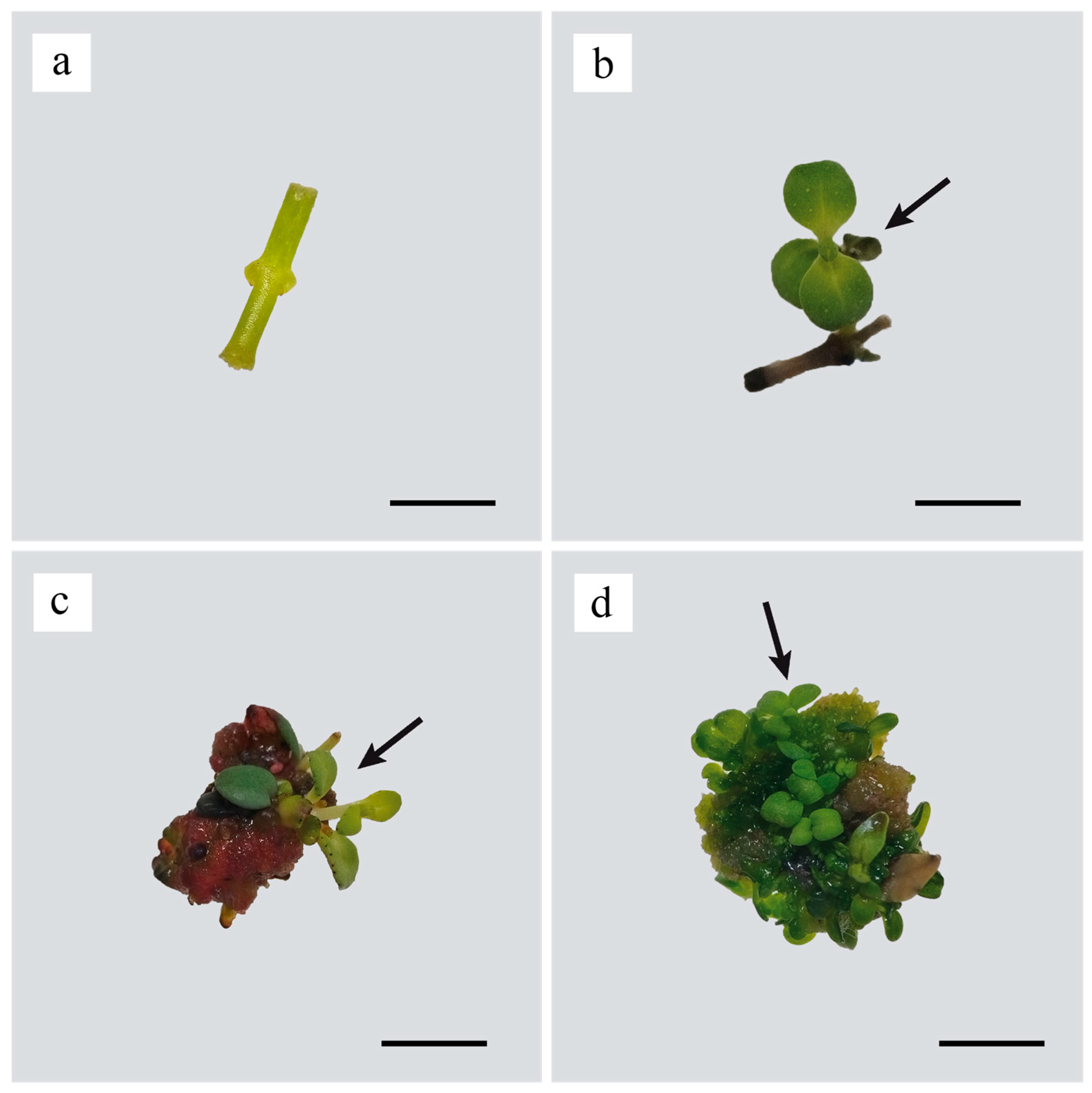
Figure 1. Shoot induction culture of H. aucheri. (a) Nodal explant on day 0; (b) shoot induced via direct shoot organogenesis in the PGRs-free medium at day 28; (c) shoot induced via indirect shoot organogenesis in 2 mg L−1 BAP at day 28; (d) shoot induced via indirect shoot organogenesis in 2 mg L−1 KIN at 28 day (arrows indicate shoots; scale bar = 1 cm).
Horticulturae 11 01069 g001
Figure 2. (a) Shoot induction responses and (b) multiplication coefficients of H. aucheri nodal explants cultured on different shoot induction media after 4 weeks of culture (Medium Number 1: PGRs-free medium, 2: 2 mg L−1 BAP, 3: 2 mg L−1 KIN, 4: 1 mg L−1 KIN and 0.5 mg L−1 2,4-D, 5: 1 mg L−1 KIN and 1 mg L−1 2,4-D, 6: 2 mg L−1 KIN and 0.5 mg L−1 2,4-D, 7: 2 mg L−1 KIN and 1 mg L−1 2,4-D, 8: 1 mg L−1 BAP and 0.5 mg L−1 2,4-D, 9: 1 mg L−1 BAP and 1 mg L−1 2,4-D, 10: 2 mg L−1 BAP and 0.5 mg L−1 2,4-D, and 11: 2 mg L−1 BAP and 1 mg L−1 2,4-D).
Horticulturae 11 01069 g002aHorticulturae 11 01069 g002b
Figure 3. Shoot development of H. aucheri during multiplication on MS medium supplemented with 2 mg L−1 KIN. (a) day 0 of the first subculture; (b) day 21 of the first subculture; (c) day 0 of the second subculture; and (d) day 21 of the second subculture (scale bar = 1 cm).
Horticulturae 11 01069 g003
Figure 4. Rooting and acclimatization of H. aucheri shoots. (a) Shoot without roots cultured on MS medium supplemented with 1 mg L−1 IAA; (b) Shoots with hairy roots on MS medium supplemented with 1 mg L−1 NAA and 1 mg L−1 PVP; (c) Shoots with well roots on MS medium supplemented with 1 mg L−1 NAA and 1 mg L−1 AC; (d) micropropagated plantlets; (e) day 0; (f) week 3; (g) week 6; and (h) week 12 of acclimatized plantlets (scale bar = 1 cm).
Horticulturae 11 01069 g004
Table 1. The content of PGRs in MS medium used for shoot induction of H. aucheri.
Medium NoPGRs (mg L−1)
BAPKIN2,4-D
1000
22.000
302.00
401.00.5
501.01.0
602.00.5
702.01.0
81.000.5
91.001.0
102.000.5
112.001.0
Table 2. Shoot multiplication data of H. aucheri shoots cultured on MS medium supplemented with 2 mg L−1 KIN.
Replicate No Number of Shoots per Explant Shoot
Length (cm)
Number of Nodes per Shoot Number of Leaves per Shoot
1 90.00 ± 9.60 a 1.49 ± 0.07 b 2.83 ± 0.18 a 3.06 ± 0.16 b
2 87.40 ± 11.11 a 1.27 ± 0.05 a 1.96 ± 0.13 b 2.30 ± 0.10 a
3 92.40 ± 16.34 a 1.39 ± 0.05 ab 2.03 ± 0.16 a 3.03 ± 0.16 b
Means 89.93 ± 6.80 1.38 ± 0.03 2.27 ± 0.10 2.80 ± 0.09
Data on the number of shoots per explant are based on 15 explants (n = 15); while other traits were measured on 30 shoots (n = 30). Values are means ± SE, and different letters within a column indicate statistically significant differences (p ≤ 0.05).
Table 3. Root response of H. aucheri shoots after 3 weeks of culture in different rooting media.
Root Induction
Medium
Rooting
Rate (%)
Number of Roots per ShootRoot
Length (cm)
Root
Morphology
1 mg L−1 IAA0.00 ± 0.00 a0.00 ± 0.00 a0.00 ± 0.00 a-
1 mg L−1 NAA
1 mg L−1 PVP
76.00 ± 9.79 b2.04 ± 0.29 b0.55 ± 0.05 bHairy
1 mg L−1 NAA
1 mg L−1 AC
100.00 ± 0.00 c2.28 ± 0.20 b4.16 ± 0.31 cNormal
Different letters within the same column indicate significant differences at p ≤ 0.05 level and values are given as mean ± SE. For each treatment, 25 shoots were randomly selected (-: no formation).
Disclaimer/Publisher’s Note: The statements, opinions and data contained in all publications are solely those of the individual author(s) and contributor(s) and not of MDPI and/or the editor(s). MDPI and/or the editor(s) disclaim responsibility for any injury to people or property resulting from any ideas, methods, instructions or products referred to in the content.