Review

Exploring Factors Influencing the Consumption of Grape Skins: A Review

College of Horticulture and Garden, Yangzhou University, Yangzhou 225009, China
*
Author to whom correspondence should be addressed.
Horticulturae 2025, 11(8), 962; https://doi.org/10.3390/horticulturae11080962 (registering DOI)
Submission received: 29 June 2025 / Revised: 7 August 2025 / Accepted: 8 August 2025 / Published: 14 August 2025
(This article belongs to the Special Issue Research Progress on Grape Genetic Diversity)

Abstract

Grapes (Vitis vinifera L.) are one of the most popular fruits globally, with studies demonstrating the numerous beneficial metabolic substances found in their skins, including anthocyanins, proanthocyanidins, stilbene (resveratrol) and so on. However, grape skins are often overlooked and discarded by consumers. To maximize the nutritional benefits of grapes, it is crucial to explore the factors influencing the edibility of grape skin and work towards enhancing consumption. This review explores the molecular mechanisms underlying these factors, focusing on cell wall modifications and tannin biosynthesis. We highlight how skin texture and astringency are influenced by enzymatic activity, genetic regulation, and environmental factors. Understanding these mechanisms provides a foundation for improving skin palatability through breeding or biotechnological approaches, enhancing the nutritional and commercial value of grapes.

1. Introduction

The grape (Vitis vinifera L.) is a significant fruit with economic importance and is cultivated globally. Grapes are consumed fresh [1] or processed into various products such as wine, grape vinegar, raisins, and other derivatives. The table grape is the main grape industry in China, accounting for 80% of total grape production [2]. Grapes are also rich in nutrients, such as B vitamins, vitamin C, calcium, phosphorus, potassium, and various amino acids essential for human health, helping to address nutritional deficiencies in the body [3]. In recent years, Xia et al. [4] found that grapes contained a large number of phenolic compounds, including anthocyanins, proanthocyanidins, stilbene (resveratrol) and phenolic acid. These compounds have been linked to the prevention of diseases caused by oxidative stress and exhibit antioxidant, anti-cancer, anti-inflammatory, anti-aging, and antibacterial properties [5]. In addition, these compounds could maintain endothelial function, enhance antioxidant capacity, protect against LDL oxidation, and inhibit cellular processes that may contribute to atherosclerosis and coronary heart disease, making them valuable as cardiac protective agents [6]. The majority of these nutrients are found in grape skin and seeds [7]. However, studies have reported that the amount of these compounds in the skin, pulp (flesh), and seeds can vary greatly, especially depending on the variety and species [8,9,10]. It is undeniable that consuming grape skin offers numerous health benefits. However, people choose to discard the grape skin for various reasons such as the unpleasant taste [11,12] and rough texture of the skin [13]. The study of the influencing factors of grape skin edibility can lay a theoretical basis for improving the texture and flavor of grape skin, thus maximizing the nutritional utilization and the commercial value of grapes. However, current research on grape skin mostly focuses on the fruit coloring and fruit storage properties. There is no in-depth study on the edibility of grape skins.
Based on the existing research, two significant factors that may influence the taste of grape skin were summarized in this review. The first factor is the thickness or hardness of the skin, which makes it difficult to chew. The second factor is the astringency of the skin after chewing. Therefore, we further analyzed and summarized the flavor and texture of grape skin by its material composition and skin structure, laying a foundation for further exploration of the main substances affecting the edibility of grape skin.

2. Development and Skin Characteristics of Grapes

2.1. Growth and Development of Grapes

The grape development period refers to the stage from flowering and fruit set to pre-veraison. It is generally divided into three phases: the initial rapid growth stage (S-I); the slow growth stage (S-II); and the second rapid growth stage (S-III). The entire berry growth follows a double sigmoid (“S-shaped”) curve [14].
The S-I stage, also known as the initial rapid growth phase, typically spans from 15 to 28 days after flowering [14]. During this critical developmental period, grape berries enter what is commonly referred to as the hard seed stage. The fruits maintain a vibrant green coloration while achieving maximum firmness [15]. Biochemically, this phase is characterized by high concentrations of organic acids and relatively low sugar content [15]. From a cellular perspective, this stage witnesses vigorous cell division activity, with the most intensive proliferation occurring between 5 to 10 days after flowering [16]. Microscopic examination reveals distinct growth patterns among different cell layers. Mesocarp cells exhibit expansion in both tangential and radial dimensions, while exocarp cells demonstrate unidirectional growth along the tangential axis accompanied by significant cell wall thickening [15]. The termination of cell division follows a well-defined spatial sequence as the stage progresses. The cessation initiates in the innermost endocarp layer, progresses through the mesocarp, and finally concludes in the outermost exocarp [17]. This sequential pattern results in the exocarp cells being the last to complete their division cycle, marking the transition to the subsequent developmental phase [17]. At this stage, the primary cell wall (CW) modifications include pectin depolymerization mediated by pectin methylesterase (PME), rhamnogalacturonan lyase (RGL), and β-galactosidase (β-Gal), as well as the loosening of the xyloglucan–cellulose network through the action of cellulases (CEL), leading to hemicellulose solubilization [16].
The S-II stage, typically occurring between 28 to 55 days after flowering, represents a period of relatively slow development [14]. During this phase, the fruits undergo gradual softening while showing no significant changes in longitudinal/transverse diameters, weight, or cellular morphology [15,16]. In the latter part of S-II, distinct cellular changes become apparent. The cross-sectional area of exocarp cells begins to increase, with epidermal cells expanding in both tangential and radial directions, and hypodermal cells exhibiting radial enlargement, contributing to subtle but important structural modifications in the developing fruit [15]. Meanwhile, at this stage, the softening process is characterized by pectin solubilization due to the activity of enzymes such as PMEs, PGs, and PLs [16,18]. Notably, pectin degradation in the middle lamella and the loss of intercellular adhesion are the primary phenomena leading to fruit softening [16].
The S-III stage generally spans from 55 to 99 days after flowering, with the veraison period (55–71 days post-flowering) marking the onset of the second rapid growth phase [14]. During this critical developmental window, the berries undergo significant physiological changes as they transition to maturity. Most notably, the fruits exhibit a visible color transformation accompanied by progressive softening of texture [16]. This chromatic shift results from the differential accumulation of anthocyanins (particularly malvidin derivatives in black varieties) or carotenoids (predominantly lutein and β-carotene in white varieties). At the cellular level, exocarp cells show growth in cross-sectional area, with the cell walls demonstrating significant thickening during early S-III, followed by progressive thinning due to hydrolytic processes as maturation progresses [15]. These coordinated cellular changes facilitate the final swelling and ripening of grape berries [16].

2.2. Structure of Grape Skin

Grape skin is composed of three anatomically different tissues, namely, the exocarp, endocarp, and mesocarp (Figure 1). Grape skin, also known as the exocarp, is composed of a waxy stratum cuticle and underlying layers of sclerenchyma-like cells named epidermal cells and subepidermal cells [19] (Figure 1). The waxy cuticle, usually called ‘fruit cream’, covers the outside of the stratum corneum in overlapping sheets, forming a powerful hydrophobic layer that assists the fruit in retaining water and acts as the primary defense against pathogen penetration [20]. Epidermal cells are generally composed of 2–3 layers of flattened cells, which are divided and neatly and tightly arranged in a pendulous peripheral direction [21]. Subepidermal cells are generally composed of 3–12 layers of oblong, thick-walled cells, which are the overlayer between the outer epidermis and mesocarp, with a cell volume much larger than that of the epidermal cells [22]. With the development of grape berries, the subepidermal cells are stretched and their cell walls thicken about 10-fold, while the cell volume expands rapidly, gradually transforming into mesocarp cells. The mesocarp, often referred to as the pulp, consists of 16–18 layers of highly vesicularized storage thin-walled cells with the cell volume of the exocarp cells [23]. It is distinguished from the exocarp by the peripheral vascular bundles; however, as the grape ripens, the peripheral vascular bundles gradually converge towards the mesocarp and the boundary between the exocarp and the mesocarp becomes progressively blurred [19].

2.3. Sensory Characteristics of Grape Skin

Grape quality traits include appearance, nutritional content, and flavor. Among them, taste quality, also referred to as flavor quality, encompasses various taste sensations influenced by chemical compounds, including taste, aroma, and texture [24]. Terpenes, C13-norisoprenoids, phenols, and non-terpenic alcohols are the most important aroma compounds in grapes; they can be found as free volatiles or glycoconjugated (bound) molecules. The texture of grape pulp and skin plays a crucial role not only in affecting the taste, storage, transportation and processing quality, but also in providing an important reference for distinguishing different grape varieties [25]. Regulating the skin texture is one of the feasible ways to improve the skin taste and edible rate. In addition, some interspecific hybrid grapes, such as Chambourcin, developed through breeding studies in recent years, may have thicker skins than Vitis vinifera varieties. However, these interspecific hybrid varieties can be preferred by consumers despite their thick skins, especially because they are more resistant to diseases and have lower risks of chemical residues [26,27].
Skin texture in grapes is primarily defined by its thickness, firmness, and crunchiness [25]. Firmness, in particular, is closely associated with the crunchiness of the fruit [28]. Table grapes with strong, easy-to-chew skins are often referred to as ‘crunchy’, which is a desirable trait in these varieties [29]. Additionally, research has indicated that the thickness of grape skin significantly impacts the taste of grapes, with thicker skin potentially making the fruit more elastic and less susceptible to rupturing [25]. Mochida et al. [11] suggested that for even-skinned edible varieties, such as ‘Sunshine Rose’, the skin should ideally be thin to avoid any lingering residue in the mouth. This implies a potential correlation between the grape skin consumption rate and factors such as skin thickness, hardness, brittleness, and other texture indicators.
Astringency is another key factor affecting the sensory characteristics of grapes [12]. The word astringency comes from the Latin word “adstringere”, which means “combination”. The main reason for astringency is that polyphenols combine with saliva protein, which makes it precipitate and aggregate, resulting in unpleasant sensations such as dryness or wrinkling in the mouth [30]. This unpleasant taste will make people unwilling to accept grape skin, thus affecting the edibility of grape skin.

3. Influencing Factors and Molecular Regulation of Skin Sensory Characteristics

Based on the existing research, two significant factors that may influence the taste of grape skin were summarized. The first factor is the thickness or hardness of the skin, making it difficult to chew. The second factor is the astringency of the skin after chewing.

3.1. Grape Skin Texture

The texture of grapes carries significant agricultural relevance, as it not only correlates with quality parameters and market requirements for table grapes, raisins, and wine grapes, but also enables transportability connections that enhance the long-term preservation capacity of these varieties. Many previous studies have demonstrated that the cell wall is one of the most important determinants of skin texture [31,32]. Cell wall decomposition, modification and reorganization during fruit growth and development lead to processes such as thickening of the cell wall and increases in the content of cell wall material in the early stages [15], as well as pectinolysis, the loss of neutral sugars, depolymerization of xylan, and cell wall relaxation in the later stages [33,34]. During these processes, the skin texture changes accordingly. In grapes, the modification of pulp and the pericarp cell wall during softening ensures flexibility during cell expansion and determines the final texture [16]. It can be inferred that a variety of cell wall-modifying enzymes and regulatory proteins lead to changes in the content of cell wall-related substances through interactions, which ultimately leads to changes in peel texture. In addition, other factors are also associated with the texture of the pericarp such as cell expansion pressure, water content, and the strength of intercellular bonds [35].

3.1.1. Composition of Grape Skin Cell Wall

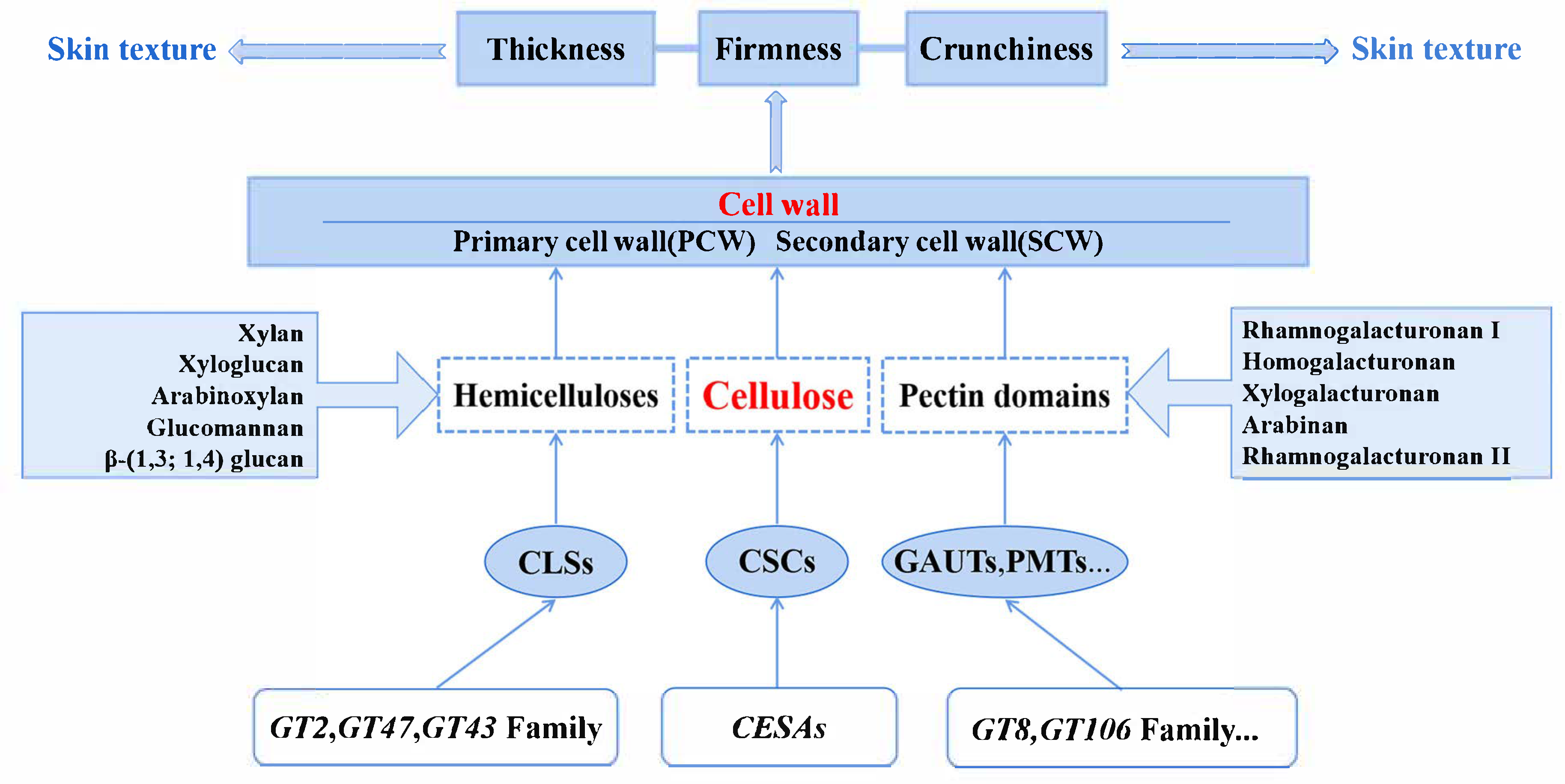
The cell wall is crucial for plant growth and development, as it determines the size and shape of the plant cell [36], provides support for its structural growth [37], and protects it from various environmental stresses [38]. Plant cell walls include the primary cell wall (PCW), secondary cell wall (SCW), and middle lamella [39] (Figure 2). PCWs are located on the inner side of the intercellular layer and synthesized during cell division and elongation. They are dynamic structures that can support cell growth and expand as the cells grow [40]. The secondary cell wall (SCW) is deposited after the cell stops expanding, typically with a unique composition and tissues. Not all cells have secondary cell walls, as they are formed by cells requiring strong mechanical strength and structural reinforcement [41]. The middle lamella is a pectin-rich layer between adjacent cells, which provides cell-to-cell connection and dissolves as the fruit matures [42]. Therefore, the PCW plays a role in cell size, while the mature SCW serves as an important determinant for the increased mechanical support of cell walls [43].
Generally, the commonly accepted cell wall structure is the pectin–cellulose–hemicellulose polysaccharide network structure, a model indicating that the cell wall is composed of pectin, cellulose, hemicellulose, and a number of structural proteins [41,44] (Figure 2). Structural changes in cell wall polysaccharides have an important influence on changes in fruit texture. Decreases in fruit firmness are often accompanied by changes in cell wall polysaccharide content and composition. Cellulose plays a central role in the mechanical strength and morphogenesis of plants [45]. It is the main component of the PCW and SCW; it is also the basis on which the plant cell wall performs physiological functions [46].
Cellulose, as the most important load-bearing component of many plant cell walls, plays a central role in plant mechanical strength and morphogenesis [45]. It is a major component of both primary and secondary plant cell walls and is the basis for the execution of physiological functions in plant cell walls [46]. Cellulose is a linear polymer composed of β-(1,4)-linked glucan chains that interact through hydrogen bonds to form microfilaments [47].
Hemicelluloses are mainly polymers composed of monosaccharides, such as xylan, xyloglucan, mannan, glucomannan, and β-(1,3;1,4)-glucan, whose backbones are all connected by β-(1,4)-glycosyl groups and have similar equatorial configurations [48] (Figure 2). In addition, hemicellulose may spontaneously associate with the surface of cellulose microfilaments and tether adjacent microfilaments together [49].
Pectin is a class of cell wall polysaccharides with α-1,4-linked galacturonic acid (GalA) in the main chain, which is divided into three types or domains: unbranched galacturonic acid (HG); rhamnose–alternating rhamnose galacturonic acid I (RG-I); and complex branched rhamnose galacturonic acid II (RG-II) [50,51] (Figure 2). HG consists of a linear chain of galacturonic acid residues; xylo-galacturonic acid is modified by the addition of wood sugar branches; and xylose galacturonic acid is modified by the addition of xylose branches [41]. RG-I is composed of alternating residues of galacturonic acid and rhamnose and may have side branches containing other pectin structural domains [52]. RG-II is a complex pectin structural domain that contains 11 different sugar residues and forms dimers via boronic esters [41]. In addition, neutral arabinogalactans and arabinogalactans have been found to be associated with acidic pectins, which enhance the flexibility of the pectin wall [53] and bind to the cellulose surface [54].

3.1.2. Transcription Factors Involved in the Regulation of Cell Wall Material Content

Cell wall synthesis is mainly associated with the synthesis of cellulose, hemicellulose, and pectin, as well as their deposition. Cellulose in most plants is produced by the cellulose synthase complexes (CSCs) formed by multiple CesA protein monomers on the plasma membrane (Figure 2). CesA protein is embedded in the plasma membrane in a hexameric array, known as granular rose [55,56]. CSCs are thought to be assembled in the Golgi apparatus and then transported to the plasma membrane (PM) through vesicular transport [56]. This hypothesis is supported by the findings of Zhang et al. [57], who identified the glycosyltransferase-like protein STELLO in the Golgi apparatus. Therefore, only CSCs localized in the PM have the ability to synthesize cellulose. In the model plant, Arabidopsis thaliana, the CesA family contained 10 genes that were expressed in different tissues and cell types [41] (Figure 2). It was reported that three different CesA genes are usually needed to manufacture functional CSCs [58,59]. CesA1, CesA3, and one CesA6-like proteins are involved in the synthesis of PCW cellulose, while CesA4, CesA7, and CesA8 are the necessary isoforms for the synthesis of secondary wall cellulose [60,61,62,63].
Most hemicellulose backbones are synthesized from cellulose synthase-like proteins (CSLs) from the GT2 family, despite differences in glycosyl residues [48], with the only exception being xylans, whose backbones are produced from type II membrane proteins from the GT47 and GT43 families [64,65,66] (Figure 2).
Pectin mainly consists of three types: homogalacturonic acid (HG) without branched chains; rhamnose-alternate rhamnogalacturonic acid I (RG-I); and complex-branched rhamnogalacturonic acid II (RG-II) [50,51]. The enzymes involved in HG pectin methylation modification include galacturonosyltransferase (GAUT) and pectin methyltransferase (PMT) [67] (Figure 2). GAUT proteins belong to the glycosyltransferase family 8 (GT8). The enzymes involved in the biosynthesis of pectin include GAUT1, GAUT7, GAUT4, and GAUT8 [50,67,68,69]. PMTs that play a role in pectin biosynthesis and influence wall structure and plant development include QUASIMODO 2 (QUA 2), QUA 3, Cotton GOLGI-EQUATED 2 (CGR 2), and CGR 3 [70,71,72,73]. RG-I consists of alternating residues of galacturonic acid and rhamnose and may also have a lateral branch that contains other pectin domains [52]. The synthesis of RG-I is associated with rhamnosyltransferases belonging to the new glycosyltransferase family 106 (GT106), which transfers rhamnose residues to RG-I oligosaccharides, whereas the galacturonosyltransferase associated with RG-I has not been identified [74]. RG-II synthesis is associated with rhamnogalacturonan polyglucosyl xylosyltransferase (RGXTs) [75,76].
The synthesis of cell walls, especially secondary cell walls, is also regulated by secondary cell wall NACs transcription factors (SWNs), including members of the transcription factor family, including: the apical meristemless tissue; NAM; the Arabidopsis transcriptional activator ATAF1/2; and the cupressus CUC2 [77]. Overexpression of NAC genes will cause secondary cell wall deposition; conversely, if the expression of this gene is suppressed, it causes a decrease in secondary cell wall thickness [78]. In Arabidopsis, it has been shown that NAC secondary cell wall thickening promoters (NST1 and NST2) and secondary cell wall NAC domain transcription factors (SND1/NST3 and SND2) can control all secondary cell wall biosynthesis processes [79,80,81]. SWNs can bind to a 19 bp segment of secondary cell wall NAC-binding element (SNBE) (T/A)NN(C/T)(T/C/G)TNNNNNNNNNNA(A/C)GN [82], which directly activates pairs of the lower layer of one NAC gene (SND3), three MYBs (MYB46, MYB83 and MYB103), and one homologous (KNAT7) [79,82,83,84]. In contrast, MYB46 and MYB83 can bind the 7 bp secondary cell wall MYB response element (SMRE) ACC(A/T)A(A/C)(T/C), which directly activates the lower transcription factors involved in secondary cell wall synthesis as well as genes for the synthesis of secondary cell wall materials [84,85]. In Arabidopsis, it has been shown that the expression of MYB46 and MYB83 promotes the biosynthesis of cell wall substances (cellulose, lignin, and xylan, etc.) and enhances the deposition of secondary cell walls [86].

3.1.3. Possible Ways to Change the Texture of Skin

According to recent research, it can be concluded that the main factors affecting the texture index of grape skin are the processes of cell wall decomposition, modification, and recombination. Therefore, the skin texture can be regulated by regulating the content of related substances and the activity of related enzymes. In the case of grapes [16,31], peaches [87], pears [88] and other fruits, it has been found that the change in berry hardness during fruit ripening is highly significant and positively correlated with cellulose content; as fruit hardness decreases, its cellulose content also tends to decrease. In grapes [16,31], bananas [89], and persimmon [24], it has been shown that changes in texture are related to changes in hemicellulose content, which tends to decrease as fruit firmness decreases. It has also been shown in cherry fruit that fruit firmness is related to the width of the molecular chain length of hemicellulose [90]. In blueberries [91], kiwifruit [92], peaches [93], pears [94], and apples [94], it has been found that changes in pectin content have a significant effect on fruit firmness during the growth and development of fruits. According to different solubilities, pectin can be categorized into insoluble protopectin as well as soluble pectin [16]. Usually, in the presence of pectin-degrading enzymes, protopectin can be solubilized into soluble pectin [16].
These processes are mainly cracked by cell wall substances (cellulose, hemicellulose and pectin) and related enzymes, including cellulose synthase complex (CSCs), cellulose synthase-like protein (CSLs), galacturonic acid transferase (GAUT), pectin methyltransferase (PMT), expansin (EXP), xyloglucan glycosyltransferase (XTHs), pectin methyl esterase (PMEs), and pectin acid (Figure 2).

3.2. Astringency of Grape Skin

Several hypotheses have been put forward to explain the phenomenon of astringency. Some researchers believe that astringency is a sensation and is the result of a change in saliva lubrication [95]. The feeling of tightening may also be related to physical movements of the mouth such as the tightening of muscle tissue and the feeling of friction [96]. Another hypothesis is that astringency is caused by the direct interaction between tannins and oral epithelial cells or receptors [97]. It has also been reported that tannin protein aggregates destroy salivary gland membranes and increase oral friction, which may lead to further interactions with oral tissues [98]. To sum up, it is generally believed that there are four main ways that astringency is generated from grape skin (Figure 3). The interaction between tannin and saliva protein, along with oral epithelial protein, is recognized as the primary factor contributing to astringency. Additionally, the bonding of tannin with gelatin is also acknowledged as a cause of astringency. Furthermore, the interaction between tannins broken down in the oral cavity and sensory receptors, particularly bitter receptors, is identified as another significant factor in the development of astringency.
Many chemicals can cause astringency, including polyphenols, multivalent cationic salts, and inorganic acids [99]. However, in most fruits, such as persimmons, grapes, pomegranates, and bananas, tannin is the substance that causes astringency.

3.2.1. Composition of Tannins

Tannins are plant polyphenols known for their astringent and bitter properties and are capable of binding and precipitating or shrinking proteins. Based on their chemical composition, tannins are classified into two categories: hydrolyzable tannins and condensed tannins [100].
Condensed tannins, also known as proanthocyanidins (PAs), are condensed from flavan-3-ol, including catechins, epicatechin, and epigallocatechin [101]. PA content is considered as a tool with which to evaluate astringency [102]. According to the polymerization mode of these monomers, PAs are mainly divided into type A and type B. Type B PAs are considered to be connected by the C4-C6 or C4-C8 flavane bond, while Type A PAs have an additional ether bond (C2-O-C7) between the two units [103]. These two precursors are thought to mainly exist in seeds and skins, but not in meat [104,105]. In addition to the diversity of polymerization, different monomer substitution modes (such as deacetylation and hydroxylation) also lead to the diversity of PAs in different fruits [106].
Hydrolyzed tannins are composed of monosaccharides, gallic acid, and its derivatives, which are easily hydrolyzed under acidic or alkaline conditions or in enzyme solutions [107]. According to the hydrolysate, HTs are further divided into galactose tannin and ellagic tannin [106]. Galactotannin is mainly composed of galactose and its derivatives; however, it is not common in the human diet. Ellagitans are appropriate gallic residues in galloylglucose molecules and have strong anti-oxidative potential [108].

3.2.2. The Biosynthetic Pathway of Tannins

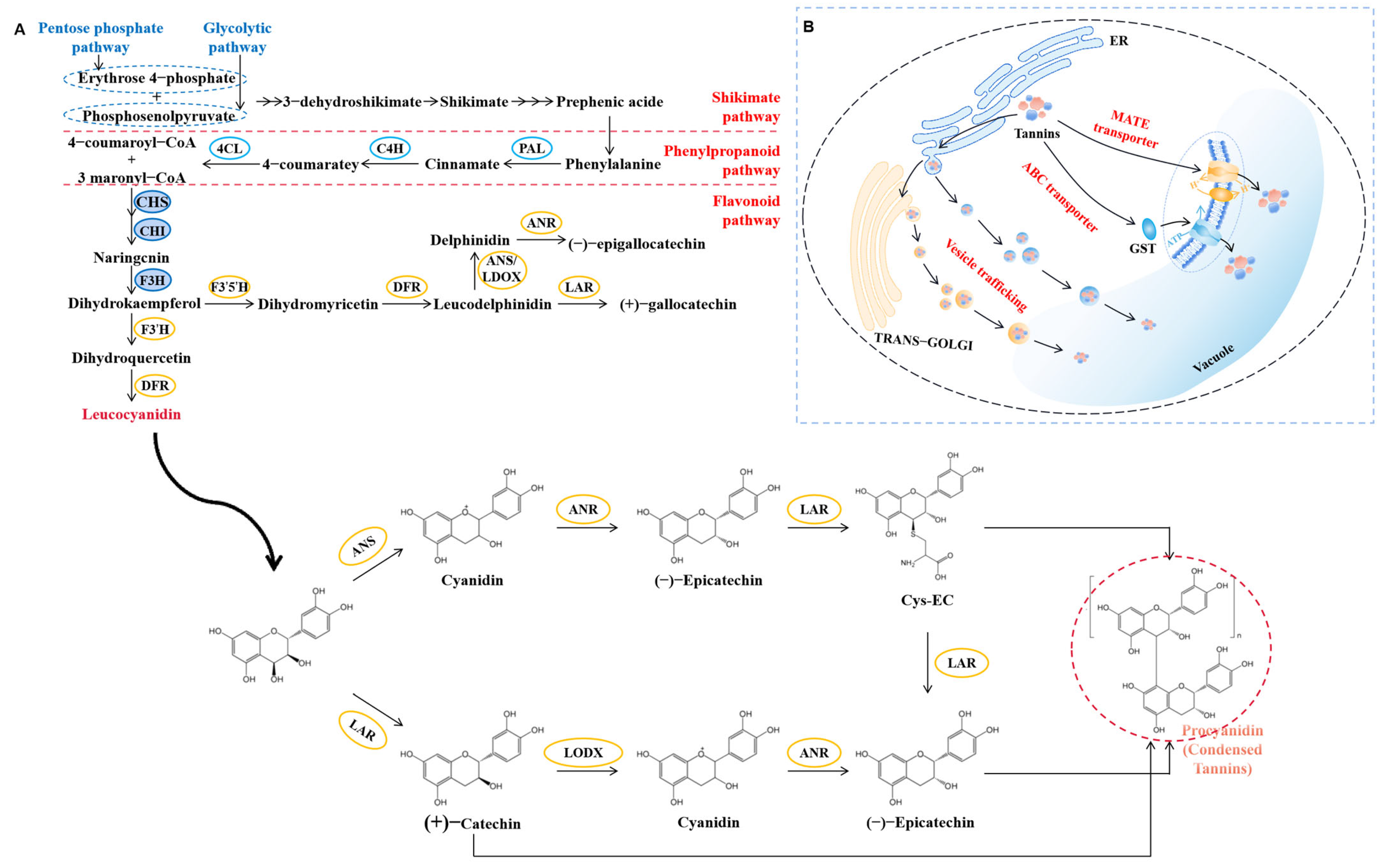
Tannin monomers are all secondary metabolites synthesized by Shikimate acid, phenylpropanoid, flavonoids, and proanthocyanidin biosynthetic pathways (Figure 4A), in which (+)-catechin and (−)-epicatechin are two common PA subunits [109]. The Shikimate pathway starts with the erythrose 4-phosphate and phosphoenolpyruvate produced by glycolysis and the pentose phosphate pathway [110]. This pathway synthesizes prephenic acid through a series of enzymatic reactions, and finally produces phenylalanine, which serves as the precursor for phenylpropanoid pathways. The phenylpropanoid pathways are catalyzed by phenylalanine ammonia-lyase (PAL), cinnamate 4-hydroxylase (C4H), and 4-coumaroyl-CoA ligase (4CL) to form 4-coumaroyl-CoA and malonyl-CoA [111]. In addition, one molecule of 4-coumaroyl-CoA and three molecules of malonyl-CoA are catalyzed by chalcone synthetase (CHS) and chalcone isomerase (CHI) to form naringenin [111]. Under the catalysis of flavanone 3-hydroxylase (F3H), flavanone 3′-hydroxylase (F3′H) or flavanone 3′5′-hydroxylase (F3′5′H), naringcnin is transformed into different dihydroflavonols. Dihydroquercetin is converted into leucocyanidin by dihydroflavonol reductase (DFR) [112]. Leucocyanidin is converted into (+)-catechin by leucocyanidin reductase (LAR) [113,114]. At the same time, anthocyanidin synthase (ANS) competes with LAR to form cyanidin. Then, (−)-epicatechin is generated by anthocyanidin reductase (ANR) [115,116]. 4β-(S-cysteinyl)-epicatechin (Cys-EC) can serve as an extension unit of (−)-epicatechin type PA and (−)-epicatechin can be produced as the second product of LAR [45]. Leucoanthocyanidin dioxygenase (LDOX) acts downstream of LAR, transforming (+)-catechin into cyanidin, and then generating (−)-epicatechin through ANR [117].

3.2.3. Transportation Pathways of Tannins

The biosynthesis of tannin monomers occurs in the endoplasmic reticulum, while the polymerization and storage of tannins occur in the vacuole. Therefore, transport from the cytoplasm to the vacuole is essential for tannin accumulation [118]. There are two possible transport routes for tannin monomers (Figure 4B); one route involves transporters or transferases, while the other route involves vesicles [119]. Current understandings of this process suggest that tannin monomers are transported to vacuoles via tonoplast-localized transport proteins [120] such as MATE transporter [121] or ABC transporter [122]. Alternatively, tannin monomers can be transported to vacuoles through endoplasmic reticulum, the Golgi apparatus, or autophagic vesicles [123].

3.2.4. Possible Ways to Remove Astringency

The unpleasant taste produced by astringency accumulation makes people unwilling to accept grape skin, which affects the edibility of grape skin. Reducing astringent substances from the skin is one of the possible ways to make grape skins edible. In plants, research on the synthesis pathway of tannin monomer has been comprehensive (Figure 4). It is possible to directly control the accumulation of tannins by regulating the expression of genes encoding key enzymes in the biosynthesis pathway. ANR and LAR are two essential enzymes that directly catalyze the synthesis of flavanols. The overexpression of VvANR in tobacco leads to the accumulation of CTs [124]. VvMYBPA1 and VvMYC1 have been shown to activate the promoter of VvANR [125]. Additionally, evidence suggests that NAC and WRKY transcription factors may be involved in the regulation of flavonoid biosynthesis in grapes [126]. For example, transient expression in grape leaves revealed that VvWRKY71, when combined with VvMYBPA1 and VvMYC1, promotes proanthocyanidin biosynthesis, whereas overexpression of VvNAC83 reduces proanthocyanidin accumulation [127].
Changes in the external environment, such as light, temperature, moisture and hormones, may directly or indirectly lead to changes in tannin content in fruits. Red light irradiation can induce the expression of MATE members involved in tannin transport in strawberries, thus affecting the content of CTs [121]. The expression of transcription factor MdMYB23 in apples can cope with cold stress, and can also activate the expression of MdANR gene, thus affecting the content of CTs [128]. Studies have also shown that the accumulation of CTs is also affected by irrigation methods. For example, grapes with sufficient water are more astringent than grapes under drought conditions [129]. Many plant hormones have also been found to regulate the content of CTs in grapes. For example, it has been reported that NAA can inhibit the expression of CHS and CHI genes in the tannin synthesis pathway, thus inhibiting the accumulation of CTs [130]. Research has shown that tannin cell development in Japanese persimmons lacking astringency ceases before fruit maturation, leading to a significantly lower number of tannin cells in mature fruit compared to pulp cells, contributing to the loss of astringency [101]. Conversely, astringent persimmons exhibit higher tannin cell density than non-astringent varieties, aligning with the astringency of their respective fruits [131]. Astringency typically manifests in the immature stage of most fruits and diminishes as they near maturity, which potentially causes tannin dilution [132]. In addition, CT content is regulated by many plant transcription factors, including MYB [133,134], bZIP [109], bHLH [135], and WRKY [88]. Therefore, it is necessary to explore the key factors regulating astringent substances, without affecting grape quality.

3.3. Other Factors

Other factors, such as the appearance of the grape skins, also affect the edibility of grape skin. Grape skin spots, particularly rust spots [136], can impact the appearance quality of grapes. Russet refers to the yellow-brown spots formed on the surface of the skins during fruit development, which essentially act as a secondary protective layer to withstand environmental challenges and safeguard the fruit, known as the cork layer [136,137]. During the early stages of grape development, when the cuticle has not yet formed, the epidermal cells are easily damaged by external factors or cuticle cracks resulting from rapid epidermal cell growth, leading to the formation of corked cell tissue, causing fruit russet [136]. Additionally, russet can be triggered by adverse environmental conditions and cultivation practices such as the use of various abiotic or biological agents. Factors, like prolonged high surface humidity [138] and pest and microbial infestations, can cause the apoptosis of epidermal guard cells, disrupting stomatal regulation and promoting the development of concentrated lignified cells in stomata. This leads to the formation of dark and thick cork lenticels, exacerbating the roughness and degree of russet on the fruit surface.

4. Conclusions

This study elucidates the molecular mechanisms governing grape skin texture and astringency—key factors limiting the wider consumption of these nutritionally rich tissues. Our findings demonstrate that skin mechanical properties are primarily determined by dynamic cell wall remodeling mediated by pectin methylesterases (PMEs) and cellulases (CELs) under NAC/MYB transcriptional regulation. Tannin biosynthesis, controlled by anthocyanidin reductase (ANR) and leucoanthocyanidin reductase (LAR) enzymes under MYB/WRKY factors, constitutes the molecular basis of astringency. These processes are further modulated by environmental conditions, including light exposure and water availability.
Based on these insights, we propose an integrated improvement strategy combining the precision breeding of cell wall-associated genes, biotechnological modulation of tannin pathways, and optimized cultivation practices. The most promising research direction involves CRISPR-Cas9 genome editing coupled with multi-omics analysis to develop novel grape cultivars with enhanced sensory properties while maintaining nutritional value. This approach could also enable the development of value-added byproducts such as nutraceutical extracts, functional food ingredients, and sustainable packaging materials.
The implementation of these findings promises to transform grape breeding and cultivation practices, simultaneously improving fresh fruit quality and creating new commercial opportunities for grape byproducts across the food, pharmaceutical, and cosmetic industries. This work establishes a foundation for maximizing both the health benefits and economic potential of grape-derived products while addressing current limitations in their utilization.

Author Contributions

S.-Y.W. conceived the paper and wrote the manuscript. Y.-M.L. and Z.-S.X. revised the manuscript. All authors have read and agreed to the published version of the manuscript.

Funding

This work was supported by grants 31872050 and 32102348 from the National Natural Science Foundation of China.

Conflicts of Interest

The authors declare no conflicts of interest.

References

  1. Atak, A. Vitis species for stress tolerance/resistance. Genet. Resour. Crop Evol. 2025, 72, 2425–2444. [Google Scholar] [CrossRef]
  2. Wu, Y.; Duan, S.; Zhao, L.; Gao, Z.; Luo, M.; Song, S.; Xu, W.; Zhang, C.; Ma, C.; Wang, S. Aroma characterization based on aromatic series analysis in table grapes. Sci. Rep. 2016, 6, 31116. [Google Scholar] [CrossRef] [PubMed]
  3. Sabra, A.; Netticadan, T.; Wijekoon, C. Grape bioactive molecules, and the potential health benefits in reducing the risk of heart diseases. Food Chem. X 2021, 12, 100149. [Google Scholar] [CrossRef]
  4. Xia, L.; Xu, C.; Huang, K.; Lu, J.; Zhang, Y. Evaluation of phenolic compounds, antioxidant and antiproliferative activities of 31 grape cultivars with different genotypes. J. Food Biochem. 2019, 43, e12626. [Google Scholar] [CrossRef]
  5. Nassiri-Asl, M.; Hosseinzadeh, H. Review of the Pharmacological Effects of Vitis vinifera (Grape) and its Bioactive Constituents: An Update. Phytother. Res. 2016, 30, 1392–1403. [Google Scholar] [CrossRef] [PubMed]
  6. Tabeshpour, J.; Mehri, S.; Shaebani Behbahani, F.; Hosseinzadeh, H. Protective effects of Vitis vinifera (grapes) and one of its biologically active constituents, resveratrol, against natural and chemical toxicities: A comprehensive review. Phytother. Res. 2018, 32, 2164–2190. [Google Scholar] [CrossRef]
  7. Lluís, L.; Muñoz, M.; Nogués, M.R.; Sánchez-Martos, V.; Romeu, M.; Giralt, M.; Valls, J.; Solà, R. Toxicology evaluation of a procyanidin-rich extract from grape skins and seeds. Food Chem. Toxicol. 2011, 49, 1450–1454. [Google Scholar] [CrossRef] [PubMed]
  8. Chengolova, Z.; Ivanov, Y.; Godjevargova, T. Comparison of Identification and Quantification of Polyphenolic Compounds in Skins and Seeds of Four Grape Varieties. Molecules 2023, 28, 4061. [Google Scholar] [CrossRef]
  9. Atak, A.; Göksel, Z.; Yılmaz, Y. Changes in Major Phenolic Compounds of Seeds, Skins, and Pulps from Various Vitis spp. and the Effect of Powdery and Downy Mildew Diseases on Their Levels in Grape Leaves. Plants 2021, 10, 2554. [Google Scholar] [CrossRef]
  10. Yilmaz, Y.; Göksel, Z.; Erdoğan, S.S.; Öztürk, A.; Atak, A.; Özer, C. Antioxidant Activity and Phenolic Content of Seed, Skin and Pulp Parts of 22 Grape (Vitis vinifera L.) Cultivars (4 Common and 18 Registered or Candidate for Registration). J. Food Process. Preserv. 2015, 39, 1682–1691. [Google Scholar] [CrossRef]
  11. Mochida, K.; Maki, S.; Onishi, A.; Uchida, Y.; Kurahashi, T. Effects of CPPU Treatment Methods on the Fruit Quality of ‘Shine Muscat’ Grape. Hortic. Res. 2013, 12, 155–163. [Google Scholar] [CrossRef]
  12. Wei, X.; Ju, Y.; Ma, T.; Zhang, J.; Fang, Y.; Sun, X. New perspectives on the biosynthesis, transportation, astringency perception and detection methods of grape proanthocyanidins. Crit. Rev. Food Sci. Nutr. 2021, 61, 2372–2398. [Google Scholar] [CrossRef]
  13. Cliff, M.A.; Dever, M.C.; Reynolds, A.G. Descriptive Profiling of New and Commercial British Columbia Table Grape Cultivars. Am. J. Enol. Vitic. 1996, 47, 301. [Google Scholar] [CrossRef]
  14. Yu, J.; Wang, R.; Ma, W.; Lei, S.; Zhu, M.; Yang, G. Pectate Lyase Gene VvPL1 Plays a Role in Fruit Cracking of Table Grapes. J. Agric. Food Chem. 2023, 71, 1643–1654. [Google Scholar] [CrossRef] [PubMed]
  15. Schlosser, J.; Olsson, N.; Weis, M.; Reid, K.; Peng, F.; Lund, S.; Bowen, P. Cellular expansion and gene expression in the developing grape (Vitis vinifera L.). Protoplasma 2008, 232, 255–265. [Google Scholar] [CrossRef]
  16. Malacarne, G.; Lagreze, J.; Rojas San Martin, B.; Malnoy, M.; Moretto, M.; Moser, C.; Dalla Costa, L. Insights into the cell-wall dynamics in grapevine berries during ripening and in response to biotic and abiotic stresses. Plant Mol. Biol. 2024, 114, 38. [Google Scholar] [CrossRef] [PubMed]
  17. Nakagawa, S.; Nanjo, Y. A Morphological Study of Delaware Grape Berries. J. Jpn. Soc. Hortic. Sci. 1965, 34, 85–95. [Google Scholar] [CrossRef]
  18. Agudelo-Romero, P.; Erban, A.; Rego, C.; Carbonell-Bejerano, P.; Nascimento, T.; Sousa, L.; Martínez-Zapater, J.M.; Kopka, J.; Fortes, A.M. Transcriptome and metabolome reprogramming in Vitis vinifera cv. Trincadeira berries upon infection with Botrytis cinerea. J. Exp. Bot. 2015, 66, 1769–1785. [Google Scholar] [CrossRef] [PubMed]
  19. Hardie, W.J.; O’Brien, T.P.; Jaudzems, V.G. Morphology, anatomy and development of the pericarp after anthesis in grape, Vitis vinifera L. Aust. J. Grape Wine Res. 1996, 2, 97–142. [Google Scholar] [CrossRef]
  20. Chang, B.-M.; Keller, M. Cuticle and skin cell walls have common and unique roles in grape berry splitting. Hortic. Res. 2021, 8, 168. [Google Scholar] [CrossRef]
  21. Ginzberg, I.; Stern, R.A. Strengthening fruit-skin resistance to growth strain by application of plant growth regulators. Sci. Hortic. 2016, 198, 150–153. [Google Scholar] [CrossRef]
  22. Cadot, Y.; Miñana-Castelló, M.T.; Chevalier, M. Anatomical, histological, and histochemical changes in grape seeds from Vitis vinifera L. cv Cabernet franc during fruit development. J. Agric. Food Chem. 2006, 54, 9206–9215. [Google Scholar] [CrossRef]
  23. Ollat, N.; Carde, J.-P.; Gaudillère, J.-P.; Barrieu, F.; Diakou-Verdin, P.; Moing, A. Grape berry development: A review. OENO One 2002, 36, 109–131. [Google Scholar] [CrossRef]
  24. Wang, H.; Chen, Y.; Lin, H.; Lin, M.; Chen, Y.; Lin, Y. 1-Methylcyclopropene containing-papers suppress the disassembly of cell wall polysaccharides in Anxi persimmon fruit during storage. Int. J. Biol. Macromol. 2020, 151, 723–729. [Google Scholar] [CrossRef]
  25. Lin, H.; Ma, L.; Guo, Q.; Liu, C.; Hou, Y.; Liu, Z.; Zhao, Y.; Jiang, C.; Guo, X.; Guo, Y. Berry texture QTL and candidate gene analysis in grape (Vitis vinifera L.). Hortic. Res. 2023, 10, uhad226. [Google Scholar] [CrossRef]
  26. Pedneault, K.; Provost, C. Fungus resistant grape varieties as a suitable alternative for organic wine production: Benefits, limits, and challenges. Sci. Hortic. 2016, 208, 57–77. [Google Scholar] [CrossRef]
  27. Atak, A. New Perspectives in Grapevine (Vitis spp.) Breeding. In Case Studies of Breeding Strategies in Major Plant Species; Wang, H., Ed.; IntechOpen: London, UK, 2022. [Google Scholar] [CrossRef]
  28. Zepeda, B.; Olmedo, P.; Ejsmentewicz, T.; Sepúlveda, P.; Balic, I.; Balladares, C.; Delgado-Rioseco, J.; Fuentealba, C.; Moreno, A.A.; Defilippi, B.G.; et al. Cell wall and metabolite composition of berries of Vitis vinifera (L.) cv. Thompson Seedless with different firmness. Food Chem. 2018, 268, 492–497. [Google Scholar] [CrossRef] [PubMed]
  29. Sato, A.; Yamada, M.; Iwanami, H. Estimation of the Proportion of Offspring Having Genetically Crispy Flesh in Grape Breeding. J. Am. Soc. Hortic. Sci. 2006, 131, 46–52. [Google Scholar] [CrossRef]
  30. Gombau, J.; Nadal, P.; Canela, N.; Gómez-Alonso, S.; García-Romero, E.; Smith, P.; Hermosín-Gutiérrez, I.; Canals, J.M.; Zamora, F. Measurement of the interaction between mucin and oenological tannins by Surface Plasmon Resonance (SPR); relationship with astringency. Food Chem. 2019, 275, 397–406. [Google Scholar] [CrossRef]
  31. Posé, S.; Paniagua, C.; Matas, A.J.; Gunning, A.P.; Morris, V.J.; Quesada, M.A.; Mercado, J.A. A nanostructural view of the cell wall disassembly process during fruit ripening and postharvest storage by atomic force microscopy. Trends Food Sci. Technol. 2019, 87, 47–58. [Google Scholar] [CrossRef]
  32. Rojas, B.; Suárez-Vega, F.; Saez-Aguayo, S.; Olmedo, P.; Zepeda, B.; Delgado-Rioseco, J.; Defilippi, B.G.; Pedreschi, R.; Meneses, C.; Pérez-Donoso, A.G.; et al. Pre-Anthesis Cytokinin Applications Increase Table Grape Berry Firmness by Modulating Cell Wall Polysaccharides. Plants 2021, 10, 2642. [Google Scholar] [CrossRef]
  33. Vicente, A.R.; Saladié, M.; Rose, J.K.C.; Labavitch, J.M. The linkage between cell wall metabolism and fruit softening: Looking to the future. J. Sci. Food Agric. 2007, 87, 1435–1448. [Google Scholar] [CrossRef]
  34. Nunan, K.J.; Davies, C.; Robinson, S.P.; Fincher, G.B. Expression patterns of cell wall-modifying enzymes during grape berry development. Planta 2001, 214, 257–264. [Google Scholar] [CrossRef]
  35. Iwatani, S.-I.; Yakushiji, H.; Mitani, N.; Sakurai, N. Evaluation of grape flesh texture by an acoustic vibration method. Postharvest Biol. Technol. 2011, 62, 305–309. [Google Scholar] [CrossRef]
  36. Szymanski, D.B.; Cosgrove, D.J. Dynamic coordination of cytoskeletal and cell wall systems during plant cell morphogenesis. Curr. Biol. 2009, 19, R800–R811. [Google Scholar] [CrossRef] [PubMed]
  37. Landrein, B.; Hamant, O. How mechanical stress controls microtubule behavior and morphogenesis in plants: History, experiments and revisited theories. Plant J. 2013, 75, 324–338. [Google Scholar] [CrossRef] [PubMed]
  38. Lionetti, V.; Raiola, A.; Mattei, B.; Bellincampi, D. The Grapevine VvPMEI1 Gene Encodes a Novel Functional Pectin Methylesterase Inhibitor Associated to Grape Berry Development. PLoS ONE 2015, 10, e0133810. [Google Scholar] [CrossRef] [PubMed]
  39. Rytioja, J.; Hildén, K.; Yuzon, J.; Hatakka, A.; de Vries, R.P.; Mäkelä, M.R. Plant-polysaccharide-degrading enzymes from Basidiomycetes. Microbiol. Mol. Biol. Rev. 2014, 78, 614–649. [Google Scholar] [CrossRef]
  40. Zhang, B.; Gao, Y.; Zhang, L.; Zhou, Y. The plant cell wall: Biosynthesis, construction, and functions. J. Integr. Plant Biol. 2021, 63, 251–272. [Google Scholar] [CrossRef]
  41. Cosgrove, D.J. Growth of the plant cell wall. Nat. Rev. Mol. Cell Biol. 2005, 6, 850–861. [Google Scholar] [CrossRef]
  42. Höfte, H.; Voxeur, A. Plant cell walls. Curr. Biol. 2017, 27, R865–R870. [Google Scholar] [CrossRef]
  43. Schuetz, M.; Smith, R.; Ellis, B. Xylem tissue specification, patterning, and differentiation mechanisms. J. Exp. Bot. 2013, 64, 11–31. [Google Scholar] [CrossRef]
  44. Somerville, C.; Bauer, S.; Brininstool, G.; Facette, M.; Hamann, T.; Milne, J.; Osborne, E.; Paredez, A.; Persson, S.; Raab, T.; et al. Toward a systems approach to understanding plant cell walls. Science 2004, 306, 2206–2211. [Google Scholar] [CrossRef]
  45. Liu, C.; Wang, X.; Shulaev, V.; Dixon, R.A. A role for leucoanthocyanidin reductase in the extension of proanthocyanidins. Nat. Plants 2016, 2, 16182. [Google Scholar] [CrossRef]
  46. Purushotham, P.; Ho, R.; Zimmer, J. Architecture of a catalytically active homotrimeric plant cellulose synthase complex. Science 2020, 369, 1089–1094. [Google Scholar] [CrossRef]
  47. Peng, L.; Kawagoe, Y.; Hogan, P.; Delmer, D. Sitosterol-beta-glucoside as primer for cellulose synthesis in plants. Science 2002, 295, 147–150. [Google Scholar] [CrossRef]
  48. Scheller, H.V.; Ulvskov, P. Hemicelluloses. Annu. Rev. Plant Biol. 2010, 61, 263–289. [Google Scholar] [CrossRef] [PubMed]
  49. Richmond, T.A.; Somerville, C.R. The cellulose synthase superfamily. Plant Physiol. 2000, 124, 495–498. [Google Scholar] [CrossRef] [PubMed]
  50. Atmodjo, M.A.; Hao, Z.; Mohnen, D. Evolving views of pectin biosynthesis. Annu. Rev. Plant Biol. 2013, 64, 747–779. [Google Scholar] [CrossRef] [PubMed]
  51. Mitsuda, N.; Seki, M.; Shinozaki, K.; Ohme-Takagi, M. The NAC transcription factors NST1 and NST2 of Arabidopsis regulate secondary wall thickenings and are required for anther dehiscence. Plant Cell 2005, 17, 2993–3006. [Google Scholar] [CrossRef]
  52. Vincken, J.-P.; Schols, H.A.; Oomen, R.J.F.J.; McCann, M.C.; Ulvskov, P.; Voragen, A.G.J.; Visser, R.G.F. If homogalacturonan were a side chain of rhamnogalacturonan I. Implications for cell wall architecture. Plant Physiol. 2003, 132, 1781–1789. [Google Scholar] [CrossRef]
  53. Jones, L.; Milne, J.L.; Ashford, D.; McQueen-Mason, S.J. Cell wall arabinan is essential for guard cell function. Proc. Natl. Acad. Sci. USA 2003, 100, 11783–11788. [Google Scholar] [CrossRef] [PubMed]
  54. Zykwinska, A.W.; Ralet, M.-C.J.; Garnier, C.D.; Thibault, J.-F.J. Evidence for in vitro binding of pectin side chains to cellulose. Plant Physiol. 2005, 139, 397–407. [Google Scholar] [CrossRef] [PubMed]
  55. Kimura, S.; Laosinchai, W.; Itoh, T.; Cui, X.; Linder, C.R.; Brown, R.M. Immunogold labeling of rosette terminal cellulose-synthesizing complexes in the vascular plant Vigna angularis. Plant Cell 1999, 11, 2075–2086. [Google Scholar] [CrossRef]
  56. Polko, J.K.; Kieber, J.J. The Regulation of Cellulose Biosynthesis in Plants. Plant Cell 2019, 31, 282–296. [Google Scholar] [CrossRef]
  57. Zhang, Y.; Nikolovski, N.; Sorieul, M.; Vellosillo, T.; McFarlane, H.E.; Dupree, R.; Kesten, C.; Schneider, R.; Driemeier, C.; Lathe, R.; et al. Golgi-localized STELLO proteins regulate the assembly and trafficking of cellulose synthase complexes in Arabidopsis. Nat. Commun. 2016, 7, 11656. [Google Scholar] [CrossRef]
  58. Arioli, T.; Peng, L.; Betzner, A.S.; Burn, J.; Wittke, W.; Herth, W.; Camilleri, C.; Höfte, H.; Plazinski, J.; Birch, R.; et al. Molecular analysis of cellulose biosynthesis in Arabidopsis. Science 1998, 279, 717–720. [Google Scholar] [CrossRef]
  59. Pear, J.R.; Kawagoe, Y.; Schreckengost, W.E.; Delmer, D.P.; Stalker, D.M. Higher plants contain homologs of the bacterial celA genes encoding the catalytic subunit of cellulose synthase. Proc. Natl. Acad. Sci. USA 1996, 93, 12637–12642. [Google Scholar] [CrossRef]
  60. Desprez, T.; Juraniec, M.; Crowell, E.F.; Jouy, H.; Pochylova, Z.; Parcy, F.; Höfte, H.; Gonneau, M.; Vernhettes, S. Organization of cellulose synthase complexes involved in primary cell wall synthesis in Arabidopsis thaliana. Proc. Natl. Acad. Sci. USA 2007, 104, 15572–15577. [Google Scholar] [CrossRef]
  61. Malinovsky, F.G.; Fangel, J.U.; Willats, W.G.T. The role of the cell wall in plant immunity. Front. Plant Sci. 2014, 5, 178. [Google Scholar] [CrossRef] [PubMed]
  62. Persson, S.; Paredez, A.; Carroll, A.; Palsdottir, H.; Doblin, M.; Poindexter, P.; Khitrov, N.; Auer, M.; Somerville, C.R. Genetic evidence for three unique components in primary cell-wall cellulose synthase complexes in Arabidopsis. Proc. Natl. Acad. Sci. USA 2007, 104, 15566–15571. [Google Scholar] [CrossRef] [PubMed]
  63. Taylor, N.G.; Howells, R.M.; Huttly, A.K.; Vickers, K.; Turner, S.R. Interactions among three distinct CesA proteins essential for cellulose synthesis. Proc. Natl. Acad. Sci. USA 2003, 100, 1450–1455. [Google Scholar] [CrossRef]
  64. Brown, D.M.; Zeef, L.A.H.; Ellis, J.; Goodacre, R.; Turner, S.R. Identification of novel genes in Arabidopsis involved in secondary cell wall formation using expression profiling and reverse genetics. Plant Cell 2005, 17, 2281–2295. [Google Scholar] [CrossRef]
  65. Brown, D.M.; Zhang, Z.; Stephens, E.; Dupree, P.; Turner, S.R. Characterization of IRX10 and IRX10-like reveals an essential role in glucuronoxylan biosynthesis in Arabidopsis. Plant J. 2009, 57, 732–746. [Google Scholar] [CrossRef]
  66. Wolf, S.; van der Does, D.; Ladwig, F.; Sticht, C.; Kolbeck, A.; Schürholz, A.-K.; Augustin, S.; Keinath, N.; Rausch, T.; Greiner, S.; et al. A receptor-like protein mediates the response to pectin modification by activating brassinosteroid signaling. Proc. Natl. Acad. Sci. USA 2014, 111, 15261–15266. [Google Scholar] [CrossRef]
  67. Du, J.; Anderson, C.T.; Xiao, C. Dynamics of pectic homogalacturonan in cellular morphogenesis and adhesion, wall integrity sensing and plant development. Nat. Plants 2022, 8, 332–340. [Google Scholar] [CrossRef]
  68. Amos, R.A.; Pattathil, S.; Yang, J.-Y.; Atmodjo, M.A.; Urbanowicz, B.R.; Moremen, K.W.; Mohnen, D. A two-phase model for the non-processive biosynthesis of homogalacturonan polysaccharides by the GAUT1:GAUT7 complex. J. Biol. Chem. 2018, 293, 19047–19063. [Google Scholar] [CrossRef]
  69. Sterling, J.D.; Atmodjo, M.A.; Inwood, S.E.; Kumar Kolli, V.S.; Quigley, H.F.; Hahn, M.G.; Mohnen, D. Functional identification of an Arabidopsis pectin biosynthetic homogalacturonan galacturonosyltransferase. Proc. Natl. Acad. Sci. USA 2006, 103, 5236–5241. [Google Scholar] [CrossRef]
  70. Kim, S.-J.; Held, M.A.; Zemelis, S.; Wilkerson, C.; Brandizzi, F. CGR2 and CGR3 have critical overlapping roles in pectin methylesterification and plant growth in Arabidopsis thaliana. Plant J. 2015, 82, 208–220. [Google Scholar] [CrossRef] [PubMed]
  71. Krupková, E.; Immerzeel, P.; Pauly, M.; Schmülling, T. The TUMOROUS SHOOT DEVELOPMENT2 gene of Arabidopsis encoding a putative methyltransferase is required for cell adhesion and co-ordinated plant development. Plant J. 2007, 50, 735–750. [Google Scholar] [CrossRef] [PubMed]
  72. Miao, Y.; Li, H.-Y.; Shen, J.; Wang, J.; Jiang, L. QUASIMODO 3 (QUA3) is a putative homogalacturonan methyltransferase regulating cell wall biosynthesis in Arabidopsis suspension-cultured cells. J. Exp. Bot. 2011, 62, 5063–5078. [Google Scholar] [CrossRef] [PubMed]
  73. Mouille, G.; Ralet, M.-C.; Cavelier, C.; Eland, C.; Effroy, D.; Hématy, K.; McCartney, L.; Truong, H.N.; Gaudon, V.; Thibault, J.-F.; et al. Homogalacturonan synthesis in Arabidopsis thaliana requires a Golgi-localized protein with a putative methyltransferase domain. Plant J. 2007, 50, 605–614. [Google Scholar] [CrossRef] [PubMed]
  74. Takenaka, Y.; Kato, K.; Ogawa-Ohnishi, M.; Tsuruhama, K.; Kajiura, H.; Yagyu, K.; Takeda, A.; Takeda, Y.; Kunieda, T.; Hara-Nishimura, I.; et al. Pectin RG-I rhamnosyltransferases represent a novel plant-specific glycosyltransferase family. Nat. Plants 2018, 4, 669–676. [Google Scholar] [CrossRef] [PubMed]
  75. Dumont, M.; Lehner, A.; Bouton, S.; Kiefer-Meyer, M.C.; Voxeur, A.; Pelloux, J.; Lerouge, P.; Mollet, J.-C. The cell wall pectic polymer rhamnogalacturonan-II is required for proper pollen tube elongation: Implications of a putative sialyltransferase-like protein. Ann. Bot. 2014, 114, 1177–1188. [Google Scholar] [CrossRef]
  76. Liu, L.; Shang-Guan, K.; Zhang, B.; Liu, X.; Yan, M.; Zhang, L.; Shi, Y.; Zhang, M.; Qian, Q.; Li, J.; et al. Brittle Culm1, a COBRA-like protein, functions in cellulose assembly through binding cellulose microfibrils. PLoS Genet. 2013, 9, e1003704. [Google Scholar] [CrossRef]
  77. Nakashima, K.; Takasaki, H.; Mizoi, J.; Shinozaki, K.; Yamaguchi-Shinozaki, K. NAC transcription factors in plant abiotic stress responses. Biochim. Et Biophys. Acta (BBA)-Gene Regul. Mech. 2012, 1819, 97–103. [Google Scholar] [CrossRef]
  78. Yin, Q.; Qin, W.; Zhou, Z.; Wu, A.-M.; Deng, W.; Li, Z.; Shan, W.; Chen, J.-Y.; Kuang, J.-F.; Lu, W.-J. Banana MaNAC1 activates secondary cell wall cellulose biosynthesis to enhance chilling resistance in fruit. Plant Biotechnol. J. 2024, 22, 413–426. [Google Scholar] [CrossRef]
  79. Zhong, R.; Demura, T.; Ye, Z.-H. SND1, a NAC domain transcription factor, is a key regulator of secondary wall synthesis in fibers of Arabidopsis. Plant Cell 2006, 18, 3158–3170. [Google Scholar] [CrossRef]
  80. Zhong, R.; Richardson, E.A.; Ye, Z.-H. Two NAC domain transcription factors, SND1 and NST1, function redundantly in regulation of secondary wall synthesis in fibers of Arabidopsis. Planta 2007, 225, 1603–1611. [Google Scholar] [CrossRef]
  81. Forlani, S.; Mizzotti, C.; Masiero, S. The NAC side of the fruit: Tuning of fruit development and maturation. BMC Plant Biol. 2021, 21, 238. [Google Scholar] [CrossRef]
  82. Zhong, R.; Lee, C.; Zhou, J.; McCarthy, R.L.; Ye, Z.-H. A battery of transcription factors involved in the regulation of secondary cell wall biosynthesis in Arabidopsis. Plant Cell 2008, 20, 2763–2782. [Google Scholar] [CrossRef]
  83. Ko, J.-H.; Kim, W.-C.; Han, K.-H. Ectopic expression of MYB46 identifies transcriptional regulatory genes involved in secondary wall biosynthesis in Arabidopsis. Plant J. 2009, 60, 649–665. [Google Scholar] [CrossRef] [PubMed]
  84. McCarthy, R.L.; Zhong, R.; Ye, Z.-H. MYB83 is a direct target of SND1 and acts redundantly with MYB46 in the regulation of secondary cell wall biosynthesis in Arabidopsis. Plant Cell Physiol. 2009, 50, 1950–1964. [Google Scholar] [CrossRef]
  85. Zhong, R.; Richardson, E.A.; Ye, Z.-H. The MYB46 transcription factor is a direct target of SND1 and regulates secondary wall biosynthesis in Arabidopsis. Plant Cell 2007, 19, 2776–2792. [Google Scholar] [CrossRef] [PubMed]
  86. Ko, J.H.; Jeon, H.W.; Kim, W.C.; Kim, J.Y.; Han, K.H. The MYB46/MYB83-mediated transcriptional regulatory programme is a gatekeeper of secondary wall biosynthesis. Ann. Bot. 2014, 114, 1099–1107. [Google Scholar] [CrossRef] [PubMed]
  87. Xu, Z.; Dai, J.; Liang, L.; Zhang, Y.; He, Y.; Xing, L.; Ma, J.; Zhang, D.; Zhao, C. Chitinase-Like Protein PpCTL1 Contributes to Maintaining Fruit Firmness by Affecting Cellulose Biosynthesis during Peach Development. Foods 2023, 12, 2503. [Google Scholar] [CrossRef]
  88. Li, G.; Liu, X.; Liang, Y.; Zhang, Y.; Cheng, X.; Cai, Y. Genome-wide characterization of the cellulose synthase gene superfamily in Pyrus bretschneideri and reveal its potential role in stone cell formation. Funct. Integr. Genom. 2020, 20, 723–738. [Google Scholar] [CrossRef]
  89. Yi, Z.; Sharif, R.; Gulzar, S.; Huang, Y.; Ning, T.; Zhan, H.; Meng, Y.; Xu, C. Changes in hemicellulose metabolism in banana peel during fruit development and ripening. Plant Physiol. Biochem. 2024, 215, 109025. [Google Scholar] [CrossRef]
  90. Lahaye, M.; Tabi, W.; Le Bot, L.; Delaire, M.; Orsel, M.; Campoy, J.A.; Quero Garcia, J.; Le Gall, S. Comparison of cell wall chemical evolution during the development of fruits of two contrasting quality from two members of the Rosaceae family: Apple and sweet cherry. Plant Physiol. Biochem. 2021, 168, 93–104. [Google Scholar] [CrossRef]
  91. Sanhueza, D.; Balic-Norambuena, I.; Sepúlveda-Orellana, P.; Siña-López, S.; Moreno, A.A.; Moya-León, M.A.; Saez-Aguayo, S. Unraveling cell wall polysaccharides during blueberry ripening: Insights into the roles of rhamnogalacturonan-I and arabinogalactan proteins in fruit firmness. Front. Plant Sci. 2024, 15, 1422917. [Google Scholar] [CrossRef]
  92. Huang, W.; Chen, M.; Zhao, T.; Han, F.; Zhang, Q.; Liu, X.; Jiang, C.; Zhong, C. Genome-Wide Identification and Expression Analysis of Polygalacturonase Gene Family in Kiwifruit (Actinidia chinensis) during Fruit Softening. Plants 2020, 9, 327. [Google Scholar] [CrossRef]
  93. Qian, M.; Xu, Z.; Zhang, Z.; Li, Q.; Yan, X.; Liu, H.; Han, M.; Li, F.; Zheng, J.; Zhang, D.; et al. The downregulation of PpPG21 and PpPG22 influences peach fruit texture and softening. Planta 2021, 254, 22. [Google Scholar] [CrossRef]
  94. Al Hinai, T.Z.S.; Vreeburg, R.A.M.; Mackay, C.L.; Murray, L.; Sadler, I.H.; Fry, S.C. Fruit softening: Evidence for pectate lyase action in vivo in date (Phoenix dactylifera) and rosaceous fruit cell walls. Ann. Bot. 2021, 128, 511–525. [Google Scholar] [CrossRef]
  95. Rossetti, D.; Yakubov, G.E.; Stokes, J.R.; Williamson, A.M.; Fuller, G.G. Interaction of human whole saliva and astringent dietary compounds investigated by interfacial shear rheology. Food Hydrocoll. 2008, 22, 1068–1078. [Google Scholar] [CrossRef]
  96. Pires, M.A.; Pastrana, L.M.; Fuciños, P.; Abreu, C.S.; Oliveira, S.M. Sensorial Perception of Astringency: Oral Mechanisms and Current Analysis Methods. Foods 2020, 9, 1124. [Google Scholar] [CrossRef] [PubMed]
  97. Ma, W.; Guo, A.; Zhang, Y.; Wang, H.; Liu, Y.; Li, H. A review on astringency and bitterness perception of tannins in wine. Trends Food Sci. Technol. 2014, 40, 6–19. [Google Scholar] [CrossRef]
  98. Rossetti, D.; Bongaerts, J.H.H.; Wantling, E.; Stokes, J.R.; Williamson, A.M. Astringency of tea catechins: More than an oral lubrication tactile percept. Food Hydrocoll. 2009, 23, 1984–1992. [Google Scholar] [CrossRef]
  99. Halsam, E.; Lilley, T.H. Natural astringency in foodstuffs—A molecular interpretation. Crit. Rev. Food Sci. Nutr. 1988, 27, 1–40. [Google Scholar] [CrossRef]
  100. Koleckar, V.; Kubikova, K.; Rehakova, Z.; Kuca, K.; Jun, D.; Jahodar, L.; Opletal, L. Condensed and hydrolysable tannins as antioxidants influencing the health. Mini Rev. Med. Chem. 2008, 8, 436–447. [Google Scholar] [CrossRef]
  101. Yonemori, K.; Matsushima, J. Property of Development of the Tannin Cells in Non-Astringent Type Fruits of Japanese Persimmon (Diospyros kaki) and Its Relationship to Natural Deastringency. J. Jpn. Soc. Hortic. Sci. 1985, 54, 201–208. [Google Scholar] [CrossRef]
  102. Basalekou, M.; Kyraleou, M.; Pappas, C.; Tarantilis, P.; Kotseridis, Y.; Kallithraka, S. Proanthocyanidin content as an astringency estimation tool and maturation index in red and white winemaking technology. Food Chem. 2019, 299, 125135. [Google Scholar] [CrossRef] [PubMed]
  103. Ferreira, D.; Slade, D. Oligomeric proanthocyanidins: Naturally occurring O-heterocycles. Nat. Prod. Rep. 2002, 19, 517–541. [Google Scholar] [CrossRef]
  104. Mané, C.; Souquet, J.M.; Ollé, D.; Verriés, C.; Véran, F.; Mazerolles, G.; Cheynier, V.; Fulcrand, H. Optimization of simultaneous flavanol, phenolic acid, and anthocyanin extraction from grapes using an experimental design: Application to the characterization of champagne grape varieties. J. Agric. Food Chem. 2007, 55, 7224–7233. [Google Scholar] [CrossRef]
  105. Verries, C.; Guiraud, J.-L.; Souquet, J.-M.; Vialet, S.; Terrier, N.; Ollé, D. Validation of an extraction method on whole pericarp of grape berry (Vitis vinifera L. cv. Shiraz) to study biochemical and molecular aspects of flavan-3-ol synthesis during berry development. J. Agric. Food Chem. 2008, 56, 5896–5904. [Google Scholar] [CrossRef] [PubMed]
  106. Soares, S.; Brandão, E.; Guerreiro, C.; Soares, S.; Mateus, N.; de Freitas, V. Tannins in Food: Insights into the Molecular Perception of Astringency and Bitter Taste. Molecules 2020, 25, 2590. [Google Scholar] [CrossRef] [PubMed]
  107. Haslam, E. Vegetable tannins-lessons of a phytochemical lifetime. Phytochemistry 2007, 68, 2713–2721. [Google Scholar] [CrossRef]
  108. Okuda, T.; Ito, H. Tannins of Constant Structure in Medicinal and Food Plants—Hydrolyzable Tannins and Polyphenols Related to Tannins. Molecules 2011, 16, 2191–2217. [Google Scholar] [CrossRef]
  109. Akagi, T.; Katayama-Ikegami, A.; Kobayashi, S.; Sato, A.; Kono, A.; Yonemori, K. Seasonal abscisic acid signal and a basic leucine zipper transcription factor, DkbZIP5, regulate proanthocyanidin biosynthesis in persimmon fruit. Plant Physiol. 2012, 158, 1089–1102. [Google Scholar] [CrossRef]
  110. Mir, R.; Jallu, S.; Singh, T.P. The shikimate pathway: Review of amino acid sequence, function and three-dimensional structures of the enzymes. Crit. Rev. Microbiol. 2015, 41, 172–189. [Google Scholar] [CrossRef]
  111. Hichri, I.; Barrieu, F.; Bogs, J.; Kappel, C.; Delrot, S.; Lauvergeat, V. Recent advances in the transcriptional regulation of the flavonoid biosynthetic pathway. J. Exp. Bot. 2011, 62, 2465–2483. [Google Scholar] [CrossRef]
  112. Petit, P.; Granier, T.; d’Estaintot, B.L.; Manigand, C.; Bathany, K.; Schmitter, J.-M.; Lauvergeat, V.; Hamdi, S.; Gallois, B. Crystal structure of grape dihydroflavonol 4-reductase, a key enzyme in flavonoid biosynthesis. J. Mol. Biol. 2007, 368, 1345–1357. [Google Scholar] [CrossRef]
  113. Tanner, G.J.; Francki, K.T.; Abrahams, S.; Watson, J.M.; Larkin, P.J.; Ashton, A.R. Proanthocyanidin biosynthesis in plants. Purification of legume leucoanthocyanidin reductase and molecular cloning of its cDNA. J. Biol. Chem. 2003, 278, 31647–31656. [Google Scholar] [CrossRef] [PubMed]
  114. Yu, K.; Jun, J.H.; Duan, C.; Dixon, R.A. VvLAR1 and VvLAR2 Are Bifunctional Enzymes for Proanthocyanidin Biosynthesis in Grapevine. Plant Physiol. 2019, 180, 1362–1374. [Google Scholar] [CrossRef]
  115. Jun, J.H.; Lu, N.; Docampo-Palacios, M.; Wang, X.; Dixon, R.A. Dual activity of anthocyanidin reductase supports the dominant plant proanthocyanidin extension unit pathway. Sci. Adv. 2021, 7, eabg4682. [Google Scholar] [CrossRef] [PubMed]
  116. Jun, J.H.; Xiao, X.; Rao, X.; Dixon, R.A. Proanthocyanidin subunit composition determined by functionally diverged dioxygenases. Nat. Plants 2018, 4, 1034–1043. [Google Scholar] [CrossRef]
  117. Yu, K.; Dixon, R.A.; Duan, C. A role for ascorbate conjugates of (+)-catechin in proanthocyanidin polymerization. Nat. Commun. 2022, 13, 3425. [Google Scholar] [CrossRef] [PubMed]
  118. Goodman, C.D.; Casati, P.; Walbot, V. A multidrug resistance-associated protein involved in anthocyanin transport in Zea mays. Plant Cell 2004, 16, 1812–1826. [Google Scholar] [CrossRef]
  119. Wu, W.; Zhu, Q.-G.; Wang, W.-Q.; Grierson, D.; Yin, X.-R. Molecular basis of the formation and removal of fruit astringency. Food Chem. 2022, 372, 131234. [Google Scholar] [CrossRef]
  120. Pérez-Díaz, R.; Madrid-Espinoza, J.; Salinas-Cornejo, J.; González-Villanueva, E.; Ruiz-Lara, S. Differential Roles for VviGST1, VviGST3, and VviGST4 in Proanthocyanidin and Anthocyanin Transport in Vitis vinífera. Front. Plant Sci. 2016, 7, 1166. [Google Scholar] [CrossRef]
  121. Chen, S.-Y.; Tang, Y.-M.; Hu, Y.-Y.; Wang, Y.; Sun, B.; Wang, X.-R.; Tang, H.-R.; Chen, Q. FaTT12-1, a multidrug and toxin extrusion (MATE) member involved in proanthocyanidin transport in strawberry fruits. Sci. Hortic. 2018, 231, 158–165. [Google Scholar] [CrossRef]
  122. Sun, L.; Huo, J.; Liu, J.; Yu, J.; Zhou, J.; Sun, C.; Wang, Y.; Leng, F. Anthocyanins distribution, transcriptional regulation, epigenetic and post-translational modification in fruits. Food Chem. 2023, 411, 135540. [Google Scholar] [CrossRef] [PubMed]
  123. Zhao, J.; Pang, Y.; Dixon, R.A. The mysteries of proanthocyanidin transport and polymerization. Plant Physiol. 2010, 153, 437–443. [Google Scholar] [CrossRef] [PubMed]
  124. Bogs, J.; Downey, M.O.; Harvey, J.S.; Ashton, A.R.; Tanner, G.J.; Robinson, S.P. Proanthocyanidin synthesis and expression of genes encoding leucoanthocyanidin reductase and anthocyanidin reductase in developing grape berries and grapevine leaves. Plant Physiol. 2005, 139, 652–663. [Google Scholar] [CrossRef]
  125. Hichri, I.; Heppel, S.C.; Pillet, J.; Léon, C.; Czemmel, S.; Delrot, S.; Lauvergeat, V.; Bogs, J. The Basic Helix-Loop-Helix Transcription Factor MYC1 Is Involved in the Regulation of the Flavonoid Biosynthesis Pathway in Grapevine. Mol. Plant 2010, 3, 509–523. [Google Scholar] [CrossRef]
  126. Amato, A.; Cavallini, E.; Walker, A.R.; Pezzotti, M.; Bliek, M.; Quattrocchio, F.; Koes, R.; Ruperti, B.; Bertini, E.; Zenoni, S.; et al. The MYB5-driven MBW complex recruits a WRKY factor to enhance the expression of targets involved in vacuolar hyper-acidification and trafficking in grapevine. Plant J. 2019, 99, 1220–1241. [Google Scholar] [CrossRef]
  127. Feng, J.; Zhang, W.; Wang, W.; Nieuwenhuizen, N.J.; Atkinson, R.G.; Gao, L.; Hu, H.; Zhao, W.; Ma, R.; Zheng, H.; et al. Integrated Transcriptomic and Proteomic Analysis Identifies Novel Regulatory Genes Associated with Plant Growth Regulator-Induced Astringency in Grape Berries. J. Agric. Food Chem. 2024, 72, 4433–4447. [Google Scholar] [CrossRef]
  128. An, J.-P.; Li, R.; Qu, F.-J.; You, C.-X.; Wang, X.-F.; Hao, Y.-J. R2R3-MYB transcription factor MdMYB23 is involved in the cold tolerance and proanthocyanidin accumulation in apple. Plant J. 2018, 96, 562–577. [Google Scholar] [CrossRef]
  129. Kyraleou, M.; Kotseridis, Y.; Koundouras, S.; Chira, K.; Teissedre, P.-L.; Kallithraka, S. Effect of irrigation regime on perceived astringency and proanthocyanidin composition of skins and seeds of Vitis vinifera L. cv. Syrah grapes under semiarid conditions. Food Chem. 2016, 203, 292–300. [Google Scholar] [CrossRef]
  130. Xu, F.; Gao, X.; Xi, Z.-M.; Zhang, H.; Peng, X.-Q.; Wang, Z.-Z.; Wang, T.-M.; Meng, Y. Application of exogenous 24-epibrassinolide enhances proanthocyanidin biosynthesis in Vitis vinifera ‘Cabernet Sauvignon’ berry skin. Plant Growth Regul. 2015, 75, 741–750. [Google Scholar] [CrossRef]
  131. Tessmer, M.A.; Kluge, R.A.; Appezzato-da-Glória, B. The accumulation of tannins during the development of ‘Giombo’ and ‘Fuyu’ persimmon fruits. Sci. Hortic. 2014, 172, 292–299. [Google Scholar] [CrossRef]
  132. Henry-Kirk, R.A.; McGhie, T.K.; Andre, C.M.; Hellens, R.P.; Allan, A.C. Transcriptional analysis of apple fruit proanthocyanidin biosynthesis. J. Exp. Bot. 2012, 63, 5437–5450. [Google Scholar] [CrossRef] [PubMed]
  133. Deluc, L.; Barrieu, F.; Marchive, C.; Lauvergeat, V.; Decendit, A.; Richard, T.; Carde, J.-P.; Mérillon, J.-M.; Hamdi, S. Characterization of a grapevine R2R3-MYB transcription factor that regulates the phenylpropanoid pathway. Plant Physiol. 2006, 140, 499–511. [Google Scholar] [CrossRef]
  134. Wang, N.; Qu, C.; Jiang, S.; Chen, Z.; Xu, H.; Fang, H.; Su, M.; Zhang, J.; Wang, Y.; Liu, W.; et al. The proanthocyanidin-specific transcription factor MdMYBPA1 initiates anthocyanin synthesis under low-temperature conditions in red-fleshed apples. Plant J. 2018, 96, 39–55. [Google Scholar] [CrossRef] [PubMed]
  135. Huang, Y.-F.; Vialet, S.; Guiraud, J.-L.; Torregrosa, L.; Bertrand, Y.; Cheynier, V.; This, P.; Terrier, N. A negative MYB regulator of proanthocyanidin accumulation, identified through expression quantitative locus mapping in the grape berry. New. Phytol. 2014, 201, 795–809. [Google Scholar] [CrossRef]
  136. Huang, Y.; Liang, D.; Xia, H.; Lin, L.-J.; Wang, J.; Lv, X.-L. Lignin and Quercetin Synthesis Underlies Berry Russeting in ‘Sunshine Muscat’ Grape. Biomolecules 2020, 10, 690. [Google Scholar] [CrossRef]
  137. Falginella, L.; Cipriani, G.; Monte, C.; Gregori, R.; Testolin, R.; Velasco, R.; Troggio, M.; Tartarini, S. A major QTL controlling apple skin russeting maps on the linkage group 12 of ‘Renetta Grigia di Torriana’. BMC Plant Biol. 2015, 15, 150. [Google Scholar] [CrossRef]
  138. Knoche, M.; Khanal, B.P.; Stopar, M. Russeting and Microcracking of ‘Golden Delicious’ Apple Fruit Concomitantly Decline Due to Gibberellin A4+7 Application. J. Amer. Soc. Hort. Sci. 2011, 136, 159–164. [Google Scholar] [CrossRef]
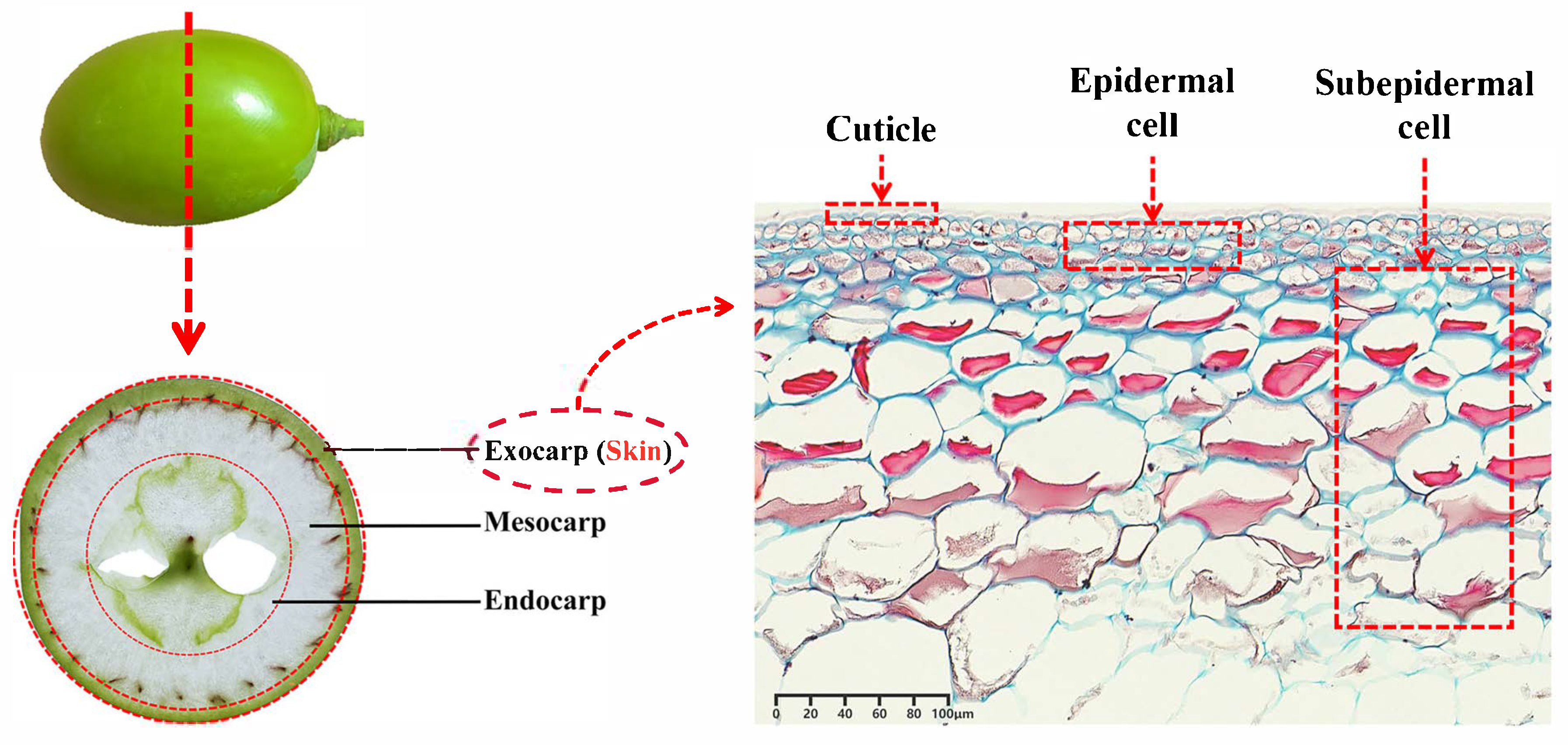
Figure 1. Outside structure of the grape.
Horticulturae 11 00962 g001
Figure 2. Theoretical model of skin texture change. CSLs, Cellulose synthase-like protein; CSCs, cellulose synthase complexes; GAUTs, galacturonosyltransferases; PMTs, pectin methyltransferase.
Horticulturae 11 00962 g002
Figure 3. The pathway for the generation of astringency.
Horticulturae 11 00962 g003
Figure 4. The pathway of tannin metabolism: (A) the biosynthesis of tannins. PAL, phenylalanine ammonia-lyase; C4H, cinnamate-4-hydroxylase; 4CL, 4-coumarate: CoA-ligase; CHS, chalcone synthase; CHI, chalcone isomerase; F3H, flavanone 3-hydroxylase; F3′H, flavonoid 3′-hydroxylase; F3′5′H, flavonoid 3′5′-hydroxylase; DFR, dihydroflavonol 4-reductase; ANS, anthocyanidin synthase; LODX, leucoanthocyanidin dioxygenase; ANR, anthocyanidin reductase; LAR, leucocyanidin reductase; Cys-EC, 4β’-(S-Cysteinyl)-epicatechin; and (B) transport mechanism of tannins. GST, glutathione-S-Transferase.
Horticulturae 11 00962 g004
Disclaimer/Publisher’s Note: The statements, opinions and data contained in all publications are solely those of the individual author(s) and contributor(s) and not of MDPI and/or the editor(s). MDPI and/or the editor(s) disclaim responsibility for any injury to people or property resulting from any ideas, methods, instructions or products referred to in the content.