Article

Delayed Starch Degradation Triggers Chromoplast Structural Aberration to Inhibit Carotenoid Cleavage: A Novel Mechanism for Flower Color Deepening in Osmanthus fragrans

1
School of Pharmacy, Xianning Medical College, Hubei University of Science and Technology, Xianning 437100, China
2
National Forestry and Grassland Administration Engineering Research Center for Osmanthus fragrans, Hubei University of Science and Technology, Xianning 437100, China
3
Public Inspection and Testing Center, Xianning 437100, China
*
Author to whom correspondence should be addressed.
These authors contributed equally to this work.
Horticulturae 2025, 11(7), 864; https://doi.org/10.3390/horticulturae11070864
Submission received: 3 June 2025 / Revised: 13 July 2025 / Accepted: 14 July 2025 / Published: 21 July 2025

Abstract

The color of flowers in Osmanthus fragrans is regulated by carotenoid metabolism. The orange-red variety, Dangui, is believed to have evolved from the yellow variety, Jingui, through a natural bud mutation. This study uses the Jingui cultivar ‘Jinqiu Gui’ (JQG) and its bud mutation cultivar ‘Huolian Jindan’ (HLJD) as materials, combining genome resequencing, ultrastructural observation, targeted metabolomics, and transcriptomic analysis to elucidate the molecular and cellular mechanisms underlying flower color variation. Phylogenetic analysis confirms that HLJD is a natural bud mutation of JQG. Ultrastructural observations reveal that during petal development, chromoplasts are transformed from proplastids. In HLJD petals, starch granules degrade more slowly and exhibit abnormal morphology, resulting in chromoplasts displaying crystalline, tubular, and fibrous composite structures, in contrast to the typical spherical plastoglobuli found in JQG. Targeted metabolomics identified 34 carotenoids, showing significant increases in the levels of ε-carotene, γ-carotene, α-carotene, and β-carotene in HLJD petals compared to JQG, with these levels continuing to accumulate throughout the flowering process, while the levels of the cleavage products α-ionone and β-ionone decrease. Transcriptomic analysis indicates that carotenoid metabolic pathway genes do not correlate directly with the phenotype; however, 49 candidate genes significantly associated with pigment accumulation were identified. Among these, the expression of genes such as glycoside hydrolases (LYG036752, etc.), sucrose synthase (LYG010191), and glucose-1-phosphate adenylyltransferase (LYG003610) are downregulated in HLJD. This study proposes for the first time the pathway of “starch degradation delay → chromoplast structural abnormalities → carotenoid cleavage inhibition” for deepening flower color, providing a new theoretical model for the metabolic regulation of carotenoids in non-photosynthetic tissues of plants. This research not only identifies key target genes (such as glycoside hydrolases) for the color breeding of O. fragrans but also establishes a theoretical foundation for the color enhancement of other ornamental plants.

1. Introduction

Osmanthus fragrans, a member of the Oleaceae family and Osmanthus genus, is an evergreen woody plant renowned as a traditional fragrant flower in China. This species is not only widely utilized for ornamental purposes in gardens but also possesses significant fragrance, is edible, and has medicinal values, offering immense potential for the development of deep-processing products that contribute importantly to the local economies in China. Flower color serves as a critical ornamental quality trait of O. fragrans, which is categorized into four groups based on flowering period and flower color traits: Sijigui, Yingui, Jingui, and Dangui, with nearly 200 cultivars currently available [1]. Carotenoids are key compounds influencing the variation in flower color among O. fragrans cultivars and serve as direct precursors in the synthesis of key aromatic compounds, such as α-ionone and β-ionone [2,3,4,5]. This is essential for enhancing the ornamental and economic value of O. fragrans. Investigating the carotenoid metabolic mechanisms in O. fragrans holds significant practical implications for the targeted cultivation of new cultivars with enhanced ornamental appeal (more vibrant flower colors) and higher economic value (stronger fragrance). Furthermore, this research enriches the theoretical foundation for the metabolic regulation of carotenoids in plant flowers, providing a universal model for flower color improvement and fragrance synthesis in other species.
The production of carotenoids relies on the methylerythritol phosphate (MEP) pathway, which provides the precursor geranylgeranyl pyrophosphate (GGPP) [6,7]. Phytoene synthase (PSY) catalyzes the conversion of GGPP into phytoene, which subsequently undergoes dehydrogenation and cyclization, leading to the formation of various carotenoids, including β-carotene and lutein [7,8]. These carotenoids can be cleaved by carotenoid cleavage dioxygenases into colored products, such as β-citraurin and crocin, or transformed into aromatic compounds like β-ionone [9,10,11]. Additionally, they may be degraded by 9-cis-epoxycarotenoid dioxygenase (NCED), resulting in the production of abscisic acid (ABA), which plays an important role in plant stress responses [12,13]. In plants, plastids serve as the sites for carotenoid synthesis and storage, categorized into proplastids, amyloplasts, chloroplasts, and chromoplasts, with chromoplasts being the primary locations for carotenoid production and accumulation [7,14]. The structural characteristics of chromoplasts vary across different species and plant organs, and they can be classified into types such as plastoglobuli, crystalline, membranous, and tubular [14]. Furthermore, chromoplasts can arise from the transformation of other plastids, with numerous studies indicating that they can develop from chloroplasts [7,15]. However, instances of amyloplasts transforming into chromoplasts are significantly less documented in petals.
Despite the conservativeness of carotenoid metabolic pathways in plants, the composition and content of carotenoids in non-green tissues across different species exhibit notable diversity [16,17]. Previous studies have indicated that α- and β-carotene predominantly accumulate in the petals of most cultivars of O. fragrans, with variations in their content leading to differences in flower color phenotypes, resulting in the formation of the cultivars Yingui, Jingui, and Dangui [3,18]. The evolutionary relationships among these three groups were clarified only after our research group completed whole-genome resequencing of over 100 O. fragrans cultivars, revealing that the flower colors of O. fragrans evolved from light to dark, with Dangui being the last to form, possibly originating from bud mutation [1,19,20]. The discovery of natural mutants of Dangui supports this conclusion [21]. However, to date, there have been no reports on the mechanisms underlying the formation of Dangui mutants. This study presents a typical case of a natural mutation from Jingui to Dangui, combining phenotypic comparisons with phylogenetic tree construction through genome resequencing. It demonstrates that ‘Huolian Jindan’ (HLJD) is a bud mutation cultivar of ‘Jinqiu Gui’ (JQG), thereby providing important natural mutant resources for studying the mechanisms of flower color variation in O. fragrans. Subsequently, ultramicroscopic observations, carotenoid metabolomics, and transcriptomic analyses of petals from JQG and HLJD at different developmental stages were conducted to explore the mechanisms of flower color mutation in O. fragrans and to identify key genes regulating floral color variation, thereby providing a theoretical basis and important genetic resources for the improvement of flower color quality in O. fragrans and other plants.

2. Materials and Methods

2.1. Plant Materials

The original mother plant (S308) and the bud mutant branch (S309) (Figure 1A) were collected from the Suzhou Lingering Garden, Jiangsu Province, China. The petals and leaves of the cultivars ‘Jinqiu Gui’ (JQG) (S310) and ‘Huolian JinDan’ (HLJD) (S314) were collected during the bud stage (S1), initial flowering stage (S2), and full flowering stage (S3) (Figure 1B) from the National Osmanthus Germplasm Resource Bank of the Landscaping Co., Ltd. in Yuhang District, Hangzhou City, Zhejiang Province. After sample collection, they were immediately frozen in liquid nitrogen and then transferred to a −80 °C freezer for storage.

2.2. Genome Resequencing

High-quality DNA was extracted from healthy leaves using the CTAB method, following the manufacturer’s protocol. The quality of the extracted DNA was evaluated using the Qsep400 system (AutoQ Biosciences, San Diego, CA, USA). Subsequently, libraries for four samples were prepared with the TruSeq Library Construction Kit and sequenced on the MGI-SEQ 2000 platform at Frasergen Bioinformatics Co., Ltd. in Wuhan, China. To investigate genetic variations, clean reads from the resequencing data of these four samples were processed. The adapter sequences and low-quality reads were filtered out using Trimmomatic (version 0.36) [22]. BWA (version 0.7.15-r1140) [23] was then employed to map the clean reads to the “Liuyejingui” reference genome [1]. GATK software (version 4.0.9.0) [24] was subsequently used to detect SNPs, with the following filtering parameters applied: QD < 2.0, MQ < 40.0, FS > 60.0, SOR > 3.0, MQRankSum < −12.5, and ReadPosRankSum < −8.0. Furthermore, a neighbor-joining (NJ) phylogenetic tree was constructed using Treebest software (v1.9.2), with a bootstrap value of 1000. The resulting tree was visualized using iTOL (version 4) [25].

2.3. Observation of Ultrastructure

Flower petals collected at various developmental stages were placed in a 4% glutaraldehyde fixative and subsequently washed with 0.1 M phosphate-buffered saline (PBS, pH 7.4). After this, the samples were treated with a prepared 1% osmium tetroxide solution in a dark environment at room temperature for 7 h. They then underwent a stepwise dehydration procedure involving ethanol, followed by infiltration with the embedding medium. The polymerization of the embedding blocks took place in an oven at 60 °C for 48 h. Ultra-thin sections were created using an ultramicrotome and stained with a saturated alcoholic solution of 2% uranyl acetate in the dark. Finally, the samples were examined and photographed utilizing a transmission electron microscope (FEI Tecnai G2 F20 S-TWIN).

2.4. Carotenoid Extraction and Determination

A total of 50 mg of dried petal powder was weighed and subsequently extracted twice using 0.5 mL of a mixed solution comprising hexane, acetone, and ethanol in equal volumes (1:1:1, v/v/v), containing 0.01% BHT (g/mL). The extracts obtained were combined and concentrated before being re-dissolved in 100 μL of a methanol and t-butyl methyl ether (MTBE) mixture (1:1, v/v). This solution was then filtered through a 0.22 μm membrane and stored in a brown injection vial for LC-MS/MS analysis utilizing a QTRAP6500+ (SCIEX, Framingham, MA, USA). Chromatographic separation was achieved using a YMC C30 column (3 μm, 100 mm × 2.0 mm i.d.). Mass spectrometry data were collected through Ultra Performance Liquid Chromatography (UPLC) utilizing an ExionLCAD in combination with Tandem Mass Spectrometry (MS/MS) on a QTRAP®6500+. The mobile phase and analytical detection parameters were modified following the protocols established by Wei et al. (2024) [3]. Qualitative analysis of the mass spectrometry data was conducted based on the creation of a standard substance MWDB (MetwareDatabase). For quantitative analysis, the Multiple Reaction Monitoring (MRM) mode of triple quadrupole mass spectrometry was employed alongside standard curves.

2.5. Determination of Volatile Components

Frozen petals (1 g) were subjected to grinding into a fine powder with the aid of liquid nitrogen, which was then transferred into a 40 mL centrifuge tube. Following this, 5 mL of n-hexane containing the internal standard methyl laurate at a final concentration of 20 ng/μL was introduced to the extraction solution, and the mixture was gently agitated for 30 min. After this agitation, the extraction solution was decanted into a fresh 50 mL centrifuge tube. An extra 5 mL of the extraction solution was added to the residual solid. Both extraction solutions were subsequently combined and allowed to rest overnight at −20 °C. The upper layer of the solvent was cautiously removed and concentrated to 2 mL using a nitrogen blow-down concentrator. The obtained solution was dried over an anhydrous Na2SO4 column and then subjected to analysis via gas chromatography-mass spectrometry (GC-MS). The chromatographic separation utilized an HP-5MS capillary column (30 m × 0.25 mm × 0.25 μm, J&W Scientific), with high-purity helium (99.999%) serving as the carrier gas at a flow rate of 1.2 mL/min. Refer to Zeng et al. (2016) [5] for the conditions of chromatographic and mass spectrometry analysis. The analysis of target analytes was performed using Agilent Mass Hunter Qualitative Analysis Navigator B.08.00 software coupled with NIST MS Search 2.3. Quantitative assessments were executed in accordance with the internal standard method utilizing methyl laurate.

2.6. Transcriptome Sequencing

RNA-seq were performed by Wuhan IGENEBOOK Biotechnology Co., Ltd. (Wuhan, China) (http://www.igenebook.com accessed on 29 October 2024). Total RNA was isolated from the petals of O. fragrans utilizing the TRNzol Universal (Tiangen Biotech Co., Ltd., Beijing, China), followed by evaluating RNA quality with the Qsep400 system (AutoQ Biosciences, San Diego, CA, USA). For the preparation of libraries, the Novogene VAHTS mRNA-seq V8 Library Prep Kit for Illumina was used, and sequencing was performed on the Illumina Novaseq 6000 platform (Illumina, San Diego, CA, USA). Clean reads were aligned to the reference genome ‘LiuyeJingui’ using Hisat2 (v2.1.0), allowing for up to two mismatches. RNA-seq raw data have been deposited into the public database of the National Center of Biotechnology Information (NCBI) BioProject PRJNA1279839 (accession number SAMN49507044- SAMN49507061). Genes identified during this procedure were compared against public protein databases, such as RefSeq non-redundant proteins (NR). Transcript levels were quantified, and gene expression was normalized to fragments per kilobase of transcript per million mapped fragments (FPKM) using Feature-count (v1.6.0). Differential expression analysis was carried out with edgeR, applying a significance threshold of FDR < 0.05 and |log2FoldChange| > 1. Furthermore, enrichment analyses for Gene Ontology (GO) and Kyoto Encyclopedia of Genes and Genomes (KEGG) were performed utilizing the Cluster Profiler in the R package (version 4.1.2.), with the groundwork for enrichment analysis based on hypergeometric distribution and a q value threshold of 0.05.

2.7. Data Analysis

The data analysis was carried out using the Oebiotech Cloud Platform (https://cloud.oebiotech.com/task/, accessed on 2 March 2025), which facilitated the creation of clustering heatmaps for metabolites and gene expression. Additionally, it performed PCA analysis, conducted Venn analysis, investigated co-expression correlations, and carried out variance analysis. The findings are reported as mean ± SD, derived from three biological replicates for every sample.

3. Results

3.1. Identification of a Bud Mutation Cultivar of O. fragrans

In the Liuyuan Garden of Suzhou, Jiangsu Province, a chimeric O. fragrans tree has been documented for many years, where bud mutation branches of the orange-red cultivar (Dan Gui) have emerged from the golden cultivar (Jin Gui). The parent tree has been preliminarily identified as ‘Jinqiu Gui’ (JQG) based on its phenotype. The bud mutation branches are primarily characterized by a change in flower color phenotype, exhibiting orange-red petals on the inside and golden edges on the outside (as shown in Figure 1A). This phenotype closely resembles that of ‘Huolian Jindan’ (HLJD), which is preserved in the National Osmanthus Germplasm Resource Bank of the Landscaping Co., Ltd. in Yuhang District, Hangzhou City, Zhejiang Province, China. To further confirm the phylogenetic relationships among JQG (S310), HLJD (S314), the parent tree from Suzhou Liuyuan Garden (S308), and the bud mutation branch (S309), DNA was extracted from the leaves of the four samples for genome resequencing. This data was subsequently utilized to construct a phylogenetic tree alongside resequencing data from 119 O. fragrans cultivars previously analyzed by our research group. As illustrated in Figure 2, O. fragrans var. Dangui is categorized into four distinct branches. The Suzhou Liu Garden bud mutation branch (S309) and HLJD (S314) cluster closely with the Suzhou Liu Garden parent (S308) and JQG (S310), as well as with the JQG cultivar sourced from germplasm banks in Wuhan, Hubei (S432) and Xianning, Hubei (S122). In contrast, these do not share the same branch as other O. fragrans cultivars of Dangui. This observation supports the conclusion that HLJD is indeed a bud mutation cultivar of JQG, and that the formation mechanism of HLJD is distinct from that of other O. fragrans cultivars of Dangui.

3.2. Ultrastructural Analysis of Plastids in O. fragrans Petals

Further observations of the ultrastructure of plastids in the petals of JGQ and HLJD at various flowering stages were conducted using transmission electron microscopy (Figure 3). During the bud stage, both JGQ and HLJD exhibited plastids containing a significant number of starch grains. At the initial flowering and full-flowering stages, the large starch grains within the plastids were observed to disappear. In JGQ, a considerable number of chromoplasts with plastoglobuli were identified, featuring round white starch grains at the center of these plastoglobuli. In contrast, HLJD displayed a highly complex internal structure of chromoplasts, characterized by a small number of irregularly shaped plastoglobuli, along with crystalline, tubular, and fibrous structures within the same chromoplast. The remaining starch grains also exhibited irregular shapes. Notably, the rate of starch grain disappearance in the petals of HLJD during the initial- and full-flowering stages was significantly slower than that in JGQ. It is speculated that the chromoplasts in O. fragrans petals have evolved from amyloplasts, and the normal hydrolysis of starch grains facilitates the formation of plastoglobuli in chromoplasts.

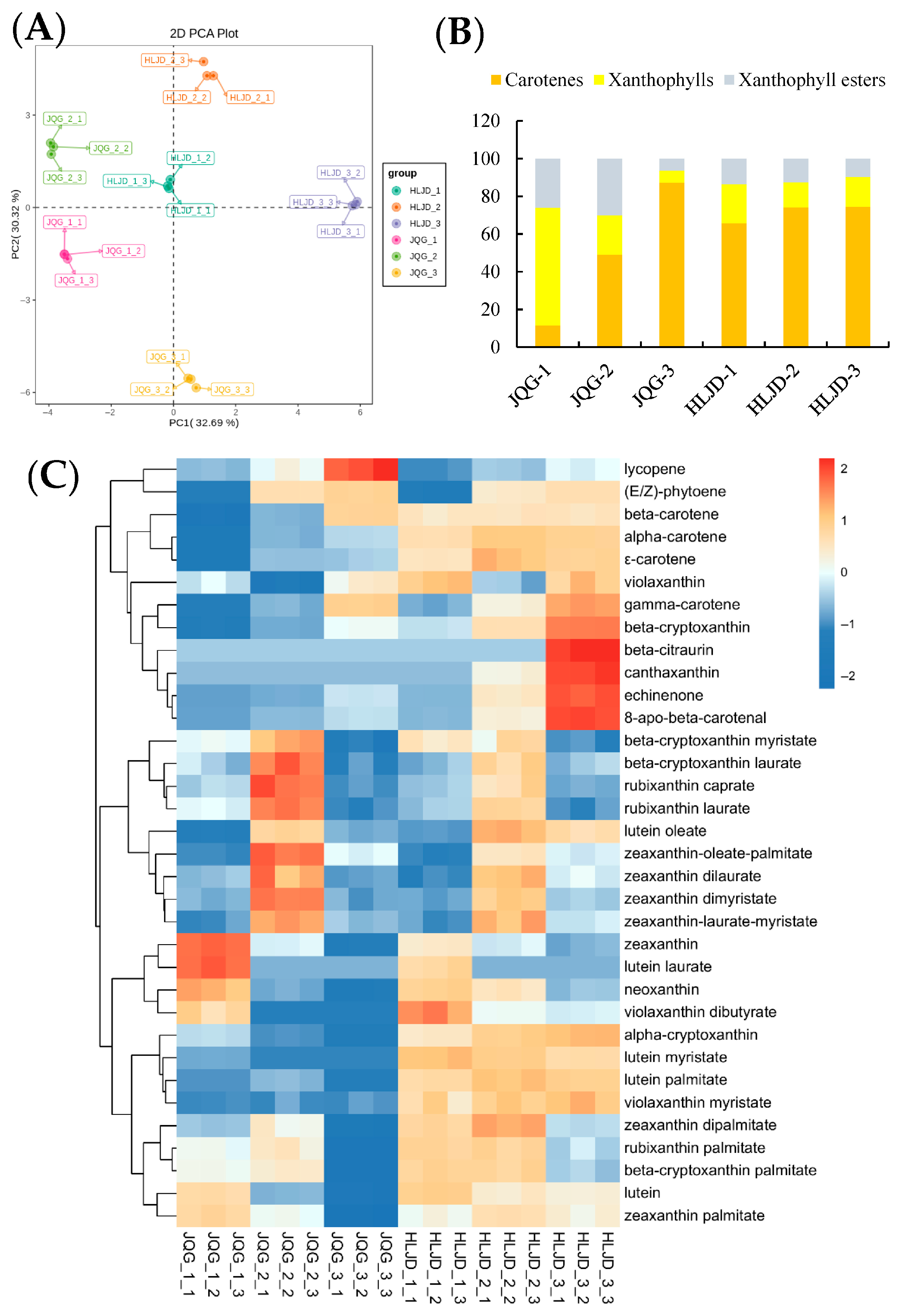
3.3. Quantitative Evaluation of Carotenoids in O. fragrans Petals

The carotenoid composition and content of petals from JQG and HLJD at different flowering stages were analyzed using UPLC-MS/MS, leading to the identification of 34 carotenoids: 6 types of carotenes, 10 types of xanthophylls, and 18 types of xanthophyll esters (see Supplementary Materials Table S1). Principal component analysis (PCA) indicated (Figure 4A) that the S1, S2, and S3 stages of HLJD are situated near the first quadrant and its boundary, whereas JQG’s S1 is located in the third quadrant, S2 in the second quadrant, and S3 in the fourth quadrant. This suggests significant differences between HLJD and JQG, as well as notable variations in the petals of JQG across different stages. From Figure 4B, it is evident that the relative content of carotene in HLJD during the three stages accounts for 60–80%, exhibiting an increasing trend as the flowers open. In JQG, the relative content of xanthophyll is highest during the S1 stage but decreases as the flowers open, while the carotene content increases. Cluster heatmap analysis reveals (Figure 4C) that the 34 carotenoids are classified into three branches: Clade I exhibits the highest content in S3; Clade II shows higher content in S2; and Clade III has the highest content in S1. Based on the floral color phenotypes of JQG and HLJD at various stages, the color deepens progressively with flower opening, with HLJD displaying a deeper flower color than JQG. The contents of β-carotene, α-carotene, and ε-carotene correspond to the observed phenotypic changes.

3.4. Transcriptome Sequencing and DEGs Analysis Between JQG and HLJD Petals of O. fragrans

To investigate the molecular mechanism underlying changes in flower color in O. fragrans, petals from the JQG and HLJD cultivars at three flowering stages were employed to create libraries for high-throughput sequencing. The quality metrics, Q20 and Q30, surpassed 95% and 90%, respectively, affirming the sequencing data’s reliability for subsequent analysis. Results from PCA analysis (Figure 5A) reveal distinct differences among varying cultivars and flowering stages. A differential gene expression analysis was performed, using FDR < 0.05 and | log2FoldChange | > 1 as criteria for selecting genes expressed at different flowering stages. The findings show (Figure 5B) that in the JQG cultivar, there were 3865 upregulated differentially expressed genes (DEGs) and 4301 downregulated DEGs in S2 compared to S1; S3 exhibited 5324 upregulated DEGs and 7133 downregulated DEGs; and in the comparison of S3 to S2, there were 2222 upregulated DEGs and 2936 downregulated DEGs. For the HLJD cultivar, relative to S1, S2 revealed 3036 upregulated DEGs and 3770 downregulated DEGs; in S3, there were 3952 upregulated DEGs and 5100 downregulated DEGs; and when comparing S3 with S2, 2337 upregulated DEGs and 2643 downregulated DEGs were identified. When comparing the DEGs between JQG and HLJD across the same stages, the results indicated 3660 upregulated DEGs and 1799 downregulated DEGs in JQG for the S1 stage; 2850 upregulated DEGs and 1875 downregulated DEGs in S2; and 1117 upregulated DEGs and 750 downregulated DEGs in S3. A Venn analysis of both upregulated and downregulated DEGs across the three stages between JQG and HLJD was performed (Figure 5C), yielding 98 and 51 DEGs, respectively.

3.5. Association Analysis Between Metabolome and Transcriptome of the Important Genes in O. fragrans

The content of various components in the carotenoid metabolic pathway across different varieties and periods was visually illustrated (Figure 6A). Throughout all three stages, the levels of (E/Z)-phytoene and lycopene were consistently greater in JQG compared to HLJD, displaying an upward trend in content as the flowers opened. Conversely, the concentrations of ε-carotene, γ-carotene, α-carotene, α-cryptoxanthin, and β-cryptoxanthin increased with flower unfolding, with HLJD consistently exhibiting higher levels than JQG during the entire flowering period. Notably, β-carotene concentrations were significantly elevated in HLJD compared to JQG during the S1 and S2 stages, whereas JQG surpassed HLJD in the S3 stage. Additionally, lutein levels were higher in HLJD than in JQG, although they gradually declined as the flowers progressed in opening. Zeaxanthin levels also diminished with the opening of flowers; however, during the S1 phase, JQG presented notably higher levels than HLJD. Considering the content and color characteristics of the various carotenoid components, it can be inferred that ε-carotene, γ-carotene, α-carotene, and β-carotene may play crucial roles in flower color mutations. Furthermore, GC-MS was utilized to analyze the contents of carotenoid cleavage products, specifically α-ionone and β-ionone, in the petals (Figure 6A). The findings demonstrated that the levels of these two critical components related to O. fragrans fragrance were more significant in JQG petals than in HLJD. A heatmap analysis of the expression levels of carotenoid metabolism pathway genes derived from transcriptome data (Figure 6B) showed no genes correlated with changes in carotenoid component levels, suggesting that genes associated with the carotenoid metabolic pathway are not the primary contributors to variations in flower color.
A correlation analysis was performed on 98 upregulated and 51 downregulated differentially expressed genes (DEGs) in relation to ε-carotene, γ-carotene, α-carotene, and β-carotene, using a significance threshold of p < 0.05. The results revealed that 68 of these genes were associated with ε-carotene, 82 with γ-carotene, 99 with α-carotene, and 60 with β-carotene. Additionally, it was found that 49 genes were related to all four carotenoid types (Figure 6C). To evaluate the relationships between these carotenoid components and the identified 49 genes, Pearson’s correlation method was utilized, as illustrated in Figure 6D. Functional annotations for these genes are detailed in Supplementary Table S2, which contains 14 genes with unknown functions. Within the gene ontology (GO) classifications, ‘hydrolase activity’ in the ‘molecular_function’ category was predominant, including four genes: LYG036752 (beta-glucosidase), LYG035009 (beta-glucosidase), LYG007921 (alpha-glucosidase), and LYG016487 (beta-galactosidase).

4. Discussion

This research initially establishes that HLJD represents a natural bud mutation variety of JQG. This finding provides direct support for the evolutionary model of floral color in O. fragrans, suggesting a transition from light to dark and reinforcing the proposition that the Dangui group evolved through bud mutations [1,21]. The distinctive phenotype of HLJD, characterized by orange-red inner petals and golden outer petals, serves as a valuable natural mutant for investigating the genetic basis of floral color development in plants. Examination of plastid structures revealed a significant presence of starch-containing plastids within the petals of both cultivars during the bud phase, with chromoplast characteristics being faint. As the plants progressed to the initial- and full-flowering stages, starch granules gradually diminished, while chromoplast features became more pronounced. However, notable structural differences in the chromoplasts were observed between the two cultivars: in JQG, only chromoplasts containing plastoglobuli were present, featuring round starch granules encased in plastoglobuli, whereas HLJD exhibited a diverse array of chromoplast structures, including crystalline, tubular, fibrillar, and membrane forms, along with a limited presence of plastoglobuli. Concurrently, the rate of starch granule degradation in HLJD was slower compared to JQG, resulting in irregular forms. This suggests that chromoplasts in the petals of O. fragrans likely originate from the transformation of amyloplasts, and that the typical hydrolysis of starch is crucial for the formation of spherical plastoglobuli within chromoplasts. Previous studies have documented the transformation of amyloplasts into chromoplasts in the roots, tubers, or fruits of plant species such as cassava, potatoes, citrus, and kiwifruit, indicating that starch degradation promotes the accumulation of the sugar precursors necessary for carotenoid synthesis [26,27,28,29,30]. In plant petals, previous studies have reported that colored plastids predominantly arise from the transformation of chloroplasts or from the direct differentiation from proplastids [31,32]. This developmental mechanism encompasses the remodeling of cellular structures, regulation of pigment metabolism, and precise coordination of gene expression [33]. This study is the first to demonstrate that the development of colored plastids in plant petals originates from amyloplasts. However, the underlying mechanisms facilitating this process require further elucidation.
Targeting metabolomics of carotenoids reveals that the concentrations of ε-carotene, γ-carotene, α-carotene, and β-carotene in HLJD petals are significantly higher compared to those in JQG, and these concentrations further increase as the flowers open. Conversely, the levels of carotenoid cleavage products, such as α-ionone and β-ionone, are markedly lower in HLJD than in JQG. This observation implies that the mutant may enhance the development of dark phenotypes by directing metabolic flow towards pigment production while simultaneously suppressing the cleavage pathway. Previous research has indicated that among the three groups of O. fragrans, the Dangui cultivar exhibits elevated levels of α-carotene and β-carotene [1,18]; however, its fragrance is less pronounced than that of Jingui and Yingui due to reduced concentrations of α-ionone and β-ionone [4,34]. It has been demonstrated that low expression or functional impairment of the CCD4 gene inhibits carotenoid cleavage [1,35,36]. In our previous investigation of the red-flowered cultivar ‘Yanzhi Hong’, decreasing expression of LYCE obstructed the synthesis of downstream α-carotene, leading to an accumulation of red lycopene [3]. This study, which integrates metabolomics and transcriptomics analyses, indicates that the genes associated with carotenoid synthesis and metabolism do not have a direct connection with the floral color mutation phenotype. This suggests that pigment accumulation may be influenced by indirect factors, such as substrate availability or conditions within storage microenvironments. Furthermore, it is noted that starch granule degradation is slower in HLJD petals compared to JQG, characterized by irregular forms, with significant crystalline, tubular, and fibrous structures present within the chromoplasts instead of plastoglobuli. It is hypothesized that the irregular degradation of starch hinders the development of spherical chromoplasts, which facilitates the cleavage of carotenoids into apocarotenoids, while crystalline, tubular, and fibrous chromoplasts are more advantageous for carotenoid storage without degradation.
A total of 49 candidate genes were identified through the correlation analysis between metabolites and transcriptomes. The genes that are highly correlated with carotenoid accumulation are associated with starch degradation and soluble sugar synthesis, including four glycoside hydrolase genes (e.g., LYG036752, LYG035009, LYG007921, LYG016487), one sucrose synthase (LYG010191), and one glucose-1-phosphate adenylyltransferase (LYG003610). These genes exhibited significantly lower transcription levels in HLJD compared to JQG. Notably, the allelic variation of the Ven1 gene in maize endosperm regulates the contents of polar and non-polar carotenoids. The increase in non-polar carotenoids delays the degradation of the amyloplast membrane, thereby hindering the interaction between protein bodies and starch granules, which affects the formation of hard endosperm [37]. This study indirectly supports the association between carotenoids and starch metabolism; however, direct evidence linking the two remains elusive. Based on our combined analysis of metabolomics and transcriptomics, we focused on the glycoside hydrolase genes involved in starch degradation. Subsequent functional analyses of these genes will provide evidence for the direct association between carotenoid and starch metabolism. Integrating these findings, we arrive at the following inference (Figure 7): the decreased expression of candidate genes, such as glycoside hydrolases, leads to a delay in the hydrolysis of starch granules. This delay subsequently causes abnormal development of chromoplast structures (e.g., atypical crystallization, tubular, and fibrous structures). Such structural abnormalities compel chromoplasts to prioritize storage over the cleavage of carotenoids. Consequently, compared to chromoplasts including spherical plastoglobuli, the likelihood of carotenoid cleavage is significantly diminished. Additionally, the cleavage pathways, including the synthesis of α-ionone and β-ionone synthesis, are inhibited, ultimately resulting in a substantial accumulation of pigments and a deepening of flower colors.

5. Conclusions

This study, based on the microstructure and multi-omics analysis of natural flower color mutants of O. fragrans, reveals for the first time a key pathway driven by delayed starch degradation that deepens flower color: “Delayed starch degradation → Abnormal chromoplast structure → Inhibition of carotenoid cleavage.” The elucidation of this pathway provides new insights into the metabolic regulation of carotenoids in the non-photosynthetic tissues of plants. Given the current relative scarcity of research on the interrelationship between starch metabolism and carotenoid metabolism, along with the limited available cases for reference, exploring the deeper molecular mechanisms of their interaction will be an important direction for future research. This study clarifies the potential core role of glycoside hydrolase genes in this pathway. Subsequent research will focus on in-depth functional validation of these genes to elucidate their specific roles in regulating starch degradation and carotenoid homeostasis. The relevant research findings are expected to identify key targets for the molecular breeding of flower color in O. fragrans and other ornamental plants, thereby promoting the breeding of high ornamental varieties.

Supplementary Materials

The following supporting information can be downloaded at: https://www.mdpi.com/article/10.3390/horticulturae11070864/s1, Table S1: Components and contents of carotenoids in petals of JQG and HLJD at three stages; Figure S1: GC chart of volatile components detection in JQG and HLJD petals; Table S2: Functional annotation of genes related to ε-carotene, γ-carotene, α-carotene, and β-carotene in O. fragrans.

Author Contributions

Conceptualization, H.C.; methodology, X.Z., Y.T., and X.W.; software, J.Y.; validation, X.C.; formal analysis, X.W.; investigation, J.Z.; resources, X.Z.; data curation, H.W.; writing—original draft, Y.T.; writing—review and editing, X.Z.; visualization, H.W.; supervision, Q.H.; project administration, X.Z. and H.C.; funding acquisition, X.Z., J.Z., J.Y., X.C. and H.C. All authors have read and agreed to the published version of the manuscript.

Funding

This research was funded by the National Natural Science Foundation of China (32101581, 32271951), Hubei Province Natural Science Foundation (2025AFB857, 2024AFB1057, 2023AFB1063) and Hubei University of Science and Technology Innovation Team (2022T02).

Data Availability Statement

Whole Genome Resequencing and RNA-seq raw data have been deposited into the public database of the National Center of Biotechnology Information (NCBI) BioProject PRJNA1279839. RNA-seq raw data were also deposited under these NCBI accession number SAMN49507044- SAMN49507061).

Acknowledgments

The authors express their sincere gratitude to Baichun Shen from the Department of Forestry of Zhejiang Province, China, for his invaluable assistance in sampling the Osmanthus fragrans ‘JQG’ and ‘HLJD’.

Conflicts of Interest

The authors declare no conflicts of interest.

References

  1. Chen, H.; Zeng, X.; Yang, J.; Cai, X.; Shi, Y.; Zheng, R.; Wang, Z.; Liu, J.; Yi, X.; Xiao, S.; et al. Whole-genome resequencing of Osmanthus fragrans provides insights into flower color evolution. Hortic. Res. 2021, 8, 98. [Google Scholar] [CrossRef] [PubMed]
  2. Zou, J.; Cai, X.; Yang, J.; Zeng, X.; Liu, D.; Huang, S.; Chen, X.; Yang, Q.; Wang, C.; Chen, H. DNA hypomethylation mediates flower opening and senescence in sweet osmanthus through auxin and ethylene responsive pathways. Postharvest Biol. Technol. 2023, 198, 112250. [Google Scholar] [CrossRef]
  3. Wei, S.; Wu, J.; Yu, P.; Tan, Y.; He, Q.; Yang, J.; Cai, X.; Zou, J.; Chen, H.; Zeng, X. Metabolomic and Transcriptomic Analysis of Unique Floral Coloration in Osmanthus fragrans Cultivars. Horticulturae 2024, 10, 801. [Google Scholar] [CrossRef]
  4. Cai, X.; Mai, R.; Zou, J.; Zhang, H.; Zeng, X.; Zheng, R.; Wang, C. Analysis of aroma-active compounds in three sweet osmanthus (Osmanthus fragrans) cultivars by GC-olfactometry and GC-MS. J. Zhejiang Univ.-Sci. B Biomed. Biotechnol. 2014, 15, 650–660. [Google Scholar] [CrossRef] [PubMed]
  5. Zeng, X.; Liu, C.; Zheng, R.; Cai, X.; Luo, J.; Zou, J.; Wang, C. Emission and accumulation of monoterpene and the key terpene synthase (TPS) associated with monoterpene biosynthesis in Osmanthus fragrans Lour. Front. Plant Sci. 2016, 6, 1232. [Google Scholar] [CrossRef] [PubMed]
  6. Iorizzo, M.; Ellison, S.; Senalik, D.; Zeng, P.; Satapoomin, P.; Huang, J.; Bowman, M.; Iovene, M.; Sanseverino, W.; Cavagnaro, P.; et al. A high-quality carrot genome assembly provides new insights into carotenoid accumulation and asterid genome evolution. Nat. Genet. 2016, 48, 657–666. [Google Scholar] [CrossRef] [PubMed]
  7. Villwock, S.S.; Li, L.; Jannink, J.L. Carotenoid-carbohydrate crosstalk: Evidence for genetic and physiological interactions in storage tissues across crop species. New Phytol. 2024, 244, 1709–1722. [Google Scholar] [CrossRef] [PubMed]
  8. Zhou, X.; Rao, S.; Wrightstone, E.; Sun, T.; Lui, A.C.W.; Welsch, R.; Li, L. Phytoene synthase: The key rate-limiting enzyme of carotenoid biosynthesis in plants. Front. Plant Sci. 2022, 13, 884720. [Google Scholar] [CrossRef] [PubMed]
  9. Sun, Q.; He, Z.; Wei, R.; Ye, J.; Chai, L.; Cheng, Y.; Xu, Q.; Deng, X. Red peel regulator 1 links ethylene response factor 25 and β-citraurin biosynthetic genes to regulate ethylene-induced peel reddening in citrus. Plant Cell 2024, 37, 10. [Google Scholar] [CrossRef] [PubMed]
  10. Zheng, X.; Mi, J.; Balakrishna, A.; Liew, K.X.; Ablazov, A.; Sougrat, R.; Al-Babili, S. Gardenia carotenoid cleavage dioxygenase 4a is an efficient tool for biotechnological production of crocins in green and non-green plant tissues. Plant Biotechnol. J. 2022, 20, 2202–2216. [Google Scholar] [CrossRef] [PubMed]
  11. Baldermann, S.; Kato, M.; Kurosawa, M.; Kurobayashi, Y.; Fujita, A.; Fleischmann, P.; Watanabe, N. Functional characterization of a carotenoid cleavage dioxygenase 1 and its relation to the carotenoid accumulation and volatile emission during the floral development of Osmanthus fragrans Lour. J. Exp. Bot. 2010, 61, 2967–2977. [Google Scholar] [CrossRef] [PubMed]
  12. Frey, A.; Effroy, D.; Lefebvre, V.; Seo, M.; Perreau, F.; Berger, A.; Sechet, J.; To, A.; North, H.M.; Marion-Poll, A. Epoxycarotenoid cleavage by NCED5 fine-tunes ABA accumulation and affects seed dormancy and drought tolerance with other NCED family members. Plant J. 2012, 70, 501–512. [Google Scholar] [CrossRef] [PubMed]
  13. Gavassi, M.A.; Silva, G.S.; Silva, C.M.S.; Thompson, A.J.; Macleod, K.; Oliveira, P.M.R.; Cavalheiro, M.F.; Domingues, D.S.; Habermann, G. NCED expression is related to increased ABA biosynthesis and stomatal closure under aluminum stress. Environ. Exp. Bot. 2021, 185, 104404. [Google Scholar] [CrossRef]
  14. Sun, T.; Yuan, H.; Cao, H.; Yazdani, M.; Tadmor, Y.; Li, L. Carotenoid metabolism in plants: The role of plastids. Mol. Plant 2018, 11, 58–74. [Google Scholar] [CrossRef] [PubMed]
  15. Ling, Q.; Sadali, N.M.; Soufi, Z.; Zhou, Y.; Huang, B.; Zeng, Y.; Rodriguez-Concepcion, M.; Jarvis, R.P. The chloroplast-associated protein degradation pathway controls chromoplast development and fruit ripening in tomato. Nat. Plants 2021, 7, 655–666. [Google Scholar] [CrossRef] [PubMed]
  16. Luan, Y.; Fu, X.; Lu, P.; Grierson, D.; Xu, W. Molecular mechanisms determining the differential accumulation of carotenoids in plant species and varieties. Crit. Rev. Plant Sci. 2020, 39, 125–139. [Google Scholar] [CrossRef]
  17. Hermanns, A.; Zhou, X.; Xu, Q.; Tadmor, Y.; Li, L. Carotenoid pigment accumulation in horticultural plants. Hortic. Plant J. 2020, 6, 343–360. [Google Scholar] [CrossRef]
  18. Wang, Y.; Zhang, C.; Dong, B.; Fu, J.; Hu, S.; Zhao, H. Carotenoid Accumulation and Its Contribution to Flower Coloration of Osmanthus fragrans. Front. Plant Sci. 2018, 9, 1499. [Google Scholar] [CrossRef] [PubMed]
  19. Xiang, Q.; Liu, Y. An Illustrated Monograph of the Sweet Osmanthus Cultivars in China; Zhejiang Science and Technology Press: Hangzhou, China, 2008. [Google Scholar]
  20. Yang, K. Chinese Osmanthus; China Forestry Publishing House: Beijing, China, 2020. [Google Scholar]
  21. Wang, Y.; Luo, Y.; Zhang, C.; Fu, J.; Hu, S.; Zhao, H. Flower Color and Pigment Composition in the Petals of Bud Mutation and its Stock Plant of Osmanthus fragrans ‘Jingui’. Acta Hortic. Sin. 2017, 44, 528–536. [Google Scholar]
  22. Simon, A. FastQC: A Quality Control Tool for High Throughput Sequence Data; Version 0.10.1; Babraham Bioinformatics: Cambridge, UK, 2010. [Google Scholar]
  23. Li, H.; Durbin, R. Fast and accurate short read alignment with Burrows-Wheeler transform. Bioinformatics 2009, 25, 1754–1760. [Google Scholar] [CrossRef] [PubMed]
  24. McKenna, A.; Hanna, M.; Banks, E.; Sivachenko, A.; Cibulskis, K.; Kernytsky, A.; Garimella, K.; Altshuler, D.; Gabriel, S.; Daly, M.; et al. The Genome Analysis Toolkit: A MapReduce framework for analyzing next-generation DNA sequencing data. Genome Res. 2010, 20, 1297–1303. [Google Scholar] [CrossRef] [PubMed]
  25. Letunic, I.; Bork, P. Interactive Tree of Life (iTOL) v4: Recent updates and new developments. Nucleic Acids Res. 2019, 47, W256–W259. [Google Scholar] [CrossRef] [PubMed]
  26. Drapal, M.; Gerrish, C.; Fraser, P.D. Changes in carbon allocation and subplastidal amyloplast structures of specialised Ipomoea batatas (sweet potato) storage root phenotypes. Phytochemistry 2022, 203, 113409. [Google Scholar] [CrossRef] [PubMed]
  27. Gutschker, S.; Ruescher, D.; Rabbi, I.Y.; Rosado-Souza, L.; Pommerrenig, B.; Pauly, M.; Robertz, S.; van Doorn, A.M.; Schlereth, A.; Neuhaus, H.E.; et al. Carbon usage in yellow-fleshed Manihot esculenta storage roots shifts from starch biosynthesis to cell wall and raffinose biosynthesis via the myo-inositol pathway. Plant J. 2024, 119, 2045–2062. [Google Scholar] [CrossRef] [PubMed]
  28. Zhou, X.; Mcquinn, R.; Fei, Z.; Wolters, A.A.; van Eck, J.; Brown, C.; Giovannoni, J.J.; Li, L. Regulatory control of high levels of carotenoid accumulation in potato tubers. Plant Cell Environ. 2011, 34, 1020–1030. [Google Scholar] [CrossRef] [PubMed]
  29. Zeng, Y.; Du, J.; Wang, L.; Pan, Z.; Xu, Q.; Xiao, S.; Deng, X. A comprehensive analysis ofchromoplastdifferentiationrevealscomplexproteinchangesassociated with plastoglobule biogenesis and remodeling of protein systems in sweet orange flesh. Plant Physiol. 2015, 168, 1648–1665. [Google Scholar] [CrossRef] [PubMed]
  30. Li, A.; Lin, J.; Zeng, Z.; Deng, Z.; Tan, J.; Chen, X.; Ding, G.; Zhu, M.; Xu, B.; Atkinson, R.G.; et al. The kiwifruit amyloplast proteome (kfALP): A resource to better understand the mechanisms underlying amyloplast biogenesis and differentiation. Plant J. 2024, 118, 565–583. [Google Scholar] [CrossRef] [PubMed]
  31. Liu, Z.; Ye, K.; Du, X.; Peng, Y.; Qin, J.; Zeng, L. Correlation analysis of petal morphology and pigment content of Rosa ‘Angela’. J. Beijing Univ. Agric. 2022, 37, 1–10. [Google Scholar]
  32. Yuan, Y.; Li, X.; Yao, X.; Fu, X.; Cheng, J.; Shan, H.; Yin, X.; Kong, H. Mechanisms underlying the formation of complex color patterns on Nigella orientalis (Ranunculaceae) petals. New Phytol. 2023, 237, 2450–2466. [Google Scholar] [CrossRef] [PubMed]
  33. May, T.S.Y.; Edwige, M. Under the rainbow: Novel insights on the mechanisms driving the development and evolution of petal pigmentation. Curr. Opin. Plant Biol. 2025, 86, 102743. [Google Scholar] [CrossRef] [PubMed]
  34. Xin, H.; Wu, B.; Zhang, H.; Wang, C.; Li, J.; Yang, B.; Li, S. Characterization of volatile compounds in flowers from four groups of sweet osmanthus (Osmanthus fragrans) cultivars. Can. J. Plant Sci. 2013, 93, 923–931. [Google Scholar] [CrossRef]
  35. Han, Y.; Wang, X.; Chen, W.; Dong, M.; Yuan, W.; Liu, X.; Shang, F. Differential expression of carotenoid-related genes determines diversified carotenoid coloration in flower petal of Osmanthus fragrans. Tree Genet. Genomes 2014, 10, 329–338. [Google Scholar] [CrossRef]
  36. Han, Y.; Lu, M.; Yue, S.; Li, K.; Dong, M.; Liu, L.; Wang, H.; Shang, F. Comparative methylomics and chromatin accessibility analysis in Osmanthus fragrans uncovers regulation of genic transcription and mechanisms of key floral scent production. Hortic. Res. 2022, 9, 96. [Google Scholar] [CrossRef] [PubMed]
  37. Wang, H.; Huang, Y.; Xiao, Q.; Huang, X.; Li, C.; Gao, X.; Wang, Q.; Xiang, X.; Zhu, Y.; Wang, J.; et al. Carotenoids modulate kernel texture in maize by influencing amyloplast envelope integrity. Nat. Commun. 2020, 11, 5346. [Google Scholar] [CrossRef] [PubMed]
Figure 1. Flower and leaf phenotypes of wild-type and bud mutant of O. fragrans (A), as well as JQG and HLJD cultivars of O. fragrans at different flowering stages (B).
Horticulturae 11 00864 g001
Figure 2. Phylogenetic tree of four samples and 119 O. fragrans cultivars. The red triangles represent the four samples that underwent genome resequencing this study.
Horticulturae 11 00864 g002
Figure 3. Observation of the ultrastructure of petal chromoplasts in JQG and HLJD cultivars of O. fragrans at different flowering stages. S—Starch granules; pg—plastoglobuli structure; T—tubular structure; C—crystalline structure; F—fibrous structure.
Horticulturae 11 00864 g003
Figure 4. Analysis of carotenoid metabolites in petals of O. fragrans JQG and HLJD cultivars at different flowering stages. (A) Principal component analysis; (B) Proportion of carotenoid components in petals; (C) Carotenoid metabolite clustering heat map.
Horticulturae 11 00864 g004
Figure 5. Transcriptomic features for petals of O. fragrans JQG and HLJD cultivars at different flowering stages. (A) Principal component analysis; (B) All DEGs statistics; (C) Venn diagram for up or down DEGs of JQG vs. HLJD at stage S1, S2, and S3.
Horticulturae 11 00864 g005aHorticulturae 11 00864 g005b
Figure 6. Conjoint analysis of carotenoid metabolome and transcriptome in O. fragrans JQG and HLJD petals. (A) Content of various components in the carotenoid metabolic pathway; (B) heat map of carotenoid metabolism pathway genes; (C) Venn map of ε-carotene, γ-carotene, α-carotene, and β-carotene related genes; (D) cluster heatmap of the co-expression correlation between carotenoid components and the 49 related genes. The correlation analysis was conducted using Pearson’s correlation method (* p ≤ 0.05, ** p ≤  0.01, *** p ≤ 0.001).
Horticulturae 11 00864 g006
Figure 7. A proposed model on floral color formation of O. fragrans.
Horticulturae 11 00864 g007
Disclaimer/Publisher’s Note: The statements, opinions and data contained in all publications are solely those of the individual author(s) and contributor(s) and not of MDPI and/or the editor(s). MDPI and/or the editor(s) disclaim responsibility for any injury to people or property resulting from any ideas, methods, instructions or products referred to in the content.

