Article

Characterization of a Gamma Radiation (60Co) Induced Mutant Population of Prickly Pear Cactus (Opuntia velutina F.A.C. Weber) Plants In Vitro Using ISSR Molecular Markers

by
Eréndira Rubio-Ochoa
1,
Eulogio De la Cruz-Torres
2,
Rosa Elena Pérez-Sánchez
3,
Héctor Eduardo Martínez-Flores
3,
Liberato Portillo
4,
Pedro Antonio García-Saucedo
1,* and
Juan Florencio Gómez-Leyva
5,*
1
Facultad de Agrobiología, Universidad Michoacana de San Nicolás de Hidalgo, Paseo Lázaro Cárdenas 2290, Emiliano Zapata, Melchor Ocampo, Uruapan 60170, Michoacán, Mexico
2
Departamento de Biología, Instituto Nacional de Investigaciones Nucleares ININ, Ocoyoacac 52750, Mexico
3
Facultad de Químico-Farmacobiología, Universidad Michoacana de San Nicolás de Hidalgo, Tzintzuntzan 173, Col. Matamoros, Morelia 58240, Michoacán, Mexico
4
Departamento de Botánica y Zoología, Centro Universitario de Ciencias Biológicas y Agropecuarias, Universidad de Guadalajara, Zapopan 44600, Jalisco, Mexico
5
Tecnológico Nacional de México-Instituto Tecnológico de Tlajomulco, DEPI, Tlajomulco de Zúñiga 45640, Jalisco, Mexico
*
Authors to whom correspondence should be addressed.
Horticulturae 2025, 11(7), 743; https://doi.org/10.3390/horticulturae11070743 (registering DOI)
Submission received: 24 May 2025 / Revised: 23 June 2025 / Accepted: 24 June 2025 / Published: 27 June 2025
(This article belongs to the Section Genetics, Genomics, Breeding, and Biotechnology (G2B2))

Abstract

The nopal cactus, a plant from the Cactaceae family, holds significant economic and nutritional value for Mexico. This study aimed to enhance the genetic diversity and morphological traits of Opuntia velutina, a species cultivated as a vegetable nopal. A total of 1050 in vitro O. velutina explants were exposed to 15 different doses of gamma radiation from 60Co gamma, ranging from 5 to 125 Gy. The lethal dose was above 50 Gy, with an LD50 of 22.8 Gy for stimulating in vitro shoot growth. Shoots derived from doses between 5 and 50 Gy were subjected to in vitro shoot proliferation across four consecutive generations to stabilize morphological traits. Cluster analysis categorized the 178 irradiated shoots into 13 distinct morphological groups (CG1–CG13). Twenty-seven shoots exhibiting significant morphological improvements, such as a 50–100% increase in cladode length, up to a six-fold increase in shoot number, and up to a seven-fold increase in root number, were selected for molecular analysis of genetic diversity. Six primers were used with the Inter Simple Sequence Repeat (ISSR) molecular markers to examine genetic uniformity, yielding 54.5% polymorphic bands, indicating a high level of genetic variation. Both a UPGMA dendrogram and STRUCTURE-based Bayesian analysis confirmed the genetic divergence among the selected mutant lines. Overall, gamma irradiation effectively enhanced both phenotypic and genotypic diversity in O. velutina. This study corroborates that in vitro mutagenesis through gamma radiation is a viable strategy for generating novel genotypes with breeding potential within the Opuntia genus.

1. Introduction

The genus Opuntia Mill. (Cactaceae) is one of the most diverse within the family, comprising nearly 150 species [1] distributed throughout the American continent, primarily in arid and semi-arid regions. The greatest diversity is found in Mexico, which is home to about 100 species [2,3]. Opuntia species are fleshy, arboreal plants, with an average height ranging from 1.5 to 3 m. They possess flat, oblong, and medium green stems known as cladodes, glochids commonly referred to as ahuates, and sessile leaves, which grow when the cladode is young and later decay. As perennial plants, they can be found at elevations from sea level up to 3400 m (above sea level) and can exist in both wild and cultivated forms [4]. Their adaptability to various soil types and their ability to survive with minimal water make them a valuable option for cultivation and use. The main cultivated species include O. ficus-indica, O. joconostle, O. megacantha, and O. streptacantha. Additionally, species such as O. robusta, O. leucotricha, O. hyptiacantha, and O. chavena are harvested from wild cactus populations in semi-arid zones [5].
Some Opuntia species have significant economic and functional value due to their use for ornamental, food, and medicinal purposes, mainly the cladodes or stems, flowers, and fruits (prickly pear) from which pigments, mucilage, and material for construction are extracted. They can also be an excellent forage crop for livestock, among other applications [6,7,8].
In Mexico, the cladodes, or young shoots of cactus, are consumed as a vegetable due to their high fiber content (15–51.4%), carbohydrates (5.6–92%), minerals (4–21%), and vitamin C and α-tocopherol [9,10]. Additionally, they contain bioactive compounds that contribute to scavenging free radicals and reducing oxidative stress, such as carotenoids, betalains, betacyanins, and polyphenols such as isorhamnetin, kaempferol, and quercetin [7]. However, the yield and quality of these crops depend on the species or variety, which in turn are influenced by abiotic factors such as climate, humidity, light, pH, and salinity, as well as biotic factors that include phytopathogenic agents such as bacteria, fungi, viruses, and nematodes. Due to these factors, efforts have been made to improve characteristics such as disease and pest resistance, cold tolerance, and a reduction in the number of thorns, glochidia, seeds, and mucilage content [11].
Currently, conventional plant breeding techniques focus on obtaining improved seeds through hybridization and domestication processes. Although these methods have been implemented to improve the qualities of cactus, none of these programs have prospered due to challenges such as slow growth, extended juvenile periods (2–4 years), genetic segregation, and the phenomenon of apomixis presented by their cultivars [11,12]. Consequently, cactus is considered a suitable candidate for use as a study model within biotechnological techniques, including induced mutagenesis.
Mutagenesis is a widely employed technique for the genetic improvement of food crops, involving the introduction of changes in the DNA chain that result in desirable transgenerational traits. This process can be initiated through the use of physical mutagenic agents, such as gamma radiation. Currently, there are approximately 3400 high-value mutant crop varieties available on the market, with about 70% having been developed directly or indirectly from gamma radiation [13], which is considered to be more efficient than other mutagenic agents. Gamma radiation has been applied to both differentiated and dedifferentiated tissues, as well as to seeds of various plant species, including agave, ginseng, rice, and so on. This has led to the development of plant lines that exhibit the enhanced production of specific secondary metabolites, such as ginsenosides, anthocyanins, and fructooligosaccharides, in addition to improved protein and amino acid contents of nutritional significance, increased tolerance to phytopathogens, drought, and high salinity, as well as higher yields and reduced cultivation periods [14,15,16,17].
Recognizing and identifying genetic variations in plant breeding programs can be enhanced by using DNA or RNA-based genetic markers, as well as understanding their relationship with a crop’s biochemical, cytological, and morphological traits [18]. Molecular markers have diverse applications in genetic plant breeding, including varietal characterization, gene labeling, mapping, and identifying phylogenetic relationships between cultivars. They offer advantages over morphological markers due to their higher polymorphism, independence from environmental factors, and their ability to automate analysis. Among the widely used molecular markers are those supported by the polymerase chain reaction (PCR), such as random amplified polymorphic DNA (RAPD), amplified fragment length polymorphism (AFLP), single-nucleotide polymorphism (SNP), and inter-simple sequence repeats (ISSRs). ISSR markers are considered neutral, as they amplify DNA regions located between microsatellite loci without targeting coding regions of the genome. They do not require prior genomic information, making them particularly useful for non-model or under-researched species such as Opuntia, where complete genomic data remain limited. ISSRs have demonstrated good reproducibility under optimized PCR conditions using longer primers [18,19,20] and have proven effective in detecting polymorphisms in materials derived from in vitro mutagenesis, particularly those induced by gamma irradiation [19,20,21,22,23,24]. In this study, ISSR markers were employed to assess the genetic diversity induced by gamma radiation in O. velutina, complementing the morphological characterization. Given the agronomic and nutritional relevance of nopal, the objective of this work was to induce genetic variation in in vitro meristems of O. velutina through 60Co gamma irradiation and to evaluate the resulting morphological and genetic diversity in the regenerated lines.

2. Materials and Methods

2.1. In Vitro Establishment of Apical Shoots

Young cladodes (10 to 15 cm in length) of prickly pear cactus (Opuntia velutina F.A.C. Weber) were collected. The specimens were tagged according to the morphological criteria and stored in the herbarium of the Instituto de Ecología, A.C., with the identification number IEB-273300. The leaves were cut in half, the thorns removed, and the stalks rinsed under running water. Subsequently, the segments were soaked for 4 h in a 5 g L−1 solution of oxytetracycline and benomyl (methyl 1-(butylcarbamoyl) benzimidazole-2-ylcarbamate), immersed in 70% (v/v) ethanol for 30 s, and then immersed in 0.4% sodium hypochlorite for 15 min. The explants were then rinsed three times with sterile water. Sections measuring 1 to 1.5 cm2 containing an areola as explant were cut and cultured in MS basal medium [25,26] supplemented with 30 g L−1 sucrose, 1.5 g L−1 activated charcoal, and 8 g L−1 agar at a pH of 5.7.

2.2. Radiation Dose and LD50 Determination

For irradiation, 70 explants were used for each dose (seven areoles per 15×90 mm Petri dish in basal MS medium). Fifteen radiation doses at 5, 10, 15, 20, 25, 30, 35, 40, 45, 50, 60, 70, 80, 100, and 125 Gy from a Cobalt-60 (60Co) source in a Gammacell irradiator (GO-220, Ottawa, ON, Canada), operating at a dose rate of 16.65 Gy/h, located at the facilities of the National Institute for Nuclear Research (ININ, Mexico), were evaluated. Irradiated explants were then cultured in MS medium supplemented with 1 mg L−1 benzylaminopurine (BAP). Cultures were maintained under a photoperiod of 16 h of light at 1000 lux and a temperature of 25 ± 2 °C for 45 days. After this period, the radiosensitivity curve was obtained by determining the percentage of sprouting using the following formula: percentage of sprouting (%) = (number of explants with sprouting after irradiation/total number of irradiated explants) × 100. The radiosensitivity curve was established through linear regression, correlating the percentage of sprouting with the radiation dose in Gy.

2.3. Post-Irradiation In Vitro Shoot Multiplication

Surviving materials were multiplied in MS medium at a 50% concentration, along with 1 g L−1 polyvinylpyrrolidone and 1 mg L−1 BAP. The shoots obtained after eight weeks were propagated in a line in the same medium until the fourth generation (V4). Thirty in vitro specimens with homogeneous physical characteristics were analyzed, measuring the length of the horizontal, vertical, and transverse axes of the main cladode, as well as counting the number of roots. The materials displaying morphological characteristics that contrasted with the control were selected for genetic analysis.

2.4. Statistical Analysis

To determine the lethal dose (LD50), a linear regression model was applied based on the percentage of in vitro sprouting and radiation dose (n = 70). The results of the lengths of horizontal, vertical, and transverse axes, the average number of shoots per explant, and the number of roots from 178 plants were recorded. The experimental design consisted of completely randomized blocks, with three replicates per line, totaling 30 individual seedlings (10 explants per replicate). The data were subjected to hierarchical clustering analysis using Ward’s method and principal component analysis (PCoA) with JMP® V11.0.0 software from SAS Institute Inc. (Cary, NC, USA), in order to distinguish materials with outstanding morphological characteristics.

2.5. DNA Extraction and PCR Amplification for ISSR Analysis

The genomic DNA from each mutant obtained in this study was isolated from in vitro young shoots using the CTAB method [27] with minor modifications. Genomic DNA was quantified using a spectrophotometer at 260 nm, and the absorbance at a 260/280 nm ratio confirmed its quality. Genetic diversity among twenty-seven lines of O. velutina, including one non-irradiated control and four external controls, was investigated using the following six ISSR primers: (GA)8YC, (GA)8YT, T(CT)7CC, (CT)8RG, (GA)8YG, and (GA)8C). Each PCR reaction was performed in a 12.5 μL volume consisting of 10 mM of Tris-HCl (pH 8), 50 mM of KCl, 2 mM of MgCl2, 0.2 mM of dNTPs, 0.5 U of Taq DNA polymerase, 10 pmol of primers, and 50 ng of DNA. The amplification reactions that took place were performed in a Select Cycle thermal cycler under the following conditions: an initial denaturation at 94 °C for five min, followed by 35 cycles of denaturation at 94 °C for 45 s, annealing at 42–52 °C for 40 s, extension at 72 °C for 90 s, and a final extension at 72 °C for 4 min. A 1.5% agarose gel was used to separate the amplified bands produced by PCR. Amplification products were separated on a 1.5% agarose gel for 90 min at 100 V. After electrophoresis, images were captured and analyzed using an image analysis system. The amplification process was repeated three times to ensure the repeatability of the primers.

2.6. Analysis of Genetic Diversity and Structure

The resulting ISSR banding patterns on the gel were manually recorded in a binary matrix of presence (1) and absence (0), thereby determining the total bands amplified (TBA), the number of polymorphic bands (NBP), and the percentage of polymorphism (%BP), using the non-irradiated material as a reference. In addition, polymorphic information content (PIC) was calculated using the following equation: PIC = 2 Pi (1–Pi), where Pi represents the frequency of occurrence of polymorphic bands for each primer and the marker index (MI) [28]. Genetic diversity analysis was performed using data from three individual plants per treatment, with each analyzed independently as a biological replicate. Genetic similarity values were determined using the unweighted paired groups with the arithmetic mean (UPGMA) method, while the Jaccard similarity coefficient was employed to generate a dendrogram using the NTSYS-PC software version 2.2 [29]. The population structure was assessed in the STRUCTURE software version 2.3.4 [30]. For each analysis, the number of genetic groups (K) evaluated ranged from 1 to 10, with 10 iterations for each K value, runs of 10,000 replicates, and a burn-in period of 3000. The admixture model was applied, considering independent allele frequencies and without incorporating prior information about the origin of the individuals [31]. The number of clusters (K) was plotted against the logarithm of the relative probability relative to the standard deviation (ΔK), and the criteria by Evanno et al. (2005) were used to estimate the optimal number of clusters (K subpopulations) [32].

3. Results and Discussion

3.1. Radiosensitivity of In Vitro Meristems

The present study demonstrated that 60Co gamma radiation significantly affects the modification of morphological parameters of O. velutina and the physiological response during in vitro shoot induction in response to plant growth regulators. The in vitro micropropagation of prickly pear cactus was initiated by inducing axillary meristems found in the areola [33]. For O. velutina, these were established in vitro, free of contaminants, and maintained as clones before exposure to 60Co gamma radiation. A total of 1050 meristems were used to induce genetic variants, evaluating morphological parameters and their response to in vitro shoot formation, serving as the first strategy for mutant selection due to the slow growth of this cactus.
Meristems irradiated with doses exceeding 50 Gy and up to 125 Gy exhibited chlorosis and subsequently reached 100% mortality. Undifferentiated tissues, such as callus or somatic embryos, were observed to be more sensitive to gamma radiation due to their high-water content, in contrast to mature tissues or lignified tissues such as shoots, seedlings, roots, or buds [17]. As the radiation dose increased, an inversely proportional effect was observed regarding the physiological response to sprouting (Figure 1). The estimated LD50 for the reduction in the in vitro meristem induction of O. velutina was 22.8 Gy, with an r2 = 0.945. LD50 values were recorded in in vitro Agave tequilana plants at 26 Gy [34] and in shoots of Citrus spp at 25–26 Gy [15]. This sensitivity to gamma radiation, in addition to the water factor, depends on the type of explants, genotype, and genome size, as well as physical factors such as oxygen, humidity, and temperature [35,36].
Mutation frequency is influenced by radiation dose, exposure rate, and tissue tolerance [37], making dosimetry evaluation crucial in mutation breeding. Radiosensitivity assays are used to estimate the median lethal dose (LD50), which is the radiation level that reduces in vitro regenerative response by 50% [37,38]. Monitoring sensitivity across different doses allows for the determination of lethality parameters (LD30 to LD60), facilitating the identification of effective doses that induce variability without compromising viability [24,35,36].
Ionizing radiation emitted by the radioisotope 60Co is capable of displacing electrons from the outer orbitals of atoms and molecules, generating ions and free radicals, particularly in aqueous environments. This primary ionization results in the formation of reactive species such as H+, H•, and OH•, along with secondary compounds including hydrogen peroxide (H2O2), superoxide (O2•), and hydronium ions. These reactive species inflict random cellular damage, with DNA being one of the most sensitive targets. Such damage can include base substitutions, strand breaks, and spontaneous purine loss, which may lead to lethal alterations [39,40], especially at doses exceeding 50 Gy, as observed in this study. However, the same damage also triggers repair mechanisms that result in stable modifications and transgenerational changes in DNA, leading to structural alterations similar to those seen in O. velutina [36,39,40].
At 45 days, a total of 260 surviving meristems were obtained from the 10 non-lethal doses of 5, 10, 15, 20, 25, 30, 35, 40, 45, and 50 Gy. These doses were micro-propagated over four consecutive cycles (V4) in MS medium supplemented with BAP at 1 mg L1. The non-irradiated meristem produced an average of 2.8 shoots per explant. All materials that lost their original characteristics for which they were selected were discarded.

3.2. Morphological Selection of Post-Irradiated In Vitro Seedlings

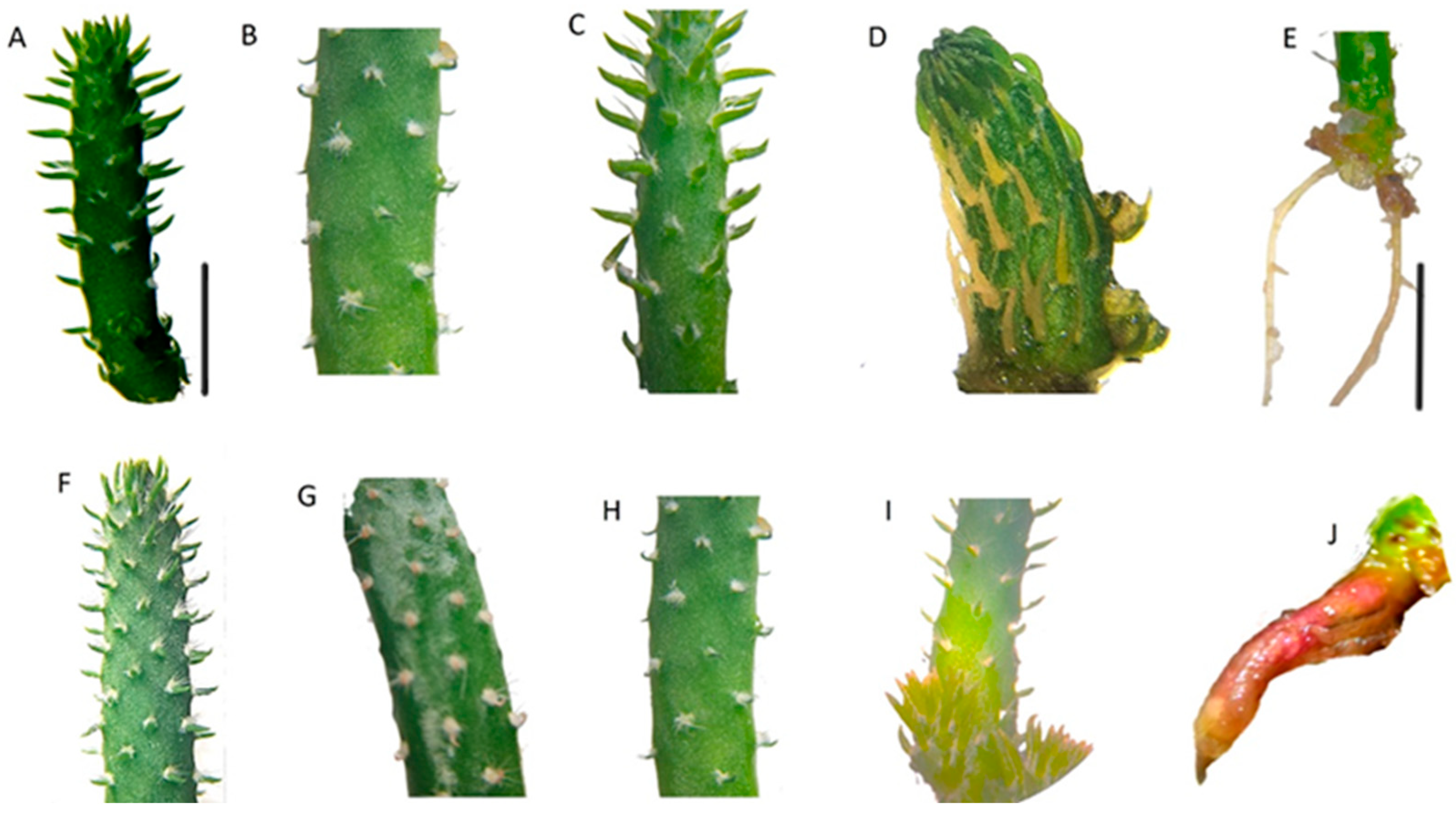
In the V4 generation, 178 clonal lines were propagated, and measurements along the longitudinal, horizontal, and transverse axes, as well as shoot formation and the number of roots per cladode, were registered using the shoots obtained from non-irradiated meristems (OUM00-00) as a reference (Figure 2A–E; Table S1). In the longitudinal axis averages, OUM10-15 (Figure 2F) stood out with a 100% increase (39.6 ± 0.6 mm) compared to the reference (18.7 ± 1.4 mm). For the horizontal axis, the OUM20-27 mutant (Figure 2G) excelled, showing a 20% increase over the non-irradiated material (4.7 ± 0.3 mm). The transverse axis exhibited increases of up to 50% with OUM25-34 (Figure 2H). The OUM05-30 mutant (Figure 2I) produced six times more shoots than the control. Among the average values for root number, OUM05-23 and OUM05-24 stood out in comparison to the control, with 2.4 ± 0.2 and 2.3 ± 0.3 roots, respectively, representing a sevenfold increase over the reference value (0.3 ± 0.05). Five mutants (OUM 05-70, OUM15-17, OUM20-45, and OUM20-35) exhibited pinkish pigmentation, possibly associated with betalain production.
Variations in plant architecture are easily observable during tissue regeneration and are therefore frequently reported in the identification of improved materials [24,36,41]. In banana (Musa acuminata cv. Berangan) mutant seedlings, increases in shoot number (48%), fresh weight (33%), stem length (47%), and root number (60%) were observed [41], as well as in Vanilla planifolia [42].
Although in vitro mutagenesis has proven to be an efficient biotechnological tool for obtaining improved varieties, it is important to consider the hormetic effect that materials subjected to low doses of irradiation commonly exhibit [43]. The hormetic effect was evident in this work, as it stimulated the development of shoots at doses lower than 20 Gy and increased toxicity or cell death at doses higher than 50 Gy, serving as a transient stage for the selection of elite lines between the third and fourth generations [23,39].

3.3. Cluster Analysis and PCoA

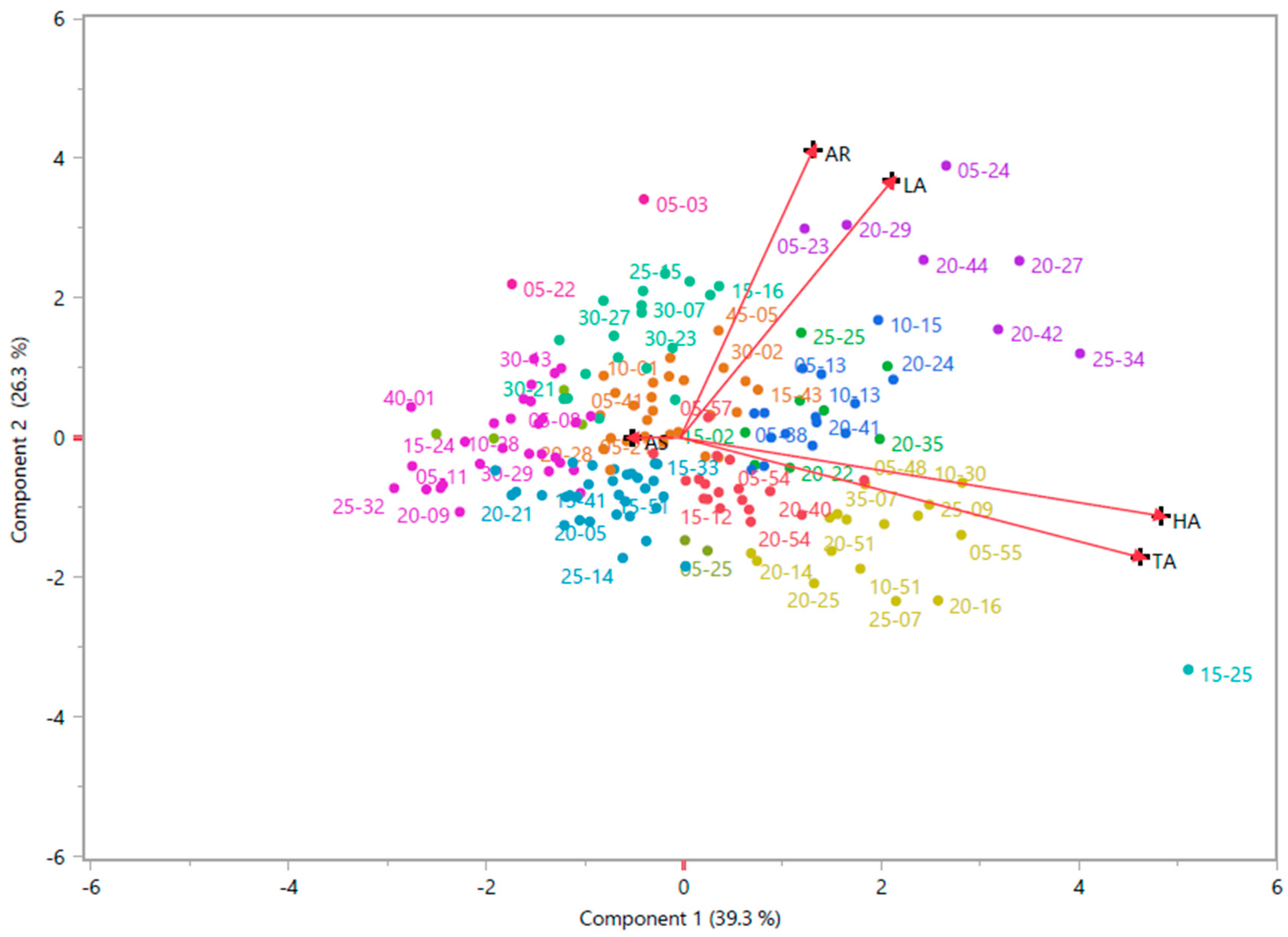
The cluster analysis of the morphological characteristics for the 178 shoots obtained at the fourth generation (Figure S1) led to the formation of the following 13 distinct groups (CG1-13), with the number of shoots indicated in parentheses: CG1 (18), CG2 (8), CG3 (15), CG4 (27), CG5 (18), CG6 (7), CG7 (16), CG8(1), CG9 (29), CG10 (4), CG11 (31 s), CG12 (2), and CG13 (2). Among the groupings, CG6, which included OUM05-23, OUM05-24, OUM20-27, OUM20-29, OUM20-42, OUM20-44, and OUM25-34, showed significant increases in length (82%), average root (fivefold), and transverse axis increases (24%) compared to the control (18.7 ± 1.4 mm; 0.3 ± 0.05; and 0.3 ± 0.05, respectively). These results were similar to CG3, which comprised OUM05-09, OUM05-13, OUM05-38, OUM10-13, OUM10-15, OUM10-17, OUM10-25, OUM15-09, OUM15-11, OUM15-23, OUM20-08, OUM20-24, OUM20-41, OUM20-53, and OUM35-08. CG3 exhibited increases in the longitudinal (60%) and transverse axis (15%), with a decrease in the number of roots (30%) and no shoot formation. CG12 (Figure 3), which comprised shoots OUM05-03 and OUM05-22, showed a 45% increase in the longitudinal axis and a two to fourfold increase in the number of shoots and roots. CG13 (OUM05-25 and OUM05-30) displayed the highest average number of outbreaks (five times higher).
PCoA analysis based on morphological traits (Figure 3) revealed a cumulative variance of 65.6%. The first axis (39.3%) showed a high correlation with the horizontal and transverse axes (r2 = 0.67, r2 = 0.64, respectively). The second axis (26.3%) was correlated with the longitudinal and average root axes. Shoots located in the positive quadrant of the first axis displayed greater lateral development, whereas those in the negative quadrant exhibited thinner and narrower structures. In contrast, shoots in the second positive quadrant contained cladodes with the greatest length and number of roots. The morphological changes observed, particularly those linked to increased proliferation or alterations in cladode shape and size, are considered indicative traits of enhanced agronomic potential in nopal cactus. Nevertheless, these phenotypes must undergo field evaluations to confirm their stability and productive value under agronomic conditions.
Currently, multivariate methods such as cluster analysis and PCoA are widely used for morphological, phytochemical, and bioactive analysis in species of the genus Opuntia [44,45,46,47]. These approaches enable the exploration of underlying information in robust datasets, enable the identification of patterns that enhance the understanding of associations or discrepancies between variables, and promote the effective grouping of analyzed materials [48,49]. In this study, the combination of multivariate methods proved useful for classifying accessions with phenotypic variations that contrast with the control, as also reported in other crops subjected to mutagenesis such as banana (Musa sp.) [48], ginger (Zingiber officinale) [23], and wheat (Triticum aestivum) [49,50]. As a result of the above analysis, the selection of 27 mutant shoots (OUM05-01, OUM05-03, OUM05-06, OUM05-09, OUM05-13, OUM05-22, OUM05-23, OUM05-24, OUM05-25, OUM05-30, OUM05-38, OUM10-13, OUM10-15, OUM15-09, OUM20-24, OUM20-27, OUM20-29, OUM20-41, OUM20-42, OUM20-44, OUM25-34, and OUM35-08), including five different materials that exhibited red pigmentation in roots (OUM05-70, OUM15-17, OUM20-35, OUM20-45, and OUM30-09), was conducted.

3.4. Analysis of the Mutants by ISSR Molecular Markers

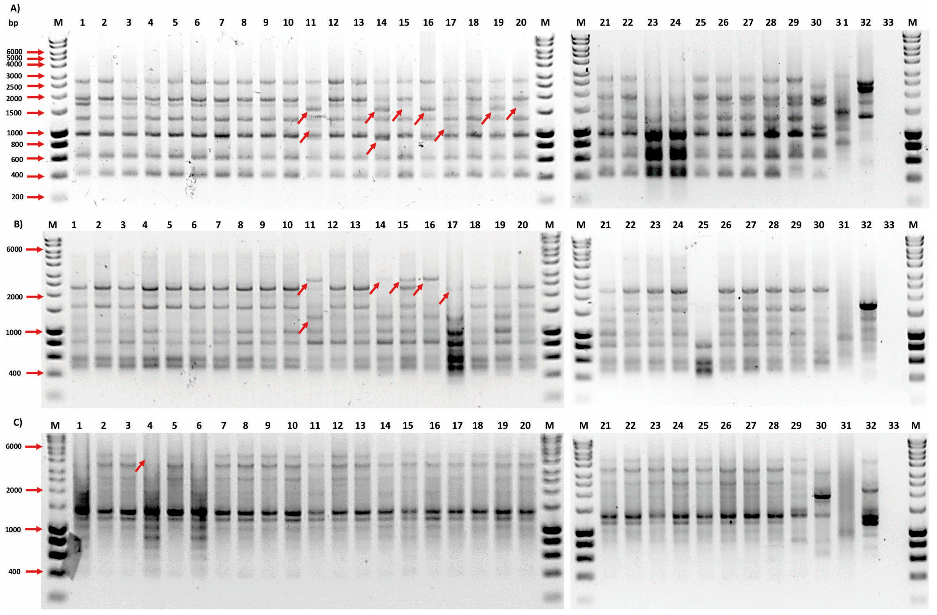
After irradiating the meristematic zone of the O. velutina areola, clonal lines were obtained and sub-cultured through a process of in vitro micropropagation, preserving their morphological characteristics. Twenty-eight clones were selected for the genetic diversity study using the ISSR molecular marker due to their contrasting morphological traits, including the red root pigmentation. ISSR markers have been successfully applied to assess the percentage of polymorphism and genetic variability since they produce multiple bands at the same locus, are highly reproducible, and do not require prior information from the plant genome, including studies of gamma-irradiated cultures. In this work, 12 primers were evaluated, of which six showed reproducible and informative amplified products. The band profiles obtained from the six ISSR primers were used to study the different mutant lines of O. velutina. Two related species and two genetically distant species generated a total of 57 bands, ranging from 13 to 5 bands per primer, with an average of 10 bands, within the range from 250 to 5000 bp (Figure 4).
The percentage of polymorphisms detected by ISSR in this study was 54.5%, which is a midpoint in relation to those reported in other studies of species within the same genus, ranging from 28.6% to 78.7% [19,51]. Additional research on tomato (Lycopersicon esculentum Mill) and banana (Musa paradisiaca cv. Sapientum) has confirmed an increase in genetic variability of 40% and 47%, respectively, following gamma ray exposure [22,52], similar to findings in the present investigation. The polymorphic information content (PIC) ranged from 0.01 to 0.48, with primers (GA)8YC and (GA)8YT yielding the highest number of polymorphic bands at 11, showing polymorphism percentages of 84.6% and 78.5%, respectively (Table 1). This aligns with the highest values for MI and PIC, which are considered strong indicators of highly informative primers and the most efficient in the current mutagenesis study [53]. The OUM00-00 control amplified a total of 47 bands. The 27 irradiated materials showed between 40 and 49 bands. Line OUM10-13 displayed the highest number of polymorphic bands at 12 compared to the control, while materials OUM05-22, OUM05-23, OUM05-25, OUM20-41, and OUM25-34 showed no polymorphism. Although these types of differences in the pattern of amplified bands are usually associated with the genetic diversity of the materials analyzed, in this study, given that we worked with a local collection such as O. velutina mutants, for which there are no reference data on existing variability, the preliminary test and subsequent selection of the primers used in the assay are key steps for achieving high levels of polymorphisms.
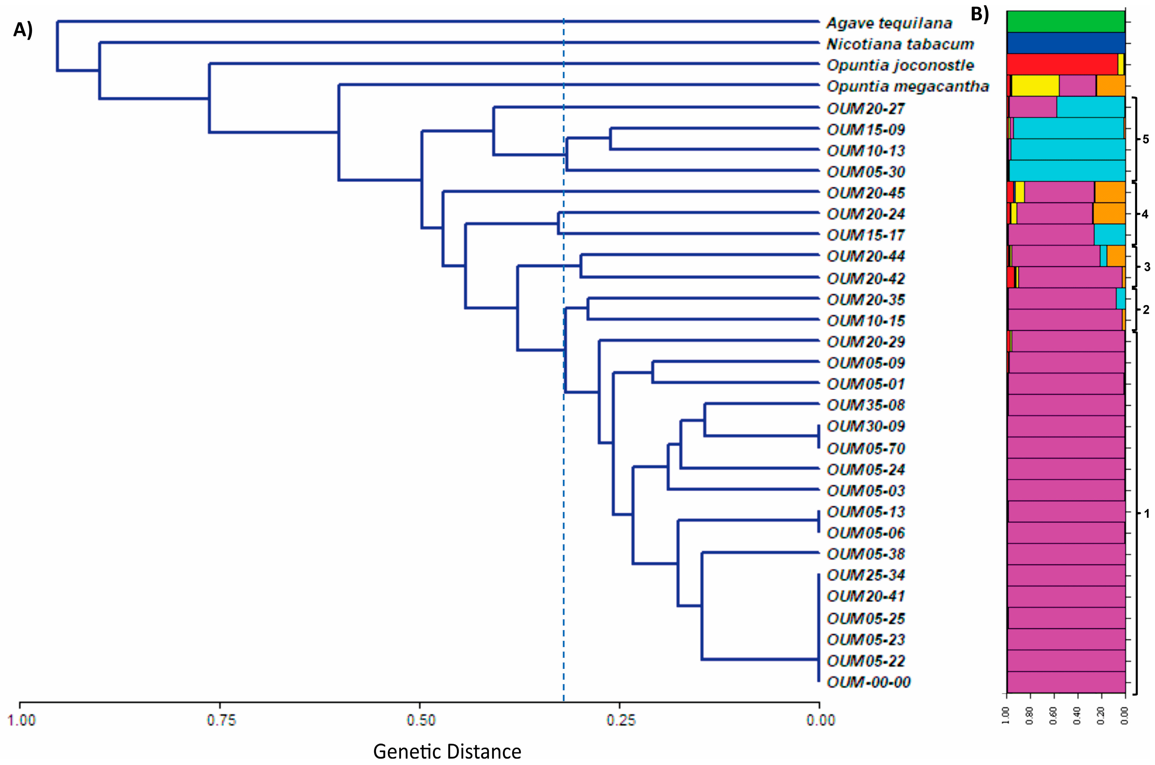
In the UPGMA dendrogram obtained for the ISSR markers using Jaccard’s coefficient, the external controls and mutant materials ranged from 0.60 to 0.96 (Figure 5A), while the distance variation between mutant lines and the non-irradiated control was from 0 to 0.49. Concurrently, a genetic diversity analysis was conducted with the STRUCTURE software to examine differences in the distribution of genetic variants among populations using a Bayesian iterative algorithm, employing the ISSR genetic data to group samples with similar patterns of variation [54]. This analysis identified the genetic mutant groups of O. velutina or subpopulations with a K = 6, which coincides with the genetic variability obtained in the UPGMA dendrogram across the different populations. Figure 5B provides a visual representation of the aforementioned subgroups and effectively illustrates the distribution of mutated populations within these subgroups. This visual breakdown improves the understanding of genetic relationships and differences between populations, providing valuable insights for future genetic research.
The results indicate that ISSR markers successfully detected genetic variations between 27 mutant clones and the different gamma irradiation doses evaluated. In the dendrogram based on the UPGMA model, with the cut-off line set at 0.3 (Figure 5A), the first group corresponds to the O. velutina control (OUM00-00) and five clones irradiated with doses ranging from 5 to 25 Gy. These clones appeared to preserve their microsatellite regions but were related to another group of irradiated plants, 65% of which corresponded to the 5 Gy dose. Generally, there was a trend of increasing genetic variability with higher doses of gamma radiation, as observed in groups 1 to 5, with group 5 being the most diverse compared to the control, which also exhibited a more distinct genetic structure (Figure 5B). This group consisted of mutant plants subjected to radiation doses of 5, 10, 15, and 20 Gy, corresponding to the tallest shoots, which measured 31 to 34 mm in height, compared to the control at 18 mm. Notably, O. velutina is related to the wild white cactus pear (O. megacantha) and xoconostle (O. joconostle), while the most genetically distant species are tobacco and agave, as expected.
In the case of the genus Opuntia, micropropagation through axillary bud break is widely used to ensure genetic homogeneity and stability while producing true plant clones. The activation of apical dominance is triggered by the action of growth regulators, such as cytokinins, which promote the sprouting and multiplication of areoles (axillary buds) and drive shoot multiplication. This propagation method is the most accurate, as it involves the initiation and development of new shoots from pre-existing meristems rather than the dedifferentiation of already differentiated cells [32,33]. Thus, it can be stated that the methodology used in the in vitro regeneration of O. velutina does not serve as a source of genetic variability that would affect the results of the ISSR analysis of the mutant lines.

4. Conclusions

For the first time, it has been reported that the use of gamma rays in nopal cactus O. velutina meristems in vitro promoted morphological changes that, when evaluated and subjected to multivariate analysis, allowed the comparison of plant materials with outstanding characteristics. The number of bands obtained by ISSR markers showed an increase in genetic variability and enabled the identification of 27 morphologically improved mutant lines of nopal cactus (O. velutina). Gamma irradiation induced significant genetic variability in O. velutina, as evidenced by the morphological and genetic variations observed in the mutant materials. The ISSR analysis revealed genetic diversity among the materials, suggesting that irradiation is an effective tool for generating variability and selecting genotypes with enhanced agronomic traits for plant breeding programs. In the future, a metabolomic analysis of the selected mutants will be conducted to evaluate their phytochemical profiles and to elucidate more precisely the modified metabolic pathways, aiming to identify elite material with relevant functional aspects.

Supplementary Materials

The following supporting information can be downloaded at: https://www.mdpi.com/article/10.3390/horticulturae11070743/s1. Table S1. Morphological measurements of 178 clonal lines O. velutina in the V4 generation post-irradiated; Figure S1. Cluster analysis of morphological characteristics in 178 O. velutina shoots following gamma irradiation.

Author Contributions

Conceptualization, J.F.G.-L. and P.A.G.-S.; formal analysis, J.F.G.-L., P.A.G.-S. and E.R.-O.; investigation, J.F.G.-L., E.D.l.C.-T. and P.A.G.-S.; methodology, J.F.G.-L., E.D.l.C.-T. and E.R.-O.; project administration, J.F.G.-L.; resources, J.F.G.-L. and P.A.G.-S.; supervision, E.D.l.C.-T., R.E.P.-S., H.E.M.-F., L.P., P.A.G.-S. and J.F.G.-L.; validation, J.F.G.-L. and P.A.G.-S.; writing—original draft, J.F.G.-L.; writing—review and editing, J.F.G.-L., P.A.G.-S. and E.R.-O. All authors have read and agreed to the published version of the manuscript.

Funding

The present research work was funded through support provided by the Tecnologico Nacional de Mexico (TecNM) and the Instituto Tecnologico de Tlajomulco. We are grateful for the support of the coordination of scientific research of the UMSNH.

Data Availability Statement

The data obtained that support the presented results of this research can be found in the Brotology Laboratory of the Faculty of Agrobiology of the Michoacan University of San Nicolas de Hidalgo, or in the Molecular Biology Laboratory of the National Institute of Technology of Mexico-Technological Institute of Tlajomulco.

Acknowledgments

We thank Patricia Hernandez Ledesma, curator of the Instituto de Ecología A. C., for her support in the identification of the prickly pear cactus as the species Opuntia velutina.

Conflicts of Interest

The authors declare no conflicts of interest.

References

  1. Korotkova, N.; Aquino, D.; Arias, S.; Eggli, U.; Franck, A.; Gómez-Hinostrosa, C.; Guerrero, P.; Hernández, H.; Kohlbecker, A.; Köhler, M.; et al. Cactaceae at Caryophyllales.org-A dynamic online species-level taxonomic backbone for the family. Willdenowia 2021, 51, 251–270. [Google Scholar] [CrossRef]
  2. Reyes-Agüero, J.A.; Aguirre Rivera, J.R.; Flores, J.L. Variación morfológica de Opuntia (Cactaceae) en relación con su domesticación en la altiplanicie meridional de México. Interciencia 2005, 30, 476–484. [Google Scholar]
  3. Villaseñor, J.L. Checklist of the native vascular plants of Mexico. Rev. Mex. Biodivrs. 2016, 87, 559–902. [Google Scholar] [CrossRef]
  4. Majure, L.C.; Puente, R.; Griffith, M.P.; Judd, W.S.; Soltis, P.S.; Soltis, D.E. Phylogeny of Opuntia s.s. (Cactaceae): Clade delineations, geographic origins, and reticulate evolution. Am. J. Bot. 2012, 99, 847–864. [Google Scholar] [CrossRef]
  5. Pedraza-Reyes, K.A.; Pérez Olvera, M.A.; Navarro-Garza, H.; Espejel García, A. El sistema familiar de producción de nopal verdura (Opuntia ficus indica L.) en San Pablo Ixquitlán, Méx. Importancia y estrategias de funcionamiento. Rev. Geogr. Agr. 2024, 72, 1–15. [Google Scholar] [CrossRef]
  6. El-Mostafa, K.; El Kharrassi, Y.; Badreddine, A.; Andreoletti, P.; Vamecq, J.; El Kebbaj, M.H.S.; Cherkaoui-Malki, M. Nopal cactus (Opuntia ficus-indica) as a source of bioactive compounds for nutrition, health and disease. Molecules 2014, 19, 14879–14901. [Google Scholar] [CrossRef]
  7. Tilahun, Y.; Welegerima, G. Pharmacological potential of cactus pear (Opuntia ficus indica): A review. J. Pharm. Phytochem. 2018, 7, 1360–1363. [Google Scholar]
  8. Das, G.; Lim, K.J.; Tantengco, O.A.G.; Carag, H.M.; Gonçalves, S.; Romano, A.; Das, S.K.; Coy Barrera, E.; Shin, H.S.; Gutierrez Grijalva, E.P.; et al. Cactus: Chemical, nutraceutical composition and potential bio-pharmacological properties. Phytother. Res. 2021, 35, 1248–1283. [Google Scholar] [CrossRef]
  9. Astello-García, M.; Santos-Díaz, M.; Reyes-Agüero, A.; Barba de la Rosa, A. Opuntia spp. as a Source of Bioactive Compounds. In Hispanic Foods: Chemistry and Bioactive Compounds; American Chemical Society: Washington, DC, USA, 2012; pp. 101–111. [Google Scholar] [CrossRef]
  10. Silva, M.A.; Albuquerque, T.G.; Pereira, P.; Ramalho, R.; Vicente, F.; Oliveira, M.B.P.; Costa, H.S. Opuntia ficus-indica (L.) Mill.: A multi-benefit potential to be exploited. Molecules 2021, 26, 951. [Google Scholar] [CrossRef]
  11. Mazri, M. Cactus pear (Opuntia spp.) breeding. In Advances in Plant Breeding Strategies: Fruits; Springer: Cham, Switzerland, 2018; pp. 307–341. [Google Scholar] [CrossRef]
  12. Omar, A.; ElSayed, A.; Mohamed, A. Genetic diversity and ecotypes of Opuntia spp. In Opuntia spp.: Chemistry, Bioactivity and Industrial Applications; Springer: Cham, Switzerland, 2021; pp. 181–199. [Google Scholar] [CrossRef]
  13. International Atomic Energy Agency (IAEA). Mutant Varieties Database. Available online: https://nucleus.iaea.org/sites/mvd/SitePages/Search.aspx (accessed on 16 October 2023).
  14. Abdelnour-Esquivel, A.; Perez, J.; Rojas, M.; Vargas, W.; Gatica-Arias, A. Use of gamma radiation to induce mutations in rice (Oryza sativa L.) and the selection of lines with tolerance to salinity and drought. In Vitro Cell. Dev. Biol. Plant. 2020, 56, 88–97. [Google Scholar] [CrossRef]
  15. Pérez-Jiménez, M.; Tallón, C.I.; Pérez-Tornero, O. Inducing mutations in Citrus spp. Sensitivity of different sources of plant material to gamma radiation. App. Radiat. Isot. 2020, 157, 109030. [Google Scholar] [CrossRef] [PubMed]
  16. Royani, J.I.; Abdullah, L.; Aisyah, S.I. Radio-sensitivity of irradiated seed, plantlets, callus, and in vitro leaves from indigofera zollingeriana Miq by gamma rays. IOP Conf. Ser. Earth Environ. Sci. 2021, 913, 012061. [Google Scholar] [CrossRef]
  17. Huerta-Olalde, A.M.; Hernández-García, A.; López-Gómez, R.; Fernández-Pavía, S.P.; Zavala-Páramo, M.G.; Salgado-Garciglia, R. In vitro selection of blackberry (Rubus fruticosus ‘Tupy’) plants resistant to botrytis cinerea using gamma ray-irradiated shoot tips. Plant Biotechnol. 2022, 22, 0312. [Google Scholar] [CrossRef]
  18. Nadeem, M.A.; Nawaz, M.A.; Shahid, M.Q.; Doğan, Y.; Comertpay, G.; Yıldız, M.; Hatipoğlu, R.; Ahmad, F.; Alsaleh, A.; Labhane, N.; et al. DNA molecular markers in plant breeding: Current status and recent advancements in genomic selection and genome editing. Biotechnol. Equip. 2018, 32, 261–285. [Google Scholar] [CrossRef]
  19. Bezerra, J.D.C.; de Andrade, A.P.; do Rêgo, M.M.; da Silva, D.S.; do Nascimento Júnior, J.R.S.; Araújo, F.; Valença, R.; do Rêgo, E.R.; Pessoa, A.M.D.; Bruno, R.D.L.A.; et al. Genetic diversity and relationships among Nopalea sp. and Opuntia spp. accessions revealed by RAPD, ISSR and ITS molecular markers. Mol. Biol. Rep. 2022, 49, 6207–6213. [Google Scholar] [CrossRef]
  20. Bhat, R.S.; Brijesh Patil, M.P.; Tilak, I.S.; Shirasawa, K. Molecular Markers for Mutant Characterization. In Mutation Breeding for Sustainable Food Production and Climate Resilience; Penna, S., Jain, S.M., Eds.; Springer: Singapore, 2023. [Google Scholar] [CrossRef]
  21. Le, K.C.; Ho, T.T.; Paek, K.Y.; Park, S.Y. Low dose gamma radiation increases the biomass and ginsenoside content of callus and adventitious root cultures of wild ginseng (Panax ginseng Mayer). Ind. Crops Prod. 2019, 130, 6–24. [Google Scholar] [CrossRef]
  22. El-Fiki, A.; Fahmy, E.; Doma, A.A.; Helmy, O.; Adly, M.; El-Metabteb, G. The genetic variation assessment of in vitro irradiated tomato (Lycopersicon esculentum Mill) by SCoT and ISSR markers. J. Microbiol. Biotechnol. Food Sci. 2021, 10, 557–565. [Google Scholar] [CrossRef]
  23. Sharma, V.; Thakur, M. Gamma irradiations induced morphological and biochemical variations in in vitro regenerated ginger (Zingiber officinale Rosc.)-an invaluable medicinal spice. Int. J. Radiat Biol. 2021, 97, 1696–1704. [Google Scholar] [CrossRef]
  24. Riviello-Flores, M.D.L.L.; Cadena-Iñiguez, J.; Ruiz-Posadas, L.D.M.; Arévalo-Galarza, M.D.L.; Castillo-Juárez, I.; Soto Hernández, M.; Castillo-Martínez, C.R. Use of gamma radiation for the genetic improvement of underutilized plant varieties. Plants. 2022, 11, 1161. [Google Scholar] [CrossRef]
  25. Abhang, S.; Sowjanya, P.R.; Singh, N.P.; Kadam, A.R.; Shinde, A.S.; Sangnure, V.R.; Mandave, A. Efficiency of RAPD, ISSR and SSR markers in assessing clonal fidelity of in vitro propagated punica granatum plantlets of cultivar Bhagwa. S. Afr. J. Bot. 2025, 179, 216–223. [Google Scholar] [CrossRef]
  26. Murashige, T.; Skoog, F. A Revised medium for rapid growth and bio assays with tobacco tissue cultures. Plant Physiol. 1962, 15, 473–497. [Google Scholar] [CrossRef]
  27. Doyle, J.J.; Doyle, J.L. Isolation of plant DNA from fresh tissue. Focus 1990, 12, 13–15. [Google Scholar]
  28. Serrote, C.M.L.; Reiniger, L.R.S.; Silva, K.B.; Rabaiolli, S.M.D.S.; Stefanel, C.M. Determining the Polymorphism Information Content of a molecular marker. Gene 2020, 726, 144175. [Google Scholar] [CrossRef] [PubMed]
  29. Rohlf, F.J. NTSYS-pc: Numerical Taxonomy and Multivariate Analysis System; Applied Biostatistics, Inc.: New York, NY, USA, 1998. [Google Scholar]
  30. Pritchardm, J.K.; Stephens, M.; Donelly, P. Inference of population structure using multilocus genotype data. Genetics 2000, 155, 945–959. [Google Scholar] [CrossRef]
  31. Falush, D.; Stephens, M.; Princhard, J. Inference of population structure using multilocus genotype data: Linked loci and correlated allele frequencies. Genetics 2003, 164, 1567–1587. [Google Scholar] [CrossRef]
  32. Evanno, G.; Regnaut, S.; Gourdet, J. Detecting the numer of clusters of individuals using the software structure: A simulation study. Mol. Ecol. 2005, 14, 2611–2620. [Google Scholar] [CrossRef]
  33. Bouzroud, S.; El_Maaiden, E.; Sobeh, M.; Devkota, K.P.; Boukcim, H.; Kouisni, L.; El-Kharrassi, Y. Micropropagation of Opuntia and other cacti species through axillary shoot proliferation: A Comprehensive Review. Front. Plant Sci. 2022, 13, 926653. [Google Scholar] [CrossRef]
  34. Ángeles-Espino, A.; Valencia-Botín, A.J.; Virgen-Calleros, G.; Ramírez-Serrano, C.; Paredes-Gutiérrez, L.; Hurtado-De la Peña, S. Determinación de la dosis letal (DL50) con Co60 en vitroplántulas de Agave tequilana var. Azul. Rev. Fitotec. Mex. 2013, 36, 381–386. [Google Scholar] [CrossRef]
  35. Spencer, L.M.M.; Forster, B.P.; Jankuloski, L. Manual de Mejoramiento Por Mutación, 3rd ed.; Food and Agriculture Organization of the United National (FAO): Roma, Italy, 2021. [Google Scholar] [CrossRef]
  36. Rubio-Ochoa, E.; De la Cruz-Torres, E.; Olalde-Portugal, V.; Pérez-Sánchez, R.E.; Gómez-Leyva, J.F.; García-Saucedo, P.A. Mutagénesis por radiación gamma para mejora genética de plantas de importancia alimentaria. Rev. Mex. Cienc. Agríc. 2024, 15, e-3747. [Google Scholar] [CrossRef]
  37. Oladosu, Y.; Rafii, M.; Abdullah, N.; Hussin, G.; Ramli, A.; Rahim, H.; Usman, M. Principle and application of plant mutagenesis in crop improvement: A review. Biotechnol. Biotechnol. Equip. 2015, 30, 1–16. [Google Scholar] [CrossRef]
  38. Penna, S.; Bhagwat, S.G. Mutagenesis and Selection: Reflections on the In Vivo and In Vitro Approaches for Mutant Development; Springer Nature: Singapore, 2023; pp. 99–127. [Google Scholar]
  39. Oliveira, N.M.; de Medeiros, A.D.; de Lima Nogueira, M.; Arthur, V.; de Araújo Mastrangelo, T.; da Silva, C.B. Hormetic effects of low-dose gamma rays in soybean seeds and seedlings: A detection technique using optical sensors. Comput. Electron. Agric. 2021, 187, 106251. [Google Scholar] [CrossRef]
  40. Saloua, K.S. Opuntia ficus indica cladode extract inhibit DNA double-strand breaks and locally multiply damaged sites induced by gamma radiation. J. Genet. Eng. Biotechnol. 2024, 22, 100425. [Google Scholar] [CrossRef]
  41. Hasim, A.A.; Shamsiah, A.; Hussein, S. Induced mutations using gamma ray and multiplication of plantlet through micro cross section culture of banana (Musa acuminata cv. Berangan). IOP Conf. Ser. Earth Environ. Sci. 2021, 757, 012007. [Google Scholar] [CrossRef]
  42. Serrano-Fuentes, M.K.; Gómez-Merino, F.C.; Cruz-Izquierdo, S.; Spinoso-Castillo, J.L.; Bello-Bello, J.J. Gamma radiation (60Co) induces mutation during in vitro multiplication of vanilla (Vanilla planifolia Jacks. ex Andrews). Horticulturae 2022, 8, 503. [Google Scholar] [CrossRef]
  43. Volkova, P.Y.; Bondarenko, E.V.; Kazakova, E.A. Radiation hormesis in plants. Curr. Opin. Toxicol. 2022, 30, 100334. [Google Scholar] [CrossRef]
  44. Astello-García, M.G.; Cervantes, I.; Nair, V.; Santos-Díaz, M.S.; Reyes-Agüero, A.; Guéraud, F.; de La Rosa, A.P.B. Chemical composition and phenolic compounds profile of cladodes from Opuntia spp. cultivars with different domestication gradient. J. Food Compos. Anal. 2015, 43, 119–130. [Google Scholar] [CrossRef]
  45. Adli, B.; Boutekrabt, A.; Touati, M.; Bakria, T.; Touati, A.; Bezini, E. Phenotypic diversity of Opuntia ficus indica (L.) MILL. in the Algerian steppe. S. Afr. J. Bot. 2017, 109, 66–74. [Google Scholar] [CrossRef]
  46. Amani, E.; Marwa, L.; Hichem, B.S.; Amel, S.H.; Ghada, B. Morphological variability of prickly pear cultivars (Opuntia spp.) established in ex-situ collection in Tunisia. Sci. Hortic. 2019, 248, 163–175. [Google Scholar] [CrossRef]
  47. Dev, R.; Mangalassery, S.; Dayal, D.; Louhaichi, M.; Hassan, S. Genetic variability, characters association and principal component study for morphological and fodder quality of Opuntia and Nopalea sp. in India. Genet. Resour. Crop Evol. 2024, 71, 2297–2310. [Google Scholar] [CrossRef]
  48. Pestanana, R.K.N.; Amorim, E.P.; Ferreira, C.F.; de Oliveira Amorim, V.B.; Oliveira, L.S.; da Silva Ledo, C.A.; de Oliveira e Silva, S. Agronomic and molecular characterization of gamma ray induced banana (Musa sp.) mutants using a multivariate statistical algorithm. Euphytica 2011, 178, 151–158. [Google Scholar] [CrossRef]
  49. Mangi, N.; Baloch, A.W.; Khaskheli, N.K.; Ali, M.; Afzal, W. Multivariate analysis for evaluation of mutant bread wheat lines using metric traits. Integr. Plant Sci. 2021, 1, 29–34. [Google Scholar]
  50. Zulfiqar, S.; Ishfaq, S.; Ikram, M.; Nawaz, M.A.; Rahman, M.U. Characterization of gamma-rays-induced spring wheat mutants for morphological and quality traits through multivariate and GT Bi-plot analysis. Agronomy 2021, 11, 2288. [Google Scholar] [CrossRef]
  51. Valadez-Moctezuma, E.; Samah, S.; Luna-Paez, A. Genetic diversity of Opuntia spp. varieties assessed by classical marker tools (RAPD and ISSR). Plant Syst. Evol. 2015, 301, 737–747. [Google Scholar] [CrossRef]
  52. Due, M.S.; Susilowati, A.R.I.; Yunus, A. The effect of gamma rays irradiation on diversity of Musa paradisiaca var sapientum as revealed by ISSR molecular marker. Biodiversitas 2019, 20, 1416–1422. [Google Scholar] [CrossRef]
  53. Alekseeva, M.; Rusanova, M.; Rusanov, K.; Atanassov, I. Aset of highlypolymorphic microsatellite markers for genetic diversity studies in the genus. Origanum. Plants 2023, 12, 824. [Google Scholar] [CrossRef]
  54. Porras-Hurtado, L.; Ruiz, Y.; Santos, C.; Phillips, C.; Carracedo, Á.; Lareu, M.V. An overview of STRUCTURE: Applications, parameter settings, and supporting software. Front. Genet. 2013, 4, 98. [Google Scholar] [CrossRef]
Figure 1. Radiosensitivity for shoot formation from gamma-irradiated meristems of in vitro Opuntia velutina.
Horticulturae 11 00743 g001
Figure 2. Effect of gamma radiation on morphological and physiological variations of Opuntia velutina in vitro. Non-irradiated plant (OUM00-00): (A) longitudinal axis, (B) horizontal axis, (C) transverse axis, (D) shoots, and (E) roots. Irradiated material: (F) OUM10-15, (G) OUM20-27, (H) OUM25-34, (I) OUM05-30, (J) OUM05-70. The first number after the letters is the radiation used in Gy. The black bar in the images corresponds to a size of 1 cm.
Horticulturae 11 00743 g002
Figure 3. Bi-plot of the first two principal components (PC1 and PC2) based on examined morphological plant materials obtained by gamma irradiation in generation V4 of Opuntia velutina. Longitudinal axis (LA), horizontal axis (HA), transverse axis (TA), average roots (AR), average shoots (AS). Materials with similar colors belong to the same group.
Horticulturae 11 00743 g003
Figure 4. Amplification pattern of bands obtained with ISSR markers using primers: (A) (GA)8 YC, (B) (GA)8 YT, and (C) (CT)8 RG. (M) Leader 1 Kb. O. velutina plants 1, OUM00-00; 2, OUM05-01; 3, OUM05-03; 4, OUM05-06; 5, OUM05-09; 6, OUM05-13; 7, OUM05-22; 8, OUM05-23; 9, OUM05-24; 10, OUM05-25; 11, OUM05-30; 12, OUM05-38; 13, OUM05-70; 14, OUM10-13; 15, OUM10-15; 16, OUM15-09; 17, OUM15-17; 18, OUM20-24; 19, OUM20-27; 20, OUM20-29; 21, OUM20-35; 22, OUM20-41; 23, OUM20-42; 24, OUM20-44; 25, OUM20-45; 26, OUM25-34; 27, OUM30-09; 28, OUM35-08; 29, Wild white cactus; 30, O. joconostle; 31, Agave; 32, Tobacco; and 33, Negative control. The red arrow indicates the presence of polymorphic bands, with respect to the non-irradiated material banding profile.
Horticulturae 11 00743 g004
Figure 5. (A) UPGMA dendrogram of ISSR markers based on the Jaccard distance coefficient in mutants of Opuntia velutina. OUM00-00 corresponds to the non-irradiated material. (B) The Bayesian population structure analysis of the 27 populations irradiated and their controls utilizing ISSR markers. Color segments within the bars indicate the membership coefficients for each population.
Horticulturae 11 00743 g005
Table 1. Inter-simple sequence repeat (ISSR) molecular markers selected to evaluate variations of Opuntia velutina at different doses of gamma irradiation with 60Co.
Primer Sequence (5′-3′)AT * (°C)TBPBBP%MSR (bp)PICMI
(GA)8YC46.7131184.6400–26000.484.23
(GA)8YT47.4141178.5500–22500.474.07
T(CT)7CC48.110440.0400–50000.110.18
(CT)8RG48.19111.1300–40000.010.00
(GA)8YG48.86466.6800–35000.320.48
(GA)8C52.05240250–30000.080.14
Average-9.65.554.5-0.251.51
* AT: annealing temperature; TB: total bands; PB: polymorphic bands; BP%: polymorphism percent; MSR: molecular size range; PIC: polymorphic information content; MI: marker index.
Disclaimer/Publisher’s Note: The statements, opinions and data contained in all publications are solely those of the individual author(s) and contributor(s) and not of MDPI and/or the editor(s). MDPI and/or the editor(s) disclaim responsibility for any injury to people or property resulting from any ideas, methods, instructions or products referred to in the content.