Article

Effect of Phenological Variation on the Phytochemical Composition and Antioxidant Activity of Different Organs of Capparis spinosa L.

by
Saeid Hazrati
1,*,
Zahra Mousavi
2,
Saeed Mollaei
3,
Hossein Rabbi Angourani
4 and
Silvana Nicola
5
1
Department of Agronomy and Plant Breeding, Faculty of Agriculture, Azarbaijan Shahid Madani University, Tabriz 5375171379, Iran
2
Department of Horticultural Science, School of Agriculture, Shiraz University, Shiraz 7194684334, Iran
3
Phytochemical Laboratory, Department of Chemistry, Faculty of Sciences, Azarbaijan Shahid Madani University, Tabriz 5375171379, Iran
4
Research Institue of Modern Biological Techniques, University of Zanjan, Zanjan 3879145371, Iran
5
Department of Agricultural, Forest, and Food Sciences—DISAFA, Horticultural Sciences—INHORTOSANITAS, Vegetable Crops and Medicinal and Aromatic Plants—VEGMAP, University of Torino, 10095 Grugliasco, Italy
*
Author to whom correspondence should be addressed.
Horticulturae 2025, 11(6), 702; https://doi.org/10.3390/horticulturae11060702
Submission received: 11 May 2025 / Revised: 12 June 2025 / Accepted: 14 June 2025 / Published: 17 June 2025

Abstract

Capparis spinosa L. (caper) is an important medicinal plant whose bioactive compounds vary significantly depending on its growth stage. This directly affects its pharmaceutical and nutritional value. Collecting C. spinosa at the optimal growth stage is essential to achieving high phytochemical quality and meeting consumer needs. This study aimed to evaluate the variation of these active compounds in the aerial parts of C. spinosa across four phenological stages (vegetative, flowering, unripe fruit, and ripe fruit). The result showed that EO content was highest in unripe fruits (0.18%) and lowest in the flowering stage (0.07%) in leaves, while extract yield was highest in leaves of the ripe fruit stage (14.65%) followed by the flowering stage in flowers (12.66%). Flowering stage leaves showed the highest total phenol (56.20 mg GAE/g) and flavonoid (17.10 mg QE/g) content, while the lowest concentrations were found in the ripe fruit stage of the leaves. EO analysis showed that methyl isothiocyanate reached the highest concentration in flowers at the flowering stage (41.6%), while isopropyl isothiocyanate reached the highest concentration in leaves at the ripe fruit stage (36.2%). Isobutyl isothiocyanate was found exclusively in fruits, with the highest concentration in ripe fruits (9.2%). Dimethyltrisulphide showed a maximum concentration in leaves at the vegetative stage, decreasing by 76.6% as the plant developed towards the ripe fruit stage. The dominant phenolic acids varied between phenological stages: cinnamic acid at the vegetative stage; rosmarinic and cinnamic acids at the flowering stage in leaves; caffeic and cinnamic acids in flowers; vanillic, cinnamic, and rosmarinic acids at the unripe fruit stage in leaves and fruits; and rosmarinic, cinnamic, and vanillic acids in ripe fruits. The results indicate that harvesting C. spinosa at the vegetative stage and in the leaves of the flowering stage is optimal for maximum secondary metabolite yield, providing valuable guidance for growers targeting food and pharmaceutical applications.

1. Introduction

For generations, the majority of the world population has relied on medicinal plants to promote health and treat disease [1]. These plants are a rich source of secondary metabolites with a variety of uses in the production of pharmaceuticals, agrochemicals, food additives, biopesticides, fragrances, flavors, and natural dyes [2,3]. Capparis spinosa L., commonly known as the caper, is a well-known edible and medicinal plant from the Capparaceae family. It grows as a thorny perennial shrub, is typically found in rocky, mountainous, and saline environments, and is highly tolerant of heat and poor soil conditions [4,5]. It also has different names in different regions and countries, such as alafeMar (Iran), wild watermelon (China), câprier (France), capparo (Italy), and alcaparra (Spain) [6,7]. Originally from the Mediterranean basin, it can be found across Italy, North Africa, Greece, Central Asia, and Iran, both in the wild and cultivated [8,9,10].
Morphologically, C. spinosa is characterized by small, alternate, fleshy, and glossy leaves; white to pinkish flowers with four petals and long purple stamens; and green oval fruits that are rich in phenolics and flavonoids, which contribute to its antioxidant potential [11,12,13,14,15,16]. It propagates through seeds and cuttings, thrives in well-drained alkaline soils, and has deep roots that tolerate drought and help prevent soil erosion [17,18,19,20]. Traditionally, its unopened flower buds (capers), have been used as flavorful condiments in various cuisines, especially in the Mediterranean region. Capers are typically pickled or salted to enhance their tangy, pungent flavor, attributed to sulfur-containing compounds like isothiocyanates [15,19,21,22,23]. The fruits and leaves are also consumed due to their high phenolic and flavonoid content, which contributes to their functional properties [1,24].
In addition to its culinary uses, C. spinosa has been used in traditional medicine since ancient times to treat ailments such as gout, rheumatism, hypertension, diabetes, malaria, and digestive disorders [9]. Contemporary studies confirm its pharmacological activities, including antioxidant, anti-inflammatory, anti-arthritic activity, antispasmodic, antidiabetic, anticancer, hepatoprotective, and antimicrobial effects [11,25,26]. Furthermore, its extracts are utilized in the food packaging industry for their natural preservative properties [27].
The phenological stage and specific plant organs can significantly impact the content and composition of essential oils (EOs), polyphenol compounds, and antioxidant activity in medicinal plants. These bioactive compounds are generally found in varying concentrations throughout a plant’s growth cycle, often peaking at specific stages such as pre-flowering, full flowering, or fruiting. Similarly, different plant organs (e.g., leaves, flowers, fruits, and seeds) contain different amounts of these compounds due to variations in metabolic activity. Early developmental stages typically exhibit higher antioxidant activity and phenolic content, while the composition of EOs can vary according to growth stage and environmental factors. Therefore, the timing of harvest and the organs of the plant selected are critical for optimizing the phytochemical and functional properties of medicinal plants [28,29,30,31,32,33].
Recent comprehensive phytochemical investigations have revealed that C. spinosa contains a variety of bioactive compounds distributed across its different organs. These include several major classes of compound, such as polyphenols, alkaloids, glucosinolates, EOs, fatty acids, and carbohydrates [9,13,21,22,34]. Furthermore, this medicinal plant is a valuable source of nutrition, providing substantial amounts of vitamins (vitamin C), highly digestible proteins, natural sugars, and essential minerals [35]. Polyphenol components are a particularly significant group of bioactive constituents that have attracted considerable research attention due to their therapeutic potential [34,36,37,38,39].
The EO profile of C. spinosa has been extensively investigated, with research findings indicating that all four aerial parts—leaves, stems, flowers, and fruit buds—are suitable sources of EOs and aliphatic ester compounds. Comprehensive studies of the EO composition of C. spinosa have revealed that the most significant volatile compounds are methyl isothiocyanate, hexadecanoic acid, dimethyl tetrasulfide, and dimethyl trisulfide. These compounds have been identified as the dominant constituents in all plant tissues [37,40,41]. Several studies have demonstrated that C. spinosa exhibits remarkable antioxidant properties, establishing it as a promising natural source of these compounds [35,38,42,43,44].
C. spinosa has attracted considerable attention in the food, pharmaceutical, and healthcare industries due to its rich profile of bioactive compounds. However, the proper time of harvest to achieve the maximum beneficial compounds of C. spinosa remains unknown and needs more research work. While previous studies have investigated variations in phytochemical composition of one or more organs, they have not considered the different phenological stages [37,45,46]. Comprehensive evaluations across different plant organs and phenological stages have been limited. For the first time, this study systematically examines the content of EOs, phenolic compounds, and flavonoids, as well as the antioxidant activities, in various organs of C. spinosa throughout its phenological development. The results will offer critical insights into the optimal harvesting time and plant part selection to maximize the accumulation of these valuable compounds.

2. Materials and Methods

2.1. Plant Material and Sampling

This study was conducted in the spring and summer of 2021, in Tabriz, East Azerbaijan Province, Iran (37°56′27″ N and 47°32′12″ E; altitude: 1750 m). The aerial organs of C. spinosa were collected at different phenological stages from fourth-year plants. A voucher specimen was maintained at the Research Institute of Modern Biological Techniques (RIMBT), University of Zanjan. Ass. Prof. Dr. Angourani deposited the voucher specimens in the botanical herbarium under the following number 1729-H-ZNU. Sampling was carried out at different stages including leaf collection at pre-flowering (vegetative stage) (10 June) leaf and flower collection at the flowering stage (20 June); leaf and unripe fruit collection at the unripe fruit stage (10 July); leaf and ripe fruit collection at the ripe fruit stage (20 August) (Figure 1). All samples were taken randomly from 10 C. spinosa bushes. At each stage, 0.5 kg of material per organ type was harvested and divided into three replicates. For all samples, the different organs were separated from each other after collection and dried in the shade at 25 °C for three weeks.

2.2. Essential Oil Isolation

Dried plant organs (leaves, fruits, and flowers) were ground separately into fine particles using a low-speed mixer. For each organ, 100 g of the dried sample was subjected to the hydrodistillation method using an all-glass Clevenger apparatus according to the European Pharmacopoeia [47] for 3 h, with three replicate distillations per organ. The extracted EOs were dehydrated over anhydrous Na2 SO4 and the EO content was estimated for each organ. The EOs were stored in sealed vials at 4 °C until further chemical analysis.

2.3. Gas Chromatography (GC)

EOs were analyzed using a gas chromatography device (model: Agilent GC 7890, Agilent Technologies Inc., Santa Clara, CA, USA). The separation was carried out using a MS HP-5 (30 m × 0.25 mm, 0.25 µm) column. The oven temperature program was set from 50 °C (3 min) to 260 °C with a ramp-up of 3 °C/min, and then held for 5 min. The detector and injector temperatures were set at 270 °C and 240 °C, respectively. Nitrogen was used as the carrier gas at a rate of 1 mL/min.

2.4. GC-MS Chromatography

In addition to GC chromatography, a gas chromatography device connected to a mass spectrometer (Model: Agilent 7890 A G Chromatograph and Agilent 5975 c Mass, Agilent Technologies Inc., Santa Clara, CA, USA), called GC-MS chromatography, was used to analyze the EOs. In this experiment, GC-MS chromatography was equipped with an HP-5 column (30 m × 0.25 mm, 0.25 µm). According to the planned program, the oven temperature remained constant at 50 °C for 3 min and remained constant for 5 min after increasing the temperature to 260 °C with a ramp-up of 3 °C/min. Here again, nitrogen was utilized as the carrier gas at a rate of 1 mL/min. The analysis was performed with scan time (30 m/z), range of analysis (600 m/z), ionization 0/6 s, 70 electron volts, and a solvent evaporation rate of 2 min. The compounds were then identified by Kovats index (KI) and Wiley Library and Nist11.

2.5. Preparation of Extracts

Phenolic and flavonoid compounds were extracted from the plant material as follows: 0.2 g of each powdered organ (in triplicate) was soaked in 10 mL of 80% aqueous methanol (v/v) and placed in an ultrasonic bath (10 min, 30 °C, 5.5 kHz). After 10 min of extraction, the samples were transferred to a centrifuge (10 min, 5000 rpm, 4 °C). After centrifugation, the supernatant was collected in clean Eppendorf tubes. The resulting extracts were evaporated under a fume hood (Fater Electronic, model CH612, Iran) for 48 h. The dried extracts were weighed to determine the yield and stored at 4 °C until further analysis.

2.6. Determination of Total Phenolic Content

Total phenolic content was quantified by the Folin–Ciocalteu method [48]. Briefly, 200 μL of crude extract (10 mg/mL) was mixed with 0.5 mL of Folin–Ciocalteu reagent (Merck, Darmstadt, Germany) and vortexed for 3 min. Then 2 mL of 20% (w/v) sodium carbonate (Sigma-Aldrich, Schnelldorf, Germany) was added to the mixture. After incubation for 30 min at room temperature in the dark, the absorbance was measured at 750 nm using a UV-Vis spectrophotometer (Agilent BioTek Epoch Microplate Spectrophotometer, Agilent Technologies, Santa Clara, CA, USA). Total phenolic content was calculated using a gallic acid standard curve (Sigma-Aldrich, Schnelldorf, Germany) and expressed as mg of gallic acid equivalent (mg GAE) per g of plant dry weight extract.

2.7. Determination of Total Flavonoid Content

Total flavonoid content was quantified using the aluminum chloride colorimetric method [49]. A reaction mixture was prepared by combining 50 µL of extract (1 mg/mL) with 10 µL of 10% aluminum chloride solution (Sigma-Aldrich, Schnelldorf, Germany) and 10 µL of 1 M sodium acetate (Sigma-Aldrich, Schnelldorf, Germany). The mixture was incubated for 15 min at room temperature in the dark and the absorbance was measured at 415 nm using a UV-Vis spectrophotometer. The results were calculated from a quercetin standard curve and expressed as mg of quercetin equivalent (mg QE) per g of dry weight extract.

2.8. Analysis of Phenolic Compounds

Individual phenolic acids were analyzed by high-performance liquid chromatography (HPLC). Analysis was performed on a Waters 2695 system (Milford, CT, USA) equipped with a photodiode array (PDA) detector, pump, and autosampler with a loop capacity of 100 μL. Chromatographic separation was performed on a Eurospher C18 column (150 × 4.6 mm, 5.0 μm particle size) at 25 °C. The mobile phase consisted of methanol (solvent A) and water (solvent B) in a gradient elution mode at a flow rate of 0.5 mL/min. Compounds were monitored at wavelengths between 280 nm. The sample injection volume was set to 100 μL.
Phenolic acids were identified by comparison of retention times with authenticated standards (≥98% purity; Sigma-Aldrich, Schnelldorf, Germany) and confirmed by sample spiking. Quantification was performed using external calibration curves constructed with nine concentration levels of standards (from 5 to 200 ppm). Data acquisition and processing were performed using Millennium 32 software. Results were expressed as μg/g dry weight extract.

2.9. Antioxidant Activity (DPPH Radical Scavenging Assay)

The antioxidant activity of the extracts was measured using the DPPH (2,2′-diphenyl-1-picrylhydrazyl; Sigma-Aldrich, Schnelldorf, Germany) free radical scavenging method described by Akroum et al. [50]. When assessing the antioxidant activity of the EOs and extracts, a control sample was used to establish a baseline for comparison. A solvent blank containing only the diluent (methanol) and no plant EO or extract was used as a common control to ensure that any observed activity in the test samples was due solely to bioactive compounds. Additionally, a positive control such as ascorbic acid was incorporated to validate the accuracy of the assay by providing a known antioxidant standard. This enabled the relative efficacy of the extract in scavenging free radicals to be evaluated, typically quantified using IC50 values or inhibition percentage. Radical scavenging activity was gauged based on the following equation: Percentage of radical scavenging activity = (Abs control − Abs sample)/Abs control × 100.

2.10. Statistical Analysis

The experiment was conducted using a randomized complete block design with three replications. The data were analyzed using Statistical Analysis System (SAS) software 9.2 and compared using one-way analysis of variance (ANOVA). Data were recorded as mean ± standard error (SE). Significant differences between means were determined by Duncan’s multiple range test, and p values < 0.05 were regarded as significant. Correlation analysis was conducted by Pearson method based on phytochemical characteristics. Moreover, principal component analysis (PCA) was performed using XLSTAT software (version 2018.1) to assess patterns in the multivariate data set.

3. Results

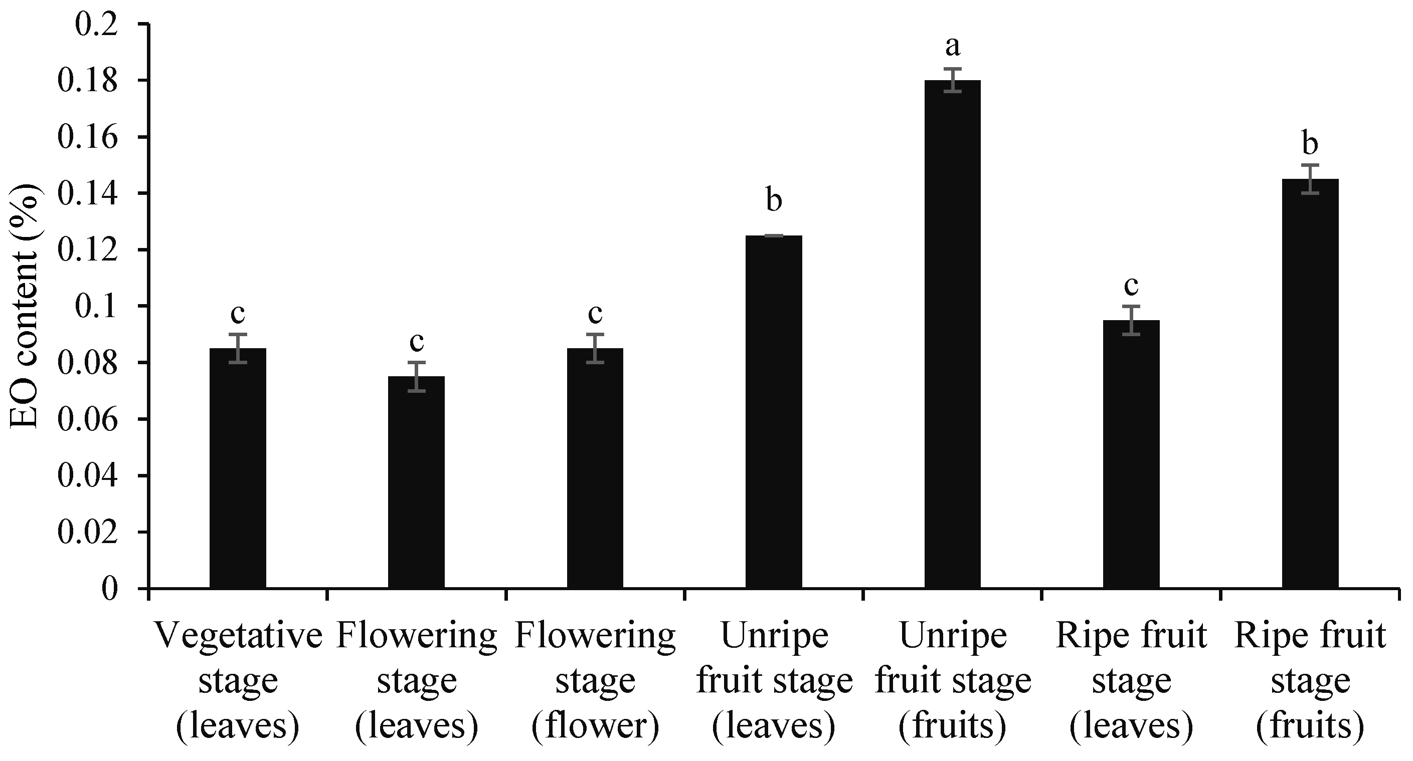
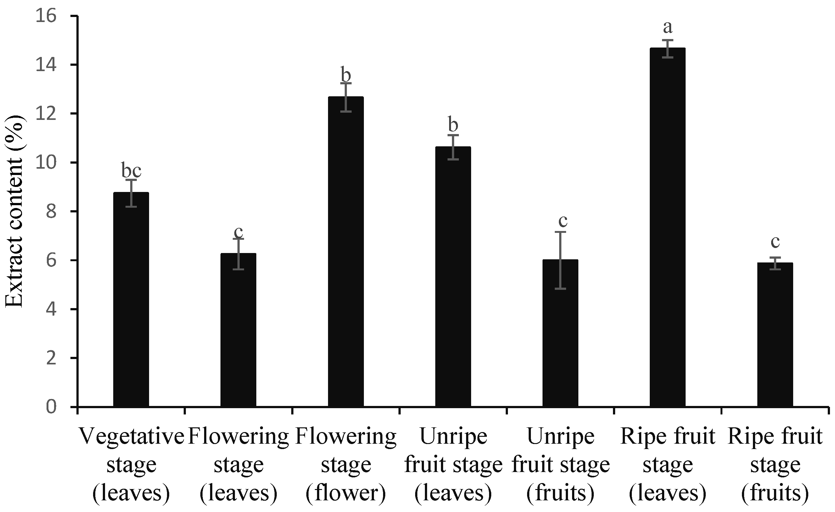
3.1. Contents of Essential Oil and Extract

The content of EOs in medicinal plants varies depending on the organ and phenological stage [33]. Given the widespread industrial applications of EOs, determining the optimal harvesting time is crucial to maximizing yield. In this study, we investigated the EO content of C. spinosa organs at different phenological stages. Our analysis revealed significant variations in EO content between different plant organs throughout the growth cycle (Figure 2). The results showed that EO accumulation varies significantly across different plant organs and phenological stages. The highest EO content observed in unripe fruits (0.18%) indicates that fruit maturation plays a key role in EO biosynthesis and accumulation. The decline in EO content as the fruit ripens could be due to metabolic changes, volatilization, or enzymatic degradation of specific EO components.
The lack of significant differences in EO content among leaves at different phenological stages (vegetative, flowering, and ripe fruits) and flowers at the flowering stage implies that EO biosynthesis in these organs remains relatively stable throughout plant development. However, the lowest EO percentage recorded in leaves during the flowering stage (0.07%) suggests a possible resource allocation shift, where the plant prioritizes EO production in flowers or fruits rather than leaves during this critical reproductive phase. Overall, the higher EO content in fruits compared to other plant organs emphasizes the importance of fruits as major sites of EO biosynthesis and storage (Figure 2).
Figure 3 shows the significant variation in extract content between different organs and phenological stages. The highest extract yield was observed in leaves during the ripe fruit stage (14.65%), followed by flowers during the flowering stage (12.66%). The extract content was significantly lower in fruits (both ripe and unripe) and leaves during the flowering stage, with no statistically significant differences observed between these samples.

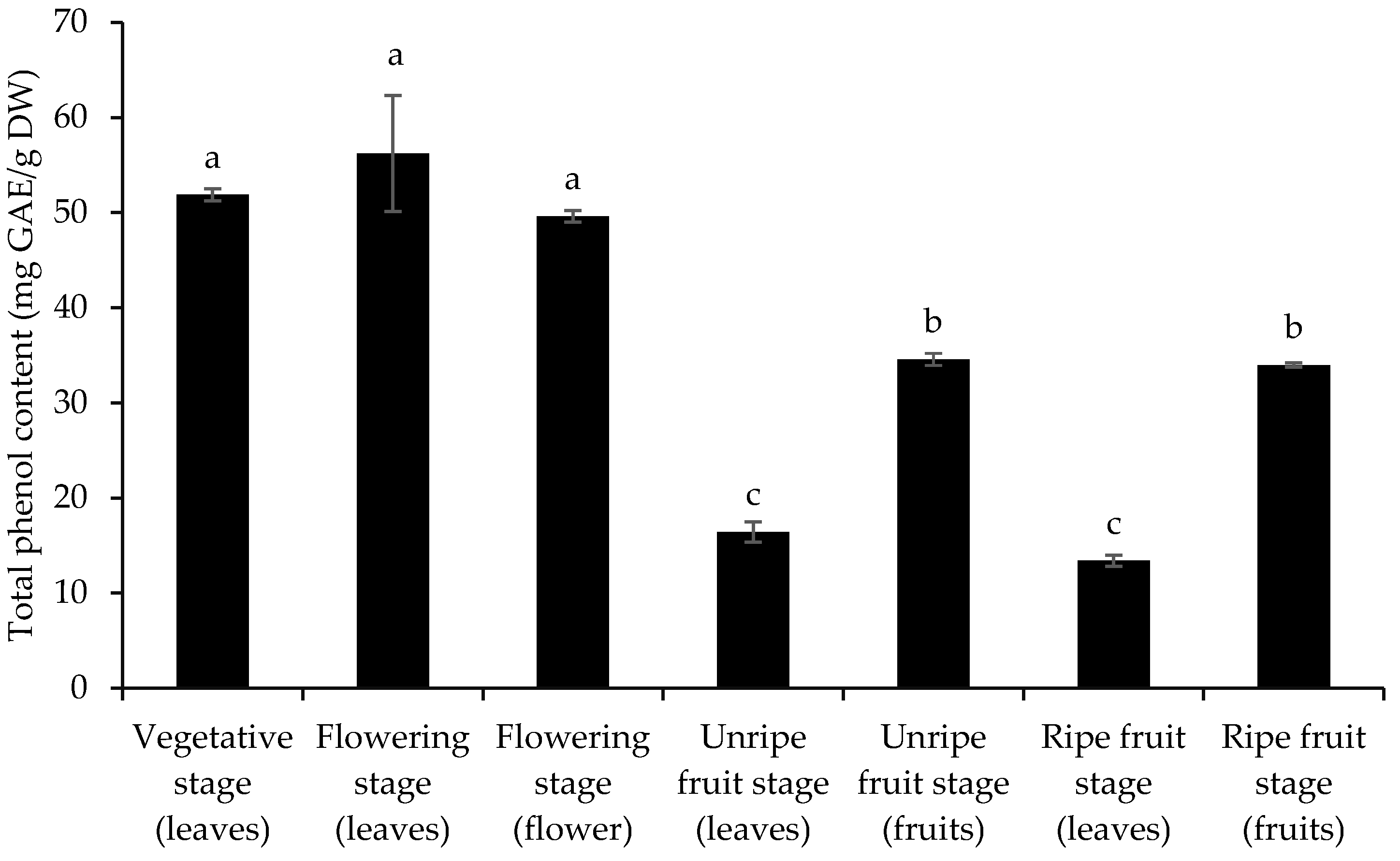
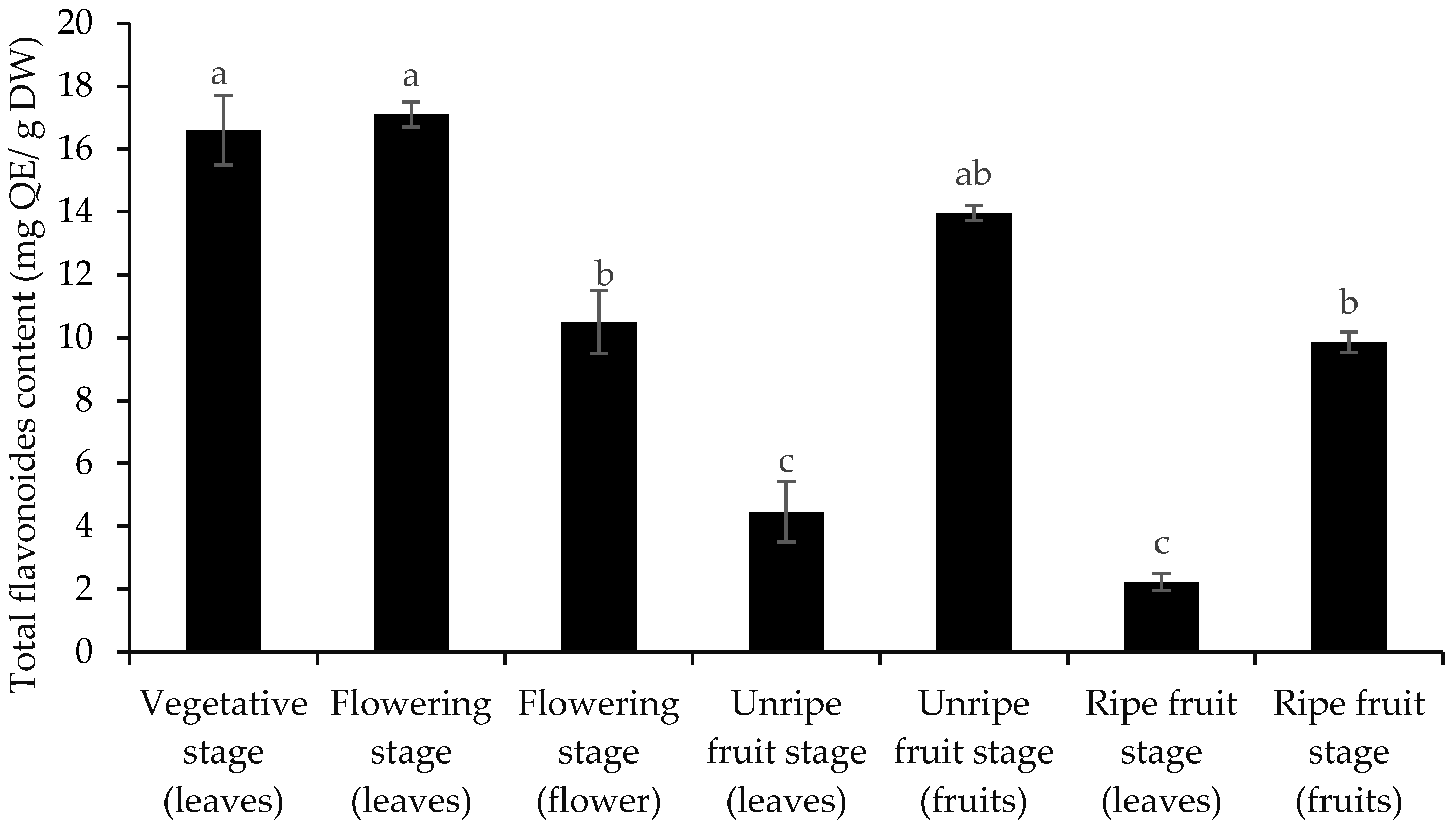
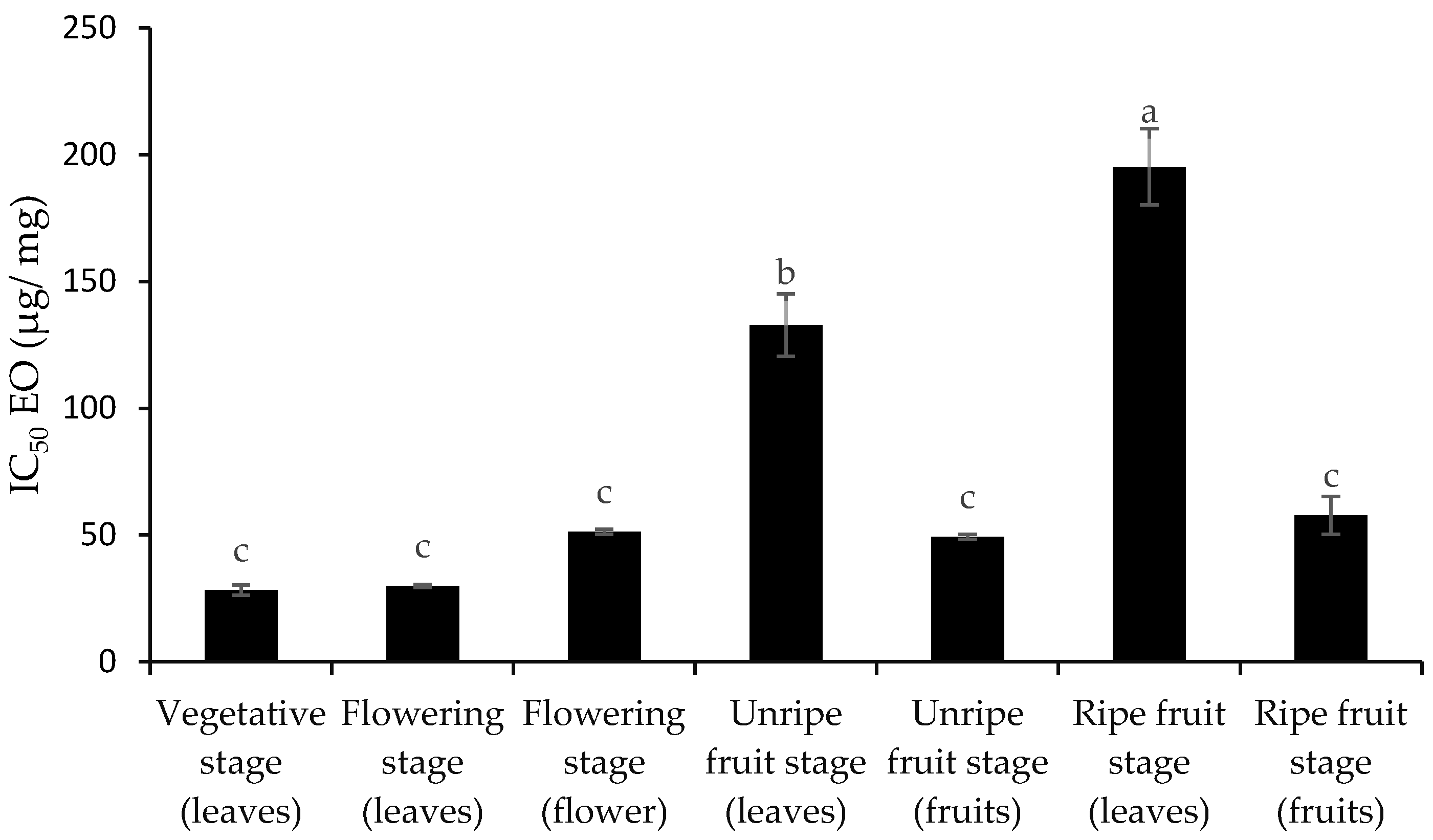
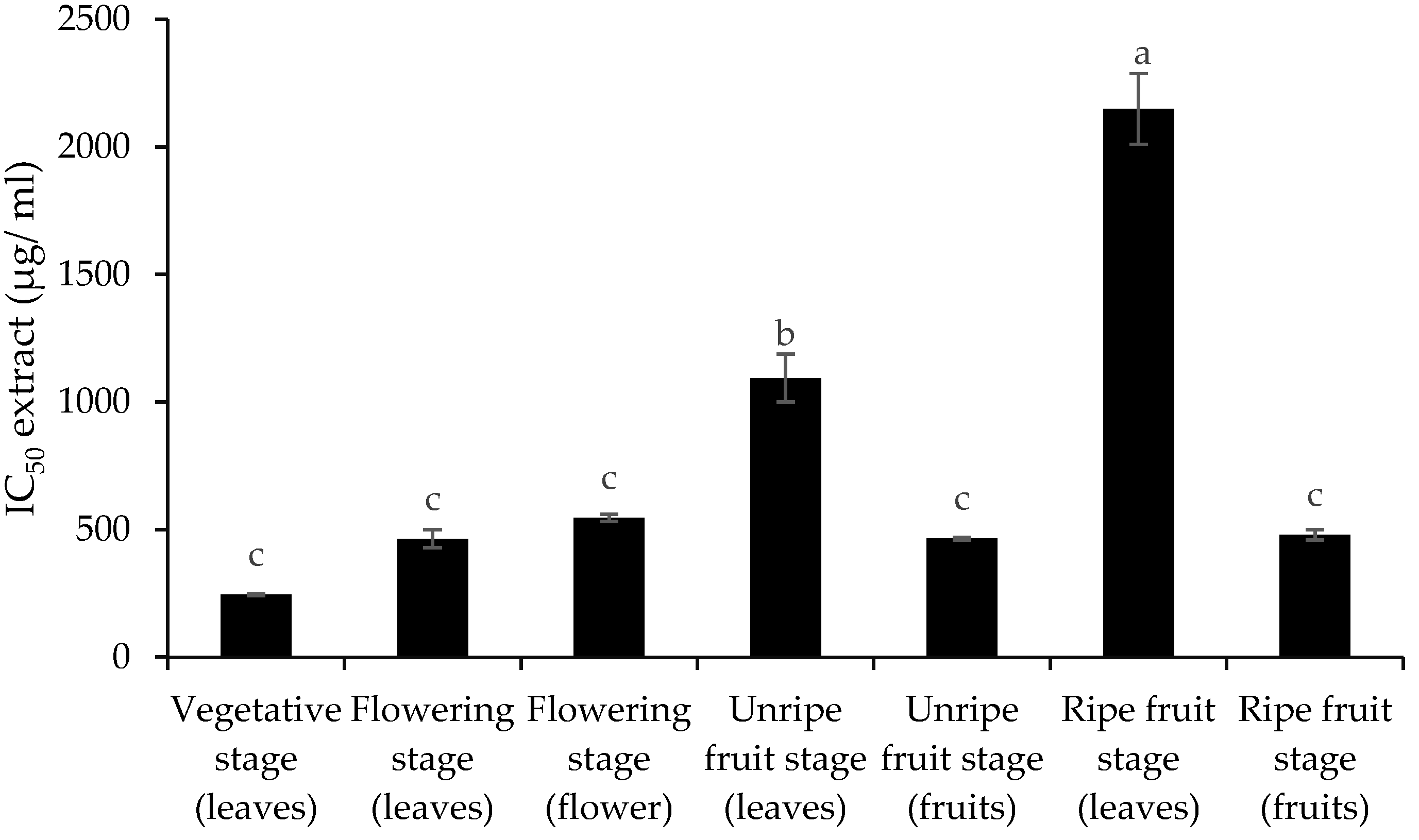
3.2. Total Phenol and Flavonoid Content and Antioxidant Activity

The results showed significant variations in the total phenol and flavonoid content of different organs of C. spinosa throughout its growth stages. The highest content was found in leaves during the flowering stage, with 56.20 mg GAE/g extract for total phenols and 17.10 mg QE/g extract for flavonoids (Figure 4).
These values were statistically comparable to those found in the leaves during the vegetative stage. Although fruits contained significant amounts of both compounds, the lowest content was found in leaves at the ripe fruit stage, with only 13.39 mg GAE/g for total phenols and 2.23 mg QE/g for flavonoids (Figure 5). Our results indicate that the levels of these compounds are higher in young leaves and decrease with age, with a greater accumulation occurring in the fruits. Figure 6 and Figure 7 show that the antioxidant activity of leaf extracts and EOs was lowest at the ripe fruit stage, as indicated by higher IC50 values, as the plant shifts its resources towards fruit ripening. Conversely, leaves during the vegetative stage exhibited the greatest antioxidant activity as they play a key role in defending against oxidative stress during the plant’s growth phase.

3.3. Essential Oils Composition

The EO constituents of the C. spinosa organs at different phenological stages, investigated by GC-FID and GC-MS (Table 1). According to the results, methyl isothiocyanate, a major sulfur-containing compound in this plant, had its highest concentration in flowers during the flowering stage (41.6%), followed by mature fruits (36.7%). The lowest concentration was found in leaves during the pre-flowering stage (27.5%).
Isopropyl isothiocyanate, another important sulfur-containing compound, reached its highest concentration in leaves during the unripe fruit stage (36.2%), followed by leaves during the ripe fruit stage (33.8%). The lowest concentration was observed in ripe fruits, showing a reduction of 86.5% compared to the highest value.
Isobutyl isothiocyanate was not detected in leaves during the flowering, unripe, and ripe fruit stages. However, it reached its highest concentration in ripe fruits (9.2%), followed by unripe fruits (6.7%). These results suggest that the production or accumulation of isobutyl isothiocyanate occurs primarily in fruits rather than in leaves, with a significant increase during fruit ripening (Table 1).
Dimethyl trisulfide showed its highest concentration in leaves during the pre-flowering stage. As the plant developed, its concentration decreased significantly by 76.6%, reaching only 1.5% in ripe fruits. This substantial reduction suggests that dimethyltrisulfide is synthesized or accumulated mainly in leaves during early growth stages and decreases as the plant transitions to fruit ripening.
Several compounds were detected exclusively in fruits (both immature and ripe), with the highest concentrations observed in ripe fruits: N-methyldodecanamide (7.0%), 3-tert-butyl-4-hydroxyanisole (5.6%), dodecanamide, N, N-diethyl (6.3%), and oleic acid (2.8%).
Dimethyl tetrasulfide reached its maximum concentration in the flowers during the flowering stage (18.3%). As development progressed, its levels gradually decreased, reaching its lowest concentration (1.8%) in ripe fruits (Table 1).

3.4. Phenolic Acids

Phenolic acid analysis revealed different patterns in different organs at phenological stages of the C. spinosa plant. In the vegetative and flowering stages, both leaves and flowers contained four types of phenolic acids. The unripe fruit stage showed the greatest diversity, with seven types in leaves and eight types in fruits, while the ripe fruit stage contained four types in leaves and six types in fruits. Quantitatively, extracts from unripe fruits contained the highest concentration of total phenolic acids (19.32 mg/g dried extract), followed by leaves from the unripe fruit stage (13.43 mg/g dried extract) and flowers from the flowering stage (8.1 mg/g dried extract). The lowest concentrations were observed in leaf extracts of both mature fruit and vegetative stages, each at 1 mg/g dried extract organ (Table 2).
The dominant phenolic acids in various phenological stages were cinnamic acid (0.5 mg/g extract) during the vegetative stage; rosmarinic acid (1.2 mg/g extract) and cinnamic acid (0.4 mg/g extract) in leaves during the flowering stage; caffeic acid (3.4 mg/g extract) and cinnamic acid (3.2 mg/g extract) in flowers during the flowering stage; vanillic acid (4.23 mg/g extract), cinnamic acid (2.8 mg/g extract), and rosmarinic acid (2.7 mg/g extract) in leaves during the unripe fruit stage; vanillic acid (5.81 mg/g extract), cinnamic acid (4.5 mg/g extract), and rosmarinic acid (2.8 mg/g extract) in unripe fruits; vanillic acid (0.3 mg/g extract) and cinnamic acid (0.3 mg/g extract) in ripe fruit leaves; and rosmarinic acid (0.7 mg/g extract), cinnamic acid (0.5 mg/g extract), and vanillic acid (0.5 mg/g extract) in fruits during the ripe fruit stage.

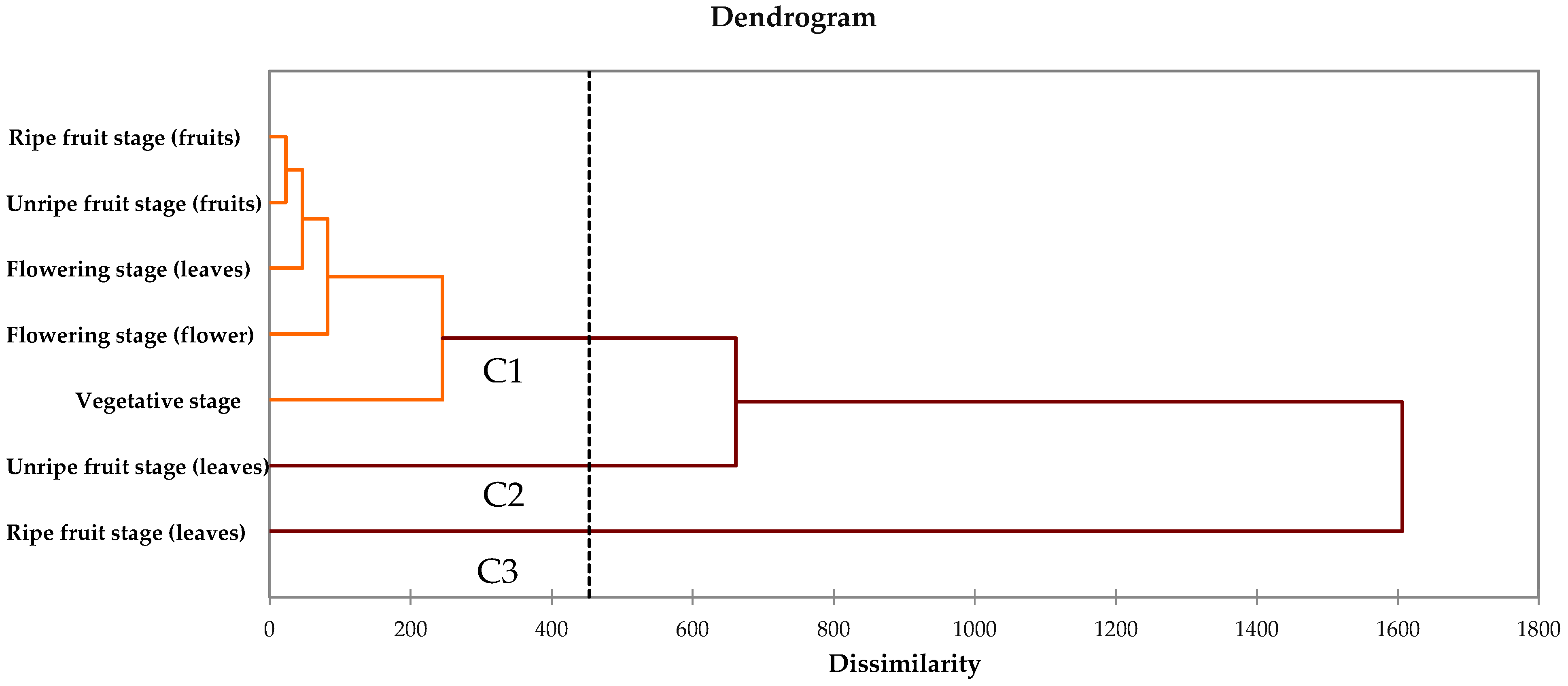
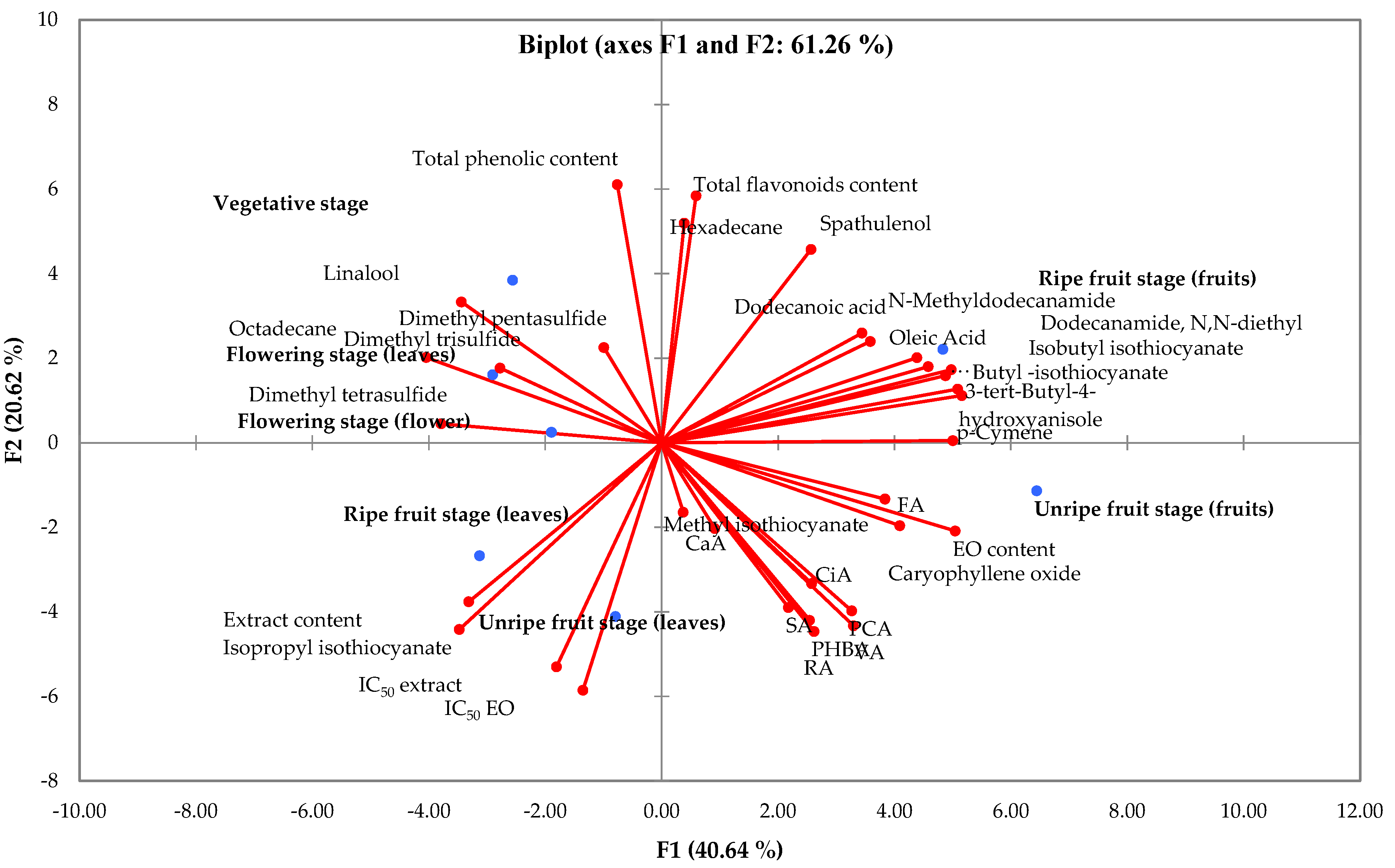
3.5. Cluster Analysis and Principal Component Analysis (PCA)

Cluster analysis was carried out to monitor the differences and similarities between the different phenological stages and different organs to determine their effect on the phytochemical composition and antioxidant activity of different parts in C. spinosa. The results of the cluster analysis showed that the four harvesting stages in eight organs could be divided into three different groups (G1, G2, G3), each identified by the similarity of their phytochemical composition. Based on the dendrogram obtained from the cluster analysis, it was observed that the vegetative stage, the flowering stage (leaves), the flowering stage (flowers), the unripe fruit stage (fruits), and the ripe fruit stage (fruits) were classified in the same group (G1), while the unripe fruit stage (leaves) was classified in group 2 (G2) and the ripe fruit stage (leaves) (Figure 8). The importance of cluster analysis goes beyond simple grouping to determine the relative distance between groups. Groups separated by short distances in this study suggest that the quality and quantity of the active compounds are largely similar. Conversely, in groups separated by greater distances, this similarity is low, indicating significant differences in the types and amounts of compounds.
Principal component analysis (PCA) was performed to visualize the relationships between different plant organs at different phenological stages and their associated phytochemical compositions (Figure 9). The analysis showed that the first principal component (PC1) explained 40.64% of the total variance, while the second principal component (PC2) accounted for 20.62%, together explaining 61.26% of the cumulative variance. The PCA biplot showed distinct clustering patterns based on plant organs and developmental stages. Upper right quadrant (positive PC1, positive PC2): ripe fruits were characterized by elevated concentrations of total flavonoids, p-cymene, 3-tert-butyl-4-hydroxyanisole, butyl isothiocyanate, oleic acid, α-cadinol, isobutyl isothiocyanate, dodecanoic acid, N-methyldodecanamide, hexadecane, and spathulenol. Upper left quadrant (negative PC1, positive PC2): vegetative stage tissues, flowering stage leaves, and flowers clustered together, showing strong associations with total phenolic content, linalool, and sulphur compounds including dimethyl trisulphide, dimethyl tetrasulphide, dimethyl pentasulphide, and octadecane. Lower right quadrant (positive PC1, negative PC2): unripe fruits had higher concentrations of EOs, methyl isothiocyanate, caryophyllene oxide, and several phenolic acids including ferulic acid (FA), cinnamic acid (CiA), p-coumaric acid (PCA), rosmarinic acid (RA), vanillic acid (VA), syringic acid (SA), caffeic acid (CaA), and p-hydroxybenzoic acid (PHBA). Lower left quadrant (negative PC1, negative PC2): leaves from both ripe and immature fruit stages were associated with higher extract, isopropyl isothiocyanate, and stronger antioxidant activity as indicated by lower IC50 values for both the extract and EOs. This distinct clustering pattern demonstrates how phytochemical profiles change significantly between different plant organs and developmental stages, providing valuable insights for optimizing harvest timing.

4. Discussion

The production and accumulation of EOs in medicinal plants is influenced by a complex interplay of genetic, environmental, and plant organ developmental factors [51]. Our results on C. spinosa show clear patterns of difference in EO accumulation across plant organs and developmental stages, consistent with basic principles of plant resource allocation and secondary metabolite production.
The significantly higher EO content observed in unripe fruits (0.18%) compared to other plant parts suggests that fruits serve as primary sites for EO biosynthesis and storage in C. spinosa. This pattern aligns with the ecological functions of EOs in fruits, which include protection against herbivores and pathogens, as well as facilitation of seed dispersal by attracting specific animals [33]. The observed decrease in EO content during fruit ripening can be attributed to several physiological processes. During ripening, fruits undergo significant biochemical transformations that may lead to the degradation of EO components or changes in their biosynthetic pathways [52]. In addition, the increased tissue permeability characteristic of ripening fruits facilitates greater volatilization of EO compounds [53]. Increased enzymatic activity during ripening, particularly of lipases and esterases, further contributes to the degradation of EO components, resulting in reduced overall EO content [54].
The relatively stable EO content in leaves at different phenological stages indicates a consistent allocation of resources to EO production in these photosynthetic organs throughout plant development. However, the slight decrease in leaf EO content during flowering (0.07%) suggests a strategic redistribution of resources, with the plant prioritizing reproductive functions over defense compound production in vegetative tissues. This pattern of resource allocation consists of optimal defense theory, which predicts that plants invest more in protecting reproductive structures than vegetative tissues [51].
The developmental regulation of EO biosynthesis observed in our study reflects broader patterns of secondary metabolite production in plants. During early stages, plants typically prioritize primary metabolism and structural development and allocate fewer resources to secondary metabolite production [55,56]. As plants mature, the expression of genes involved in EO biosynthesis increases in response to developmental signals, leading to enhanced production in specific organs [36,57]. Furthermore, EO biosynthesis shares metabolic precursors with primary metabolic pathways, leading to trade-offs in resource allocation that vary with developmental stage and environmental conditions [58].
These results have important implications for optimizing harvesting strategies for C. spinosa. The peak EO content in unripe fruits suggests that harvesting at this specific stage of development would maximize EO yield for commercial applications. Understanding these temporal and spatial patterns of EO accumulation provides valuable insights for the sustainable management and utilization of this medicinal plant resource.
Examining the EO composition of C. spinosa using GC-FID across various phenological stages revealed significant changes in the content of key bioactive constituents, notably isothiocyanates and sulfur-containing compounds. These fluctuations highlight the adaptive nature of EO biosynthesis, reflecting the influence of developmental progression across different plant organs. The highest concentration of methyl isothiocyanate (41.6%) was found in flowers during the flowering stage, followed by mature fruits (36.7%). This pattern is consistent with the plant’s reproductive strategy, given that flowering is a critical period of vulnerability requiring increased chemical defense. During this period, plants typically increase the biosynthesis of glucosinolates, which serve as precursors to methyl isothiocyanate [59]. This compound plays a key role in the plant’s defense system, enabling it to combat herbivorous insects and microbial pathogens. When the plant experiences physical damage, such as from insect feeding or mechanical stress, it activates a sophisticated enzymatic response. At the core of this reaction is myrosinase, an enzyme responsible for breaking down glucosinolates, a class of sulfur-containing secondary metabolites. This enzymatic hydrolysis leads to the production of methyl isothiocyanate, a volatile compound that acts as an effective deterrent against biological threats [59,60,61].
Isopropyl isothiocyanate showed a different distribution pattern, reaching its highest concentration (36.2%) in leaves at the unripe fruit stage, closely followed by leaves at the ripe fruit stage (33.8%). Previous studies on the C. spinosa and other Capparis species have reported high levels of this compound in the leaves [39,42,62]. The distribution of defensive compounds within the plant suggests a strategic allocation aimed at protecting developing fruits. The markedly higher concentrations of isopropyl isothiocyanate in leaves than in fruits indicate that leaves are the primary site of their biosynthesis during fruit development. This defensive strategy ensures optimal protection, particularly during vulnerable stages. During the unripe fruit stage, the elevated presence of isopropyl isothiocyanate aligns with the heightened susceptibility of the plant to herbivory. Immature fruits represent a significant metabolic investment and therefore demand strong chemical defense mechanisms to deter potential predators. As part of this defense strategy, glucosinolates—the precursors of isothiocyanates—accumulate in higher concentrations within plant tissues during critical developmental windows, thereby reinforcing protection. As fruits mature, there is a notable decline in the concentration of isopropyl isothiocyanate, with an observed reduction of 86.5% in ripe fruits compared to leaves. This trend reflects the plant’s adaptive resource reallocation, shifting its focus from chemical defense to reproductive success and seed dispersal efficiency. The decline in defensive compound levels suggests that energy is prioritized for fruit maturation, seed viability, and dispersal mechanisms, rather than being invested in continued chemical deterrents. This intricate biochemical regulation highlights the dynamic balance between defense and reproduction in plant survival strategies. The ability to modulate secondary metabolite levels based on developmental needs highlights the plant’s remarkable adaptability to environmental pressures and predation risks [63,64].
The organ-specific accumulation of isobutyl isothiocyanate provides further insight into the plant’s defense strategy. Its absence in leaves during flowering, unripe, and ripe fruit stages, in contrast to its highest concentration in ripe fruits, suggests specialized biosynthetic pathways that are activated predominantly during fruit ripening. This compound probably contributes to seed protection and may play a role in discouraging premature consumption of fruits before seed ripening is complete. The temporal dynamics of dimethyl trisulfide and dimethyl tetra-sulfide further illustrate the plant’s adaptive chemical defense mechanisms. Dimethyl trisulfide reached its maximum concentration in leaves during pre-flowering and then decreased by 76.6% during development to ripe fruits. Similarly, dimethyl tetra sulfide peaked in flowers (18.3%) and gradually declined to 1.8% in ripe fruits. These patterns suggest that sulfur volatiles play a critical role during the early stages of development but become less important as the fruits mature and other defense compounds take precedence.
Comparative analysis with previous studies reveals both consistencies and variations in the EO composition of C. spinosa. Merlino et al. [41], reported methyl isothiocyanate and indole-3-methanol as the predominant glucosinolate derivatives in C. spinosa leaf EO emulsions, while ethyl, isopropyl, butyl, and isobutyl isothiocyanates were present in trace amounts (below 0.1%). This contrasts with our findings of substantial isopropyl isothiocyanate concentrations in leaves, suggesting a possible influence of environmental factors, genetic variation or extraction methods on isothiocyanate profiles. Kulisic et al. [65], identified methyl isothiocyanate as the dominant component (92.06%) in EOs from caper leaves and flower buds extracted by hydrodistillation, a significantly higher proportion than observed in our study. Meanwhile, Afsharypuor et al. [66], reported a more diverse EO composition with thymol (26.4%), isopropyl isothiocyanate (11%), 2-hexenal (10.2%), and butyl isothiocyanate (6.3%) as major components in leaf EOs, whereas fruit EOs contained mainly methyl isothiocyanate (41.6%) and isopropyl isothiocyanate (52.2%). These differences between studies highlight the influence of genetic, environmental and methodological factors on the chemical composition of C. spinosa EOs [67].
Various environmental factors, such as temperature, light intensity, soil water availability, and soil fertility, can significantly impact the physiological and biochemical responses of medicinal plants, as well as the biosynthesis pathways of secondary metabolites [68]. These abiotic environmental stressors can inhibit or promote secondary metabolite production [69], which may explain the variations observed in our study depending on the phenological stage. Water availability in particular affects the composition of phenolic compounds in medicinal plants [70], and the varying soil moisture content across the different phenological stages in our experiment likely contributed to the observed differences in levels of bioactive compounds. Furthermore, phenolic compounds act as a UV protection mechanism, with their synthesis being closely linked to photosynthetic processes that provide essential precursors. Therefore, seasonal changes in light exposure during different growth stages may also influence the observed metabolite profiles in C. spinosa organs. It seems reasonable to conclude that this is because, during the growth period of the plant, the characteristics of the soil around the roots may change the diversity in terms of production of EOs at different phenological stages.
The exclusive detection of certain compounds in fruits, in particular N-methyldodecanamide, 3-tert-butyl-4-hydroxyanisole, dodecanamide, N,N-diethyl and oleic acid in ripe fruits, suggests that specialized metabolic pathways are activated during fruit ripening. These compounds may contribute to seed protection, fruit palatability for seed dispersers, or antimicrobial properties that prevent fruit spoilage. Taken together, these findings demonstrate the sophisticated chemical adaptation of C. spinosa throughout its developmental cycle, with distinct chemotypes emerging at different phenological stages. The strategic allocation of defense compounds across plant organs and developmental stages reflects an evolutionary optimization of resource investment in chemical defense relative to growth and reproductive priorities.
The analysis of total phenolic and flavonoid contents in different organs of C. spinosa throughout its phenological development revealed significant variations, reflecting the plants adaptive biochemical responses to changing physiological needs and environmental conditions. During the late vegetative and flowering stages, C. spinosa showed a peak accumulation of phenolic compounds and flavonoids in the leaves. This increased biosynthesis serves several critical functions beyond simple defense. The contents of phenols and flavonoids found in the organs studied in this research were higher than in previous studies [71,72]. Phenolic compounds at these stages primarily support structural development through lignification, a process essential for plant architecture and vascular integrity [73]. The phenylpropanoid pathway plays a key role in plant defense and development. Its activity increases significantly during reproduction. This pathway synthesizes essential phenolic compounds, such as flavonoids, lignins, and phenolic acids, which are vital for plant survival. The key enzyme in this process is phenylalanine ammonia-lyase (PAL), which catalyzes the conversion of phenylalanine to cinnamic acid in the first step of phenylpropanoid metabolism. During reproduction, PAL expression increases in response to developmental and environmental triggers, ensuring a sufficient supply of precursors for phenolic synthesis. Phenylpropanoid metabolism fluctuates in response to external stressors, including UV exposure, nutrient availability, and pathogen attacks. Hormonal signaling, particularly involving salicylic and jasmonic acids, regulates PAL activity and phenolic accumulation. This aligns metabolic output with reproductive success. The strategic upregulation of PAL demonstrates how plants can optimize their biochemical resources by balancing growth, defense, and reproductive efficiency [74,75,76].
The flowering stage is a period of increased vulnerability and specialized metabolic requirements for C. spinosa. During this phase, plants typically experience increased exposure to solar radiation, particularly UV light. Flavonoids act as natural UV screens, absorbing harmful UV radiation and protecting sensitive reproductive tissues from photodamage [77]. This photoprotective function is particularly important for the preservation of genetic material in pollen and ovules. At the same time, the flowering stage triggers increased metabolic activity, generating elevated levels of reactive oxygen species (ROS). The abundant phenolic compounds, with their exceptional radical scavenging capacity, provide a robust antioxidant defense system that prevents oxidative damage to cellular components [78].
The strategic accumulation of flavonoids during the pre-flowering and flowering stages further highlights their multifunctional roles in C. spinosa reproduction. Beyond UV protection, these compounds contribute to flower pigmentation, attract pollinators through visual cues, and participate in scent production through precursor relationships with volatile phenylpropanoids [79,80]. In addition, flavonoids act as defensive compounds against flower-specific herbivores and pathogens, protecting the plants reproductive investment. The observed developmental trajectory of phenolic and flavonoid content in C. spinosa is consistent with the well-established pattern that flavonoid accumulation peaks in young tissues and declines after flowering [81]. This decline coincides with a fundamental metabolic shift as plants transition from active synthesis of secondary metabolites to cellular differentiation and reproductive development. The dramatic reduction in leaf phenolic content during the ripe fruit stage exemplifies this metabolic reprioritization. The compartmentalization of phenolic synthesis primarily within the mesophyll tissue of leaves explains the organ-specific distribution patterns observed in C. spinosa. As the plant progresses through its phenological stages, carbohydrates—which serve as primary precursors for phenolic compounds—are preferentially transported via the phloem to developing reproductive structures. Importantly, phenolic compounds themselves cannot be mobilized via the phloem transport system [82,83], requiring fruits to synthesize these compounds independently rather than receiving them from the leaves. This developmental redistribution of metabolic resources reflects an evolutionary optimization to balance growth, reproduction, and defense. The increased accumulation of phenolic compounds in fruits, despite decreasing levels in mature leaves, suggests a selective pressure to protect reproductive structures carrying genetic material. In addition, fruit phenolics may contribute to seed dispersal strategies by influencing fruit palatability and providing antioxidant protection to embryonic tissues during dormancy. These findings not only elucidate the complex biochemical adaptations of C. spinosa but also have practical implications for harvesting strategies aimed at maximizing the yield of bioactive compounds for medicinal applications. The identification of optimal harvesting times—particularly during flowering for leaves and at appropriate maturity for fruits—may significantly enhance the therapeutic potential of C. spinosa-derived natural products.
Analysis of antioxidant activity in different phenological stages and plant organs of C. spinosa revealed remarkable variations that correlate with developmental priorities and resource allocation strategies of the plant. Leaf extracts and EOs showed minimum antioxidant capacity during the ripe fruit stage, as evidenced by elevated IC50 values, indicating a strategic redistribution of metabolic resources towards fruit development and ripening. In contrast, leaves in the vegetative stage exhibited superior antioxidant properties, reflecting their critical role in defending against oxidative stress during periods of active growth and photosynthesis.
These dynamic fluctuations in antioxidant capacity across developmental stages and plant organs are consistent with findings from numerous studies on medicinal and aromatic plants [75,81,84,85,86]. This pattern underscores a fundamental principle in plant physiology—the dynamic regulation of antioxidant defenses in response to metabolic demands and environmental pressures at different developmental stages. The ability of C. spinosa to fine-tune its phenolic metabolism highlights how plants balance growth, defense, and reproduction through precise biochemical adjustments. Throughout its life cycle, C. spinosa modulates the production and distribution of phenolic compounds, ensuring that antioxidant mechanisms align with physiological requirements. During active growth, high antioxidant activity in leaves safeguards against oxidative stress caused by intense photosynthesis and environmental fluctuations. As the plant enters flowering and fruiting, metabolic focus shifts toward reproductive success, leading to redistribution of secondary metabolites—including phenolics—toward seed formation rather than sustained antioxidant defense. The antioxidant capacity of C. spinosa is directly tied to its phenolic composition, influencing both quantitative and qualitative aspects of defense. Phenolic concentration fluctuates across different plant organs, with leaves generally exhibiting higher antioxidant levels during vegetative stages compared to fruits. The specific types of phenolic acid compounds, determine the potency and function of antioxidant defenses. This biochemical flexibility allows C. spinosa to optimize its defense mechanisms based on environmental conditions [87]. Stress-induced phenolic biosynthesis ensures resilience, particularly in arid or nutrient-limited ecosystems, where oxidative damage poses significant risks [88]. Understanding these metabolic strategies provides valuable insights into plant stress responses, ecological adaptations, and potential applications in medicinal and agricultural fields. The direct correlation between peak phenolic content and maximum antioxidant activity in vegetative stage leaves demonstrates the critical contribution of these compounds to the plant’s redox homeostasis. The decline in antioxidant capacity observed in leaves during the ripe fruit stage is likely to reflect not only reduced phenolic content but also changes in the specific composition of the phenolic profile. Certain high-potency antioxidant phenolics may be preferentially synthesized during early developmental stages, when oxidative challenges are most pronounced, while others may predominate during reproductive stages to perform specialized functions beyond antioxidant protection [89].
A comprehensive analysis of phenolic acid profiles in different organs and phenological stages of C. spinosa revealed complex patterns of accumulation and distribution, reflecting developmental priorities and ecological adaptations of the plant. The remarkable diversity and concentration of phenolic acids observed during the unripe fruit stage—with eight different compounds in fruits and seven in leaves—highlights this critical developmental stage as a period of intensive secondary metabolite biosynthesis.
Cinnamic acid was found to be ubiquitous at all stages of development, demonstrating its fundamental role in the metabolism of C. spinosa. Its concentration peaked impressively in unripe fruits (4.5 mg/g extract) and progressively decreased to its lowest level in leaves during the ripe fruit stage (0.3 mg/g extract). This pattern suggests that cinnamic acid, as a central precursor in the phenylpropanoid pathway, undergoes strategic remobilization towards reproductive structures during fruit development. As an essential intermediate for several phenolic compounds, the increased presence of cinnamic acid in unripe fruits reflects its dual role in defense mechanisms and as a biosynthetic building block for subsequent metabolites [90,91]. Rosmarinic acid also showed a consistent presence at all phenological stages, with the highest concentration in unripe fruits (2.8 mg/g extract) and the lowest in leaves at the vegetative and ripe fruit stages (0.2 mg/g extract). This distribution pattern suggests a specialized function during fruit development. The potent antioxidant capacity of rosmarinic acid is thought to help protect developing fruits from oxidative damage during rapid cell division and expansion. Its diverse bioactivities—including anti-inflammatory, antimicrobial, and anti-allergic properties—provide comprehensive protection to vulnerable immature fruits against various biotic stressors [92,93]. Vanillic acid showed the most pronounced stage-dependent accumulation pattern, with extraordinary concentrations during the unripe fruit stage (5.81 mg/g extract in fruits and 4.23 mg/g extract in leaves), while remaining at minimal levels during other developmental stages. This dramatic stage-specific increase suggests a highly specialized ecological function, possibly involving targeted defense against herbivores or pathogens that particularly threaten developing fruits. The synchronized accumulation of vanillic acid in both fruits and associated leaves suggests a coordinated whole-plant response to protect reproductive investment during this vulnerable developmental window.
The exceptional accumulation of caffeic acid in flowers during the flowering stage (3.4 mg/g extract) represents another organ-specific specialization. This selective concentration suggests caffeic acid plays crucial roles in reproductive processes, potentially including pollinator attraction, UV protection for genetic material, or defense against flower-specific herbivores. Caffeic acid documented antioxidant and anti-tumor properties may serve to protect the delicate reproductive tissues from oxidative damage during pollen development and fertilization [94]. During early fruit development, plants deploy a highly regulated biochemical defense system to maximize survival during this critical reproductive phase. Unripe fruits, which house the plant’s genetic material and represent a significant metabolic investment, are particularly susceptible to herbivory and pathogen attack. To counter these threats, the plant strategically accumulates multiple phenolic acids, forming a synergistic chemical shield with diverse protective functions. The presence of cinnamic, rosmarinic, and vanillic acids in developing fruits reflects an intricate defense network where each compound plays a distinct yet complementary role. Cinnamic acid functions as a precursor in the phenylpropanoid pathway, essential for synthesizing lignin, flavonoids, and other defensive secondary metabolites. It exhibits antioxidant properties, neutralizing ROS generated during metabolic activity. Rosmarinic acid demonstrates broad-spectrum antimicrobial activity, inhibiting bacterial and fungal pathogens that could compromise fruit integrity. It acts as a free radical scavenger, reinforcing oxidative stress defense and cellular protection. Vanillic acid possesses anti-herbivore properties, potentially deterring predation through its interaction with insect and mammalian digestive enzymes. It contributes to structural reinforcement, integrating into polymeric compounds that fortify fruit tissues [90,95,96]. Beyond their ecological significance, these phenolic acids contribute substantially to the medicinal value of C. spinosa. Cinnamic acid’s diverse bioactivities—encompassing antioxidant, antimicrobial, anticancer, neuroprotective, and anti-inflammatory effects—make it a valuable therapeutic compound with applications across multiple health domains. Its industrial importance in cosmetics, flavoring agents, toiletries, and detergents further enhances its economic significance [90,91].
PCA and cluster analyses revealed that organ type and phenological stage were the primary drivers of PC1, accounting for the significant chemical variations observed in C. spinosa. The eigenvector values clearly separated from the developmental stages, with leaves in the vegetative and fruiting phases clustering distinctly compared to other organs. This statistical approach effectively validated our biochemical findings: leaves exhibited higher extract yields, elevated isopropyl isothiocyanate content, and superior antioxidant activity, as indicated by lower IC50 values for both extracts and EOs. PCA and cluster analysis confirmed that organ- and stage-specific variations are critical determinants of bioactive compound accumulation. This supports the use of targeted harvesting strategies to optimize the therapeutic potential of C. spinosa. These results are consistent with previous phytochemical studies in which PCA successfully distinguished between plant organs and growth stages [30,31,32,97,98]. This reinforces the value of PCA as a data reduction tool for understanding complex biochemical patterns in medicinal plants [99].
The stage-dependent variations in phenolic acid profiles observed in this study have significant implications for optimizing harvesting strategies to maximize specific bioactive compounds. The unripe fruit stage clearly represents the optimal harvesting period for obtaining maximum concentrations of several phenolic acids, particularly vanillic, cinnamic, and rosmarinic acids. Conversely, the flowering stage would be preferable for the isolation of caffeic acid. These findings provide valuable guidance for the targeted extraction and formulation of C. spinosa-derived therapeutic products with enhanced efficacy for specific health applications.

5. Conclusions

This study provides a comprehensive analysis of the phytochemical composition of C. spinosa organs at different phenological stages, revealing significant stage-dependent and organ-specific variations in bioactive compounds with considerable industrial potential. Our investigation shows that the EO content varies significantly between plant organs, with unripe fruits containing the highest concentration (0.18%). Chemical profiling identified methyl isothiocyanate as the predominant component in flowers (41.6%) and ripe fruits (36.7%), while isopropyl isothiocyanate dominated in leaves at both unripe (36.2%) and ripe fruits (33.8%) stages. These isothiocyanates, derived from glucosinolates, not only serve as natural defense compounds against herbivores and pathogens but also represent valuable bioactive molecules with antimicrobial, anti-inflammatory, and potential anticancer properties for pharmaceutical applications. Total phenol and flavonoid contents were highest in flowering stage leaves and decreased with leaf age, consistent with their role in UV protection, flower pigmentation, and pest defense. Correspondingly, antioxidant activity was highest in vegetative stage leaves and lowest in ripe fruit stage leaves, reflecting the changing priorities in metabolite production as the plant matures. Phenolic acid analysis revealed that unripe fruits contained the highest diversity and concentration of these compounds, with cinnamic acid, rosmarinic acid, and vanillic acid being the predominant compounds at different stages. These bioactive compounds, known for their antioxidant, antimicrobial, and anti-inflammatory properties, were particularly concentrated in unripe fruits, probably as a protective mechanism during this vulnerable stage of development. Overall, this study provides comprehensive insights into the organ-specific and stage-dependent production of bioactive compounds in C. spinosa, highlighting the optimal harvesting times to maximize yield and quality of specific compounds. These findings provide valuable information for pharmaceutical, cosmetic, and food industries interested in exploiting the bioactive potential of this medicinal plant. Future research could explore the molecular mechanisms underlying these metabolic shifts and investigate the therapeutic applications of the identified compounds.

Author Contributions

Conceptualization, S.H.; methodology, S.H., Z.M., S.M. and H.R.A.; validation, S.H. and S.N.; formal analysis, S.H., S.M., H.R.A. and Z.M.; investigation, S.H., S.M., S.N. and Z.M.; resources, S.H.; data curation, S.H., S.M. and Z.M.; writing—original draft preparation, S.H. and Z.M.; writing—review and editing, S.H., Z.M., S.N., S.M. and H.R.A.; visualization, S.N.; supervision, S.H.; project administration, S.H. and Z.M. All authors have read and agreed to the published version of the manuscript.

Funding

This research was funded by Azarbaijan Shahid Madani University, grant number 1402/1510.

Data Availability Statement

The data presented in this study are available on request from the corresponding author.

Acknowledgments

The support of Azarbaijan Shahid Madani University is greatly appreciated.

Conflicts of Interest

The authors declare no conflicts of interest.

References

  1. Zhang, H.; Ma, Z.F. Phytochemical and pharmacological properties of Capparis spinose as a medicinal plants. Nutrients 2018, 10, 116. [Google Scholar] [CrossRef] [PubMed]
  2. Bhatla, S.C.; Lal, M.A. Secondary metabolites. In Plant Physiology, Development and Metabolism; Springer Nature: Singapore, 2023; pp. 765–808. [Google Scholar]
  3. Elshafie, H.S.; Camele, I.; Mohamed, A.A. A comprehensive review on the biological, agricultural and pharmaceutical properties of secondary metabolites based-plant origin. Int. J. Mol. Sci. 2023, 24, 3266. [Google Scholar] [CrossRef] [PubMed]
  4. Raissi, A.; Arbabi, M.; Ahmad, R.M. Capparis spinosa L., an important medicinal plant from Sistan and Baloochestan Province, Iran. In Proceedings of the Global Conference on New Approaches in Agriculture and Environment with the Focus on Sustainable Development and Safe Production, Shiraz, Iran, 28 January 2016. [Google Scholar]
  5. Sun, W.; Shahrajabian, M.H. Therapeutic potential of phenolic compounds in medicinal plants—Natural health products for human health. Molecules 2023, 28, 1845. [Google Scholar] [CrossRef] [PubMed]
  6. Rivera, D.; Inocencio, C.; Obón, C.; Alcaraz, F. Review of food and medicinal uses of Capparis L. Subgenus Capparis (capparidaceae). Econ. Bot. 2003, 57, 515–534. [Google Scholar] [CrossRef]
  7. Liu, Y.T.; Sun, Y.T.; Cheng, X.M.; Wang, C.H.; Zhou, H.; Yang, T. Advances on the investigation of chemical constituents and pharmacological activities of Capparis spinosa L. Modernization of Traditional Chinese Medicine and Materia Materia. World Sci. Technol. 2019, 12, 2599–2608. [Google Scholar]
  8. Bina, F.; Bostani, A. Evaluation of the phenotypic variation in a caper (Capparis spinosa L.) population growing in south of Tehran using multivariate analysis. J. BioSci. Biotechnol. 2016, 5, 117–123. [Google Scholar]
  9. Shahrajabian, M.H.; Sun, W.; Cheng, Q. Plant of the Millennium, Caper (Capparis spinosa L.), chemical composition and medicinal uses. Bull. Natl. Res. Cent. 2021, 45, 131. [Google Scholar] [CrossRef]
  10. Zarei, M.; Seyedi, N.; Maghsoudi, S.; Shahbi Nejad, M.; Sheibani, H. Green synthesis of Ag nanoparticles on the modified graphene oxide using Capparis spinosa fruit extract for catalytic reduction of organic dyes. Inorg. Chem. Commun. 2021, 123, 108327. [Google Scholar] [CrossRef]
  11. Kdimy, A.; El Yadini, M.; Guaadaoui, A.; Bourais, I.; El Hajjaji, S.; Le, H.V. Phytochemistry, Biological Activities, Therapeutic Potential, and Socio-Economic Value of the Caper Bush (Capparis spinosa L.). Chem. Biodivers. 2022, 19, 202200300. [Google Scholar] [CrossRef]
  12. Kulisic-Bilusic, T.; Schmöller, I.; Schnäbele, K.; Siracusa, L.; Ruberto, G. The anticarcinogenic potential of essential oil and aqueous infusion from caper (Capparis spinosa L.). Food. Chem. 2012, 132, 261–267. [Google Scholar] [CrossRef]
  13. Olas, B. The Current State of Knowledge about the Biological Activity of Different Parts of Capers. Nutrients 2023, 15, 623. [Google Scholar] [CrossRef] [PubMed]
  14. Jagannath, R. Phytochemical and Pharmacological Screening on Roots of Capparis spinosa F: Capparidaceae. Ph.D. Thesis, Rajiv Gandhi University of Health Sciences, Bengaluru, India, 2010. [Google Scholar]
  15. Al-Snafi, A.E. Chemical constituents and pharmacological activities of Ammi majus and Ammi visnaga: A review. Int. J. Pharm. Ind. Res. 2013, 3, 257–265. [Google Scholar]
  16. Rezzan, A.; Gonca, T.; Nurettin, Y.; Emre, E. Characterisation of volatile compounds by spme and gc-fid/ms of capers (Capparis spinosa L.). Afr. J. Agric. Res. 2015, 10, 2213–2217. [Google Scholar]
  17. Koufan, M.; Belkoura, I.; Mazri, M.A. In vitro propagation of caper (Capparis spinosa L.): A review. Horticulturae 2022, 8, 737. [Google Scholar] [CrossRef]
  18. Afzali, S.F.; Sadeghi, H.; Taban, A. A comprehensive model for predicting the development of defense system of Capparis spinosa L.: A novel approach to assess the physiological indices. Sci. Rep. 2023, 13, 12413. [Google Scholar] [CrossRef]
  19. Moghaddasi, S.M.; Haddad Kashani, H.; Azarbad, Z. Capparis spinosa L. propagation and medicinal uses. Life Sci. J. 2012, 9, 684–686. [Google Scholar]
  20. Sun, Y.; Yang, T.; Wang, C. Capparis spinosa L. as a potential source of nutrition and its health benefits in foods: A comprehensive review of its phytochemistry, bioactivities, safety, and application. Food Chem. 2023, 409, 135258. [Google Scholar] [CrossRef]
  21. Annaz, H.; Sane, Y.; Bitchagno, G.T.M.; Ben Bakrim, W.; Drissi, B.; Mahdi, I.; El Bouhssini, M.; Sobeh, M. Caper (Capparis spinosa L.): An updated review on its phytochemistry, nutritional value, traditional uses, and therapeutic potential. Front. Pharmacol. 2022, 13, 878749. [Google Scholar] [CrossRef]
  22. Wojdyło, A.; Nowicka, P.; Grimalt, M.; Legua, P.; Almansa, M.S.; Amoros, A.; Carbonell-Barachina, A.A.; Hernandez, F. Polyphenol compounds and biological activity of caper (Capparis spinosa L.) flowers buds. Plants 2019, 8, 539. [Google Scholar] [CrossRef]
  23. Zhu, P.; Wang, Y.; Zhang, X. Preparation and characterization of electrospun nanofibre membranes incorporated with an ethanol extract of Capparis spinosa L. as a potential packaging material. Food Packag. Shelf Life 2022, 32, 100851. [Google Scholar] [CrossRef]
  24. Bacchetti, T.; Campagna, R.; Sartini, D.; Cecati, M.; Morresi, C.; Bellachioma, L.; Martinelli, E.; Rocchetti, G.; Lucini, L.; Ferretti, G.; et al. spinosa L. subsp. rupestris Phytochemical Profile and Effect on Oxidative Stress in Normal and Cancer Cells. Molecules 2022, 27, 6488. [Google Scholar] [CrossRef] [PubMed]
  25. Anwar, F.; Muhammad, G.; Hussain, M.A.; Zengin, G.; Alkharfy, K.M.; Ashraf, M.; Gilani, A.H. Capparis spinosa L.: A Plant with High Potential for Development of Functional Foods and Nutraceuticals/Pharmaceuticals. Int. J. Pharmacol. 2016, 12, 201–219. [Google Scholar] [CrossRef]
  26. Nabavi, S.F.; Maggi, F.; Daglia, M.; Habtermariam, S.; Rstrelli, L.; Nabavi, S.M. Pharmacological effects of Capparis spinosa L. Phytother. Res. 2016, 30, 1733–1744. [Google Scholar] [CrossRef]
  27. Lo Bosco, F.; Guasrrasi, V.; Moschetti, M.; Germana, M.A.; Butera, D.; Corana, F.; Papetti, A. Nutraceutical value of pantelleria capers (Capparis spinosa L.). J. Food Sci. 2019, 84, 2337–2346. [Google Scholar] [CrossRef]
  28. Esmaeili, H.; Karami, A.; Maggi, F. Essential oil composition, total phenolic and flavonoids contents, and antioxidant activity of Oliveria decumbens Vent. (Apiaceae) at different phenological stages. J. Clean. Prod. 2018, 198, 91–95. [Google Scholar] [CrossRef]
  29. Grevsen, K.; Fretté, X.C.; Christensen, L.P. Content and composition of volatile terpenes, flavonoids and phenolic acids in Greek Oregano (Origanum vulgare L. ssp. hirtum) at different development stages during cultivation in cool temperate climate. Eur. J. Hortic. Sci. 2009, 74, 193–203. [Google Scholar] [CrossRef]
  30. Hazrati, S.; Mollaei, S.; Rabbi Angourani, H.; Hosseini, S.J.; Sedaghat, M.; Nicola, S. How do essential oil composition and phenolic acid profile of Heracleum persicum fluctuate at different phenological stages. Food Sci. Nutr. 2020, 8, 6192–6206. [Google Scholar] [CrossRef]
  31. Hazrati, S.; Rowshan, V.; Hosseini, S.J.; Sedaghat, M.; Mohammadi, H. Variation of essential oil composition and antioxidant activity in aerial parts of Stachys schtschegleevi Sosn at different growing stages. J. Essent. Oil Bear. Plants 2020, 23, 1054–1071. [Google Scholar] [CrossRef]
  32. Hazrati, S.; Hosseini, S.J.; Ebadi, M.T.; Nicola, S. Evolution of phytochemical variation in Myrtle (Myrtus communis L.) organs during different phenological stages. Horticulturae 2022, 8, 757. [Google Scholar] [CrossRef]
  33. Hazrati, S.; Mousavi, Z.; Nicola, S. Harvest time optimization for medicinal and aromatic plant secondary metabolites. Plant Physiol. Biochem. 2024, 212, 108735. [Google Scholar] [CrossRef]
  34. Mohaddab, M.; Genva, M.; Malika, F.; El-Goumi, Y.; Zeroual, A.; Fauconnier, M.L. Capparis spinosa: A rich source of phenolic compounds—A comprehensive review of its phytochemistry, health benefits, and biotechnological applications. Biocatal. Agric. Biotechnol. 2024, 61, 103409. [Google Scholar] [CrossRef]
  35. Grimalt, M.; Hernández, F.; Legua, P.; Almansa, M.S.; Amorós, A. Physicochemical composition and antioxidant activity of three Spanish caper (Capparis spinosa L.) fruit cultivars in three stages of development. Sci. Hortic. 2018, 240, 509–515. [Google Scholar] [CrossRef]
  36. Grimalt, M.; Sánchez-Rodríguez, L.; Hernández, F.; Legua, P.; Carbonell-Barrachina, Á.A.; Almansa, M.S.; Amorós, A. Volatile profile in different aerial parts of two caper cultivars (Capparis spinosa L.). J. Food Qual. 2021, 2021, 6620776. [Google Scholar] [CrossRef]
  37. Vahid, H.; Rakhshandeh, H.; Ghorbani, A. Antidiabetic properties of Capparis spinosa L. and its components. Biomed. Pharm. 2017, 92, 293–302. [Google Scholar] [CrossRef]
  38. Yahia, Y.; Benabderrahim, M.A.; Tlili, N.; Hannachi, H.; Ayadi, L.; Elfalleh, W. Comparison of three extraction protocols for the characterization of caper (Capparis spinosa L.) leaf extracts: Evaluation of phenolic acids and flavonoids by liquid chromatography–electrospray ionization–tandem mass spectrometry (LC–ESI–MS) and the antioxidant activity. Anal. Lett. 2020, 53, 1366–1377. [Google Scholar]
  39. Sonmezdag, A.S.; Kelebek, H.; Selli, S. Characterization of aroma-active compounds, phenolics, and antioxidant properties in fresh and fermented capers (Capparis spinosa) by GC-MS-olfactometry and LC-DAD-ESI-MS/MS. J. Food Sci. 2019, 84, 2449–2457. [Google Scholar] [CrossRef]
  40. Bakr, R.O.; El Bishbishy, M.H. Profile of bioactive compounds of Capparis spinosa var. aegyptiaca growing in Egypt. Rev. Bras. Farmacogn. 2016, 26, 514–520. [Google Scholar] [CrossRef]
  41. Alkhaibari, A.M.; Alanazi, A.D. Chemical composition and insecticidal, antiplasmodial, and anti-leishmanial activity of Capparis spinosa essential oil and its main constituents. Evid. Based Complement. Altern. Med. 2022, 2022, 6371274. [Google Scholar] [CrossRef]
  42. Merlino, M.; Condurso, C.; Cincotta, F.; Nalbone, L.; Ziino, G.; Verzera, A. Essential oil emulsion from caper (Capparis spinosa L.) leaves: Exploration of its antibacterial and anti-oxidant properties for possible application as a natural food preservative. Antioxidants 2024, 13, 718. [Google Scholar] [CrossRef]
  43. AlMousa, L.A.; AlFaris, N.A.; Alshammari, G.M.; ALTamimi, J.Z.; Alsyadi, M.M.; Alagal, R.I.; Yahya, M.A. Antioxidant and antimicrobial potential of two extracts from Capparis spinosa L. and Rumex nervosus and molecular docking investigation of selected major compounds. Saudi J. Biol. Sci. 2022, 29, 103346. [Google Scholar] [CrossRef]
  44. El Finou, H.; Salhi, N.; Zaid, A.; El Rhaffari, L. Phytotoxicity, antioxidant activity and chemical profile of aqueous extracts from Moroccan caper (Capparis spinosa L.). Sci. Afr. 2024, 24, 02176. [Google Scholar] [CrossRef]
  45. Rajhi, I.; Hernandez-Ramos, F.; Abderrabba, M.; Ben Dhia, M.T.; Ayadi, S.; Labidi, J. Antioxidant, Antifungal and Phytochemical Investigations of Capparis spinosa L. Agriculture 2021, 11, 1025. Agriculture 2021, 11, 1025. [Google Scholar]
  46. Ouhammou, M.; Adnany, E.M.E.; Mourjane, A.; Hammou, H.A.; Bouchdoug, M.; Jaouad, A.; Mahrouz, M. Physi-co-chemical analysis and antioxidant activity of Moroccan caper leaves (Capparis spinosa L.). Euro-Mediterranean. J. Environ. Integr. 2022, 7, 407–414. [Google Scholar] [CrossRef]
  47. Arrar, L.; Benzidane, N.; Krache, I.; Charef, N.; Khennouf, S.; Baghiani, A. Comparison between polyphenol contents and antioxidant activities of different parts of Capparis spinosa L. Pharmacogn. Commun. 2013, 3, 70. [Google Scholar]
  48. British Pharmacopoeia. Published on the Recommendation of the Medicines Commission Pursuant to the Medicines Act; Her Majesty’s Stationary Office: London, UK, 1998. [Google Scholar]
  49. Singleton, V.L.; Orthofer, R.; Lamuela-Raventós, R.M. Analysis of Total Phenols and Other Oxidation Substrates and Antioxidants by Means of Folin-Ciocalteu Reagent. In Methods in Enzymology; Academic Press: Cambridge, MA, USA, 1999; Volume 299, pp. 152–178. [Google Scholar]
  50. Chang, C.; Yang, M.; Wen, H.; Chern, J. Estimation of total flavonoid content in propolis by two complementary colorimetric methods. J. Food Drug Anal. 2002, 10, 178–182. [Google Scholar]
  51. Akroum, S.; Bendjeddou, D.; Satta, D.; Lalaoui, K. Antibacterial, antioxidant and acute toxicity tests on flavonoids extracted from some medicinal plants. Int. J. Green Pharm. 2010, 4, 165–169. [Google Scholar] [CrossRef]
  52. Chrystal, P.; Pereira, A.C.; Fernandes, C.C.; Souza, J.M.D.; Martins, C.H.G.; Potenza, J.; Crotti, A.E.M.; Miranda, M.L.D. Essential oil from Psidium cattleianum Sabine (Myrtaceae) fresh leaves: Chemical characterization and in vitro antibacterial activity against endodontic pathogens. Braz. Arch. Biol. Technol. 2020, 63, e20190196. [Google Scholar] [CrossRef]
  53. Gundewadi, G.; Reddy, V.R.; Bhimappa, B.B. Physiological and biochemical basis of fruit development and ripening—A review. J. Hill Agric. 2018, 9, 7–21. [Google Scholar] [CrossRef]
  54. Bhatla, S.C.; Lal, M.A. Fruit Development and Ripening. In Plant Physiology, Development and Metabolism; Springer: Singapore, 2023. [Google Scholar]
  55. Ayour, J.; Harrak, H.; Benichou, M. Cell wall enzymatic activity control: A reliable technique in the fruit ripening process. In New Discoveries in the Ripening Processes; IntechOpen: London, UK, 2023. [Google Scholar]
  56. Şanli, A.; Karadoğan, T.; Tosun, B.; Tonguç, M.; Erbaş, S. Growth Stage and drying methods affect essential oil content and composition of Pickling Herb (Echinophora tenuifolia subsp. sibthorpiana Tutin). J. Appl. Nat. Sci. 2016, 20, 143–149. [Google Scholar] [CrossRef]
  57. Chang, T.G.; Zhu, X.G. Source–sink interaction: A century old concept under the light of modern molecular systems biology. J. Exp. Bot. 2017, 68, 4417–4431. [Google Scholar] [CrossRef]
  58. Soltani Howyzeh, M.; Sadat Noori, S.A.; Shariati, J.V. Essential oil profiling of Ajowan (Trachyspermum ammi) industrial medicinal plant. Ind. Crop. Prod. 2018, 119, 255–259. [Google Scholar] [CrossRef]
  59. Daghbouche, S.; Ammar, I.; Rekik, D.M.; Djazouli, Z.E.; Zebib, B.; Merah, O. Effect of phenological stages on essential oil composition of Cytisus triflorus L’Her. J. King Saud Univ. Sci. 2020, 32, 2383–2387. [Google Scholar] [CrossRef]
  60. Chrétien, L.T.; Khalil, A.; Gershenzon, J.; Lucas-Barbosa, D.; Dicke, M.; Giron, D. Plant metabolism and defence strategies in the flowering stage: Time-dependent responses of leaves and flowers under attack. Plant Cell Environ. 2022, 45, 2841–2855. [Google Scholar] [CrossRef]
  61. Winde, I.; Wittstock, U. Insect herbivore counteradaptations to the plant glucosinolate–myrosinase system. Phytochemistry 2011, 72, 1566–1575. [Google Scholar] [CrossRef]
  62. Rahimi, F.; Rahmanpour, S. Overcoming glucosinolate-myrosinase-isothiocyanate defense system by plant pathogenic fungi. Int. J. Second. Metab. 2020, 7, 19–27. [Google Scholar] [CrossRef]
  63. Alsharif, B.; Babington, G.A.; Radulović, N.; Boylan, F. Volatiles of Capparis cartilaginea Decne. from Saudi Arabia. Plants 2022, 11, 2518. [Google Scholar] [CrossRef]
  64. Agrawal, A.A.; Kurashige, N.S. A role for isothiocyanates in plant resistance against the specialist herbivore Pieris rapae. J. Chem. Ecol. 2003, 29, 1403–1415. [Google Scholar] [CrossRef]
  65. Olayanju, J.B.; Bozic, D.; Naidoo, U.; Sadik, O.A. A Comparative Review of Key Isothiocyanates and Their Health Benefits. Nutrients 2024, 16, 757. [Google Scholar] [CrossRef]
  66. Kulisic-Bilusic, T.; Blažević, I.; Dejanović, B.; Miloš, M.; Pifat, G. Evaluation of the antioxidant activity of essential oils from caper (Capparis spinosa) and sea fennel (Crithmum maritimum) by different methods. J. Food Biochem. 2010, 34, 286–302. [Google Scholar] [CrossRef]
  67. Afsharypuor, S.; Jeiran, K.; Jazy, A.A. First investigation of the flavour profiles of the leaf, ripe fruit and root of Capparis spinosa var. mucronifolia from Iran. Pharm. Acta Helv. 1998, 72, 307–309. [Google Scholar] [CrossRef]
  68. Mollica, A.; Stefanucci, A.; Macedonio, G.; Locatelli, M.; Luisi, G.; Novellino, E.; Zengin, G. Chemical composition and biological activity of Capparis spinosa L. from Lipari Island. S. Afr. J. Bot. 2019, 120, 135–140. [Google Scholar] [CrossRef]
  69. Pant, P.; Pandey, S.; Dall’Acqua, S. The influence of environmental conditions on secondary metabolites in medicinal plants: A literature review. Chem. Biodivers. 2021, 18, 2100345. [Google Scholar] [CrossRef]
  70. Qaderi, M.M.; Martel, A.B.; Strugnell, C.A. Environmental Factors Regulate Plant Secondary Metabolites. Plants 2023, 12, 447. [Google Scholar] [CrossRef]
  71. Albergaria, E.T.; Oliveira, A.F.M.; Albuquerque, U.P. The effect of water deficit stress on the composition of phenolic compounds in medicinal plants. S. Afr. J. Bot. 2020, 131, 12–17. [Google Scholar] [CrossRef]
  72. Tlili, N.; Mejri, H.; Anouer, F.; Saadaoui, E.; Khaldi, A.; Nasri, N. Phenolic Profile and Antioxidant Activity of Capparis spinosa Seeds Harvested from Different Wild Habitats. Ind. Crop. Prod. 2015, 76, 930–935. [Google Scholar] [CrossRef]
  73. Ghafoor, K.; Juhaimi, F.A.; Özcan, M.M.; Uslu, N.; Babiker, E.E.; Ahmed, I.A.M. Bioactive Properties and Phenolic Compounds in Bud, Sprout, and Fruit of Capparis Spp. Plants. J. Food Process. Preserv. 2020, 44, e14357. [Google Scholar] [CrossRef]
  74. Naghiloo, S.; Movafeghi, A.; Delazar, A.; Nazemiyeh, H.; Asnaashari, S.; Dadpour, M.R. Ontogenetic variation of total phenolics and antioxidant activity in roots, leaves and flowers of Astragalus compactus Lam. (Fabaceae). BioImpacts BI 2012, 2, 105. [Google Scholar]
  75. Jiang, X.; Liu, Y.; Li, W.; Zhao, L.; Meng, F.; Wang, Y.; Tan, H.; Yang, H.; Wei, C.; Wan, X.; et al. Tissue-specific, development-dependent phenolic compounds accumulation profile and gene expression pattern in tea plant [Camellia sinensis]. PLoS ONE 2013, 8, 62315. [Google Scholar] [CrossRef]
  76. Saffaryazdi, A.; Ganjeali, A.; Farhoosh, R.; Cheniany, M. Variation in phenolic compounds, α-linolenic acid and linoleic acid contents and antioxidant activity of purslane (Portulaca oleracea L.) during phenological growth stages. Physiol. Mol. Biol. Plants 2020, 26, 1519–1529. [Google Scholar] [CrossRef]
  77. Feduraev, P.; Riabova, A.; Skrypnik, L.; Pungin, A.; Tokupova, E.; Maslennikov, P.; Chupakhina, G. Assessment of the role of PAL in lignin accumulation in wheat (Triticum aestivum L.) at the early stage of ontogenesis. Int. J. Mol. Sci. 2021, 22, 9848. [Google Scholar] [CrossRef]
  78. Ferreyra, M.L.F.; Serra, P.; Casati, P. Recent advances on the roles of flavonoids as plant protective molecules after UV and high light exposure. Physiol. Plant 2021, 173, 736–749. [Google Scholar] [CrossRef] [PubMed]
  79. Platzer, M.; Kiese, S.; Tybussek, T.; Herfellner, T.; Schneider, F.; Schweiggert-Weisz, U.; Eisner, P. Radical scavenging mechanisms of phenolic compounds: A quantitative structure-property relationship (QSPR) study. Front. Nutr. 2022, 9, 882458. [Google Scholar] [CrossRef] [PubMed]
  80. Mathesius, U. Flavonoid functions in plants and their interactions with other organisms. Plants 2018, 7, 30. [Google Scholar] [CrossRef] [PubMed]
  81. Dehghanian, Z.; Habibi, K.; Dehghanian, M.; Aliyar, S.; Lajayer, B.A.; Astatkie, T.; Minkina, T.; Keswani, C. Reinforcing the bulwark: Unravelling the efficient applications of plant phenolics and tannins against environmental stresses. Heliyon 2022, 8, e09094. [Google Scholar] [CrossRef]
  82. Biswas, A.; Dey, S.; Xiao, A.; Huang, S.; Birhanie, Z.M.; Deng, Y.; Liu, L.; Li, D. Phytochemical content and antioxidant activity of different anatomical parts of Corchorus olitorius and C. capsularis during different phenological stages. Heliyon 2023, 9, e16494. [Google Scholar] [CrossRef]
  83. Knoblauch, M.; Peters, W.S. What actually is the Münch hypothesis? A short history of assimilate transport by mass flow. J. Integr. Plant Biol. 2017, 59, 292–310. [Google Scholar] [CrossRef]
  84. Savina, T.; Lisun, V.; Feduraev, P.; Skrypnik, L. Variation in phenolic compounds, antioxidant and antibacte-rial activities of extracts from different plant organs of meadowsweet (Filipendula ulmaria (L.) Maxim.). Molecules 2023, 28, 3512. [Google Scholar] [CrossRef]
  85. Vlaisavljević, S.; Kaurinović, B.; Popović, M.; Vasiljević, S. Profile of phenolic compounds in Trifolium pratense L. extracts at different growth stages and their biological activities. Int. J. Food Prop. 2017, 20, 3090–3101. [Google Scholar] [CrossRef]
  86. Farhadi, N.; Babaei, K.; Farsaraei, S.; Moghaddam, M.; Pirbalouti, A.G. Changes in essential oil compositions, total phenol, flavonoids and antioxidant capacity of Achillea millefolium at different growth stages. Ind. Crop. Prod. 2020, 152, 112570. [Google Scholar] [CrossRef]
  87. Grimalt, M.; Hernández, F.; Legua, P.; Amorós, A.; Almansa, M.S. Antioxidant activity and the physicochemical composition of young caper shoots (Capparis spinosa L.) of different Spanish cultivars. Sci. Hortic. 2022, 293, 110646. [Google Scholar] [CrossRef]
  88. Zagoskina, N.V.; Zubova, M.Y.; Nechaeva, T.L.; Kazantseva, V.V.; Goncharuk, E.A.; Katanskaya, V.M.; Baranova, E.N.; Aksenova, M.A. Polyphenols in plants: Structure, biosynthesis, abiotic stress regulation, and practical applications. Int. J. Mol. Sci. 2023, 24, 13874. [Google Scholar] [CrossRef] [PubMed]
  89. Beltrán, S.B.; Sierra, L.J.; Fernández-Alonso, J.L.; Romero, A.K.; Martínez, J.R.; Stashenko, E.E. Antioxidant Properties and Secondary Metabolites Profile of Hyptis colombiana at Various Phenological Stages. Molecules 2023, 28, 6767. [Google Scholar] [CrossRef]
  90. Cheynier, V.; Comte, G.; Davies, K.M.; Lattanzio, V.; Martens, S. Plant phenolics: Recent advances on their biosynthesis, genetics, and ecophysiology. Plant Physiol. Biochem. 2013, 72, 1–20. [Google Scholar] [CrossRef]
  91. Ruwizhi, N.; Aderibigbe, B.A. Cinnamic acid derivatives and their biological efficacy. Int. J. Mol. Sci. 2020, 21, 5712. [Google Scholar] [CrossRef]
  92. Feng, L.S.; Cheng, J.B.; Su, W.Q.; Li, H.Z.; Xiao, T.; Chen, D.A.; Zhang, Z.L. Cinnamic acid hybrids as anticancer agents: A mini-review. Arch. Pharm. 2022, 355, 2200052. [Google Scholar] [CrossRef]
  93. Nadeem, M.; Imran, M.; Aslam Gondal, T.; Imran, A.; Shahbaz, M.; Muhammad Amir, R.; Wasim Sajid, M.; Batool Qaisrani, T.; Atif, M.; Hussain, G.; et al. Therapeutic potential of rosmarinic acid: A comprehensive review. Appl. Sci. 2019, 9, 3139. [Google Scholar] [CrossRef]
  94. Marchev, A.S.; Vasileva, L.V.; Amirova, K.M.; Savova, M.S.; Koycheva, I.K.; Balcheva-Sivenova, Z.P.; Vasileva, S.M.; Georgiev, M.I. Rosmarinic acid-From bench to valuable applications in food industry. Trends Food Sci. Technol. 2021, 117, 182–193. [Google Scholar] [CrossRef]
  95. Alam, M.; Ahmed, S.; Elasbali, A.M.; Adnan, M.; Alam, S.; Hassan, M.I.; Pasupuleti, V.R. Therapeutic implications of caffeic acid in cancer and neurological diseases. Front. Oncol. 2022, 12, 860508. [Google Scholar] [CrossRef]
  96. De-la-Cruz Chacón, I.; Riley-Saldaña, C.A.; González-Esquinca, A.R. Secondary metabolites during early development in plants. Phytochem. Rev. 2013, 12, 47–64. [Google Scholar] [CrossRef]
  97. Vuolo, M.M.; Lima, V.S.; Maróstica, M.R., Jr. Chapter 2—Phenolic Compounds: Structure, Classification, and Antioxidant Power. Bioact. Compd. 2019, 33–50. [Google Scholar] [CrossRef]
  98. Hazrati, S.; Ebadi, M.T.; Mollaei, S.; Khurizadeh, S. Evaluation of volatile and phenolic compounds, and antioxidant activity of different parts of Ferulago angulata (schlecht.) Boiss. Ind. Crop. Prod. 2019, 140, 111589. [Google Scholar] [CrossRef]
  99. Ringnér, M. What is principal component analysis? Nat. Biotechnol. 2008, 26, 303–304. [Google Scholar] [CrossRef]
Figure 1. C. spinosa organs at different phenological stages.
Horticulturae 11 00702 g001
Figure 2. Changes in EO content (% w/w) of C. spinosa organs at different phenological stages. Means (columns) and standard errors (vertical bars) of three replicates are depicted. Columns with different letters were significantly different at p < 0.05 (Duncan’s test).
Horticulturae 11 00702 g002
Figure 3. Changes in extract content (% w/w) of C. spinosa organs at different phenological stages. Means (columns) and standard errors (vertical bars) of three replicates are depicted. Columns with different letters were significantly different at p < 0.05 (Duncan’s test).
Horticulturae 11 00702 g003
Figure 4. Changes in total phenol content of C. spinosa organs at different phenological stages. Means (columns) and standard errors (vertical bars) of three replicates are depicted. Columns with different letters were significantly different at p < 0.05 (Duncan’s test).
Horticulturae 11 00702 g004
Figure 5. Changes in total flavonoid content of C. spinosa organs at different phenological stages. Means (columns) and standard errors (vertical bars) of three replicates are depicted. Columns with different letters were significantly different at p < 0.05 (Duncan’s test).
Horticulturae 11 00702 g005
Figure 6. Changes in IC50 EOs of C. spinosa organs at different phenological stages. Means (columns) and standard errors (vertical bars) of three replicates are depicted. Columns with different letters were significantly different at p < 0.05 (Duncan’s test).
Horticulturae 11 00702 g006
Figure 7. Changes in IC50 extract of C. spinosa organs at different phenological stages. Means (columns) and standard errors (vertical bars) of three replicates are depicted. Columns with different letters were significantly different at p < 0.05 (Duncan’s test).
Horticulturae 11 00702 g007
Figure 8. Hierarchical cluster analysis based on all studied traits during the different phenological stages of C. spinosa organs. (G1 (group 1), G2 (group 2), G3 (group 3).
Horticulturae 11 00702 g008
Figure 9. Principal component analysis of all studied traits during the different phenological stages in different organs of C. spinosa.
Horticulturae 11 00702 g009
Table 1. The EO composition (%) of C. spinosa organs at different phenological stages.
NoCompoundsKIVegetative Stage (Leaves)Flowering Stage (Leaves)Flowering Stage (Flower)Unripe Fruit Stage (Leaves)Unripe Fruit Stage (Fruits)Ripe Fruit Stage (Leaves)Ripe Fruit Stage (Fruits)
1Methyl isothiocyanate70527.532.741.632.532.835.736.7
2Isopropyl isothiocyanate83513.232.913.236.29.433.84.9
3Isobutyl isothiocyanate9190.5nd1nd6.7nd9.2
4Dimethyl trisulfide9436.43.95.93.24.24.21.5
5Butyl-isothiocyanate975ndnd0.7nd2.8nd3.2
6p-Cymene10200.80.71.10.66.10.83.3
7Linalool10954.45.40.51.7nd1.70.6
8Dimethyl tetrasulfide120412.410.418.39.55.28.51.8
9Dimethyl pentasulfide142612.32.93.76.55.32.92.4
10N-Methyldodecanamide1486ndndndnd1.2nd7
113-tert-Butyl-4-hydroxyanisole1496ndndndnd5.1nd5.6
12Dodecanamide, N,N-diethyl1504ndndndnd2.6nd6.3
13Dodecanoic acid15650.2ndndnd0.9nd6.3
14Spathulenol15772.3nd0.6nd1.8nd1.3
15Caryophyllene oxide15820.51.21.11.14.61.31.4
16Hexadecane16008.41.35.9nd4.4nd2.8
17α-Cadinol16551.1ndndnd2.7nd1.4
18Octadecane18003.53.12.12.11.72.30.8
19Oleic acid2132ndndndnd1.8nd2.8
Total-93.594.595.793.499.391.299.3
Note. “nd”—not detected; KI: Kovats index. The Kovats index (KI) is a standardized measurement used to identify compounds based on their retention times in relation to a series of reference compounds (typically alkanes). In this article, the C5–C24 n-alkane series was used to calculate the KI under identical thermal conditions.
Table 2. Phenolic acid compounds (mg/g dried extract) of C. spinosa organs at different phenological stages.
Different Organs in Different Phenological StagesPCAPHBAVACaAFACiARASATotal
Vegetative stagend0.2ndndnd0.50.20.11
Flowering stage (leaves)nd0.180.05ndnd0.41.2nd1.83
Flowering stage (flower)ndndnd3.4nd3.21.30.28.1
Unripe fruit stage (leaves)1.30.84.231.2nd2.82.70.413.43
Unripe fruit stage (fruits)2.10.815.811.91.14.52.80.319.32
Ripe fruit stage (leaves)nd0.20.3ndnd0.30.2nd1
Ripe fruit stage (fruits)nd0.180.50.1nd0.50.70.12.08
PCA: Protocatechuic acid; PHBA: p-Hydroxybenzoic acid; VA: Vanillic acid; CaA: Caffeic acid; FA: Ferulic acid; CiA: Cinnamic acid; RA: Rosmarinic acid; SA: Salicylic acid. Results were given as means ± SE. Different letters for the same columns indicate significant differences at p < 0.05 (Duncan’s test); nd: not detected.
Disclaimer/Publisher’s Note: The statements, opinions and data contained in all publications are solely those of the individual author(s) and contributor(s) and not of MDPI and/or the editor(s). MDPI and/or the editor(s) disclaim responsibility for any injury to people or property resulting from any ideas, methods, instructions or products referred to in the content.

