Article

Hormonal and Storage Metabolic Regulation of Germination in Toona sinensis

1
College of Forestry and Grassland, Nanjing Forestry University, 159 Longpan Road, Xuanwu District, Nanjing 210037, China
2
Co-Innovation Center for Sustainable Forestry in Southern China, Southern Tree Inspection Center National Forestry Administration, 159 Longpan Road, Xuanwu District, Nanjing 210037, China
3
Coastal Protection Forest Experimental Station, Jiangsu Academy of Forestry, Nanjing 211153, China
*
Authors to whom correspondence should be addressed.
These authors contributed equally to this work. Yongbao Shen is the first corresponding author, Yu Wu is the second corresponding author.
Horticulturae 2025, 11(6), 685; https://doi.org/10.3390/horticulturae11060685
Submission received: 16 May 2025 / Revised: 11 June 2025 / Accepted: 13 June 2025 / Published: 15 June 2025
(This article belongs to the Collection Seed Dormancy and Germination of Horticultural Plants)

Abstract

Toona sinensis (A. Juss.) Roem, classified under the Toona genus of the Meliaceae family, is a fast-growing, woody species endemic to China, valued as both a vegetable crop and medicinal plant. Its seeds achieve rapid germination through a cascade of interconnected physiological, metabolic, and hormonal adaptations. Initially, physiological hydration is driven and accelerated by only two distinct phases of water imbibition. This hydration surge triggers storage reserve mobilization, with soluble sugars, proteins, and lipids undergoing rapid degradation during imbibition, while starch catabolism proceeds gradually—a pattern mirrored by progressive increases in enzymatic activities (amylase, protease, and acid phosphodiesterase (ACP)) that correlate with reserve reallocation. Concurrently, a metabolic shift from glycolysis to the pentose phosphate pathway (PPP) optimizes energy utilization, supporting germination acceleration. These biochemical changes are orchestrated by hormonal coordination: elevated gibberellin A3 (GA3), zeatin riboside (ZR), and indole-3-acetic acid (IAA) levels, coupled with rising GA3/ABA, IAA/ABA, and ZR/ABA ratios, temporally aligned with germination progression. Finally, structural evidence confirms successful germination completion, as cotyledon lipid droplet breakdown and starch granule synthesis directly correlate with embryonic elongation. Together, these mechanisms underscore T. sinensis’ adaptive strategy, integrating physiological plasticity, metabolic flexibility, and endocrine precision to ensure efficient germination.

1. Introduction

Toona sinensis (A. Juss.) Roem belongs to the Toona genus of the Meliaceae family, which is a unique tree species in China. T. sinensis grows rapidly, its wood is hard, and its texture is fine and aesthetically pleasing, making it an excellent material for furniture making and construction [1]. In addition, it is a woody plant that can be used for both medicine and food. Its tender leaves are rich in nutrients and can be eaten raw, cooked, and pickled [2]. Its leaves, roots, skin, and fruits can all be used as medicine, with functions such as dispelling wind, diuresis, hemostasis, and pain relief [3]. With its distinctive economic value, T. sinensis has emerged as a preferred crop for poverty alleviation in certain regions, creating an urgent market demand for rapid seedling propagation techniques [4]. Conventionally, T. sinensis is propagated mainly by seed [5]. However, its low germination rate due to low seed vigor is a major impediment to the natural propagation of this tree [6]. Seed germination is crucial stage in plant development and can be considered as a determinant for plant productivity [7]. Physiological and biochemical changes followed by morphological changes during germination are strongly related to seedling survival rate and vegetative growth, which consequently affect yield and quality [8]. Early seed germination thereby contributes to seed and seedling performance, which are important for plant establishment in natural and agricultural ecosystems [9].
In this study, the hormonal and storage metabolic regulation of germination in Toona sinensis is revealed. Physiological hydration is driven and accelerated by water imbibition in two distinct phases. This surge initiates storage reserve mobilization, with soluble sugars, proteins, and lipids undergoing rapid degradation during early imbibition, while starch catabolism proceeds gradually, a pattern temporally linked to progressive elevations in hydrolytic enzyme activities (amylase, protease, and acid phosphodiesterase (ACP)). Metabolic reprogramming concurrently occurs, shifting from glycolysis to the pentose phosphate pathway (PPP) to optimize NADPH production and energy efficiency. Hormonal crosstalk underpins these changes: elevated gibberellin A3 (GA3), zeatin riboside (ZR), and indole-3-acetic acid (IAA) levels, alongside increasing GA3/ABA, IAA/ABA, and ZR/ABA ratios, synchronize with germination progression, likely mediating dormancy release. Structurally, ultrastructural analysis confirms germination completion, as cotyledon lipid droplet breakdown and starch granule accumulation correlate with embryonic axis elongation. Collectively, T. sinensis exemplifies adaptive synergy, combining physiological plasticity (hydration kinetics), metabolic flexibility (pathway switching), and endocrine precision (hormonal balancing) to ensure efficient, synchronized germination under suboptimal conditions.

2. Materials and Methods

2.1. Plant Materials

The fruits of Toona sinensis (Figure 1A) were collected in November 2022 from Pingshan Forest Farm (118°81′ E, 33°44′ N) in Luhe District, Nanjing City, Jiangsu Province, China. After collection, the fruits were transferred to a shaded, well-ventilated area for natural drying. Upon complete dehiscence of the capsules, seeds were manually extracted (Figure 1B). Subsequent quality assessments conducted by the Southern Forest Seed Inspection Center revealed the following parameters: the thousand-grain weight was 13.04 g, the water content was 11.59%, and the germination percentage was 85%. Plump T. sinensis seeds were selected by an air-selecting clarity meter (FJ-I, Qianjiang Instrument & Equipment Co., Ltd., Hangzhou, China) and then stored in a refrigerator at 4 °C.
All chemicals, unless indicated otherwise, were obtained from Sinopharm Chemical Reagent Co., Ltd. (Shanghai, China).

2.2. Seed Cultivation During the Initial Stages of Germination

Ninety-six hundred (9600) plump T. sinensis seeds were used for seed cultivation. Two hundred seeds were placed on each germination bed in 48 germination boxes. For each time point, treatments were set up with three replicate groups, each containing 400 seeds (seeds of two germination boxes). Each germination bed was lined with degreased cotton soaked in deionized water, followed by the placement of a thin layer of additional degreased cotton over the seeds to ensure uniform water absorption. All germination boxes were placed in a full light incubator at 25 °C. The seeds were sprayed daily with deionized water to ensure adequate water absorption. The end of cultivation was marked by the radicle breaking through the seed coat by 1 mm. The sampling time points were 0, 9, 18, 27, 36, 45, 54, 63, and 81 h. After each sampling, the surface water was wiped off with absorbent paper, and an appropriate number of seeds were taken for respiration rate determination (three replicates), water content determination (three replicates), and electron microscopic observations of cells. The remaining seeds were sealed in airtight bags and stored in a −80 °C ultra-low temperature refrigerator for the determination of other indicators (each determination contained three replicates, and the average value was calculated).

2.3. Seed Water Content and Respiration Rate

The water content (WC) of a seed was determined using the high-constant-temperature drying method described by the International Seed Testing Association (ISTA) [10]. Three replicates each of 10 g seeds were oven dried at 130 °C for 4 h to a constant weight. WC was calculated using the following equation:
WC   ( % ) = M 2 M 3 M 2 M 1   ×   100
where M1 = weight (g) of the container and cover; M2 = weight (g) of the container, cover, and contents before drying; and M3 = weight (g) of the container, cover, and contents after drying. The sampling time points were 0, 9, 18, 27, 36, 45, 54, 63, and 81 h.
The respiration rate during the initial stage of germination was tested with a photosynthetic respirometer (Li-6400, LinCor, Lincoln, NE, USA). The respiration rate Pn (μmol·CO2·g−1 FW·min−1) is expressed as the CO2 released by the respiration of 1 g of seeds in 1 min. Then, 5 g randomly selected seeds from each treatment group were examined at 0, 9, 18, 27, 36, 45, 54, 63, and 81 h. The moisture on the surface of the seeds was absorbed by absorbent paper. Then, the seeds were placed in the sample chamber. The two ends of the sample chamber were, respectively, connected to the air inlet and the air outlet. The measurement time was 2 min, and it was recorded every 2 s. The final data point was its average value. The photosynthesis respirometer was in the analysis mode (light intensity = 0 and ambient temperature = 25 °C).

2.4. Assessment of Starch, Soluble Sugar, Soluble Protein, and Fat Contents

2.4.1. Determination of Starch and Soluble Sugar Content

To measure starch and soluble sugars, we used a 200 mg sample of dried embryos. We repeatedly extracted this sample four separate times using 20 mL of 80% ethanol at room temperature (RT), followed by centrifugation at 10,200× g for 20 min at 4 °C, leading to the separation of the supernatant, which was then evaporated at 50 °C under vacuum. The residue was dissolved in 20 mL of distilled water containing 0.5 g polyvinylpyrrolidone. Subsequently, the solution was subjected to centrifugation at 10,000× g for 10 min at 4 °C. The supernatant was collected and the total soluble sugars were determined by the 3,5-Dinitrosalicylic acid (DNS) method [11]. The optical density at 550 nm was measured using a spectrophotometer (DU800, Beckman Coulter, Inc., Brea, CA, USA). Glucose content was determined from a glucose standard curve (0–1 mg·mL−1).
The residue of extracted soluble sugar was transferred to a test tube, 10 mL of distilled water was added, and a boiling water bath was used for 15 min, to which 2 mL of 52% HCLO4 was added to terminate the reaction. Then, the reaction was heated in a boiling water bath for 15 min, cooled to room temperature, and then the extract was filtered and condensed to 25 mL. The phenol-sulphuric acid method was used to estimate the sugar content of the filtered perchloric acid extract [12]. Absorbance at 490 nm was measured (DU800, Beckman Coulter, Inc., Brea, CA, USA), and glucose content was determined from a glucose standard curve (0–100 μg·mL−1).
Soluble   sugar   content   ( % ) = C   ×   V   ×   N 10 6   ×   V S   ×   W   ×   100
C = sugar content converted from standard curve (μg), V = total volume of constant volume solution (mL), N = dilution multiple, Vs = volume of constant solution absorbed during determination (mL), and W = weight of sample (g).
Starch   content   ( % ) = C   ×   V   × 0 . 9 10 3   ×   V S   ×   W   ×   100
C = sugar content converted from the standard curve equation (mg), V = total volume of extraction solution (mL), 0.9 = the coefficient for converting glucose to starch, Vs = the volume of constant solution absorbed during determination (mL), and W = weight of sample (g).

2.4.2. Determination of Soluble Protein Content

The determination method for soluble protein used the Coomassie brilliant blue method [13]. Initially, 0.4 g of fresh embryo was ground with 2 mL of distilled water. This mixture was subsequently centrifuged at 7000× g for 25 min 4 °C and filtered. A volume of 1 mL from the filtrate’s supernatant was mixed with 5 mL of Coomassie Brilliant Blue solution (G250, BioFroxx, Einhausen, Germany). The absorbance was measured at 595 nm immediately (DU800, Beckman Coulter, Inc., Brea, CA, USA). A protein standard curve (0–1000 μg·mL−1) was used to find the corresponding protein content.
Soluble   protein   content   ( mg · g 1   FW ) = C   ×   V T 10 3   ×   W   ×   V S
C = protein content obtained by checking the standard curve (μg), VT = total volume of extract solution (mL), VS = volume of sample (mL), and W = fresh weight of endosperm (g).

2.4.3. Determination of Fat Content

Fat content was determined using the Soxhlet Extractor SER (VELP Scientifica, Usmate Velate, Italy) by the Soxhlet extraction method. First, 0.5 g of dried embryo was enveloped in fat-free filter paper and positioned within a Soxhlet extractor. Petroleum ether was added into the system, and extraction proceeded at a stable water bath temperature of 40 °C for 16 h. Then, the filter paper packet was removed, dried in an oven set at 105 °C to evaporate the residual petroleum ether, and cooled in a desiccator before weighing.
Fat   content   ( % ) = W 1 + W 2   W 3 W 2   ×   100
W1 (g) = weight of filter paper, W2 (g) = dry weight of embryo, and W3 (g) = weight of extracted embryo.

2.5. Assessment of Metabolic Enzyme Activity (Amylase, Protease, Acid Phosphatase)

2.5.1. Determination of Amylase Activity

The total amylase activity, α-amylase activity, and β-amylase activity were determined by the salicylic acid method [14]. In total, 1 g of seeds was placed in a mortar, and a small amount of quartz sand and 2 mL of distilled water were added. The mixture was ground into a homogenate in an ice bath. A total of 25 mL of citrate buffer was used to extract at RT for 20 min, then centrifuged at 3000× g for 10 min at 4 °C. The supernatant was collected and diluted to 100 mL with citrate buffer for enzyme activity determination. The original amylase solution was heated in a 70 °C water bath for 15 min to inactivate β-amylase, and then cooled to 40 °C. The absorbance was measured at 540 nm (DU800, Beckman Coulter, Inc., Brea, CA, USA) and carried over to the maltose standard curve (0–2 mg·mL−1) to find the corresponding maltose content for the determination of the total amylase and α-amylase activities. The difference between the total amylase activity and α-amylase activity was the β-amylase activity.
α - amylase   activity   ( mg · g 1 · min 1 ) = C   ×   V T W   ×   t   ×   V S
C = maltose content obtained by the standard curve equation (mg), VT = volume of amylase extract solution (mL), N = dilution multiple, VS = volume of enzyme solution required for the reaction (mL), W = sample weight (g), and t = reaction time (min).

2.5.2. Determination of Protease Activity

Protease activity was measured according to the Folin phenol method [15]. First, 0.4 g of embryo was ground in 8 mL of Tris-HCl buffer (0.02 M, pH 7.5) in an ice bath. The ground material was centrifuged at 7000× g for 35 min at 4 °C and then filtered (Whatman, 0.45 μm). A 1 mL aliquot of the filtered supernatant was added to 2 mL pf casein solution (5 g·L−1) and incubated at 37 °C in a water bath for 15 min. The reaction was terminated by adding 3 mL of trichloroacetic acid (0.1 g·mL−1), and then the solution was filtered (Whatman, 0.45 μm). A 1 mL aliquot of the filtered solution was mixed with 5 mL of Na2CO3 solution and 1 mL of Folin phenol reagent. The absorbance at 680 nm was immediately recorded (DU800, Beckman Coulter, Inc., Brea, CA, USA). It was carried over to the tyrosine standard curve (0–100 μg·mL−1) to find the corresponding tyrosine content.
Protease   activity   ( μ mol · g 1 · h 1 ) = C   ×   V T   ×   50 W   ×   V S   ×   t
where C is the tyrosine content determined using a standard curve (μg), VT is the total volume of the extract (mL), VS is the sample volume (mL), W is the embryo fresh weight (g), 50 is the total volume of the enzyme suspension (mL), and t is the reaction time (min).

2.5.3. Determination of ACP Activity

ACP activity was determined using the benzene disodium phosphate colorimetric method [16]. First, 0.3 g excised embryos were homogenized in buffer solution [5 mL Tris-HCl, pH 7.5] and then centrifuged at 6000 r/min for 20 min at 4 °C. The supernatant was incubated with 5 mM para-acetate for 10 min in an incubator. The supernatant was incubated with 1 mL of 0.1 mol·L−1 potassium acetate buffer and 0.1 mL of 0.018 mol·L−1 sodium p-nitrophenol phosphate at 30 °C for 10 min, and then the reaction was terminated with 1 mL of 0.5 mol·L−1 NaOH. Absorbance was measured at 400 nm (DU800, Beckman Coulter, Inc., Brea, CA, USA). ACP activity was calculated using the following formula:
ACP   activity   ( μ mol ·   ( mg · min ) 1 ) = O D 400   ×   3 . 1   ×   v 0 . 019   ×   W   ×   t
where 0.019 is the extinction coefficient of p-nitrophenol at pH = 14 (1 μmol·L−1), 3.1 is the volume of reaction mixture (mL), and V is the volume of enzyme solution used for each measurement (mL). W is the milligrams of protein per milliliter of extract, (g), and t is the reaction time (min).

2.6. Assessment of Key Oxidative Pathway Enzyme Activity (PGI, MDH, G-6-PDH, and 6-PGDH)

2.6.1. Determination of Phosphohexose Isomerase (PGI) Activity

The amount of fructose-6-phosphate (6-P-F) substrate reflects PGI activity [17]. The activity of PGI was assayed according to the protocol described by Brown and Wary [18]. First, 1 g of embryo was ground with 5 mL of Tris-HCl buffer (0.05 mol L−1, pH 7.4) in an ice bath, and the homogenate was subsequently centrifuged at 5000× g for 30 min at 4 °C before filtration. Then, a mixture was prepared by combining 0.5 mL of filtered supernatant with 1.0 mL of 15 mmol L−1 glucose-6-phosphate (6-P-G), and this was maintained at 30 °C for 5 min. Next, 0.5 mL of 10% trichloroacetic acid was added to the mixture, followed by centrifugation at 5000× g for 35 min at 4 °C. Afterwards, 1 mL of the resulting supernatant, combined with 6 mL of 3% HCl and 2 mL of 0.1% resorcinol, was maintained at 80 °C for 8 min. Finally, the optical density at 520 nm was recorded using a spectrophotometer (DU800, Beckman Coulter, Inc., Brea, CA, USA).
PGI   activity   ( OD · g 1 · FW ) = A × V T W × V S
where A = OD value, W = fresh weight (g), VS = volume of enzyme solution (mL), and VT = total volume of extract (mL).

2.6.2. Determination of Malate Dehydrogenase (MDH) Activity

MDH activity was assessed according to the method described by Bergmeyer [19]. Initially, 0.4 g of embryo was ground in an ice bath with 2 mL of Tris-HCl buffer (0.1 mol L−1, pH 7.4). The mixture was then centrifuged at 7000× g for 30 min at 4 °C and filtered. Next, 0.1 mL of the filtered supernatant was combined with 1.35 mL of MDH assay solution (0.05 mol L−1, containing 0.191 g of NADH, 0.066 g of oxaloacetic acid, 0.102 g of MgCl2·6H2O, and 0.077 g of glutathione). The absorbance was promptly measured at 340 nm (DU800, Beckman Coulter, Inc., Brea, CA, USA).
MDH   activity   ( μ · g 1 · min 1 ) =   ( A 2     A 1 )   ×   V T W   ×   t   ×   0 . 01   ×   V S
where A2 = OD value at the end of the reaction, A1 = OD value at the start of the reaction, W = fresh weight (g), VT = total volume of extract (mL), VS = volume of enzyme solution (mL), and t = reaction time (min).

2.6.3. Determination of Glucose-6-Phosphate Dehydrogenase, and 6-Phosphogluconate Dehydrogenase (G-6-PDH and 6 PGDH) Activity

To examine the activity of G-6-PDH and 6-PGDH, we followed the method described by Brown and Wary [18]. In summary, 1 g of embryo was ground in an ice bath with 5 mL of pre-cooled potassium phosphate buffer (0.05 mol L−1, pH 6.8, containing 0.25 mol L−1 of sucrose, 0.005 mol L−1 of EDTA, and 1 mg L−1 of bovine serum albumin). The ground mixture was then centrifuged at 10,000× g for 15 min at 4 °C, with the supernatant being discarded afterward. Next, 5 mL of 0.05 mol L−1 pH 7.6 Tris-HCl buffer was added to the precipitate, and 0.1 mL of the dissolved fraction was transferred into 0.9 mL of the reaction solution (containing 5 mmol L−1 6-P-G, 5 mmol L−1 MgCl2, and 5 mmol L−1 pH 7.4 Tris-HCl). The absorbance was promptly measured at 340 nm (DU800, Beckman Coulter, Inc., Brea, CA, USA).
G - 6 - PDH + 6 - PGDH   activity   ( μ · g 1 · min 1 ) =   ( A 2     A 1 )   ×   V T W   ×   t   ×   0 . 01   ×   V S
where A2 = OD value at the end of the reaction, A1 = OD value at the start of the reaction, W = fresh weight (g), VT = total volume of extract (mL), VS = volume of enzyme solution (mL), and t = reaction time (min).

2.7. Assessment of Endogenous Hormones (GA3, ZR, IAA, ABA)

GA3, ZR, IAA, and ABA were extracted, purified, and quantified using an enzyme-linked immunosorbent assay (ELISA), as described by Zhao et al. [20] and Wang et al. [21]. Approximately 0.3 g of fresh embryo was collected from the seeds in each treatment group at 0, 9, 18, 27, 36, 45, 54, 63, and 81 h using kits provided by the Plant Endogenous Hormone Laboratory (China Agricultural University, see He, 1993) [22]. Hormones were extracted in 10 mL of cold 80% (v/v) methanol extraction solution containing 1 mM butylated hydroxytoluene as an antioxidant. The extracts were incubated for 4 h at 4 °C and then centrifuged at 1000× g for 15 min at 4 °C. The supernatants were passed through Sep-Pak C18 columns (Waters Corp., Milford, MA, USA) that had been prewashed with 10 mL of 100% (w/v) methanol and then 5 mL of 80% (v/v) methanol. The hormone fractions were eluted from the columns using 10 mL of 100% (v/v) methanol and 10 mL of ether and then dried using N2 gas. The dried material was dissolved in 2 mL of phosphate-buffered saline containing 0.1% (v/v) Tween 20 and 0.1% (w/v) gelatin (pH 7.5) for the ELISA. All experiments were replicated three times, and mean values (with standard errors) were calculated. Data are recorded as the mean ± standard deviation.

2.8. Transmission Electron Microscopy

Transmission electron microscopy (TEM) was used to test the cell morphologies of the cotyledon. Five randomly selected seeds were observed at 0, 9, 18, 27, 36, 45, 54, 63, and 81 h. Each seed’s cotyledons were cut into fragments measuring 1 mm3, promptly immersed in a 2.5% glutaraldehyde fixing aqueous solution (Servicebio, Wuhan, China) for 2 h, subsequently transferred to 1% OsO4 for 5 h, dehydrated in an ethanol series, and embedded in acetone blocks. The acetone blocks were cut to a thickness of 60 nm on a slicer with a glass knife, and the slicer was treated with stains consisting of 5% uranyl acetate and 0.5% lead citrate, observed under an HT7700 transmission electron microscope (Hitachi, Tokyo, Japan). TEM images were processed and analyzed using ImageJ software (version 1.54f; Java-based public image processing software developed by National Institutes of Health, Bethesda, MD, USA).

2.9. Statistical Analysis

A rational function model was employed to fit the relationship between the dependent variable and time. Model parameters and goodness-of-fit (R2) were estimated via nonlinear least-squares regression. This iterative optimization procedure minimizes the residual sum of squares (RSS) to estimate parameters in nonlinear static models. The coefficient of determination (R2) quantifies the model’s explanatory power, where R2 approaching unity indicates a higher fitting accuracy, while lower values reflect a poorer model performance. All figures were drawn by Origin 2021 software (the software for drawing and data analysis developed by OriginLab, Northampton, MA, USA).

3. Results

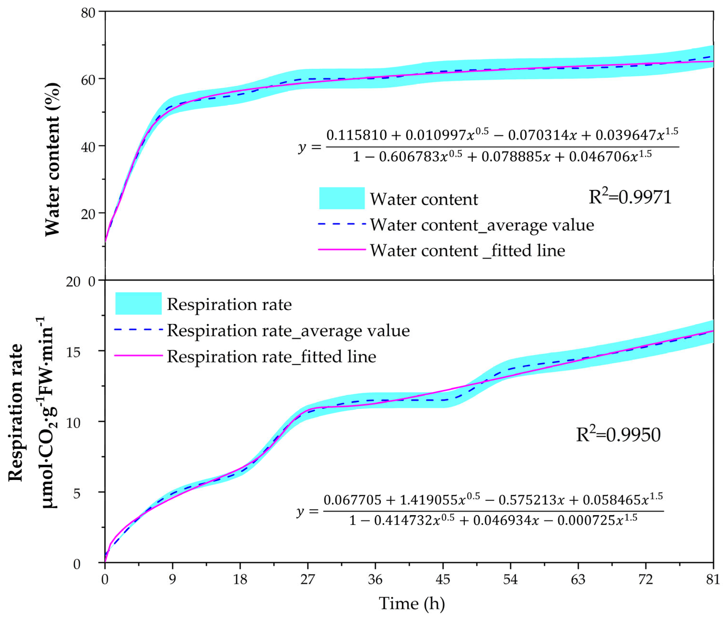
3.1. Changes in Water Content and Respiration Rate of Seeds

At 0–9 h, the water content of the Toona sinensis seeds increased significantly and was in the period of rapid water absorption, with the water content reaching 51.94 ± 2.6%, 4.39 times that of the dry seeds (Figure 2). The significant increase in seed water content during this period was mainly dependent on the swelling of colloidal substances. The substances constituting the seed cells contained proteins, pentosans, starch, and cellulose in protoplasts and cell walls, which were highly hydrophilic. Between 9 and 27 h, the water content increased to 59.86 ± 2.99% and the magnitude of water absorption by the seeds became smaller (Figure 2). After 9 h, the degree of hydration of protoplasts tended to be saturated, and vesicles, as well as a large number of new protoplasts, had not been formed. Therefore, the seeds lacked the impetus to absorb water, and the rate of water absorption slowed down. After 27 h, the water content of the seeds gradually stabilized. In the late stage of seed culture, the cells of seed embryos began to divide, the embryos elongated rapidly, the cell volume increased, and the organic matter stored in the seeds was hydrolyzed into highly permeable small molecules on a large scale. The water absorption at this time was metabolism-related osmotic water absorption.
Generally, the respiratory metabolism is continuously strengthened during seed germination. T. sinensis seeds also follow this law, and the change in their respiration rate can be mainly divided into the following three stages: during 0–27 h, the respiration rate of the seeds increased rapidly, from 0.55 ± 0.03 μmol·CO2·g−1 FW·min−1 at the beginning to 10.65 ± 0.53 μmol·CO2·g−1 FW·min−1. With this increase in the water content of the seeds, the enzyme system was continuously repaired and activated, and the respiration rate of the originally “quiescent” seeds was enhanced significantly. During 27–45 h, due to the limitations of the seed coat and the lack of significant formation of new respiratory enzymes and mitochondria, the seed respiration rate changed gently. After 45 h, the respiration rate increased dramatically, reaching a maximum value of 16.39 ± 0.82 μmol·CO2·g−1 FW·min−1 at 81 h (Figure 2). During this period, the embryonic axis grew, new mitochondrial synthesis intensified, respiratory enzymes were activated, respiration increased dramatically, and the rate of respiration increased rapidly.

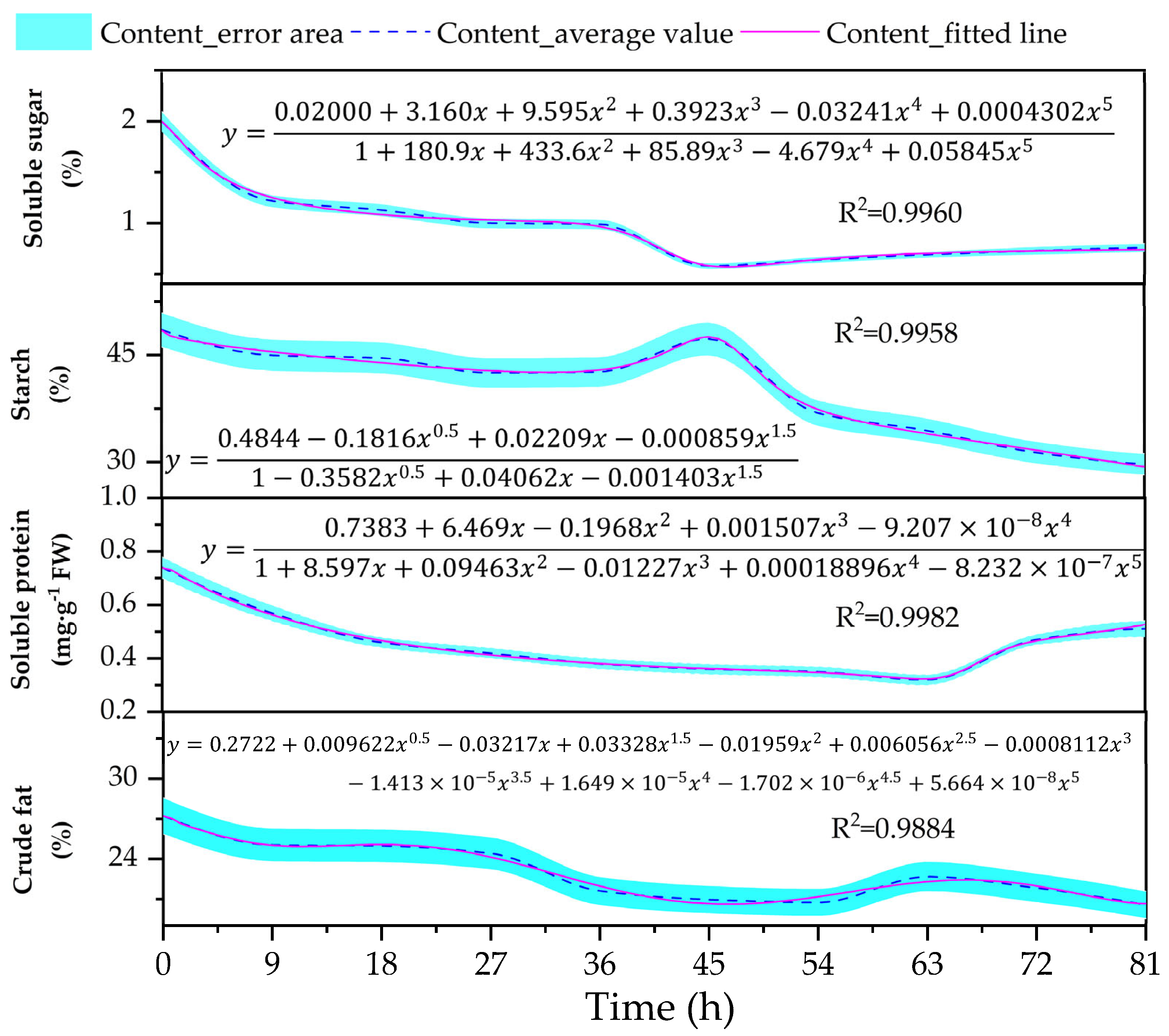
3.2. Changes in the Content of Reserve Materials (Soluble Sugar, Starch, Soluble Protein, and Crude Fat) in the Embryos

The soluble sugar content in the seed embryos of T. sinensis showed a tendency to decrease and then increase (Figure 3). At 9 h, the soluble sugar content decreased significantly. The soluble sugar content declined relatively slowly between 9 and 18 h, and then decreased sharply again after 27 h to a minimum value of 0.64% at 45 h, a decrease of 71%. After 45 h, the soluble sugar content began to rebound and continued until 81 h. There was a general trend of gradual decline in starch content within the seed embryos (Figure 3). During the first 27 h, the starch content showed a decreasing trend, and the difference between 27 h and 0 h was significant. During the period of 27–45 h, the starch rebounded, and after 45 h, the starch content decreased sharply again, reaching the lowest value at 81 h, with a decrease of 41.35%. The soluble protein content decreased continuously from 0 h to 63 h and reached the lowest value of 0.32% at 64 h (Figure 3), and then increased again until the end of the nursery period, with a significant difference between 63 h and 72 h. The crude fat in the seed embryos of T. sinensis was degraded continuously during germination, and decreased significantly at 9 h. From 9 h to 27 h, the crude fat content decreased slowly, and the content change was not significant. Then, it decreased rapidly at 36 h, and although there was a brief rebound at 54 h, the change was not significant (Figure 3).

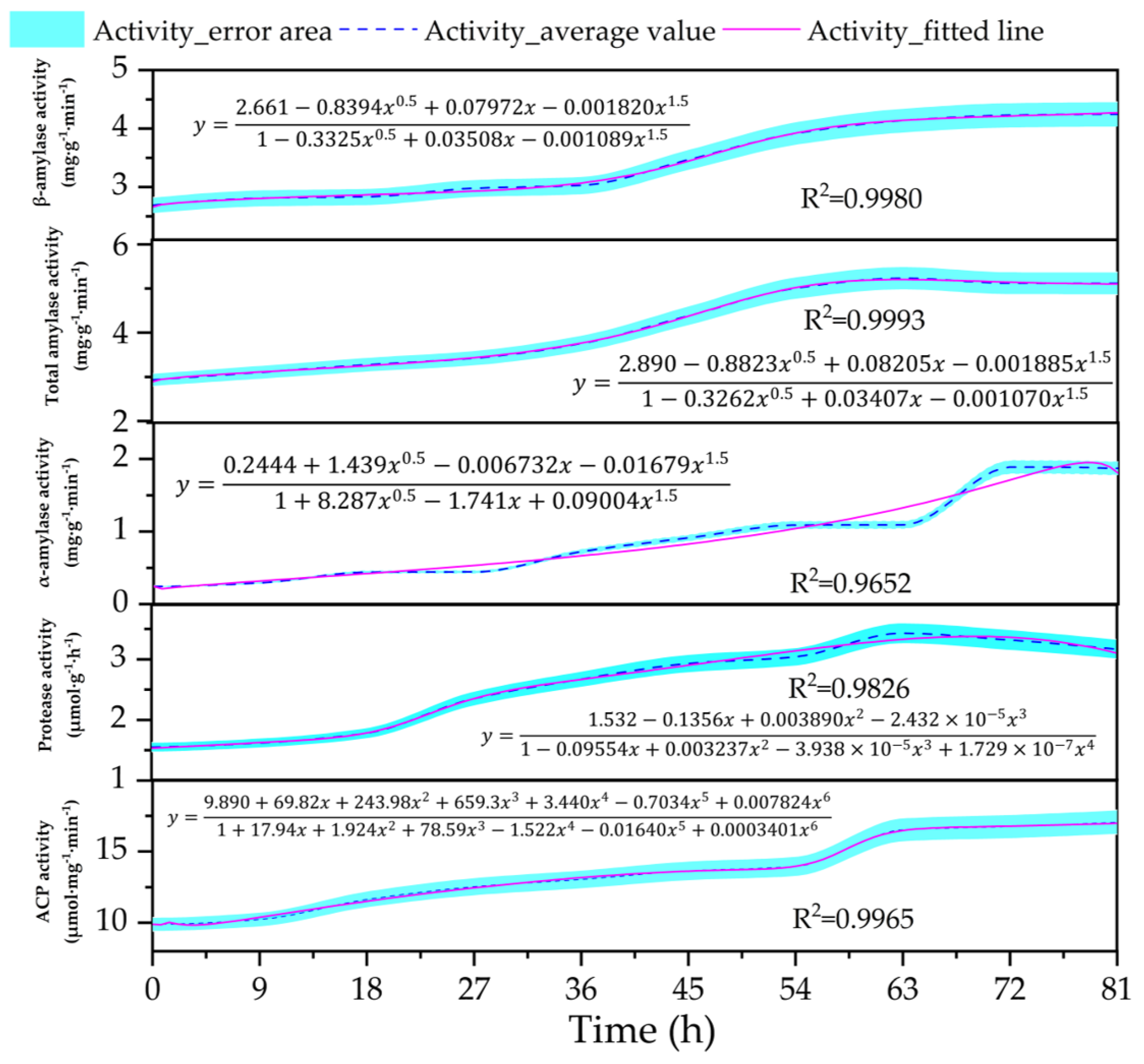
3.3. Changes in the Content of Metabolic Enzyme Activity (Amylase, Protease, Acid Phosphatase) in the Embryos

With the extension of incubation time, the activities of α-amylase and β-amylase increased and the total amylase activity also showed an upward trend (Figure 4). At 18 h, at which time seed metabolic activity began to intensify, α-amylase activity rose rapidly, reaching a peak at 72 h (1.89 ± 0.10 mg·g−1·min−1), and then there was a small decrease in the late stage of radicle breakthrough (72–81 h), but it was still in a high state of activation (Figure 4). β-amylase activity showed an increasing trend from the beginning of seed water absorption to germination and reached a maximum value at 81 h (4.25 ± 0.21 mg·g−1·min−1) (Figure 4). When the seed radicle and germ were about to break through the skin, the branched-chain starch was mainly hydrolyzed to produce monosaccharides, supplying the growth and development of the seedling, so the β-amylase activity reached its maximum. In addition, it was found by comparison that the enzyme degrading starch in the germination process of T. sinensis seeds was mainly β-amylase, whose activity was generally higher than that of α-amylase. Protease activity showed a tendency to increase and then decrease, the increment in protease activity was not large during the first 18 h of seed culture, and after 18 h of culture, the protease activity increased rapidly. The nutrients required by the plumule were maximized at 63 h, when the protease activity was the highest (3.43 ± 0.17 μmol·g−1·h−1). After 63 h, the activity began to decrease until the end of culture (Figure 4). The ACP activity in the seed embryos increased continuously with an increase in incubation time: this increase in activity was large from 0 to 27 h; relatively flat from 27 to 54 h; rapidly increased after 54 h, and reached a peak of 17.06 ± 0.85 μmol·mg−1·min−1 at 81 h (Figure 4). ACP was closely related to the metabolism of material and energy in the embryos, which required a rapid elevation of its activity to accommodate embryo growth.

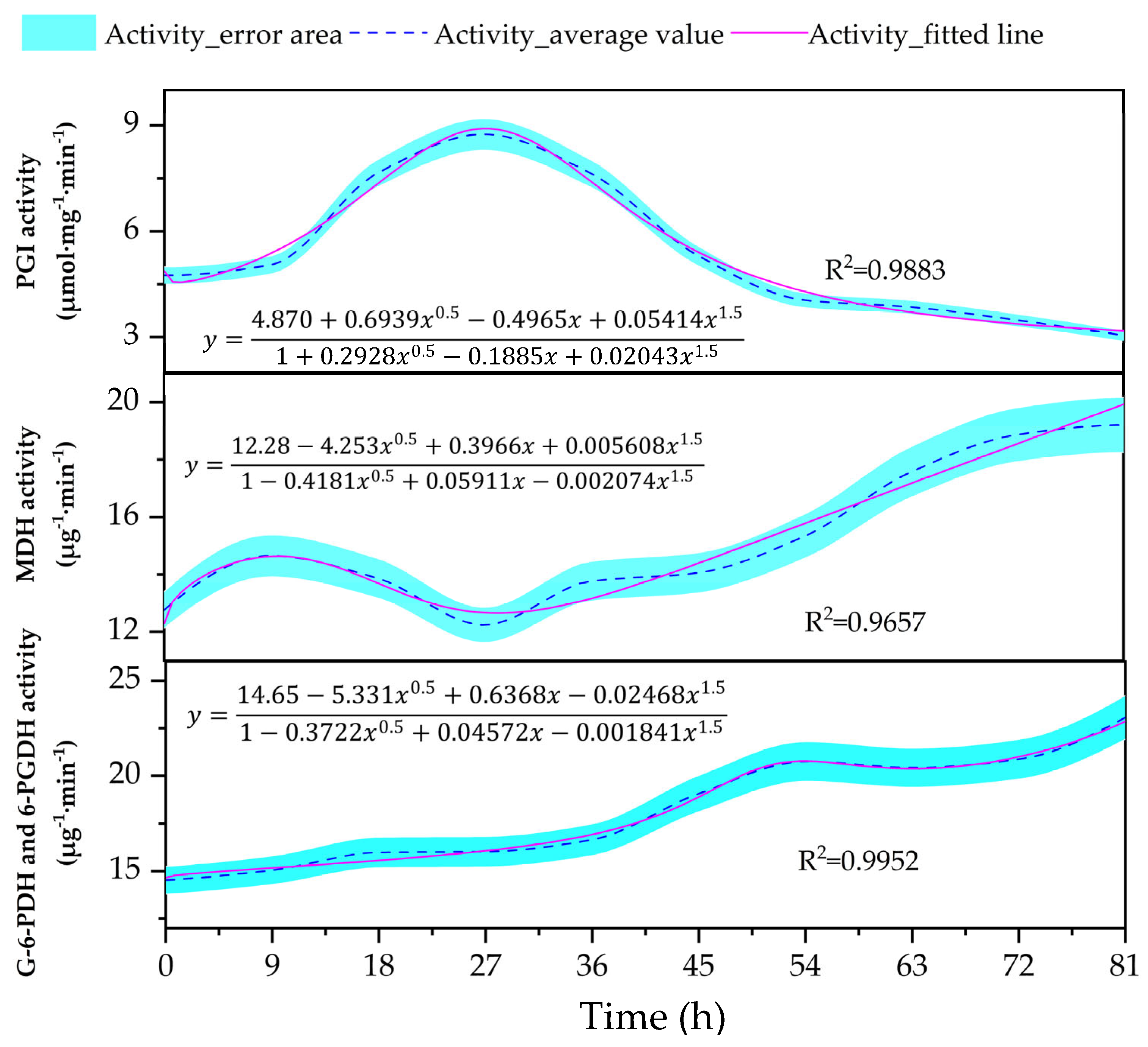
3.4. Changes in the Content of Key Oxidative Pathway Enzyme Activity (PGI, MDH, G-6-PDH, and 6-PGDH) in the Embryos

As shown in Figure 5, the changes in PGI activity in the seed embryos at the initial stage of seed germination can be divided into the following two stages: a 0–27 h rising stage and a 27–81 h declining stage. The PGI activity in the seed embryos increased rapidly after seed incubation and reached a maximum value of 8.75 μmol·mg−1·min−1 at 27 h, 84.12% higher than that at 0 h. Following 27 h, the PGI activity declined rapidly again and decreased to the lowest value of 3.04 μmol·mg−1·min−1. The activity of MDH in the seed embryos was generally on the rise during the period of 0–27 h. The activity of MDH firstly increased and then decreased, and the difference was significant at 9 h compared with that at 0 and 27 h. At 36 h, MDH activity increased significantly and reached a maximum value of 19.21 u·g−1·min−1 at 81 h (Figure 5). The activity of G-6-PDH and 6-PGDH gradually increased with the prolongation of incubation time. The activities of G-6-PDH and 6-PGDH in the seed embryos increased gradually. Before 27 h, the activity of the co-enzymes did not increase significantly, and at 36 h, the co-enzyme activity reached the highest value of 23.07 u·g−1 min−1 when the radicle broke through the seed shell and the auxin activity reached a maximum value of 23.07 u·g−1 min−1, 58.88% higher than that of dry seeds (Figure 5).

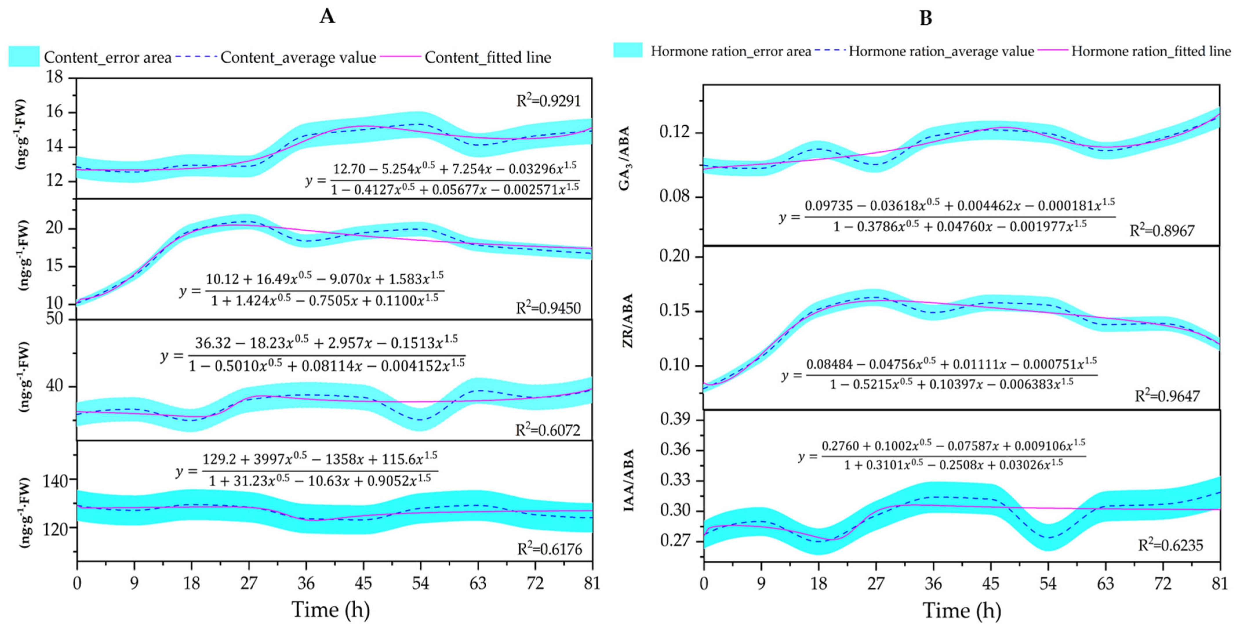
3.5. Changes in the Content of Endogenous Hormones (GA3, ZR, IAA, and ABA) and Ratios (GA3/ABA, IAA/ABA, and ZR/ABA) in the Embryos

With the prolongation of incubation time, the changes in GA3 content in the seed embryos of T. sinensis can be divided into the following three stages: the GA3 content of the seed embryos did not change much during the period of 0–27 h, basically remaining at 12.57–12.96 ng·g−1·FW. It rose significantly at 36 h and peaked at 54 h (15.32 ng·g−1·FW), declined briefly, and then continued to climb until the end of the incubation, but there was no significant difference between 54 h and several subsequent time points (Figure 6A). The ZR content in the embryo showed a general upward trend, rising sharply from 0 to 27 h, with a first peak of 20.98 ng·g−1·FW at 27 h, then rising again after a brief decline to reach a second peak of 19.98 ng·g−1·FW at 54 h, after which the content gradually fell back to 16.76 ng·g−1·FW at the end of incubation. This was 64.96% higher than the initial stage (0 h) (Figure 6A). The IAA levels in the seed embryos showed a fluctuating trend from 0 to 18 h with little overall change. There was a significant increase at 36 h compared to 18 h, a decrease at 54 h followed by a significant increase, and a maximum value of 39.43 ng·g−1·FW at 81 h (Figure 6A). However, there was no significant difference between the time points of ABA during the germination of the T. sinensis seeds, and the content generally showed a decreasing trend (Figure 6A).
With the prolongation of seed culture time, the ratios of GA3/ABA, IAA/ABA, and ZR/ABA in the seed embryos of T. sinensis showed different degrees of elevation, where the ratio of IAA/ABA was always higher than those of GA3/ABA and ZR/ABA and was at a relatively high level (Figure 6B). The IAA/ABA ratio was small in the first 18 h, then gradually increased after 18 h, and then continued to increase after a short drop at 54 h, reaching the maximum value at the end of culture. The ZR/ABA ratio gradually increased within 27 h, and then decreased in a fluctuating manner. The GA3/ABA ratio did not change significantly in 27 h, then gradually increased, dropped slightly at 54–63 h, and then continued to increase until it peaked at the end of the incubation period.

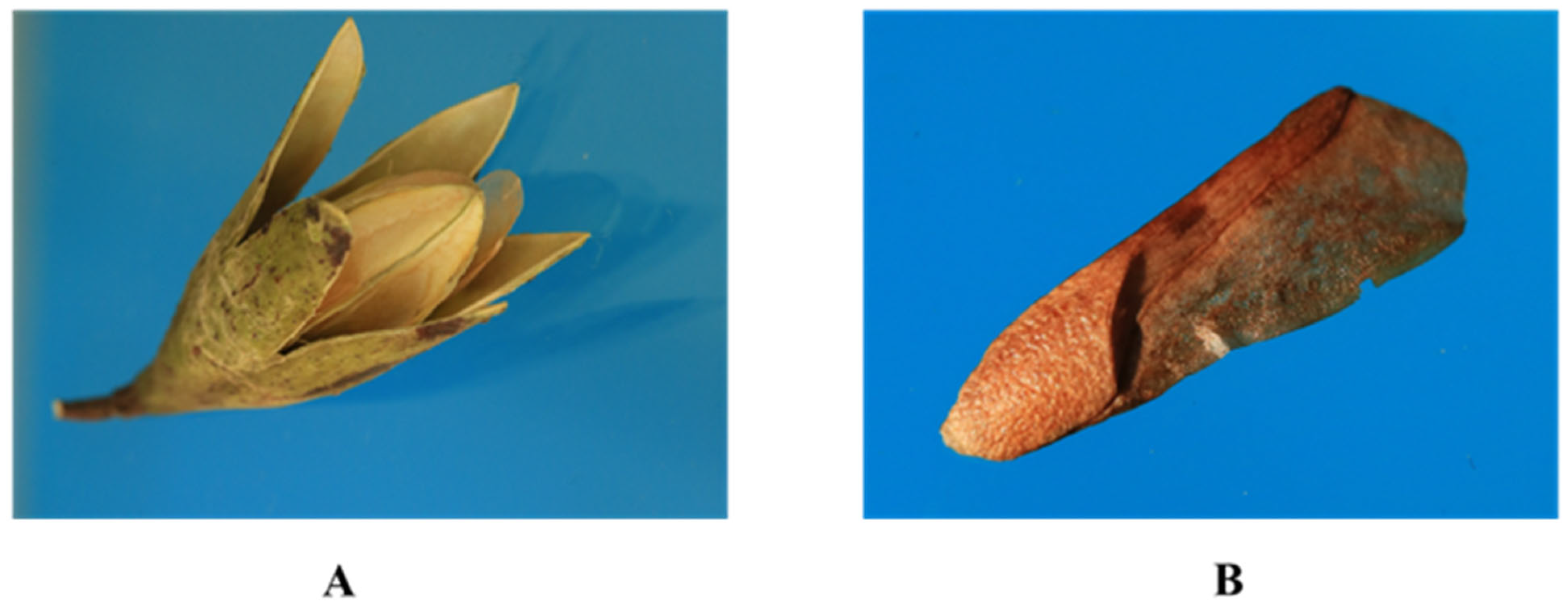
3.6. Morphological Changes in the Contents of Cotyledon Cells

The endosperm cells were composed of lipid droplets, starch granules, and dense substances of different shapes at 0 h. At 9 h of incubation, the lipid droplets were regularly arranged around the dense substances of different shapes, and the starch granules were distributed close to the cell wall. At 18 h of incubation, the dense substances were degraded and the cell volume was significantly reduced; the lipid droplets were degraded and their content was significantly reduced; and the starch granules were distributed around the nucleus. At 27 h and 36 h, the lipid droplets and dense substances were fused with each other, and the lipid droplets were completely ablated, leaving behind white vacuoles. Small fragments of incompletely degraded lipid droplets were distributed in some of the white vacuoles. The starch granules were distributed close to the nucleus. At 45 h and 54 h of incubation, the dense substances were ablated and became less abundant, and the number of white vacuoles increased significantly after the degradation of the lipid droplets. The starch granules were basically distributed close to the nucleus and also free in the cell. At 63 h of incubation, the dense substances fused and disintegrated into flocculent or small fragments. The lipid droplets were partially fused. The content of starch granules increased. At 72 h of incubation, the dense substances were almost completely fused. The lipid droplets were mostly fused. The content and volume of starch granules increased significantly. At 81 h, the dense substances and lipid droplets were fused. The content of starch granules decreased, but their volume increased significantly. The nuclei were degraded (Figure 7).

4. Discussion

4.1. Changes in Seed Water Content and Respiration Rate

Water serves as both the medium and regulatory factor for seed metabolic processes, enabling enzymatic reactions and reserve mobilization [23]. Traditional seed imbibition models describe the following three phases: rapid water uptake, metabolic plateau, and secondary rapid hydration [24]. However, T. sinensis seeds exhibited an atypical hydration profile characterized by an initial 9 h rapid hydration phase followed by stabilization without subsequent re-rapid uptake. This pattern aligns with observations in Camellia oleifera [25], Gentiana veitchiorum [26], and Cucumis anguria [27], supporting the notion that triphasic imbibition is not universally applicable. This exceptional hydration capacity, potentially adaptive to warm humid climates, coincides with early metabolic activation: unlike traditional models attributing early imbibition to passive physical swelling, T. sinensis seeds demonstrated simultaneous respiratory surges during hydration, indicating the premature activation of biochemical pathways (e.g., substrate mobilization and enzyme synthesis) that prime germination processes. Concurrently, respiration activity conformed to the three-stage rule of the general theory [28]. Notably, hydration and respiration exhibited temporal decoupling: water uptake plateaued at 9 h while respiration peaked at 45 h. This asynchronous physiology suggests T. sinensis employs a dual adaptive strategy, where rapid hydration ensures immediate metabolic substrate availability while the delayed respiratory peak optimizes energy allocation for germination completion. Such coordination likely contributes to the species’ high germination success in subtropical environments, where rapid water acquisition and metabolic flexibility confer competitive advantages.

4.2. Changes in Reserve Materials Within Embryos

During seed germination, the nutrients (sugars, proteins, and fats) stored in endosperms or cotyledons were metabolized into simple organics to sustain embryonic growth [29]. T. sinensis seeds, classified as starch-type seeds, primarily store nutrients in cotyledons. During germination, all reserves generally decreased, with distinct utilization patterns. In the early phase (0–36 h), soluble sugar, crude fat, and soluble protein levels dropped sharply, indicating rapid mobilization for initial metabolic demands. This is similar to rapeseed seeds. Batool et al. found that the active metabolism of storage compounds, including sugars, lipids, and proteins, significantly contributes to seed germination processes and may play an important role in rapeseed seeds [30]. In the mid-phase (27–45 h), starch temporarily accumulated due to transient anabolism, ensuring energy reserves [31,32]. This is similar to red rice seeds. Gianinetti et al. found that the putative reconstruction of general metabolism prompted a preferred usage of carbon and nitrogen resources for biosynthetic processes in the plastid in red rice seeds during imbibition, including starch and PA accumulation [33]. In the late phase (>45 h), starch degradation accelerated, while soluble protein rebounded (63–81 h) via amino acid recycling, supporting elongation growth. Similarly, Galland et al. found that in the imbibition phase of Arabidopsis seed germination, storage proteins were degraded to provide the nutrients and energy required [34]. Notably, soluble sugar replenishment after 45 h may occur through starch hydrolysis and glyoxylate-cycle-mediated fat conversion, highlighting metabolic adaptability [35,36]. Similar to this, Penfield found et al. that germinating Arabidopsis thaliana seeds broke down fatty acids through peroxisomal β-oxidation and converted carbon into soluble carbohydrates through the glyoxylate cycle and gluconeogenesis [37].

4.3. Changes in the Activity of Hydrolytic Enzymes Within Embryos

During seed germination, enzymes orchestrate the breakdown of stored reserves to fuel embryonic growth [38], with the amylase, protease, and acid phosphatase (ACP) activities in T. sinensis embryos dynamically reflecting substrate mobilization. Initially, dry seeds exhibited low metabolic activity, but upon imbibition, enzymes were rapidly activated or synthesized. α-Amylase, synthesized de novo during germination, remained at low levels until 27 h post-imbibition due to minimal starch mobilization; thereafter, its activity rose sharply, driven by protein-derived amino acids or gibberellic acid (GA) secreted by the embryo, which induced its synthesis (GA3 in Figure 6 and α-amylase in Figure 4 were positively correlated) [39]. β-Amylase, an exo-acting glucanase, acted sequentially after α-amylase to hydrolyze amylopectin [40], with its activity increasing gradually as starch degradation progressed. Li et al. pointed out that the germination of wheat seeds depends largely on their β-amylase activity, which is related to the rate of germination of plant seeds [41]. It was also found that the activity of β-amylase was significantly higher than that of α-amylase at different germination periods, indicating that T. sinensis seeds hydrolyzed starch and glycogen mainly by β-amylase. Protease activity, initially low (0–18 h), surged as the embryos’ demand for nitrogenous nutrients escalated, facilitating the degradation of structural proteins to support organogenesis and growth. After 63 h, most of the proteins in the embryo were degraded, and the protease lost its action substrate, so the activity began to decrease. Concurrently, ACP activity—a regulator of organic phosphate hydrolysis—remained low during early germination (0–54 h), but increased markedly thereafter as the embryos’ energy and nutrient demands escalated, aligning with its role in accelerating the breakdown of crude fats, proteins, and soluble sugars [25]. From Figure 2 and Figure 4, it can already be seen that the changes in ACP activity were also basically consistent with the changes in seed respiration rate, verifying the direct correlation between ACP and the energy metabolism of cells. These coordinated enzyme dynamics underscore the transition from dormancy to active growth, with α-amylase and protease activity surges marking starch and protein hydrolysis, respectively, while ACP activity escalates to sustain energy and nutrient supply for embryogenesis.

4.4. Changes in the Activities of Key Enzymes of Oxidative Pathways Within Embryos

During seed germination, respiratory metabolism transitions through the following three primary pathways: glycolytic pathway (EMP) (carbohydrate metabolism hub), tricarboxylic acid cycle (TCA) (energy/intermediate supply), and PPP (NADPH/raw material provider) [32,42,43]. Phosphohexose isomerase (PGI), malate dehydrogenase (MDH), glucose-6-phosphate dehydrogenase, and 6-phosphogluconate dehydrogenase (G-6-PDH and 6-PGDH) are among the key enzymes of these three metabolic pathways, respectively, and their activities can reflect changes in the status of these three respiratory pathways during seed germination. Initially (0–27 h), high PGI activity indicated the dominance of the EMP pathway, meeting minimal energy demands of early embryo growth. As germination progressed (>27 h), PGI activity plummeted while MDH (TCA-linked) and G-6-PDH/6-PGDH (PPP-linked) surged, reflecting a metabolic shift to PPP/TCA to support escalating energy/material requirements. Notably, PPP became predominant in mid-to-late germination, as evidenced by sustained G-6-PDH/6-PGDH superiority over MDH and corroborated by studies linking PPP activation to storage protein mobilization and dormancy release. This EMP-to-PPP transition, critical for successful germination in T. sinensis, aligns with hypotheses that PPP dominance ensures an adequate reducing power and metabolic intermediates for embryogenic growth. This is similar to Phyllostachys edulis seed, but not exactly the same. Li et al. found that the EMP pathway and TCA cycle were enhanced during Phyllostachys edulis seed germination [28]. Meanwhile, this is also similar to T. miqueliana. Wu et al. found that EMP is the primary respiratory pathway in dormant seeds, while the TCA cycle and PPP pathway become critical in non-dormant seeds [44].

4.5. Changes in Endogenous Hormone Content in Embryos

Plant hormones—gibberellins (GAs), cytokinins (CTKs), auxins (IAAs), and abscisic acid (ABA)—orchestrate seed dormancy, germination, and early growth through dynamic concentration shifts and hormonal cross-talk [45]. In Toona sinensis seeds, GA3 (the most active gibberellin) was synthesized by the embryo and accumulated during germination, promoting enzyme activities (e.g., α-amylase and protease) that hydrolyzed endosperm reserves and accelerated embryonic cell division [46]. Cytokinins, such as zeatin riboside (ZR), enhance embryo cell proliferation and organogenesis through their role in membrane permeability and protein synthesis [47]. We hypothesized that early ZR elevation could promote the continuous division and growth of young embryos of T. sinensis and play a key regulatory role in the pre-emergence stage of seeds. IAA levels fluctuated and increased, but remained low. Some studies have found that low concentrations of IAA can promote seed germination, because IAA can activate cell wall relaxation proteins and promote the growth of radicles [48]. ABA can hinder germination by inhibiting radicle elongation, cell division, enzyme activity (e.g., α-amylase suppression), and the related transcription regulated by GAs [49,50]. Liu et al. showed that ABA levels decreased from the onset of imbibition, and this decrease in ABA levels was mainly related to an increased expression of the OsABA8ox gene [51]. In this study, the ABA content in the cotyledons of T. sinensis seeds exhibited a fluctuating downward trend, which was beneficial for successful seed germination. In addition, ABA is involved in water and fertilizer uptake within plants [52,53]. A transient rebound in ABA content was also observed during 45–63 h, which may have been due to the seeds’ urgent need for sufficient nutrients and water to maintain normal cellular metabolism [54]. The changes in endogenous hormone content observed in this study are consistent with the findings of Zhang [55].
The balance between growth-promoting (GA3, ZR, and IAA) and growth-inhibiting (ABA) hormones dictates germination success, as evidenced by the elevated GA3/ABA, IAA/ABA, and ZR/ABA ratios during early T. sinensis germination. The GA/ABA ratio is recognized as a key factor affecting seed germination [56]. It has been shown that, in rice seeds, the AP2 structural domain TF Os AP2-39 regulates germination by regulating the balance between ABA and GA levels [57]. Yan et al. suggested that the decrease in ABA content during the lifting of seed dormancy in Zelkova schneideriana led to an increase in the levels of the hormones IAA and CTKs, and the signaling mediated by them was gradually activated [58]. Wang et al. found that high levels of ABA did not inhibit the germination of Suaeda acuminata seeds, because high concentrations of ZR may counteract the negative effects of ABA [59]. This further illustrates the importance of hormone balance during seed germination. In the pre-culture period, the changes in the ratios of GA3/ABA and IAA/ABA were not significant, while the ratio of ZR/ABA increased rapidly, indicating that the antagonistic effects of ZR and ABA mainly dominated in the pre-culture period during the initial stage of T. sinensis seed germination, whereas the interactions of GA3, IAA, and ABA played dominant roles mainly in the mid-to late-stage. These findings indicate that increases in the ratios of growth-promoting hormones and growth-inhibiting hormones positively promote seed germination. These experimental results are in general agreement with Si et al. [60].

4.6. Ultrastructural Changes in Cotyledon Cell Contents

Transmission electron microscopy (TEM) of T. sinensis cotyledon cells revealed dynamic ultrastructural changes during germination, characterized by the degradation of high-electron-density osmiophilic substances (primarily lipoproteins) and lipid droplets, alongside the accumulation of starch granules. These alterations align with the nutrient mobilization required for embryonic growth, as lipids and proteins were catabolized to fuel germination. Wu et al. studied the ultrastructural changes in endosperm inclusions of Tilia miqueliana, and found that with the release of seed dormancy of Tilia miqueliana, the lipid droplets and high-electron-dense substances in the endosperm cells were gradually degraded and ablated, and finally completely disintegrated [44]. The lipoproteins in T. sinensis cotyledons underwent dual-mode (internal and peripheral) degradation, contrasting with the threefold mechanisms (internal, peripheral, and combined) reported in Populus euphratica [61]. Lipid droplets, initially swollen by water uptake, remained intact for the first 18 h of incubation, but were progressively degraded thereafter, coinciding with radicle emergence. Lipid breakdown likely proceeded via mitochondrial β-oxidation and the glyoxylate cycle, which converts fatty acids to sugars that accumulate as starch in plastids [62]. Simultaneously, starch granules—classified as compounds due to their aggregated structure—emerged after 18 h and proliferated until radicle protrusion, paralleling the degradation of lipids and proteins. This coordinated metabolic shift, where triglycerides were hydrolyzed by lipase and channeled into starch biosynthesis via the glyoxylate cycle, mirrors findings in the Coix lacryma-jobi scutellar epithelium and underscores the critical role of ultrastructural remodeling in sustaining early seedling vigor [63].

5. Conclusions

This study reveals T. sinensis seeds employ a biphasic hydration strategy of rapid water uptake followed by stabilization, omitting the re-rapid phase of classical models, suggesting adaptive specialization for humid subtropical niches. Respiration followed a triphasic, temporally decoupled pattern (surge, stagnation, and secondary elevation), indicating independent metabolic and hydraulic control. As a starch-dominant seed, it prioritized the immediate mobilization of soluble sugars, proteins, and lipids while conserving starch reserves, with enzyme kinetics (amylase, protease, and phosphatase) precisely tracking substrate depletion. Metabolic flux shifted from glycolysis dominance (EMP, early stage) to PPP dominance (mid–late stage), optimizing the NADPH/ATP balance for rapid germination. Hormonal crosstalk, notably early ZR surges and GA3/ABA ratio elevations, triggered reserve breakdown and radicle emergence, confirmed by lipid-to-starch transitions in cotyledons. These findings enable the following targeted interventions: developing seed priming protocols using osmotic agents to mimic rapid hydration phases, potentially accelerating germination in nurseries; formulating hormone-balanced growth regulators to synchronize metabolic and respiratory peaks during large-scale propagation; and integrating metabolic flux analysis into breeding programs to select for PPP pathway efficiency. Future priorities include the CRISPR-based editing of seed coat permeability genes, longitudinal ecological monitoring of assisted restoration sites, and deciphering the epigenetic regulation of starch–lipid transition dynamics under climate change scenarios.

Author Contributions

Conceptualization, L.L. and Y.W.; methodology, L.L.; software, Z.W.; formal analysis, L.L.; investigation, L.L. and Z.W.; resources, Y.W.; data curation, L.L. and Z.W.; writing—original draft preparation, L.L.; writing—review and editing, Y.W. and Y.S.; funding acquisition, Y.S. All authors have read and agreed to the published version of the manuscript.

Funding

This research was funded by the Innovation and popularization of forest technology in Jiangsu Province, Long-term scientific research base for the in vitro conservation of ray native tree germplasm resources in Jiangsu Province, grant number LYKJ [2021] 03.

Data Availability Statement

The data presented in this study are available on request from the corresponding author.

Conflicts of Interest

The authors declare no conflicts of interest.

References

  1. Chen, C.J.; Wang, Q.; Lin, C.W. Progress on the pharmacological effects of Toona sinensis leaves. China Pharm. 2011, 22, 277–280. [Google Scholar]
  2. Zhao, Q.; Zhong, X.-L.; Zhu, S.-H.; Wang, K.; Tan, G.-F.; Meng, P.-H.; Zhang, J. Research advances in Toona sinensis, a traditional Chinese medicinal plant and popular vegetable in China. Diversity 2022, 14, 572. [Google Scholar] [CrossRef]
  3. Li, W.Z.; Ding, J.X.; Zhang, X.P.; Tian, J.Z. Advances in chemical composition, extraction methods and bioactivity of Toona sinensis. J. Pharm. Res. 2012, 31, 37–39. [Google Scholar]
  4. Yang, M.; Zhong, G.Y.; Gao, Y.H.; Lv, J.C.; Wang, R.Q.; Hang, R.Q.; Huang, P.; Li, H.X. Current status and research advances in propagation techniques of Toona sinensis. Bull. Agric. Sci. Technol. 2021, 11, 217–220. [Google Scholar]
  5. Fang, J.-Y.; Zhu, Y.; Wang, C.-Y.; Ye, K.-K.; Gao, W.-D.; Zhang, Z.-H.; Yan, J.-J.; Li, Q.-M. Physiological and biochemical changes of Toona sinensis seeds during artificial aging. For. Res. 2020, 33, 163–169. [Google Scholar]
  6. Hou, J.; Wang, D.; Su, P.; Ding, S.; Wu, L. In vitro large-scale propagation and genetic fidelity of Toona sinensis (Juss.) M. Roem., an economically important vegetable tree. Plant Cell Tissue Organ. Cult. 2022, 151, 275–291. [Google Scholar] [CrossRef]
  7. Hasanuzzaman, M.; Nahar, K.; Alam, M.M.; Roychowdhury, R.; Fujita, M. Physiological, biochemical, and molecular mechanisms of heat stress tolerance in plants. Int. J. Mol. Sci. 2013, 14, 9643–9684. [Google Scholar] [CrossRef]
  8. Ali, A.S.; Elozeiri, A.A. Metabolic processes during seed germination. Adv. Seed Biol. 2017, 2017, 141–166. [Google Scholar]
  9. Weitbrecht, K.; Müller, K.; Leubner-Metzger, G. First off the mark: Early seed germination. J. Exp. Bot. 2011, 62, 3289–3309. [Google Scholar] [CrossRef]
  10. Bewley, J.D.; Black, M. Seeds: Physiology of Development and Germination; Springer Science & Business Media: Berlin/Heidelberg, Germany, 2013. [Google Scholar]
  11. Ghose, T. Measurement of cellulase activities. Pure Appl. Chem. 1987, 59, 257–268. [Google Scholar] [CrossRef]
  12. Taylor, K.A. A modification of the phenol/sulfuric acid assay for total carbohydrates giving more comparable absorbances. Appl. Biochem. Biotechnol. 1995, 53, 207–214. [Google Scholar] [CrossRef]
  13. Bradford, M.M. A rapid and sensitive method for the quantitation of microgram quantities of protein utilizing the principle of protein-dye binding. Anal. Biochem. 1976, 72, 248–254. [Google Scholar] [CrossRef] [PubMed]
  14. Liu, Z.F. Seed Science Laboratory Manual; Chemical Industry Press: Beijing, China, 2011. [Google Scholar]
  15. Lowry, O.H.; Rosebrough, N.J.; Farr, A.L.; Randall, R.J. Protein measurement with the Folin phenol reagent. J. Biol. Chem. 1951, 193, 265–275. [Google Scholar] [CrossRef]
  16. Younessi-Hamzekhanlu, M.; Izadi-Darbandi, A.; Malboobi, M.A.; Ebrahimi, M.; Abdipour, M.; Sparvoli, F.; Paolo, D. Agrobacterium rhizogenes transformed soybeans with AtPAP18 gene show enhanced phosphorus uptake and biomass production. Biotechnol. Biotechnol. Equip. 2018, 32, 865–873. [Google Scholar] [CrossRef]
  17. Zhang, S.L. Effects of Salicylic Acid on Respiratory Pathway of Huang-Guan Pear; Agricultural University of Heibei: Baoding, China, 2008. [Google Scholar]
  18. Brown, A.; Wray, J. Correlated changes of some enzyme activities and cofactor and substrate contents of pea cotyledon tissue during germination. Biochem. J. 1968, 108, 437–444. [Google Scholar] [CrossRef]
  19. Bergmeyer, H. Methods of Enzymatic Analysis; Elsevier: Amsterdam, The Netherlands, 1956. [Google Scholar]
  20. Zhao, J.; Li, G.; Yi, G.-X.; Wang, B.-M.; Deng, A.-X.; Nan, T.-G.; Li, Z.-H.; Li, Q.X. Comparison between conventional indirect competitive enzyme-linked immunosorbent assay (icELISA) and simplified icELISA for small molecules. Anal. Chim. Acta 2006, 571, 79–85. [Google Scholar] [CrossRef]
  21. Wang, Y.; Li, B.; Du, M.; Eneji, A.E.; Wang, B.; Duan, L.; Li, Z.; Tian, X. Mechanism of phytohormone involvement in feedback regulation of cotton leaf senescence induced by potassium deficiency. J. Exp. Bot. 2012, 63, 5887–5901. [Google Scholar] [CrossRef] [PubMed]
  22. He, Z. Guidance to Experiment on Chemical Control in Crop Plants; Beijing Agricultural University Publishers: Beijing, China, 1993; pp. 60–68. [Google Scholar]
  23. Szczerba, A.; Płażek, A.; Pastuszak, J.; Kopeć, P.; Hornyák, M.; Dubert, F. Effect of low temperature on germination, growth, and seed yield of four soybean (Glycine max L.) cultivars. Agronomy 2021, 11, 800. [Google Scholar] [CrossRef]
  24. Bewley, J.D.; Bradford, K.; Hilhorst, H.; Nonogaki, H. Seeds: Physiology of Development, Germination and Dormancy, 3rd ed.; Springer: New York, NY, USA, 2012. [Google Scholar]
  25. Long, W.; Yao, X.H.; Lv, L.Y.; Wang, K.L. An analysis of seed traits and endogenous hormone levels after seed soakings in Camellia oleifera. J. Nanjing For. Univ. 2020, 44, 148–156. [Google Scholar]
  26. Nie, X.Y.; Bai, F.H.; Li, Z.; Zhang, S.S.; Chu, H.; Cai, S.J.; Li, C.H.; Xu, T.L. Study on the seed biological characteristics of two types of Gentiana plant seed in Qinghai. J. Qinghai Univ. 2024, 42, 27–33. [Google Scholar]
  27. Araújo, P.C.; Torres, S.B.; Benedito, C.P.; Paiva, E.P.d. Condicionamento fisiológico e vigor de sementes de maxixe. Rev. Bras. Sementes 2011, 33, 482–489. [Google Scholar] [CrossRef]
  28. Li, J.; Li, X.; Bai, Y.; Xie, Y.; Li, L.; Mu, S.; Gao, J. Transcriptome analysis of energy supply process during seed germination in Phyllostachys edulis. Plant Mol. Biol. Report. 2023, 41, 489–511. [Google Scholar] [CrossRef]
  29. Bewley, J.; Black, M. Seeds: Physiology of Germination and Development; Plenum Press: New York, NY, USA, 1985. [Google Scholar]
  30. Batool, M.; El-Badri, A.M.; Wang, C.; Mohamed, I.A.; Wang, Z.; Khatab, A.; Bashir, F.; Xu, Z.; Wang, J.; Kuai, J. The role of storage reserves and their mobilization during seed germination under drought stress conditions of rapeseed cultivars with high and low oli contents. Crop Environ. 2022, 1, 231–240. [Google Scholar] [CrossRef]
  31. Chen, Y.-S.; David Ho, T.-H.; Liu, L.; Lee, D.H.; Lee, C.-H.; Chen, Y.-R.; Lin, S.-Y.; Lu, C.-A.; Yu, S.-M. Sugar starvation-regulated MYBS2 and 14-3-3 protein interactions enhance plant growth, stress tolerance, and grain weight in rice. Proc. Natl. Acad. Sci. USA 2019, 116, 21925–21935. [Google Scholar] [CrossRef]
  32. Zhu, M.; Zang, Y.; Zhang, X.; Shang, S.; Xue, S.; Chen, J.; Tang, X. Insights into the regulation of energy metabolism during the seed-to-seedling transition in marine angiosperm Zostera marina L.: Integrated metabolomic and transcriptomic analysis. Front. Plant Sci. 2023, 14, 1130292. [Google Scholar] [CrossRef]
  33. Gianinetti, A.; Finocchiaro, F.; Bagnaresi, P.; Zechini, A.; Faccioli, P.; Cattivelli, L.; Valè, G.; Biselli, C. Seed dormancy involves a transcriptional program that supports early plastid functionality during imbibition. Plants 2018, 7, 35. [Google Scholar] [CrossRef] [PubMed]
  34. Galland, M.; Huguet, R.; Arc, E.; Cueff, G.; Job, D.; Rajjou, L. Dynamic proteomics emphasizes the importance of selective mRNA translation and protein turnover during Arabidopsis seed germination. Mol. Cell. Proteom. 2014, 13, 252–268. [Google Scholar] [CrossRef]
  35. Sharma, S.; Gambhir, S.; Munshi, S.K. Changes in lipid and carbohydrate composition of germinating soybean seeds under different storage conditions. Asian J. Plant Sci. 2007, 6, 502–507. [Google Scholar]
  36. Rosental, L.; Nonogaki, H.; Fait, A. Activation and regulation of primary metabolism during seed germination. Seed Sci. Res. 2014, 24, 1–15. [Google Scholar] [CrossRef]
  37. Penfield, S.; Graham, S.; Graham, I. Storage reserve mobilization in germinating oilseeds: Arabidopsis as a model system. Biochem. Soc. Trans. 2005, 33, 380–383. [Google Scholar] [CrossRef]
  38. Joshi, R. Role of enzymes in seed germination. Int. J. Creat. Res. Thoughts 2018, 6, 1481–1485. [Google Scholar]
  39. Damaris, R.N.; Lin, Z.; Yang, P.; He, D. The rice alpha-amylase, conserved regulator of seed maturation and germination. Int. J. Mol. Sci. 2019, 20, 450. [Google Scholar] [CrossRef]
  40. Yamaguchi, J.; Itoh, S.; Saitoh, T.; Ikeda, A.; Tashiro, T.; Nagato, Y. Characterization of β-amylase and its deficiency in various rice cultivars. Theor. Appl. Genet. 1999, 98, 32–38. [Google Scholar] [CrossRef]
  41. Li, Y.-J.; Hu, Q.-P. Studying of the promotion mechanism of Bacillus subtilis QM3 on wheat seed germination based on β-amylase. Open Life Sci. 2020, 15, 553–560. [Google Scholar] [CrossRef] [PubMed]
  42. Song, Y.; Gao, X.; Wu, Y. Key Metabolite differences between Korean Pine (Pinus koraiensis) seeds with primary physiological dormancy and no-dormancy. Front. Plant Sci. 2021, 12, 767108. [Google Scholar] [CrossRef]
  43. Botha, F.; Potgieter, G.; Botha, A.-M. Respiratory metabolism and gene expression during seed germination. Plant Growth Regul. 1992, 11, 211–224. [Google Scholar] [CrossRef]
  44. Wu, Y.; Bao, W.Q.; Hu, H.; Zhou, Y.J.; Shen, Y.B. Effects of H2SO4 and Gibberellin A3 on Dormancy Release of Tilia miqueliana Seeds. J. Plant Growth Regul. 2022, 41, 796–807. [Google Scholar] [CrossRef]
  45. Gong, D.; He, F.; Liu, J.; Zhang, C.; Wang, Y.; Tian, S.; Sun, C.; Zhang, X. Understanding of hormonal regulation in rice seed germination. Life 2022, 12, 1021. [Google Scholar] [CrossRef]
  46. Guo, H.; Li, J.; Liu, Y.; Fernández-Pascual, E. Lipid metabolism during seed germination of Pistacia chinensis and its response to gibberellic acid. Plant Physiol. Biochem. 2025, 219, 109371. [Google Scholar] [CrossRef]
  47. Chen, L.P. Studies on the Changes of Physiological and Biochemical Characteristics of Pinus tabuliformis Seeds During Initial Germinating Stages; Nanjin Forestry University: Nanjing, China, 2009. [Google Scholar]
  48. Liu, J.; Qiu, S.; Xue, T.; Yuan, Y. Physiology and transcriptome of Sapindus mukorossi seeds at different germination stages. Genomics 2024, 116, 110822. [Google Scholar] [CrossRef]
  49. Shun, G.Z.; Xiao, S.H. Abscisic Acid and Seed Dormancy. Plant Physiol. Commun. 2004, 40, 115–120. [Google Scholar]
  50. Gao, Y.; Zhu, M.; Wang, H.; Li, S. Dynamic changes to endogenous germination inhibitors in Cercis chinensis seeds during dormancy release. HortScience 2021, 56, 557–562. [Google Scholar] [CrossRef]
  51. Liu, Y.; Fang, J.; Xu, F.; Chu, J.F.; Yan, C.Y.; Schläppi, M.R.; Wang, Y.P.; Chu, C.C. Expression Patterns of ABA and GA Metabolism Genes and Hormone Levels during Rice Seed Development and Imbibition: A Comparison of Dormant and Non-Dormant Rice Cultivars. J. Genet. Genom. 2014, 41, 327–338. [Google Scholar] [CrossRef]
  52. Chen, K.; Li, G.J.; Bressan, R.A.; Song, C.P.; Zhu, J.K.; Zhao, Y. Abscisic acid dynamics, signaling, and functions in plants. J. Integr. Plant Biol. 2020, 62, 25–54. [Google Scholar] [CrossRef]
  53. Luo, X.; Chen, Z.; Gao, J.; Gong, Z. Abscisic acid inhibits root growth in Arabidopsis through ethylene biosynthesis. Plant J. 2014, 79, 44–55. [Google Scholar] [CrossRef] [PubMed]
  54. Liu, J.; Qiu, S.; Xue, T.; Yuan, Y. Physiology and transcriptome of Eucommia ulmoides seeds at different germination stages. Plant Signal. Behav. 2024, 19, 2329487. [Google Scholar] [CrossRef]
  55. Zhang, X.Y.; Yan, X.; Li, T.H.; Li, Y.L.; Zhang, Y.B.; Xing, L.; Guo, S.Y. Physiological responses of seed dormancy and germination to cold stratification in Phoebe hui Cheng ex Yang. Plant Sci. J. 2022, 40, 398–407. [Google Scholar]
  56. Tuan, P.A.; Rohit, K.; Rehal, P.K.; Toora, P.K.; Ayele, B.T. Molecular Mechanisms Underlying Abscisic Acid/Gibberellin Balance in the Control of Seed Dormancy and Germination in Cereals. Front. Plant Sci. 2018, 9, 668. [Google Scholar] [CrossRef]
  57. Yaish, M.W.; Ashraf, E.K.; Tong, Z.; Beatty, P.H.; Good, A.G.; Yong-Mei, B.; Rothstein, S.J.; Copenhaver, G.P. The APETALA-2-Like Transcription Factor OsAP2-39 Controls Key Interactions between Abscisic Acid and Gibberellin in Rice. PLoS Genet. 2010, 6, e1001098. [Google Scholar] [CrossRef]
  58. Yan, F.; Wei, T.; Yang, C.; Yang, Y.; Luo, Z.; Jiang, Y. Combined Analysis of Untargeted Metabolomics and Transcriptomics Revealed Seed Germination and Seedling Establishment in Zelkova schneideriana. Genes 2024, 15, 488. [Google Scholar] [CrossRef]
  59. Wang, H.L.; Wang, L.; Tian, C.Y.; Huang, Z.Y. Germination dimorphism in Suaeda acuminata: A new combination of dormancy types for heteromorphic seeds. South. Afr. J. Bot. 2012, 78, 270–275. [Google Scholar] [CrossRef]
  60. Si, Q.; Ma, Y.; Zang, D. The causes of dormancy and the changes of endogenous hormone content in Cephalotaxus sinensis seeds. Agric. Sci. 2016, 7, 834. [Google Scholar]
  61. Ge, C.H.; Ji, Q.L.; Liu, Y.P.; Wang, X.H. The Changes of Protein Bodies’ Ultrastructure in Cell of Seeds of Populus euphratic Oliv in the Process of Storage. Seed 2007, 5, 27–29. [Google Scholar]
  62. Qin, Z.; Wang, T.; Zhao, Y.; Ma, C.; Shao, Q. Molecular Machinery of Lipid Droplet Degradation and Turnover in Plants. Int. J. Mol. Sci. 2023, 24, 16039. [Google Scholar] [CrossRef]
  63. Dong, J.H.; Cui, D.C.; Shao, Z.Z.; Jin, L.F. Ultrastructural Changes and Localization of Acid Phosphatase in Scutellar Epi Thelium of Coix lacryma-jobi During Germination. Acta Biol. Exp. Sin. 1996, 29, 71–79. [Google Scholar]
Figure 1. Digital photos of T. sinensis’ fruit (A) and seed (B).
Horticulturae 11 00685 g001
Figure 2. Changes in water content and respiration rate of seeds during the initial stage of germination. The value at each time point is the average of three repetitions.
Horticulturae 11 00685 g002
Figure 3. Changes in soluble sugar, starch, soluble protein, and crude fat content in embryos during the initial stage of germination. The value at each time point is the average of three repetitions.
Horticulturae 11 00685 g003
Figure 4. Changes in total amylase, α-amylase activity, β-amylase activity, protease activity, and ACP activity content in embryos during the initial stage of germination. The value at each time point is the average of three repetitions.
Horticulturae 11 00685 g004
Figure 5. Changes in PGI, MDH, G-6-PDH, and 6-PGDH activity content in embryos during the initial stage of germination. The value at each time point is the average of three repetitions.
Horticulturae 11 00685 g005
Figure 6. (A) Changes in GA3, ZR, IAA, and ABA content in embryos during the initial stage of germination; (B) Changes in GA3/ABA, IAA/ABA, and ZR/ABA in embryos during the initial stage of germination. The value at each time point is the average of three repetitions.
Horticulturae 11 00685 g006
Figure 7. Morphological changes in the contents of cotyledon cells of T. sinensis seeds during initial germination stages (0, 9, 18, 27, 36, 45, 54, 63, 72, and 81 h). CW—cell wall, L—lipid droplet, N—cell nucleus, SG—starch granule. Bar = 1.25 μm.
Horticulturae 11 00685 g007
Disclaimer/Publisher’s Note: The statements, opinions and data contained in all publications are solely those of the individual author(s) and contributor(s) and not of MDPI and/or the editor(s). MDPI and/or the editor(s) disclaim responsibility for any injury to people or property resulting from any ideas, methods, instructions or products referred to in the content.

