Article

De Novo Assembly and Comparative Analysis of the Mitochondrial Genomes for Six Rubus Species

1
Zhejiang Key Laboratory for Restoration of Damaged Coastal Ecosystems, School of Life Sciences, Taizhou University, Taizhou 318000, China
2
Zhejiang Provincial Key Laboratory of Plant Evolutionary Ecology and Conservation, School of Life Sciences, Taizhou University, Taizhou 318000, China
3
Zhejiang International Science and Technology Cooperation Base for Biomass Resources Development and Utilization, School of Life Sciences, Taizhou University, Taizhou 318000, China
4
Institute of Horticulture, Taizhou Academy of Agricultural Sciences, Taizhou 318000, China
5
State Key Laboratory of Subtropical Silviculture, College of Forestry and Biotechnology, Zhejiang A&F University, Hangzhou 311300, China
*
Author to whom correspondence should be addressed.
Horticulturae 2025, 11(5), 559; https://doi.org/10.3390/horticulturae11050559
Submission received: 10 April 2025 / Revised: 19 May 2025 / Accepted: 20 May 2025 / Published: 21 May 2025
(This article belongs to the Special Issue Fruit Tree Physiology and Molecular Biology)

Abstract

:
Rubus is a genus of small berry-producing shrubs, valued for their medicinal properties and as a food source. This genus is a large, globally distributed group that includes over 700 species. Despite numerous plastid and nuclear genomes having been reported for Rubus, there is a notable lack of research on its mitogenomes. We utilized PMAT to assemble the mitogenomes of six Rubus species according to long-read HiFi reads and annotated them through homologous alignment. Subsequently, we compared their characteristic differences within Rubus mitogenomes. The complete mitogenomes of R. parviflorus, R. spectabilis, R. idaeus, R. armeniacus, and R. caesius all exhibit master circle structures, with lengths ranging from 360,869 bp to 447,754 bp. However, R. chamaemorus displays a double-circle structure composed of two small circular molecules, spanning 392,134 bp. These mitogenomes encode a total of 54–61 genes, including 33–34 PCGs, 17–24 tRNAs, and 3 rRNA genes. Compared to the other five Rubus species, R. chamaemorus has fewer sequence repeats. These six species exhibit similar codon usage patterns. A large number of gene transfers were detected between organellar genomes of six Rubus species. Additionally, two phylogenetic trees were constructed using 31 mitogenomes and 94 chloroplast genomes, revealing a minor conflict within Rubus. Overall, this study clarifies the mitogenome characteristics of Rubus and provides valuable insights into the evolution of the genus.

1. Introduction

Rubus L. is a genus in the family Rosaceae, comprising approximately 700 species, with 208 of those being found in China. Of these 208, 67% are endemic, making China, along with Western Europe and Eastern North America, geographic hotspots [1,2]. Fruits of Rubus species are edible when fresh [3] and abundant in phenols, terpenoids, minerals, vitamin C, and anthocyanins [4,5]. The most widely grown cultivars are selections of red raspberries (R. idaeus) and blackberries from the subgenus Rubus. There are also medicinal uses for certain Rubus species. For instance, the dried fruit of R. chingii serves as a traditional Chinese medicine [6], while the roots, stems, and leaves of some Rubus species are reported to be used to treat diarrhea and aid in wound healing [7,8]. Furthermore, phenomena such as hybridization, apomixis, polyploidization, and introgression are common in Rubus [9,10]. Ploidy levels vary significantly among different subgenera and species, with chromosome numbers ranging from diploid to fourteen-ploid (2n = 14–98) [11]. Focke categorized the genus Rubus into twelve subgenera [12] (Chamaemorus, Dalibarda, Chamaebatus, Comaropsis, Cylactis, Orobatus, Dalibardastrum, Malachobatus, Anoplobatus, Idaeobatus, Lampobatus, and Rubus), whereas the Flora of China recognized eight groups (Chamaemorus, Chamaebatus, Cylactis, Dalibardastrum, Malachobatus, Idaeobatus, Lampobatus, and Rubus) within the genus [13]. Consequently, a worldwide, unified classification system for Rubus is still lacking. Different classification systems exhibit considerable discrepancies in grouping and the delineation of subordinate taxa. However, current classification systems also face issues such as incorrect classification of species and varieties, questionable assignment of certain taxa, and ambiguous descriptions of species morphology. Additionally, a multitude of existing research outcomes, based on palynology [14], cytology, and molecular taxonomy [15,16], often conflict with the subordinate divisions of the classification systems, indicating a need for further refinement. This, in turn, hampers the development and utilization of the genus.
Mitochondria are semi-autonomous organelles found within eukaryotic cells. They play a crucial role in cellular respiration and energy conversion [17]. Mitochondria are primarily responsible for regulating cell growth, division, apoptosis, and the synthesis and breakdown of certain compounds [18]. These key metabolic processes exhibit semi-autonomous characteristics and possess their own genetic material and regulatory mechanisms. Their genetic variation can be valuable for studying plant phylogeny and patterns of diversity and can serve as a key marker for variant identification and development [19,20]. Plant mitogenomes consistently exhibit complex and dynamic structures. For instance, they display extreme variation in genome structure and size, a profusion of repetitive sequences, and the incorporation of foreign DNA sequences [21,22]. These factors have hindered the study of mitogenomes in relation to that of other plastid genomes. Thus, the majority of current plant systematics research focuses on nuclear and chloroplast genomes, with the complete assembly of plant mitochondria persisting as a bottleneck in evolutionary biology. With the advancement of high-throughput sequencing technology and the emergence of next-generation genomics, software programs suitable for sequencing and assembling the mitogenome have been developed, such as GetOrganelle v1.7.5 [23], GSAT v1.12 [24], and PMAT v2.1.0 [25]. These advances have made the sequencing and assembly of mitogenomes more accurate and efficient, providing valuable tools to deepen our understanding of the genetic characteristics and phylogenies of plants.
In the family Rosaceae, 34 mitogenomes have been reported and comparatively analyzed [26]. The results indicate that repetitive sequences primarily influence dynamic changes in the mitogenome structure through homologous recombination and genomic rearrangement. Currently, genomic research on Rubus species primarily concentrates on the chloroplast and nuclear genomes [1,2,5,27,28,29,30,31,32], with relatively few studies on its mitogenomes [3,26,33]. This paucity of mitogenome data has impeded phylogenetic studies within this genus. Consequently, there is an urgent need to analyze the mitogenomes of additional Rubus species. This will facilitate a comprehensive understanding of diversity within the genus, aid in advancing future phylogenetic research, and may support more effective protection and utilization of these genetic resources.
We assembled and annotated the mitogenomes of six Rubus species, conducted comparative analysis, and further analyzed their phylogeny. Together with 31 other mitogenomes and 94 plastid genomes, the evolutionary relationships and genetic backgrounds of Rubus were clarified. For examining the topological differences in the phylogenetic trees derived from plastids and mitochondria, we conducted multiple collinearity analyses within Rubus and identified a substantial number of gene rearrangements. The diversity of the mitogenomes within Rubus species and the complexity of their phylogeny may be closely related to frequent recombination and introgression. The purpose of this study is to analyze the mitogenome and provide a baseline for further exploration of the genetic variation, phylogeny, and molecular breeding of Rubus.

2. Materials and Methods

2.1. Data Acquisition, Genome Assembly, and Annotation

Based on published genome data, we downloaded PacBio sequencing data for nine Rubus species from the NCBI database, including R. caesius (ERR12875186), R. armeniacus (SRR30502861), R. parviflorus (SRR30533059), R. spectabilis (SRR30534156), R. argutus (SRR2161389), R. idaeus (SRR18212518), R. leucanthus (SRR29481259), R. occidentalis (SRR6675257), and R. chamaemorus (SRR30534586).
According to HiFi reads, we used PMAT v2.1.0 software to assemble plant mitogenomes with the “autoMito” model [25,34]. The initial assembly sequence was visualized using Bandage v0.8.1 software [35], and the contigs of chloroplast and nuclear genes were manually deleted. The HiFi reads were used to map the mitogenomes of Rubus to identify repetitive sequences through the Minimap v2.24 tool [36]. After uncovering these repeat sequences, six complete mitogenomes were generated (R. caesius, R. armeniacus, R. parviflorus, R. spectabilis, R. idaeus, and R. chamaemorus), while the remaining three species (R. argutus, R. leucanthus, and R. occidentalis) did not produce complete mitogenomes due to insufficient HiFi sequence data. Furthermore, the chloroplast genomes of four Rubus species (R. spectabilis, R. armeniacus, R. caesius, and R. chamaemorus) were also assembled using PMAT v2.1.0 software with the default parameters.
The complete mitogenomes of six Rubus species were annotated through the PMGA (http://47.96.249.172:16084/annotate.html, accessed on 10 January 2025) and GeSeq (https://chlorobox.mpimp-golm.mpg.de/geseq.html, accessed on 10 January 2025) tools [37,38], and two existing Rubus mitogenomes (R. chingii and R. suavissimus) were downloaded from the NCBI database as BLAST v2.9.0 reference sequences in GeSeq. The annotation of chloroplast genomes used CPGAVAS2 (http://47.96.249.172:16019/analyzer/home, accessed on 10 January 2025) and GeSeq online tools (https://chlorobox.mpimp-golm.mpg.de/geseq.html, accessed on 10 January 2025) [38,39]. Furthermore, tRNA and rRNA in the mitochondrial and chloroplast genomes were annotated with the tRNAscan-SE v2.0 tool and BLASTN v2.9.0 software [40,41], respectively. Since there may be errors in the annotation information, we used Geneious v21.0.7 software [42] to visually compare the information annotated using different tools and manually corrected them one by one. Finally, six complete mitogenomes were visualized via the PMGmap tool (http://47.96.249.172:16086/home/, accessed on 10 January 2025) [43], and all assembled sequences were uploaded to the GenBank database to obtain accession numbers. Details for all species accession numbers are recorded in Table S1.

2.2. Repeat Sequence Identification

We identified three types of repeat sequences in six complete mitogenomes, including simple sequence repeats (SSRs), dispersed sequence repeats, and tandem repeats (TRs). SSRs were detected using MISA-web (https://webblast.ipk-gatersleben.de/misa/index.php?action=1, accessed on 10 January 2025) [44], and the minimum number of repeats for SSR motifs with lengths of 1–6 were set to 10, 5, 4, 3, 3, and 3, respectively. TRs were identified via the Tandem Repeat Finder (https://tandem.bu.edu/trf/trf.html, accessed on 10 January 2025) [45] using the default parameters. And the REPuter tool (https://bibiserv.cebitec.uni-bielefeld.de/reputer, accessed on 10 January 2025) [46] was used to identify dispersed sequence repeats (including forward repeats, reverse repeats, palindromic repeats, and complementary repeats) with a Hamming distance of 3, and the minimum repeat size and maximum repeat time were set to 30 and 5000, respectively.

2.3. RNA Editing Predictions

All protein coding genes (PCGs) from six Rubus mitogenomes were extracted with the PhyloSuite v1.2.3 tool [47]. Then, we utilized the Deepred-Mt tool (http://47.96.249.172:16084/deepredmt.html, accessed on 10 January 2025) [48] to predict all potential editing sites for C to U, employing the default parameters, and ultimately set the threshold for the results to be greater than 0.9.

2.4. Ka/Ks Value Analysis

To evaluate selection pressures on PCGs in the Rubus mitogenomes, we used the PhyloSuite tool to extract shared PCGs in the mitogenomes of R. armeniacus, R. caesius, R. chamaemorus, R. idaeus, R. parviflorus, R. spectabilis, R. chingii, and R. suavissimus, with Arabidopsis thaliana as an outgroup. Then, the MAFFT v7.149b tool [49] was used to align sequences, and the KaKs_Calculator v2.0 tool [50] was used to determine the Ka and Ks ratio of each PCG.

2.5. Codon Usage Bias Analysis

The relative synonymous codon usage (RSCU) value was evaluated using the Genepionee cloud platform (http://cloud.genepioneer.com:9929/#/, accessed on 10 January 2025), which was also used to calculate the number of codon usages corresponding to each amino acid. The RSCU is the ratio of the frequency of codon observations to the frequency of codon usage. When the value of RSCU is less than 1, then codon usage is less frequent than expected, and the converse is also true. Finally, a circular heat map for the total number of codons used was plotted with the Chiplot online tool (https://www.chiplot.online/, accessed on 10 January 2025).

2.6. Mitochondrial and Plastid Sequence Migration Analysis

Six homologous sequences between Rubus mitochondria and plastids (MTPTs) were examined via BLAST, with the parameters set as identity > 70%, e-value < 1 × 10⁻⁵, and length greater than 30 bp. We analyzed the homologous sequence regions to determine the length, number, and annotation information of the migrated sequences. Sequence migration maps of the mitochondrial and plastid genomes were drawn using the “Advanced Circos” program in TBtools v2.210 [51].

2.7. Phylogenetic Analysis

To explore the evolutionary relationships of Rubus taxa in a broader context, we downloaded 25 plant mitogenomes from the NCBI database along with our six newly assembled mitogenomes for analysis, including four Magnoliids (Cinnamomum camphora, Magnolia liiflora, Machilus yunnanensis, and Liriodendron tulipifera), four Monocots (Oryza sativa, Zea mays, Allium cepa, and Cocos nucifera), and 23 Eudicots. The PCGs common to all species were extracted through PhyloSuite v1.2.3 software, the MAFFT v7.149b tool was used for multiple sequence alignment, and the trimAI tool was used for sequence trimming. Subsequently, we used IQ-TREE v2.2.0.3 to build a maximum likelihood (ML) tree with 5000 bootstrap replicates, using Magnoliids as the outgroup. Moreover, due to the limited number of mitogenomes of Rubus, we obtained all chloroplast genomes for this genus from the NCBI database, comprising 90 Rubus species. An ML phylogenetic tree based on chloroplast PCGs was constructed using the same method as above. Finally, the ML phylogenetic trees of both mitochondria and plastids were visualized via the Chiplot online tool (https://www.chiplot.online/, accessed on 10 January 2025) [52].

2.8. Collinearity Analysis of Mitochondrial Genome

In order to compare collinearity between Rubus mitogenomes, we detected homologous fragments between R. caesius and six other Rubus taxa by using MUMmer v4 with the default parameters [53]; only homologous fragments with alignment lengths greater than 500 bp were retained for analysis. The collinearity results were visualized using NGenomeSyn v1.39 [54].

3. Results

3.1. Characteristics of Rubus Mitogenomes

Based on HiFi sequencing data, we successfully assembled six complete mitogenomes of Rubus. Interestingly, five mitogenomes conformed to a single master circle (Figure 1A and Figures S1 and S2), while the mitogenome of R. chamaemorus had a double-circle conformation (Figure 1B). There were significant differences in their sizes, with R. armeniacus having the longest mitogenome, at 447,754 bp, whereas R. spectabilis had the shortest, spanning only 360,869 bp (Table S2). The GC content of the six mitogenomes was relatively stable, ranging from 43.8% to 44.6%, with a mean of 44.3%. Furthermore, we compared the annotated information for these six mitogenomes with two previously reported mitogenomes (R. chingii and R. suavissimus) [33], revealing that R. armeniacus had the most genes (61), including 34 PCGs, 24 tRNAs, and 3 rRNAs; R. spectabilis and R. chingii had fewer annotated genes (54). The mitogenomes of the eight Rubus species all contained 24 core PCGs, including ATP synthesis genes (atp1/4/6/8/9), cytochrome c biogenesis genes (ccmB/C/FC/FN), a ubiquinol cytochrome c reductase gene (cob), cytochrome c oxidase genes (cox1/2/3), maturases (matR), and transport membrane protein (mttB) and NADH dehydrogenase genes (nad1/2/3/4/4L/5/6/7/9), but there were certain differences in the copy numbers (Table S3). Among them, atp1, atp4, cox3, and nad4L genes had two copies in some species (Figure 1C). Among the variable genes, there were large differences among species, e.g., R. caesius lacked the rps12 gene, R. parviflorus lacked rps14, R. suavissimus lacked rps3, and R. chingii lacked both rpl16 and rps7.

3.2. Repeat Sequence Analysis and RNA Editing Prediction

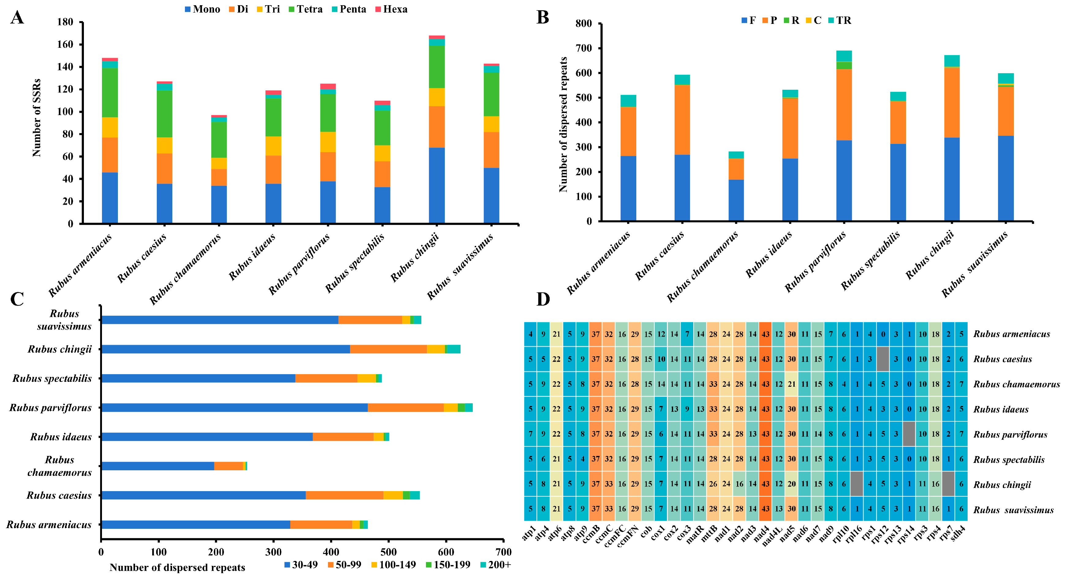
Repeat sequences included SSRs, dispersed repeats, and tandem repeats. A total of 1037 SSRs were detected in the mitogenomes of eight Rubus species (Figure 2A). Among them, R. chingii had the most SSRs, with 168 loci, while R. chamaemorus had the least, with 97. Although the SSR numbers among the eight Rubus species varied significantly, they all contained six types of nucleotide repeats. Among seven species, mononucleotide repeats dominated, ranging from 33 to 50 loci (accounting for 0.30% to 0.40% of the total), whereas R. caesius had the largest number of tetranucleotide repeats (accounting for 0.33% of the total). Furthermore, we detected tandem repeats and dispersed repeats in the mitogenomes of all eight species (Figure 2B). The number of tandem repeats ranged from 29 (R. chamaemorus) to 47 (R. chingii), with large differences among species, with their numbers trending in a pattern consistent with those of SSRs. For dispersed repeats, we detected a total of 4089 sequences, ranging from 254 (R. chamaemorus) to 646 (R. parviflorus), and most species had four dispersed repeat types. However, R. armeniacus and R. caesius lacked complementary repeats, and R. chamaemorus lacked reverse repeats. Within this general class, forward repeats (169–346 sequences) predominated in seven species, but palindromic repeats were the most common R. caesius sequences (at 281, accounting for 51% of all). Although the dispersed sequence repeats varied in number and type among the eight species, trends in the distribution of their lengths were similar, mainly distributed between 30 and 49 bp, accounting for 64.26 to 77.56% of the total (Figure 2C). There were one and two dispersed repeats with spans greater than 5000 bp in R. armeniacus and R. caesius mitogenomes, respectively, with sizes of 5341 bp, 7700 bp, and 10,422 bp (Figure 1A and Figure S1). Furthermore, R. chamaemorus and R. spectabilis had fewer repeats than other Rubus species, which may be the main reason for their smaller mitogenomes.
In this study, we predicted 34 PCGs to further understand gene expression in eight Rubus mitogenomes (Figure 2D) and identified 3890 RNA editing sites, 465 to 497 within each genome. Although there were some differences in overall number of editing sites, trends for the editing sites of specific genes were similar, with nad4 having the most RNA editing sites at 43, followed by ccmB with 37, while most ribosomal proteins usually had fewer editing sites. Additionally, rpl16 and rps7 had only one editing site in some species, and rps12 and rps14 sometimes even had none. It is worth noting that the number of editing sites for the second codon was dominant in all eight species, ranging from 298 to 308 (61.57 to 62.60% of the total). However, the number of editing sites for the third codon was much lower than that for the first and second codons (Figures S3 and S4). These RNA editing events often lead to amino acid changes that contribute to improved protein stability [55].

3.3. Analysis of Selection Pressure on Shared PCGs Among Rubus Species

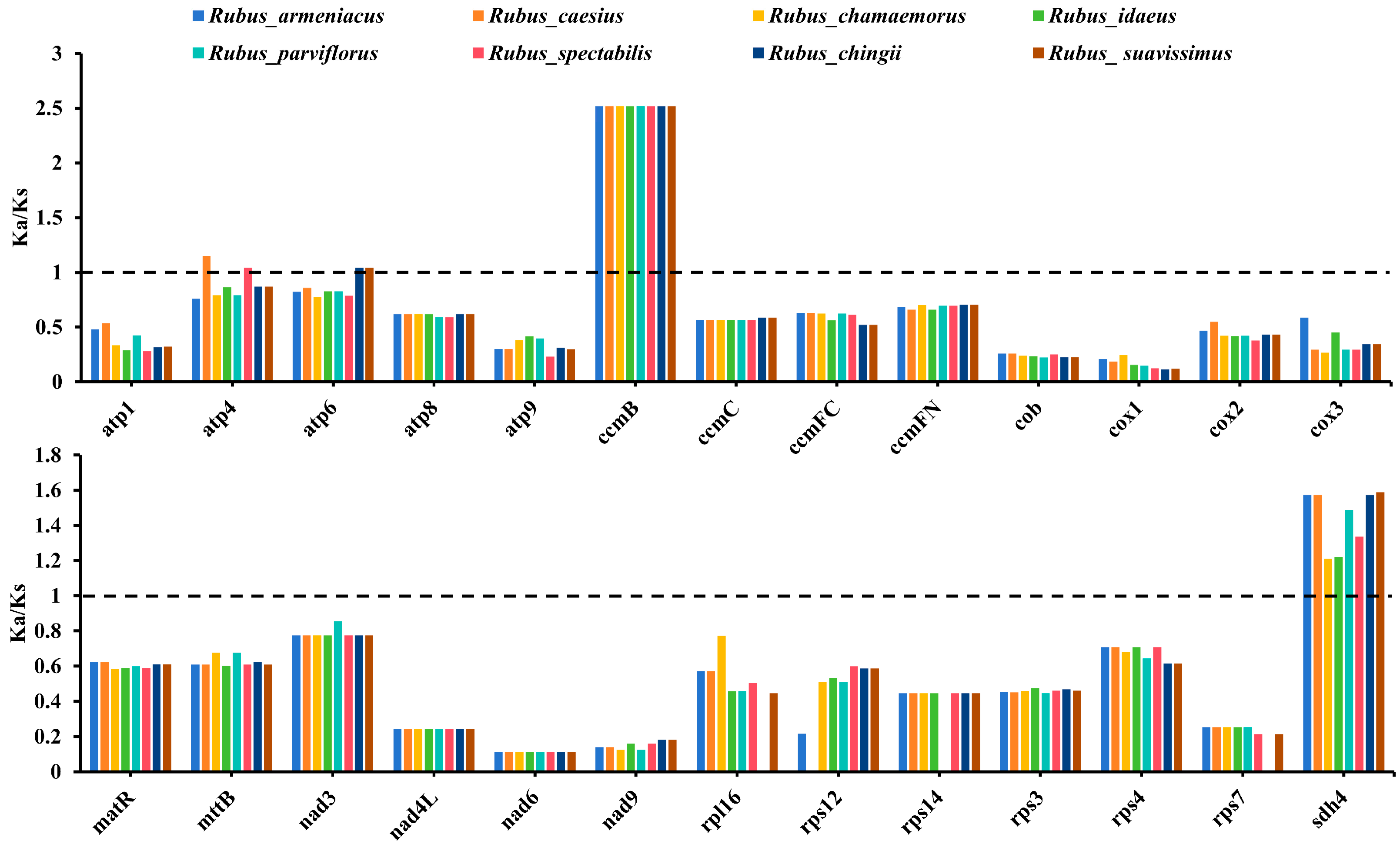
To investigate the impact of selection pressure on the evolution of mitogenomes, we used 26 PCGs shared in eight Rubus mitogenomes to calculate the ratio of non-synonymous substitutions (Ka) to synonymous substitutions (Ks) (Figure 3). Our results showed that the Ka/Ks ratio distribution trends of 26 PCGs were similar across these taxa, with a majority of the genes undergoing purifying selection (Ka/Ks ratios < 1). Notably, the Ka/Ks values for ccmB and sdh4 were >1 in all Rubus species, while atp4 and atp6 exceeded 1 in two species, meaning that they had likely undergone positive selection in response to environmental changes or other selective pressures.

3.4. Codon Usage Bias Analysis of Rubus mitogenomes

To compare the codon usage bias of PCGs in the Rubus mitogenomes, we calculated the total number of codons in each Rubus mitogenome; they varied between 9369 and 10,020 (Figure 4B). There were 64 different codon types, encoding 20 amino acids and 1 stop codon, of which UUU was most commonly used at 342 to 373 copies. Among the 20 amino acids, leucine was the most common, ranging from 998 to 1078 copies (10.60 to 10.77% of the total), followed by serine (840 to 911 copies, 8.97 to 9.15% of the total), while cysteine had the fewest codons in R. armeniacus, R. caesius, R. parviflorus, and R. spectabilis (144–156), and tryptophan had the fewest (140–153) in the other species. RSCU values reflect the results of genetic drift in natural selection, mutation, and codon-use preferences [18]. We estimated RSCU values for the 64 codon types by species (Figure 4A), and the results showed that 31 codons were used more frequently than expected, i.e., RCSU values > 1, with methionine (AUG) bias being the strongest. Usage frequencies of the remaining 32 codons were lower than expected (RSCU < 1), but codon types with the lowest values varied by species. Notably, there was no codon-usage preference for tryptophan (UGG) (RSCU = 1). Therefore, except tryptophan, the codons of all amino acids showed preference for usage, and methionine, like tryptophan, had only one codon, while most amino acids had at least two. Arginine, serine, and leucine each had six different codons.

3.5. Sequence Migration Analysis

We analyzed homologous fragments of the mitochondrial and chloroplast genomes from six Rubus species, and fragments with a similarity of >70% were identified as organelle migrating sequences (MTPTs), thus identifying 55 MTPTs (R. armeniacus), 43 MTPTs (R. idaeus), 36 MTPTs (R. caesius), 30 MTPTs (R. spectabilis), 42 (R. parviflorus), and 35 MTPTs (R. chamaemorus). These MTPTs varied greatly in total length, ranging from 23,446 (R. spectabilis) to 67,012 bp (R. armeniacus), accounting for 6.50 to 14.97% of the total mitogenome (Figure 5A,B). Among them, the longest MTPT in R. armeniacus spanned 13,207 bp, which was much larger than the longest MTPTs in the other five species (between 5878 and 8958 bp) (Figure S5). Further genetic annotation of these MTPTs showed that most contained homologous sequences of chloroplast genes, but most PCGs lost their integrity during the transfer from chloroplasts to mitochondria, retaining only partial sequences, and the numbers of complete, intact genes were fairly small. For example, 47 complete genes were detected in the MTPTs of R. armeniacus, including 27 PCGs (accD, atpB/E/F/H/I, ccsA, ndhC/J/K, petG, psaA/I/J/C, psbZ, rbcL, rpl20/32/33, rps2/7/14/18, and ycf3/4), 3 rRNA genes, and 17 tRNA genes. The fewest complete genes (17) were detected in the MTPTs of R. spectabilis, containing only 8 PCGs and 9 tRNA genes (Table S4). In addition, we compared the distribution of these complete genes in six species. Interestingly, we found that 10 complete migration genes coexisted in six Rubus mitogenomes, namely trnW-CCA, trnP-UGG, trnN-GUU, trnM-CAU, trnI-CAU, trnH-GUG, trnD-GUC, rps14, psbZ, and petG (Figure 5C). Furthermore, some genes existed only in one taxon. For example, the atpA gene existed only in R. spectabilis; the rpl23 gene existed only in R. idaeus; and the psbE/F/J/L genes existed only in R. parviflorus. These genes are closely related to plant photosynthesis and self-repair [56] and may be preserved by organellar genomes of Rubus species during the adaptive evolution process [33].

3.6. Multidimensional Systemic Generation Analysis

To further explore the maternal phylogeny of Rubus species, an ML evolutionary tree was constructed based on 20 conserved mitochondrial PCGs from 31 plant species (Figure 6A). Phylogenetic analysis showed that eight Rubus species formed an independent branch and had a close genetic relationship to Glycine max. Among the eight Rubus taxa, R. idaeus and R. spectabilis formed a clade, and R. chingii and R. suavissimus formed a clade, which together formed a branch with R. armeniacus and then formed a large clade with R. caesius and R. parviflorus. Rubus chamaemorus was located basally with a relatively distant relationship to the other Rubus species, which is also reflected in the different conformation of its mitogenome (Figure 1B). The reconstructed ML phylogenetic tree helps clarify evolutionary relationships among Rubus species, and its overall topology was reasonably consistent with the APG Ⅳ classification system [57].
To explore phylogenetic relationships within Rubus in greater detail, we reconstructed an ML evolutionary tree based on 65 conserved PCGs from the chloroplast genomes of 90 Rubus species (Figure 6B). Based on the subgenera system proposed by predecessors [1,2], we could assign these 90 species to 9 subgenera, of which R. subg. Malachobatus contained the most species (28 species), and R. subg. Batothamnus contained 26 species, while R. subg. Anoplobatus and R. subg. Chamaerubus both contained only 1 species. Among the six newly assembled genomes, R. chamaemorus was located basally within the genus, consistent with our mitochondrial ML phylogeny, indicating that R. chamaemorus may be an ancient taxon. Notably, the maternal phylogenetic relationships involving R. caesius were inconsistent between the two trees. In the mitochondrial phylogeny, R. caesius and R. parviflorus were sister groups, while in the chloroplast-based phylogeny, R. caesius and R. armeniacus had a close genetic relationship, consistent with their membership in subg. Rubus. This inconsistency could arise through differential evolutionary rates between the mitogenome and the chloroplast genome or through the complexities of reticulate evolution, especially since R. caesius is known to hybridize with members of other subgenera [9].

3.7. Collinearity Analysis of Mitochondrial Genome

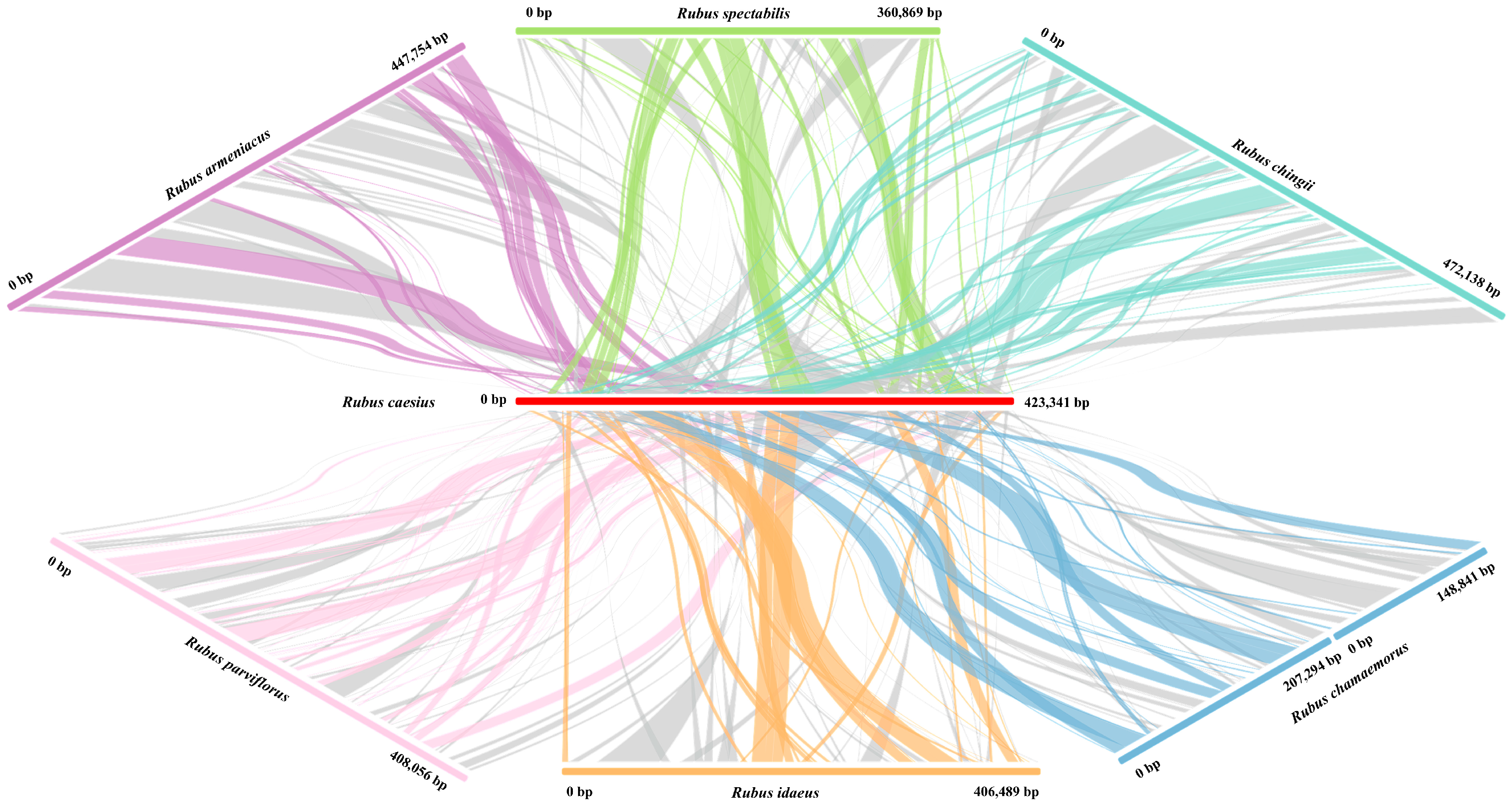
In this study, we used the mitogenome of R. caesius as a reference and conducted a collinearity analysis with the mitogenomes of six other Rubus taxa to elucidate conflicts in the relationship between R. caesius and these other Rubus species (Figure 7). The results showed that 44, 75, 60, 54, 54, and 74 fragments homologous to R. caesius were identified in R. armeniacus, R. parviflorus, R. spectababilis, R. idaeus, R. chamaemorus, and R. chingii, respectively. Rubus caesius and R. chamaemorus had the shortest homologous fragments, spanning 259,071 bp, accounting for 61.20% of the R. caesius mitogenome, which was consistent with the phylogenetic trees. Noteworthily, R. caesius and R. armeniacus had the longest homologous fragments, spanning 343,826 bp, accounting for 81.22% of the R. caesius mitogenome, while the homologous fragments with R. parviflorus had a length of 276,356 bp, which was consistent with the phylogenetic tree constructed from the chloroplast genomes. Additionally, we discovered a substantial number of sequence rearrangements within the mitogenomes of Rubus species, contributing to the diversity and complexity of the genomes. Consequently, to further refine the phylogenetic relationships of this genus, it is essential to expand the mitogenome database for Rubus species.

4. Discussion

Mitochondria are essential organelles within eukaryotic cells, fulfilling the energy demands of physiological processes. Their genomes exhibit complex structural features, with notable variations, including linear, circular, branched, and network structures [3]. The widespread occurrence of recombination mediated by repetitive sequences enables multiple structures to coexist within the same species [58,59]. Typically, the structure of plant mitogenomes consists of master circular DNA molecules. However, numerous studies in recent years have clarified the diversity of plant mitogenome conformations. For instance, the mitogenome of Spodiopogon sagittifolius exhibits a typical master circular structure spanning 500,699 bp [60], whereas the mitogenome of Echinacanthus longipes comprises five chromosomes, including three linear and two circular chromosomes, with significant size differences [61]. The mitogenome of Cymbidium ensifolium is extremely complex, with a total length of 560,647 bp, comprising 19 circular chromosomes ranging in size from 21,995 to 48,212 bp [18]. Remarkably, even closely related plants of the same genus may evolve mitogenomes with different conformations. In Mikania, the mitogenome of M. cordata is a branched linear DNA molecule, but M. micrantha possesses a typical circular structure [59]. The mitogenomes of R. armeniacus, R. caesius, R. spectrabilis, R. idaeus, and R. parviflorus all presented typical master circular structures, which were the same as most species of Rosaceae, whereas R. chamaemorus displayed a double-ring structure. This implied that the length and structure of the small genome may have undergone convergent evolution to some extent. Additionally, the mitogenomes of angiosperms are in a state of dynamic change [62], influenced by recombination, the accumulation of repetitive sequences, and gene transfer, resulting in genomes with varying conformations and sizes [63]. Past studies have described very broad variations in the size of plant mitogenomes, typically ranging from 66 kb to 12 Mb [64,65]. To date, in Rubus, the mitogenome size may vary much less, ranging only from 360,869 bp in R. spectabilis to 472,138 bp in R. chingii, a phenomenon similar to that observed in Silene [21]. As far as Rubus mitogenomes are currently described, they exhibit a moderate length within the family Rosaceae (ranging 277.76 to 535.73 kb) [3]. Size differences in the mitogenomes of these congeneric species may be attributed to the accumulation of repetitive sequences or the integration of foreign DNA [66,67]. Notably, the GC content is a key indicator to assess species evolution and amino acid composition [68]. In the mitogenomes of Rubus species, the GC content ranged from 43.8 to 44.6%, indicating that despite the diversity in the structure and size of the Rubus mitogenomes throughout evolution, the GC content remained relatively stable. Similar results have been observed in other species of the family Rosaceae [3].
Generally, the rps10 gene is absent from the mitogenome of most plants, with its function being supplanted by other nuclear-encoded genes. This phenomenon is also observed in model plants such as Arabidopsis and Populus [34,69]. In this study, we determined that our six studied Rubus mitogenomes also lack it, along with the rps2 and rps11 genes, consistent with the mitogenomes of M. micrantha [59] and Mentha spicata [70]. It is speculated that these genes may have been lost early in the evolution of vascular plants [71]. Additionally, the mitogenomes of R. caesius, R. parviflorus, and R. suavissimus lacked the rps12, rps14, and rps3 genes, respectively, and R. chingii was missing both the rpl16 and rps7 genes. The number of tRNAs varied more significantly among these species, ranging from 17 to 24 genes. This indicates that during the evolution of Rubus species, there were varying degrees of gene loss or retention among individual taxa.
Codons are the fundamental units responsible for accurately identifying and transmitting genetic information [72]. There are significant differences in the usage of the 64 codons among different species, which is believed to be the result of long-term evolutionary selection [73]. By studying codon-usage patterns, we can uncover the evolutionary patterns of genes and predict the regulatory mechanisms involved in gene expression [74]. In general, ATG serves as a common start codon in all plants, although a few PCGs in the mitochondrial genome utilize ACG or CTG as the initiation codon [75]. In our study, the nad1, nad4L, and rps4 genes in the mitogenomes of six Rubus taxa begin with ACG, as well as the cox1 genes in R. idaeus, R. armeniacus, R. caesius, and R. chamaemorus. Comparable findings have been reported in other plants, including Echinacanthus longipes [61], Ilex rotunda [76], Lycopodium japonica [77], Prunus pedunculata [78], and P. davidiana [79]. This phenomenon may be attributed to an RNA editing event. For Rubus species, the distribution of the amino acid composition is comparable to that of other angiosperms [34,59,80]. Our analysis revealed that within the mitogenomes of eight Rubus taxa, 31 codons had an RSCU value greater than 1, suggesting a significant enrichment of A/T bases at the third codon position of these gene sequences. A strong bias towards A/T bases at the third codon position is a common phenomenon in most plant mitogenomes [81,82].
It is well established that the ratio of the synonymous substitution rate (Ka) to the non-synonymous substitution rate (Ks) of a gene can reflect its relative molecular evolution rate. A Ka/Ks ratio greater than 1, equal to 1, and less than 1 indicates that the gene has undergone positive selection, neutral selection, and negative selection, respectively [83]. In our study, most genes within the mitogenomes of Rubus species appeared to be under negative selection, indicating that these PCGs and key functional genes are highly conserved. A few genes exhibited signs of positive selection, with slight differences among different species. Notably, the Ka/Ks values of the ccmB and sdh4 genes in all Rubus species were greater than 1, while the Ka/Ks value of the ccmB gene was the highest, suggesting that it is an important functional gene involved in adaptation. This selection model has similar findings in other plant species [61,84]. For instance, sdh4 exhibits the highest Ka/Ks value in the mitogenomes of both P. davidiana and Taihangia rupestris var. ciliata, whereas ccmFN has the highest value in Rosa chinensis [78,79,85].
The mitogenomes of higher plants contain numerous repetitive sequences, comprising three types: simple sequence repeats, tandem repeats, and dispersed sequence repeats. These can be utilized to develop molecular markers for studying phylogeny [59,84]. These sequences are crucial for plant evolution, influencing mitogenome structure, resulting in size variations, intermolecular recombination, heterodimerization, and isomerization [59,86,87]. Among the eight Rubus taxa, the longest repeat sequence ranged widely from 237 (R. chamaemorus) to 10,422 bp (R. caesius). Furthermore, R. caesius exhibited the highest proportion of such sequences in its mitogenome (at 13.51%), and R. chamaemorus had the lowest proportion at only 3.39%. The proportion of repeat sequences in four other taxa was relatively consistent, ranging from 7.19 to 9.73%. There was a positive correlation between the proportion of these repetitive sequences and the size of the mitogenome, except that the mitogenome of R. caesius was smaller than that of R. armeniacus or R. chingii, yet its proportion of repetitive sequences was much larger, containing a substantial number of longer repetitive sequences. Consequently, the amplification of mitogenome size was not solely due to the accumulation of repeat sequences but was also influenced by factors such as gene sequence migration and gene loss [88,89].
Sequence migration between organellar genomes is frequent, encompassing the transfer of sequence fragments from plastids to mitochondria and the movement of gene fragments from nuclei to mitochondria [66,89]. Understanding transfer relationships is crucial for elucidating the evolution of plant mitogenomes. In this study, we identified homologous genes shared between the plastids and mitochondrial genomes (MTPTs) of six Rubus species. These MTPTs constituted 6.50% (R. spectabilis), 8.09% (R. idaeus), 8.28% (R. chamaemorus), 8.50% (R. parviflorus), and 8.62% (R. caesius) of five mitogenomes, respectively, well within the range observed in most plants (1–12%) [90]. But the MTPT sequences of R. armeniacus constituted 14.97% of the mitogenome, a proportion higher than that observed in most plants, yet comparable to the MTPT proportion reported in R. suavissimus (13.16%) [33] and lower than that in R. chingii (16.34%). Generally, a substantial number of tRNA genes in angiosperms have been transferred from the plastid genome to the mitogenome. We annotated MTPTs in Rubus, and as observed in other plants such as M. micrantha [59], S. sagittifolius [60], T. rupestris var. ciliata, and P. davidiana [79,85], we also identified plastid-derived tRNA genes in six Rubus mitogenomes. To preserve functionality within the mitogenome, tRNA genes typically exhibit a high level of integrity, signifying their strong conservation and potentially indispensable roles in the mitogenome. In contrast, transferred PCGs and rRNA genes display less integrity than tRNA genes. The occurrence of plastid-derived tRNAs in plant mitogenomes is a well-documented phenomenon that may offer insights into early gene transfer events [91]. For example, trnM-CAU and trnD-GUC were identified in gymnosperms [92] and dicotyledonous plants [59], respectively. These genes were also present in the mitogenomes of six Rubus species. However, some plastid-derived genes were found only in one species, such as trnV-UAC, which was unique to R. armeniacus.
RNA editing events encompass post-transcriptional modifications of RNA sequences, including substitutions, deletions, or insertions [66]. They are prevalent in the mitochondrial and chloroplast genomes and play a crucial role in plant gene expression. Current research indicates that substitution is the most common type of editing, primarily involving the conversion of C to U [66,77]. In this study, we examined RNA editing sites in eight Rubus mitogenomes, identifying C-to-U editing sites ranging in number from 465 (R. chingii) to 497 (R. parviflorus), similar to other angiosperms such as M. micrantha (473) and cordata (492) [59], Oryza sativa (491) [93], A. thaliana (441) [33,94], P. davidiana (502) [79], and T. rupestris var. ciliata (470) [85]. Among all the RNA editing events in Rubus mitogenomes, the nad4 gene exhibited the highest editing efficiency. This is consistent with past findings for R. suavissimus and C. camphora [33,66]. This editing may affect the stability of enzymes within the mitochondrial respiratory chain complex [33,66]. Additionally, the second position in the codon triplet has been identified as the most prevalent site for RNA editing, which we observed consistently across all Rubus mitogenomes.
Mitogenomes help reveal the unique evolutionary paths of angiosperms [95]. The content, structure, and genetic arrangement of plastids are crucial for exploring the evolutionary relationships among plants [96]. In most terrestrial plants, the chloroplast genome size remains relatively constant, and gene losses are rare during biological evolution [97]. On the other hand, the mitogenomes are relatively complex and vary greatly in size, but their PCGs exhibit low substitution rates and high homogeneity [98]. These characteristics make mitogenome analysis better at uncovering older and more fundamental phylogenetic relationships within the tree of life [99]. For some taxa, there are clear topological differences between phylogenies based on the distinct evolutionary paths of plastid and mitochondrial genomes [76,100]. Several studies have investigated the intricate phylogeny within Rubus. For instance, Howarth et al. [101] conducted a phylogenetic analysis of the ndhF gene and concluded that Hawaiian Islands endemics (R. hawaiensis and R. macraei) originated from distinct ancestors, a finding that challenges the results of morphological studies. Wang et al. [15] employed multiple chloroplast and nuclear genes to examine the phylogenetic relationships among 142 Rubus taxa, revealing that evolutionary events among different subgenera and species exhibited reticulate patterns. Carter et al. [1] constructed phylogenetic trees for 87 wild Rubus taxa based on nearly 1000 target genes. The findings suggested that the low resolution and topological conflicts observed among different subgenera were not due to insufficient molecular signals but rather to hybridization and incomplete lineage sorting (ILS).
Our study investigated the phylogeny of Rubus by examining both mitochondria and plastids. The phylogenetic tree constructed from mitochondrial PCGs largely aligns with the APG IV classification system [57]. However, there was a certain conflict regarding the phylogenetic placement of R. caesius within the genus, as reflected in discrepancies between mitochondrial and plastid data, which may be attributed to ILS. In the phylogenetic tree constructed using mitochondrial data, R. caesius and R. parviflorus were depicted as sister groups, whereas the plastid tree suggested a closer genetic relationship between R. caesius and R. armeniacus. To further investigate the reasons behind this conflict, we performed a collinearity analysis involving R. caesius and six other Rubus species. Collinearity analysis is essential for clarifying evolutionary relationships among species [102]. The results indicated that R. caesius and R. parviflorus possessed a greater number of homologous fragments, whereas the collinear sequences with R. armeniacus were more extensive.
Thus, the entire mitogenome of R. caesius exhibited greater homology with R. armeniacus, whereas R. caesius shared a more similar collinear relationship with R. parviflorus concerning the PCGs of the mitogenome. This observation led to the construction of a phylogenetic tree based on PCGs from the mitogenome, which indicated a closer genetic affinity between R. caesius and R. parviflorus. Generally, the mitogenome holds significant value for the phylogeny of species. However, to obtain more accurate phylogenetic relationships, comprehensive analysis is necessary, which should be conducted using multiple datasets and algorithms. Moreover, the mitogenome exhibits a high degree of variability, indicating that it has undergone numerous instances of genetic recombination over the course of its extensive evolutionary history. Previous research has identified gene rearrangement as a key factor in the reconfiguration of the mitogenome within the family Rosaceae [26]. Similar results have been observed in Rubus mitogenomes, which may be a potential driving force for their adaptation and evolution.

5. Conclusions

For the first time, this study analyzed the structures and annotations of the mitogenomes of six Rubus species. The complete mitogenomes of R. parviflorus, R. spectabilis, R. idaeus, R. armeniacus, and R. caesius were all master circle structures with lengths ranging from 360,869 to 447,754 bp. In contrast, the mitogenome of R. chamaemorus had a double-ring structure, consisting of two small circular structures with a span of 392,134 bp. The mitogenomes of the six Rubus taxa encoded 33–34 PCGs, 17–24 tRNAs, and 3 rRNA genes. The difference in the number of PCGs among these species was mainly due to a lack of rps12 and rps14 genes in R. caesius and R. parviflorus, respectively. Further comparative analysis showed that R. chamaemorus had a lower number of SSRs, tandem repeats, and dispersed repeats than other studied Rubus taxa. However, all of these Rubus species showed similar codon-usage patterns. The Ka/Ks ratios for most coding genes were less than 1, indicating that these genes had been subjected to negative selection, highlighting the conservation of mitochondrial genes in these species. We also conduct comparative analysis of RNA editing, genome collinearity, and sequence migration among Rubus taxa, providing a broader understanding of their mitochondrial genetic characteristics. Maternal phylogeny of Rubus species was clarified based on 31 mitogenomes and 94 plastid genomes. This study not only elucidated the general characteristics of Rubus mitogenomes but also contributed to the phylogeny of the genus. However, due to frequent hybridization, asexual fusion, polyploidy, and introgression within this genus, further clarification of its classification requires more extensive sampling and analyses of additional taxa.

Supplementary Materials

The following are available online at https://www.mdpi.com/article/10.3390/horticulturae11050559/s1, Figure S1. Mitochondrial genome annotation circle maps of Rubus armeniacus and Rubus idaeus. Figure S2. Mitochondrial genome annotation circle maps of Rubus parviflorus and Rubus spectabilis. Figure S3. Prediction of RNA editing sites for 34 protein coding genes in Rubus. Figure S4. Prediction of RNA editing sites for 34 protein coding genes in Rubus. Figure S5. Migration sequence analysis between mitochondrial and plastid genomes. Table S1. Accession numbers for plant mitochondrial and chloroplast genomes. Table S2. Characteristic information on the mitochondrial genomes of 8 Rubus plants. Table S3. Functional annotation of mitochondrial genome of 8 Rubus species. Table S4. The homologous DNA fragments between mitochondrial genome and chloroplast genome of 6 Rubus plants. Table S5. Collinear fragments of the mitogenome of Rubus caesius and the mitogenomes of six remaining Rubus plants.

Author Contributions

Y.S., Z.C., J.J., Q.L. and W.Z. conceived and performed the original research project. Y.S., Z.C., Q.L. and J.J. designed the experiments and analyzed the data. Y.S. refined the project and wrote the manuscript with contributions from all authors. Z.C. and W.Z. supervised the experiments and revised the writing. Z.C. and W.Z. obtained the funding for the research project. All authors have read and agreed to the published version of the manuscript.

Funding

This work was funded by Taizhou 500 talent program (W.Z., grant number: Z2024136), the Basic Public Welfare Research Project of Zhejiang Province (Z.C., grant number: LGN22C020001), and Startup Funding of Taizhou University for the Biomass Polysaccharide Metabolism Institute (W.Z., grant number: T20231801002).

Data Availability Statement

The mitochondrial genomes of R. parviflorus (PV329689), R. spectabilis (PV353732), R. idaeus (PV339482), R. armeniacus (PV340594), R. caesius (PV296168), and R. chamaemorus (PV242989; PV242990) are accessioned in GeneBank. The chloroplast genomes of R. spectabilis (PV330320), R. armeniacus (PV282406), R. caesius (PV282407), and R. chamaemorus (PV282408) are also accessioned in GeneBank.

Conflicts of Interest

The authors declare that they have no conflicts of interest.

References

  1. Carter, K.A.; Liston, A.; Bassil, N.V.; Alice, L.A.; Bushakra, J.M.; Sutherland, B.L.; Mockler, T.C.; Bryant, D.W.; Hummer, K.E. Target Capture Sequencing Unravels Rubus Evolution. Front. Plant Sci. 2019, 10, 1615. [Google Scholar] [CrossRef] [PubMed]
  2. Huang, T.; Chen, J.; Hummer, K.E.; Alice, L.A.; Wang, W.; He, Y.; Yu, S.; Yang, M.; Chai, T.; Zhu, X.; et al. Phylogeny of Rubus (Rosaceae): Integrating Molecular and Morphological Evidence into an Infrageneric Revision. Taxon 2023, 72, 278–306. [Google Scholar] [CrossRef]
  3. Zhang, H.; Yan, M.; Li, L.; Jiang, Z.; Xiong, Y.; Wang, Y.; Akogwu, C.O.; Tolulope, O.M.; Zhou, H.; Sun, Y.; et al. Assembly and Comparative Analysis of the Complete Mitochondrial Genome of Red Raspberry (Rubus idaeus L.) Revealing Repeat-Mediated Recombination and Gene Transfer. BMC Plant Biol. 2025, 25, 85. [Google Scholar] [CrossRef] [PubMed]
  4. Huang, X.; Wu, Y.; Zhang, S.; Yang, H.; Wu, W.; Lyu, L.; Li, W. Overexpression of RuFLS2 Enhances Flavonol-Related Substance Contents and Gene Expression Levels. Int. J. Mol. Sci. 2022, 23, 14230. [Google Scholar] [CrossRef] [PubMed]
  5. Yang, Y.; Zhang, K.; Xiao, Y.; Zhang, L.; Huang, Y.; Li, X.; Chen, S.; Peng, Y.; Yang, S.; Liu, Y.; et al. Genome Assembly and Population Resequencing Reveal the Geographical Divergence of Shanmei (Rubus corchorifolius). Genom. Proteom. Bioinform. 2022, 20, 1106–1118. [Google Scholar] [CrossRef]
  6. Hua, Y.; Dai, B.; Luo, Y.; Ding, Y. Integrated Analysis of Multiple Metabolome and Transcriptome Revealed the Accumulation of Flavonoids and Associated Molecular Regulation Mechanisms in Rubus chingii Hu at Different Developmental Stages. Plant Physiol. Biochem. 2023, 204, 108085. [Google Scholar] [CrossRef]
  7. Kumar, V.; Sharma, A.; Sharma, N.; Saini, R.; Dev, K.; El-Shazly, M.; Bari, A.B.A. A Review of Botany, Traditional Applications, Phytochemistry, Pharmacological Applications, and Toxicology of Rubus ellipticus Smith Fruits. Naunyn-Schmiedeberg’s Arch. Pharmacol. 2024, 397, 4483–4497. [Google Scholar] [CrossRef]
  8. Tao, Y.; Bao, J.; Zhu, F.; Pan, M.; Liu, Q.; Wang, P. Ethnopharmacology of Rubus idaeus Linnaeus: A Critical Review on Ethnobotany, Processing Methods, Phytochemicals, Pharmacology and Quality Control. J. Ethnopharmacol. 2023, 302, 115870. [Google Scholar] [CrossRef]
  9. Alice, L.; Campbell, C. Hybridization and Gene Flow Between Distantly Related Species of Rubus (Rosaceae): Evidence from Nuclear Ribosomal DNA Internal Transcribed Spacer Region Sequences. Syst. Bot. 2001, 26, 769–778. [Google Scholar]
  10. Sochor, M.; Vašut, R.J.; Sharbel, T.F.; Trávníček, B. How Just a Few Makes a Lot: Speciation via Reticulation and Apomixis on Example of European Brambles (Rubus subgen. Rubus, Rosaceae). Mol. Phylogenetics Evol. 2015, 89, 13–27. [Google Scholar] [CrossRef]
  11. Thompson, M.M.; Zhao, C.M. Chromosome Numbers of Rubus species in southwest China. Acta Hortic. 1993, 352, 493–502. [Google Scholar] [CrossRef]
  12. Focke, W.O. Species Ruborum: Monographiae Generis Rubi Prodromus; Botanica, B., Ed.; Schweizerbart: Stuttgart, Germany, 1914. [Google Scholar]
  13. Lu, L.; David, E.B. Flora of China; Rubus Linnaeus; 1: 492. 1753; Science Press: Beijing, China, 2003; Volume 9, pp. 195–285. [Google Scholar]
  14. Xiong, X.-H.; Zhou, X.-M.; Li, M.; Xu, B.; Deng, H.-N.; Yu, Q.; Gao, X.-F. Pollen Morphology in Rubus (Rosaceae) and Its Taxonomic Implications. Plant Syst. Evol. 2019, 305, 705–716. [Google Scholar] [CrossRef]
  15. Wang, Y.; Chen, Q.; Chen, T.; Tang, H.; Liu, L.; Wang, X. Phylogenetic Insights into Chinese Rubus (Rosaceae) from Multiple Chloroplast and Nuclear DNAs. Front. Plant Sci. 2016, 7, 968. [Google Scholar] [CrossRef] [PubMed]
  16. Wang, Y.; Wang, X.; Chen, Q.; Zhang, L.; Tang, H.; Luo, Y.; Liu, Z. Phylogenetic Insight into Subgenera Idaeobatus and Malachobatus (Rubus, Rosaceae) Inferring from ISH Analysis. Mol. Cytogenet. 2015, 8, 11. [Google Scholar] [CrossRef]
  17. Birky, C.W. The Inheritance of Genes in Mitochondria and Chloroplasts: Laws, Mechanisms, and Models. Annu. Rev. Genet. 2001, 35, 125–148. [Google Scholar] [CrossRef]
  18. Shen, B.; Shen, A.; Liu, L.; Tan, Y.; Li, S.; Tan, Z. Assembly and Comparative Analysis of the Complete Multichromosomal Mitochondrial Genome of Cymbidium ensifolium, an Orchid of High Economic and Ornamental Value. BMC Plant Biol. 2024, 24, 255. [Google Scholar] [CrossRef]
  19. Rydin, C.; Wikström, N.; Bremer, B. Conflicting Results from Mitochondrial Genomic Data Challenge Current Views of Rubiaceae Phylogeny. Am. J. Bot. 2017, 104, 1522–1532. [Google Scholar] [CrossRef]
  20. Gray, M.W.; Cedergren, R.; Abel, Y.; Sankoff, D. On the Evolutionary Origin of the Plant Mitochondrion and Its Genome. Proc. Natl. Acad. Sci. USA 1989, 86, 2267–2271. [Google Scholar] [CrossRef]
  21. Wu, Z.; Liao, X.; Zhang, X.; Tembrock, L.R.; Broz, A. Genomic Architectural Variation of Plant Mitochondria—A Review of Multichromosomal Structuring. J. Syst. Evol. 2022, 60, 160–168. [Google Scholar] [CrossRef]
  22. Kozik, A.; Rowan, B.A.; Lavelle, D.; Berke, L.; Schranz, M.E.; Michelmore, R.W.; Christensen, A.C. The Alternative Reality of Plant Mitochondrial DNA: One Ring Does Not Rule Them All. PLoS Genet. 2019, 15, e1008373. [Google Scholar] [CrossRef]
  23. Jin, J.-J.; Yu, W.-B.; Yang, J.-B.; Song, Y.; dePamphilis, C.W.; Yi, T.-S.; Li, D.-Z. GetOrganelle: A Fast and Versatile Toolkit for Accurate de Novo Assembly of Organelle Genomes. Genome Biol. 2020, 21, 241. [Google Scholar] [CrossRef]
  24. He, W.; Xiang, K.; Chen, C.; Wang, J.; Wu, Z. Master Graph: An Essential Integrated Assembly Model for the Plant Mitogenome Based on a Graph-Based Framework. Brief. Bioinform. 2023, 24, bbac522. [Google Scholar] [CrossRef] [PubMed]
  25. Bi, C.; Shen, F.; Han, F.; Qu, Y.; Hou, J.; Xu, K.; Xu, L.; He, W.; Wu, Z.; Yin, T. PMAT: An Efficient Plant Mitogenome Assembly Toolkit Using Low-Coverage HiFi Sequencing Data. Hortic. Res. 2024, 11, uhae023. [Google Scholar] [CrossRef]
  26. Sun, M.; Zhang, M.; Chen, X.; Liu, Y.; Liu, B.; Li, J.; Wang, R.; Zhao, K.; Wu, J. Rearrangement and Domestication as Drivers of Rosaceae Mitogenome Plasticity. BMC Biol. 2022, 20, 181. [Google Scholar] [CrossRef] [PubMed]
  27. VanBuren, R.; Bryant, D.; Bushakra, J.M.; Vining, K.J.; Edger, P.P.; Rowley, E.R.; Priest, H.D.; Michael, T.P.; Lyons, E.; Filichkin, S.A.; et al. The Genome of Black Raspberry (Rubus occidentalis). Plant J. 2016, 87, 535–547. [Google Scholar] [CrossRef] [PubMed]
  28. Wang, L.; Lei, T.; Han, G.; Yue, J.; Zhang, X.; Yang, Q.; Ruan, H.; Gu, C.; Zhang, Q.; Qian, T.; et al. The Chromosome-scale Reference Genome of Rubus chingii Hu Provides Insight into the Biosynthetic Pathway of Hydrolyzable Tannins. Plant J. 2021, 107, 1466–1477. [Google Scholar] [CrossRef]
  29. Wang, Y.; Guan, J.; Zhang, Q. Chromosome-Scale Genome, Together with Transcriptome and Metabolome, Provides Insights into the Evolution and Anthocyanin Biosynthesis of Rubus rosaefolius Sm. (Rosaceae). Hortic. Res. 2024, 11, uhae064. [Google Scholar] [CrossRef]
  30. Price, R.J.; Davik, J.; Fernandéz Fernandéz, F.; Bates, H.J.; Lynn, S.; Nellist, C.F.; Buti, M.; Røen, D.; Šurbanovski, N.; Alsheikh, M.; et al. Chromosome-Scale Genome Sequence Assemblies of the ‘Autumn Bliss’ and ‘Malling Jewel’ Cultivars of the Highly Heterozygous Red Raspberry (Rubus idaeus L.) Derived from Long-Read Oxford Nanopore Sequence Data. PLoS ONE 2023, 18, e0285756. [Google Scholar] [CrossRef]
  31. Wu, W.; Wang, L.; Huang, W.; Zhang, X.; Li, Y.; Guo, W. A High-Quality Genome Assembly Reveals Adaptations Underlying Glossy, Wax-Coated Leaves in the Heat-Tolerant Wild Raspberry Rubus leucanthus. DNA Res. 2024, 31, dsae024. [Google Scholar] [CrossRef]
  32. Brůna, T.; Aryal, R.; Dudchenko, O.; Sargent, D.J.; Mead, D.; Buti, M.; Cavallini, A.; Hytönen, T.; Andrés, J.; Pham, M.; et al. A Chromosome-Length Genome Assembly and Annotation of Blackberry (Rubus argutus, cv. “Hillquist”). G3 2023, 13, jkac289. [Google Scholar] [CrossRef]
  33. Shi, Y.; Chen, Z.; Jiang, J.; Wu, W.; Xin, Y.; Zeng, W. Assembly and Comparative Analysis of the Complete Mitogenome of Rubus chingii var. suavissimus, an Exceptional Berry Plant Possessing Sweet Leaves. Front. Plant Sci. 2024, 15, 1504687. [Google Scholar] [CrossRef]
  34. Bi, C.; Qu, Y.; Hou, J.; Wu, K.; Ye, N.; Yin, T. Deciphering the Multi-Chromosomal Mitochondrial Genome of Populus simonii. Front. Plant Sci. 2022, 13, 914635. [Google Scholar] [CrossRef]
  35. Wick, R.R.; Schultz, M.B.; Zobel, J.; Holt, K.E. Bandage: Interactive Visualization of de Novo Genome Assemblies. Bioinformatics 2015, 31, 3350–3352. [Google Scholar] [CrossRef] [PubMed]
  36. Li, H. New Strategies to Improve Minimap2 Alignment Accuracy. Bioinformatics 2021, 37, 4572–4574. [Google Scholar] [CrossRef]
  37. Li, J.; Ni, Y.; Lu, Q.; Chen, H.; Liu, C. PMGA: A Plant Mitochondrial Genome Annotator. Plant Commun. 2025, 6, 101191. [Google Scholar] [CrossRef] [PubMed]
  38. Tillich, M.; Lehwark, P.; Pellizzer, T.; Ulbricht-Jones, E.S.; Fischer, A.; Bock, R.; Greiner, S. GeSeq—Versatile and Accurate Annotation of Organelle Genomes. Nucleic Acids Res. 2017, 45, W6–W11. [Google Scholar] [CrossRef] [PubMed]
  39. Shi, L.; Chen, H.; Jiang, M.; Wang, L.; Wu, X.; Huang, L.; Liu, C. CPGAVAS2, an Integrated Plastome Sequence Annotator and Analyzer. Nucleic Acids Res. 2019, 47, W65–W73. [Google Scholar] [CrossRef]
  40. Chan, P.P.; Lin, B.Y.; Mak, A.J.; Lowe, T.M. tRNAscan-SE 2.0: Improved Detection and Functional Classification of Transfer RNA Genes. Nucleic Acids Res. 2021, 49, 9077–9096. [Google Scholar] [CrossRef]
  41. Chen, Y.; Ye, W.; Zhang, Y.; Xu, Y. High Speed BLASTN: An Accelerated MegaBLAST Search Tool. Nucleic Acids Res. 2015, 43, 7762–7768. [Google Scholar] [CrossRef]
  42. Kearse, M.; Moir, R.; Wilson, A.; Stones-Havas, S.; Cheung, M.; Sturrock, S.; Buxton, S.; Cooper, A.; Markowitz, S.; Duran, C.; et al. Geneious Basic: An Integrated and Extendable Desktop Software Platform for the Organization and Analysis of Sequence Data. Bioinformatics 2012, 28, 1647–1649. [Google Scholar] [CrossRef]
  43. Zhang, X.; Chen, H.; Ni, Y.; Wu, B.; Li, J.; Burzyński, A.; Liu, C. Plant Mitochondrial Genome Map (PMGmap): A Software Tool for the Comprehensive Visualization of Coding, Noncoding and Genome Features of Plant Mitochondrial Genomes. Mol. Ecol. Resour. 2024, 24, e13952. [Google Scholar] [CrossRef] [PubMed]
  44. Beier, S.; Thiel, T.; Münch, T.; Scholz, U.; Mascher, M. MISA-Web: A Web Server for Microsatellite Prediction. Bioinformatics 2017, 33, 2583–2585. [Google Scholar] [CrossRef]
  45. Benson, G. Tandem Repeats Finder: A Program to Analyze DNA Sequences. Nucleic Acids Res. 1999, 27, 573–580. [Google Scholar] [CrossRef]
  46. Kurtz, S. REPuter: The Manifold Applications of Repeat Analysis on a Genomic Scale. Nucleic Acids Res. 2001, 29, 4633–4642. [Google Scholar] [CrossRef]
  47. Zhang, D.; Gao, F.; Jakovlić, I.; Zou, H.; Zhang, J.; Li, W.X.; Wang, G.T. PhyloSuite: An Integrated and Scalable Desktop Platform for Streamlined Molecular Sequence Data Management and Evolutionary Phylogenetics Studies. Mol. Ecol. Resour. 2020, 20, 348–355. [Google Scholar] [CrossRef] [PubMed]
  48. Edera, A.A.; Small, I.; Milone, D.H.; Sanchez-Puerta, M.V. Deepred-Mt: Deep Representation Learning for Predicting C-to-U RNA Editing in Plant Mitochondria. Comput. Biol. Med. 2021, 136, 104682. [Google Scholar] [CrossRef]
  49. Katoh, K. MAFFT: A Novel Method for Rapid Multiple Sequence Alignment Based on Fast Fourier Transform. Nucleic Acids Res. 2002, 30, 3059–3066. [Google Scholar] [CrossRef] [PubMed]
  50. Wang, D.; Zhang, Y.; Zhang, Z.; Zhu, J.; Yu, J. KaKs_Calculator 2.0: A Toolkit Incorporating Gamma-Series Methods and Sliding Window Strategies. Genom. Proteom. Bioinform. 2010, 8, 77–80. [Google Scholar] [CrossRef]
  51. Chen, C.; Wu, Y.; Li, J.; Wang, X.; Zeng, Z.; Xu, J.; Liu, Y.; Feng, J.; Chen, H.; He, Y.; et al. TBtools-II: A “One for All, All for One” Bioinformatics Platform for Biological Big-Data Mining. Mol. Plant 2023, 16, 1733–1742. [Google Scholar] [CrossRef]
  52. Xie, J.; Chen, Y.; Cai, G.; Cai, R.; Hu, Z.; Wang, H. Tree Visualization By One Table (tvBOT): A Web Application for Visualizing, Modifying and Annotating Phylogenetic Trees. Nucleic Acids Res. 2023, 51, W587–W592. [Google Scholar] [CrossRef]
  53. Marçais, G.; Delcher, A.L.; Phillippy, A.M.; Coston, R.; Salzberg, S.L.; Zimin, A. MUMmer4: A Fast and Versatile Genome Alignment System. PLoS Comput. Biol. 2018, 14, e1005944. [Google Scholar] [CrossRef] [PubMed]
  54. He, W.; Yang, J.; Jing, Y.; Xu, L.; Yu, K.; Fang, X. NGenomeSyn: An Easy-to-Use and Flexible Tool for Publication-Ready Visualization of Syntenic Relationships across Multiple Genomes. Bioinformatics 2023, 39, btad121. [Google Scholar] [CrossRef]
  55. Liu, M.; Fan, R.; Wang, C.; Dai, L.; Chu, S. Complete Analysis and Phylogenetic Analysis of Polygonatum sibiricum Mitochondria. BMC Plant Biol. 2025, 25, 471. [Google Scholar] [CrossRef] [PubMed]
  56. Huang, L.; Wen, J.; Crabbe, M.J.C.; Chen, C.; Ren, Z. Complete Chloroplast Genome Characterization of Three Plagiomnium Species and the Phylogeny of Family Mniaceae. Genetica 2025, 153, 6. [Google Scholar] [CrossRef]
  57. Angiosperm Phylogeny Group; Chase, M.W.; Christenhusz, M.J.; Fay, M.F.; Byng, J.; Judd, W.; Soltis, D.; Mabberley, D.; Sennikov, A.; Soltis, P. An Update of the Angiosperm Phylogeny Group Classification for the Orders and Families of Flowering Plants: APG IV. Bot. J. Linn. Soc. 2016, 181, 1–20. [Google Scholar] [CrossRef]
  58. Wang, H.; Wu, Z.; Li, T.; Zhao, J. Highly Active Repeat-Mediated Recombination in the Mitogenome of the Aquatic Grass Hygroryza aristata. BMC Plant Biol. 2024, 24, 644. [Google Scholar] [CrossRef]
  59. Wang, Z.; Wang, R.; Sang, Y.; Wang, T.; Su, Y.; Liao, W. Comparative Analysis of Mitochondrial Genomes of Invasive Weed Mikania micrantha and Its Indigenous Congener Mikania cordata. Int. J. Biol. Macromol. 2024, 281, 136357. [Google Scholar] [CrossRef]
  60. Xu, C.; Bi, W.; Ma, R.; Li, P.; Liu, F.; Liu, Z. Assembly and Comparative Analysis of the Complete Mitochondrial of Spodiopogon sagittifolius, an Endemic and Protective Species from Yunnan, China. BMC Plant Biol. 2025, 25, 373. [Google Scholar] [CrossRef]
  61. Gao, C.; Wang, S.; Huang, Y.; Deng, Y. Assembly and Comparative Analysis of the Complete Mitochondrial Genome of Echinacanthus longipes (Acanthaceae), Endemic to the Sino-Vietnamese Karst Flora. BMC Genom. 2025, 26, 251. [Google Scholar] [CrossRef]
  62. Mower, J.P. Variation in Protein Gene and Intron Content among Land Plant Mitogenomes. Mitochondrion 2020, 53, 203–213. [Google Scholar] [CrossRef]
  63. Alverson, A.J.; Rice, D.W.; Dickinson, S.; Barry, K.; Palmer, J.D. Origins and Recombination of the Bacterial-Sized Multichromosomal Mitochondrial Genome of Cucumber. Plant Cell 2011, 23, 2499–2513. [Google Scholar] [CrossRef] [PubMed]
  64. Skippington, E.; Barkman, T.J.; Rice, D.W.; Palmer, J.D. Miniaturized Mitogenome of the Parasitic Plant Viscum scurruloideum Is Extremely Divergent and Dynamic and Has Lost All Nad Genes. Proc. Natl. Acad. Sci. USA 2015, 112, E3515–E3524. [Google Scholar] [CrossRef] [PubMed]
  65. Putintseva, Y.A.; Bondar, E.I.; Simonov, E.P.; Sharov, V.V.; Oreshkova, N.V.; Kuzmin, D.A.; Konstantinov, Y.M.; Shmakov, V.N.; Belkov, V.I.; Sadovsky, M.G.; et al. Siberian larch (Larix sibirica Ledeb.) Mitochondrial Genome Assembled Using Both Short and Long Nucleotide Sequence Reads Is Currently the Largest Known Mitogenome. BMC Genom. 2020, 21, 654. [Google Scholar] [CrossRef]
  66. Han, F.; Bi, C.; Zhao, Y.; Gao, M.; Wang, Y.; Chen, Y. Unraveling the Complex Evolutionary Features of the Cinnamomum camphora Mitochondrial Genome. Plant Cell Rep. 2024, 43, 183. [Google Scholar] [CrossRef] [PubMed]
  67. Dong, S.; Chen, L.; Liu, Y.; Wang, Y.; Zhang, S.; Yang, L.; Lang, X.; Zhang, S. The Draft Mitochondrial Genome of Magnolia biondii and Mitochondrial Phylogenomics of Angiosperms. PLoS ONE 2020, 15, e0231020. [Google Scholar] [CrossRef]
  68. Du, M.-Z.; Liu, S.; Zeng, Z.; Alemayehu, L.A.; Wei, W.; Guo, F.-B. Amino Acid Compositions Contribute to the Proteins’ Evolution under the Influence of Their Abundances and Genomic GC Content. Sci. Rep. 2018, 8, 7382. [Google Scholar] [CrossRef]
  69. Kubo, T.; Newton, K.J. Angiosperm Mitochondrial Genomes and Mutations. Mitochondrion 2008, 8, 5–14. [Google Scholar] [CrossRef]
  70. Jiang, M.; Ni, Y.; Zhang, J.; Li, J.; Liu, C. Complete Mitochondrial Genome of Mentha spicata L. Reveals Multiple Chromosomal Configurations and RNA Editing Events. Int. J. Biol. Macromol. 2023, 251, 126257. [Google Scholar] [CrossRef]
  71. Fajardo, D.; Schlautman, B.; Steffan, S.; Polashock, J.; Vorsa, N.; Zalapa, J. The American Cranberry Mitochondrial Genome Reveals the Presence of Selenocysteine (tRNA-Sec and SECIS) Insertion Machinery in Land Plants. Gene 2014, 536, 336–343. [Google Scholar] [CrossRef]
  72. Li, Y.; Hu, X.; Xiao, M.; Huang, J.; Lou, Y.; Hu, F.; Fu, X.; Li, Y.; He, H.; Cheng, J. An Analysis of Codon Utilization Patterns in the Chloroplast Genomes of Three Species of Coffea. BMC Genom. Data 2023, 24, 42. [Google Scholar] [CrossRef]
  73. Liu, D.; Guo, H.; Zhu, J.; Qu, K.; Chen, Y.; Guo, Y.; Ding, P.; Yang, H.; Xu, T.; Jing, Q.; et al. Complex Physical Structure of Complete Mitochondrial Genome of Quercus acutissima (Fagaceae): A Significant Energy Plant. Genes 2022, 13, 1321. [Google Scholar] [CrossRef] [PubMed]
  74. Parvathy, S.T.; Udayasuriyan, V.; Bhadana, V. Codon Usage Bias. Mol. Biol. Rep. 2022, 49, 539–565. [Google Scholar] [CrossRef] [PubMed]
  75. Bock, H.; Brennicke, A.; Schuster, W. Rps3 and Rpll6 Genes Do Not Overlap in Oenothera Mitochondria: GTG as a Potential Translation Initiation Codon in Plant Mitochondria? Plant Mol. Biol. 1994, 24, 811–818. [Google Scholar] [CrossRef] [PubMed]
  76. Wang, Y.; Cui, G.; He, K.; Xu, K.; Liu, W.; Wang, Y.; Wang, Z.; Liu, S.; Bi, C. Assembly and Comparative Analysis of the Complete Mitochondrial Genome of Ilex rotunda Thunb. Forests 2024, 15, 1117. [Google Scholar] [CrossRef]
  77. Sun, N.; Han, F.; Wang, S.; Shen, F.; Liu, W.; Fan, W.; Bi, C. Comprehensive Analysis of the Lycopodium japonicum Mitogenome Reveals Abundant tRNA Genes and Cis-Spliced Introns in Lycopodiaceae Species. Front. Plant Sci. 2024, 15, 1446015. [Google Scholar] [CrossRef]
  78. Liu, Q.; Wu, Z.; Tian, C.; Yang, Y.; Liu, L.; Feng, Y.; Li, Z. Complete Mitochondrial Genome of the Endangered Prunus pedunculata (Prunoideae, Rosaceae) in China: Characterization and Phylogenetic Analysis. Front. Plant Sci. 2023, 14, 1266797. [Google Scholar] [CrossRef]
  79. Zhang, Y.; Guo, X.; Ma, R.; Yu, M.; Xu, J.; Shen, Z. Assembly and Analysis of the Complete Mitochondrial Genome of Prunus davidiana (Rosaceae). Front. Plant Sci. 2025, 16, 1558619. [Google Scholar] [CrossRef]
  80. Sloan, D.B.; Wu, Z.; Sharbrough, J. Correction of Persistent Errors in Arabidopsis Reference Mitochondrial Genomes. Plant Cell 2018, 30, 525–527. [Google Scholar] [CrossRef]
  81. Bi, C.; Lu, N.; Xu, Y.; He, C.; Lu, Z. Characterization and Analysis of the Mitochondrial Genome of Common Bean (Phaseolus vulgaris) by Comparative Genomic Approaches. Int. J. Mol. Sci. 2020, 21, 3778. [Google Scholar] [CrossRef]
  82. Sun, S.; Xiao, N.; Sha, Z. Complete mitochondrial genomes of four deep-sea echinoids: Conserved mitogenome organization and new insights into the phylogeny and evolution of Echinoidea. PeerJ. 2022, 10, e13730. [Google Scholar] [CrossRef]
  83. Wang, D.; Liu, F.; Wang, L.; Huang, S.; Yu, J. Nonsynonymous Substitution Rate (Ka) Is a Relatively Consistent Parameter for Defining Fast-Evolving and Slow-Evolving Protein-Coding Genes. Biol. Direct 2011, 6, 13. [Google Scholar] [CrossRef]
  84. Zhou, P.; Zhang, Q.; Li, F.; Huang, J.; Zhang, M. Assembly and Comparative Analysis of the Complete Mitochondrial Genome of Ilex metabaptista (Aquifoliaceae), a Chinese Endemic Species with a Narrow Distribution. BMC Plant Biol. 2023, 23, 393. [Google Scholar] [CrossRef]
  85. Li, Z.-Z.; Wang, Y.; He, X.-Y.; Li, W.-G. The Taihangia Mitogenome Provides New Insights into Its Adaptation and Organelle Genome Evolution in Rosaceae. Planta 2025, 261, 59. [Google Scholar] [CrossRef] [PubMed]
  86. Ma, Q.; Wang, Y.; Li, S.; Wen, J.; Zhu, L.; Yan, K.; Du, Y.; Ren, J.; Li, S.; Chen, Z.; et al. Assembly and Comparative Analysis of the First Complete Mitochondrial Genome of Acer truncatum Bunge: A Woody Oil-Tree Species Producing Nervonic Acid. BMC Plant Biol. 2022, 22, 29. [Google Scholar] [CrossRef]
  87. Christensen, A.C. Plant Mitochondrial Genome Evolution Can Be Explained by DNA Repair Mechanisms. Genome Biol. Evol. 2013, 5, 1079–1086. [Google Scholar] [CrossRef] [PubMed]
  88. Yisilam, G.; Liu, Z.; Turdi, R.; Chu, Z.; Luo, W.; Tian, X. Assembly and Comparative Analysis of the Complete Mitochondrial Genome of Isopyrum anemonoides (Ranunculaceae). PLoS ONE 2023, 18, e0286628. [Google Scholar] [CrossRef]
  89. Yong, Y.; Wang, Y.; Wang, D.; Yuan, X.; Zhang, Q. The Organelle Genomes of the Endangered Seagrass Zostera caespitosa Reveal Sequence Divergences, Massive Gene Transfer, and Uncommon RNA Editing Types. Front. Plant Sci. 2025, 16, 1550467. [Google Scholar] [CrossRef] [PubMed]
  90. Wang, X.-C.; Chen, H.; Yang, D.; Liu, C. Diversity of Mitochondrial Plastid DNAs (MTPTs) in Seed Plants. Mitochondrial DNA Part A 2018, 29, 635–642. [Google Scholar] [CrossRef]
  91. Bi, C.; Paterson, A.H.; Wang, X.; Xu, Y.; Wu, D.; Qu, Y.; Jiang, A.; Ye, Q.; Ye, N. Analysis of the Complete Mitochondrial Genome Sequence of the Diploid Cotton Gossypium raimondii by Comparative Genomics Approaches. BioMed Res. Int. 2016, 2016, 5040598. [Google Scholar] [CrossRef]
  92. Gao, C.; Ren, X.; Mason, A.S.; Liu, H.; Xiao, M.; Li, J.; Fu, D. Horizontal Gene Transfer in Plants. Funct. Integr. Genom. 2014, 14, 23–29. [Google Scholar] [CrossRef]
  93. Clifton, S.W.; Minx, P.; Fauron, C.M.-R.; Gibson, M.; Allen, J.O.; Sun, H.; Thompson, M.; Barbazuk, W.B.; Kanuganti, S.; Tayloe, C.; et al. Sequence and Comparative Analysis of the Maize NB Mitochondrial Genome. Plant Physiol. 2004, 136, 3486–3503. [Google Scholar] [CrossRef] [PubMed]
  94. Unseld, M.; Marienfeld, J.R.; Brandt, P.; Brennicke, A. The Mitochondrial Genome of Arabidopsis thaliana Contains 57 Genes in 366,924 Nucleotides. Nat. Genet. 1997, 15, 57–61. [Google Scholar] [CrossRef] [PubMed]
  95. Li, Y.-X.; Li, Z.-H.; Schuiteman, A.; Chase, M.W.; Li, J.-W.; Huang, W.-C.; Hidayat, A.; Wu, S.-S.; Jin, X.-H. Phylogenomics of Orchidaceae Based on Plastid and Mitochondrial Genomes. Mol. Phylogenetics Evol. 2019, 139, 106540. [Google Scholar] [CrossRef] [PubMed]
  96. Yang, Z.; Ferguson, D.K.; Yang, Y. New Insights into the Plastome Evolution of Lauraceae Using Herbariomics. BMC Plant Biol. 2023, 23, 387. [Google Scholar] [CrossRef]
  97. Liu, C.; Chen, H.-H.; Tang, L.-Z.; Khine, P.K.; Han, L.-H.; Song, Y.; Tan, Y.-H. Plastid Genome Evolution of a Monophyletic Group in the Subtribe lauriineae (Laureae, Lauraceae). Plant Divers. 2022, 44, 377–388. [Google Scholar] [CrossRef]
  98. Dong, W.; Xu, C.; Cheng, T.; Lin, K.; Zhou, S. Sequencing Angiosperm Plastid Genomes Made Easy: A Complete Set of Universal Primers and a Case Study on the Phylogeny of Saxifragales. Genome Biol. Evol. 2013, 5, 989–997. [Google Scholar] [CrossRef] [PubMed]
  99. Qiu, Y.-L.; Li, L.; Wang, B.; Xue, J.-Y.; Hendry, T.A.; Li, R.-Q.; Brown, J.W.; Liu, Y.; Hudson, G.T.; Chen, Z.-D. Angiosperm Phylogeny Inferred from Sequences of Four Mitochondrial Genes. J. Syst. Evol. 2010, 48, 391–425. [Google Scholar] [CrossRef]
  100. Pearl, S.A.; Welch, M.E.; McCauley, D.E. Mitochondrial Heteroplasmy and Paternal Leakage in Natural Populations of Silene vulgaris, a Gynodioecious Plant. Mol. Biol. Evol. 2009, 26, 537–545. [Google Scholar] [CrossRef]
  101. Howarth, D.G.; Gardner, D.E.; Morden, C.W. Phylogeny of Rubus subgenus Idaeobatus (Rosaceae) and Its Implications Toward Colonization of the Hawaiian Islands. Syst. Bot. 1997, 22, 433–441. [Google Scholar] [CrossRef]
  102. Wang, X.; Wang, J.; Jin, D.; Guo, H.; Lee, T.-H.; Liu, T.; Paterson, A.H. Genome Alignment Spanning Major Poaceae Lineages Reveals Heterogeneous Evolutionary Rates and Alters Inferred Dates for Key Evolutionary Events. Mol. Plant 2015, 8, 885–898. [Google Scholar] [CrossRef]
Figure 1. Mitochondrial genome assembly and annotation. (A) The mitogenome map of R. caesius. (B) The mitogenome map of R. chamaemorus. Genes with different functions are given different colors. The colored curves in the circles represent dispersed sequence repeats. (C) Comparison of the number of protein-coding genes in eight Rubus mitogenomes.
Horticulturae 11 00559 g001
Figure 2. Repeated sequence analysis and prediction of RNA editing sites in the mitogenomes. (A) Identification of SSR loci in eight Rubus mitogenomes; (B) identification of dispersed sequence repeats and tandem repeats in eight Rubus mitogenomes; (C) distribution of dispersed sequence repeats in eight Rubus mitogenomes; (D) prediction of RNA editing sites for PCGs of eight Rubus mitogenomes. From blue to red represents the number of RNA editing sites from fewer to more.
Horticulturae 11 00559 g002
Figure 3. Ka/Ks ratios for 26 PCGs in the mitogenomes of eight Rubus species. Different species have different colors.
Horticulturae 11 00559 g003
Figure 4. Codon usage bias analysis of mitogenomes from eight Rubus species. (A) Comparison of relative synonymous codon usage (RSCU) values for different species. Each thin bar from left to right represents a species, namely R. armeniacus, R. caesius, R. chamaemorus, R. idaeus, R. parviflorus, R. spectabilis, R. chingii, and R. suavissimus. A to Y on the X-axis represent alanine, cysteine, aspartic acid, glutamic acid, phenylalanine, glycine, histidine, isoleucine, lysine, leucine, methionine, asparagine, proline, glutamine, arginine, serine, threonine, valine, tryptophan, and tyrosine. (B) Total numbers of amino acid codons in protein-coding genes of different species.
Horticulturae 11 00559 g004
Figure 5. Migration sequence analysis between mitochondrial and plastid genomes. (A) Homogeneous fragments between the mitochondrial and chloroplast genomes of R. armeniacus. (B) Homogeneous fragments between the mitochondrial and chloroplast genomes of R. spectabilis. Two arcs of different lengths represent the mitochondrial and plastid genome, the histogram in the inner circle represents the length of MTPTs, and the lines between the two arcs represents MTPTs. Detailed information is recorded in Table S4. (C) Comparative analysis of complete genes in MTPT sequences from six Rubus species.
Horticulturae 11 00559 g005
Figure 6. Phylogenetic analysis. (A) The ML phylogenetic tree constructed based on PCGs shared in the mitogenomes of 31 species; (B) the ML phylogenetic tree constructed based on PCGs shared in the plastid genomes of 94 species. Support rates for different nodes are displayed on branches. The red stars represent the newly assembled genomes for this study.
Horticulturae 11 00559 g006
Figure 7. Collinearity analysis among the mitogenomes of Rubus caesius and six other Rubus species. Different colored bars represent the mitogenomes of different taxa. Among them, the colored lines between the mitogenome of R. caesius and the other species indicate good similarity, and the gray lines indicate reversal. All collinear fragments were greater than 500 bp.
Horticulturae 11 00559 g007
Disclaimer/Publisher’s Note: The statements, opinions and data contained in all publications are solely those of the individual author(s) and contributor(s) and not of MDPI and/or the editor(s). MDPI and/or the editor(s) disclaim responsibility for any injury to people or property resulting from any ideas, methods, instructions or products referred to in the content.