Article

RrLBD40 Enhances Salt Tolerance in Rosa rugosa via Promoting Root Development

College of Horticulture and Landscape Architecture, Yangzhou University, Yangzhou 225009, China
*
Authors to whom correspondence should be addressed.
These authors contributed equally to this work.
Horticulturae 2025, 11(11), 1379; https://doi.org/10.3390/horticulturae11111379
Submission received: 14 September 2025 / Revised: 10 November 2025 / Accepted: 14 November 2025 / Published: 15 November 2025

Abstract

LATERAL ORGAN BOUNDARIES DOMAIN (LBD) genes encode plant specific transcription factors that regulate various developmental processes and abiotic stresses. In this study, we characterized RrLBD40 from Rosa rugosa as a nucleus-localized transcription factor harboring a conserved LOB domain. We generated RrLBD40-overexpressing Rosa rugosa plants and compared them with control plants in terms of physiological indices, root architecture, and Na+ homeostasis. The results showed that RrLBD40 overexpression significantly increased peroxidase activity and reduced malondialdehyde content in the roots, indicating enhanced antioxidant capacity under salt stress. Furthermore, RrLBD40 overexpression markedly promoted root growth and development, a similar phenotype consistently observed in RrLBD40 transgenic Arabidopsis plants. Propidium iodide staining and analysis of the Na+ flux rates of root tips revealed that the barrier function of the Casparian strip was compromised in both the RrLBD40-overexpression and control plants under salt stress. This disruption of endodermal selectivity permitted Na+ influx into the vascular cylinder. Furthermore, neither plants exhibited significant Na+ efflux capacity. Taken together, these findings demonstrate that RrLBD40 enhances salt tolerance in Rosa rugosa by primarily promoting root growth and development, rather than modulating Na+ homeostasis.

1. Introduction

Salt stress is one of the most severe abiotic stresses that impair normal plant growth and development. Excessive soil salt concentration induces high osmotic stress, ion imbalance, and oxidative damage in plant cells, resulting in stunted growth, reduced biomass, leaf injury, and even plant death [1,2]. To cope with salt stress, plants have evolved complex tolerance mechanisms over long-term evolution. For example, they accumulate organic osmolytes (such as proline, betaine, and soluble sugars) and inorganic ions (such as K+ and Ca2+) to maintain cell turgor and enzyme activity, thereby ensuring normal metabolic function [3,4]. Plants also activate antioxidant systems to scavenge reactive oxygen species and mitigate oxidative stress, and establish ion compartmentalization to prevent metabolic disruption caused by excessive Na+ [5,6]. The establishment of these adaptive responses involves the mobilization of numerous functional genes in plant cells, among which transcription factors play a critical role in regulating their expression.
LATERAL ORGAN BOUNDARIES DOMAIN (LBD) family transcription factors are characterized by a highly conserved LOB domain at the N-terminus and play important roles in lateral organ formation [7,8,9], establishment of adaxial–abaxial leaf polarity [10,11], plant regeneration [12,13], and secondary growth [14,15]. LBDs such as ZmLBD2 [16], ZmLBD33 [17], MdLBD37 [18], MdLBD3 [19], and PheLBD29 [20] are involved in the abiotic stress of plants. These LBDs enhance plant drought resistance by influencing morphology, augmenting antioxidant enzyme activity, and scavenging reactive oxygen species. Moreover, LBD genes are implicated in regulating hormonal signaling pathways in response to drought stress. For instance, ZmLBD5 negatively regulates drought tolerance by inhibiting ABA biosynthesis [21]. In tomato, the expression of SlLBD40 is dependent on the JA signaling pathway and acts as a negative regulator of drought resistance [22]. Although drought and salt stress lead to similar plant damage phenotypes, research on the role of LBD genes in salt tolerance remains relatively limited. Previous studies have indicated that PvLBD12 enhanced salt tolerance in switchgrass by increasing proline accumulation, improving K+ accumulation, and decreasing reactive oxygen species level in switchgrass [23]. In apple callus, the overexpression of MdLBD37 significantly improved salt tolerance, which was characterized by a substantial increase in biomass and a concurrent decrease in malondialdehyde levels [18].
Rosa rugosa (R. rugosa), a deciduous shrub within the family Rosaceae and genus Rosa, is globally valued as a source of natural fragrance and is also traditionally used in China as both a medicinal and edible flower [24]. The flowers contain abundant bioactive compounds, including flavonoids, volatile oils, phenolic acids, and amino acids [25]. Unlike roses, which are primarily cultivated for ornamental purposes, R. rugosa is valued mainly for the commercial processing of its by-products. As a result, it is typically grown using open-field cultivation, whereas roses are often produced under protected facility conditions. However, increasing soil salinization poses a serious threat to its cultivation. Most cultivated varieties of R. rugosa exhibit limited salt tolerance, and open-field cultivation further increases their exposure to salt stress. In recent years, several transcription factors, such as GT [26], WRKY1 [27], C2H2-8 [28], and bHLH5 [29], have been reported to be involved in salt stress response. However, the functional roles of LBD family genes remain largely unexplored.
Wild R. rugosa grows in the high-salinity coastal areas of northeastern China, the Russian Far East, the Korean Peninsula, and Japan. They exhibit greater salt tolerance compared with cultivated varieties. Previously, the transcriptome analysis of salt-treated wild R. rugosa identified a strongly salt-induced RrLBD40 gene. However, its functional role remains unexplored [30]. In this study, we generated RrLBD40-overexpressing transgenic R. rugosa plants. Through comprehensive comparisons of the physiological indices, root system architecture, Na+ distribution, and flux rates between the transgenic and control plants, we demonstrated that RrLBD40 enhances salt tolerance by promoting root growth and development. These findings establish a basis for future molecular investigations into the salt tolerance mechanisms in R. rugosa.

2. Materials and Methods

2.1. Plant Materials and Growth Conditions

The R. rugosa cuttings used for the genetic transformation experiment mediated by Agrobacterium rhizogenes were obtained from the Rose Resource Garden of Yangzhou University. Infected R. rugosa cuttings were cultivated in a greenhouse under controlled conditions (24 °C, 16 h light/8 h dark).
Nicotiana benthamiana plants were also cultured under the same above-mentioned conditions as the infected R. rugosa cuttings. Leaves from 1-month-old plants were used for subcellular localization assays.
The Arabidopsis thaliana plants were cultivated in growth chambers maintained at 22 °C under long-day conditions (16 h light/8 h dark cycle).

2.2. Subcellular Localization

The coding sequences of RrLBD40 were cloned into the pENTR D-TOPO entry vector (Invitrogen, Carlsbad, CA, USA) and then recombined via LR reaction into the pFAST-R05 vectors (with GFP tag) to construct the overexpression vectors (35S:RrLBD40). The 35S:RrLBD40 plasmids were transformed into cells of Agrobacterium tumefaciens strain GV3101 for injection into Nicotiana benthamiana leaves. Following agroinfiltration, the plants were incubated overnight in the dark at 25 °C and then transferred to the greenhouse for further cultivation. Green fluorescence signals were observed within 48–72 h post-infiltration using a confocal laser scanning microscope (LSM 880NLO, Carl Zeiss, Oberkochen, Germany). GFP fluorescence was excited at 488 nm and detected at 500–535 nm.

2.3. Arabidopsis Thaliana Transformation and NaCl Treatment

The recombinant plasmids 35S:RrLBD40 were first introduced into GV3101 cells of Agrobacterium tumefaciens and then transformed into the Columbia (Col-0) wild-type (WT) seedlings of Arabidopsis thaliana by following the flower-dipping protocol [31]. The transgenic lines were identified by RT-qPCR.
WT and transgenic seeds were sown on 1/2MS medium (pH = 5.8) and transferred to a medium containing 100 mM NaCl after germination. Three independent biological replicates were performed. Following a 2-week growth period, the longest root was measured for each line. The final data were derived from the average value of all the data from each experiment.

2.4. R. rugosa Transformation Mediated by Agrobacterium rhizogenes

The recombinant plasmids 35S:RrLBD40 (35S:Empty as control) were introduced into Agrobacterium rhizogenes K599. R. rugosa cuttings (lower end of morphology) were then immersed in the bacterial suspension and subsequently planted in a greenhouse under controlled conditions. Hairy roots emerged approximately three weeks post-inoculation.
The 35S:RrLBD40 and control plants were treated with 200 mM NaCl solution. After 10 days, the chlorophyll fluorescence was imaged and Fv/Fm were recorded using PlantView230F (Biolight Biotechnology, Guangzhou, China). Furthermore, root samples were collected for RT-qPCR analysis, root configuration observation, and physiological parameter measurements.
The root images and related data (including total length, surface area and lateral root number) were acquired using a flat-bed scanner (perfection V800, Epson, Nagano, Japan).
Each experiment was performed with three biological repetitions. The final data were derived from the average value of three independent biological replicates.
The physiological indicators of peroxidase (POD), superoxide dismutase (SOD), proline (PRO) and malondialdehyde (MDA) were quantified using Peroxidase Assay Kit (A084-3-1), Superoxide Dismutase (SOD) Assay Kit (A001-3-2), Total PROtein Quantitative Assay Kit (A045-2-2), and Malondialdehyde (MDA) Assay Kit (TBA method) (A003-1-2), respectively (Jiancheng, Nanjing, China). The final data for each physiological indicator were derived from the average of three independent biological replicates, and each biological replicate with three technical replicates.

2.5. Propidium Iodide (PI) Staining

A PI penetration assay was performed as previously described [32], with slight modifications. The roots were first rinsed with clean water and subsequently incubated in a 0.6% MES buffer solution containing 200 mM NaCl for 72 h. Following this treatment, they were transferred to a 50 μM CoroNa™ Green AM solution and stained for 3 h at room temperature in darkness. Then, root tip sections were immersed in 10 μg/mL PI solution and kept in the dark for 10 min for observation with a laser-scanning confocal microscope (TCS SP8x, Leica Microsystems, Mannheim, Germany). Observations were carried out using a laser scanning confocal microscope. The PI red fluorescence signal was detected under 535 nm excitation, and Na+ imaging was performed using the green fluorescence signal under 488 nm excitation.

2.6. Measurement of Na+ Flux of Root Tips

The net Na+ fluxes at the root tips of isolated roots were measured using a non-invasive micro-test technique (NMT 100 Series, Xuyue (Beijing) Sci. & Tech. Co. Ltd., Beijing, China). Ion-selective microelectrodes (model XY-CGQ-01) were prepared according to the manufacturer’s instructions by filling with a perfusion buffer containing 200 mM NaCl and a Na+-selective backfilling solution (Liquid Ion Exchanger, LIX234). Prior to each measurement, the Na+-selective electrodes were calibrated using standard solutions with increasing Na+ concentrations (0.01, 0.1, and 1 mM NaCl) against a reference electrode. The roots were equilibrated for 5 min in a measuring buffer containing 0.1 mM NaCl before flux recordings were conducted. Measurements were taken at three positions located 800 μm from the root tip, with each recording lasting 10 min. Net ion fluxes were derived from five biologically independent replicates (n = 5) and analyzed using imFluxes V3.0 software.

2.7. RT-qPCR

Total RNA was extracted from the roots of 35S:RrLBD40 and control plants using the FastPure Plant Total RNA Isolation Kit (Vazyme, Nanjing, China) following the manufacturer’s protocol. First-strand cDNA was synthesized using the HiScript III RT SuperMix for qPCR (+gDNA wiper) (Vazyme). Fluorescent quantitative PCR primers were designed based on the cDNA sequences and standard primer design principles. RT-qPCR was conducted using the CFX Connect Real-Time PCR Detection System (CFX96, Bio-Rad, California, USA) with SYBR qPCR Master Mix (Vazyme) and gene-specific primers (Table S1). Gene expression levels were analyzed using the 2−ΔΔCt method [33], with Rr5.8s serving as the internal reference gene [30]. Relative gene expression was represented by fold changes (times), with the experimental control setting to 1. Each experiment was performed with three biological replicates, and each biological repeat with 3 technical replicates.

2.8. Statistical Analysis

Statistical analysis of the experimental data was conducted using statistical software of SPSS 17. Data were compared using the paired Student’s t test (* p < 0.05; ** p < 0.01; *** p < 0.001).

3. Results

3.1. Characterization of RrLBD40

Our previous study on the LBD gene family in R. rugosa demonstrated that RrLBD40 exhibits a significant response to salt stress [30]. However, its functional role remains uncharacterized. Molecular cloning and bioinformatics analysis determined that the RrLBD40 gene encodes a 300-amino acid protein with a molecular weight of 32.27 kDa. Comparative sequence analysis from different species revealed a conserved LOB domain at the N-terminus of RrLBD40 (Figure 1). Furthermore, subcellular localization assays confirmed its nuclear localization (Figure 2).

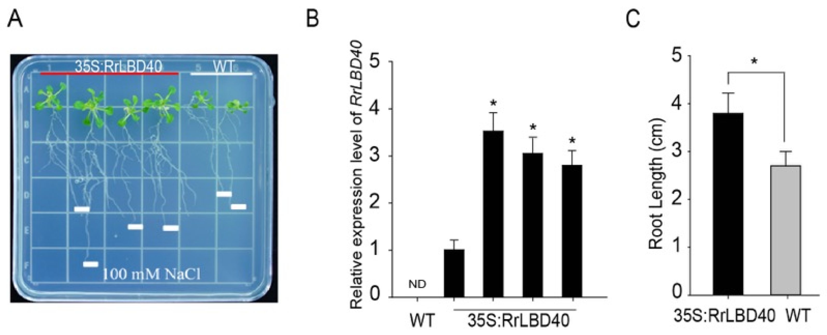
3.2. Ectopic Expression of RrLBD40 Enhances salt Tolerance in Arabidopsis

Given that RrLBD40 is significantly induced by salt stress [30], we investigated how RrLBD40 responds to salt stress. We heterologously overexpressed RrLBD40 in Arabidopsis and confirmed its overexpression level via RT-qPCR (Figure 3B). The germinated WT and 35S:RrLBD40 Arabidopsis seedlings were grown on 1/2 MS medium with 100 mM NaCl for two weeks. Then, we counted the root lengths of the 35S:RrLBD40 and WT lines (Figure 3A). The results showed that the 35S:RrLBD40 lines exhibited significantly longer roots than WT under salt stress conditions (Figure 3C), demonstrating that RrLBD40 overexpression reduces sensitivity to NaCl in Arabidopsis.

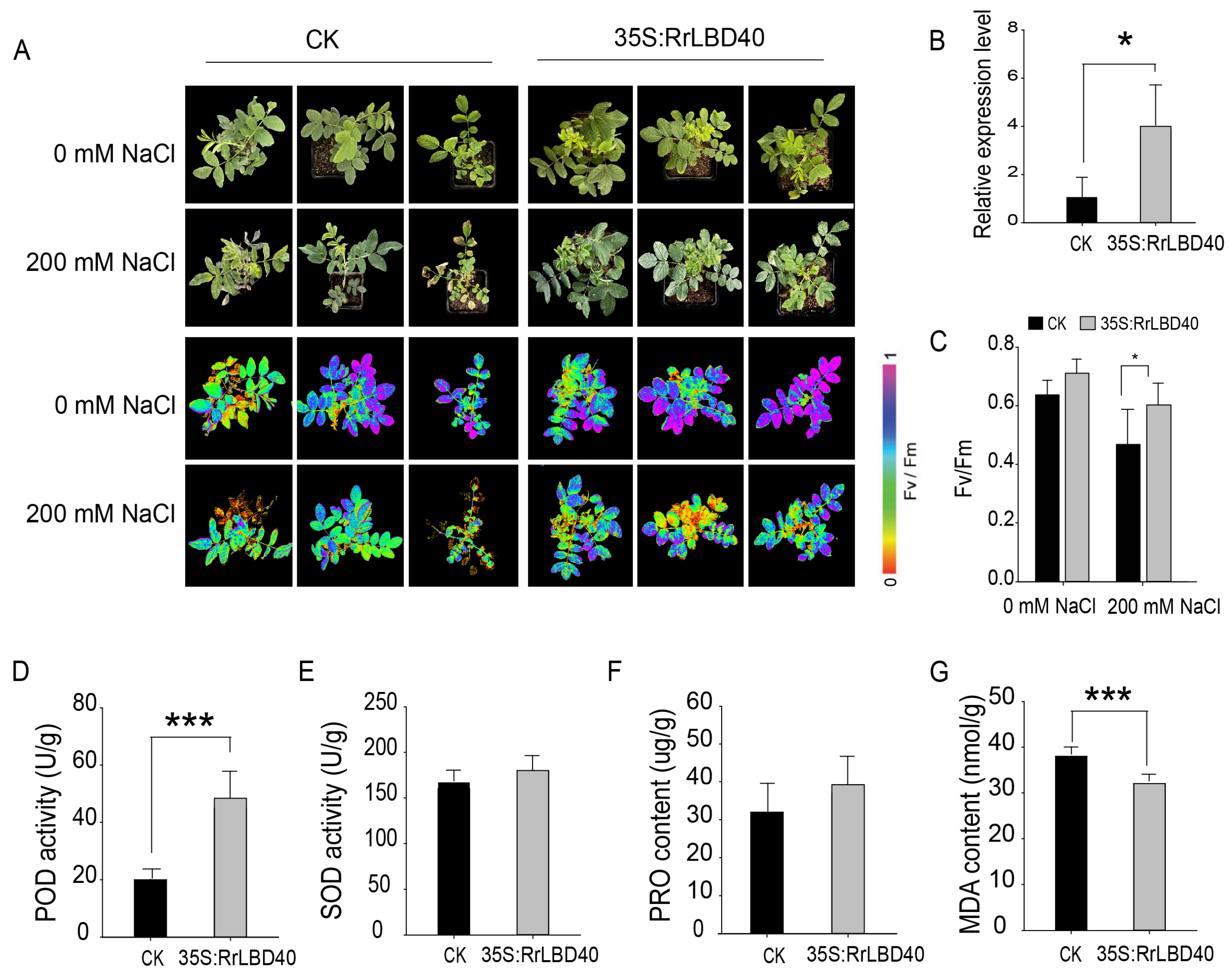
3.3. RrLBD40 Positively Regulates Salt Tolerance in R. rugosa

To clarify the functions of RrLBD40 in R. rugosa, we used Agrobacterium rhizogenes to obtain composite 35S:RrLBD40 R. rugosa plants with transgenic roots, and the transgenic plants were confirmed by RT-qPCR analysis (Figure 4B). Under 200 mM NaCl for 10 days treatment, phenotypic observations and chlorophyll fluorescence imaging revealed that 35S:RrLBD40 plants exhibited less damage, and had a higher Fv/Fm ratio compared with the controls (Figure 4A,C). We further assessed key physiological and biochemical parameters in the CK and transgenic lines, including POD and SOD activities, PRO and MDA contents, and the results showed that the 35S:RrLBD40 lines exhibited significantly higher POD activity and lower MDA content. However, the SOD activity and PRO content in the root tissues were not significantly different compared with the controls (Figure 4D–G). These results suggest that RrLBD40 enhances the antioxidant defense capacity, thereby reducing oxidative damage in R. rugosa under salt stress.

3.4. Overexpression of RrLBD40 Does Not Affect the Na+ Homeostasis in R. rugosa Roots

The Casparian strip is a band-like structure located on the inner epidermal cell wall of the root, which acts as a barrier preventing harmful ions (such as Na+ and Al3+) from freely entering the vascular cylinder and subsequently being transported to the shoot or leaves. Thus, the barrier function is a key mechanism contributing to plant stress resistance [34,35,36].
To investigate whether RrLBD40 modulates salt tolerance in R. rugosa via this structure, we analyzed Na+ distribution in roots under salt stress. The results revealed that Na+ entered the vascular cylinder through the Casparian strip, with no significant difference in distribution patterns between the control and 35S:RrLBD40 plant roots (Figure 5A). This result suggests that the barrier function of the Casparian strip was compromised in all roots. Furthermore, we measured the Na+ efflux rates in the roots using non-invasive microelectrode ion flux estimation (NMT), the results showed that the efflux rates in both the 35S:RrLBD40 and control plants were nearly zero (Figure 5B,C), suggesting roots do not have the ability of Na+ exclusion capacity. Overall, these findings demonstrate that RrLBD40 does not enhance salt tolerance in R. rugosa by modulating Na+ homeostasis.

3.5. Overexpression of RrLBD40 Promotes Root Development in R. rugosa

The root system is the primary organ for sensing salt stress and can mount adaptive responses through alterations in root morphology and architecture. Moreover, the LBD transcription factor family plays significant roles in lateral root development. Accordingly, we analyzed the root architectures of various transgenic plants. As shown in Figure 6, the RrLBD40-overexpressing lines exhibited significantly greater total root length, root surface area, and number of lateral roots compared with the controls, suggesting that RrLBD40 enhances salt tolerance by modulating root architecture.

4. Discussion

Research on the salt tolerance mechanism in roses has primarily emphasized physiological and biochemical aspects. At the molecular level, studies have largely been confined to the screening and identification of stress-resistant genes, whereas functional characterization of the resulting candidate genes remains considerably understudied [37,38,39]. In this study, we functionally characterized RrLBD40, a gene previously identified through screening [30]. Heterologous RrLBD40 overexpression in Arabidopsis revealed its role in modulating root length. Furthermore, homologous transformation in R. rugosa demonstrated that RrLBD40 enhances salt tolerance by regulating root architecture rather than by maintaining Na+ homeostasis. These findings provide a molecular basis for further elucidating the salt tolerance mechanisms in Rosaceae plants.
We previously performed a genome-wide analysis of the RrLBD gene family, which classified RrLBD40 as a member of the Class II subfamily. In this study, conserved domain analysis revealed that RrLBD40 contains a characteristic N-terminal LOB domain (Figure 1), and subcellular localization experiments confirmed its nuclear localization (Figure 2), clearly defining its attributes as a transcription factor. Genetic evidence proved that RrLBD40 acts as a positive regulator of salt tolerance (Figure 3, Figure 4, Figure 5 and Figure 6). Homologs of LBD40 have also been functionally characterized in other species. For instance, in Arabidopsis and citrus, LBD40 is involved in somatic embryogenesis [40,41]. Recent studies have shown that LBD40 also regulates the fatty acid profile in seeds in Camelina [42]. In tomato, SlLBD40 serves multifaceted roles and functions as a positive regulator of fruit cell expansion and growth [43]. Conversely, it acts as a negative regulator of drought resistance, despite being significantly induced by drought, salt, and jasmonic acid (JA) [22]. In the JA signaling pathway, SlLBD40 and SlLBD42 form homodimers or heterodimers that compete with SlMYC2 for binding to downstream disease-resistant genes, thereby suppressing their expression. This mechanism likely originates from the role of SlLBD40/42 as key developmental regulators, which attempt to balance resource allocation between growth and defense by repressing SlMYC2-mediated defense signaling during pathogen invasion [44]. These functional divergences among homologous genes highlight the evolutionary diversity of LBD gene functions.
Plants respond to and cope with salt stress via a range of physiological and biochemical adaptations. In this study, we investigated the physiological mechanisms by which RrLBD40 positively regulates salt tolerance from multiple perspectives. (i) To maintain cellular osmotic balance under salt stress, plants accumulate compatible solutes (such as proline and soluble sugar) and enhance antioxidant enzyme activity (e.g., POD and SOD) to scavenge reactive oxygen species [1,2]. Compared with those in the control, POD activity was significantly elevated whereas MDA content was markedly reduced in the RrLBD40-overexpressing plants (Figure 4D,G). These results indicate that RrLBD40 enhances the antioxidant defense capacity, thereby mitigating oxidative damage in R. rugosa under salt stress. Consistent with these physiological changes, the RrLBD40-overexpressing plants also exhibited improved photosynthetic performance in the aerial parts (Figure 4A,C). (ii) Casparian strip is considered to play a pivotal role as the barrier to apoplastic transport in roots [45,46]. However, Na+ distribution did not differ significantly between the 35S:RrLBD40 and control lines under salt stress. A clear accumulation of Na+ was observed in the vascular cylinder, indicating an impaired barrier function of the Casparian strip. Furthermore, no difference in Na+ efflux rate was detected. (iii) As the primary organ for salt perception, roots orchestrate adaptive responses through alterations in their morphology and architecture [47]. Analysis of the roots revealed that 35S:RrLBD40 significantly enhanced the total root length, surface area, and lateral root number relative to the control plants, underscoring the functional involvement of RrLBD40 in the regulation of root system architecture. These findings indicate that RrLBD40 confers salt tolerance not through mechanisms related to Na+ exclusion or compartmentalization, but rather primarily via remodeling of the root architecture.
The LBD transcription factor family plays significant roles in lateral root development and stress resistance [20,21,23,48,49,50]. Although changes in root morphology and configuration are one of the adaptive changes in plants under stress conditions, few studies have directly demonstrated that LBD genes confer stress tolerance specifically through the regulation of root system architecture. In this study, we functionally characterized RrLBD40 as a transcription factor that enhances salt stress tolerance by promoting root development. However, the precise molecular mechanisms underlying this regulation remain to be elucidated.

5. Conclusions

In this study, we demonstrated that RrLBD40 is localized to the nucleus and contains a conserved LOB domain, confirming its identity as a transcription factor. Overexpression of RrLBD40 significantly enhanced salt tolerance in both Arabidopsis and R. rugosa. Comparative analysis of physiological stress indicators revealed that RrLBD40 overexpression resulted in increased SOD activity and decreased MDA content. Moreover, RrLBD40 overexpression significantly promoted root development, as reflected by increases in root number, length, and surface area. In contrast, no significant differences in Na+ distribution or flux were detected compared with the control. These results suggest that RrLBD40 positively regulates salt tolerance by enhancing root development. This work establishes a foundation for further elucidating the molecular mechanisms underlying RrLBD40-mediated salt stress adaptation.

Supplementary Materials

The following supporting information can be downloaded at: https://www.mdpi.com/article/10.3390/horticulturae11111379/s1, Table S1: List of related primer sequences used in the paper.

Author Contributions

M.B. wrote the manuscript, Y.W. and Y.S. executed most of the experiments, Y.S. revised the format, J.W. and L.F. conceived and designed the experiments. All authors have read and agreed to the published version of the manuscript.

Funding

This research was funded by the National Natural Science Foundation of China NSFC (grant number, 32302587) and the Natural Science Foundation of the Jiangsu Higher Education Institutions of China Program—General Program (grant number, 24KJB210026).

Data Availability Statement

The original contributions presented in this study are included in the article/Supplementary Materials. Further inquiries can be directed to the corresponding authors.

Conflicts of Interest

The authors declare no conflicts of interest.

References

  1. van Zelm, E.; Zhang, Y.; Testerink, C. Salt tolerance mechanisms of plants. Annu. Rev. Plant Biol. 2020, 71, 403–433. [Google Scholar] [CrossRef]
  2. Munns, R.; Tester, M. Mechanisms of salinity tolerance. Annu. Rev. Plant Biol. 2008, 59, 651–681. [Google Scholar] [CrossRef]
  3. Cavusoglu, E.; Sari, U.; Tiryaki, I. Genome-wide identification and expression analysis of Na+/H+ antiporter (NHX) genes in tomato under salt stress. Plant Direct 2023, 7, e543. [Google Scholar] [CrossRef] [PubMed]
  4. Yoshida, T.; Mogami, J.; Yamaguchi-Shinozaki, K. ABA-dependent and ABA-independent signaling in response to osmotic stress in plants. Curr. Opin. Plant Biol. 2014, 21, 133–139. [Google Scholar] [CrossRef] [PubMed]
  5. Apel, K.; Hirt, H. Reactive oxygen species: Metabolism, oxidative stress, and signal transduction. Annu. Rev. Plant Biol. 2004, 55, 373–399. [Google Scholar] [CrossRef] [PubMed]
  6. Roppolo, D.; De Rybel, B.; Denervaud Tendon, V.; Pfister, A.; Alassimone, J.; Vermeer, J.E.; Yamazaki, M.; Stierhof, Y.D.; Beeckman, T.; Geldner, N. A novel protein family mediates Casparian strip formation in the endodermis. Nature 2011, 473, 380-U564. [Google Scholar] [CrossRef]
  7. Okushima, Y.; Fukaki, H.; Onoda, M.; Theologis, A.; Tasaka, M. ARF7 and ARF19 regulate lateral root formation via direct activation of LBD/ASL genes in Arabidopsis. Plant Cell 2007, 19, 118–130. [Google Scholar] [CrossRef]
  8. Lee, H.W.; Cho, C.; Kim, J. Lateral organ boundaries domain16 and 18 act downstream of the AUXIN1 and LIKE-AUXIN3 auxin influx carriers to control lateral root development in Arabidopsis. Plant Physiol. 2015, 168, 1792-U1177. [Google Scholar] [CrossRef]
  9. Porco, S.; Larrieu, A.; Du, Y.; Gaudinier, A.; Goh, T.; Swarup, K.; Swarup, R.; Kuempers, B.; Bishopp, A.; Lavenus, J.; et al. Lateral root emergence in Arabidopsis is dependent on transcription factor LBD29 regulation of auxin influx carrier LAX3. Development 2016, 143, 3340–3349. [Google Scholar] [CrossRef]
  10. Semiarti, E.; Ueno, Y.; Tsukaya, H.; Iwakawa, H.; Machida, C.; Machida, Y. The ASYMMETRIC LEAVES2 gene of Arabidopsis thaliana regulates formation of a symmetric lamina, establishment of venation and repression of meristem-related homeobox genes in leaves. Development 2001, 128, 1771–1783. [Google Scholar] [CrossRef]
  11. Lin, W.C.; Shuai, B.; Springer, P.S. The Arabidopsis LATERAL ORGAN BOUNDARIES-domain gene ASYMMETRIC LEAVES2 functions in the repression of KNOX gene expression and in adaxial-abaxial patterning. Plant Cell 2003, 15, 2241–2252. [Google Scholar] [CrossRef] [PubMed]
  12. Fan, M.Z.; Xu, C.Y.; Xu, K.; Hu, Y. LATERAL ORGAN BOUNDARIES DOMAIN transcription factors direct callus formation in Arabidopsis regeneration. Cell Res. 2012, 22, 1169–1180. [Google Scholar] [CrossRef] [PubMed]
  13. Xu, C.; Cao, H.; Zhang, Q.; Wang, H.; Xin, W.; Xu, E.; Zhang, S.; Yu, R.; Yu, D.; Hu, Y. Control of auxin-induced callus formation by bZIP59-LBD complex in Arabidopsis regeneration. Nat. Plants 2018, 4, 108–115. [Google Scholar] [CrossRef] [PubMed]
  14. Liu, C.; Yu, H.; Li, L.G. SUMO modification of LBD30 by SIZ1 regulates secondary cell wall formation in Arabidopsis thaliana. PloS Genet. 2019, 15, e1007928. [Google Scholar] [CrossRef]
  15. Han, Z.; Yang, T.; Guo, Y.; Cui, W.H.; Yao, L.J.; Li, G.; Wu, A.M.; Li, J.H.; Liu, L.J. The transcription factor PagLBD3 contributes to the regulation of secondary growth in Populus. J. Exp. Bot. 2021, 72, 7092–7106. [Google Scholar] [CrossRef]
  16. Jiao, P.; Wei, X.; Jiang, Z.; Liu, S.; Guan, S.; Ma, Y. ZmLBD2 a maize (Zea mays L.) lateral organ boundaries domain (LBD) transcription factor enhances drought tolerance in transgenic Arabidopsis thaliana. Front. Plant. Sci. 2022, 13, 1000149. [Google Scholar] [CrossRef]
  17. Xiong, J.; Mi, X.; Du, L.J.; Wang, X. The LBD Transcription Factor ZmLBD33 Confers Drought Tolerance in Transgenic Arabidopsis. Plants 2025, 14, 1305. [Google Scholar] [CrossRef]
  18. Li, D.; Chen, X.Z.; Feng, S.Q. The Class II LBD protein MdLBD37 positively regulates the adaptability of apples to drought and salt stress. Biochem. Bioph. Res. Commun. 2025, 754, 151528. [Google Scholar] [CrossRef]
  19. Liu, Y.; An, X.H.; Liu, H.; Zhang, T.; Li, X.; Liu, R.; Li, C.; Tian, Y.; You, C.; Wang, X.F. Cloning and functional identification of apple LATERAL ORGAN BOUNDARY DOMAIN 3 (LBD3) transcription factor in the regulation of drought and salt stress. Planta 2024, 259, 125. [Google Scholar] [CrossRef]
  20. Wu, M.; He, W.; Wang, L.; Zhang, X.; Wang, K.; Xiang, Y. PheLBD29, an LBD transcription factor from Moso bamboo, causes leaf curvature and enhances tolerance to drought stress in transgenic Arabidopsis. J. Plant Physiol. 2023, 280, 153865. [Google Scholar] [CrossRef]
  21. Feng, X.; Xiong, J.; Zhang, W.; Guan, H.; Zheng, D.; Xiong, H.; Jia, L.; Hu, Y.; Zhou, H.; Wen, Y.; et al. ZmLBD5, a class-II LBD gene, negatively regulates drought tolerance by impairing abscisic acid synthesis. Plant J. 2022, 112, 1364–1376. [Google Scholar] [CrossRef]
  22. Liu, L.; Zhang, J.; Xu, J.; Li, Y.; Guo, L.; Wang, Z.; Zhang, X.; Zhao, B.; Guo, Y.D.; Zhang, N. CRISPR/Cas9 targeted mutagenesis of SlLBD40, a lateral organ boundaries domain transcription factor, enhances drought tolerance in tomato. Plant Sci. 2020, 301, 110683. [Google Scholar] [CrossRef]
  23. Guan, C.; Wu, B.; Ma, S.; Zhang, J.; Liu, X.; Wang, H.; Zhang, J.; Gao, R.; Jiang, H.; Jia, C. Genome-wide characterization of LBD transcription factors in switchgrass (Panicum virgatum L.) and the involvement of PvLBD12 in salt tolerance. Plant Cell Rep. 2023, 42, 735–748. [Google Scholar] [CrossRef]
  24. Bai, M.; Liu, J.; Fan, C.; Chen, Y.; Chen, H.; Lu, J.; Sun, J.; Ning, G.; Wang, C. KSN heterozygosity is associated with continuous flowering of Rosa rugosa Purple branch. Hortic. Res. 2021, 8, 26. [Google Scholar] [CrossRef]
  25. Wei, G.; Chen, Y.; Wang, J.; Feng, L. Molecular cloning and characterization of farnesyl diphosphate synthase from Rosa rugosa Thunb associated with salinity stress. PeerJ 2024, 12, e16929. [Google Scholar] [CrossRef] [PubMed]
  26. Wang, J.; Cheng, Y.; Shi, X.; Feng, L. GT transcription factors of Rosa rugosa Thunb. involved in salt stress response. Biology 2023, 12, 176. [Google Scholar] [CrossRef] [PubMed]
  27. Zang, F.; Wu, Q.; Li, Z.; Li, L.; Xie, X.; Tong, B.; Yu, S.; Liang, Z.; Chu, C.; Zang, D.; et al. RrWRKY1, a transcription factor, is involved in the regulation of the salt stress response in Rosa rugosa. Plants 2024, 13, 2973. [Google Scholar] [CrossRef] [PubMed]
  28. Xu, Y.; Shi, Y.; Zhang, W.; Zhu, K.; Feng, L.; Wang, J. C2H2 zinc finger protein family analysis of Rosa rugosa identified a salt-tolerance regulator, RrC2H2-8. Plants 2024, 13, 3580. [Google Scholar] [CrossRef]
  29. Bao, M.Y.; Xu, Y.; Wei, G.; Bai, M.J.; Wang, J.W.; Feng, L.G. The MYC Gene RrbHLH105 Contributes to Salt Stress-Induced Geraniol in Rose by Regulating Trehalose-6-Phosphate Signalling. Plant Cell Environ. 2025, 48, 1947–1962. [Google Scholar] [CrossRef]
  30. Wang, J.; Zhang, W.; Cheng, Y.; Feng, L. Genome-Wide Identification of LATERAL ORGAN BOUNDARIES DOMAIN (LBD) Transcription Factors and Screening of Salt Stress Candidates of Rosa rugosa Thunb. Biology 2021, 10, 992. [Google Scholar] [CrossRef]
  31. Clough, S.J.; Bent, A.F. Floral dip: A simplified method for Agrobacterium-mediated transformation of Arabidopsis thaliana. Plant J. 1998, 16, 735–743. [Google Scholar] [CrossRef]
  32. Wang, Z.; Yamaji, N.; Huang, S.; Zhang, X.; Shi, M.; Fu, S.; Yang, G.; Ma, J.F.; Xia, J. OsCASP1 Is Required for Casparian Strip Formation at Endodermal Cells of Rice Roots for Selective Uptake of Mineral Elements. Plant Cell 2019, 31, 2636–2648. [Google Scholar] [CrossRef]
  33. Livak, K.J.; Schmittgen, T.D. Analysis of relative gene expression data using real-time quantitative PCR and the 2−ΔΔCT method. Methods 2001, 25, 402–408. [Google Scholar] [CrossRef]
  34. Nenadić, M.; Vermeer, J.E.M. How to establish a GAPLESS Casparian strip. Nat. Plants 2023, 9, 1585–1586. [Google Scholar] [CrossRef] [PubMed]
  35. Mahiwal, S.; Andersen, T.G.; Shen, D.F. Establishment and functions of the Casparian strip. Mol. Plant 2025, 18, 1249–1252. [Google Scholar] [CrossRef] [PubMed]
  36. Wu, Q.; Feng, Z.H.; Tsukagoshi, H.; Yang, M.Y.; Ao, Y.; Fujiwara, T.; Kamiya, T. Early differentiation of Casparian strip mediated by nitric oxide is required for efficient K transport under low K conditions in Arabidopsis. Plant J. 2023, 116, 467–477. [Google Scholar] [CrossRef] [PubMed]
  37. Feng, L.G.; Ding, H.; Wang, J.; Wang, M.; Xia, W.; Zang, S.; Sheng, L.X. Molecular cloning and expression analysis of RrNHX1 and RrVHA-c genes related to salt tolerance in wild Rosa rugosa. Saudi J. Biol. Sci. 2015, 22, 417–423. [Google Scholar] [CrossRef]
  38. Ren, H.R.; Yang, W.J.; Jing, W.K.; Shahid, M.O.; Liu, Y.M.; Qiu, X.H.; Choisy, P.; Xu, T.; Ma, N.; Gao, J.P.; et al. Multi-omics analysis reveals key regulatory defense pathways and genes involved in salt tolerance of rose plants. Hortic. Res. 2024, 11, uhae068. [Google Scholar] [CrossRef]
  39. Chen, F.; Su, L.Y.; Hu, S.Y.; Xue, J.Y.; Liu, H.; Liu, G.H.; Jiang, Y.F.; Du, J.K.; Qiao, Y.S.; Fan, Y.N.; et al. A chromosome-level genome assembly of rugged rose (Rosa rugosa) provides insights into its evolution, ecology, and floral characteristics. Hortic. Res. 2021, 8, 141. [Google Scholar] [CrossRef]
  40. Joshi, S.; Hill, K.; Chakrabarti, M.; Perry, S.E. Regulatory mechanisms of the LBD40 transcription factor in Arabidopsis thaliana somatic embryogenesis. Plant Direct 2023, 7, e547. [Google Scholar] [CrossRef]
  41. Feng, M.Q.; Jiang, N.; Wang, P.B.; Liu, Y.; Xia, Q.M.; Jia, H.H.; Shi, Q.F.; Long, J.M.; Xiao, G.A.; Yin, Z.P.; et al. miR171-targeted SCARECROW-LIKE genes CsSCL2 and CsSCL3 regulate somatic embryogenesis in citrus. Plant Physiol. 2023, 192, 2838–2854. [Google Scholar] [CrossRef] [PubMed]
  42. Qiao, P.F.; Saleem, N.; Zhao, J.L.; Zhao, C.Z.; Zhang, M. Pan-genome analysis of the LATERAL ORGAN BOUNDARIES domain family in camelina and function investigation of LATERAL ORGAN BOUNDARIES domain 40 in fatty acid synthesis. Int. J. Biol. Macromol. 2025, 330 Pt 3, 148232. [Google Scholar] [CrossRef] [PubMed]
  43. Liu, L.; Zhang, J.L.; Xu, J.Y.; Li, Y.F.; Lv, H.M.; Wang, F.; Guo, J.X.; Lin, T.; Zhao, B.; Li, X.X.; et al. SlMYC2 promotes SlLBD40-mediated cell expansion in tomato fruit development. Plant J. 2024, 118, 1872–1888. [Google Scholar] [CrossRef] [PubMed]
  44. Zhang, J.L.; Dong, D.H.; Jia, C.Y.; Li, H.X.; Liu, L.; Xu, J.Y.; Cui, H.; Zhang, N.; Guo, Y.D. Fine-tuning of MYC2-mediated Botrytis defense response by the LBD40/42-CRL3BPM4 module in tomato. Plant Cell 2025, 24, koaf258. [Google Scholar] [CrossRef]
  45. Karahara, I.; Shibaoka, H. The Casparian strip in pea epicotyls: Effects of light on its development. Planta 1994, 192, 269–275. [Google Scholar] [CrossRef]
  46. Karahara, I.; Ikeda, A.; Kondo, T.; Uetake, Y. Development of the Casparian strip in primary roots of maize under salt stress. Planta 2004, 219, 41–47. [Google Scholar] [CrossRef]
  47. West, G.; Inze, D.; Beemster, G.T. Cell cycle modulation in the response of the primary root of Arabidopsis to salt stress. Plant Physiol. 2004, 135, 1050–1058. [Google Scholar] [CrossRef]
  48. Zhang, F.; Wang, J.; Ding, T.; Lin, X.; Hu, H.; Ding, Z.; Tian, H. MYB2 and MYB108 regulate lateral root development by interacting with LBD29 in Arabidopsis thaliana. J. Integr. Plant Biol. 2024, 66, 1675–1687. [Google Scholar] [CrossRef]
  49. Geng, L.P.; Tan, M.F.; Deng, Q.Y.; Wang, Y.J.; Zhang, T.; Hu, X.S.; Ye, M.M.; Lian, X.M.; Zhou, D.X.; Zhao, Y. Transcription factors WOX11 and LBD16 function with histone demethylase JMJ706 to control crown root development in rice. Plant Cell 2024, 36, 1777–1790. [Google Scholar] [CrossRef]
  50. Zhang, F.; Tao, W.; Sun, R.; Wang, J.; Li, C.; Kong, X.; Tian, H.; Ding, Z. Correction: PRH1 mediates ARF7-LBD dependent auxin signaling to regulate lateral root development in Arabidopsis thaliana. PLoS Genet. 2022, 18, e1010125. [Google Scholar] [CrossRef]
Figure 1. RrLBD40 conservation domain analysis. LBD protein sequence alignment from Rosa chinensis (RcLBD40), R. rugosa (RrLBD40), Arabidopsis thaliana (AtLBD41), Fragaria vesca (FvLBD), Malus domestica (MdLBD), and Prunus persica (PpLBD). The red line represents the LOB domain.
Horticulturae 11 01379 g001
Figure 2. RrLBD40 is located in the cell nucleus. The cell nucleus was stained blue using DAPI as a control. Chloroplasts exhibited red chlorophyll autofluorescence. Scale bars = 40 μm.
Horticulturae 11 01379 g002
Figure 3. Ectopic expression of RrLBD40 enhances salt tolerance in Arabidopsis. (A) The phenotypes of WT and 35S:RrLBD40 transgenic lines after two weeks of growth on MS agar plates supplemented with 100 mM NaCl. (B) Expression analysis of RrLBD40 in WT and 35S:RrLBD40 transgenic lines. (C) Statistics of root length for WT and 35S:RrLBD40 transgenic lines. Values represent the means ± SD from three replicates. ND: No Detected. Asterisks indicate significant differences determined by Student’s t tests (* p < 0.05).
Horticulturae 11 01379 g003
Figure 4. RrLBD40 enhances salt tolerance in R. rugosa. (A) Growth phenotypes and corresponding chlorophyll fluorescence images of CK and 35S:RrLBD40 transgenic R. rugosa plants with 0 Mm NaCl and 200 mM NaCl treatment for 10 days. (B) Expression levels of RrLBD40 in the CK and 35S:RrLBD40 transgenic R. rugosa roots. (C) Fv/Fm of CK and 35S:RrLBD40 transgenic R. rugosa plants with 200 mM NaCl treatment for 10 days. Activities of POD (D) and SOD (E), and contents of PRO (F) and MDA (G) and in CK and 35S:RrLBD40 transgenic R. rugosa roots with 200 mM NaCl treatment for 10 days. Values represent the means ± SD from three replicates. Asterisks indicate significant differences determined by Student’s t tests (* p < 0.05, *** p < 0.001).
Horticulturae 11 01379 g004
Figure 5. Overexpression of RrLBD40 does not affect the Na+ homeostasis in the roots. (A) Na+ fluorescence distribution in CK and 35S:RrLBD40 transgenic R. rugosa roots under 200 mM NaCl treatment. The image on the right is a close-up of the area in the red frame. The white arrow indicates the Casparian strip, and the red arrow indicates the vascular cylinder. Scale bars = 100 μm. (B) Statistics of Na+ flow rate within ten minutes of CK and 35S:RrLBD40 transgenic roots under 200 mM NaCl treatment. (C) The average Na+ flow rate within ten minutes of CK and 35S:RrLBD40 transgenic roots under 200 mM NaCl treatment. Each experiment was conducted with five biological replicates. The significant differences were determined using the Student’s t tests, ns: non-significant.
Horticulturae 11 01379 g005
Figure 6. Overexpressing of RrLBD40 promotes the root development of R. rugosa. (A) Root phenotype of CK and 35S:RrLBD40 transgenic R. rugosa plants under 200 mM NaCl treatment for 10 days. Scale bars = 5 cm. Statistics of Total length (B), Surface area (C), and Lateral root number (D) of CK and 35S:RrLBD40 transgenic R. rugosa plants. Three biological replicates were performed for each experiment. Asterisks above the bars indicate significant differences as determined by Student’s t tests (* p < 0.05).
Horticulturae 11 01379 g006
Disclaimer/Publisher’s Note: The statements, opinions and data contained in all publications are solely those of the individual author(s) and contributor(s) and not of MDPI and/or the editor(s). MDPI and/or the editor(s) disclaim responsibility for any injury to people or property resulting from any ideas, methods, instructions or products referred to in the content.

