Article

Physiological Responses of Anoectochilus roxburghii to Salt Stress

School of Biological and Food Engineering, Suzhou University, Suzhou 234000, China
*
Author to whom correspondence should be addressed.
Horticulturae 2025, 11(10), 1254; https://doi.org/10.3390/horticulturae11101254
Submission received: 22 September 2025 / Revised: 7 October 2025 / Accepted: 15 October 2025 / Published: 17 October 2025
(This article belongs to the Section Biotic and Abiotic Stress)

Abstract

Salt stress is a significant environmental factor influencing plant growth and development. Anoectochilus roxburghii is a valuable medicinal plant, but it is still unclear how it responds to salinity. In this study, A. roxburghii was used as experimental material to investigate its physiological mechanisms underlying salt stress resistance. Seedlings were subjected to various NaCl concentrations (0, 50, 100, 150, and 200 mmol/L), and changes in key physiological parameters were subsequently analyzed. The results indicated that under NaCl-induced salt stress, the activities of superoxide dismutase (SOD), peroxidase (POD), catalase (CAT), and ascorbate peroxidase (APX), as well as soluble protein content, initially increased and then decreased, with peak levels observed between 100 and 150 mmol/L. Malondialdehyde (MDA) content exhibited a steady increase with rising salt concentration. Total chlorophyll content declined progressively, while anthocyanin content increased initially but decreased significantly when NaCl concentration exceeded 100 mmol/L. Additionally, the contents of total flavonoids and total phenolics decreased markedly at salt concentrations above 100 mmol/L. These findings suggest that A. roxburghii can tolerate salt stress up to 100 mmol/L for 24 h without exhibiting substantial physiological or morphological damage. This study provides a theoretical basis for analyzing the salt tolerance mechanism of A. roxburghii.

1. Introduction

Anoectochilus roxburghii (Wall.) Lindl., commonly known as “Jinxianlian”, is a rare perennial medicinal herb belonging to the genus Anoectochilus in the family Orchidaceae [1]. The entire plant of A. roxburghii is used medicinally and possesses various pharmacological properties, including clearing heat and cooling blood, dispelling wind, promoting diuresis, detoxifying, reducing swelling, protecting the liver, lowering blood sugar, and anti-inflammatory, antibacterial, antioxidant, sedative, analgesic, and tumor-inhibiting effects [2]. It is highly valued as the “divine medicine” and “king of medicine” in many Asian countries [1]. This species typically grows in the understory of evergreen broad-leaved forests or on shaded slopes and cliffs in deep mountain valleys at higher altitudes. It thrives in fertile, well-drained soils and exhibits optimal growth under strongly acidic conditions [3].
According to data from the Food and Agriculture Organization of the United Nations, saline-alkali land covers approximately 1.38 billion hectares globally, accounting for 10.7% of the world’s total land area [4]. In China, about 100 million hectares are affected by salinization [5]. With increasing soil salinization, salt stress has become a major constraint on plant growth and poses a significant challenge to agricultural productivity. Numerous studies have shown that salt stress not only reduces plant growth rates and crop yields but also negatively affects crop quality and nutritional value [6]. When exposed to salt stress, plants initiate complex physiological and biochemical responses to adapt to adverse conditions. One key consequence of salt stress is the accumulation of reactive oxygen species (ROS), leading to oxidative damage. To counteract this, plants activate both enzymatic antioxidants—such as superoxide dismutase (SOD), peroxidase (POD), catalase (CAT), and ascorbate peroxidase (APX)—and non-enzymatic antioxidant systems [7]. In recent years, extensive research has been conducted on the physiological responses of various plants to salt stress [8,9,10,11]. Many species exhibit varying degrees of salt tolerance. For example, African nightshade (Solanum scabrum Mill.) can be cultivated in soils with NaCl concentrations up to 75 mM [8], while certain cultivars of water dropwort (Oenanthe javanica) demonstrate efficient tolerance to 100 mM NaCl [9]. Similarly, in cucumber, exposure to 100 mM NaCl reduces water potential and impairs water uptake, thereby inhibiting seed germination [10]. Among pitaya varieties—red, white, and hybrid—red pitaya exhibited the strongest salt tolerance following treatment with 150 mM NaCl [11]. Flavonoids are widely present in plants and belong to the secondary metabolites of polyphenols. They play a key role in various physiological processes of plants, such as flower color formation, pollination, and reproductive development [12]. In A. roxburghii, flavonoids are regarded as the main active ingredients that exert liver-protective effects [13]. A certain degree of salt stress can promote the accumulation of total flavonoids and help plants resist various environmental stresses in their leaves [14].
Although plant salt tolerance has been widely studied, the effects of salt stress on A. roxburghii physiology have been little studied. Given the unique value of A. roxburghii as a rare and endangered medicinal plant, as well as the cultivation challenge of increasingly salinized soil, and in order to promote the accumulation of its bioactive components, this study selected A. roxburghii as the experimental material and employed NaCl to simulate salt stress. The effects of varying NaCl concentrations on multiple physiological parameters in A. roxburghii seedlings were systematically examined, revealing the salt tolerance threshold and providing a theoretical foundation for understanding the salt tolerance mechanisms in this species.

2. Materials and Methods

2.1. Experimental Materials

All experimental plants were 5-month-old tissue-cultured seedlings of A. roxburghii, variety “Sharp-leaf”, obtained from a production base in Zhangzhou City, Fujian Province, China. They were selected as experimental materials for seedlings with uniform growth, with 6 plants per replicate. All analyses were performed in three biological replicates. The seedlings were thoroughly rinsed and transferred to distilled water in an artificial climate chamber (Topper Instrument Co., Ltd. Hangzhou, China) with light at 25 ± 0.5 °C and a relative humidity of 75% for two weeks of acclimation. The light intensity for all treatments was set at 3500 lx, and the photoperiod was 12 h/12 h (day/night).

2.2. Experimental Design

Uniformly healthy seedlings exhibiting consistent growth and free from mechanical injury or pest infestation were selected and individually placed in 250 mL culture bottles. Each bottle was filled with 150 mL of half-strength Hoagland nutrient solution, including Hoagland reagent and calcium salt reagent (Phygene Life Sciences Company, Fuzhou, China), and the nutrient solution was replaced daily. After 7 days, five NaCl salt stress treatments were set up at concentrations of 0, 50, 100, 150, and 200 mmol/L, labeled as CK, T1, T2, T3, and T4, respectively. Each treatment was based on Hoagland solution and had three replicates. After 24 h of NaCl solution treatment, fresh seedlings should be taken immediately to measure the physiological indicators of their leaves and stems. All analyses were conducted with three biological replicates, and the results are expressed as mean ± standard deviation.

2.3. Measurement of Stem Diameter and Leaf Thickness

Healthy seedlings without mechanical damage or pests and with consistent growth were selected. The stem diameter was measured with a vernier caliper. The second leaf from the middle of each plant was selected to measure the leaf thickness with a vernier caliper. The measurements were repeated three times, and the data were recorded.

2.4. Measurement of Physiological Indicators

Fresh leaves and stems from the same position of the treated seedlings were used to measure physiological indicators. Superoxide dismutase (SOD) activity was measured with the nitrogen blue tetrazole (NBT) photoreduction method of Giannopolitis [15]. Peroxidase (POD) and catalase (CAT) were assessed, following the protocol established by Abdel [16]. Ascorbate peroxidase (APX) activity was estimated following the protocol of Cakmak and Marschner [17]. The soluble protein content (SPC) was measured according to Bradford [18] using bovine serum albumin as the standard, and the result was presented as milligrams of bovine serum albumin (mg of BSA) per gram of fresh weight (FW) in samples. Malondialdehyde (MDA) content was determined by the thiobarbituric reaction via spectrophotometric (Allsheng Instruments Co., Ltd., Hangzhou, China) analysis, and the result was presented as micromoles of malondialdehyde per gram (μmol/g) of fresh weight (FW) in samples [19]. Chlorophyll content (CHC) was measured according to the methods by Jiang [20], and the result was presented as milligrams of chlorophyll (mg/g) per gram of fresh weight (FW) in samples.

2.5. Measurement of Quality Indicators

The anthocyanin contents (ANC) were analyzed following the method of Li [21] with minor modifications. Briefly, fresh tissues were cut into small pieces, and 0.1 g of sample was accurately weighed and transferred to a test tube. Then, 5 mL of acidic methanol solution (1% HCl in methanol, v/v = 1:99) was added for extraction. The mixture was incubated in the dark at 4 °C for 24 h. After centrifugation, the supernatant was collected, and absorbance was measured at 530 nm for anthocyanin contents with cyanidin-3-glucoside as a standard, and the result was presented as nanomoles of cyanidin-3-glucoside (nmol/g) per gram of fresh weight (FW) in samples. The supernatant was subsequently filtered through qualitative filter paper and used for determination of total phenolic (TPC) and flavonoid contents (TFC) according to the referenced protocols by Li [21]. The total phenolic content (TPC) was determined using the Folin–Ciocalteu method, and the result was presented as milligrams of gallic acid equivalent (mg of GAE) per gram of fresh weight (FW) in samples. The flavonoid contents were determined using the colorimetric method with rutin equivalents (RE) as a standard, and the result was presented as milligrams of rutin equivalents (mg of RE) per gram of fresh weight (FW) in samples.

2.6. Data Statistical Analysis

The data were organized and graphed by Microsoft Excel 2010. All data were analyzed using SPSS 20.0 (IBM, Armonk, NY, USA) for analysis of variance (ANOVA), and Duncan’s multiple comparison method was used for significant difference analysis (p < 0.05). The heatmap of Pearson’s correlation was generated by an online website (https://mypdftools.cn/heatmap (accessed on 15 September 2025)). All results were expressed as mean ± SD, with three replicates for each treatment (n = 3).

3. Results

3.1. Effects of Salt Stress on Leaf Thickness and Stem Diameter of A. roxburghii

Following salt stress treatment, visible morphological changes were observed in the stems of A. roxburghii seedlings (as shown in Figure 1). As the NaCl concentration increased, the degree of stem bending intensified. Exposure to increasing concentrations of NaCl solution resulted in a general reduction in both leaf thickness and stem diameter of A. roxburghii seedlings (as shown in Figure 2). Specifically, as illustrated in Figure 2A, leaf thickness decreased significantly under NaCl treatment, with values following the order T1 > T2 > T3 > T4, reaching the lowest measurement at T4 (200 mmol/L). Similarly, as shown in Figure 2B, stem diameter progressively declined across the treatment groups from T1 to T4, with all treated groups exhibiting smaller diameters than the control (CK), and the most pronounced reduction observed at the highest salt concentration. Taken together, these results indicate that NaCl stress exerts a significant inhibitory effect on the growth of A. roxburghii seedlings, inducing clear physiological stress responses.

3.2. Effects of Salt Stress on Antioxidant Enzyme Activities in A. roxburghii

As shown in Figure 3, under different concentrations of NaCl stress, the activities of SOD, CAT, POD, and APX activity in the leaves and stems of A. roxburghii all showed an initial increase followed by a decrease, indicating that salt treatment caused oxidative stress to the plant. Among them, the SOD activity in both leaves and stems of all treatment groups was significantly higher than that of the control group (CK), with the T2 group (100 mmol/L) having the highest activity (Figure 3A). The CAT activity in leaves of T1–T4 groups was significantly higher than CK, reaching the highest in the T3 group (150 mmol/L); in stems, only the T2 and T3 groups were significantly higher than CK, with the peak also appearing in the T3 group (Figure 3B). The POD activity in leaves was the highest in the T1 and T2 groups; in stems, the T1–T3 groups were significantly higher than CK, with the T1 group (50 mmol/L) having the highest activity (Figure 3C). The APX activity in leaves was significantly higher than CK in the T1–T3 groups, with the T3 group having the highest; in stems, only the T1 and T2 groups were significantly higher than CK, with the T2 group reaching the peak (Figure 3D). These results suggest that an appropriate concentration of NaCl stress (50–150 mmol/L) can significantly activate the antioxidant enzyme system of A. roxburghii, enhancing the ability to scavenge reactive oxygen species, thereby alleviating membrane damage and improving stress resistance. However, when salt concentration exceeds the plant’s tolerance threshold, enzyme activity declines, indicating that severe salt stress may cause irreversible physiological damage.

3.3. Effects of Salt Stress on Membrane Lipid Peroxidation and Osmotic Adjustment Substances in A. roxburghii

As shown in Figure 4, the contents of soluble protein and malondialdehyde (MDA) in leaves and stems of A. roxburghii responded differently under varying NaCl concentrations. The content of soluble protein in the leaves first increased and then decreased, with the T4 group being the highest and significantly higher than the others; in the stems, it continuously increased with the increase of salt concentration, and the T2–T4 groups were significantly higher than the others (Figure 4A). This indicates that moderate salt stress (T2–T3) can induce the synthesis and accumulation of soluble protein in A. roxburghii, thereby maintaining cellular osmotic balance and enhancing salt tolerance. At the same time, the content of MDA in the leaves and stems continuously increased with the increase of salt concentration, reaching the maximum in the T4 group, and all treatment groups were significantly higher than CK (Figure 4B). This result indicates that salt stress induces the accumulation of reactive oxygen species and membrane lipid peroxidation, causing damage to the cell membrane system, and the degree of damage increases with the increase of salt concentration. High salt stress (T4) causes severe damage to the stability of the plasma membrane.

3.4. Effects of Salt Stress on Pigment Content in A. roxburghii

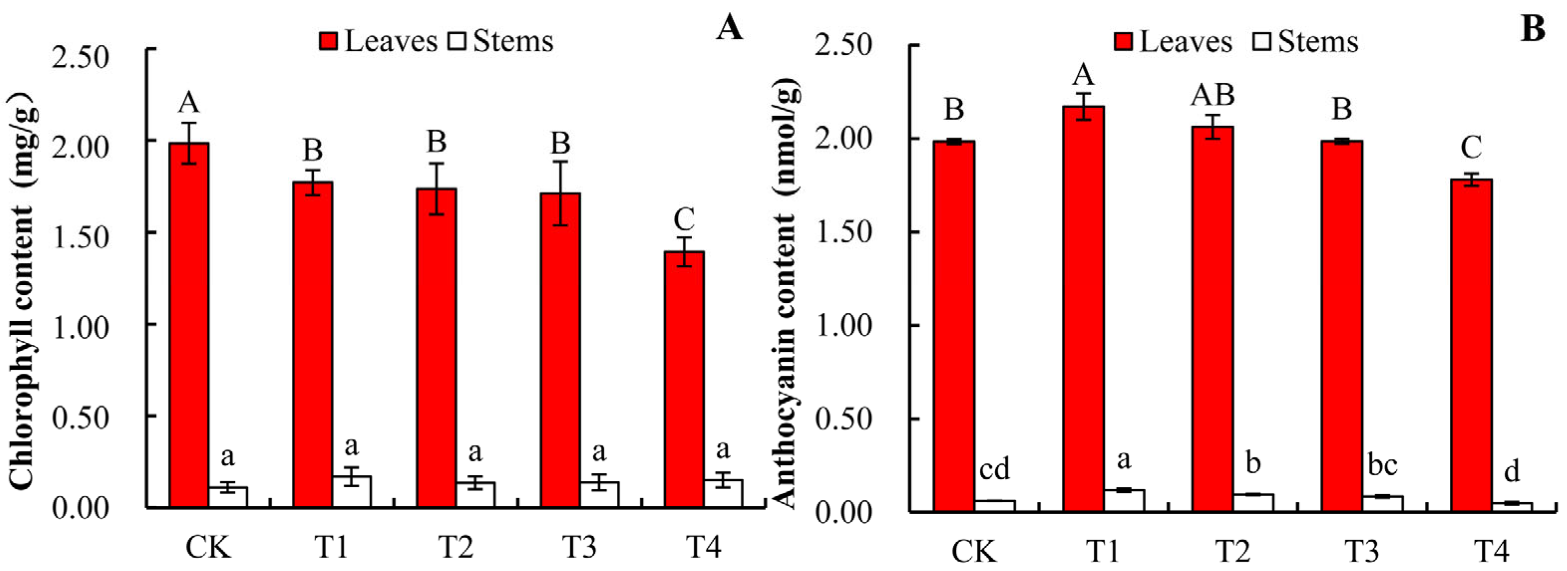
As shown in Figure 5A, with the increase of NaCl concentration, the chlorophyll content in the leaves of A. roxburghii showed a decreasing trend, and each salt treatment group was significantly lower than the control group (CK). The chlorophyll content in the stem tissue was extremely low, and no significant changes were observed among the treatment groups. This indicates that as the salt concentration increases, salt stress significantly inhibits the biosynthesis of chlorophyll in the leaves, thereby affecting the photosynthetic capacity of the plant. As shown in Figure 5B, after treatment with different concentrations of NaCl, the anthocyanin accumulation in the leaves reached its peak at the T1 treatment, while in the stems, the anthocyanin content was the highest in the T3 group and significantly higher than that in other treatment groups. This response suggests that A. roxburghii enhances its antioxidant capacity by promoting the accumulation of secondary metabolites such as anthocyanins to respond to salt stress environments.

3.5. Effects of Salt Stress on Contents of Flavonoids and Total Phenols in A. roxburghii

As shown in Figure 6A, the total flavonoid content of A. roxburghii decreased overall with the increase of NaCl treatment concentration. In leaves, the T1 treatment group had the highest total flavonoid content, which was significantly higher than that of the other groups; in stems, except for the T2 group having the lowest content, there was no significant difference among the other treatment groups. Generally speaking, the increase of salt concentration had a relatively small impact on the total flavonoid content in stems. As can be seen from Figure 6B, the total phenol content in leaves and stems of A. roxburghii under NaCl stress showed different change patterns with the increase of salt concentration. In leaves, the total phenol content in the T1 group was the lowest, and there was no significant difference among the other groups; in stems, the total phenol content in the T3 and T4 groups was significantly lower than that of the other treatment groups. In conclusion, with the increase of salt concentration, salt stress led to a decrease in the total flavonoid and total phenol content in the leaves of A. roxburghii, while having a relatively small impact on the content of these two substances in stems.

3.6. Correlation Analysis of Different NaCl Concentrations in Leaves and Stems of A. roxburghii

The Pearson correlation heatmap illustrates complex interactions among physiological and biochemical indices for salt stress in A. roxburghii (Figure 7). All the observed variables were divided in this study, namely osmoregulatory substances, ROS and antioxidant enzyme systems, pigment parameters, and growth and quality indicators in leaves (Figure 7A) and stems (Figure 7B) of A. roxburghii. The results revealed significant correlations among various parameters (p < 0.01 or p < 0.05). In leaves, leaf thickness (LT) was found to have a highly significant negative correlation with CAT, SPC, and MDA. Among them, the correlation with MDA was the strongest (r = −0.960), while it exhibited a significant positive correlation with CHC. The activities of four antioxidant enzymes, namely SOD, POD, CAT, and APX, were all significantly positively correlated with each other (r > 0.6). Among these, the correlation between SOD and POD was the highest (r = 0.677). As a marker of membrane lipid peroxidation, MDA was strongly correlated with CAT (r = 0.910). SPC was significantly positively correlated with CAT and APX (r = 0.550–0.886). Anthocyanin was positively correlated with chlorophyll and TFC (r = 0.569), and TFC was significantly positively correlated with SOD, POD, and APX, and the correlation with SOD was the strongest (r = 0.888). TPC was significantly positively correlated with SOD, CAT, APX, and TFC (r > 0.53). In addition, in stems, stem thickness (ST) was significantly negatively correlated with CAT, POD, soluble protein, MDA, and TPC. The strongest correlation was observed with MDA (r = −0.909). SOD, POD, CAT, and APX maintained significant positive correlations with each other (the highest correlation was between SOD and APX, r = 0.933). SPC was significantly positively correlated with CAT, POD, MDA, and TPC (r > 0.717). ANC was positively correlated with SOD, POD, and APX (r > 0.626), and chlorophyll was significantly positively correlated with TFC.

4. Discussion

Salt stress represents a critical environmental factor that constrains plant growth and agricultural productivity on a global scale [22]. It exerts adverse effects on plant development through multiple physiological and biochemical mechanisms, including osmotic stress, ion toxicity, and nutrient imbalance, ultimately leading to a reduction in crop yield and quality [23]. In this study, it was observed that salt stress significantly inhibits the morphological development of A. roxburghii seedlings, as indicated by the decrease in leaf and stem thickness (Figure 2), with the most pronounced inhibitory effect occurring under 200 mmol/L NaCl treatment. This morphological alteration is a typical adaptive response of plants to saline conditions. High salinity reduces soil water potential, inducing osmotic stress that impedes water uptake and results in physiological drought [24]. Concurrently, salt stress diminishes root hydraulic conductivity, thereby impairing water transport to the aerial parts of the plant, which may directly hinder cell expansion and division [25], thereby causing the stem thickness of the A. roxburghii to decrease and the leaves to become thinner.
In this study, it was found that under salt stress, the activities of antioxidant enzymes, including SOD, CAT, POD, and APX, in the leaves and stems of A. roxburghii increased significantly (Figure 3). Previous studies have shown that salt stress with appropriate concentration could reduce seedling growth but increase antioxidant enzyme activity and osmotic compound levels [26,27]. This synergistic mechanism effectively maintains the cellular redox homeostasis of ROS [28]. Notably, peak enzyme activities occurred at different salt concentrations, suggesting potential differences in their regulatory mechanisms. For instance, SOD activity peaked at 100 mmol/L NaCl, whereas CAT reached its maximum at 150 mmol/L. This sequential activation pattern may reflect a hierarchical defense strategy employed by plants against oxidative stress [29]. However, when salt concentration exceeded the plant’s tolerance threshold (150 mmol/L), the activities of all antioxidant enzymes declined, indicating that the antioxidant defense system was overwhelmed. The content of soluble proteins in leaves exhibited an initial increase followed by a decline, whereas in stems, it showed a continuous upward trend (Figure 4A). This organ-specific response indicates that the plant employs distinct osmotic adjustment strategies across different tissues [30]. This observation aligns with the trend in malondialdehyde (MDA) content, which increased continuously with rising salt concentration (Figure 4B), reflecting progressive membrane lipid peroxidation. These findings are consistent with those reported in Hollyhock (Alcea rosea L.), suggesting that the activation and subsequent collapse of the antioxidant system represent conserved mechanisms in plant responses to salt stress [31]. In this study, it was observed that chlorophyll content decreased with increasing salt concentration, which is closely related to the damage of photosynthetic apparatus [22]. Salt stress may affect chlorophyll metabolism through multiple mechanisms: on the one hand, it directly inhibits the activity of enzymes related to chlorophyll synthesis; on the other hand, it promotes the degradation process mediated by chlorophyllase [32,33]. In contrast, anthocyanin content significantly accumulated at specific salt concentrations (50 mmol/L in leaves and 150 mmol/L in stems), which may be an adaptive response. As effective antioxidants, anthocyanins can scavenge ROS, reduce oxidative damage, and prevent photoinhibition by absorbing excess light energy [33]. Additionally, among secondary metabolites, the contents of flavonoids and total phenols first increased and then decreased with the increase of salt concentration. When plants are subjected to salt stress (50–150 mmol/L), they may recognize it as a “stress signal”. To cope with this stress, plants promote the synthesis of secondary metabolites with strong antioxidant capacity, such as flavonoids and total phenols. When the salt concentration is too high (100 mmol/L), exceeding the tolerance range of plants, physiological functions are impaired, and the synthesis capacity decreases, leading to a decrease in their contents. This finding is similar to the results in vegetable amaranth (Amaranthus lividus), indicating that appropriate salt stress can enhance the accumulation of high phenolic and flavonoid compounds in plant cells [34].
In the correlation analysis, the significant positive correlations among antioxidant enzymes (SOD, POD, CAT, APX) in leaves and stems confirmed the theoretical framework of multi-enzyme collaborative clearance of ROS. Among these enzymes, the high correlation between SOD and POD highlights their key roles as core components in maintaining oxidative balance [35]. Notably, the strong negative correlation between leaf thickness and MDA reveals an antagonistic relationship between morphological traits and membrane lipid peroxidation. Furthermore, the significant positive correlation between flavonoids and antioxidant enzymes supports the hypothesis that phenolic compound accumulation contributes to oxidative stress resistance and enhanced plant tolerance, which is consistent with previous findings [36].
This study demonstrates that A. roxburghii employs an integrated strategy involving morphological modulation, antioxidant activation, and metabolic reprogramming to cope with salt stress. However, in this experiment, the plants were subjected to salt treatment in the short term (24 h). When the salt concentration exceeded the critical threshold (100–150 mmol/L NaCl), these adaptive mechanisms were insufficient to counteract the stress, resulting in growth inhibition and physiological dysfunction. In order to have a more comprehensive understanding of the physiological response of A. roxburghii to salt stress, we will focus on including biomass, root morphology, and photosynthetic performance in subsequent work to further improve the systematic analysis of the stress response mechanism. Meanwhile, future research could further investigate the molecular regulatory networks underlying the salt stress response in A. roxburghii using transcriptomic and proteomic approaches or explore the potential of cultivation management practices—such as exogenous substance application and water regulation—to mitigate salt stress effects.

5. Conclusions

In conclusion, excessively high NaCl concentrations significantly inhibit the growth and physiological performance of A. roxburghii. This study demonstrates that A. roxburghii exhibits moderate tolerance to salt stress, effectively withstanding NaCl concentrations up to 100 mmol/L. Within this threshold, the plant activates its antioxidant defense system, as evidenced by increased activities of SOD, POD, CAT, and APX, and accumulates osmoregulatory substances such as soluble proteins, thereby mitigating oxidative damage under saline conditions. However, beyond this concentration, these protective mechanisms become compromised, resulting in decreased antioxidant enzyme activity, reduced accumulation of beneficial metabolites, and a significant increase in oxidative stress, as indicated by elevated MDA levels. The observed declines in chlorophyll, anthocyanin, total flavonoid, and total phenolic contents further confirm the adverse effects of high salinity. These findings not only elucidate the physiological basis of salt tolerance in A. roxburghii but also provide a valuable theoretical foundation for the selection and breeding of salt-tolerant genotypes. Therefore, in practical cultivation, it is recommended to grow A. roxburghii in low-salt environments with NaCl concentrations not exceeding 100 mmol/L, along with the implementation of scientific management practices.

Author Contributions

Conceptualization, M.L.; methodology, H.R. and M.L.; validation, M.L. and H.L.; formal analysis, M.L., H.R., and N.L.; data curation, M.L., H.R., H.L., and N.L.; writing—original draft preparation, M.L.; writing—review and editing, M.L. and Y.J. All authors have read and agreed to the published version of the manuscript.

Funding

This research was financially supported by the Doctoral (Post) Research Initiation Fund Project of Suzhou University (grant numbers 2021BSK020, 2021BSK022, and 2022BSK036), the Anhui Province Higher Education Science Research Project (grant number 2024AH051823), the Social Practice Course Project in Anhui Province (grant number 2024shkc005), and the Key Scientific Research Project of Suzhou University (grant number 2024yzd01).

Data Availability Statement

The original contributions presented in this study are included in the article. Further inquiries can be directed to the corresponding author.

Acknowledgments

The authors sincerely thank the reviewers and editors for their valuable feedback and efforts. They also appreciate the support of the north region of Anhui province’s specialized agricultural planting research and development center (2021XJPT36ZC) for providing an important research platform for this study.

Conflicts of Interest

The authors declare no conflicts of interest.

References

  1. Ye, S.; Shao, Q.; Zhang, A. Anoectochilus roxburghii: A review of its phytochemistry, pharmacology, and clinical applications. J. Ethnopharmacol. 2017, 209, 184–202. [Google Scholar] [CrossRef] [PubMed]
  2. Wu, T.; Li, S.; Huang, Y.; He, Z.; Zheng, Y.; Antony, S.; Shao, Q.; Lin, D. Structure and pharmacological activities of polysaccharides from Anoectochilus roxburghii (Wall.) Lindl. J. Funct. Foods 2021, 87, 104815. [Google Scholar] [CrossRef]
  3. Gao, Y.; Wu, J.; Pan, Y.; Zhao, J.; Zou, X. Effects of Different pH Values on the Growth and Physiological Characteristics of Anoectochilus roxburghii. J. Plant Growth Regul. 2025, 44, 4477–4487. [Google Scholar] [CrossRef]
  4. FAO. Global Status of Salt-Affected Soils-Main Report; [R/OL]; [2025-4-2]; FAO: Rome, Italy, 2024. [Google Scholar]
  5. Devkota, K.P.; Devkota, M.; Rezael, M.; Ezaei, M.; Roland Oosterbaan, R. Managing salinity for sustainable agricultural production in salt-affected soils of irrigated drylands. Agric. Syst. 2022, 198, 103390. [Google Scholar] [CrossRef]
  6. Kumar, M.; Kesawat, M.S.; Du, X.; Siddique, K.H.M.; Kant, S.; Chung, S.M. In silico analysis and expression profiling reveal the presence of abiotic stress and developmental stage specific Aconitase genes in rice (Oryza sativa L.). Plant Stress 2024, 11, 100416. [Google Scholar] [CrossRef]
  7. Zhao, S.; Tariq, F.; Ma, C. Integrative dynamics of cell wall architecture and plant growth under salt stress. Front. Plant Sci. 2025, 16, 1644412. [Google Scholar] [CrossRef]
  8. Musyimi, D.M.; Chemisto, J.K.; Buyela, D.K. Growth and physiological response of African nightshades (Solanum Scabrum Mill.) to Sodium chloride salinity stress. Int. J. Food Saf. Public Health 2021, 8, 1–8. [Google Scholar]
  9. Kumar, S.; Li, G.; Yang, J.; Huang, X.; Ji, Q.; Liu, Z.; Hou, H. Effect of salt stress on growth, physiological parameters, and ionic concentration of water dropwort (Oenanthe javanica) cultivars. Front. Plant Sci. 2021, 12, 660409. [Google Scholar] [CrossRef]
  10. Chen, C.; Yu, W.; Xu, X.; Wang, Y.; Wang, B.; Xu, S.; Lan, Q.; Wang, Y. Research advancements in salt tolerance of cucurbitaceae: From salt response to molecular mechanisms. Int. J. Mol. Sci. 2024, 25, 9051. [Google Scholar] [CrossRef]
  11. Dias, M.V.; Rodrigues, F.A.; Ribeiro, M.S.; Dambroz, C.; Dória, J.; Pasqual, M. Physiological and morphological responses of Selenicereus species to salt stress in vitro. Plant Cell Tiss. Org. 2025, 162, 26. [Google Scholar] [CrossRef]
  12. Cui, M.; Liang, Z.; Liu, Y.; Sun, Q.; Wu, D.; Luo, L.; Hao, Y. Flavonoid profile of Anoectochilus roxburghii (Wall.) Lindl. Under short-term heat stress revealed by integrated metabolome, transcriptome, and biochemical analyses. Plant Physiol. Biochem. 2023, 201, 107896. [Google Scholar] [CrossRef] [PubMed]
  13. Gam, D.T.; Khoi, P.H.; Ngoc, P.B.; Linh, L.K.; Hung, N.K.; Anh, P.T.L.; Ha, C.H. LED lights promote growth and flavonoid accumulation of Anoectochilus roxburghii and are linked to the enhanced expression of several related genes. Plants 2020, 9, 1344. [Google Scholar] [CrossRef]
  14. Xu, N.; Liu, S.; Lu, Z.; Pang, S.; Wang, L.; Wang, L.; Li, W. Gene expression profiles and flavonoid accumulation during salt stress in Ginkgo biloba seedlings. Plants 2020, 9, 1162. [Google Scholar] [CrossRef]
  15. Giannopolitis, C.N.; Ries, S.K. Superoxide dismutases: I. occurrence in higher plants. Plant Physiol. 1977, 59, 309–314. [Google Scholar] [CrossRef]
  16. AbdelAty, A.M.; Salama, W.H.; Fahmy, A.S.; Mohamed, S.A. Impact of Germination on Antioxidant Capacity of Garden Cress: New Calculation for Determination of Total Antioxidant Activity. Sci. Hortic. 2019, 246, 155–160. [Google Scholar] [CrossRef]
  17. Cakmak, I.; Marschner, H. Magnesium deficiency and high light intensity enhance activities of superoxide dismutase, ascorbate peroxidase, and glutathione reductase in bean leaves. Plant Physiol. 1992, 98, 1222–1227. [Google Scholar] [CrossRef]
  18. Bradford, M.M. A rapid and sensitive method for the quantitation of microgram quantities of protein utilizing the principle of protein-dye binding. Anal. Biochem. 1976, 72, 248–254. [Google Scholar] [CrossRef]
  19. Yan, K.; Cui, M.; Zhao, S.; Chen, X.; Tang, X. Salinity stress is beneficial to the accumulation of chlorogenic acids in honeysuckle (Lonicera japonica Thunb.). Front. Plant Sci. 2016, 7, 1563. [Google Scholar] [CrossRef]
  20. Jiang, Y.; Li, M.; Qian, Y.; Rong, H.; Xie, T.; Wang, S.; Zhao, H.; Yang, L.; Wang, Q.; Cao, Y. Analysis of the Transcriptome Provides Insights into the Photosynthate of Maize Response to Salt Stress by 5-Aminolevulinic Acid. Int. J. Mol. Sci. 2025, 26, 786. [Google Scholar] [CrossRef]
  21. Li, M. Study on the Molecular Mechanism of Petal Fading of Brunfelsia acuminata and the Technology of Adjusting Light and Temperature; Fujian Agriculture and Forestry University: Fuzhou, China, 2020. [Google Scholar]
  22. Zhu, Y.; Li, M.; Wang, T.; Wang, J.; Zhou, H.; Lin, Y.; Yin, C. Research advances of salt exclusion, salt sequestration, salt secretion, and salt signaling regulation in plants. Plant Stress 2025, 17, 100952. [Google Scholar] [CrossRef]
  23. Shahid, M.A.; Sarkhosh, A.; Khan, N.; Balal, R.M.; Ali, S.; Rossi, L.; Gómez, C.; Mattson, N.; Nasim, W.; Garcia-Sanchez, F. Insights into the Physiological and Biochemical Impacts of Salt Stress on Plant Growth and Development. Agronomy 2020, 10, 938. [Google Scholar] [CrossRef]
  24. Saddiq, M.S.; Iqbal, S.; Hafeez, M.B.; Ibrahim, A.M.H.; Raza, A.; Fatima, E.M.; Baloch, H.; Jahanzai, B.; Woodrow, P.; Ciarmiello, L.F. Effect of Salinity Stress on Physiological Changes in Winter and Spring Wheat. Agronomy 2021, 11, 1193. [Google Scholar] [CrossRef]
  25. Fu, Y.; Li, P.; Mounkaila, H.A.K.; Wan, S.; Gao, Y.; Wang, X. Effects of Single and Combined Drought and Salinity Stress on the Root Morphological Characteristics and Root Hydraulic Conductivity of Different Winter Wheat Varieties. Plants 2023, 12, 2694. [Google Scholar] [CrossRef]
  26. Xu, Y.; Bu, W.; Xu, Y.; Fei, H.; Zhu, Y.; Ahmad, I.; Zhu, G. Effects of salt stress on physiological and agronomic traits of rice genotypes with contrasting salt tolerance. Plants 2024, 13, 1157. [Google Scholar] [CrossRef]
  27. Liu, P.; Gao, C.; Gao, Y.; Wang, C.; Jiao, Z.; Xu, A.; Dong, Y.; Sun, J. Investigating Salt Tolerance in Melon During Germination and Early Seedling Stages. Horticulturae 2025, 11, 397. [Google Scholar] [CrossRef]
  28. Nawaz, K.; Hussain, K.; Majeed, A.; Khan, F.; Afghan, S.; Ali, K. Fatality of salt stress to plants: Morphological, physiological and biochemical aspects. Afr. J. Biotechnol. 2010, 9, 5475–5480. [Google Scholar]
  29. Sharma, P.; Jha, A.B.; Dubey, R.S.; Pessarakli, M. Reactive oxygen species, oxidative damage, and antioxidative defense mechanism in plants under stressful conditions. J. Bot. 2012, 2012, 217037. [Google Scholar] [CrossRef]
  30. Akram, M.; Sajid, Z.; Farooq, A.B.U.; Ahmad, I.; Jamal, A.; Rizwana, H.; Almunqedhi, B.M.; Ronga, D. Characterization of Physiological and Biochemical Attributes of Neem (Azadirachta indica A. Juss) under Salinity Stress. Horticulturae 2024, 10, 702. [Google Scholar] [CrossRef]
  31. Sadeghi, A.; Razmjoo, J.; Karimmojeni, H.; Baldwin, T.C. Differential responses of Hollyhock (Alcea rosea L.) varieties to salt stress in relation to physiological and biochemical parameters. Sci. Rep. 2024, 14, 8105. [Google Scholar] [CrossRef] [PubMed]
  32. Ma, X.; Jia, Z.; Liu, Y. Study on Photosynthesis and Chlorophyll Metabolism of Avena sativa Lunder Salt Stress. Chin. Qinghai J. Anim. Vet. Sci. 2021, 51, 7–14. [Google Scholar]
  33. Eryılmaz, F. The relationships between salt stress and anthocyanin content in higher plants. Biotechnol. Biotechnol. Equip. 2006, 20, 47–52. [Google Scholar] [CrossRef]
  34. Hossain, M.N.; Sarker, U.; Raihan, M.S.; Al-Huqail, A.A.; Siddiqui, M.H.; Oba, S. Influence of salinity stress on color parameters, leaf pigmentation, polyphenol and flavonoid contents, and antioxidant activity of Amaranthus lividus leafy vegetables. Molecules 2022, 27, 1821. [Google Scholar] [CrossRef] [PubMed]
  35. Zhang, J.; Zhang, X.; Zhang, Z. Arabidopsis LOS5 gene enhances chilling and salt stress tolerance in cucumber. J. Integr. Agric. 2013, 12, 825–834. [Google Scholar] [CrossRef]
  36. Rao, M.; Zheng, B. The role of polyphenols in abiotic stress tolerance and their antioxidant properties to scavenge reactive oxygen species and free radicals. Antioxidants 2025, 14, 74. [Google Scholar] [CrossRef] [PubMed]
Figure 1. Morphological changes of A. roxburghii seedlings under different NaCl concentrations (CK–T4: 0 mmol/L, 50 mmol/L, 100 mmol/L, 150 mmol/L, 200 mmol/L).
Horticulturae 11 01254 g001
Figure 2. The effects of NaCl on leaf thickness (A) and stem thickness (B) in A. roxburghii seedlings. (CK–T4: 0 mmol/L, 50 mmol/L, 100 mmol/L, 150 mmol/L, 200 mmol/L). Different lowercase letters in this figure indicate significant differences (p < 0.05). Data are expressed as mean ± SD (n = 3).
Horticulturae 11 01254 g002
Figure 3. The effects of NaCl on leaf and stem of antioxidant enzyme activities ((A): SOD activity; (B): CAT activity; (C): POD activity; (D): APX activity) in A. roxburghii. (CK–T4: 0 mmol/L, 50 mmol/L, 100 mmol/L, 150 mmol/L, 200 mmol/L). Data are expressed as mean ± SD (n = 3). Different uppercase letters indicate significant differences in different salt concentrations of leaves (p < 0.05), and different lowercase letters indicate significant differences in salt concentration of stems (p < 0.05). Mean values in the column marked by different letters are significantly different within the same group (p < 0.05; ANOVA and Duncan test).
Horticulturae 11 01254 g003
Figure 4. The effects of soluble protein content (A) and MDA content (B) in leaves and stems of A. roxburghii under different NaCl concentrations. (CK–T4: 0 mmol/L, 50 mmol/L, 100 mmol/L, 150 mmol/L, 200 mmol/L). Different uppercase letters indicate significant differences in different salt concentrations of leaves (p < 0.05), and different lowercase letters indicate significant differences in salt concentration of stems (p < 0.05). Mean values in the column marked by different letters are significantly different within the same group (p < 0.05; ANOVA and Duncan test).
Horticulturae 11 01254 g004
Figure 5. Effects of the contents of chlorophyll (A) and anthocyanin (B) in A. roxburghii under different NaCl concentrations. (CK–T4: 0 mmol/L, 50 mmol/L, 100 mmol/L, 150 mmol/L, 200 mmol/L). Different uppercase letters indicate significant differences in different salt concentrations of leaves (p < 0.05), and different lowercase letters indicate significant differences in salt concentration of stems (p < 0.05). Mean values in the column marked by different letters are significantly different within the same group (p < 0.05; ANOVA and Duncan test).
Horticulturae 11 01254 g005
Figure 6. Effects of contents of total flavonoids (A) and total phenols (B) in leaves and stems of A. roxburghii under different NaCl concentrations. (CK–T4: 0 mmol/L, 50 mmol/L, 100 mmol/L, 150 mmol/L, 200 mmol/L). Different uppercase letters indicate significant differences in different salt concentrations of leaves (p < 0.05), and different lowercase letters indicate significant differences in salt concentration of stems (p < 0.05). Mean values in the column marked by different letters are significantly different within the same group (p < 0.05; ANOVA and Duncan test).
Horticulturae 11 01254 g006
Figure 7. Correlation analysis of different NaCl concentrations in leaves (A) and stems (B) of A. roxburghii. An asterisk (*) denotes statistically significant differences at p < 0.05. Two asterisks (**) denote statistically significant differences at p < 0.01. LT = stem thickness; ST = stem thickness; CAT = catalase activity; SOD = superoxide dismutase activity; POD = peroxidase activity; APX = Ascorbate peroxidase activity; SPC = soluble protein content; MDA = malondialdehyde content; ANC = Anthocyanin content; CHC = chlorophyll content; TPC = total phenols content; TFC = total flavonoids content.
Horticulturae 11 01254 g007
Disclaimer/Publisher’s Note: The statements, opinions and data contained in all publications are solely those of the individual author(s) and contributor(s) and not of MDPI and/or the editor(s). MDPI and/or the editor(s) disclaim responsibility for any injury to people or property resulting from any ideas, methods, instructions or products referred to in the content.