Article

Genome-Wide Analysis of Grapevine Ascorbate Oxidase Genes Identifies VaAAO7 in Vitis amurensis as a Positive Regulator of Botrytis cinerea Resistance

1
College of Horticulture, Henan Agricultural University, Zhengzhou 450046, China
2
Henan Provincial Engineering Research Center for Apple Germplasm Innovation and Utilization, Zhengzhou 450046, China
3
College of Agricultural Sciences, Zhengzhou University, Zhengzhou 450001, China
*
Authors to whom correspondence should be addressed.
These authors contributed equally to this work.
Horticulturae 2025, 11(10), 1211; https://doi.org/10.3390/horticulturae11101211
Submission received: 13 September 2025 / Revised: 5 October 2025 / Accepted: 6 October 2025 / Published: 8 October 2025
(This article belongs to the Collection New Insights into Developmental Biology of Fruit Trees)

Abstract

Ascorbate oxidases (AAOs) are key regulators of extracellular redox homeostasis and plant stress responses, but their roles in grapevine defense remain unclear. Here, we performed a genome-wide analysis and characterization of the AAO gene family in grapevine Vitis amurensis, identifying 10 VaAAO genes that are unevenly distributed across six chromosomes, with notable clustering on chromosome 7. Promoter analysis revealed multiple phytohormone- and stress-responsive cis-elements (e.g., ARE, STRE, and TCA-element) and transcription factor binding sites (e.g., MYC/MYB, and WRKY), suggesting involvement in redox- and stress-related signaling pathways. Analysis of previously published transcriptomic data under Botrytis cinerea infection identified VaAAO7 as a key pathogen-responsive gene. VaAAO7 was rapidly induced by H2O2, and its transient ectopic overexpression in susceptible V. vinifera ‘Red Globe’ leaves significantly reduced lesion development. Together, these results demonstrate that VaAAO7 functions as a positive regulator of B. cinerea resistance and highlight its potential for genetic engineering to enhance systemic defense and develop disease-resistant grapevine cultivars.

1. Introduction

Grapevine, a globally significant horticultural crop with high economic value, experiences substantial yield losses (20–60%) and quality reduction caused by the necrotrophic fungus Botrytis cinerea-induced gray mold [1,2]. This pathogen has a broad host range and frequently infects grapevines during production and post-harvest storage, posing significant challenges to vineyard management, thereby necessitating effective control strategies [3]. Resistance varies among germplasm accessions, with certain Vitis cultivars, such as V. amurensis ‘Shuangyou’, V. yanshanensis ‘Yanshan-1’, and V. quinquangularis ‘Pingli-5’, offering valuable traits for resistance breeding [4,5].
Plants defend against pathogens through a two-tiered innate immune system. Pattern-triggered immunity (PTI), activated by pattern recognition receptors (PRRs), detects pathogen-associated molecular patterns (PAMPs) and initiates defense responses including Ca2+ influx, reactive oxygen species (ROS) bursts, and activation of defense genes. Effector-triggered immunity (ETI), mediated by nucleotide-binding leucine-rich repeat (NLR) proteins, activates a complex network involving transcription factors (TFs), hormone signaling, and ROS regulation [6,7,8]. ROS are key signaling molecules in both PTI and ETI against Botrytis cinerea infection [9]. This pathogen possesses a strong capacity for ROS detoxification and actively manipulates host programmed cell death to facilitate its colonization, highlighting the importance of precise spatial and temporal regulation of ROS during plant defense. In the apoplast, ROS generated by ascorbate oxidases (AAO), Class III peroxidases, and NADPH oxidases act as critical defense signals [10]. Susceptible grapevine genotypes typically show excessive ROS accumulation and reduced antioxidant capacity after infection, whereas resistant genotypes maintain a more balanced ROS homeostasis [11].
AAO (EC 1.10.3.3), a member of the multicopper oxidase family, is widely distributed in plants and fungi. They catalyze the oxidation of reduced ascorbate (AsA) to monodehydroascorbate (MDHA), concurrently reducing molecular oxygen to water. MDHA is spontaneously converted to dehydroascorbate (DHA), subsequently transported to the cytoplasm, and reduced back to AsA via the ascorbate–glutathione (AsA-GSH) cycle. By regulating the apoplastic redox status, AAOs are key regulators of plant growth and mediators of responses to biotic and abiotic stimuli [12,13].
AAOs play divergent roles across plant species. AAO silencing in Solanum lycopersicum enhances abiotic stress tolerance [14]; overexpression of an AAO gene in Nicotiana tabacum increases susceptibility to fungal pathogens [15]; and AAOs overexpression in Oryza sativa compromises salinity tolerance [16]. By contrast, in Ammopiptanthus nanus, cold stress upregulates AnAAOs, and heterologous AnAAO5 expression enhances cold tolerance in Arabidopsis thaliana [17]. Moreover, enhanced AAO activity in O. sativa promotes basal ROS accumulation and strengthens antiviral defense [18].
Research on AAO gene families has been conducted across multiple plant species, such as A. thaliana, A. nanus, O. sativa, Gossypium hirsutum, Zea mays, Triticum aestivum, Glycine max, Beta vulgaris, and Citrus sinensis [19]. However, knowledge of AAOs in grapevine remains limited. In the present study, we carried out a genome-wide identification of the AAO gene family in the resistant species V. amurensis. Furthermore, the biological function of VaAAO7 was investigated through ectopic transient overexpression during B. cinerea infection in the susceptible species V. vinifera. Collectively, these findings provide new insights into the functional roles of VaAAOs in plant responses to environmental stress, which may facilitate the development of novel strategies for enhancing disease resistance in grapevine breeding programs.

2. Materials and Methods

2.1. Plant Materials and Treatments

Grapevines were grown in the vineyard of the research base of Henan Agricultural University. Dormant branches of V. amurensis ‘Shuangyou’ (SY), as well as V. vinifera ‘Red Globe’ (RG), were collected in January 2025 and were cultured in 1 L containers containing sterile water supplemented with 1/8 Murashige and Skoog medium, with the water level maintained between the first and second basal bud nodes. No additional pest and disease control was applied. Under these conditions, branches developed newly emerged shoots elongated to more than 20 cm. Plants were maintained in a growth chamber set at 26 °C, with a 16 h light/8 h dark photoperiod and 75% relative humidity. Young leaves were harvested randomly from the third to fifth nodes below the apex.

2.2. Total RNA Extraction, cDNA Synthesis, and qRT-PCR

RNA was isolated from 50 to 100 mg of grapevine leaf tissue using the E.Z.N.A.® Plant RNA Kit (Omega Bio-Tek, Norcross, GA, USA). RNA integrity was evaluated by 1.2% agarose gel electrophoresis, and RNA concentration was measured by a NanoDrop 2000 spectrophotometer (Thermo Fisher Scientific, Shanghai, China). Genomic DNA removal and reverse transcription were carried out using the HiScript III qRT SuperMix for qRT-PCR (+gDNA wiper) kit (Vazyme Biotech Co., Ltd., Nanjing, China). The resulting cDNA was diluted to 150 ng/µL and stored at −20 °C for subsequent experiments.
qRT-PCR was carried out on an ABI 7500 Fast instrument (Applied Biosystems, Carlsbad, CA, USA) with ChamQ Universal SYBR qRT-PCR Master Mix (Vazyme Biotech, Nanjing, China). Gene expression levels were determined using the double delta Ct method [20]. Three biological replicates were analyzed per treatment, each with three technical replicates.

2.3. VaAAO Gene Family Identification

The genome sequences and annotation of V. amurensis, Malus domestica, Prunus persica, O. sativa, and A. thaliana were obtained from the published datasets [21], the Ensembl Plants database, and the TAIR database, correspondingly. The Hidden Markov Model (HMM; PF07731, PF07732, and PF00394) profiles for copper-oxidase domains were downloaded from the Pfam database [22]. All potential VaAAO family members were identified using HMMER (version 3.3.1) [23] in combination with BLASTP searches against reported AAO family members of A. thaliana, O. sativa, Z. mays, N. tabacum, Cucumis sativus, Cucurbita pepo, Cucurbita maxima, and Brassica napus [24]. Candidate VaAAO genes were manually curated in IGV-GSAman [25] based on previously published transcriptome data [26]. Candidate VaAAO proteins were examined for conserved domains using the NCBI CDD database [27].
Chromosomal positions were determined from genome annotation files, and the genes were renamed according to their chromosomal order. The genomic distribution of VaAAO genes was visualized on the chromosomes using TBtools (v2.336) software [28]. The isoelectric points (pI) and molecular weights (Mw) of VaAAO proteins were predicted using the ExPASy 3.0 Compute pI/Mw tool [29], while their subcellular localization was predicted with WoLF PSORT (https://wolfpsort.hgc.jp/; accessed on 6 September 2025).

2.4. Phylogenetic and Synteny Analysis

A phylogenetic tree was generated in MEGA v7.0 [30] with the maximum likelihood (ML) method and 1000 bootstrap replicates. AtAAO protein sequences of A. thaliana were obtained from the TAIR database, and the sequences of O. sativa, N. tabacum, C. sativus, C. pepo, C. maxima, and B. napus were obtained from UniProt KB (https://www.uniprot.org/uniprotkb; accessed on 6 September 2025). Full-length AAO protein sequences from V. amurensis and the above species were used for phylogenetic tree construct, which was subsequently visualized using the iTOL online platform [31]. Synteny analyses between V. amurensis and A. thaliana, O. sativa, M. domestica, and P. persica were performed using the Multiple Collinearity Scan toolkit in TBtools II software (v2.336) with default parameters [28].

2.5. Analyses of Gene Structure, Conserved Motif, and Cis-Acting Element

VaAAO gene structures were visualized using TBtools (v2.336) [28] based on the genome sequence and annotation data. VaAAO protein motifs were analyzed by MEME Suite [32], and conserved domains were confirmed with NCBI CDD [27]. For cis-element analysis, 2 kb promoter sequences upstream of the start codon of VaAAO genes were examined using PlantCARE (https://bioinformatics.psb.ugent.be/webtools/plantcare/html/; accessed on 3 September 2025) [33].

2.6. Genes Cloning and Plasmid Construction

The VaAAO7 coding sequence was amplified from ‘SY’ cDNA using primers designed with Primer Premier 5.0 (Table S1). Subsequently, this sequence was cloned into pCAMBIA2300-35S-GFP for subcellular localization and overexpression using the ClonExpress® II One Step Cloning Kit (Vazyme Biotech Co., Ltd., Nanjing, China). Recombinant plasmids were confirmed by colony PCR and sequencing.

2.7. Subcellular Localization Analysis of VaAAO7 in N. benthamiana Leaves

Agrobacterium tumefaciens GV3101 harboring the pCAMBIA2300-35S-VaAAO7-GFP plasmid was cultivated in selective LB liquid medium (rifampicin and kanamycin), harvested by centrifugation at 12,000 rpm for 10 min, resuspended to an OD600 of 0.7–0.8, and kept at room temperature for 3 h. After infiltrating the suspension into the abaxial side of 5-week-old N. benthamiana leaves with a needleless syringe, plants were maintained at 22 °C under a 16 h light/8 h dark photoperiod and 60% relative humidity before the leaves were assessed using a Leica confocal laser scanning microscope (model TCS-SPE) after 48 h [34,35].

2.8. Transient Overexpression of VaAAO7 in V. vinifera

For transient expression in V. vinifera ‘RG’ leaves, A. tumefaciens GV3101 carrying pCAMBIA2300-35S-VaAAO7-GFP (VaAAO7-OE) or the empty vector was prepared as described above, adjusted to an OD600 of 0.7–0.8 with acetosyringone (150 µM), and incubated at room temperature for 3–5 h. Healthy leaves were immersed (abaxial side up) in the suspension, subjected to vacuum infiltration at 0.085 MPa for 30 min. After infiltration, the leaves were blotted dry, and the petioles were wrapped with moist cotton. The leaves were maintained at >90% humidity and 23 °C, incubated in darkness for 24 h, and then cultured under a 12 h light/12 h dark photoperiod for subsequent analyses.

2.9. H2O2 Treatment and B. cinerea Inoculation

Given that H2O2 is a key ROS mediating plant defense, we investigated VaAAO7 expression in healthy ‘SY’ grapevine leaves sprayed with 100 mM H2O2, with samples collected at 0, 3, 6, 12, 24, and 48 h post application, flash-frozen in liquid nitrogen, and stored at −80 °C.
B. cinerea spores were suspended in sterile distilled water, filtered through gauze, counted with a hemocytometer, and adjusted to 1.5 × 10⁶ spores mL−1. Healthy grapevine leaves were rinsed with sterile distilled water; petioles were wrapped in moistened cotton and placed in trays. Wild type (WT) and VaAAO7-OE leaves were sprayed with the spore suspension (treatment) or sterile distilled water (mock). All trays were sealed to maintain >90% relative humidity and incubated at 23 °C in darkness for 24 h, after which they were transferred to a 12 h light/12 h dark photoperiod. Leaf samples collected at 0, 4, 8, 18, and 36 h post-inoculation (hpi) were flash-frozen in liquid nitrogen and stored at −80 °C.
For each treatment and control, 8–10 leaves were collected per replicate, with three biological replicates.

2.10. Data Analysis

Lesion area and leaf area were measured by ImageJ software (v1.54r) [36]. Statistical analyses and relevant graphs generation were performed using GraphPad Prism Software (Version 10.4.0; GraphPad Software Inc., Boston, MA, USA). Different lowercase letters denote statistically significant differences among treatments (p < 0.05).

3. Results

3.1. Genome-Wide Identification and Characterization of the VaAAO Gene Family in V. amurensis

The V. amurensis cultivar ‘Zuoshan 1’ genome (~522 Mb, contig N50 = 2.51 Mb, scaffold N50 = 26.52 Mb, 615 contigs anchored to 19 pseudochromosomes) [20] provided a high-quality reference for gene identification [21]. Based on this genome annotation, we searched for members of the VaAAO gene family. A total of ten VaAAO genes were identified in the genome and were designated VaAAO1 to VaAAO10 based on their chromosomal locations (Table 1 and Table S2). The encoded proteins varied in length from 535 to 1202 amino acids, corresponding to molecular weights of 59.98 to 134.94 kDa. Four proteins were acidic (theoretical isoelectric points (pI) < 7) and six were basic (pI > 7), with VaAAO9 (pI 5.29) and VaAAO8 (pI 9.46) representing the extremes. Localization predictions indicated distinct subcellular localization: VaAAO1 and 4 in the extracellular matrix, VaAAO2, 3, and 6 in the vacuole, VaAAO7 in the cytoplasm, and VaAAO5, 8, 9, and 10 in the plasma membrane (Table 1).
Chromosomal localization analysis revealed an uneven distribution of VaAAOs across six chromosomes, with a notable cluster of six genes on chromosome 7 (Chr07; Table 1 and Figure 1A). Phylogenetic analysis of 28 AAO proteins from nine plant species resolved four distinct groups (Figure 1B). Groups I–III contained orthologs from both monocots (O. sativa and Z. mays) and dicots (V. amurensis, B. napus, N. tabacum, A. thaliana, C. sativus, C. maxima, and C. pepo), whereas Group IV contained only V. amurensis AAO proteins, indicating that VaAAO family differentiation occurred prior to monocot-dicot divergence during angiosperm evolution. Notably, VaAAO7 clustered with A. thaliana AtAAO1, O. sativa OsAAO4, 5, and Z. mays ZmAAO1–3.

3.2. Collinearity Analysis of VaAAO Family Genes in V. amurensis

To investigate potential gene duplication, we performed an intraspecific collinearity analysis within the V. amurensis genome. No duplication events involving VaAAO genes were detected on any chromosomes (Figure 2A). We further conducted intergeneric collinearity analysis between grapevine and four representative species, including two herbaceous plants (A. thaliana and O. sativa) and two woody plants (M. domestica and P. persica). Nine collinear AAO gene pairs were identified between V. amurensis and peach (P. persica) (Figure 2B), while no collinear AAO gene pairs were found with the other three species (Supplementary Figure S1). These results suggest a closer evolutionary relationship between grapevine and peach compared with the other species analyzed.

3.3. Motif, Structural and Cis-Regulatory Elements Analysis of VaAAO Family Members in V. amurensis

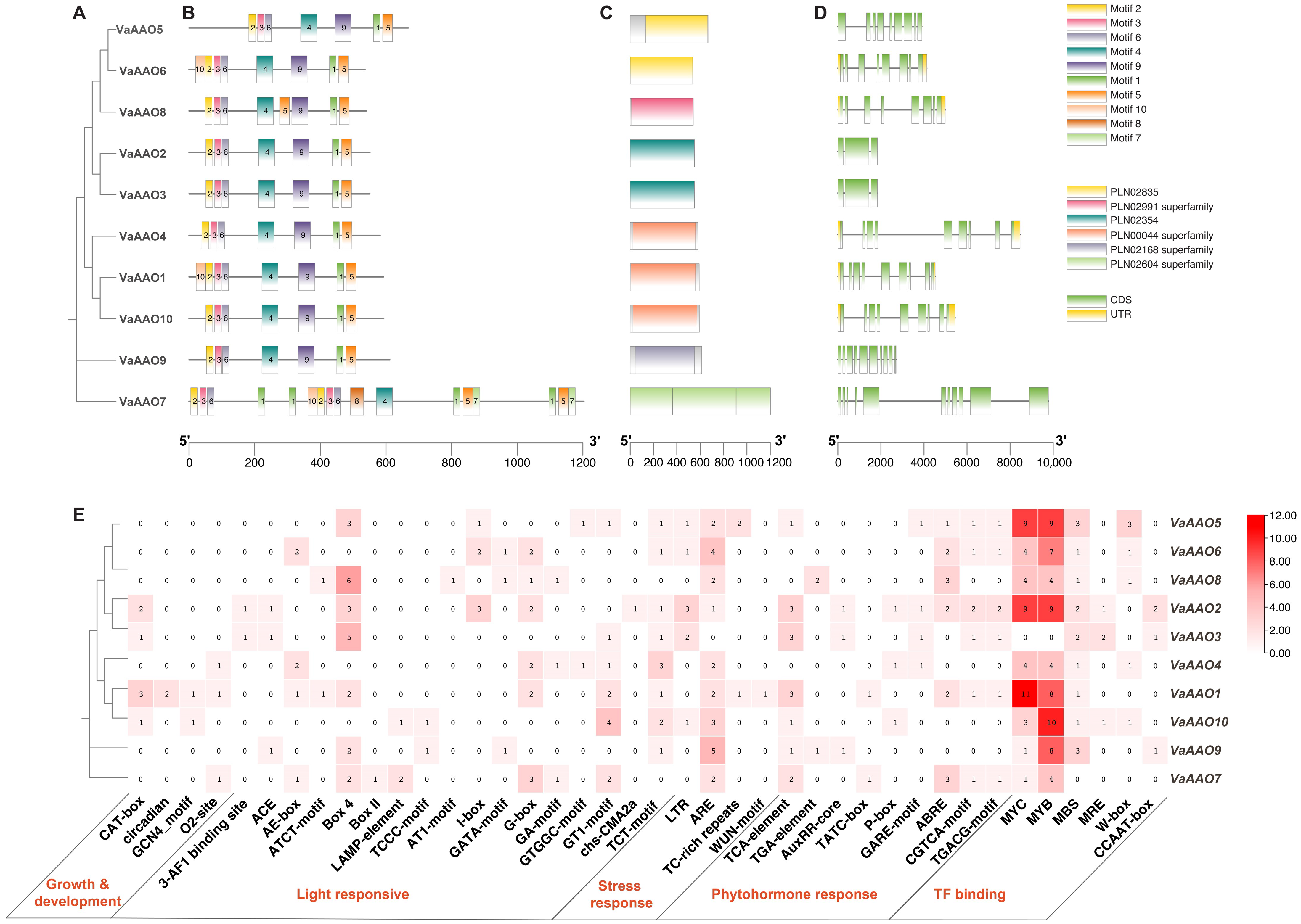
The distribution of conserved motifs within the VaAAO proteins was analyzed using the MEME suite. Overall, the VaAAO family exhibited a highly conserved motif composition and arrangement, except for repeated motifs 1, 5, and 7 in VaAAO7 (Figure 3A,B). All identified VaAAO proteins contained the oxidoreductase domain (Figure 3C). Further exon-intron structure analysis revealed variation in gene structure, with exon numbers ranging from three to eleven (Figure 3D).
Cis-acting element analysis of 2 kb promoter regions identified over 40 distinct regulatory elements, which were grouped into five major functional categories (Figure 3E). Among these, light-responsive elements (e.g., Box 4) were the most abundant. In addition, promoters contained multiple stress-responsive elements (e.g., TC-rich repeats for defense and stress response, ARE for anaerobic induction), phytohormone-responsive elements (e.g., TCA-element for salicylic acid (SA), ABRE for abscisic acid (ABA), and CGTCA/TGACG motifs for methyl jasmonate), and TF (e.g., MYB, MYC, and WRKY) binding sites. Notably, stress- and phytohormone-responsive elements, including ARE and TCA, were present in the majority of VaAAO promoters. MYC and MYB TFs binding sites were also widely distributed, whereas W-box motifs (WRKY-binding sites) [37] were absent from VaAAO1–3, VaAAO7, and VaAAO9.

3.4. Expression Patterns of VaAAO Family Genes During B. cinerea Infection

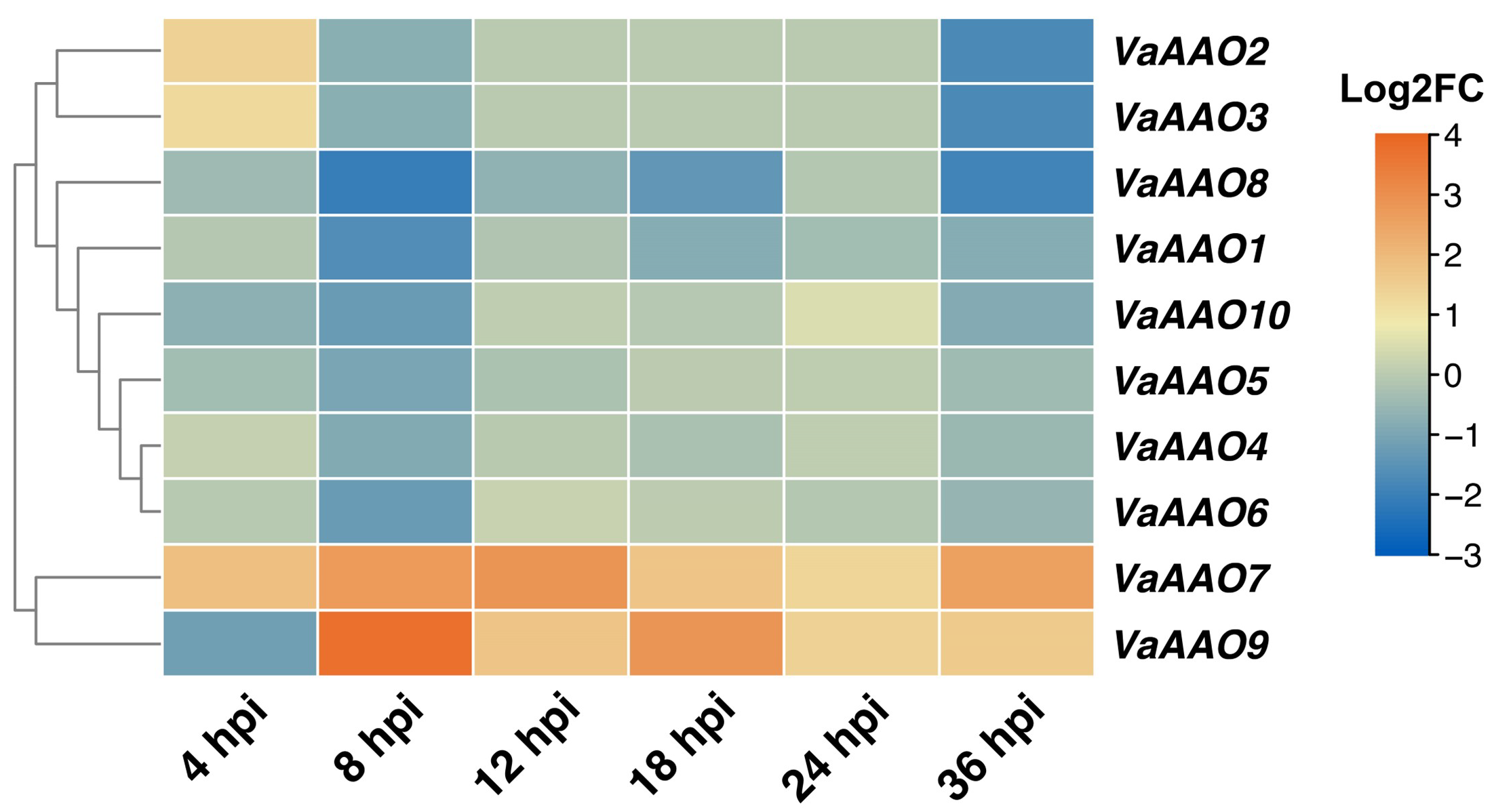
Using previously published RNA-seq data [5], we examined the expression dynamics of VaAAO genes in the B. cinerea-resistant wild grapevine V. amurensis ‘SY’ following inoculation at 4, 8, 12, 18, 24, and 36 hpi. The analysis revealed distinct stress-responsive expression patterns (Figure 4). VaAAO2, VaAAO3, and VaAAO7 were rapidly induced as early as 4 hpi compared with mock-treated controls. While VaAAO7 and VaAAO9 maintained consistently high expression from 8 to 36 hpi, suggesting their potential roles in the sustained defense response of grapevine against B. cinerea.

3.5. H2O2 Treatment Triggers Rapid Induction of VaAAO7 in ‘SY’ Leaves

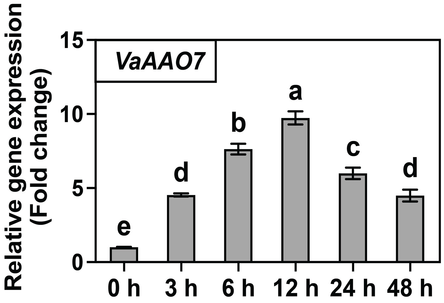
Following the genome-wide identification of VaAAO family members, VaAAO7 was selected for functional characterization because it clustered phylogenetically with AtAAO1, OsAAO4/5, and ZmAAO1–3, and VaAAO7 was rapidly and strongly induced at all time points induced during B. cinerea infection (Figure 4). Since ROS, including H2O2, play key roles in mediating host defense [38], we examined VaAAO7 expression in ‘SY’ leaves following exogenous treatment with 100 mM H2O2 at 0, 3, 6, 12, 24, and 48 h. VaAAO7 transcription was rapidly induced, showing a significant increase at 3 h and reaching a peak at 12 h, approximately 10-fold higher than at 0 h. Although its expression declined thereafter, the VaAAO7 transcript level remained elevated compared with untreated controls (Figure 5). These results suggest that VaAAO7 is ROS-responsive and may contribute to the grapevine defense response.

3.6. Transient Overexpression of VaAAO7 Enhanced Resistance to B. cinerea in Susceptible V. vinifera

The VaAAO7 protein was predicted to be localized in the cytoplasm (Table 1). To examine VaAAO7 subcellular localization, a 35S:VaAAO7-GFP fusion construct was introduced into A. tumefaciens strain and transiently expressed in N. benthamiana leaves. Confocal microscopy revealed GFP fluorescence predominantly at the cytoplasm and at the cell periphery, suggesting potential extracellular localization of the VaAAO7 protein (Figure 6A).
To investigate the role of VaAAO7 in the B. cinerea defense response, we transiently overexpressed VaAAO7 in leaves of the susceptible V. vinifera cultivar ‘RG’ (VaAAO7-OE) and subsequently inoculated them with B. cinerea. WT leaves were inoculated in parallel and served as controls. The susceptible ‘RG’ was selected to assess VaAAO7’s contribution to defense, since the resistant ‘SY’ exhibits strong basal resistance. qRT-PCR analysis confirmed that VaAAO7 transcript levels were consistently higher in VaAAO7-OE leaves compared with WT at 48, 72, and 120 hpi (Figure 6B).
Phenotypic evaluation showed that disease symptoms appeared at 48 hpi. At this stage, the lesion area in VaAAO7-OE leaves was 3.4%, significantly lower than the 12.7% observed in WT leaves. At 72 hpi, lesions in VaAAO7-OE leaves expanded to 6.1%, which is still substantially smaller than the 42.2% in WT. By 120 hpi, the lesion area in VaAAO7-OE leaves remained low at 11.5%, compared with 65.0% in WT (Figure 6C,D). Throughout the observation period, both symptom onset and lesion expansion were markedly reduced in VaAAO7-OE leaves, indicating that transient overexpression of VaAAO7 significantly enhanced resistance to B. cinerea in ‘RG’.

4. Discussion

AAOs are pivotal enzymes in the ASA-GSH cycle, regulating extracellular ascorbate and oxygen levels and integrating multiple signaling pathways under both biotic and abiotic stresses [39,40]. AAO transcription is highly responsive to oxidative stimuli (e.g., ROS, hormones, and mechanical injury). Induction of AAO depletes apoplastic AsA pools, disrupting redox homeostasis, which in turn triggers antioxidant defenses through transcriptional reprogramming to restore equilibrium [41]. Despite their significance, AAO genes have not been systematically identified in grapevine. Here, we present the genome-wide identification and characterization of the grapevine V. amurensis AAO gene family, highlighting their potential roles in plant immunity and stress adaptation. Ten VaAAO members were identified in this study, consistent with the AAO family member scale (3–16) in other plant species [19]. Comparative analysis revealed strong collinearity with P. persica (Figure 2B), but no syntenic relationships were detected with the perennial M. domestica or model herbaceous plants (A. thaliana and O. sativa), suggesting lineage-specific retention of VaAAO genes in closely related woody perennials.
Promoter analysis revealed that VaAAO genes are enriched in light-, stress-, and hormone- responsive cis-elements, as well as binding sites for key TFs. These features suggest that VaAAOs may be differentially activated under diverse environmental cues, and their regulation is likely integrated into hormone and transcription factor networks, particularly WRKY, MYB, and MYC, which are central regulators of stress responses [42,43,44,45]. ARE elements were present in nine of ten VaAAOs, linking them to anaerobic induction. Since B. cinerea infection creates local hypoxia that stabilizes ERF-VII proteins and alters redox and elicitor metabolism [46], VaAAOs may be involved in oxidative and hypoxic responses during pathogen attack. In addition, the presence of ABRE and TCA elements in the VaAAO promoters, particularly in VaAAO7, indicates that VaAAOs may participate in ABA- and SA-mediated regulation and suggests potential crosstalk between ROS and multiple hormone signaling pathways in defense against necrotrophic pathogens [47,48,49].
ROS homeostasis is central to regulating plant growth, development, and adaptive responses, such as the induction of stomatal closure to enhance stress tolerance [50,51,52]. AAOs are widely distributed in higher plants and are predominantly localized in the apoplast, either as soluble enzymes secreted into the extracellular space or bound to the cell wall matrix. They catalyze the reduction of H2O2 to water through the formation of an enzyme–peroxide complex [40]. Genetic evidence supports the role of AAOs in defense regulation: silencing AtAAO1 in A. thaliana increases susceptibility to spider mite Tetranychus urticae, while its overexpression affects genes involved in cell wall biosynthesis [53,54], highlighting the cell wall’s role as a primary barrier against pathogen invasion [55]. VaAAO7 shares high sequence similarity with AtAAO1, which is also homologous to rice OsAAO4 (Figure 1B). In rice, OsAAO4 cooperates with OsAAO3 to fine-tune apoplastic ROS balance and immunity through interaction with the rice blast fungus Magnaporthe oryzae effector MoAo1 [56], indicating a potential role for VaAAO7 in modulating extracellular ROS and defense responses in grapevine.
VaAAO7 was shown to be localized to the cytoplasm and cell periphery (Figure 4), while its precise subcellular localization requires further validation (e.g., measuring apoplastic ascorbate oxidase activity). The rapid induction of VaAAO7 upon B. cinerea infection and H2O2 treatment supports a role in defense against pathogen-triggered oxidative bursts. Transient overexpression of VaAAO7 in the susceptible V. vinifera cultivar ‘RG’ significantly reduced lesion area and delayed symptom progression (Figure 5 and Figure 6). The presence of four MYC/MYB binding sites in the VaAAO7 promoter highlights the need to identify upstream regulators and to elucidate their integration into hormone-mediated broader stress responses. Additionally, functional redundancy among VaAAO family members may exist, and stable transgenics or gene-editing approaches [57] will clarify the specific roles of VaAAO7 and related genes.
In N. benthamiana, cucumber mosaic virus-induced NbAAO expression disrupted dimer formation and weakened defense, whereas silencing of another NbAAO gene increased viral susceptibility, indicating isoform-specific functions and highlighting the need to identify potential VaAAO7 interactors [41]. Prolonged overexpression of one NtAAO in tobacco shifted the AsA pool toward oxidation, resulting in enhanced vulnerability to stress [15]. Notably, foliar AO application has been shown to induce systemic nematode resistance in rice against Meloidogyne graminicola [58] and in sugar beet against Heterodera schachtii [59]. These findings suggest that VaAAO7 could serve not only as a target for genetic manipulation (e.g., promoter modification and precise genome editing), but also as a candidate for foliar application to enhance systemic defense in grapevine against a broad spectrum of pathogens beyond B. cinerea. Overall, our results emphasize the potential of wild Vitis germplasm, such as V. amurensis, for breeding disease-resistant and stress-tolerant grapevine cultivars.

5. Conclusions

This study provided a comprehensive characterization of the AAO gene family in V. amurensis, identifying ten VaAAO genes grouped into five clades with close evolutionary relationships to P. persica. Promoter analyses revealed abundant light-, phytohormone-, and stress-responsive elements, as well as transcription factor binding sites. VaAAO7 localized to the cytoplasm and cell periphery, and its transcription was rapidly induced by H2O2 and B. cinerea. Overexpression of VaAAO7 enhanced B. cinerea resistance in susceptible V. vinifera, establishing it as a promising candidate for improving grapevine disease resistance and providing a solid foundation for future functional and mechanistic studies.

Supplementary Materials

The following supporting information can be downloaded at: https://www.mdpi.com/article/10.3390/horticulturae11101211/s1. Figure S1: Intergeneric collinearity between V. amurensis and three representative species. Table S1: List of primers employed for PCR assays. Table S2: Amino acid sequences of VaAAO family proteins.

Author Contributions

Conceptualization, Y.S., Z.Y., R.W. and X.Z.; formal analysis, Y.S., Z.Y. and L.Z.; visualization, Y.S., Z.Y., L.Z., J.S., J.J. and R.W.; investigation, Y.S., Z.Y., L.Z., Y.L., M.W., K.Z. and P.H.; resources, R.W., Y.Z., Y.L., L.C., T.B. and C.S.; writing—original draft, Y.S., Z.Y. and R.W.; writing—review and editing, Y.S., R.W. and X.Z.; funding acquisition, R.W. and X.Z. All authors have read and agreed to the published version of the manuscript.

Funding

This research was funded by the National Natural Science Foundation of China (32302495).

Data Availability Statement

The original contributions presented in this study are included in the article and the Supplementary Materials; further inquiries can be directed to the corresponding authors.

Conflicts of Interest

The authors declare no conflicts of interest.

References

  1. Martínez-Romero, D.; Guillén, F.; Valverde, J.M.; Bailén, G.; Zapata, P.; Serrano, M.; Castillo, S.; Valero, D. Influence of Carvacrol on Survival of Botrytis cinerea Inoculated in Table Grapes. Int. J. Food Microbiol. 2007, 115, 144–148. [Google Scholar] [CrossRef]
  2. Hou, X.; Zhang, G.; Han, R.; Wan, R.; Li, Z.; Wang, X. Ultrastructural Observations of Botrytis cinerea and Physical Changes in Resistant and Susceptible Grapevines. Phytopathology 2022, 112, 387–395. [Google Scholar] [CrossRef] [PubMed]
  3. Dean, R.; Van Kan, J.A.L.; Pretotius, Z.A.; Hammond-Kosack, K.E.; Di Pietro, A.; Spanu, P.D.; Rudd, J.J.; Dickman, M.; Kahmann, R.; Ellis, J.; et al. The Top 10 Fungal Pathogens in Molecular Plant Pathology. Mol. Plant Pathol. 2012, 13, 414–430. [Google Scholar] [CrossRef]
  4. Wan, R.; Hou, X.; Wang, X.; Qu, J.; Singer, S.D.; Wang, Y.; Wang, X. Resistance Evaluation of Chinese Wild Vitis Genotypes against Botrytis cinerea and Different Responses of Resistant and Susceptible Hosts to the Infection. Front. Plant Sci. 2015, 6, 854. [Google Scholar] [CrossRef] [PubMed]
  5. Wan, R.; Guo, C.; Hou, X.; Zhu, Y.; Gao, M.; Hu, X.; Zhang, S.; Jiao, C.; Guo, R.; Li, Z.; et al. Comparative Transcriptomic Analysis Highlights Contrasting Levels of Resistance of Vitis vinifera and Vitis amurensis to Botrytis cinerea. Hortic. Res. 2021, 8, 103. [Google Scholar] [CrossRef]
  6. Dodds, P.N.; Chen, J.; Outram, M.A. Pathogen Perception and Signaling in Plant Immunity. Plant Cell 2024, 36, 1465–1481. [Google Scholar] [CrossRef]
  7. Huang, S.; Wang, J.; Song, R.; Jia, A.; Xiao, Y.; Sun, Y.; Wang, L.; Mahr, D.; Wu, Z.; Han, Z.; et al. Balanced Plant Helper NLR Activation by a Modified Host Protein Complex. Nature 2025, 639, 447–455. [Google Scholar] [CrossRef] [PubMed]
  8. Ma, C.; Tian, X.; Dong, Z.; Li, H.; Chen, X.; Liu, W.; Yin, G.; Ma, S.; Zhang, L.; Cao, A.; et al. An Aegilops Longissima NLR Protein with Integrated CC-BED Module Mediates Resistance to Wheat Powdery Mildew. Nat. Commun. 2024, 15, 8281. [Google Scholar] [CrossRef]
  9. Yuan, M.; Jiang, Z.; Bi, G.; Nomura, K.; Liu, M.; Wang, Y.; Cai, B.; Zhou, J.; He, S.; Xin, X. Pattern-Recognition Receptors Are Required for NLR-Mediated Plant Immunity. Nature 2021, 592, 105–109. [Google Scholar] [CrossRef]
  10. Veloso, J.; van Kan, J.A.L. Many Shades of Grey in Botrytis–Host Plant Interactions. Trends Plant Sci. 2018, 23, 613–622. [Google Scholar] [CrossRef]
  11. Rahman, M.U.; Hanif, M.; Wan, R.; Hou, X.; Ahmad, B.; Wang, X. Screening Vitis Genotypes for Responses to Botrytis cinerea and Evaluation of Antioxidant Enzymes, Reactive Oxygen Species and Jasmonic Acid in Resistant and Susceptible Hosts. Molecules 2019, 24, 5. [Google Scholar] [CrossRef]
  12. Karpinska, B.; Zhang, K.; Rasool, B.; Pastok, D.; Morris, J.; Verrall, S.R.; Hedley, P.E.; Hancock, R.D.; Foyer, C.H. The Redox State of the Apoplast Influences the Acclimation of Photosynthesis and Leaf Metabolism to Changing Irradiance. Plant Cell Environ. 2018, 41, 1083–1097. [Google Scholar] [CrossRef]
  13. Foyer, C.H.; Kyndt, T.; Hancock, R.D. Vitamin C in Plants: Novel Concepts, New Perspectives, and Outstanding Issues. Antioxid. Redox Signal. 2020, 32, 463–485. [Google Scholar] [CrossRef]
  14. Abdelgawad, K.; El-Mogy, M.; Mohamed, M.; Garchery, C.; Stevens, R. Increasing Ascorbic Acid Content and Salinity Tolerance of Cherry Tomato Plants by Suppressed Expression of the Ascorbate Oxidase Gene. Agronomy 2019, 9, 51. [Google Scholar] [CrossRef]
  15. Fotopoulos, V.; Sanmartin, M.; Kanellis, A.K. Effect of Ascorbate Oxidase Over-Expression on Ascorbate Recycling Gene Expression in Response to Agents Imposing Oxidative Stress. J. Exp. Bot. 2006, 57, 3933–3943. [Google Scholar] [CrossRef] [PubMed]
  16. Wang, M.; Guo, W.; Li, J.; Pan, X.; Pan, L.; Zhao, J.; Zhang, Y.; Cai, S.; Huang, X.; Wang, A.; et al. The miR528-AO Module Confers Enhanced Salt Tolerance in Rice by Modulating the Ascorbic Acid and Abscisic Acid Metabolism and ROS Scavenging. J. Agric. Food Chem. 2021, 69, 8634–8648. [Google Scholar] [CrossRef] [PubMed]
  17. Zhu, M.; Liu, Q.; Liu, F.; Zheng, L.; Bing, J.; Zhou, Y.; Gao, F. Gene Profiling of the Ascorbate Oxidase Family Genes under Osmotic and Cold Stress Reveals the Role of AnAO5 in Cold Adaptation in Ammopiptanthus nanus. Plants 2023, 12, 677. [Google Scholar] [CrossRef]
  18. Wu, J.; Yang, R.; Yang, Z.; Yao, S.; Zhao, S.; Wang, Y.; Li, P.; Song, X.; Jin, L.; Zhou, T.; et al. ROS Accumulation and Antiviral Defence Control by microRNA528 in Rice. Nat. Plants 2017, 3, 16203. [Google Scholar] [CrossRef]
  19. Xu, X.; Miao, X.; Deng, N.; Liang, M.; Wang, L.; Jiang, L.; Zeng, S. Identification of Ascorbate Oxidase Genes and Their Response to Cold Stress in Citrus sinensis. Agriculture 2024, 14, 1643. [Google Scholar] [CrossRef]
  20. Livak, K.J.; Schmittgen, T.D. Analysis of Relative Gene Expression Data Using Real-Time Quantitative PCR and the 2−ΔΔCT Method. Methods 2001, 25, 402–408. [Google Scholar] [CrossRef]
  21. Wang, P.; Meng, F.; Yang, Y.; Ding, T.; Liu, H.; Wang, F.; Li, A.; Zhang, Q.; Li, K.; Fan, S.; et al. De Novo Assembling a High-Quality Genome Sequence of Amur Grape (Vitis amurensis Rupr.) Gives Insight into Vitis Divergence and Sex Determination. Hortic. Res. 2024, 11, uhae117. [Google Scholar] [CrossRef]
  22. Paysan-Lafosse, T.; Andreeva, A.; Blum, M.; Chuguransky, S.R.; Grego, T.; Pinto, B.L.; Salazar, G.A.; Bileschi, M.L.; Llinares-López, F.; Meng-Papaxanthos, L.; et al. The Pfam Protein Families Database: Embracing AI/ML. Nucleic Acids Res. 2025, 53, D523–D534. [Google Scholar] [CrossRef] [PubMed]
  23. Potter, S.C.; Luciani, A.; Eddy, S.R.; Park, Y.; Lopez, R.; Finn, R.D. HMMER Web Server: 2018 Update. Nucleic Acids Res. 2018, 46, W200–W204. [Google Scholar] [CrossRef] [PubMed]
  24. Glenzel, V.H.; Filgueiras, J.P.C.; Zolet, A.C.T.; Kulcheski, F.R. Evolutionary and Functional Insights into Ascorbate Oxidase Genes in the Fabaceae Plant Family. Plant Gene 2025, 44, 100538. [Google Scholar] [CrossRef]
  25. Robinson, J.T.; Thorvaldsdottir, H.; Turner, D.; Mesirov, J.P. Igv.Js: An Embeddable JavaScript Implementation of the Integrative Genomics Viewer (IGV). Bioinformatics 2023, 39, btac830. [Google Scholar] [CrossRef]
  26. Ma, X.; Zhao, F.; Su, K.; Lin, H.; Guo, Y. Discovery of Cold-Resistance Genes in Vitis amurensis Using Bud-Based Quantitative Trait Locus Mapping and RNA-Seq. BMC Genom. 2022, 23, 551. [Google Scholar] [CrossRef]
  27. Marchler-Bauer, A.; Bo, Y.; Han, L.; He, J.; Lanczycki, C.J.; Lu, S.; Chitsaz, F.; Derbyshire, M.K.; Geer, R.C.; Gonzales, N.R.; et al. CDD/SPARCLE: Functional Classification of Proteins via Subfamily Domain Architectures. Nucleic Acids Res. 2017, 45, D200–D203. [Google Scholar] [CrossRef]
  28. Chen, C.; Wu, Y.; Li, J.; Wang, X.; Zeng, Z.; Xu, J.; Liu, Y.; Feng, J.; Chen, H.; He, Y.; et al. TBtools-II: A “One for All, All for One” Bioinformatics Platform for Biological Big-Data Mining. Mol. Plant 2023, 16, 1733–1742. [Google Scholar] [CrossRef]
  29. Duvaud, S.; Gabella, C.; Lisacek, F.; Stockinger, H.; Ioannidis, V.; Durinx, C. Expasy, the Swiss Bioinformatics Resource Portal, as Designed by Its Users. Nucleic Acids Res. 2021, 49, W216–W227. [Google Scholar] [CrossRef]
  30. Kumar, S.; Stecher, G.; Tamura, K. MEGA7: Molecular Evolutionary Genetics Analysis Version 7.0 for Bigger Datasets. Mol. Biol. Evol. 2016, 33, 1870–1874. [Google Scholar] [CrossRef] [PubMed]
  31. Letunic, I.; Bork, P. Interactive Tree of Life (iTOL) v6: Recent Updates to the Phylogenetic Tree Display and Annotation Tool. Nucleic Acids Res. 2024, 52, W78–W82. [Google Scholar] [CrossRef]
  32. Bailey, T.L.; Boden, M.; Buske, F.A.; Frith, M.; Grant, C.E.; Clementi, L.; Ren, J.; Li, W.W.; Noble, W.S. MEME SUITE: Tools for Motif Discovery and Searching. Nucleic Acids Res. 2009, 37, W202–W208. [Google Scholar] [CrossRef]
  33. Lescot, M. PlantCARE, a Database of Plant Cis-Acting Regulatory Elements and a Portal to Tools for in Silico Analysis of Promoter Sequences. Nucleic Acids Res. 2002, 30, 325–327. [Google Scholar] [CrossRef]
  34. Withers, J.; Yao, J.; Mecey, C.; Howe, G.A.; Melotto, M.; He, S.Y. Transcription Factor-Dependent Nuclear Localization of a Transcriptional Repressor in Jasmonate Hormone Signaling. Proc. Natl. Acad. Sci. USA 2012, 109, 20148–20153. [Google Scholar] [CrossRef]
  35. Xie, J.; He, C.; Li, Z.; Li, M.; He, S.; Qian, J.; Tan, B.; Zheng, X.; Cheng, J.; Wang, W.; et al. A Rapid and Efficient Agrobacterium-Mediated Transient Transformation System in Grape Berries. Protoplasma 2024, 261, 819–830. [Google Scholar] [CrossRef]
  36. Schneider, C.A.; Rasband, W.S.; Eliceiri, K.W. NIH Image to ImageJ: 25 Years of Image Analysis. Nat. Methods 2012, 9, 671–675. [Google Scholar] [CrossRef] [PubMed]
  37. Li, G.Z.; Wang, Z.Q.; Yokosho, K.; Ding, B.; Fan, W.; Gong, Q.Q.; Li, G.X.; Wu, Y.R.; Yang, J.L.; Ma, J.F.; et al. Transcription Factor WRKY22 Promotes Aluminum Tolerance via Activation of OsFRDL4 Expression and Enhancement of Citrate Secretion in Rice (Oryza Sativa). New Phytol. 2018, 219, 149–162. [Google Scholar] [CrossRef]
  38. Ding, L.; Wu, Z.; Xiang, J.; Cao, X.; Xu, S.; Zhang, Y.; Zhang, D.; Teng, N. A LlWRKY33-LlHSFA4-LlCAT2 Module Confers Resistance to Botrytis cinerea in Lily. Hortic. Res. 2024, 11, uhad254. [Google Scholar] [CrossRef] [PubMed]
  39. Zhang, N.; Li, L.; Zhang, L.; Li, J.; Fang, Y.; Zhao, L.; Ren, Y.; Chen, F. Abscisic Acid Enhances Tolerance to Spring Freeze Stress and Regulates the Expression of Ascorbate–Glutathione Biosynthesis-Related Genes and Stress-Responsive Genes in Common Wheat. Mol. Breed. 2020, 40, 108. [Google Scholar] [CrossRef]
  40. Liu, C.; Mao, B.; Zhang, Y.; Tian, L.; Ma, B.; Chen, Z.; Wei, Z.; Li, A.; Shao, Y.; Cheng, G.; et al. The OsWRKY72–OsAAT30/OsGSTU26 Module Mediates Reactive Oxygen Species Scavenging to Drive Heterosis for Salt Tolerance in Hybrid Rice. J. Integr. Plant Biol. 2024, 66, 709–730. [Google Scholar] [CrossRef]
  41. Mellidou, I.; Kanellis, A.K. Revisiting the Role of Ascorbate Oxidase in Plant Systems. J. Exp. Bot. 2024, 75, 2740–2753. [Google Scholar] [CrossRef] [PubMed]
  42. Zheng, H.; Dong, L.; Han, X.; Jin, H.; Yin, C.; Han, Y.; Li, B.; Qin, H.; Zhang, J.; Shen, Q.; et al. The TuMYB46L-TuACO3 Module Regulates Ethylene Biosynthesis in Einkorn Wheat Defense to Powdery Mildew. New Phytol. 2020, 225, 2526–2541. [Google Scholar] [CrossRef]
  43. Li, G.; Liu, J.; Wang, Y.; Han, A.; Liu, H.; Guo, T.; Han, Q.; Kang, G. TaWRKY24-1D, Interacts with TaERFL1a, Regulates DHAR-Mediated ASA-GSH Biosynthesis to Enhance Drought Tolerance in Wheat. Plant Growth Regul. 2024, 104, 713–725. [Google Scholar] [CrossRef]
  44. Ren, Z.; Zhang, P.; Su, H.; Xie, X.; Shao, J.; Ku, L.; Tian, Z.; Deng, D.; Wei, L. Regulatory Mechanisms Used by ZmMYB39 to Enhance Drought Tolerance in Maize (Zea Mays) Seedlings. Plant Physiol. Biochem. 2024, 211, 108696. [Google Scholar] [CrossRef]
  45. Zhang, X.; Pan, Y.; Hao, X.; Guo, C.; Wang, X.; Yan, X.; Guo, R. Overexpression of a Grapevine VqWRKY2 Transcription Factor in Arabidopsis Thaliana Increases Resistance to Powdery Mildew. Plant Cell Tissue Organ Cult. (PCTOC) 2024, 157, 16. [Google Scholar] [CrossRef]
  46. Valeri, M.C.; Novi, G.; Weits, D.A.; Mensuali, A.; Perata, P.; Loreti, E. Botrytis cinerea Induces Local Hypoxia in Arabidopsis Leaves. New Phytol. 2021, 229, 173–185. [Google Scholar] [CrossRef]
  47. Chen, J.; Clinton, M.; Qi, G.; Wang, D.; Liu, F.; Fu, Z.Q. Reprogramming and Remodeling: Transcriptional and Epigenetic Regulation of Salicylic Acid-Mediated Plant Defense. J. Exp. Bot. 2020, 71, 5256–5268. [Google Scholar] [CrossRef] [PubMed]
  48. Qi, G.; Chen, H.; Wang, D.; Zheng, H.; Tang, X.; Guo, Z.; Cheng, J.; Chen, J.; Wang, Y.; Bai, M.; et al. The BZR1-EDS1 Module Regulates Plant Growth-Defense Coordination. Mol. Plant 2021, 14, 2072–2087. [Google Scholar] [CrossRef]
  49. Meng, Y.; Zhang, Z.; Zhang, D.; Chen, X.; Xia, Z. Transcriptomic and Physiological Analyses Reveal That Jasmonic Acid and Abscisic Acid Coordinately Regulate Cold Stress Response in Myriophyllum aquaticum. Environ. Exp. Bot. 2024, 219, 105645. [Google Scholar] [CrossRef]
  50. Mhamdi, A.; Van Breusegem, F. Reactive Oxygen Species in Plant Development. Development 2018, 145, dev164376. [Google Scholar] [CrossRef]
  51. Qin, H.; Yang, W.; Liu, Z.; Ouyang, Y.; Wang, X.; Duan, H.; Zhao, B.; Wang, S.; Zhang, J.; Chang, Y.; et al. Mitochondrial VOLTAGE-DEPENDENT ANION CHANNEL 3 Regulates Stomatal Closure by Abscisic Acid Signaling. Plant Physiol. 2024, 194, 1041–1058. [Google Scholar] [CrossRef]
  52. Yang, X.; Zhang, L.; Wei, J.; Liu, L.; Liu, D.; Yan, X.; Yuan, M.; Zhang, L.; Zhang, N.; Ren, Y.; et al. A TaSnRK1α-TaCAT2 Model Mediates Resistance to Fusarium Crown Rot by Scavenging ROS in Common Wheat. Nat. Commun. 2025, 16, 2549. [Google Scholar] [CrossRef] [PubMed]
  53. Santamaría, M.E.; Arnaiz, A.; Velasco-Arroyo, B.; Grbic, V.; Diaz, I.; Martinez, M. Arabidopsis Response to the Spider Mite Tetranychus Urticae Depends on the Regulation of Reactive Oxygen Species Homeostasis. Sci. Rep. 2018, 8, 9432. [Google Scholar] [CrossRef] [PubMed]
  54. Ishida, K.; Yamamoto, S.; Makino, T.; Tobimatsu, Y. Expression of Laccase and Ascorbate Oxidase Affects Lignin Composition in Arabidopsis Thaliana Stems. J. Plant Res. 2024, 137, 1177–1187. [Google Scholar] [CrossRef]
  55. Houston, K.; Tucker, M.R.; Chowdhury, J.; Shirley, N.; Little, A. The Plant Cell Wall: A Complex and Dynamic Structure as Revealed by the Responses of Genes under Stress Conditions. Front. Plant Sci. 2016, 7, 984. [Google Scholar] [CrossRef]
  56. Hu, J.; Liu, M.; Zhang, A.; Dai, Y.; Chen, W.; Chen, F.; Wang, W.; Shen, D.; Telebanco-Yanoria, M.J.; Ren, B.; et al. Co-Evolved Plant and Blast Fungus Ascorbate Oxidases Orchestrate the Redox State of Host Apoplast to Modulate Rice Immunity. Mol. Plant 2022, 15, 1347–1366. [Google Scholar] [CrossRef]
  57. Zhang, D.; Zhang, Z.; Unver, T.; Zhang, B. CRISPR/Cas: A Powerful Tool for Gene Function Study and Crop Improvement. J. Adv. Res. 2021, 29, 207–221. [Google Scholar] [CrossRef]
  58. Singh, R.R.; Verstraeten, B.; Siddique, S.; Tegene, A.M.; Tenhaken, R.; Frei, M.; Haeck, A.; Demeestere, K.; Pokhare, S.; Gheysen, G.; et al. Ascorbate Oxidation Activates Systemic Defence against Root-Knot Nematode Meloidogyne Graminicola in Rice. J. Exp. Bot. 2020, 71, 4271–4284. [Google Scholar] [CrossRef] [PubMed]
  59. Singh, R.R.; Nobleza, N.; Demeestere, K.; Kyndt, T. Ascorbate Oxidase Induces Systemic Resistance in Sugar Beet Against Cyst Nematode Heterodera Schachtii. Front. Plant Sci. 2020, 11, 591715. [Google Scholar] [CrossRef]
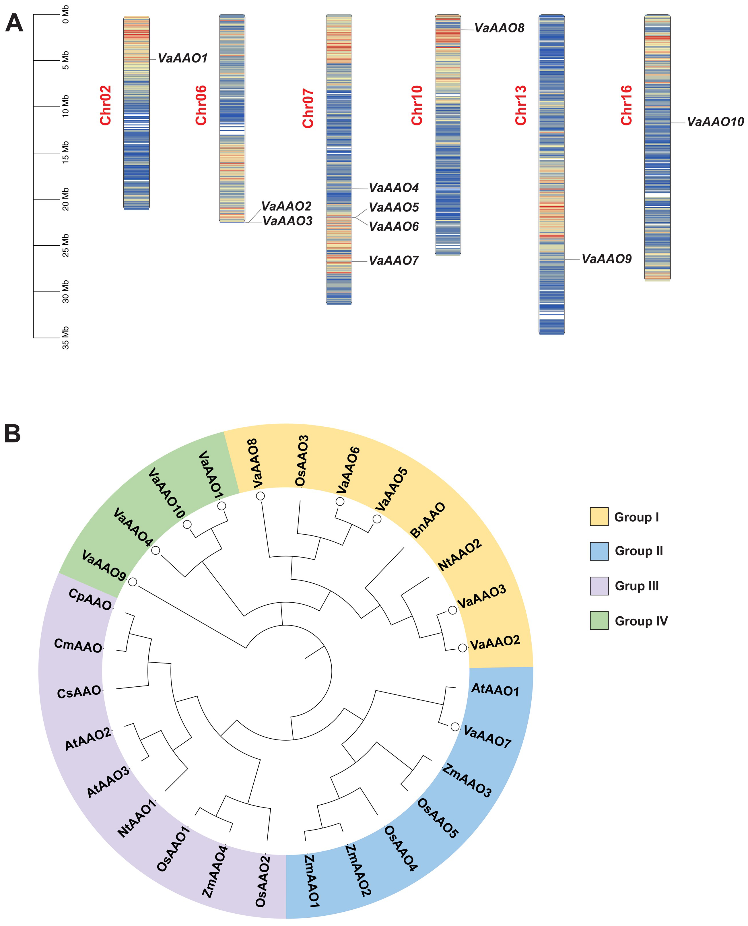
Figure 1. Genomic distribution and phylogenetic analysis of VaAAO genes in Vitis amurensis. (A) Chromosomal positions of VaAAO genes. Scale bar represents megabases (Mb). (B) Phylogenetic tree of AAO proteins from V. amurensis (Va), O. sativa (Os), B. napus (Bn), N. tabacum (Nt), A. thaliana (At), Z. mays (Zm), C. sativus (Cs), C. maxima (Cm), and C. pepo (Cp). VaAAOs are indicated by circular markers.
Horticulturae 11 01211 g001
Figure 2. Intraspecific and intergeneric collinearity of VaAAO genes in V. amurensis. (A) Circos plot showing intraspecific collinearity within the grapevine genome. The innermost ring represents the 19 chromosomes, followed by rings showing gene density. VaAAO genes are marked on their respective chromosomes. (B) Intergeneric collinearity between V. amurensis and peach (P. persica). Gray lines denote genome-wide syntenic relationships, while red lines indicate syntenic pairs involving VaAAO genes.
Horticulturae 11 01211 g002
Figure 3. Phylogenetic relationships, conserved motifs and domains, gene structures, and cis-element analyses of VaAAO family members. (A) Phylogenetic tree of VaAAO proteins generated in MEGA. (B) Conserved protein motifs and (C) conserved domains identified among VaAAO proteins. (D) Exon-intron structures of VaAAO genes. (E) Predicted cis-acting elements in the 2 kb promoter region of each gene. The heatmap shows the number of elements in each promoter, grouped by functional categories; grid values indicate element counts, and color intensity reflects their abundance. CDS, coding sequence; UTR, untranslated region.
Horticulturae 11 01211 g003
Figure 4. Expression dynamics of VaAAO family genes in response to B. cinerea infection in V. amurensis ‘SY’. Heatmap showing log2fold-change (log2FC) values relative to mock-inoculated controls at indicated hours post-inoculation (hpi).
Horticulturae 11 01211 g004
Figure 5. Expression profile of VaAAO7 in ‘SY’ leaves upon H2O2 treatment. VaAAO7 expression in the leaves of B. cinerea resistant cultivar ‘SY’ at different time points following 100 mM H2O2 treatment. Significant differences (p < 0.05; one-way ANOVA) among the means (±SD; n = 3) are indicated by different lowercase letters.
Horticulturae 11 01211 g005
Figure 6. VaAAO7 overexpression enhances resistance to B. cinerea in ‘RG’ leaves. (A) Subcellular localization of VaAAO7-GFP in N. benthamiana epidermal cells. Confocal microscopy shows GFP signal in the cytoplasm and at the cell periphery. Scale bar = 50 μm. (B) qRT-PCR analysis of VaAAO7 expression in VaAAO7-OE leaves relative to WT at the corresponding time points (mean ± SD, n = 3). One-way ANOVA was applied to determine significant differences. (C) Representative images showing lesion development in WT and VaAAO7-OE leaves at different time points after B. cinerea inoculation. (D) Quantification of lesion area as a percentage of total leaf area. Distinct lowercase letters denote statistically significant differences (p < 0.05; two-way ANOVA).
Horticulturae 11 01211 g006
Table 1. Gene information of the VaAAO gene family.
Gene NameSequence IDChromosomeAmino Acid No.Molecular Weight (Kda)pISubcellular
Localization
Gene Location
VaAAO1VAG0101936.1Chr0259165.878.78Extr4,650,177–4,654,698
VaAAO2VAG0108170.1Chr0655062.098.84Vacu22,520,739–22,522,577
VaAAO3VAG0108171.1Chr0655062.098.84Vacu22,540,213–22,542,051
VaAAO4VAG0109268.1Chr0758165.145.8Extr18,851,122–18,859,588
VaAAO5VAG0109429.1Chr0766773.818.89Plas21,969,204–21,973,103
VaAAO6VAG0109430.1Chr0753559.989.23Vacu21,974,570–21,978,699
VaAAO7VAG0109859.1Chr071202134.946.36Cyto26,729,891–26,739,673
VaAAO8VAG0113138.1Chr1054060.809.46Plas1,630,999–1,635,987
VaAAO9VAG0118612.1Chr1361167.475.29Plas26,514,513–26,517,220
VaAAO10VAG0122405.1Chr1659265.706.42Plas11,685,114–11,690,564
Note: Extr, extracellular; Vacu, vacuole; Plas, plasma membrane; Cyto, cytoplasm; pI, isoelectric points.
Disclaimer/Publisher’s Note: The statements, opinions and data contained in all publications are solely those of the individual author(s) and contributor(s) and not of MDPI and/or the editor(s). MDPI and/or the editor(s) disclaim responsibility for any injury to people or property resulting from any ideas, methods, instructions or products referred to in the content.