Article

Melatonin Application Induced Physiological and Molecular Changes in Carnation (Dianthus caryophyllus L.) under Heat Stress

by
Mohamed S. Elmongy
* and
Mohaned M. Abd El-Baset
Department of Vegetable and Floriculture, Faculty of Agriculture, Mansoura University, Mansoura 35516, Egypt
*
Author to whom correspondence should be addressed.
Horticulturae 2024, 10(2), 122; https://doi.org/10.3390/horticulturae10020122
Submission received: 12 December 2023 / Revised: 23 January 2024 / Accepted: 25 January 2024 / Published: 26 January 2024
(This article belongs to the Special Issue Tolerance and Response of Ornamental Plants to Abiotic Stress)

Abstract

:
Carnation is one of the most important ornamental plants worldwide; however, heat stress is a problem, which affects carnation cultivation. The harmful effects of heat stress include impaired vegetative development and reduced floral induction. In this study, to enhance carnation growth under conditions of heat stress, various concentrations of melatonin were added to in vitro culture media. The mechanism by which melatonin reduced heat stress damage was then studied by taking measurements of morphological parameters, levels of reactive oxygen species (ROS), antioxidant enzymes, and malondialdehyde (MDA), as well as differential gene expression, in carnation plants during in vitro culture. These data revealed that untreated carnation plants were more harmed by conditions of heat stress than plants treated with melatonin. Melatonin at concentrations of 5 and 10 mM increased chlorophyll content, fresh weight, and plant height to a greater extent than other concentrations. Melatonin may, thus, be used to alleviate damage to carnations caused by heat stress. The application of melatonin was also found to reduce oxidative damage and enhance antioxidant defense mechanisms. In addition, the expression of heat-related genes was found to be upregulated; in melatonin-treated plants, an upregulation was recorded in the expression of GAPDH, DcPOD1, DcPOD2, DcPOD3, Gols1, MBF1c, HSF30, HSP101, HSP70, and sHSP (MT) genes. In short, we found that melatonin treatment increased heat tolerance in carnation plants. The data presented here may serve as a reference for those seeking to enhance the growth of plants in conditions of heat stress.

1. Introduction

Carnation (Dianthus caryophyllus L.) is one of the most important cultivated plants; it is grown worldwide for use as a cut flower [1]. Carnation is a perennial herbaceous plant that is relatively well adapted to cold seasons but is very sensitive to high temperatures [2]. The best temperature ranges for growing carnations are 13–15 °C in summer and 10–11 °C in winter [3,4]. In recent years, the problem of climate change has made it necessary to better understand the effects of heat stress on plant growth and how any negative effects might be alleviated [5]. Indeed, this is now a critical issue worldwide, because heat stress is known to have an inhibitory effect on the growth of most crops [6], including lower productivity levels in the cultivation of ornamental plants [3,7].
Melatonin (N-acetyl-5-methoxytryptamine) has been shown to enhance plant production under conditions of heat stress [8]. Melatonin participates in many biological processes, including root development, shoot differentiation, leaf senescence, cell elongation, and regulation of the process of photosynthesis [9,10]. The ability of melatonin to enhance the tolerance of plants to abiotic stresses, such as high temperature, salinity, and cold stress, has also been noted [11,12]. The antioxidant effect of melatonin appears to be a direct result of the activation of antioxidant enzymes and scavenging ROS [13,14]. The authors of [15] reported that the mechanism by which melatonin achieved such effects was by increasing the efficiency of mitochondrial electron transport. Researchers have also found that melatonin stimulates molecular pathways in several plants [8,13,14,16]. It has also been reported that, in some ornamental plants, melatonin plays a key role during heat stress through its effect on genes, which are directly related to heat tolerance [7,17].
In the floriculture industry, the search for a protocol to minimize the damage caused to carnations by high temperatures is a pressing issue [17]. The treatment of plants with substances under in vitro and ex vitro conditions is one of the most important methods used for reducing heat stress in plants [18]. Previous studies of carnations have shown that heat stress transcription factors (Hsfs) and heat shock proteins (Hsps) both enhance the tolerance of plants to high temperatures [19,20,21]. Considering the achievement of such effects using exogenous chemicals, researchers have shown that melatonin helps plants recover from heat stress [22]; however, it remains uncertain whether heat tolerance in carnations may be enhanced by melatonin. Heat stress factors (Hsfs) are important regulatory variables that can directly activate the transcription of downstream Hsps. They are essential for delivering heat stress information and enhancing heat tolerance in plants [23].
In the present study, we evaluated the effects of different levels of melatonin on carnations subjected to heat stress. The relationships between the amounts of melatonin added to in vitro media and physiological parameters, such as antioxidant enzymes, MDA, and ROS, were studied. In addition, at the molecular level, differential expressions of related genes were also investigated. This is the first study conducted to determine the best level of melatonin to be added to in vitro culture media for the cultivation of carnations.

2. Materials and Methods

2.1. Plant Material and Culture Conditions

Carnation (Dianthus caryophyllus L.) seeds were imported from Japan. The experiments were conducted at the Laboratory of Tissue Culture of Vegetable and Ornamental Plants, Horticulture Division, Mansoura University, Mansoura, Egypt. Sodium hypochlorite (2%) was used for sterilizing seed surfaces. Seeds were then rinsed three times with double-distilled sterile water. Subsequently, glass jars (250 mL) containing MS media (30 g L−1 sucrose and 8 g L−1 agar (Difco Bacto TM Agar, Carolina, Burlington, NC, USA)) were used for seeds without adding any growth regulators to the media [24]. In vitro cultures were grown at 26 ± 1 °C under a long-day photoperiod (16/8 h light/dark) provided by lamps with a light intensity of 2500 lux.

2.2. Heat Application and Morphological Traits

Seedlings of height 4–7 cm were used for heat treatments. Absolute ethanol (99.99%) was used for dissolving the melatonin (Sigma-Aldrich, St. Louis, MO, USA); stock solutions at a concentration of 100 mM were then prepared. To test the melatonin (MT) application under heat stress, all seedlings (40 days old) were assigned to new jars containing MS medium fortified with MT at concentrations of 0 (control), 1, 5, and 10 mM. Four days after transfer to media, seedlings were moved to a growth chamber with a temperature of 42 °C for 1, 3, 6, 12, and 24 h. Samples were directly taken at each time point. Seedling microshoots were harvested, then immersed in liquid nitrogen and stored at −80 °C for subsequent physiological and molecular analyses. To obtain measurements of fresh weight and plant height, the carnation seedlings were first moved to a growth chamber with a temperature of 26 ± 1 °C. The indicated parameters were then obtained after a recovery period of 3 days.

2.3. Photosynthetic Chlorophyll Quantification

Chlorophyll (Chl) content was determined according the method of Frank et al. [25] with some modifications. Briefly, a 100 mg of carnation leaves was ground with a sterile pestle. An amount of 2 mL of ethanol (97%) was added to the ground leaves, and the mixture was allowed to incubate at 4 °C for 2 days. The mixture was then centrifuged at 12,000× g at 4 °C for 1 min, and the OD value of the supernatant was assayed by spectrophotometer (Shimadzu, Kyoto, Japan) at 665 nm and 649 nm wavelengths.

2.4. Reactive Oxygen Species

Samples of carnation plants (approximately 0.5 g = 3–4 explants) were taken for the purpose of detecting hydrogen peroxide (H2O2) levels [26]. Shoots were ground in liquid nitrogen, then dissolved in 5.0 mL of 0.1% trichloroacetic acid (TCA). Ground samples were then centrifuged at 10,000× g for 15 min. The supernatant was mixed with 10 mM potassium phosphate buffer. H2O2 levels were detected using a standard curve. Explant samples of 0.5 g weight were also used to determine levels of hydroxyl radical (OH) content. Samples were mixed with 15 mM 2-deoxy-D-ribose at 37–38 °C for 3 h (pH 7.5). Samples of the resulting mixture (0.7–0.8 mL) were then mixed with 0.5% (w/v) thiobarbituric acid (TBA at concentration 1%, dissolved in 10 mM NaOH) and 1 mL glacial acetic acid, then placed in a water bath (100 °C) (Fisher Scientific Isotemp Digital-Control Water Bath, Kuala Lumpur, Malaysia) for 45 min. Samples were then immediately cooled at 4 °C for 15 min and levels of hydroxyl radicals were detected according to the method of Halliwell et al. [27]. Superoxide radical (O2) concentrations were detected according to the method of Elstner and Heupel [28]. Following a pH 7.7 adjustment, 0.5 g of each microshoot sample was combined with potassium phosphate buffer, and the mixture was centrifuged at 4000× g for 12 min. The reaction was then maintained at 25 °C for 24 h, and the supernatant’s absorbance was measured at 530 nm.

2.5. Detection and Quantification of ROS

Levels of accumulated ROS in carnation leaves after heat stress were determined according to the method of Fukao et al. [29]. After the 42 °C heat treatment, samples were taken from plants subjected to different levels of melatonin application for the purpose of detecting hydrogen peroxide. Excised aerial vegetative samples were treated with 1 mg/mL DAB (3,3′diaminobenzidine tetra-hydrochlorride) in 50 mM Tris Acetate buffer, pH 5.0, and then incubated at 25 °C for 24 h under darkness in order to detect hydrogen peroxide. After staining, each plant’s uppermost leaf was boiled in 95% v/v ethanol for 20 min to eliminate chlorophyll and then rehydrated by incubation in 40% v/v glycerol for 16 h at 25 °C. At least seven distinct plants were used in each experiment’s replication, and representative photos are provided.

2.6. Antioxidant Enzyme Extraction and Malondialdehide Estimation

Amounts of approximately 0.5 g of fully fresh tissues were collected every 7 days during the whole period of transfer to the acclimatization treatments. The plant material was homogenized in 4 mL of 0.1 M phosphate buffer (pH 7.0, contained 2 mM EDTA + 1% PVP at 4 °C). Then, it was centrifuged at 12,000× g for 10 min at a temperature of 4 °C. The supernatant was stored at 4 °C for the purpose of measuring enzyme activity. Three biological replicates were used to measure all enzymes.
Peroxidase (POD, EC 1.11.1.7) was detected using guaiacol (Sigma-Aldrich, Burlington, MA, USA) in a mixture of 3 mL [30] consisting of 2.7 mL phosphate buffer (25 μM, pH 7.0) with 0.1 mL H2O2 (0.4%), 0.1 mL guaiacol (1.5%), and 0.1 mL of enzyme extract. The absorbance was calculated at 470 nm. By measuring how quickly l M of guaiacol oxidized g−1 FW min−1 at 25 ± 2 °C, POD enzyme activity was determined.
Catalase (CAT, EC. 1.11.1.6) activity was measured following the method of Góth [31]. The total volume of the reaction mixture was 3 mL; this consisted of 0.1 mL enzyme extract and 0.1 mL H2O2 (0.4%), in addition to 2.8 mL phosphate buffer (25 mM, pH 7.0). The reduction in absorbance was measured at 240 nm.
Superoxide dismutase (SOD, EC 1.15.1.1) enzyme activity was checked by measuring the inhibition of the amount of nitro blue tetrazolium (NBT) photochemical reduction (Sigma-Aldrich, Burlington, MA, USA) following the method of Sheteiwy et al. [32]. The total volume of the reaction mixture was 3.1 mL, consisting of 0.1 mL of enzyme extract and 3 mL NBT solution. Reaction tubes were placed under 15 W fluorescent lamps for 15 min after the addition of 2 µmol L−1 riboflavin. The control treatment was the reaction mixture without any enzyme extract. One unit of SOD was determined to be the volume of extract that caused 50% inhibition of NBT reduction. The photoreduction of NBT was measured at 560 nm.
Malondialdehyde (MDA) concentration was estimated using TBA reactive metabolites, according to the method of Heath and Packer [33], so that 1.5 mL of extract solution was added to 2.5 mL of 5% TBA formed in 5% TCA, then subjected to a temperature of 95 °C for 15 min before rapid cooling on ice. The MDA was measured at 532 nm after centrifugation of the supernatant at 5000× g for 10 min. Correction of nonspecific turbidity was measured at 600 nm.

2.7. Real-Time Quantitative PCR Analysis

Using Trizol reagent (Invitrogen, Carlsbad, CA, USA), total RNA was isolated from carnation microshoots and then processed with RNase-free DNase (Promega, Madison, WI, USA). Using reverse transcriptase (TOYOBO, Otsu, Japan), five micrograms of DNA-free total RNA (about 500 ng/μL) was reverse-transcribed into first-strand cDNA. Total RNA was used to synthesize the first-strand cDNA using the PrimeScript RT Reagent Kit with gDNA Eraser (Perfect Real Time) (Takara, Otsu, Japan). PCR was performed with TB Green® Premix Ex Taq (Takara, Otsu, Japan) on a CFX Connect Real-Time System. The thermal cycle program was as follows: 95 °C for 5 min, 45 cycles of 95 °C for 10 s and 60 °C for 30 s; 95 °C for 5 min, 65 °C for 5 s, 95 °C for 1 min program. Analysis of each sample was conducted with three biological replicates. The relative expression levels of genes were obtained by the 2−ΔΔCT method [34]. Primers for qRT-PCR are shown in Table 1.

2.8. Statistical Analyses

The experiments were prepared in a completely randomized design. For each treatment, about 30 explants were used, and all experiments were repeated twice. The data were subjected to analysis of variance (ANOVA) using SPSS version 16 (SPSS Inc., Chicago, IL, USA). Duncan’s multiple range test was used to test the significance of differences between means (<0.05). The results were presented as mean ± SD.

3. Results

3.1. Effects of Melatonin Application on Chlorophyll, Plant Height, and Fresh Weight

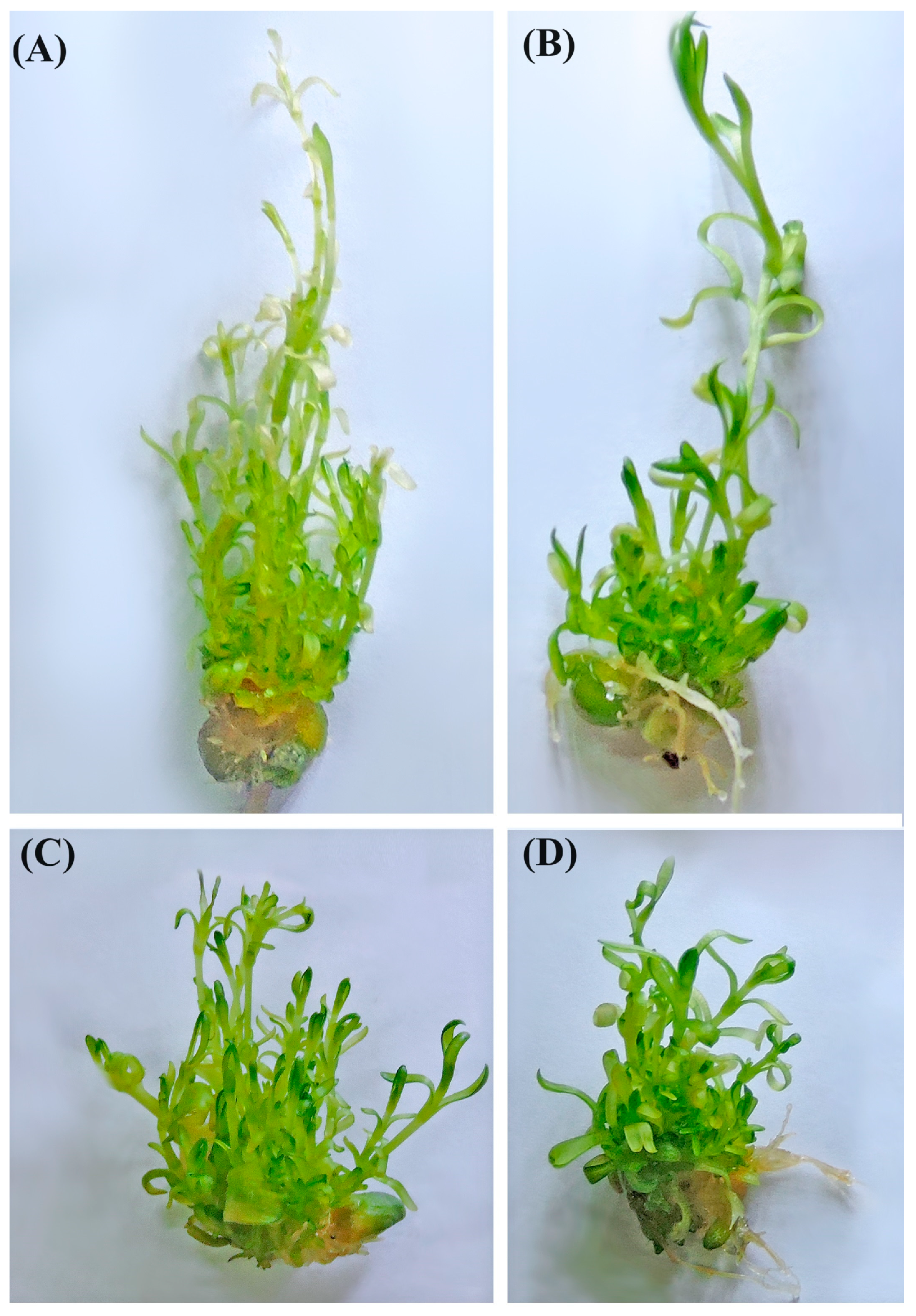
It can be seen from the data in Figure 1 that heat treatments reduced the levels of chlorophyll in carnation microshoots; however, higher levels of chlorophyll were recorded in media supplemented with melatonin, compared with control treatments (Figure 1A). In addition, when treated with melatonin at a concentration of 5 mM, carnation microshoots exhibited levels of chlorophyll, which were significantly higher than for any other concentration. After 6–24 h of heat treatment, the heights of carnation plants were lower, compared with earlier time points, but plants treated with melatonin exhibited significant differences compared with control plants (Figure 1B), with the best values recorded for melatonin concentrations of 5 and 10 mM. At all time points, there were no significant differences between melatonin treatments and untreated plants in terms of fresh weight measurements, but weights were higher at all times in melatonin-treated plants compared with controls (Figure 1C). These data indicated that melatonin can alleviate the harmful effects of high temperature in carnations (Figure 2).

3.2. Effects of Melatonin Application on Accumulation of Reactive Oxygen Species (ROS) in Carnation Plants under Heat Stress

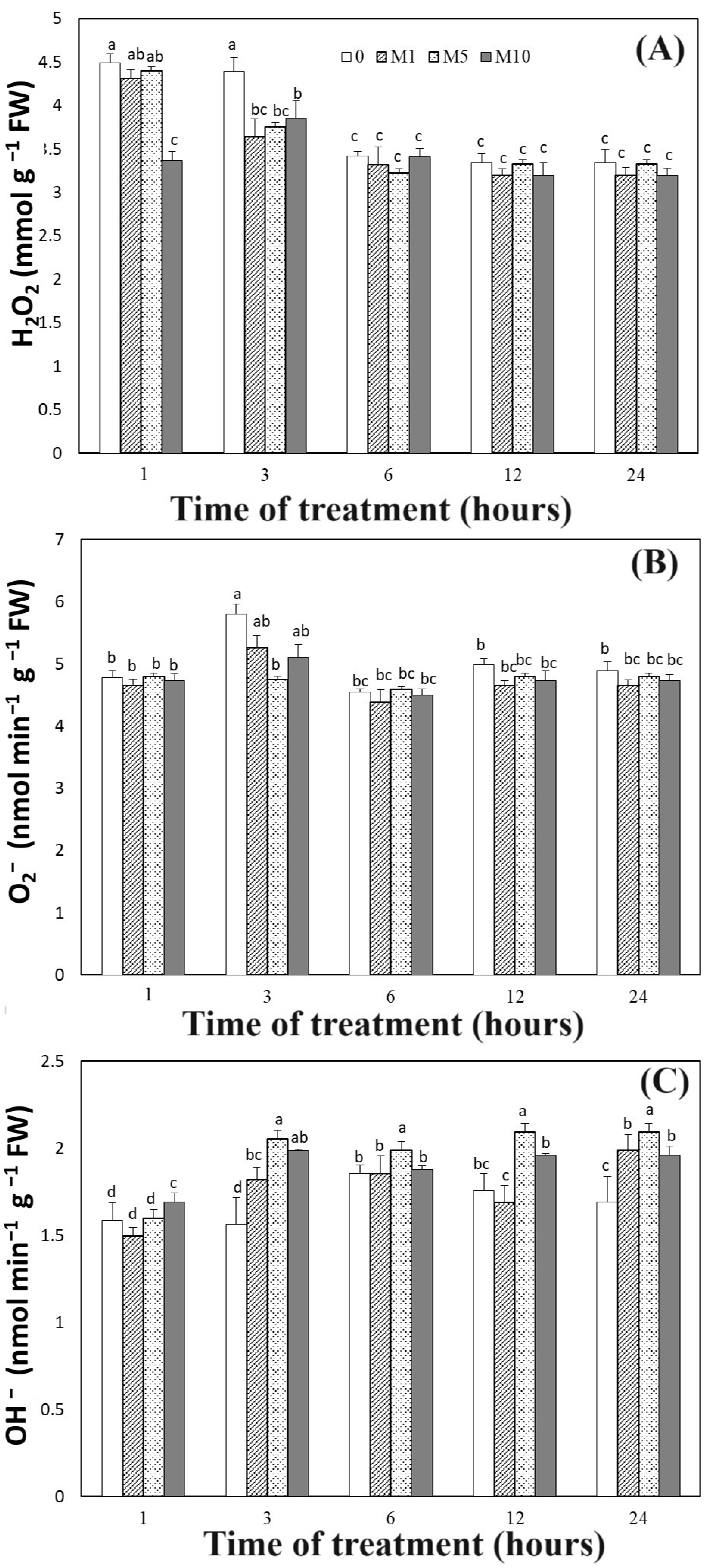
Melatonin was found to reduce the levels of H2O2 content in carnation plants at all testing times (Figure 3A). However, the levels of H2O2 in carnation plants declined more notably during the later periods of the experiment. Furthermore, after three hours of heat treatment, control treatment samples showed the highest H2O2 levels. The O2 levels were assessed in order to determine how melatonin influences ROS metabolisms in carnations during heat stress. DAB solution was used to identify the accumulation of ROS (H2O2) in carnation leaves (Figure 4). The observations demonstrated that ROS were formed as a heat shock reaction; however, the application of melatonin at a concentration of 5 mM reduced the buildup of ROS to a greater degree than any other melatonin concentration, including controls. In control samples, levels of superoxide content significantly increased under heat stress after 3 h (Figure 3B). However, after 24 h, carnation plants treated with melatonin exhibited lower levels of superoxide content than control plants. These support the idea that melatonin can positively affect superoxide generation under high-temperature conditions. The data in Figure 3 also showed that application of melatonin increased OH levels to a greater degree than control treatments (Figure 3C).

3.3. Effects of Melatonin Application on Antioxidant Enzyme Activities and MDA in Carnation Plants under Heat Stress

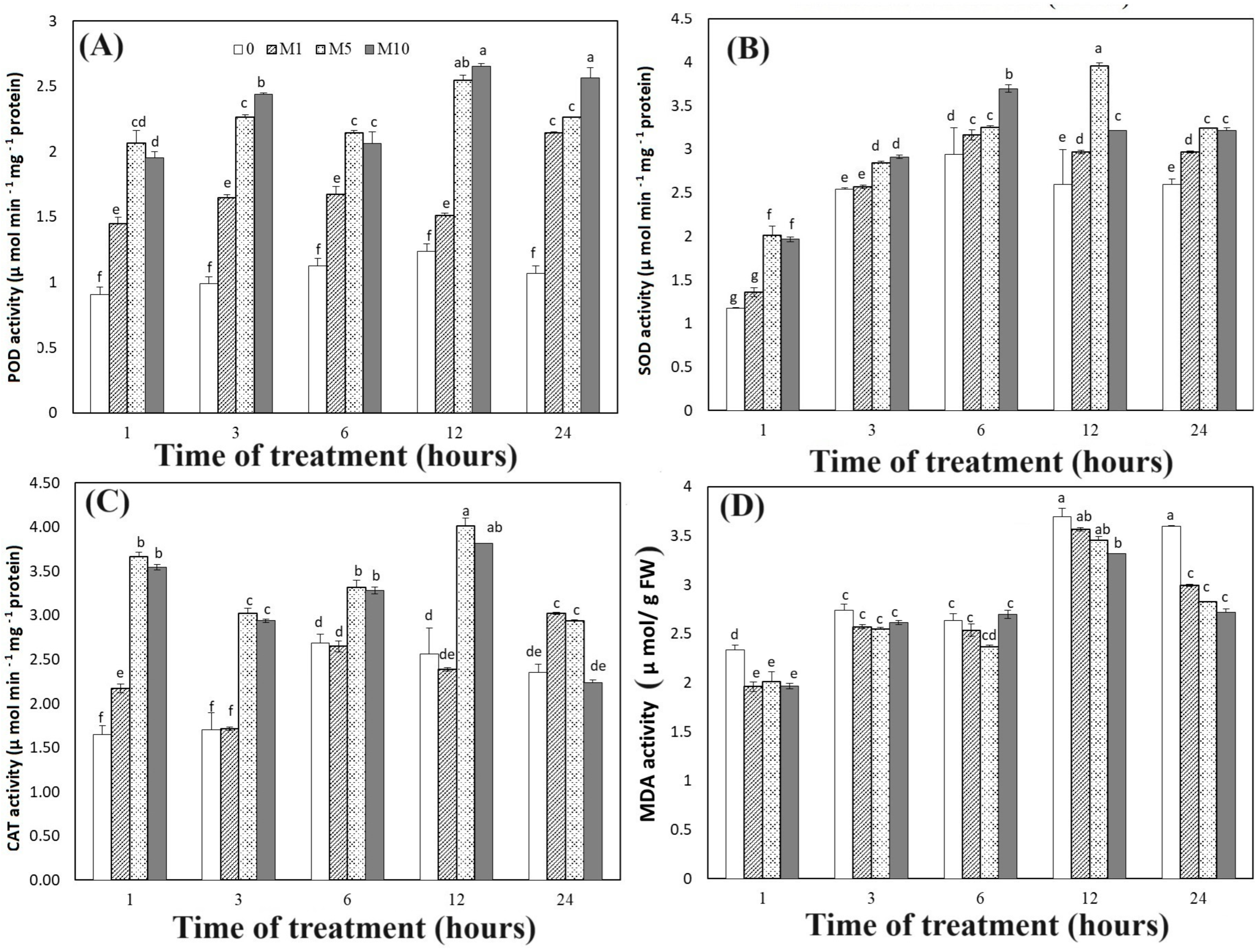
A relationship between increased antioxidant enzyme activities and reduced ROS levels was clearly evident in our data (Figure 5). The effects of melatonin on carnation plants under high-temperature stress were demonstrated by our finding that POD activity was significantly increased at all testing times with melatonin concentrations of 5 and 10 mM. The lowest levels of POD activity were recorded for control treatments. In addition, POD activity was increased more notably at 12 and 24 h than at earlier times (1–3 and 6 h). SOD enzymes exhibited a similar trend; however, the greatest increase in SOD activity was recorded at 12 h after melatonin application. The lowest value of SOD activity was recorded for untreated carnation plants. Under heat stress, melatonin applications at concentrations of 5 and 10 mM both resulted in significantly increased CAT activity after 1 and 12 h. The application of melatonin at a concentration of 1 mM did not significantly affect CAT in the first hours after treatment (Figure 5C). High-temperature treatment also caused the levels of malondialdehide (MDA) in carnation plants to significantly increase after 1, 12, and 24 h. However, plants which received melatonin application had lower levels of MDA at all tested times compared with untreated plants. Finally, greater increases in MDA activity were recorded at later time points (Figure 5D).

3.4. Effects of Melatonin Application on Gene Expression in Carnation Plants under Heat Stress

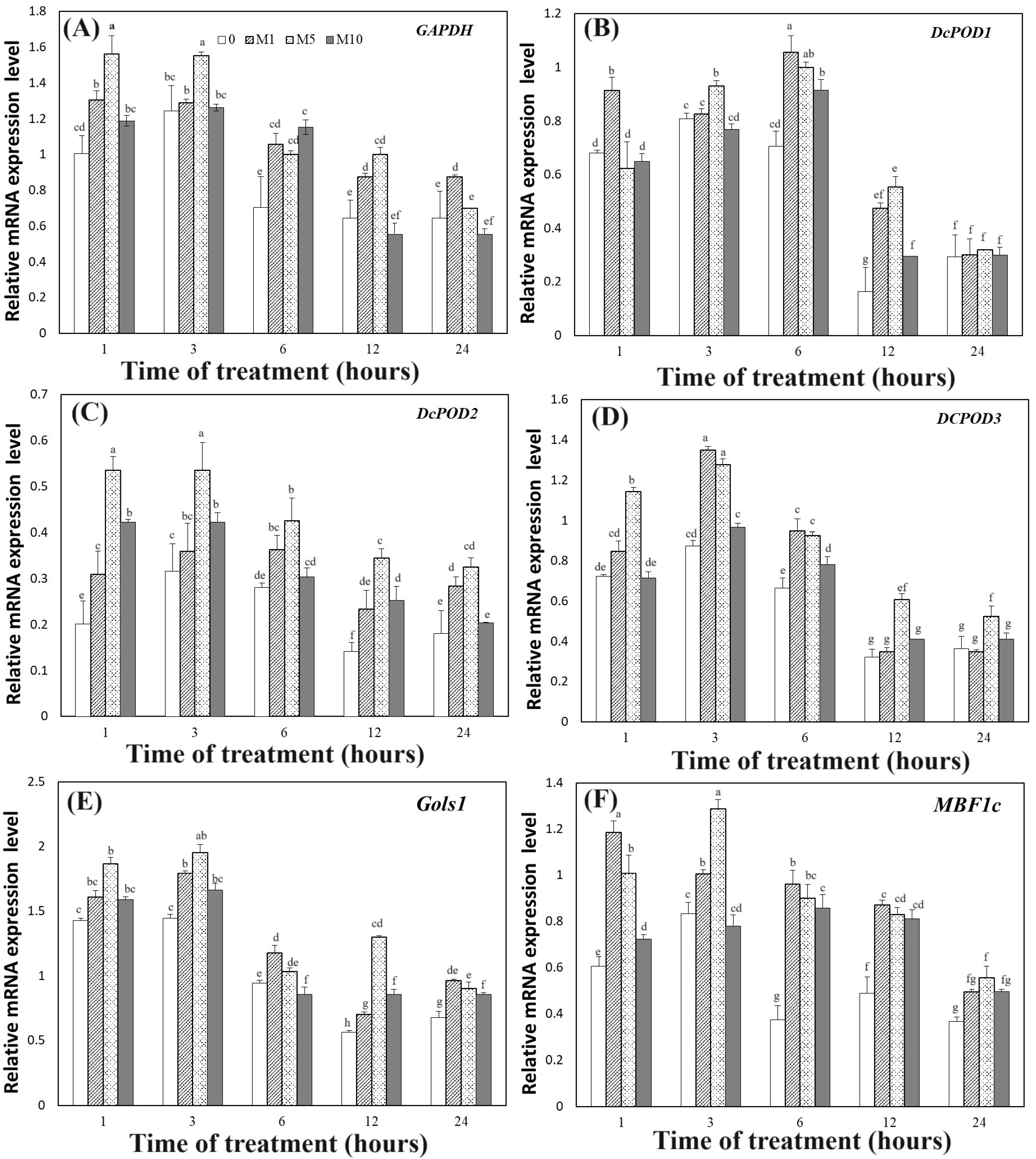
In order to study molecular changes in plants treated with melatonin under in vitro heat stress, the levels of expression of genes participating in heat stress were studied. Ten genes were subjected to qRT-PCR analysis. The obtained data shown in Figure 6 show that GAPDH was downregulated under heat stress but upregulated when melatonin was applied, especially at concentrations of 1 and 5 Mm. Additionally, the expression of the GAPDH gene was higher after 1 and 3 h of heat stress than at any than other time points. The relative expression of DcPOD1 increased gradually between the first and sixth hours of heat treatments but then declined, as measured at 12 and 24 h (Figure 6B).
However, treatments with 1 or 5 Mm of melatonin increased the expression of the DcPOD1 relative gene more than other concentrations, including controls. Similarly, the expressions of DcPOD2 and DcPOD3 both decreased in untreated plants at all time points but increased in most plants subjected to melatonin application (Figure 6C,D). In the case of the DcPOD2 relative gene, at all time points, expression was best in plants treated with a melatonin concentration of 5 Mm. For the DcPOD3 relative gene, the best results were obtained with concentrations of both 1 and 5 mM. Treatment of carnations with melatonin in vitro increased the relative expression of the Gols1 gene compared with untreated plants. This increase in expression of the Gols1 relative gene was greater after 1 and 3 h, compared with other time points (Figure 6E). Similarly, the relative transcript level of MBF1c was upregulated in heat-stressed plants treated with melatonin compared with control samples (Figure 6F).
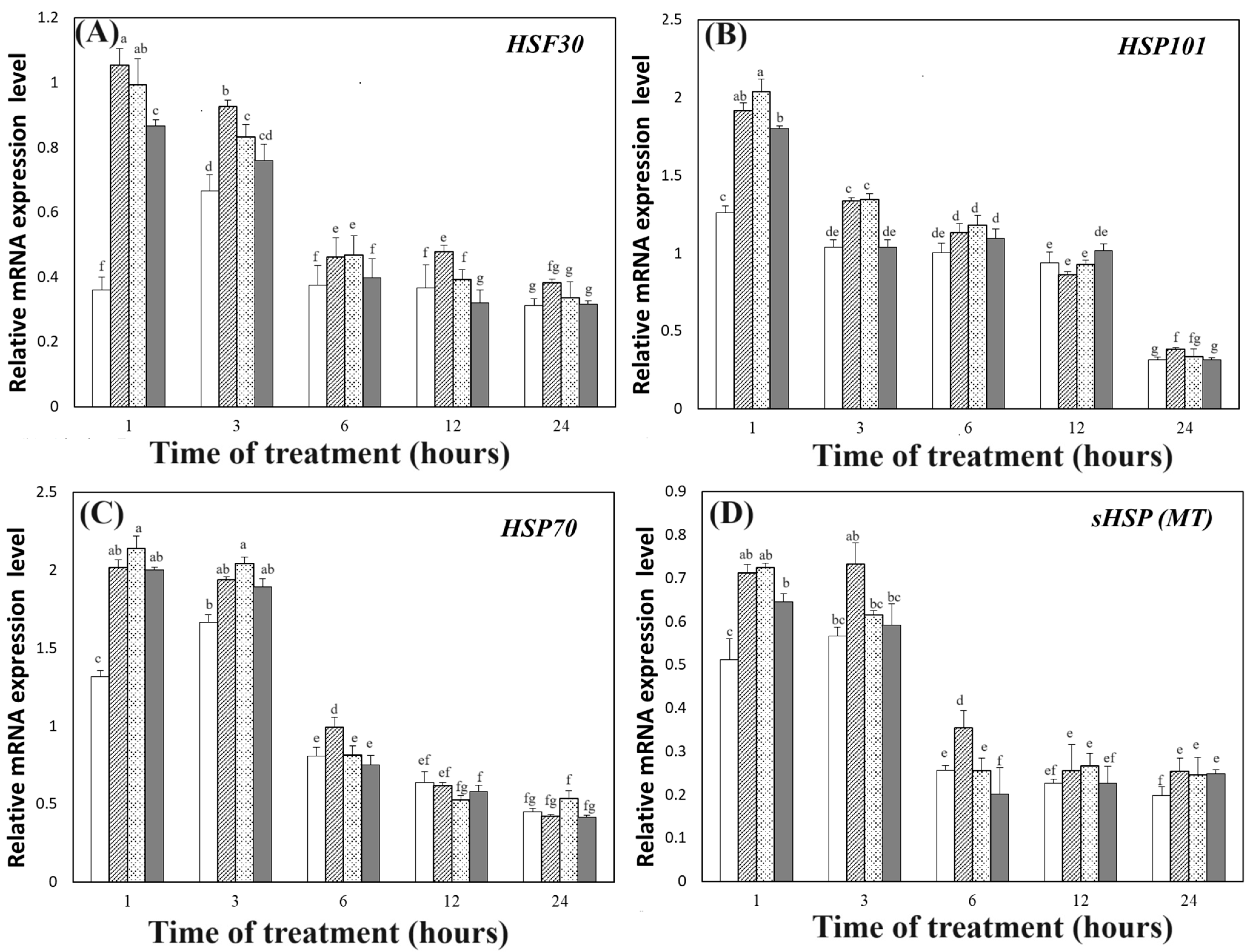
The data presented in Figure 7 show that two relative genes (HSF30 and HSP101) were upregulated in melatonin-treated plants, with the greatest increases recorded for the one- and three-hour time points. Similar data were obtained for the relative genes HSP70 and sHSP (MT), as both these genes were upregulated in melatonin-treated samples at the one- and three-hour points (Figure 7C,D). Overall, we recorded increases in the expression of the GAPDH, DcPOD1, DcPOD2, DcPOD3, Gols1, MBF1c, HSF30, HSP101, HSP70, and sHSP (MT) genes (Figure 6 and Figure 7), indicating that these genes may be putative targets of melatonin involved in response to heat stress at the transcriptional level.

4. Discussion

Heat stress inhibits the growth and development of plants. However, melatonin application has been reported to alleviate damage caused by heat stress in plants, such as tall fescue [7], strawberry (Fragaria × ananassa) [35], and tomato [36]. The present study is the first to evaluate the effects of melatonin application in vitro on carnation plants under heat stress. To assess the effects of melatonin treatments on heat tolerance in carnation plants, measurements of a number of morphological indices, physiological parameters, and molecular changes were obtained and analyzed.
It is well known that leaf senescence reduces green color in plants; low chlorophyll levels in plants may, therefore, be seen as an indicator of leaf senescence [37]. During the photosynthesis process, chlorophyll protects plants from light damage and is involved in the absorption and transmission of light energy [38]. Previous studies have reported that melatonin plays an effective role in increasing chlorophyll levels in several plants, including maize, tomato, tall fescue, Medicago sativa, and Malus hupehensis, thus protecting plants from leaf damage and the shock caused by high temperatures [7,37,38,39,40,41].
Treatment with melatonin has also been reported to enhance fresh weight, dry weight, and plant height in various plants subjected to heat stress conditions [11,42,43]. In carnations, the data obtained in the present study indicated that melatonin application resulted in increases in chlorophyll content, fresh weight, and plant height, suggesting that substances like melatonin might have a vital role to play in alleviating the effects of heat stress in carnations.
Under heat stress conditions, the accumulation of ROS, such as OH–, H2O2, and O2, and free radicals causes increases in MDA and the leakage of electrolytes, which may be scavenged by plants under stress conditions through the stimulation of the activities of antioxidant enzymes. For example, SOD can inhibit the superoxide radical (O2) by converting it to H2O2, and the catalase enzyme is also able to inhibit it by releasing H2O. It has previously been shown that melatonin treatment alleviates oxidative disfiguration under abiotic stresses by conserving ROS [39,44]. Dewir et al. [2] found that POD enzyme activity was enhanced in carnation plants under heat stress and that this was associated with scavenging ROS in treated plants. In addition, POD enzymes can reduce ROS activity by enhancing secondary metabolites in plants; however, these secondary metabolites protect the plants from heat stress damage by supplying mechanical support to the cultured cells through their participation in the maintenance of cell membrane integrity, further demonstrating that POD plays an important role in lignin synthesis under heat stress conditions [45,46]. Additionally, plants, such as tea plant (Camellia sinensis), eggplant (Solanum melongena), and carnation, which were treated with melatonin, have all been shown to exhibit low levels of MDA content under heat stress [17,47,48]. In terms of the parameters of minimal fluorescence (F0) and maximal fluorescence (Fm), from which we may obtain maximal variable fluorescence (Fv = Fm − F0) and the photochemical efficiency of PSII (Fv/Fm), we note that melatonin has been shown to play a vital role in repressing heat stress, by reducing leaf curling, blotching, and fraying and increasing the value of Fv/Fm [17,49,50]. A series of metabolic changes are involved in heat stress responses in plants [51]. These include the following: overproduction of ROS and reactive nitrogen species (RNS); lipid peroxidation, production of end products, such as MDA; photoinhibition; protein denaturation and degradation; and accumulation of compatible solutes [52]. Lipid peroxidation is caused by abiotic stress [53]. The end products of lipid peroxidation are reactive aldehydes such as malondialdehide (MDA) and 4-hydroxynonenal (HNE) [54]. In the present study, heat treatments significantly increased MDA content in control carnation plants; contrarily, melatonin application resulted in decreased MDA production. These findings indicate that melatonin alleviates cell membrane damage caused by heat stress. We also found that the content levels of ROS and MDA decreased in carnation plants treated with melatonin (Figure 4 and Figure 5), but levels of the enzymes POD, CAT, and SOD all increased (Figure 5). Taken together, the data reported here confirm the significant effect of melatonin on morphological traits and chlorophyll content (Figure 3); they are in line with the findings of previous studies, which showed that oxidative stress in plants may be alleviated by melatonin treatment [7,8,10,16,55,56].
High temperatures may cause the denaturation of proteins. High-temperature conditions may also cause cell cytotoxicity [57]. Previous studies have reported that, in cells exposed to heat stress, protein homeostasis can be safeguarded and protein aggregation reduced by the interaction of chaperones with stress-denatured proteins [3,17,58]. In addition, heat-induced genes have been found to be the most effective genes in encoding molecular chaperones such as heat shock proteins [19,59]. These genes have been found to be upregulated and induced when cells are exposed to high or low temperatures [3,7,60,61]. In the present study, we evaluated the expression of ten genes and found that heat shock proteins were upregulated under melatonin application. Previous studies have also found that the HSP40 family plays a vital role in enhancing ATPase activity [3]. Exogenous substances can effectively increase the expression of Hsfs and Hsps; this has been found to improve the thermotolerance in creeping bent grass (Agrostis stolonifera) [62] and strawberry [35]. The genes that we evaluated in our study have been reported as participating in protein folding and unfolding, and this may enhance the ability of cells to tolerate heat stress [43].

5. Conclusions

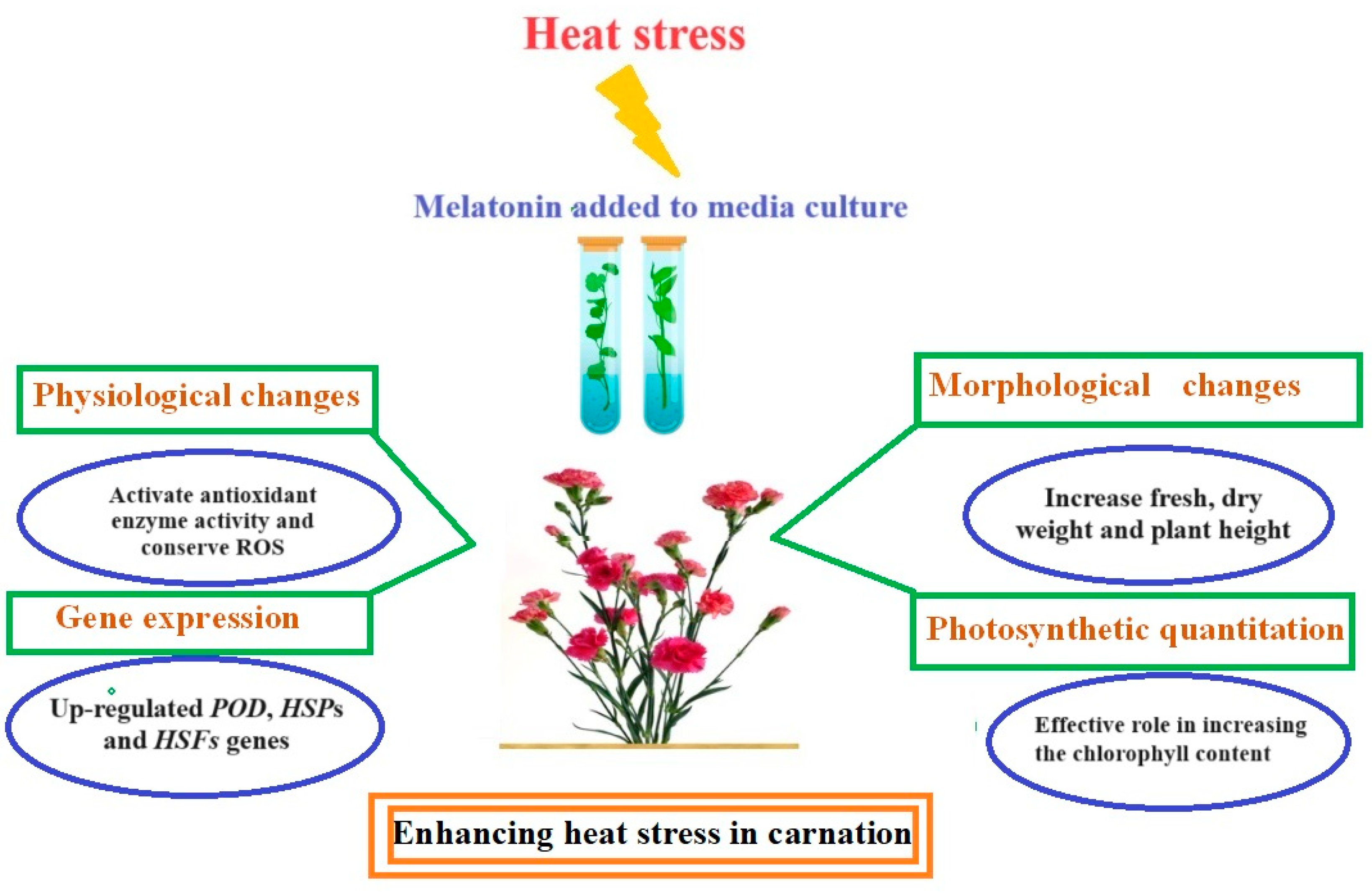
In the present study, an experiment was performed to determine whether the addition of melatonin to in vitro media would enhance the response of carnation microshoots to heat stress (Figure 8). Based on the measured effects of melatonin on chlorophyll content, fresh weight, and plant height in carnations under heat stress, we may say that melatonin enhances the response to heat stress. We also found that melatonin application improved thermotolerance in carnations by lowering the content levels of ROS and MDA while increasing antioxidant enzyme activities. Through such means, melatonin exerts an inhibitory effect upon heat stress. We also assessed the relationship between melatonin application and the expression of related heat stress genes. We found that the mechanism by which heat stress damage is alleviated is strongly associated with the upregulation of the POD, HSP, and HSF genes. We also found that treatment with melatonin at concentrations of 5 and 10 mM promoted growth and development in carnation plants exposed to 42 °C, in comparison with untreated plants. The supplementation of media with melatonin reduced oxidative damage through scavenging ROS and inhibiting MDA synthesis; it also increased the activities of antioxidant enzymes and upregulated genes involved in heat tolerance. From the results reported here, we may state that melatonin plays a significant role in improving the tolerance of plants to conditions of abiotic stress, especially heat stress.

Author Contributions

Both authors designed the experiment, conducted the experiment, wrote the manuscript. All authors have read and agreed to the published version of the manuscript.

Funding

The current work was funded by Mansoura University, Faculty of Agriculture, Egypt.

Data Availability Statement

Data are contained within the article.

Acknowledgments

The authors extend their gratitude to the anonymous reviewers and editors for their helpful reviews and critical comments. The Corresponding author would like to thank Xiuyun Wang of Zhejiang University’s College of Agriculture and Biotechnology for her strong scientific support.

Conflicts of Interest

The authors declare no conflicts of interest.

References

  1. Navarro, A.; Elia, A.; Conversa, G.; Campi, P.; Mastrorilli, M. Potted mycorrhizal carnation plants and saline stress: Growth, quality and nutritional plant responses. Sci. Hortic. 2012, 140, 131–139. [Google Scholar] [CrossRef]
  2. Dewir, Y.H.; Yu, R.; Fan, Y.; Ali, M.B.; Paek, K.Y. Effect of Temperature and gamma-Radiation on Antioxidant Defense System in Suspension Cell Culture of Carnation (Dianthus caryophyllus). Korean J. Hortic. Sci. 2005, 46, 329. [Google Scholar]
  3. Wan, X.L.; Zhou, Q.; Wang, Y.Y.; Wang, W.E.; Bao, M.Z.; Zhang, J.W. Identification of heat-responsive genes in carnation (Dianthus caryophyllus L.) by RNA-seq. Front. Plant Sci. 2015, 6, 519. [Google Scholar] [CrossRef] [PubMed]
  4. In, B.C.; Binder, B.M.; Falbel, T.G.; Patterson, S.E. Analysis of gene expression during the transition to climacteric phase in carnation flowers (Dianthus caryophyllus L.). J. Exp. Bot. 2013, 64, 4923–4937. [Google Scholar] [CrossRef] [PubMed]
  5. Zahra, N.; Hafeez, M.B.; Ghaffar, A.; Kausar, A.; Zeidi, M.A.; Siddique, K.H.M.; Farooq, M. Plant photosynthesis under heat stress: Effects and management. Environ. Exp. Bot. 2023, 206, 105178. [Google Scholar] [CrossRef]
  6. Wilson, R.A.; Sangha, M.; Banga, S.; Atwal, A.; Gupta, S. Heat stress tolerance in relation to oxidative stress and antioxidants in Brassica juncea. J. Environ. Biol. 2014, 35, 383. [Google Scholar]
  7. Alam, M.N.; Zhang, L.; Yang, L.; Islam, M.R.; Liu, Y.; Luo, H.; Yang, P.; Wang, Q.; Chan, Z. Transcriptomic profiling of tall fescue in response to heat stress and improved thermotolerance by melatonin and 24-epibrassinolide. BMC Genom. 2018, 19, 224. [Google Scholar] [CrossRef]
  8. Li, Y.; Zhang, Z.; He, C.; Zhu, K.; Xu, Z.; Ma, T.; Tao, J.; Liu, G. Melatonin protects porcine oocyte in vitro maturation from heat stress. J. Pineal Res. 2015, 59, 365–375. [Google Scholar] [CrossRef]
  9. Hu, Z.; Fan, J.; Xie, Y.; Amombo, E.; Liu, A.; Gitau, M.M.; Khaldun, A.B.M.; Chen, L.; Fu, J. Comparative photosynthetic and metabolic analyses reveal mechanism of improved cold stress tolerance in bermudagrass by exogenous melatonin. Plant Physiol. Biochem. 2016, 100, 94–104. [Google Scholar] [CrossRef]
  10. Zhang, N.; Zhang, H.J.; Zhao, B.; Sun, Q.Q.; Cao, Y.Y.; Li, R.; Wu, X.X.; Weeda, S.; Li, L.; Ren, S. The RNA-seq approach to discriminate gene expression profiles in response to melatonin on cucumber lateral root formation. J. Pineal Res. 2014, 56, 39–50. [Google Scholar] [CrossRef]
  11. Shi, H.; Jiang, C.; Ye, T.; Tan, D.-X.; Reiter, R.J.; Zhang, H.; Liu, R.; Chan, Z. Comparative physiological, metabolomic, and transcriptomic analyses reveal mechanisms of improved abiotic stress resistance in bermudagrass [Cynodon dactylon (L). Pers.] by exogenous melatonin. J. Exp. Bot. 2014, 66, 681–694. [Google Scholar] [CrossRef]
  12. Bajwa, V.S.; Shukla, M.R.; Sherif, S.M.; Murch, S.J.; Saxena, P.K. Role of melatonin in alleviating cold stress in A rabidopsis thaliana. J. Pineal Res. 2014, 56, 238–245. [Google Scholar] [CrossRef]
  13. Galano, A.; Tan, D.X.; Reiter, R.J. On the free radical scavenging activities of melatonin’s metabolites, AFMK and AMK. J. Pineal Res. 2013, 54, 245–257. [Google Scholar] [CrossRef] [PubMed]
  14. Purushothaman, A.; Sheeja, A.A.; Janardanan, D. Hydroxyl radical scavenging activity of melatonin and its related indolamines. Free Radic. Res. 2020, 54, 373–383. [Google Scholar] [CrossRef] [PubMed]
  15. Tomás-Zapico, C.; Coto-Montes, A. A proposed mechanism to explain the stimulatory effect of melatonin on antioxidative enzymes. J. Pineal Res. 2005, 39, 99–104. [Google Scholar] [CrossRef] [PubMed]
  16. Wei, W.; Li, Q.T.; Chu, Y.N.; Reiter, R.J.; Yu, X.M.; Zhu, D.H.; Zhang, W.K.; Ma, B.; Lin, Q.; Zhang, J.S.; et al. Melatonin enhances plant growth and abiotic stress tolerance in soybean plants. J. Exp. Bot. 2015, 66, 695–707. [Google Scholar] [CrossRef]
  17. Hu, D.; Zhang, X.; Xue, P.; Nie, Y.; Liu, J.; Li, Y.; Wang, C.; Wan, X. Exogenous melatonin ameliorates heat damages by regulating growth, photosynthetic efficiency and leaf ultrastructure of carnation. Plant Physiol. Biochem. 2023, 198, 107698. [Google Scholar] [CrossRef]
  18. Sarwar, M.; SALEEM, M.F.; Ali, B.; Saleem, M.H.; Rizwan, M.; Usman, K.; KEBLAWY, A.E.; Ali, A.; Afzal, M.; Sheteiwy, M.S. Application of potassium, zinc and boron as potential plant growth modulators in Gossypium hirsutum L. under heat stress. Turk. J. Agric. For. 2022, 46, 567–584. [Google Scholar] [CrossRef]
  19. Sun, Y.; Hu, D.; Xue, P.; Wan, X. Identification of the DcHsp20 gene family in carnation (Dianthus caryophyllus) and functional characterization of DcHsp17. 8 in heat tolerance. Planta 2022, 256, 2. [Google Scholar] [CrossRef] [PubMed]
  20. Wan, X.; Sun, Y.; Feng, Y.; Bao, M.; Zhang, J. Heat stress transcription factor DcHsfA1d isolated from Dianthus caryophyllus enhances thermotolerance and salt tolerance of transgenic Arabidopsis. Biol. Plant 2022, 66, 29. [Google Scholar] [CrossRef]
  21. Xue, P.; Sun, Y.; Hu, D.; Zhang, J.; Wan, X. Genome-wide characterization of DcHsp90 gene family in carnation (Dianthus caryophyllus L.) and functional analysis of DcHsp90-6 in heat tolerance. Protoplasma 2023, 260, 807–819. [Google Scholar] [CrossRef]
  22. Ulhassan, Z.; Huang, Q.; Gill, R.A.; Ali, S.; Mwamba, T.M.; Ali, B.; Hina, F.; Zhou, W. Protective mechanisms of melatonin against selenium toxicity in Brassica napus: Insights into physiological traits, thiol biosynthesis and antioxidant machinery. BMC Plant Biol. 2019, 19, 507. [Google Scholar] [CrossRef]
  23. Gong, Z.; Xiong, L.; Shi, H.; Yang, S.; Herrera-Estrella, L.R.; Xu, G.; Chao, D.-Y.; Li, J.; Wang, P.-Y.; Qin, F. Plant abiotic stress response and nutrient use efficiency. Sci. China Life Sci. 2020, 63, 635–674. [Google Scholar] [CrossRef]
  24. Murashige, T.; Skoog, F. A revised medium for rapid growth and bio assays with tobacco tissue cultures. Physiol. Plant. 1962, 15, 473–497. [Google Scholar] [CrossRef]
  25. Frank, W.; Ratnadewi, D.; Reski, R. Physcomitrella patens is highly tolerant against drought, salt and osmotic stress. Planta 2005, 220, 384–394. [Google Scholar] [CrossRef] [PubMed]
  26. Perveen, S.; Anis, M. Physiological and biochemical parameters influencing ex vitro establishment of the in vitro regenerants of Albizia lebbeck. Agrofor. Syst. 2015, 89, 721–733. [Google Scholar] [CrossRef]
  27. Halliwell, B.; Gutteridge, J.M.; Aruoma, O.I. The deoxyribose method: A simple “test-tube” assay for determination of rate constants for reactions of hydroxyl radicals. Anal. Biochem. 1987, 165, 215–219. [Google Scholar] [CrossRef] [PubMed]
  28. Elstner, E.F.; Heupel, A. Inhibition of nitrite formation from hydroxylammoniumchloride: A simple assay for superoxide dismutase. Anal. Biochem. 1976, 70, 616–620. [Google Scholar] [CrossRef] [PubMed]
  29. Fukao, T.; Yeung, E.; Bailey-Serres, J. The submergence tolerance regulator SUB1A mediates crosstalk between submergence and drought tolerance in rice. Plant Cell 2011, 23, 412–427. [Google Scholar] [CrossRef] [PubMed]
  30. Elmongy, M.S.; Zhou, H.; Cao, Y.; Liu, B.; Xia, Y. The effect of humic acid on endogenous hormone levels and antioxidant enzyme activity during in vitro rooting of evergreen azalea. Sci. Hortic. 2018, 227, 234–243. [Google Scholar] [CrossRef]
  31. Góth, L. A simple method for determination of serum catalase activity and revision of reference range. Clin. Chim. Acta 1991, 196, 143–151. [Google Scholar] [CrossRef] [PubMed]
  32. Sheteiwy, M.; Shen, H.; Xu, J.; Guan, Y.; Song, W.; Hu, J. Seed polyamines metabolism induced by seed priming with spermidine and 5-aminolevulinic acid for chilling tolerance improvement in rice (Oryza sativa L.) seedlings. Environ. Exp. Bot. 2017, 137, 58–72. [Google Scholar] [CrossRef]
  33. Heath, R.L.; Packer, L. Photoperoxidation in isolated chloroplasts: I. Kinetics and stoichiometry of fatty acid peroxidation. Arch. Biochem. Biophys. 1968, 125, 189–198. [Google Scholar] [CrossRef] [PubMed]
  34. Livak, K.J.; Schmittgen, T.D. Analysis of relative gene expression data using real-time quantitative PCR and the 2− ΔΔCT method. Methods 2001, 25, 402–408. [Google Scholar] [CrossRef] [PubMed]
  35. Manafi, H.; Baninasab, B.; Gholami, M.; Talebi, M.; Khanizadeh, S. Exogenous melatonin alleviates heat-induced oxidative damage in strawberry (Fragaria × ananassa Duch. cv. Ventana) Plant. J. Plant Growth Regul. 2022, 41, 52–64. [Google Scholar] [CrossRef]
  36. El-Yazied, A.A.; Ibrahim, M.F.; Ibrahim, M.A.; Nasef, I.N.; Al-Qahtani, S.M.; Al-Harbi, N.A.; Alzuaibr, F.M.; Alaklabi, A.; Dessoky, E.S.; Alabdallah, N.M. Melatonin mitigates drought induced oxidative stress in potato plants through modulation of osmolytes, sugar metabolism, ABA homeostasis and antioxidant enzymes. Plants 2022, 11, 1151. [Google Scholar] [CrossRef]
  37. Jahan, M.S.; Shu, S.; Wang, Y.; Hasan, M.M.; El-Yazied, A.A.; Alabdallah, N.M.; Hajjar, D.; Altaf, M.A.; Sun, J.; Guo, S. Melatonin pretreatment confers heat tolerance and repression of heat-induced senescence in tomato through the modulation of ABA-and GA-mediated pathways. Front. Plant Sci. 2021, 12, 650955. [Google Scholar] [CrossRef]
  38. Ahmad, S.; Kamran, M.; Ding, R.; Meng, X.; Wang, H.; Ahmad, I.; Fahad, S.; Han, Q. Exogenous melatonin confers drought stress by promoting plant growth, photosynthetic capacity and antioxidant defense system of maize seedlings. PeerJ 2019, 7, e7793. [Google Scholar] [CrossRef]
  39. Antoniou, C.; Chatzimichail, G.; Xenofontos, R.; Pavlou, J.J.; Panagiotou, E.; Christou, A.; Fotopoulos, V. Melatonin systemically ameliorates drought stress-induced damage in M edicago sativa plants by modulating nitro-oxidative homeostasis and proline metabolism. J. Pineal Res. 2017, 62, e12401. [Google Scholar] [CrossRef]
  40. Wassie, M.; Zhang, W.; Zhang, Q.; Ji, K.; Cao, L.; Chen, L. Exogenous salicylic acid ameliorates heat stress-induced damages and improves growth and photosynthetic efficiency in alfalfa (Medicago sativa L.). Ecotoxicol. Environ. Saf. 2020, 191, 110206. [Google Scholar] [CrossRef]
  41. Wang, P.; Sun, X.; Chang, C.; Feng, F.; Liang, D.; Cheng, L.; Ma, F. Delay in leaf senescence of Malus hupehensis by long-term melatonin application is associated with its regulation of metabolic status and protein degradation. J. Pineal Res. 2013, 55, 424–434. [Google Scholar] [CrossRef]
  42. Kotak, S.; Larkindale, J.; Lee, U.; von Koskull-Doring, P.; Vierling, E.; Scharf, K.D. Complexity of the heat stress response in plants. Curr. Opin. Plant Biol. 2007, 10, 310–316. [Google Scholar] [CrossRef]
  43. Li, S.; Zhou, X.; Chen, L.; Huang, W.; Yu, D. Functional characterization of Arabidopsis thaliana WRKY39 in heat stress. Mol. Cells 2010, 29, 475–483. [Google Scholar] [CrossRef]
  44. Meng, J.F.; Xu, T.F.; Wang, Z.Z.; Fang, Y.L.; Xi, Z.M.; Zhang, Z.W. The ameliorative effects of exogenous melatonin on grape cuttings under water-deficient stress: Antioxidant metabolites, leaf anatomy, and chloroplast morphology. J. Pineal Res. 2014, 57, 200–212. [Google Scholar] [CrossRef]
  45. Chaitanya, K.; Sundar, D.; Masilamani, S.; Ramachandra Reddy, A. Variation in heat stress-induced antioxidant enzyme activities among three mulberry cultivars. Plant Growth Regul. 2002, 36, 175–180. [Google Scholar] [CrossRef]
  46. Otter, T.; Polle, A. The influence of apoplastic ascorbate on the activities of cell wall-associated peroxidase and NADH oxidase in needles of Norway spruce (Picea abies L.). Plant Cell Physiol. 1994, 35, 1231–1238. [Google Scholar] [CrossRef]
  47. Li, J.; Yang, Y.; Sun, K.; Chen, Y.; Chen, X.; Li, X. Exogenous melatonin enhances cold, salt and drought stress tolerance by improving antioxidant defense in tea plant (Camellia sinensis (L.) O. Kuntze). Molecules 2019, 24, 1826. [Google Scholar] [CrossRef]
  48. Song, L.; Tan, Z.; Zhang, W.; Li, Q.; Jiang, Z.; Shen, S.; Luo, S.; Chen, X. Exogenous melatonin improves the chilling tolerance and preharvest fruit shelf life in eggplant by affecting ROS-and senescence-related processes. Hortic. Plant J. 2023, 9, 523–540. [Google Scholar] [CrossRef]
  49. Zhang, L.; Hu, T.; Amombo, E.; Wang, G.; Xie, Y.; Fu, J. The alleviation of heat damage to photosystem II and enzymatic antioxidants by exogenous spermidine in tall fescue. Front. Plant Sci. 2017, 8, 1747. [Google Scholar] [CrossRef]
  50. Fatma, M.; Iqbal, N.; Sehar, Z.; Alyemeni, M.N.; Kaushik, P.; Khan, N.A.; Ahmad, P. Methyl jasmonate protects the PS II system by maintaining the stability of chloroplast D1 protein and accelerating enzymatic antioxidants in heat-stressed wheat plants. Antioxidants 2021, 10, 1216. [Google Scholar] [CrossRef]
  51. Fan, X.; Zhao, J.; Sun, X.; Zhu, Y.; Li, Q.; Zhang, L.; Zhao, D.; Huang, L.; Zhang, C.; Liu, Q. Exogenous melatonin improves the quality performance of rice under high temperature during grain filling. Agronomy 2022, 12, 949. [Google Scholar] [CrossRef]
  52. Sanoubar, R.; Cellini, A.; Gianfranco, G.; Spinelli, F. Osmoprotectants and antioxidative enzymes as screening tools for salinity tolerance in radish (Raphanus sativus). Hortic. Plant J. 2020, 6, 14–24. [Google Scholar] [CrossRef]
  53. Talaat, N.B.; Shawky, B.T. Synergistic Effects of Salicylic Acid and Melatonin on Modulating Ion Homeostasis in Salt-Stressed Wheat (Triticum aestivum L.) Plants by Enhancing Root H+-Pump Activity. Plants 2022, 11, 416. [Google Scholar] [CrossRef] [PubMed]
  54. Karumannil, S.; Khan, T.A.; Kappachery, S.; Gururani, M.A. Impact of exogenous melatonin application on photosynthetic machinery under abiotic stress conditions. Plants 2023, 12, 2948. [Google Scholar] [CrossRef] [PubMed]
  55. Xu, W.; Cai, S.Y.; Zhang, Y.; Wang, Y.; Ahammed, G.J.; Xia, X.J.; Shi, K.; Zhou, Y.H.; Yu, J.Q.; Reiter, R.J. Melatonin enhances thermotolerance by promoting cellular protein protection in tomato plants. J. Pineal Res. 2016, 61, 457–469. [Google Scholar] [CrossRef] [PubMed]
  56. Yadava, P.; Kaushal, J.; Gautam, A.; Parmar, H.; Singh, I. Physiological and biochemical effects of 24-epibrassinolide on heat-stress adaptation in maize (Zea mays L.). Nat. Sci. 2016, 8, 171–179. [Google Scholar]
  57. Mittler, R.; Vanderauwera, S.; Suzuki, N.; Miller, G.; Tognetti, V.B.; Vandepoele, K.; Gollery, M.; Shulaev, V.; Van Breusegem, F. ROS signaling: The new wave? Trends Plant Sci. 2011, 16, 300–309. [Google Scholar] [CrossRef]
  58. Qu, A.-L.; Ding, Y.-F.; Jiang, Q.; Zhu, C. Molecular mechanisms of the plant heat stress response. Biochem. Biophys. Res. Commun. 2013, 432, 203–207. [Google Scholar] [CrossRef] [PubMed]
  59. Scarpeci, T.E.; Zanor, M.I.; Valle, E.M. Investigating the role of plant heat shock proteins during oxidative stress. Plant Signal. Behav. 2008, 3, 856–857. [Google Scholar] [CrossRef] [PubMed]
  60. Chauhan, H.; Khurana, N.; Nijhavan, A.; Khurana, J.P.; Khurana, P. The wheat chloroplastic small heat shock protein (sHSP26) is involved in seed maturation and germination and imparts tolerance to heat stress. Plant Cell Environ. 2012, 35, 1912–1931. [Google Scholar] [CrossRef]
  61. Reddy, P.S.; Thirulogachandar, V.; Vaishnavi, C.; Aakrati, A.; Sopory, S.K.; Reddy, M.K. Molecular characterization and expression of a gene encoding cytosolic Hsp90 from Pennisetum glaucum and its role in abiotic stress adaptation. Gene 2011, 474, 29–38. [Google Scholar] [CrossRef]
  62. Huang, C.; Tian, Y.; Zhang, B.; Hassan, M.J.; Li, Z.; Zhu, Y. Chitosan (CTS) Alleviates heat-induced leaf senescence in creeping Bentgrass by regulating chlorophyll metabolism, antioxidant defense, and the heat shock pathway. Molecules 2021, 26, 5337. [Google Scholar] [CrossRef]
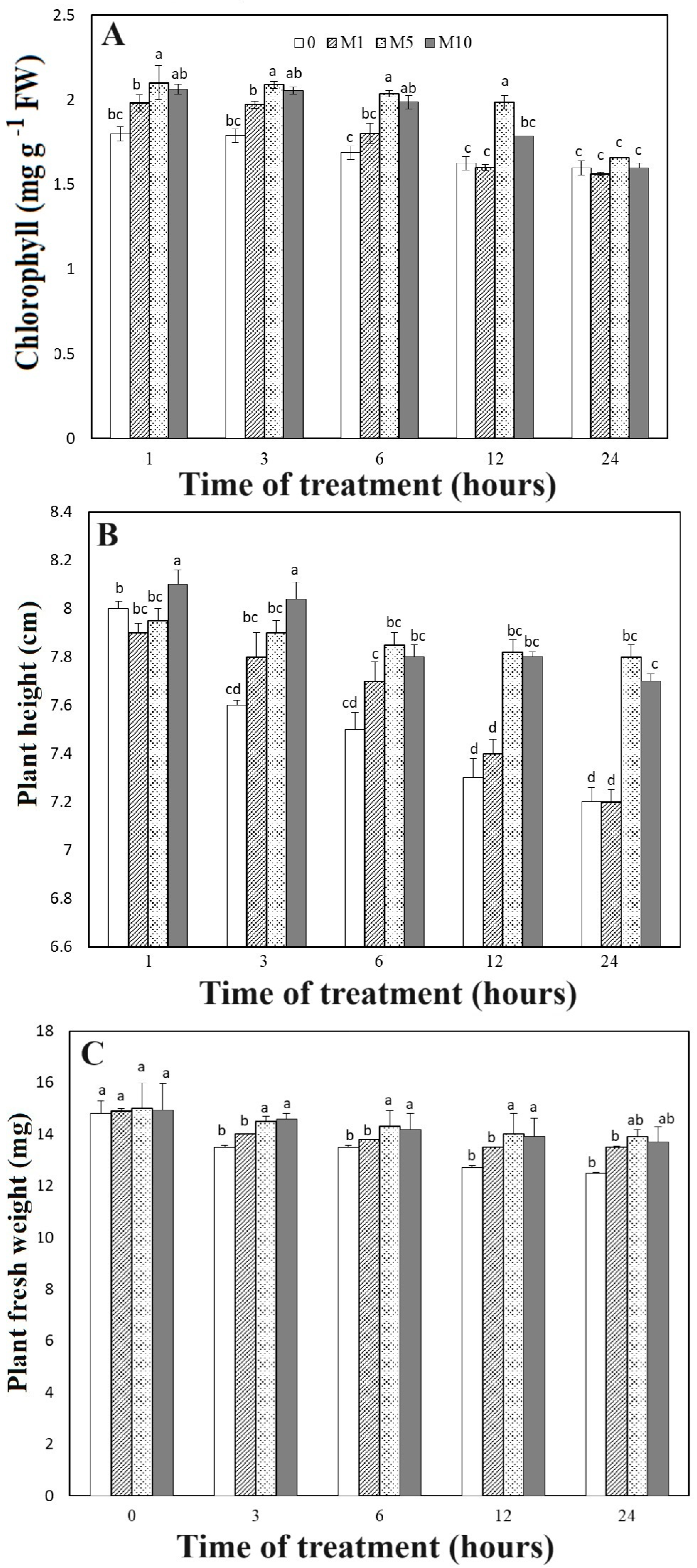
Figure 1. Effect of melatonin at (0, 1, 5, and 10 mM) on the carnation physiological parameters (A) chlorophyll content, (B) Plant height, and plant fresh weight (C). The plants were treated with in vitro heat stress on the growth chamber with 42 °C temperature for 1, 3, 6, 12, and 24 h. Different lowercase letters represent significant differences according to Duncan’s tests (p < 0.05).
Horticulturae 10 00122 g001
Figure 2. Effects of melatonin application on the morphological traits of in vitro carnation plants under heat stress condition after 24 h. Microshoots of carnation plants subjected to free medium (control) (A). The microshoots’ morphological traits under melatonin application at concentration of 1 (B), 5 (C), and 10 mM (D) after 24 h of culture.
Horticulturae 10 00122 g002
Figure 3. Effect of melatonin at (0, 1, 5, and 10 mM) on hydrogen peroxide ((A); H2O2), superoxide radical ((B); O2) and hydroxyl ((C); −OH) contents of carnation plants during heat stress condition. Means expressed the average of three replicates ± SE, and means within each graph denoted by the same lowercase letter did not significantly differ according to Duncan test at p < 0.05.
Horticulturae 10 00122 g003
Figure 4. Accumulation of H2O2. Carnation leaves were collected after 12 h after melatonin application at concentration of 0 (control) (A), 1 mM (B), 5 mM (C), and 10 mM (D). Carnation leaves treated with DAB solution to measure the hydrogen peroxide, then transferred in darkness one day.
Horticulturae 10 00122 g004
Figure 5. Effect of melatonin at (0, 1, 5, and 10 mM) on hydrogen peroxide on major antioxidant enzymes POD (A), SOD (B), CAT (C) and MDA (D) under heat stress condition of carnation plants. Means expressed the average of three replicates ± SE, and means within each graph denoted by the same lowercase letter did not significantly differ according to Duncan test at p < 0.05.
Horticulturae 10 00122 g005
Figure 6. Effects melatonin at different concentration on expression pattern of (A) GAPDH, (B) DcPOD1, (C) DcPOD2, (D) DcPOD3, (E) Gols1 and (F) MBF1c in carnation plants under heat stress condition by qRT-PCR. Microshoots were transferred to MS medium containing melatonin at concentration of 0, 1, 5, and 10 mM), and then heat stress (42 °C) was applied for indicated time. Microshoots were collected immediately for RNA extraction. Notably, 10 genes highly induced by heat stress were used for qPCR analysis and most of them showed significant changes after melatonin applications. Data are the means of three replicates with standard division shown by vertical bars indicates significant differences at p < 0.05. Different lowercase letters represent significant differences according to Duncan’s tests (p < 0.05).
Horticulturae 10 00122 g006
Figure 7. Effects of melatonin at different concentration on expression pattern of (A) HSF30, (B) HSP101, (C) HSP70, and (D) sHSP (MT) in carnation plants under heat stress condition by qRT-PCR. Microshoots were transferred to MS medium containing melatonin at concentration of 0, 1, 5, and 10 mM), and then heat stress (42 °C) was applied for indicated time. Microshoots were collected immediately for RNA extraction. Notably, 10 genes highly induced by heat stress were used for qPCR analysis and most of them showed significant changes after melatonin applications. Data are the means of three replicates with standard division shown by vertical bars indicates significant differences at p < 0.05. Different lowercase letters represent significant differences according to Duncan’s tests (p < 0.05).
Horticulturae 10 00122 g007
Figure 8. The mechanism of melatonin’s role in increasing carnation heat tolerance.
Horticulturae 10 00122 g008
Table 1. Sequences of oligonucleotide primers used in RT-PCR.
Gene NameAccession NumberForward PrimerReverse PrimerProduct Length
GAPDHDc49995CACTCCATCACAGCCACACAACACGGAAAGCCATACCAGTCA190
DcPOD1DT214806GTGTAGTCTCGTGTGCCGATCTTCGGGGATTTTGCCTTGC144
DcPOD2DT214807AGCAACCCTTTACCAGCAACTCGTCTTCCAACCCAGTGGA170
DCPOD3CF259499CTGAACGGTAAAGGGTTGCTGAACAAAACCATGGCCCTAGC131
Gols1Dc14879GGGGTCAAAGCCGTGGAGATCTCTAAAGGGCTCCAGTTTCGT155
MBF1cDc_15682TGAATGCCCGGAAACTCGACGACCGCCTTTCCATTCTCGT179
HSF30Dc83420CCGGAGCTAGACAGGCTAATGTCGATTTCTGGGGGCATTGA128
HSP101Dc_70612AGGTGGTGACTGAACTGTCGGATCGACCATCCCTCCGTTT126
HSP70Dc_37984CAGGCGAAGAGAGAAGCCATCTGAGTCACCCCGGTTTCAA168
sHSP (MT)Dc_89619TCTCCGGCAGTAATGTCGTCGTTCCTCTCAGAGCGGTCG137
Disclaimer/Publisher’s Note: The statements, opinions and data contained in all publications are solely those of the individual author(s) and contributor(s) and not of MDPI and/or the editor(s). MDPI and/or the editor(s) disclaim responsibility for any injury to people or property resulting from any ideas, methods, instructions or products referred to in the content.