Article

In Vitro Propagation, Evaluation of Antioxidant Activities, and Phytochemical Profiling of Wild and In Vitro-Cultured Plants of Curcuma larsenii Maknoi & Jenjitikul—A Rare Plant Species in Thailand

by
Surapon Saensouk
1,2,
Supacha Benjamin
3,
Theeraphan Chumroenphat
2,4 and
Piyaporn Saensouk
2,3,*
1
Walai Rukhavej Botanical Research Institute, Mahasarakham University, Maha Sarakham 44150, Thailand
2
Diversity of Family Zingiberaceae and Vascular Plant for Its Applications Research, Mahasarakham University, Maha Sarakham 44150, Thailand
3
Department of Biology, Faculty of Science, Mahasarakham University, Maha Sarakham 44150, Thailand
4
Aesthetic Sciences and Health Program, Faculty of Thai Traditional and Alternative medicine, Ubon Ratchathani Rajabhat University, Ubon Ratchathani 34000, Thailand
*
Author to whom correspondence should be addressed.
Horticulturae 2024, 10(11), 1181; https://doi.org/10.3390/horticulturae10111181
Submission received: 26 September 2024 / Revised: 25 October 2024 / Accepted: 6 November 2024 / Published: 8 November 2024
(This article belongs to the Section Propagation and Seeds)

Abstract

:
Curcuma larsenii Maknoi & Jenjitikul is a member of the Zingiberaceae family, possessing significant pharmacological potential, although it has become endangered through the abuse of resources. This research article delineates the findings of the in vitro propagation, transplantation, and phytochemical profiles of C. larsenii, a rare plant species in Thailand. Microshoots measuring 1 cm in length were used as explants for the induction of shoots and roots in both solid and liquid Murashige and Skoog medium, incorporating various concentrations of cytokinins (6-benzylamino-purine (BA), 6-furfurylaminopurine (kinetin), thidiazuron (TDZ)) and auxins (1-naphthaleneacetic acid (NAA) and indole-3-acetic acid (IAA)) over a duration of 8 weeks. This study assessed the total phenolic content, total flavonoid content, and antioxidant activity via the 2,2-diphenyl-1-picrylhydrazyl (DPPH) and ferric reducing antioxidant power (FRAP) assays and conducted high-performance liquid chromatography (HPLC) analysis. The highest number of shoots was recorded in solid and liquid media containing MS medium enriched with 2 mg/L BA and 0.5 mg/L NAA, as well as 2 mg/L BA and 0.5 mg/L IAA, yielding 5.40 and 8.80 shoots/explant, respectively. The biggest roots/explant induction of 9.20 was attained using the liquid MS medium supplemented with 4 mg/L BA and 0.5 mg/L IAA. The highest survival rate (100%) was recorded when tissue culture plantlets were transplanted into a mixture of sand and soil (1:1). In vitro-cultivated plants exhibited superior total phenolic content relative to wild plants. Leaf extracts of C. larsenii exhibited markedly superior antioxidant activity compared to other plant organs from both in vitro and wild specimens. C. larsenii wild plants and in vitro plants generated phenolic acids and flavonoids and exhibited antioxidant activity, demonstrating a biotechnological alternative for the acquisition of bioactive substances.

1. Introduction

Curcuma larsenii Maknoi & Jenjittikul, a rare and endemic species of the tribe Zingibereae belonging to the Zingiberaceae family, holds particular significance due to its restricted distribution and unique botanical characteristics [1]. Like many species in the Curcuma genus, C. larsenii has a historical role in local culinary practices, where its aromatic rhizomes are valued for their flavor and potential medicinal properties [1]. These rhizomes, a hallmark of the ginger family, have been traditionally used in various remedies due to their bioactive compounds, which are now being further investigated for their therapeutic potential.
Recent phytochemical studies [2,3,4,5,6,7,8,9] have demonstrated that Curcuma plants synthetize several important secondary metabolites, including essential oils, flavonoids, and curcuminoids. Curcuminoids, in particular, are well recognized for their anti-inflammatory, antioxidant, and potentially disease-modifying properties. These compounds are known to combat oxidative stress and inflammation, both of which are key factors in a range of chronic diseases. The presence of such bioactive compounds suggests that C. larsenii may have medicinal properties similar to Curcuma longa L. (turmeric), which is widely studied and utilized for its health benefits. From a conservation perspective, the limited natural range of C. larsenii makes it a priority for biodiversity preservation efforts. Protecting this species is crucial not only for ecological reasons but also for maintaining its potential future use in medicine and food industries. Conservation strategies might include protecting its natural habitat, promoting sustainable harvesting, and exploring ex situ conservation techniques such as propagation through tissue culture. The traditional vegetative propagation of C. larsenii relies on rhizome division, a common method in many Curcuma species. However, this technique presents challenges for large-scale cultivation due to its slow multiplication rate and susceptibility to soil-borne pathogens. In response to these limitations, tissue culture has emerged as a promising alternative for mass propagation. Tissue culture offers rapid, year-round growth and the ability to produce large numbers of disease-free plants, making it an attractive solution for scaling up the production of C. larsenii. Several studies have focused on optimizing tissue culture techniques for different Curcuma species, with specific attention given to the roles of plant growth regulators like auxins and cytokinins in enhancing shoot multiplication and root induction. Research on Curcuma aeruginosa Roxb. [10,11,12], C. amada Roxb. [13,14], C. angustifolia Roxb. [15,16], C. aromatica Salisb. [17], C. caesia Roxb. [18,19,20], and C. longa [21,22,23] has demonstrated that adjusting the concentrations of these hormones can significantly impact both the number and quality of shoots and roots produced.
The present study on C. larsenii seeks to build on this existing body of knowledge by developing a reliable protocol for in vitro shoot multiplication, root induction, and successful acclimation to soil. Additionally, the antioxidant activity and phytochemical profiles of both wild and in vitro-derived plants will be compared, aiming to confirm whether the propagated plants retain the same bioactive properties as their wild counterparts. By combining mass propagation techniques with phytochemical analysis, this research not only contributes to the conservation of C. larsenii but also explores its potential for therapeutic applications.

2. Materials and Methods

2.1. In Vitro Micropropagation

Source of plant material: axillary buds of C. larsenii, selected for their vitality, were harvested from Ubon Ratchathani Province, Thailand, to be utilized as explants for in vitro multiplication.
Explant preparation and disinfection: Axillary buds were collected from plants cultivated in the field and subjected to a rigorous sterilization protocol. Initially, the buds underwent a preliminary washing step, where they were rinsed thoroughly under running tap water for one hour to remove surface debris. For initial surface sterilization, the buds were then immersed in a 70% (v/v) ethanol solution for one minute. To ensure a more comprehensive disinfection, the buds were transferred to a sterile environment within a laminar flow chamber. Under these aseptic conditions, the buds were treated with sodium hypochlorite (NaOCl) solutions at concentrations of 20% and 15%. These treatments were applied for 20 min and 15 min, respectively, in order to effectively eliminate microbial contaminants. Following these NaOCl treatments, the buds were rinsed thoroughly three times with sterilized distilled water, each rinse lasting five minutes, to remove any residual disinfectant. Post sterilization, the axillary buds were excised into small sections, approximately 3–4 mm in length, for further in vitro cultivation. The excised bud segments were inoculated onto Murashige and Skoog (MS) medium, a well-established plant tissue culture medium first described by Murashige and Skoog in 1962 [24]. The MS medium was supplemented with 2.0 mg/L of 6-benzylamino-purine (BA), a cytokinin known to promote cell division and shoot proliferation, along with 0.5 mg/L of 1-naphthaleneacetic acid (NAA), an auxin that facilitates root induction and overall morphogenesis. Throughout the experiment, explants were transferred to fresh MS medium at regular intervals, with subculturing performed every four weeks to maintain optimal growth conditions. After two months of growth, the developing microshoots were transferred to MS medium devoid of plant growth regulators (PGRs). This step allowed the shoots to stabilize and continue growing in a hormone-free environment. After an additional month, microshoots that reached a minimum length of 1 cm were selected as explants for further experimental evaluation and testing. This methodology highlights the importance of a meticulous sterilization process to prevent microbial contamination in tissue culture. The combination of BA and NAA in the MS medium facilitated effective shoot and root development, while the subsequent removal of PGRs in later stages allowed the natural growth and acclimation of the microshoots. By employing these strategies, this protocol provides a reliable approach for the in vitro propagation of plant species, with particular applicability to species like C. larsenii, which require mass propagation for conservation and study.
Medium and culture conditions: For in vitro germination and propagation, axillary buds were cultivated using a standard Murashige and Skoog (MS) medium, which was enriched with 30 g/L of sucrose to provide the necessary carbon source for plant growth. The medium was solidified by adding 7 g/L of bacto agar, ensuring proper consistency for solid culture conditions. Additionally, various concentrations and combinations of PGRs were incorporated into the medium to optimize shoot and root development based on the specific requirements of the experiment. Microshoots were individually cultured in solid media within 120 mL glass culture vessels. A single microshoot was placed in each vessel, and 20 vessels were used for each experimental treatment, ensuring adequate replication. In parallel, a separate set of microshoots was grown in liquid media. This method utilized 250 mL Erlenmeyer flasks, with two microshoots inoculated per flask, and 10 flasks were prepared for each treatment. Liquid cultures allow for more uniform exposure of the microshoots to the nutrients and growth regulators in the medium, which can promote different developmental outcomes compared to solid media. The pH of all culture media was adjusted to between 5.7 and 5.8 to maintain a favorable environment for plant tissue growth. This pH calibration was achieved using either 1 N sodium hydroxide (NaOH) or 1 N hydrochloric acid (HCl) prior to sterilization. The sterilization process involved autoclaving the media at 121 °C for 15 min, ensuring that all materials were free of contaminants before inoculation with plant tissue. All cultures were maintained under controlled environmental conditions to support optimal growth. A photoperiod of 16 h of light was provided, with the temperature kept at 25 ± 2 °C to mimic natural conditions favorable for plant development. The light intensity was set to 27 µmol s−1m−2, delivered by fluorescent white light tubes, which supplied the energy necessary for photosynthesis and other light-dependent physiological processes in the microshoots. These environmental conditions were crucial in maintaining consistent growth and development throughout the in vitro propagation process. This approach to in vitro germination and propagation, with the use of both solid and liquid media and the inclusion of PGRs, demonstrates the importance of tailoring the growth environment to the specific needs of the plant tissue. It also highlights the precision required in maintaining consistent culture conditions, such as the pH, temperature, and light, to achieve successful propagation in tissue culture systems.
In vitro shoot multiplication: The microshoots were transferred to MS medium, where they were exposed to various concentrations of PGRs to optimize shoot and root development. Specifically, BA was applied at concentrations of 0, 1.0, 2.0, 3.0, 4.0, and 5.0 mg/L, in combination with NAA at concentrations of 0, 0.1, and 0.5 mg/L in solid medium. Similarly, a parallel set of treatments was conducted using kinetin, a cytokinin known for its role in cell division, at the same concentration range (0, 1.0, 2.0, 3.0, 4.0, and 5.0 mg/L), also combined with NAA at 0, 0.1, and 0.5 mg/L. In addition to solid media cultures, microshoots were cultivated in liquid media to assess the effects of different PGR concentrations in a more dynamic environment. These liquid cultures were prepared with MS agar medium supplemented with varying concentrations of BA or kinetin (0, 1.0, 2.0, 3.0, 4.0, and 5.0 mg/L), combined with indole-3-acetic acid (IAA) at two levels: 0 and 0.5 mg/L. The liquid cultures were maintained under continuous agitation on a rotary shaker set at 120 rpm, which enhances nutrient and PGR uptake by the microshoots through the constant movement of the medium. The microshoots were cultured under these conditions for a total duration of eight weeks. Throughout the experiment, several key growth parameters were measured to evaluate the effectiveness of each PGR treatment. These parameters included the average number of shoots per plantlet, average shoot length (measured in centimeters), average number of roots, and average root length (also measured in centimeters). These metrics provided a comprehensive analysis of both the shoot proliferation and root induction capabilities of the different PGR combinations. The results helped determine the optimal concentrations and combinations of BA, kinetin, NAA, and IAA for maximizing the in vitro propagation of the microshoots.
Acclimatization and transplantation: Following an 8-week cultivation period in MS medium, the plantlets were subjected to a 2-week acclimatization phase under room temperature to facilitate their adjustment to the ex vitro environment. The in vitro-propagated shoots, which had achieved a length of 5–6 cm and exhibited both well-formed shoots and roots, were carefully extracted from the culture containers. To remove any residual culture medium clinging to the roots, the plantlets were thoroughly rinsed under running tap water. After cleaning, the plantlets were transplanted into disposable pots containing different growth substrates, including soil, sand, and a 1:1 mixture of soil and sand (w/w). The pots were placed in a greenhouse located at the Department of Biology, Faculty of Science, Mahasarakham University, Thailand, where the plantlets underwent an 8-week acclimatization period. During this phase, the environmental conditions were carefully controlled, and daily watering with tap water ensured adequate hydration. Throughout the acclimatization process, key growth parameters were monitored to assess the performance and adaptability of the plantlets to the new growing conditions. These parameters included the survival rate of the plantlets, the average number of shoots produced per plant, and the shoot length (measured in centimeters), as well as leaf-related metrics such as the number of leaves, leaf width, and leaf length (all measured in centimeters). This detailed monitoring provided valuable insights into the plantlets’ growth and adaptation during the transition from in vitro to ex vitro conditions.
Experimental design and statistical analysis for in vitro micropropagation: The experimental investigations were structured utilizing a completely randomized design (CRD) to ensure robust and unbiased results. Each treatment group included a total of twenty microshoots, and each treatment was replicated three times to provide sufficient statistical power and reproducibility. The outcomes are presented as the mean values accompanied by the standard error (SE) to reflect the variability within the data. For the analysis of the experimental data, statistical significance was determined using analysis of variance (ANOVA), which allowed for the comparison of differences among treatment means. To further examine specific differences between group means, Duncan’s Multiple Range Test (DMRT) was employed, with a significance level set at 5% (p < 0.05). This post hoc test is particularly effective in identifying which treatment groups significantly differ from each other while controlling for error. The statistical analyses were conducted using SPSS software (version 22), a widely recognized tool for executing comprehensive statistical evaluations. By employing ANOVA and DMRT, this study ensured that the data were rigorously analyzed to identify significant trends and differences, enhancing the reliability of the conclusions drawn from the research findings.

2.2. Phytochemical Profiling Analysis

Plant material: The native specimens were collected from Ubon Ratchathani Province, Thailand, in June 2021. The identification and authentication of the plant material were conducted by Associate Professor Dr. Surapon Saensouk at the Walai Rukhavej Botanical Research Institute, Mahasarakham University, Thailand. A voucher specimen (Saensouk P10) was deposited at the Mahasarakham University Herbarium to ensure proper documentation and future reference. Specimens of native plant species were collected for this study, encompassing various plant parts such as the leaves, pseudostems, rhizomes, and roots. These were gathered alongside leaves from tissue culture plants cultivated on MS medium under different conditions: one group grown on MS medium without PGRs and another on MS medium supplemented with 2 mg/L BA and 0.1 mg/L NAA. Additionally, root specimens were obtained from aseptic cultures both on PGR-free MS medium and MS medium fortified with 2 mg/L BA and 0.1 mg/L NAA. For subsequent analyses, both wild and in vitro-derived plant samples were meticulously rinsed with tap water to remove any contaminants. Following this, the samples were frozen at −20 °C to preserve their biochemical integrity, and they were subjected to freeze-drying for 24 h to remove moisture while maintaining the stability of heat-sensitive compounds. Once freeze-dried, the samples were finely ground into powder using an herb grinder, facilitating uniformity for subsequent chemical analysis. Soil samples from the collection sites were similarly stored at −20 °C until they were processed for further analysis.
Extraction: The extraction process was carried out following a modified version of the protocols established by Chumroenphat et al. [25] and Siriamornpun and Kaewseejan [26]. For this procedure, a finely powdered plant sample weighing 1.00 g was used as the starting material. The extraction was performed using 40 mL of methanol as the solvent. The sample and solvent mixture was placed in an incubator shaker set to 37 °C and agitated at a constant speed of 150 rpm for a period of 15 h to maximize the dissolution of bioactive compounds. After that, the mixture was filtrated using Whatman No. 1 filter paper. The filtrate, which contained the methanol-soluble compounds, was collected and stored at −20 °C to preserve its integrity for subsequent phytochemical and antioxidant activity analyses.
Determination of total phenolic contents: The total phenolic content (TPC) was quantified using a modified approach derived from Chumroenphat et al. [25]. In this procedure, a 0.30 mL aliquot of the plant extract was combined with 2.25 mL of Folin–Ciocalteu reagent, which had been diluted at a ratio of 1:10 with distilled water. The mixture was left to react for 5 min at room temperature. After the reaction, 2.25 mL of a 6% (w/v) sodium carbonate solution was added to the mixture, and it was incubated in the dark for 90 min at ambient temperature to allow the development of the colorimetric reaction. Following the incubation period, the absorbance of the reaction mixture was measured at 725 nm using a UV–visible spectrophotometer (Shimadzu Corp., Kyoto, Japan). For quantification purposes, gallic acid was employed as a standard reference compound, with calibration concentrations prepared at 6.25, 12.50, 25, 50, 100, and 150 mg/L. The total phenolic content of the plant samples was then expressed in terms of milligrams of gallic acid equivalents (GAEs) per gram of dry weight (DW), providing a standardized measure of phenolic concentration across samples.
Determination of total flavonoid contents: The total flavonoid content (TFC) was quantified using a modified protocol described by Chumroenphat et al. (2019) [25]. In this method, a 0.5 mL aliquot of the plant extract was transferred to a centrifuge tube, followed by the addition of 0.15 mL of a 5% (w/v) sodium nitrite (NaNO2) solution. The mixture was allowed to incubate in the dark for 6 min to facilitate the formation of a complex. Following this incubation, 0.3 mL of a 10% (w/v) aluminum chloride hexahydrate (AlCl3·6H2O) solution was introduced, and the mixture was kept in the dark for an additional 5 min to promote further reaction. After this period, 1.0 mL of 1 M sodium hydroxide (NaOH) was added to the mixture to develop a stable color complex. The absorbance of the resulting solution was measured at 510 nm using a UV–visible spectrophotometer. Rutin was utilized as the standard reference for calibration, with concentrations prepared at 6.25, 12.50, 25, 50, 100, and 150 mg/L. The total flavonoid content was expressed as milligrams of rutin equivalents (REs) per gram of DW, providing a standardized measure of flavonoid concentration in the plant samples.
Determination of DPPH free radical scavenging assay: The free radical scavenging activity of the samples was assessed using the 2,2-diphenyl-1-picrylhydrazyl (DPPH) assay, as outlined in the methodologies of Chumroenphat et al. [25] and Siriamornpun and Kaewseejan [26]. A stock solution of DPPH was prepared by dissolving the compound in methanol, which was then stored at −20 °C for stability. For the assay, the working solution was created by mixing 100 µL of the plant extract with 3.0 mL of a 0.004% DPPH solution. This mixture was incubated in the dark for 30 min to allow for adequate reaction time. The absorbance of the solution was subsequently measured at 517 nm using a UV–visible spectrophotometer to determine the extent of free radical scavenging. Trolox served as the reference standard for quantification, and the results were expressed as milligrams of Trolox equivalents (TEs) per gram of DW.
Ferric reducing antioxidant power (FRAP) assay: The antioxidant capacity of the samples was assessed using the ferric reducing antioxidant power (FRAP) assay, as described by Siriamornpun et al. [27]. In this procedure, the samples were first diluted tenfold with ethanol and subsequently transferred into a centrifuge tube. To this, 180 µL of deionized water was added, followed by the introduction of 1.8 mL of the FRAP reagent. The resulting mixture was then incubated at 37 °C in a water bath for a duration of 4 min to allow for the reduction reaction to occur. Following the incubation period, the absorbance of the solution was measured at 593 nm using a UV–visible spectrophotometer to determine the reducing power of the samples. Ferrous sulfate (FeSO4) served as the standard reference for calibration, and the antioxidant capacity was expressed as milligrams of ferrous sulfate equivalents (FeSO4) per gram of DW.
Determination of phenolic acid and flavonoid content by HPLC analysis: For the subsequent biochemical analyses, phenolic acid detection was performed using high-performance liquid chromatography (HPLC), with a range of phenolic standards employed for comparison. The standards included gallic acid, protocatechuic acid, p-hydroxybenzoic acid, chlorogenic acid, vanillic acid, caffeic acid, syringic acid, p-coumaric acid, ferulic acid, and sinapic acid. In addition to phenolic acids, flavonoid content was also evaluated using rutin, myricetin, quercetin, and apigenin as reference standards. Extracts from various parts of C. larsenii, including the leaves, pseudostems, rhizomes, and roots from wild specimens, as well as leaf samples from in vitro cultures grown on MS medium with and without PGRs, were prepared for analysis. Each sample (0.2 g) was ground into a fine powder and placed into vials. The extraction process, adapted from methodologies established by Chumroenphat et al. 2021 [28] involved the use of a hydrochloric acid/methanol solvent (1:100, v/v). The samples were incubated for 15 h at 37 °C on a shaker incubator set at 150 rpm, protected from light to prevent degradation of sensitive compounds. Following extraction, the solutions were filtered through a 0.22 µm nylon membrane filter to remove particulate matter prior to high-performance liquid chromatography (HPLC) analysis. The HPLC setup included an ODS-3 C18 column (4.6 mm × 250 mm, 5 µm), safeguarded by an Inersil ODS-3 guard column (10 mm × 4 mm, 5 µm). The chromatographic method employed gradient elution as per the protocol detailed by Siriamornpun et al. [27]. The flow rate was consistently maintained at 0.8 mL/min, and the injection volume was set to 20 µL. The detection of phenolic compounds was achieved at specific wavelengths: 280 nm for hydroxybenzoic acids, 320 nm for hydroxycinnamic acids, and 370 nm for flavonoids. Spectra were captured across a wavelength range of 200 to 800 nm. The identification of phenolic acids and flavonoids within the extracts was conducted by comparing their relative retention times (RTs) against those of established external standards, ensuring the accurate quantification and characterization of these bioactive constituents.

2.3. Statistical Analysis for Phytochemical Profiling

All measurements were reported as the mean ± standard error (SE) derived from three independent replicates. To assess the significance of the results, one-way analysis of variance (ANOVA) was employed, followed by Duncan’s Multiple Range Test (DMRT) for post hoc comparisons, with a significance threshold set at p < 0.05. Statistical analyses were conducted using SPSS software (version 22). Additionally, Pearson’s correlation coefficient (r) was computed using Microsoft Excel 2021 to evaluate the strength and direction of relationships between variables.

3. Results

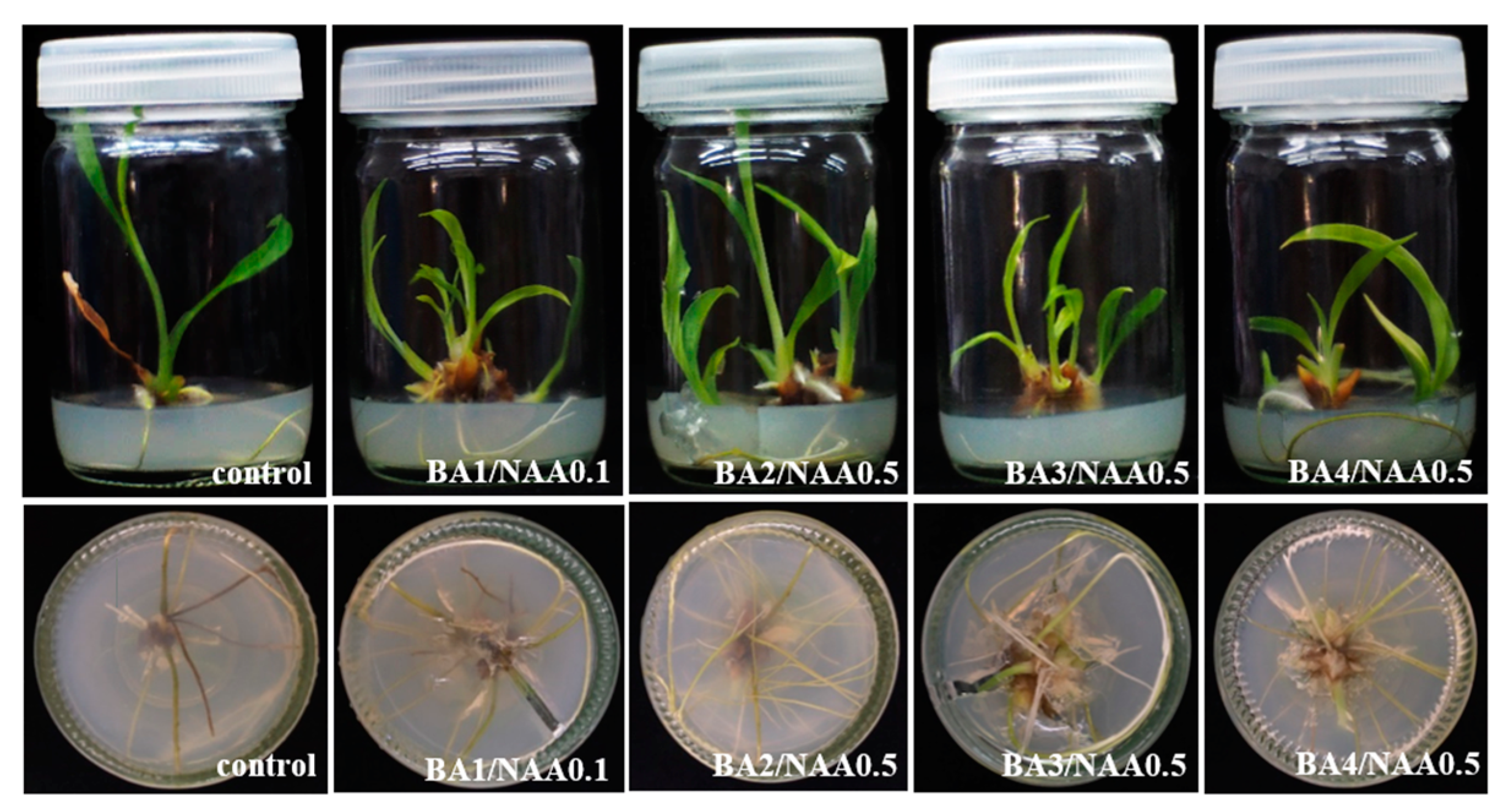
3.1. In Vitro Plant Regeneration

3.1.1. In Vitro Shoot Multiplication and Root Induction

Microshoots of C. larsenii, measuring 1 cm in length, were cultivated on both solid and liquid MS media over an 8-week period. The media were supplemented with various concentrations of cytokinins (BA and kinetin) in conjunction with auxins (NAA and IAA) to enhance shoot multiplication and root induction. The shooting response of the explants was significantly influenced by the type and concentration of the cytokinins applied (Table 1). Explants grown on the control medium, which lacked PGRs, produced the fewest shoots. The optimal combination of BA at 2 mg/L and NAA at 0.5 mg/L resulted in the highest shoot production, yielding an average of 5.40 shoots per explant after eight weeks of culture on solid MS medium. However, the average shoot length recorded was 3.67 cm, which was notably lower than the 5.18 cm achieved on the control medium without PGRs. Roots began to develop within two weeks for the regenerated shoots cultured on solid MS medium, irrespective of whether PGRs were included (Table 1 and Figure 1). In contrast, the combination of kinetin and NAA led to the growth of significantly fewer shoots compared to the BA and NAA treatment. Notably, while the kinetin and NAA combination produced longer shoots, the overall quantity of shoots was lower. Specifically, microshoots grown on solid MS medium enriched with 2 mg/L kinetin and 0.5 mg/L NAA exhibited the highest metrics for shoot number, shoot length, root quantity, and root length, with averages of 3.10 shoots/explant, 5.93 cm, 8.50 roots/explant, and 4.54 cm, respectively (Table 2 and Figure 2). When comparing the efficacy of the two cytokinins in liquid MS medium supplemented with 0.5 mg/L IAA, BA was found to be more effective in promoting shoot proliferation than kinetin (p < 0.05). The optimal results were achieved with varying concentrations of BA (ranging from 1 to 5 mg/L) combined with 0.5 mg/L IAA, with the highest shoot production of 8.80 shoots/explant observed at 2 mg/L BA. However, an increase in BA concentration to 4 mg/L or greater led to a decrease in the average shoot count (Table 3 and Figure 3).

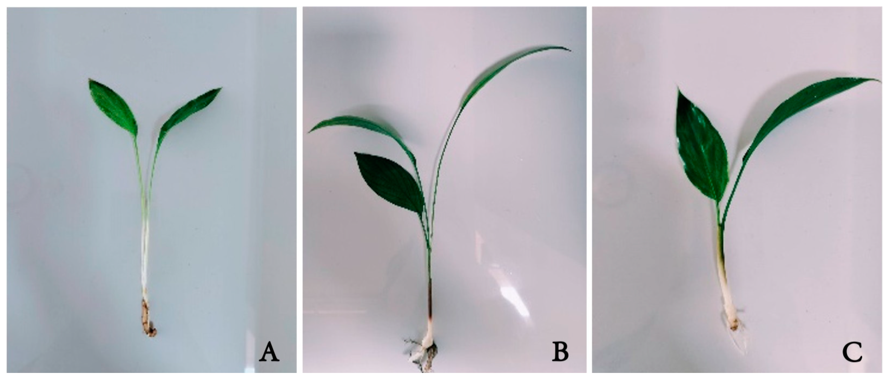
3.1.2. Acclimatization and Transplantation of In Vitro-Grown Plantlets

All C. larsenii plantlets were acclimatized for two weeks in the media preparation room, where the light intensity and temperature were not controlled. The selected plantlets, characterized by a minimum of two leaves, heights exceeding 5 cm, root lengths greater than 1 cm, and the absence of morphological defects, were subsequently transferred to various potting media. The plantlets were removed from the agar medium within the culture bottles and carefully transplanted into plastic pots containing a 1:1 (w/w) mixture of soil and sand. This acclimatization occurred during the rainy season in a greenhouse setting at the Department of Biology, Faculty of Science, Mahasarakham University, Thailand, and lasted for eight weeks.
Upon completion of the acclimatization period, C. larsenii plantlets exhibited a range of shoot sizes and quantities depending on the type of potting medium used. Among the three types of planting materials evaluated, the soil–sand mixture yielded the highest survival rate of 100%. The highest metrics for shoot number (4.74 shoots/explant), shoot length (8.78 cm), number of leaves (7.26 leaves/explant), leaf width (1.95 cm), and leaf length (10.07 cm) were recorded in plantlets grown in soil. Importantly, no significant morphological differences were observed between the regenerated plantlets and the parent plants of C. larsenii, indicating that the in vitro-derived plants retained morphological similarities to those propagated through conventional means (Table 4 and Figure 4).

3.2. Phytochemical Profiling Analysis

3.2.1. Total Phenolic Contents and Total Flavonoid Contents

The quantification of the total phenolic content (TPC) and total flavonoid content (TFC) was performed on methanolic extracts derived from various mother plant and in vitro plant parts of C. larsenii. The samples analyzed included aerial components such as roots, rhizomes, pseudostems, and leaves, along with in vitro-grown roots and leaves cultured on MS medium, as well as MS medium supplemented with 2 mg/L BA and 0.1 mg/L NAA. The TPC of the extracts ranged from 10.29 to 317.33 mg gallic acid equivalent (GAE) per gram of dry weight (DW). Notably, leaves cultivated on MS medium without PGRs exhibited the highest TPC values (p < 0.05), followed by leaves grown on MS medium enriched with 2 mg/L BA and 0.1 mg/L NAA, roots on MS medium devoid of PGRs, and roots grown with 2 mg/L BA and 0.1 mg/L NAA. Aerial parts, including leaves and pseudostems from natural conditions, demonstrated lower TPC levels, with rhizomes from natural sources exhibiting the lowest TPC content, as presented in Table 5. The TFC ranged from 10.49 to 34.86 mg rutin equivalent (RE) per gram of DW. Leaves in their natural habitat showed significantly elevated TFC yields (p < 0.05), followed closely by leaves cultured on MS medium without PGRs and those grown on MS medium with 2 mg/L BA and 0.1 mg/L NAA. Roots from natural conditions, pseudostems from the wild, and roots grown on both MS media (with and without PGRs) exhibited progressively lower TFC values, while rhizomes in their natural state displayed the lowest TFC yield, as detailed in Table 5.

3.2.2. Antioxidant Activity

The antioxidant capacity of C. larsenii, evaluated through DPPH scavenging activity and the ferric reducing antioxidant power (FRAP) assay, demonstrated significant variations based on the growing methods (wild or in vitro) and the specific plant sections analyzed, as summarized in Table 5. According to the DPPH assay results, leaves from in vitro-regenerated plants cultivated on MS medium without PGRs exhibited the highest antioxidant capacity, measuring 4.45 mg Trolox equivalent (TE) per gram of dry weight (DW). This was closely followed by wild plant leaves at 4.38 mg TE/g DW and the pseudostems of wild plants, which recorded 4.33 mg TE/g DW. In vitro leaves grown on MS medium supplemented with 2 mg/L BA and 0.1 mg/L NAA showed an antioxidant capacity of 3.99 mg TE/g DW. Conversely, the lowest DPPH scavenging activity was observed in the rhizomes of wild plants, at 1.50 mg TE/g DW. The radical scavenging percentages further indicated that the leaves and pseudostems of wild plants exhibited the highest values, at 93.54% and 92.09%, respectively. In comparison, the leaves from in vitro-derived plants cultivated on MS medium demonstrated radical scavenging percentages of 85.93% when grown without PGRs and 76.04% with PGRs. In the FRAP assay, the rhizome of wild plants displayed the lowest antioxidant activity, quantified at 0.02 mg FeSO4/g DW. Notably, vanillic acid emerged as the predominant phenolic acid across various samples, particularly in the roots (both with and without PGRs) and in the leaves of in vitro-regenerated plants, as detailed in Table 5.
The correlation analysis Pearson test was used to determine the correlation coefficients (r) between the mean values of each parameter obtained in this study. A strong positive correlation was found between FRAP and TPC (r = 0.883, p < 0.01) and %inhibition (DPPH), FRAP, and DPPH (r = 0.974, p < 0.01; r = 0.433, p < 0.05) (Table 6).

3.2.3. Determination of Phenolic Acid and Flavonoid Content by HPLC Analysis

The analysis of C. larsenii extracts and its various cultured organs was conducted using high-performance liquid chromatography (HPLC). Previous investigations into the phytochemical profiles of Zingiberaceae species have highlighted the presence of phenolic acids, flavonoids, and notable antioxidant activity. In this study, the identification of significant bioactive compounds in C. larsenii was facilitated by comparing the extracts to several reference standards. The standards for detecting phenolic acids included gallic acid, protocatechuic acid, p-hydroxybenzoic acid, chlorogenic acid, vanillic acid, caffeic acid, syringic acid, p-coumaric acid, ferulic acid, and sinapic acid. For the analysis of flavonoids, rutin, myricetin, quercetin, and apigenin were utilized as reference compounds, as detailed in Table 7 and Table 8. The results revealed that vanillic acid and sinapic acid were consistently present across all plant extracts, including those derived from both wild and in vitro cultures. However, p-hydroxybenzoic acid and chlorogenic acid were notably absent in the in vitro samples. Vanillic acid was identified as the predominant phenolic acid, especially concentrated in the roots (both with and without PGRs) and the leaves of in vitro-cultivated plants. The highest concentration of vanillic acid, measured at 952.81 µg/g, was found in the roots of in vitro-regenerated plants cultured on MS medium without any PGRs. Additionally, amounts of gallic acid, protocatechuic acid, p-hydroxybenzoic acid, chlorogenic acid, and caffeic acid were detected in all plant samples. Among the flavonoids, myricetin was present in all extracts, whereas quercetin was exclusively detected in the pseudostems of wild plants at a concentration of 16.87 µg/g. The highest concentration of myricetin, recorded at 1042.97 µg/g, was found in leaf extracts from natural plants. Notably, the leaf extract from wild C. larsenii plants exhibited the highest level of rutin, quantified at 441.29 µg/g. These findings underscore the rich phytochemical composition of C. larsenii and its potential for further pharmacological exploration.

4. Discussion

This study contributes to the understanding of how hormonal regulation influences plant tissue culture, with specific applicability to improving propagation protocols for the rare and endemic species C. larsenii. The response of explants in C. larsenii to shoot induction was significantly affected by the type and concentration of cytokinins applied. Our findings indicate that when microshoots were cultured on a hormone-free medium, shoot production remained minimal, which aligns with observations in other Zingiberaceae species. For instance, axillary bud explants of Curcuma zedoaria (Christm.) Roscoe did not show any proliferation on MS medium devoid of plant growth regulators [29]. This research focuses on enhancing shoot multiplication through the supplementation of MS medium with cytokinins, which are widely recognized for their role in promoting shoot induction and proliferation. The stimulatory effects of cytokinins on shoot development have been well documented across various members of the Zingiberaceae family, including Curcuma caesia Roxb. [6], C. sparganiifolia Gagnep. [30], Globba globulifera Gagnep. [31], and Kaempferia siamensis Sirirugsa [32]. For in vitro propagation, BA, kinetin (an adenine derivative), and thidiazuron (TDZ, a phenylurea derivative) are frequently employed for effective shoot induction and multiplication. Among the cytokinins tested, BA demonstrated superior efficacy in promoting the formation of multiple shoots compared to kinetin. The optimal results were observed with MS medium containing 2 mg/L BA combined with 0.5 mg/L NAA, established through a series of experiments with BA concentrations ranging from 0 to 5 mg/L paired with 0.1 and 0.5 mg/L NAA. This aligns with the work of Haida et al. [6] who reported that MSB5 medium supplemented with 15 µM BAP and 6 µM IBA achieved significant shoot multiplication, resulting in 100% shoot induction with an average of 3.53 shoots/explant and an average shoot length of 10.81 cm. Conversely, Alizah et al. [12] documented an impressive average of 18 shoots/explant from immature buds of C. aeruginosa Roxb. in liquid MS medium enriched with 4 mg/L BA. In this study, the most effective treatment was identified as MS medium supplemented with 2 mg/L BA and 0.5 mg/L NAA, surpassing all other tested conditions. The preference for BA over other cytokinins may be attributed to its favorable metabolism into nucleotides and ribosides, enhancing its biological activity [33]. The beneficial impact of BA on shoot multiplication has been corroborated in various plant species, including Amomum subulatum, Curcuma angustifolia, C. aromatica, C. caesia, C. longa, and C. zedoaria [15,17,18,34,35,36,37,38].
This study investigated the influence of varying concentrations of BA, kinetin, and IAA on in vitro shoot multiplication and root induction in C. larsenii cultivated in liquid MS medium. The highest proliferation rate was recorded at an average of 8.8 shoots/explant in liquid MS medium supplemented with 2 mg/L BA and 0.5 mg/L IAA. Furthermore, plantlets cultured in liquid medium enriched with BA and IAA exhibited superior performance in terms of shoot quantity, shoot length, root number, and root length compared to those supplemented with kinetin and IAA. These results corroborate findings by Mohanty et al. [14], who noted the maximum shoot proliferation (3.80 shoots/explant) in solid MS medium augmented with 2 mg/L BA and 0.5 mg/L IAA in C. amada Roxb. The advantages of liquid medium in explant cultivation include enhanced nutrient absorption and consistent exposure to the growth medium, promoting improved growth compared to solid media, which may restrict nutrient access and light availability, potentially limiting overall development. Once the plantlets reached approximately 5 cm in height, two randomly selected leaves from those with well-developed roots were carefully rinsed to eliminate residual agar and transplanted into greenhouse conditions. The regenerated plantlets displayed no morphological defects and achieved a survival rate of 95–100%. Notably, the high survival rate of plantlets grown in sand (100%) can be attributed to the substrate’s superior porosity and aeration properties, which facilitated root health and enhanced survival compared to other substrates. These observations align with reports by Saensouk et al. [30] that documented a 100% survival rate for C. sparganifolia plantlets in sand. Similarly, Saensouk et al. [39] found a survival rate of 100% for Kaempferia angustifolia plantlets transplanted into both sand and sand–soil mixtures. In contrast, Haida et al. [6] reported a lower survival rate of 77.78% for Curcuma caesia when cultivated in cocopeat. This methodology highlights the importance of a meticulous sterilization process to prevent microbial contamination in tissue culture. The combination of BA and NAA in the MS medium facilitated effective shoot and root development, while the subsequent removal of PGRs in later stages allowed a natural growth and acclimation of the microshoots. By employing these strategies, this protocol provides a reliable approach for the in vitro propagation of plant species, with particular applicability to species like C. larsenii, which require mass propagation for conservation and study.
This study marks the first comprehensive examination of the phytochemical composition of C. larsenii from both wild and in vitro sources. Notably, the rhizome of C. larsenii in its natural habitat exhibited the lowest total phenolic content (TPC), total flavonoid content (TFC), and antioxidant activity, as measured by DPPH and FRAP assays. Conversely, the leaves of in vitro-propagated C. larsenii plants exhibited the highest TPC, while the wild counterparts displayed the highest TFC. Significant antioxidant activity was recorded in the leaves across both DPPH and FRAP assays, indicating that extracts from both natural and in vitro-cultivated plants may serve as valuable resources for phytochemical applications. HPLC analysis revealed a variety of phenolic acids and flavonoids in this study, supporting earlier findings by Chumroenphat et al. [25], who identified phenolic acids such as gallic acid, protocatechuic acid, vanillic acid, caffeic acid, p-coumaric acid, ferulic acid, and sinapic acid in the rhizome extracts of Curcuma angustifolia and C. singularis Gagnep. Notably, p-hydroxybenzoic acid, chlorogenic acid, and syringic acid were absent. Flavonoids, including apigenin, kaempferol, myricetin, quercetin, and rutin, were also documented.
In a related study, Chumroenphat et al. [28] reported the presence of phenolic acids and five flavonoids in the rhizome extract of Curcuma longa. Additionally, various drying methods—such as fresh, freeze-dried, hot-air-dried, and sun-dried—were found to significantly impact the levels of phenolic compounds and flavonoids in C. longa. Further analysis by Nonthalee et al. [32] explored the concentrations of phenolic acids and flavonoids in the leaves and rhizomes of Kaempferia grandifolia Saensouk and Jenjitt. and K. siamensis under both in vitro and greenhouse conditions. Their findings highlighted that p-coumaric acid, ferulic acid, and quercetin were predominant, with quercetin levels in rhizome extracts from wild plants exceeding those from greenhouse specimens. While vanillic acid was absent in wild samples, it was detected in greenhouse cultivars. This study noted that the vanillic acid concentrations in the roots and leaves of in vitro-cultured plants, regardless of the presence of PGRs, surpassed those in wild specimens. The insights gained from this research highlight the potential application of C. larsenii in cosmetics, dietary supplements, and the pharmaceutical industry.

5. Conclusions

This research represents the inaugural investigation into the micropropagation, transplantation, antioxidant activity, and phytochemical profiles of C. larsenii. Optimal shoot regeneration was achieved using liquid MS medium supplemented with 2 mg/L BA and 0.5 mg/L IAA with a mean of 8.80 shoots/explant. Acclimatized, rooted plantlets exhibited a 100% survival rate upon transplantation. The use of sand as a substrate produced superior outcomes regarding survival rate, shoot quantity, shoot length, leaf count, leaf width, and leaf length compared to other planting materials. Importantly, no significant morphological differences were observed between in vitro- and conventionally cultivated plants of C. larsenii, confirming that in vitro-derived plants were morphologically comparable to their maternal counterparts. HPLC analysis further validated the presence of phenolic acids, flavonoids, and antioxidant activity in both wild and in vitro-cultivated specimens. Future studies should investigate the biological activities of C. larsenii, particularly its antimicrobial, anti-inflammatory, and anticancer properties.

Author Contributions

Conceptualization, P.S. and S.S.; methodology, P.S., S.S. and T.C.; software, P.S., T.C. and S.B.; validation, P.S., S.S. and T.C.; formal analysis, P.S., S.S., T.C. and S.B.; investigation, P.S., S.S., T.C. and S.B.; resources, S.S. and P.S.; data curation, P.S., S.S., T.C. and S.B.; writing—original draft preparation, P.S.; writing—review and editing, P.S. and S.S.; visualization, P.S.; supervision, P.S. and S.S.; project administration, P.S. and S.S.; funding acquisition, P.S. All authors have read and agreed to the published version of the manuscript.

Funding

This research project was financially supported by Mahasarakham University.

Data Availability Statement

Data is contained within the article.

Acknowledgments

The authors would like to thank the Faculty of Science, Department of Biology, Mahasarakham University, Thailand, for providing facilities during this research.

Conflicts of Interest

The authors declare no conflicts of interest.

References

  1. Maknoi, C.; Jenjittikul, T. A new species of Curcuma L. (Zingiberaceae) from Southeast Asia. Gard. Bull. Singap. 2006, 58, 41–46. [Google Scholar]
  2. Akter, J.; Hossain, M.A.; Takara, K.; Islam, M.Z.; Hou, D.X. Antioxidant activity of different species and varieties of turmeric (Curcuma spp): Isolation of active compounds. Comp. Biochem. Physiol. C 2019, 215, 9–17. [Google Scholar] [CrossRef] [PubMed]
  3. Borah, A.; Paw, M.; Gogoi, R.; Loying, R.; Sarma, N.; Munda, S.; Pandey, S.K.; Lal, M. Chemical composition, antioxidant, anti-inflammatory, anti-microbial and in-vitro cytotoxic efficacy of essential oil of Curcuma caesia Roxb. leaves: An endangered medicinal plant of North East India. Ind. Crops Prod. 2019, 129, 448–454. [Google Scholar] [CrossRef]
  4. Burapan, S.; Kim, M.; Paisooksantivatana, Y.; Eser, B.E.; Han, J. Thai Curcuma species: Antioxidant and bioactive compounds. Foods 2020, 9, 1219. [Google Scholar] [CrossRef]
  5. Sabir, S.M.; Zeb, A.; Mahmood, M.; Abbas, S.R.; Ahmad, Z.; Iqbal, N. Phytochemical analysis and biological activities of ethanolic extract of Curcuma longa rhizome. Braz. J. Bot. 2021, 81, 737–740. [Google Scholar] [CrossRef]
  6. Haida, Z.; Sinniah, U.R.; Nakasha, J.J.; Hakiman, M. Shoot induction, multiplication, rooting and acclimatization of black turmeric (Curcuma caesia Roxb.): An important and endangered Curcuma species. Horticulturae 2022, 8, 740. [Google Scholar] [CrossRef]
  7. Jain, A.; Jain, P.; Bajaj, S.; Majumdar, A.; Soni, P. Chemoprofiling and antioxidant activity of edible Curcuma species. Food Humanit. 2023, 1, 1027–1039. [Google Scholar] [CrossRef]
  8. Yurasbe, N.Q.; Din, N.A.; Palaniveloo, K.; Manikam, S.; Nagappan, T. Phytochemical diversity and biological activities of Curcuma species from the East Coast of Peninsular Malaysia. Biodiversitas 2023, 24, 4243–4252. [Google Scholar] [CrossRef]
  9. Monton, C.; Theanphong, O.; Pathompak, P.; Suksaeree, J.; Chankana, N. Curcuminoid contents in rhizomes of some Zingiberaceous plants sold via online platforms: Influence of species and cultivation location. Int. J. Food Sci. 2024, 5929119. [Google Scholar] [CrossRef]
  10. Balachandran, S.M.; Bhat, S.R.; Chandel, K.P.S. In vitro clonal multiplication of turmeric (Curcuma spp.) and ginger (Zingiber officinale Rosc.). Plant Cell Rep. 1990, 8, 521–524. [Google Scholar] [CrossRef]
  11. Theanphong, O.; Songsak, T.; Kirdmanee, C. Effect of plant growth regulators on micropropagation of Curcuma. aeruginosa Roxb. Thai J. Bot. 2010, 2, 135–142. [Google Scholar]
  12. Alizah, Z.; Nurulaishah, Y.; Adilah, A. In vitro propagation of Cucurma aeruginosa Roxb in liquid culture. S. Asian Res. J. Biol. Appl. Biosci. 2019, 1, 87–89. [Google Scholar] [CrossRef]
  13. Prakash, S.; Elangomathavan, R.; Seshadri, S.; Kathiravan, K.; Ignacimuthu, S. Efficient regeneration of Curcuma amada Roxb. plantlets from rhizome and leaf sheath explants. Plant Cell Tissue Organ Cult. 2004, 78, 159–165. [Google Scholar] [CrossRef]
  14. Mohanty, S.; Joshi, R.K.; Subudhi, E.; Sahoo, S.; Nayak, S. Genetic stability assessment of micropropagated mango ginger (Curcuma amada Roxb.) through RAPD and ISSR markers. Res. J. Med. Plant 2012, 6, 529–536. [Google Scholar] [CrossRef]
  15. Shukla, S.K.; Shukla, S.; Koche, V.; Mishra, S.K. In vitro propagation of tikhur (Curcuma angustifolia Roxb.): A starch yielding plant. Indian J. Biotechnol. 2007, 6, 274–276. [Google Scholar]
  16. Jena, S.; Ray, A.; Sahoo, A.; Sahoo, S.; Kar, B.; Panda, P.C.; Nayak, S. High-frequency clonal propagation of Curcuma angustifolia ensuring genetic fidelity of micropropagated plants. Plant Cell Tissue Organ Cult. 2018, 135, 473–486. Available online: https://link.springer.com/article/10.1007/s11240-018-1480-z (accessed on 4 June 2024). [CrossRef]
  17. Nayak, S. In vitro multiplication and microrhizome induction in Curcuma aromatica Salisb. Plant Growth Regul. 2000, 32, 41–47. [Google Scholar] [CrossRef]
  18. Bharalee, R.; Das, A.; Kalita, M.S. In vitro clonal propagation of Curcuma caesia Roxb. and Curcuma zedoaria Rosc. from rhizome bud explant. J. Plant Biochem. Biotechnol. 2005, 14, 61–63. [Google Scholar] [CrossRef]
  19. Shahinozzaman, M.; Ferdous, M.M.; Faruq, M.O.; Azad, M.A.K.; Amin, M.N. Micropropagation of black turmeric (Curcuma caesia Roxb.) through in vitro culture of rhizome bud explants. J. Cent. Eur. Agric. 2013, 14, 110–115. [Google Scholar] [CrossRef]
  20. Chowdhury, S.; Pal, K.; Chakraborty, M.; Chakraborty, S.; Mandal, S.; Pandit, G.K.; Maitra, S.; Sahana, N. Conservation and in vitro propagation of an endangered wild turmeric (Curcuma caesia Roxb.) species from sub-himalayan terai region of west Bengal. Int. J. Curr. Microbiol. Appl. Sci. 2020, 9, 2132–2140. [Google Scholar] [CrossRef]
  21. Prathanturarug, S.; Soonthornchareonnon, N.; Chuakul, W.; Phaidee, Y.; Saralamp, P. Rapid micropropagation of Curcuma longa using bud explants pre-culture in thidiazuron-supplemented liquid medium. Plant Cell Tissue Organ Cult. 2005, 80, 347–351. [Google Scholar] [CrossRef]
  22. Jala, A. Effects of NAA, BA and sucrose on shoot induction and rapid micropropagation by trimming shoot of Curcuma longa L. Int. Transact. J. Eng. Manag. Appl. Sci. Technol. 2012, 3, 101–109. [Google Scholar]
  23. Ghosh, A.; Chatterjee, P.; Ghosh, P. A protocol for rapid propagation of genetically true to type Indian turmeric (Curcuma longa L.) through in vitro culture technique. Adv. Appl. Sci. Res. 2013, 4, 39–45. [Google Scholar]
  24. Murashige, T.; Skoog, F. A revised medium for rapid growth and bioassay tobacco tissue culture. Physiol. Plant 1962, 15, 473–497. [Google Scholar] [CrossRef]
  25. Chumroenphat, T.; Somboonwatthanakul, I.; Saensouk, S.; Siriamornpun, S. The diversity of biologically active compounds in the rhizomes of recently discovered Zingiberaceae plants native to North Eastern Thailand. Pharmacogn. J. 2019, 11, 1014–1022. [Google Scholar] [CrossRef]
  26. Siriamornpun, S.; Kaewseejan, N. Quality, bioactive compounds and antioxidant capacity of selected climacteric fruits with relation to their maturity. Sci. Hortic. 2017, 221, 33–42. [Google Scholar] [CrossRef]
  27. Siriamornpun, S.; Tangkhawanit, E.; Kaewseejan, N. Reducing retrogradation and lipid oxidation of normal and glutinous rice flours by adding mango peel powder. Food Chem. 2016, 201, 160–167. [Google Scholar] [CrossRef]
  28. Chumroenphat, T.; Somboonwatthanakul, I.; Saensouk, S.; Siriamornpun, S. Changes in curcuminoids and chemical components of turmeric (Curcuma longa L.) under freeze-drying and low-temperature drying methods. Food Chem. 2021, 339, 128121. [Google Scholar] [CrossRef]
  29. Jena, S.; Ray, A.; Sahoo, A.; Sahoo, S.; Dash, B.; Kar, B.; Nayak, S. Rapid plant regeneration in industrially important Curcuma zedoaria revealing genetic and biochemical fidelity of the regenerants. Biotechnology 2020, 10, 17. [Google Scholar] [CrossRef]
  30. Saensouk, S.; Phookabhin, B.; Muangsan, N.; Chumroenphat, T.; Saensouk, P. In vitro propagation of Curcuma sparganifolia Gagnep., a rare plant species from Thailand. J. Anim. Plant Sci. 2023, 33, 367–377. [Google Scholar] [CrossRef]
  31. Yaowachai, W.; Saensouk, S.; Saensouk, P. In vitro propagation and determination of total phenolic compounds, flavonoid contents and antioxidative activity of Globba globulifera Gagnep. Pharmacogn. J. 2020, 12, 1740–1747. [Google Scholar] [CrossRef]
  32. Nonthalee, S.; Maneechai, S.; Saensouk, S.; Saensouk, P. In vitro propagation, microrhizome induction, and evaluation of genetic variation by RAPD markers of Kaempferia siamensis Sirirugsa. Propag. Ornam. Plants 2022, 22, 11–22. Available online: https://www.journal-pop.org/2022_22_1_11-22.html (accessed on 8 March 2024).
  33. Khajuria, A.K.; Bisht, N.S.; Bhagat, N. In vitro organogenesis and plant regeneration of Thymus serpyllum L.: An important aromatic medicinal plant. In Vitro Cell Dev. Biol. Plant 2020, 56, 652–661. [Google Scholar] [CrossRef]
  34. Purohit, S.; Nandi, S.K.; Paul, S.; Tariq, M.; Palni, L.M.S. Micropropagation and genetic fidelity analysis in Amomum subulatum Roxb.: A commercially important Himalayan plant. J. Appl. Res. Med. Aroma Plants 2017, 4, 21–26. [Google Scholar] [CrossRef]
  35. Nasirujjaman, K.; Uddin, M.S.; Zaman, S.; Reza, M.A. Micropropagation of turmeric (Curcuma longa L.) through in vitro rhizome bud culture. J. Biol. Sci. 2005, 5, 490–492. [Google Scholar] [CrossRef]
  36. Ma, X.; Gang, D.R. Metabolic profiling of turmeric (Curcuma longa L.) plants derived from in vitro micropropagation and conventional greenhouse cultivation. J. Agric. Food Chem. 2006, 54, 9573–9583. [Google Scholar] [CrossRef]
  37. Panda, M.K.; Mohanty, S.; Subudhi, E.; Acharya, L.; Nayak, S. Assessment of genetic stability of micropropagated plants of Curcuma longa L. by cytophotometry and RAPD analyses. Int. J. Integr. 2007, 1, 189–195. [Google Scholar]
  38. Tyagi, R.K.; Agrawal, A.; Mahalakshmi, C.; Hussain, Z.; Tyagi, H. Low-cost media for in vitro conservation of turmeric (Curcuma longa L.) and genetic stability assessment using RAPD markers. In Vitro Cell Dev. Biol. Plant 2007, 43, 51–58. [Google Scholar] [CrossRef]
  39. Saensouk, S.; Yaowachai, W.; Chumroenphat, T.; Nonthalee, S.; Saensouk, P. In vitro regeneration, transplantation and phytochemical profiles of Kaempferia angustifolia Roscoe. Not. Bot. Horti Agrobot. 2023, 51, 13190. [Google Scholar] [CrossRef]
Figure 1. Multiple shoots with roots of C. larsenii induced in solid MS medium supplemented with BA in combination with NAA after 8 weeks of incubation.
Horticulturae 10 01181 g001
Figure 2. Multiple shoots with roots of C. larsenii induced in solid MS medium supplemented with kinetin in combination with NAA after 8 weeks of incubation.
Horticulturae 10 01181 g002
Figure 3. Multiple shoots with roots of C. larsenii induced in liquid MS medium supplemented with BA, kinetin, and IAA after 8 weeks of incubation.
Horticulturae 10 01181 g003
Figure 4. Acclimatization of C. larsenii plantlets 8 weeks after transfer to pots in a greenhouse: (A) soil; (B) sand; (C) soil–sand (1:1).
Horticulturae 10 01181 g004
Table 1. Effect of different concentrations of BA and NAA on in vitro shoot multiplication and root induction of C. larsenii after 8 weeks of incubation (means followed by the same letters within each column are not significantly different according to DMRT at p < 0.05).
BA
(mg/L)
NAA
(mg/L)
Average No. of
Shoots/Explant
Mean ± SE
Average Shoot
Length (cm)
Mean ± SE
Average No. of
Roots/Explant
Mean ± SE
Average Root
Length (cm)
Mean ± SE
001.00 ± 0.00 d5.18 ± 0.49 a4.70 ± 0.49 c2.45 ± 0.43 c
10.13.40 ± 0.60 b3.51 ± 0.29 b5.50 ± 0.69 b3.77 ± 0.31 b
20.12.80 ± 0.40 c3.50 ± 0.39 b5.00 ± 0.39 b4.16 ± 0.34 a
30.12.70 ± 0.42 c2.46 ± 0.24 c5.30 ± 0.45 b3.91 ± 0.47 b
40.12.80 ± 0.30 c3.73 ± 0.34 b5.70 ± 0.50 a,b4.21 ± 0.49 a
50.12.90 ± 0.35 c3.17 ± 0.38 b,c5.00 ± 0.33 b4.43 ± 0.61 a
10.53.20 ± 0.26 b3.16 ± 0.18 b,c5.50 ± 0.96 b4.77 ± 0.24 a
20.55.40 ± 0.40 a3.67 ± 0.32 b6.60 ± 1.09 a4.78 ± 0.23 a
30.54.70 ± 0.22 a,b3.56 ± 0.34 b5.80 ± 1.02 a,b5.01 ± 0.37 a
40.54.20 ± 0.43 a,b3.73 ± 0.27 b5.80 ± 0.65 a,b4.61 ± 0.42 a
50.53.90 ± 0.27 b3.29 ± 0.26 b,c5.40 ± 0.73 b4.58 ± 0.31 a
Table 2. Effect of different concentrations of kinetin and NAA on in vitro shoot multiplication and root induction of C. larsenii after 8 weeks of incubation (means followed by the same letters within each column are not significantly different according to DMRT at p < 0.05).
Kinetin
(mg/L)
NAA
(mg/L)
Average No. of
Shoots/Explant
Mean ± SE
Average Shoot
Length (cm)
Mean ± SE
Average No. of
Roots/Explant
Mean ± SE
Average Root
Length (cm)
Mean ± SE
002.00 ± 0.61 b5.17 ± 0.72 a5.10 ± 0.60 c3.52 ± 0.47 b
10.12.00 ± 0.33 b4.26 ± 0.29 b5.50 ± 0.96 c3.38 ± 0.47 b
20.12.80 ± 0.55 a,b4.83 ± 0.60 a,b6.60 ± 1.09 b,c3.35 ± 0.55 b
30.12.50 ± 0.54 b5.03 ± 0.46 a5.80 ± 1.02 c3.92 ± 0.45 b
40.12.40 ± 0.34 b5.63 ± 0.39 a5.80 ± 0.65 c4.28 ± 0.30 a
50.12.10 ± 0.35 b4.70 ± 0.67 a,b5.40 ± 0.73 c3.75 ± 0.24 b
10.52.34 ± 0.24 b5.21 ± 0.18 a5.70 ± 0.28 c4.23 ± 0.31 a
20.53.10 ± 0.27 a5.93 ± 0.42 a8.50 ± 0.24 a4.54 ± 0.25 a
30.52.80 ± 0.32 a,b5.88 ± 0.41 a7.50 ± 0.32 b4.92 ± 0.21 a
40.52.60 ± 0.23 a,b5.74 ± 0.29 a7.20 ± 0.23 b4.80 ± 0.26 a
50.52.40 ± 0.235 b4.95 ± 0.27 a,b6.80 ± 0.22 b,c4.35 ± 0.23 a
Table 3. Effect of different concentrations of BA, kinetin, and IAA on in vitro shoot multiplication and root induction of C. larsenii when cultured on liquid MS medium after 8 weeks of incubation (means followed by the same letters within each column are not significantly different according to DMRT at p < 0.05).
BA
(mg/L)
Kinetin (mg/L)IAA
(mg/L)
Average No. of
Shoots/Explant
Mean ± SE
Average Shoot
Length (cm)
Mean ± SE
Average No. of
Roots/Explant
Mean ± SE
Average Root
Length (cm)
Mean ± SE
0002.30 ± 0.24 e5.39 ± 0.45 d6.90 ± 0.82 c2.62 ± 0.18 b
100.53.70 ± 0.15 d10.28 ± 1.30 a7.30 ± 0.76 a,b3.92 ± 0.44 a
200.58.80 ± 0.31 a10.60 ± 0.71 a8.80 ± 0.33 a,b3.98 ± 0.11 a
300.57.40 ± 0.28 a,b8.65 ± 1.29 b8.20 ± 0.71 b3.83 ± 0.09 a
400.56.50 ± 0.28 b6.79 ± 0.77 c9.20 ± 1.03 a3.49 ± 0.28 a
500.55.30 ± 0.30 c7.20 ± 0.65 c6.60 ± 0.72 c3.21 ± 0.24 a,b
010.53.30 ± 0.34 d6.65 ± 1.02 c6.20 ± 0.57 c2.83 ± 0.10 a,b
020.55.50 ± 0.31 c7.92 ± 0.78 b,c7.30 ± 0.43 a,b2.49 ± 0.21 b
030.54.70 ± 0.29 c,d7.54 ± 0.28 c6.50 ± 0.52 c2.21 ± 0.17 b
040.54.40 ± 0.36 c,d8.02 ± 0.35 b6.20 ± 0.57 c2.83 ± 0.18 a,b
050.54.70 ± 0.28 c,d7.02 ± 0.46 c6.30 ± 0.41 c2.56 ± 0.19 b
Table 4. Effect of plant material on plantlet performance of C. larsenii after 8 weeks of acclimatization in greenhouse conditions (means followed by the same letters within each column are not significantly different according to DMRT at p < 0.05).
Plant MaterialsPercentage of Surviving Plantlets (%)Average No. of
Shoots/Explant
Mean ± SE
Average Shoot
Length (cm)
Mean ± SE
Average No. of
Leaves/Explant
Mean ± SE
Average Width of
Leaves/Explant (cm)
Mean ± SE
Average Length of
Leaves/Explant (cm)
Mean ± SE
Soil903.24 ± 0.12 b6.34 ± 0.27 c6.54 ± 0.27 b1.59 ± 0.04 a7.93 ± 0.14 b
Sand1004.74 ± 0.15 a8.78 ± 0.23 a7.26 ± 0.21 a1.95 ± 0.04 a10.07 ± 0.18 a
Soil–sand1003.89 ± 0.12 b7.35 ± 0.20 b6.46 ± 0.23 b1.70 ± 0.03 a9.42 ± 0.14 a
Table 5. TPC, TFC, DPPH, and FRAP values in methanolic extracts from different explant parts of C. larsenii (means followed by the same letters within each column are not significantly different according to DMRT at p < 0.05).
ConditionsExplantsTPC
(mg GAE/g DW) Mean ± SE
TFC
(mg RE/g DW) Mean ± SE
DPPH
(mg TE/g DW)
Mean ± SE
DPPH
(% Inhibition)
Mean ± SE
FRAP
(mg FeSO4/
g DW)
Mean ± SE
Mother plantLeaves115.74 ± 0.46 d34.86 ± 0.05 a4.38 ± 0.01 a,b93.54 ± 0.28 a0.06 ± 0.00 d
Pseudostem100.98 ± 0.53 d23.81 ± 0.03 b,c4.33 ± 0.02 a,b92.09 ± 0.64 a0.06 ± 0.00 d
Rhizome10.29 ± 0.14 e10.49 ± 0.43 e1.50 ± 0.11 e16.48 ± 0.23 f0.02 ± 0.01 d
Root30.08 ± 0.18 e25.25 ± 1.59 b,c1.83 ± 0.05 e22.30 ± 1.44 e0.03 ± 0.00 d
In vitroLeaves (MS)317.33 ± 1.45 a28.19 ± 0.99 b4.54 ± 0.31 a85.93 ± 2.24 b6.24 ± 0.37 a
Leaves (MS + 2BA + 0.1NAA)302.63 ± 2.39 b26.04 ± 1.89 b,c3.99 ± 0.01 b,c76.04 ± 0.21 c5.71 ± 0.24 b
Root (MS)265.00 ± 0.77 c20.45 ± 4.95 c,d3.63 ± 0.31 c73.56 ± 1.15 c3.41 ± 0.08 c
Root (MS + 2BA + 0.1NAA)263.79 ± 4.48 c15.22 ± 0.54 d,e3.08 ± 0.03 d59.84 ± 0.60 d3.27 ± 0.17 c
Table 6. Correlations between DPPH scavenging and FRAP activities, %inhibition (DPPH), total phenolic contents, and total flavonoid contents in C. larsenii (** correlation is significant at the 0.01 level (2-tailed); * correlation is significant at the 0.05 level (2-tailed)).
TPCTFCDPPH%Inhibition (DPPH)FRAP
TPC10.2260.1260.0450.883 **
TFC 1−0.045 *−0.543 **0.119
DPPH 10.974 **0.433 *
%inhibition (DPPH) 10.332
FRAP 1
Table 7. Phenolic content from mother plants and in vitro-derived plants of C. larsenii determined by HPLC analysis (ND = not detected; means followed by the same letters within each column are not significantly different according to DMRT at p ≤ 0.05).
ConditionsExplantsPhenolic Content (µg/g)
Gallic AcidProtocatechuic Acidp-Hy-
droxy-
benzoic
Acid
Chlorogenic AcidVanillic AcidCaffeic AcidSyringic Acidp-Coumaric AcidFerulic AcidSinapic AcidTotal Phenolics
Mother plantLeafND11.11 ± 0.18 aNDND18.14 ± 0.09 e6.57 ± 0.09 a71.79 ± 0.14 a 33.89 ± 0.17 b57.54 ± 0.09 a86.33 ± 0.31 a285.38 ± 1.08
Pseudostem0.49 ± 0.04 de7.77 ± 0.12 bND2.98 ± 0.23 a8.97 ± 0.13 f0.36 ± 0.03 e4.59 ± 0.09 b5.03 ± 0.15 d2.59 ± 0.12 d5.26 ± 0.22 f38.02 ± 1.13
Rhizome0.55 ± 0.03 d3.69 ± 0.19 c5.86 ± 0.02 aND2.51 ± 0.06 h0.52 ± 0.03 d0.53 ± 0.04 c,d11.54 ± 0.15 c5.54 ± 0.11 b6.87 ± 0.03 c37.61 ± 0.67
Root1.84 ± 0.03 a1.56 ± 0.09 e5.53 ± 0.09 b0.08 ± 0.00 b3.02 ± 0.19 g0.72 ± 0.01 c0.48 ± 0.03 d50.91 ± 0.22 a2.02 ± 0.01 e1.28 ± 0.07 h67.46 ± 0.73
In vitroLeaf (MS)0.89 ± 0.05 bNDNDND592.85 ± 0.04 bNDND0.10 ± 0.00 gND5.73 ± 0.09 e599.57 ± 3.65
Leaf (MS + 2BA + 0.1NAA)0.67 ± 0.03 c2.07 ± 0.09 dNDND293.27 ± 0.06 dND0.67 ± 0.03 cND3.54 ± 0.05 c2.53 ± 0.00 g302.75 ± 6.33
Root (MS)0.91 ± 0.03 bNDNDND952.81 ± 0.09 a1.68 ± 0.05 bND0.34 ± 0.01 fND6.28 ± 0.09 d962.02 ± 9.61
Root (MS + 2BA + 0.1NAA)0.46 ± 0.04 eNDNDND582.38 ± 0.78 c0.67 ± 0.04 cND1.67 ± 0.04 e1.71 ± 0.01 f8.60 ± 0.09 b595.49 ± 7.97
Table 8. Flavonoid content from mother plants and in vitro-derived plants of C. larsenii determined by HPLC analysis.
ConditionsExplantsFlavonoid Content (µg/g)
RutinMyrecetinQuercetinApigeninTotal Flavonoids
Mother plant Leaf441.29 ± 3.94 a1042.97 ± 7.07 aND69.04 ± 0.59 c1553.31 ± 11.61
Pseudostem6.86 ± 0.20 b1042.97 ± 7.07 a16.87 ± 0.11 aND1066.71 ± 7.39
Rhizome2.56 ± 0.07 d206.75 ± 3.08 bND20.65 ± 0.41 e227.40 ± 3.49
RootND68.15 ± 1.22 dNDND70.71 ± 1.29
In vitroLeaf (MS)6.29 ± 0.07 b,c32.13 ± 0.07 eND57.26 ± 0.02 d95.67 ± 0.17
Leaf (MS + 2BA + 0.1NAA)3.55 ± 0.02 c,d22.26 ± 0.09 fND57.58 ± 0.03 d83.39 ± 0.16
Root (MS)2.84 ± 0.00 d99.23 ± 0.09 cND88.31 ± 0.01 a192.01 ± 0.12
Root (MS + 2BA + 0.1NAA)4.91 ± 0.06 b,c,d99.70 ± 0.05 cND87.40 ± 0.01 b 190.37 ± 0.11
ND = not detected; means followed by the same letters within each column are not significantly different according to DMRT at p ≤ 0.05.
Disclaimer/Publisher’s Note: The statements, opinions and data contained in all publications are solely those of the individual author(s) and contributor(s) and not of MDPI and/or the editor(s). MDPI and/or the editor(s) disclaim responsibility for any injury to people or property resulting from any ideas, methods, instructions or products referred to in the content.

