Article

Effects of Exogenous Oral Infusion of Volatile Fatty Acids on Ileal Microbiome Profiling and Epithelial Health in Goats

1
Laboratory of Metabolic Manipulation of Herbivorous Animal Nutrition, College of Animal Science and Technology, Yangzhou University, Yangzhou 225009, China
2
State Key Laboratory of Sheep Genetic Improvement and Healthy Production, Xinjiang Academy of Agricultural Reclamation Sciences, Shihezi 832000, China
*
Author to whom correspondence should be addressed.
These authors contributed equally to this work.
Fermentation 2023, 9(9), 801; https://doi.org/10.3390/fermentation9090801
Submission received: 24 July 2023 / Revised: 23 August 2023 / Accepted: 29 August 2023 / Published: 30 August 2023
(This article belongs to the Special Issue In Vitro Fermentation, 3rd Edition)

Abstract

:
The role of volatile fatty acids (VFAs) in ruminal fermentation is well studied, but their effects on the ileal microbiome and epithelial health remain less explored. In this study, we investigated the impact of the exogenous oral infusion of three VFAs, sodium acetate (NaAc), propionate (NaPr), and butyrate (NaBu), on the VFA concentrations in ileal contents, as well as ileal microbiome profiling and epithelial health parameters (inflammatory cytokine and tight junctions) in goats. The data demonstrated that the oral infusion of three VFAs can enhance VFA production by increasing the proportions of each individual VFA and the total VFAs. Then, the microbiome revealed distinct microbial succession patterns and altered microbial diversities in response to the oral infusion of VFA treatments. Moreover, the oral infusion of each VFA had unique effects on the ileal bacterial community, with potential implications for epithelial health. Notably, the oral infusion of VFAs demonstrated potential anti-inflammatory effects, as evidenced by reduced IL-6 levels in the NaPr group and increased IL-10 levels in the NaAc group. Notably, the oral infusion of VFAs did not directly impact the tight junction concentrations, such as Claudin1, Occludin, and ZO-1. Lastly, the correlation analyses identified specific relationships between the ileal bacteria and epithelial health parameters, and Prevotella was positively correlated with IL-6 and IL-1β, while Bifidobacterium was positively correlated with IL-10. These results highlighted the crosstalk between VFAs, the ileal microbiome, and the health of the small intestine. Our findings had significant implications for optimizing ruminant nutrition, enhancing epithelial health, and promoting sustainable livestock production practices.

1. Introduction

Ruminants possess unique digestive systems that enable them to efficiently digest plant fibers through the gastrointestinal tract (GIT), such as the rumen, small intestine, and hindgut [1,2]. This intricate cooperation results in the production of volatile fatty acids (VFAs) through microbial fermentation in the rumen and hindgut [3]. Acetate, propionate, and butyrate are the major VFAs, constituting approximately 80% of the total VFA production. These VFAs serve as substantial energy sources for ruminants, contributing up to 70% of their total energy requirements [4]. The availability of VFAs ensures proper nutrient absorption and utilization, allowing for ruminants to thrive on fibrous plant-based diets. Apart from their energetic role, VFAs also play critical roles in promoting GIT development and maintaining the homeostasis status of the gut environment [5]. Research conducted on ruminants demonstrated that dietary supplementation with VFAs, particularly butyrate, not only enhances microbial fermentation in the GIT tract and stimulates the development of rumen papillae and epithelial cells of neonatal or preweaning ruminants [6,7,8], but also exhibits anti-inflammatory, antitumorigenic, and antimicrobial effects [9], thus maintaining the homeostasis of the gut environment for the health and productivity of ruminants [10]. Thus, the stability between the production and absorption of VFAs in the GIT are fundamental factors influencing the overall health and productivity of ruminants.
The essential roles of VFAs extend beyond providing energy sources and enhancing the development of the GIT; these metabolites also play a crucial role in modulating the microbial ecosystem in the whole GIT of ruminants [8,11]. VFAs act as signaling molecules, influencing the growth, activity, and composition of microorganisms in the rumen and hindgut, and they are essential in maintaining a balanced microbial community [12,13]. VFAs impact nutrient synthesis by spurring specialized bacteria to degrade intricate plant fibers, releasing vital nutrients like sugars and organic acids, and further generating essential microbial proteins and vitamins, ultimately enriching the host’s nutrient supply and contributing to the health of ruminants [14,15]. Moreover, VFAs also exert regulatory effects on the metabolic processes in ruminants. Propionate, for instance, is a key precursor for gluconeogenesis in the liver, contributing to the synthesis of glucose and other energy-yielding compounds [16,17]. This pathway is crucial during periods of low energy availability, such as early lactation or under dietary energy restriction [18,19].
Previous studies on nutritional metabolism and absorption have primarily concentrated on the ruminal microbiota because it plays a key role in energy production and nutrient supply in the host. Although the small intestine is the important site of post-ruminal digestion and the absorption of nutrients, only a few studies have explored the relationship between the roles of VFAs and microbial populations in the small intestine in ruminants [20,21]. The small intestine is a critical site for nutrient absorption and serves as a dynamic habitat for a diverse microbial community [22]. It is responsible for the absorption of nutrients, such as sugars, amino acids, and fatty acids, which derive from ruminal fermentation and microbial activities. Even though the small intestinal epithelial cells use glutamate, glucose, and ketones but do not use VFAs very well for energy in ruminants [23,24], VFAs still have their significant roles in regulating small intestinal health and functions [25]. For example, VFAs have been implicated in modulating the functions of the gut barrier and the expression levels of inflammatory cytokines in the jejunum epithelial cells [26]. These effects could potentially influence the integrity of the intestinal barrier and overall gut health.
In consideration of the distinctive digestive systems of ruminants and the critical role of VFAs, it is important to explore the effects of the exogenous oral infusion of VFAs on both microecological homeostasis and epithelial health not only in the rumen, but also in the small intestine. Our hypothesis was that the oral infusion of three key VFAs, acetate, propionate, and butyrate, will reshape the composition of the ileal microbiome and maintain the health and functionality of the epithelial lining in the small intestine. To address this hypothesis, we generated comprehensive data encompassing the concentrations of VFAs and microbiome profiling in ileal contents, as well as the concentrations of inflammatory cytokines and tight junctions in ileal epithelium tissue using a goat model. The objective of our study was to promote the healthy production of ruminants via the precise nutrition regulation of VFAs in the whole GIT.

2. Materials and Methods

2.1. Ethics Statement

All animal experiments were performed according to the ethical policies and procedures approved by the Animal Care and Use Committee of Yangzhou University, Jiangsu, China (approval no. 202203-512).

2.2. Animals, Diets, and Experimental Design

This experiment conducted in the small intestine was based on our previous study of VFA infusion on rumen microbial metabolism and rumen development [27]. Briefly, 24 female goats at the early lactation period with an average body weight of 47.44 ± 3.38 kg were selected. All goats were provided with a standardized diet and fed twice a day, and detailed information about the ingredient and chemical composition of the feed can be found in Table 1 (in the basis of dry matter). The goats were individually housed, and they had unrestricted access to the water and feed throughout the study duration. After an initial period of 14 days of continuous ad libitum stable feeding to acclimate the goats, we randomly divided them into four groups (n = 6/group). These groups were assigned to receive different oral infusion treatments as follows: the NaAc group received a sodium acetate infusion at 0.8 g/kg body weight (BW); the NaPr group received a sodium propionate infusion at 0.8 g/kg BW; and the NaBu group received a sodium butyrate infusion at 0.5 g/kg BW. For each goat, a total volume of 1.0 L of the respective infusion solution was slowly administered orally before morning feeding once a day, while the control (Ctrl) group received a saline infusion at 1.0 L/day. The concentrations of VFA infusion solutions and the methods for oral administration were referred to several previously described protocols [8,28,29]. Meanwhile, as we previously reported, oral VFA infusion increased the ruminal proportion of each individual VFA (i.e., NaAc increased ruminal acetate proportion), which proved the high reliability of our experimental design. This infusion process continued for a duration of 12 days to observe the effects of the different treatments.

2.3. Sample Collection

All 24 goats in each group underwent a 12 h fasting period on the last day of their respective infusion treatments. After that, the goats were humanely sacrificed for sample collection following a previous reported protocol [20,30]. Briefly, the whole GIT was removed immediately after the goats were sacrificed, and the small intestine was separated. After, the small intestine was then divided into the duodenum (with the end determined by the ligament of Treitz), the jejunum (with the end determined based on the ileocecal fold), and the ileum (with the end at the ileocecal junction) after gently separating the mesentery [20]. The ileal contents were collected after the midpoint of the ileum was scraped using a microscope slide, and the ileal contents were then stored in liquid nitrogen and subsequently stored at −80 °C for the subsequent analysis of concentrations of VFAs and microbiome profiling. Finally, the cross-section of the whole ileal epithelial tissue was harvested after it was emptied from the digesta, rinsed in phosphate-buffered saline solution (PBS), and then rapidly frozen using liquid nitrogen and stored at −80 °C for the measurement of inflammatory cytokines and tight junctions using the enzyme-linked immunosorbent assay (ELISA) methods.

2.4. Measurement of the Concentrations of VFAs in Ileal Contents

The detailed protocol for the determination of VFAs was described previously [31]. Briefly, the concentrations of VFAs (acetate, butyrate, and propionate) in the ileal contents were determined using gas chromatography (GC-14B, Shimadzu, Kyoto, Japan) with a 30 m × 0.32 mm × 0.25 mm thick film capillary column. The chromatography was run at temperatures of 180 °C, 110 °C, and 180 °C for the injection, column, and detector, respectively, using crotonic acid as an internal standard [32].

2.5. ELISA Assay of Inflammatory Cytokines and Tight Junctions of Ileal Epithelium

To measure the concentrations of the total protein (TP), inflammatory cytokines, and tight junctions, 0.1 g of ileal epithelial tissue sample from each goat was accurately weighed and immediately extracted and homogenized with 1 mL PBS buffer. The homogenized tissue was then subjected to centrifugation at 13,400× g at 4 °C for 10 min to collect the supernatant for subsequent analysis. The concentration of TP was measured using the BCA Protein Assay Kit (PC0020, Beijing Solarbio Science & Technology Co., Ltd., Beijing, China) with a microplate reader (SpectraMax M5, Molecular Devices, Sunnyvale, CA, USA) at 562 nm. Subsequently, the concentrations of inflammatory cytokines, such as interleukin-1β (IL-1β), interleukin-6 (IL-6), interleukin-10 (IL-10), tumor necrosis factor α (TNF-α), and tight junction proteins, including Claudin1, Occludin, and tight junction protein 1 (ZO-1), were measured using the ELISA method with a microplate reader (SpectraMax M5) following a previously reported protocol [33].

2.6. Microbial DNA Extraction and 16S rRNA Gene Sequencing

The total microbial DNA from the ileal contents of each goat was extracted utilizing the FastPure Bacteria DNA Isolation Mini Kit (cat no. DC103, Vazyme Biotech Co., Ltd., Nanjing, China) in accordance with the manufacturer’s 16S rRNA sequencing protocol. To ensure DNA quality, agarose gel electrophoresis was employed, and the DNA was quantified using a UV spectrophotometer.
For the high-throughput 16S rDNA sequencing, we utilized the Illumina NovaSeq PE250 platform (Illumina, San Diego, CA, USA). To target the specific V3-V4 region of the 16S rRNA gene, we employed specific primers, namely 341F (5′-CCTACGGGNGGCWGCAG-3′) and 805R (5′-GACTACHVGGGTATCTAATCC-3′), as previously described in a study [34]. The sequencing process was conducted at LC-Bio Technology Co., Ltd. (Hangzhou, China), ensuring accurate and reliable results for subsequent analysis.

2.7. Bioinformatic Analysis of Ileal Microbiome Data

For the bioinformatic analysis of ileal microbiome data, the paired-end reads obtained from the Illumina platform were initially processed and merged using FLASH software (version 1.2.7) [35]. To ensure high data quality, raw reads underwent quality filtering with specific conditions, resulting in the generation of high-quality clean reads using Fqtrim software (version 0.9.4). The subsequent step involved filtering out chimeric sequences using VSEARCH software (version 2.3.4) to ensure the integrity of the data [36]. Next, feature table and feature sequences were obtained after dereplication using DADA2 software (version 1.10.1) [37].
To assess the microbial beta diversity, we performed principal coordinate analysis (PCoA) based on unweighted Unifrac distance, utilizing the QIIME2 software (version 2029.4) [38]. Additionally, microbial alpha diversity parameters, including Chao1, Shannon, and Simpson indices, were utilized to evaluate the complexity of species diversity for each sample, which were also analyzed in QIIME2 software. To gain insight into amplicon sequence variants (ASVs) profiling and the microbial structure and composition, QIIME2 software was used, and the taxonomic classification was aligned to the SILVA 16S rRNA database (version 132) using a confidence threshold of 0.7 [39]. The visualization of bacteria community was created using R software (version 4.2), specifically through the ComplexHeatmap and circlize packages. Finally, for the identification of significant taxonomic differences of ileal microbiome, we used linear discriminant analysis effect size (LEfSe), considering an LDA score greater than 3 as significant [40]. The visualization of the core differential microbes was achieved using the R software (version 4.2) Pheatmap package.

2.8. Statistical Analysis

The data were presented as mean ± standard deviation (SD), and significance and extreme significance were determined at p < 0.05 and p < 0.01 in our study. For the comparison of differences of the concentrations of VFAs in ileal contents, and the concentrations of inflammatory cytokines and tight junctions in ileal epithelial tissue, as well as microbial alpha diversity parameters, we performed one-way ANOVA or Kruskal–Wallis tests using SPSS 25.0 software (IBM-SPSS Inc., Chicago, IL, USA), and graphics were generated using GraphPad Prism 6.0 software (GraphPad Software Inc., San Diego, CA, USA). Lastly, Spearman correlation coefficients were calculated to assess the relationships between variables to analyze the correlations.

3. Results

3.1. Concentrations of VFAs in Ileal Contents

The concentrations of the total VFAs, acetate, propionate, and butyrate, as well as the ratio of acetate to propionate, were measured in ileal contents of four groups. Firstly, the data indicated that the concentrations of total VFAs and acetate were the highest in the NaAc and NaPr groups, followed by the NaBu group, and were the lowest in the Ctrl group (p < 0.05) (Figure 1A,C). Then, the oral infusion of three VFAs significantly increased the concentrations of propionate and butyrate compared with the Ctrl group (p < 0.05) (Figure 1C). Lastly, the ratio of acetate to propionate was significantly decreased in the NaPr and NaBu groups compared with the Ctrl group (p < 0.05), except for the NaAc group (p < 0.05) (Figure 1B).

3.2. The Concentrations of Inflammatory Cytokines and Tight Junctions in Ileal Epithelial Tissue

The protein concentrations of inflammatory cytokines and tight junctions were measured using the ELISA method. Firstly, in terms of inflammatory cytokines, the oral infusion of three VFAs significantly affected their concentrations in the ileal epithelial tissue (Figure 2A). In detail, the NaPr group decreased the concentration of IL-6 compared with the Ctrl group (p < 0.05), while no effect was found for the NaAc and NaBu groups (p > 0.05). Additionally, the NaAc group significantly increased the concentration of IL-10 compared with the Ctrl group (p < 0.05), except for the NaPr and NaBu groups (p > 0.05). It was also found that the oral infusion of three VFAs had no impact on the concentrations of IL-1β and TNF-α compared with the Ctrl group (p > 0.05). Then, in terms of tight junctions, the data suggested that the oral infusion of three VFAs did not impact the concentrations of Claudin1, Occludin, and ZO-1 compared with the Ctrl group (p > 0.05) (Figure 2B).

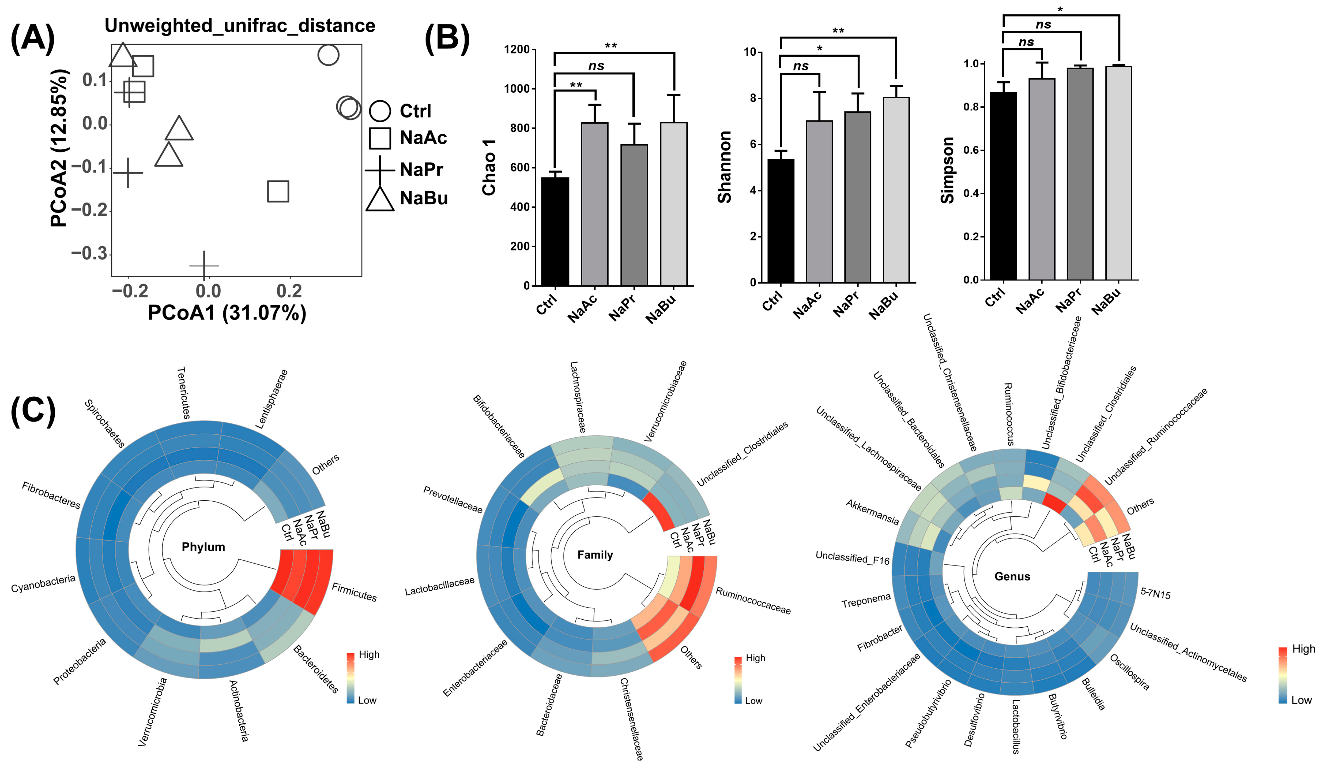
3.3. Distribution of the Microbial Succession Patterns and Microbial Diversities and Abundances in Ileal Contents

Firstly, the distribution of the microbial succession pattern was calculated based on the analysis of PCoA using the unweighted Unifrac distance. Our data demonstrated that the oral infusion of three VFAs significantly altered the bacterial successions compared with the Ctrl group; however, there was no significant separation among the three VFAs’ oral infusion groups (Figure 3A). Then, the differences of the microbial alpha diversity parameters, including the Chao1, Shannon, and Simpson indexes, among the four groups were calculated (Figure 3B). The data indicated that the NaAc and NaBu groups significantly increased the Chao1 index compared with the Ctrl group (p < 0.01), except for the NaPr group (p > 0.05). Then, the Shannon index was significantly increased in the NaPr and NaBu groups compared with the Ctrl group (p < 0.05 or p < 0.01), except for the NaAC group (p > 0.05). Lastly, in terms of the Simpson index, no obvious differences were found in the NaAC and NaPr groups compared with the Ctrl group (p > 0.05), while the Simpson index was significantly increased in the NaBu group compared with the Ctrl group (p < 0.05).
Lastly, the bacterial abundances at the phylum, family, and genus classifications of four groups were shown in Figure 3C. In detail, the most abundant phyla mainly included Firmicutes (63.26%), Bacteroidetes (10.85%), Actinobacteria (7.67%), Verrucomicrobia (7.05%), and Proteobacteria (1.83%). At the family level, the domain ileal bacteria mainly included Ruminococcaceae (26.76%), Unclassified_Clostridiales (14.92%), Lachnospiraceae (9.80%), Christensenellaceae (5.10%), Bifidobacteriaceae (4.84%), Verrucomicrobiaceae (6.71%), and Bacteroidaceae (2.54%). Lastly, ileal bacteria at the genus level were mainly involved with Unclassified_Ruminococcaceae (17.06%), Unclassified_Clostridiales (14.93%), Unclassified_Lachnospiraceae (8.11%), Ruminococcus (6.97%), Akkermansia (6.75%), Unclassified_Bacteroidales (5.25%), Unclassified_Christensenellaceae (5.06%), Unclassified_Bifidobacteriaceae (4.76%), Oscillospira (2.64%), Lactobacillus (0.83%), Fibrobacter (0.72%), and Butyrivibrio (0.66%).

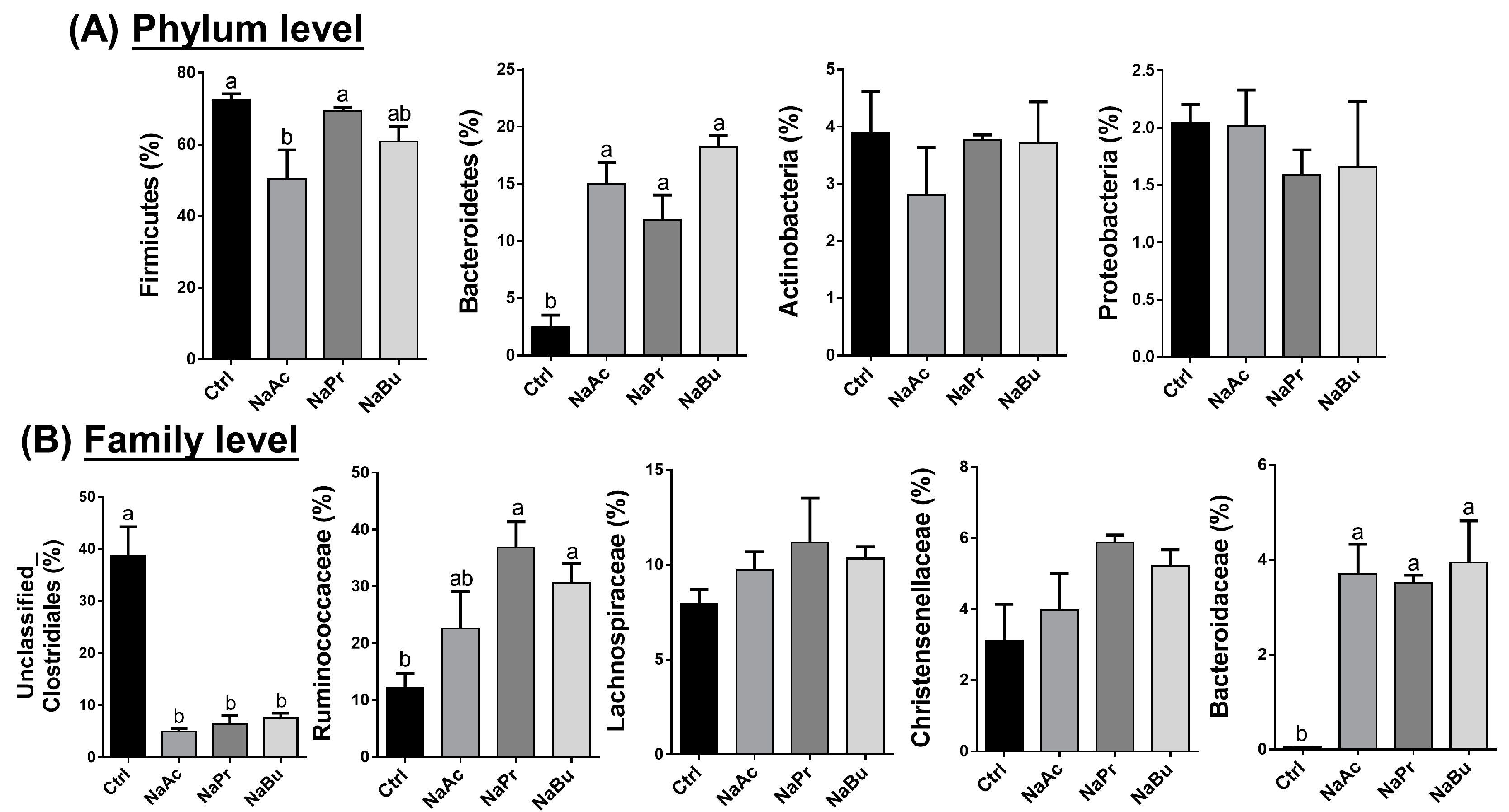
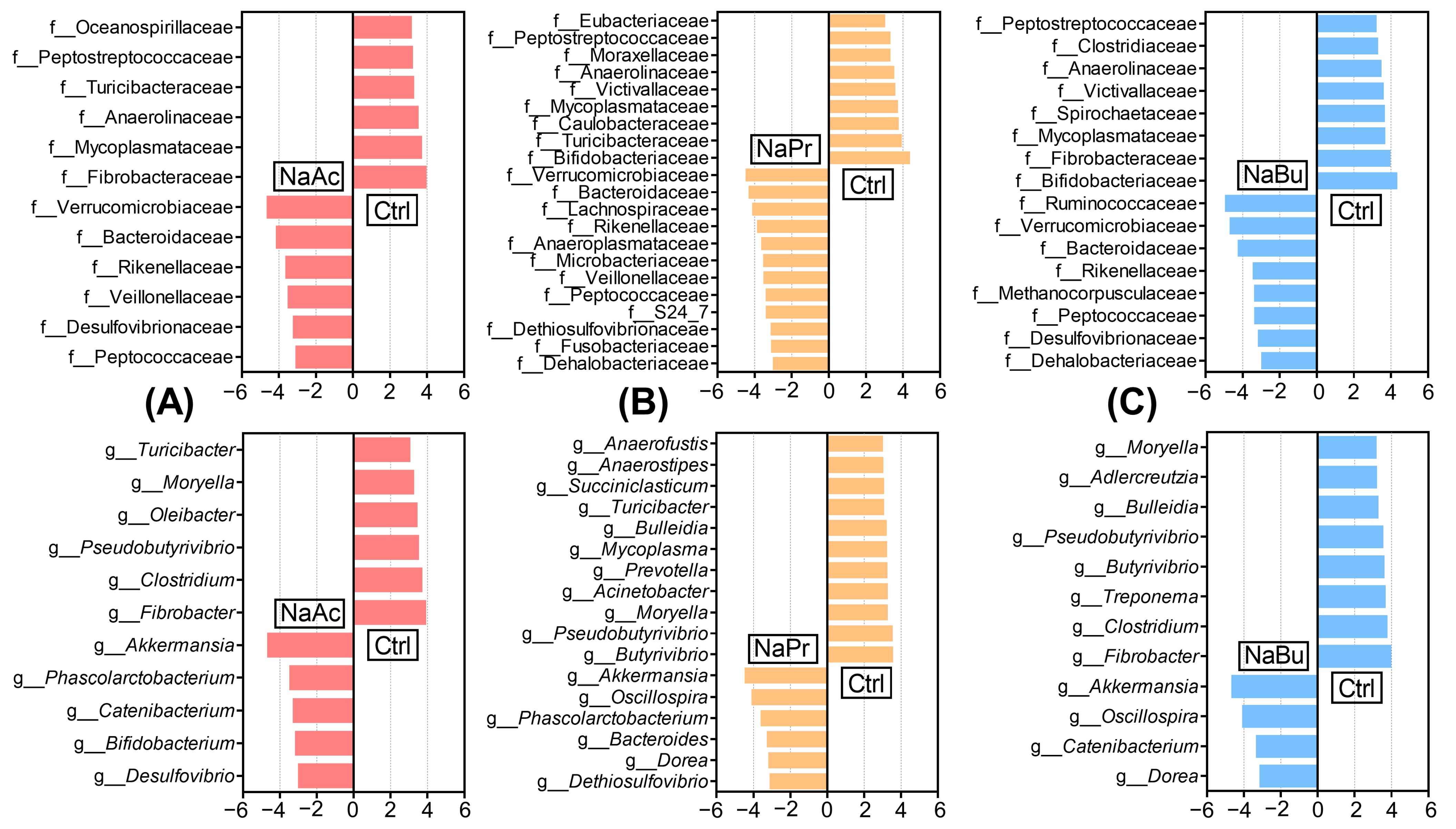
3.4. Selection of Significant Taxonomic Differences under Different Levels in Ileal Contents

The data indicated that different treatments had similar domain ileal bacterial communities, but their abundances were different. For example, under the phylum level, the relative abundance of Firmicutes significantly decreased in the NaAc group compared with the other three groups (p < 0.05) (Figure 4A). Meanwhile, the oral infusion of three VFAs significantly increased the relative abundance of Bacteroidetes compared with the Ctrl group (p < 0.05), but had no impact on the relative abundances of Actinobacteria and Proteobacteria (p > 0.05) (Figure 4A). Then, under the family level, the oral infusion of three VFAs significantly decreased the relative abundance of Unclassified_Clostridiales but increased the relative abundance of Bacteroidaceae compared with the Ctrl group (p < 0.05) (Figure 4B). Additionally, the infusion of NaPr and NaBu increased the relative abundance of Ruminococcaceae compared with the Ctrl group (p < 0.05) (Figure 4B). Lastly, different treatments did not impact the relative abundances of Lachnospiraceae and Christensenellaceae compared with the Ctrl group (p > 0.05) (Figure 4B).
Then, the different microbial taxonomies under the family and genus classifications of the four groups were further screened using the LEfSe method, and only LDA scores of over 3 were marked. Firstly, between the NaAc and Ctrl groups, the abundances of Verrucomicrobiaceae, Bacteroidaceae, Rikenellaceae, Veillonellaceae, Desulfovibrionaceae, and Peptococcaceae were increased, while the abundances of Fibrobacteraceae, Mycoplasmataceae, Anaerolinaceae, Turicibacteraceae, Peptostreptococcaceae, and Oceanospirillaceae were decreased under the family level; the abundances of Akkermansia, Phascolarctobacterium, Catenibacterium, Bifidobacterium, and Desulfovibrio were increased, while the abundances of Fibrobacter, Clostridium, Pseudobutyrivibrio, Oleibacter, Moryella, and Turicibacter were decreased under the genus level (Figure 5A). Additionally, between the NaPr and Ctrl groups, the abundances of ileal bacteria such as Verrucomicrobiaceae, Bacteroidaceae, Lachnospiraceae, Rikenellaceae, etc., were increased, while the abundances of Bifidobacteriaceae, Turicibacteraceae, Caulobacteraceae, Moraxellaceae, and Peptostreptococcaceae were decreased under the family level; meanwhile, under the genus level, the abundances of bacteria such as Akkermansia, Oscillospira, Bacteroides, and Dorea were increased, while the abundances of Butyrivibrio, Pseudobutyrivibrio, Moryella, and Prevotella were decreased (Figure 5B). Lastly, between the NaBu and Ctrl groups, ileal bacteria such as Ruminococcaceae, Verrucomicrobiaceae, Bacteroidaceae, and Rikenellaceae were enriched in the NaBu group, while Bifidobacteriaceae, Fibrobacteraceae, Spirochaetaceae, and Clostridiaceae were enriched in the Ctrl group under the family level. Akkermansia, Oscillospira, Catenibacterium, and Dorea were enriched in the NaBu group, while Fibrobacter, Clostridium, Treponema, and Butyrivibrio were enriched in the Ctrl group under the genus level (Figure 5C).

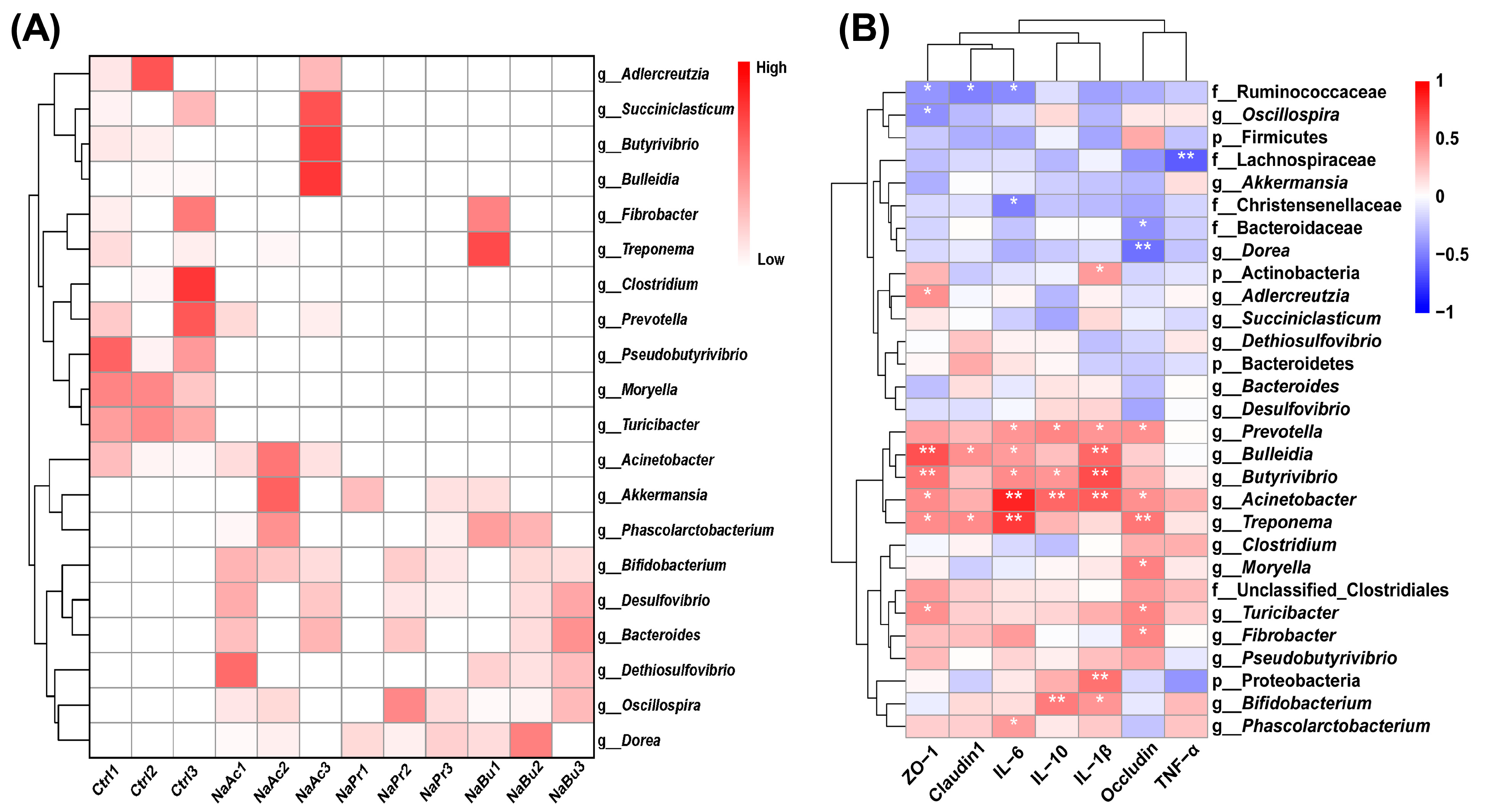
3.5. Core Differential Microbes in Ileal Contents of Four Groups

The core differential microbes in relation to the oral infusion of three VFAs in the ileal contents were selected under the genus classification using the cluster heatmap (Figure 6A). It can be found that the oral infusion of three different VFAs can simultaneously up- or down-regulate the abundances of several ileal bacteria. For example, the up-regulated marker bacteria mainly included Akkermansia, Bifidobacterium, Desulfovibrio, Bacteroides, Oscillospira, and Dorea, and the down-regulated marker bacteria mainly included Prevotella, Turicibacter, Moryella, Clostridium, Treponema, and Butyrivibrio.

3.6. The Correlations between Ileal Bacteria and Gut Health Parameters

Lastly, we conducted a correlation analysis between the ileal bacteria (under phylum, family, and genus levels) and gut health parameters (concentrations of inflammatory cytokines and tight junctions) using Spearman correlation coefficient (Figure 6B). Our data corroborated that these gut bacteria were correlated with the gut health parameters. In detail, under the phylum level, Actinobacteria and Proteobacteria were positively correlated with IL-1β (p < 0.05 or p < 0.01); under the family level, Christensenellaceae was negatively correlated with IL-6 (p < 0.05); and lastly, under the genus level, ileal bacteria such as Prevotella, Bulleidia, Butyrivibio, Acinetobacter, and Bifidobacterium were positively correlated with IL-1β, IL-6, IL-10, Claudin1, Occludin, and ZO-1 (p < 0.05 or p < 0.01).

4. Discussion

Our previous study demonstrated that the oral administration of different VFAs promoted microbial fermentation in the rumens of goats, increasing the concentration of each individual VFA and the total VFA concentration [27]. Not only that, oral administration of VFAs can also promote their transport and absorption in the rumen epithelium by increasing the expression levels of epithelial transporters [27]. These results not only provided a basis for substance studies on the small intestine, but also revealed that the oral infusion of different VFAs could affect the concentrations of VFAs in the ileum. As we expected, the data indicated that the oral infusion of three VFAs can enhance VFA production in the ileum by increasing the proportions of each individual VFA and the total VFAs. The possible reason for this was that the oral infusion of different VFAs promoted rumen microbial fermentation to produce large numbers of VFAs in the rumen, and parts of them cannot be absorbed by the rumen epithelium and then entered the small intestine, which may have further impacts on ileal microbiome profiling and epithelial health.
Next, our results revealed distinct microbial succession patterns in the ileal contents following oral VFA infusion groups compared to the Ctrl group based on the PCoA analysis. For example, the infusion of NaAc was associated with an increase in the relative abundances of Akkermansia and Bifidobacterium, which are known for their beneficial roles in mucin degradation and in promoting gut barrier function and homeostatic immunity [41,42,43]. On the other hand, the oral infusions of NaPr and NaBu were linked to increased abundances of Verrucomicrobiaceae, Bacteroidaceae, and Ruminococcaceae families, which are known for their contributions to the production of VFAs and immunomodulation [12,44,45]. However, there was no significant separation observed among the oral VFA infusion groups, indicating that the overall microbial community structure in the ileum remained relatively stable across the oral infusion of different VFAs. This stability in the core bacterial community may be due to the fact that certain bacterial abundances were highly adapted to the gut environment and were less susceptible to short-term dietary changes. Nevertheless, these subtle shifts in specific bacterial groups following the oral infusion of VFAs could have important functional implications for the nutrient metabolism, immune modulation, and overall gut health in goats.
The assessment of microbial alpha diversity parameters, including Chao1, Shannon, and Simpson indexes, also provided further insights into the effects of VFA infusion on the ileal microbiome. Our data indicated that the NaAc and NaBu groups significantly increased the microbial richness (Chao1 index) compared to the Ctrl group. This increase in richness may be attributed to the ability of certain bacterial species, such as Bifidobacterium in the NaAc group, to utilize VFAs as energy sources, promoting their growth and proliferation [46]. Moreover, the Shannon index was significantly increased in the NaPr and NaBu groups compared to the Ctrl group, which indicated that the oral infusions of NaPr and NaBu might promoted a more even distribution of microbial diversity in the ileum, potentially contributing to a more stable and healthier microbial ecosystem [6,47]. Lastly, the NaBu group exhibited a significantly increased Simpson index compared with the Ctrl group, suggesting that the oral infusion of NaBu might lead to changes in the dominance of certain microbial taxonomy in the ileum. For example, the increased dominance of Ruminococcaceae, which is known for its role in fermenting complex carbohydrates and producing VFAs, may be linked to the beneficial effects of butyrate on gut health in goats and piglets [48,49].
These taxonomic differences suggested that each VFA might exert unique effects on the ileal microbial community, leading to specific functional changes that could impact gut health and host metabolism differently. The enriched abundances of beneficial bacteria such as Akkermansia, Bifidobacterium, and Verrucomicrobiaceae following VFA infusion indicate potential positive effects on the gut barrier integrity, immune modulation, and metabolic homeostasis [12,41,42,43,44,45]. Conversely, the decrease in certain bacteria such as Prevotella in the NaPr group might be associated with reduced inflammatory responses and an improved gut health [50].
Furthermore, the concentrations of inflammatory cytokines and tight junctions in the ileal epithelial tissue were measured to assess the impact of oral VFA infusion on gut health parameters. Our results showed that the NaPr group significantly reduced the concentration of IL-6 compared to the Ctrl group, suggesting a potential anti-inflammatory effect of propionate in the ileum in ruminants [51]. On the other hand, the NaAc group was associated with an increased IL-10 concentration, indicating a potential role in modulating the anti-inflammatory response in goats [52]. Our findings were aligned with previous studies that demonstrated the anti-inflammatory properties of specific VFAs in various gut compartments [53,54,55]. However, no significant changes were observed in the concentrations of IL-1β and TNF-α following VFA infusion. Moreover, tight junctions, such as Claudin1, Occludin, and ZO-1, were measured to evaluate the impact of VFA infusion on the gut barrier integrity. Interestingly, no significant differences were observed in the concentration of tight junctions between the VFA infusion groups and the Ctrl group, suggesting that the oral infusion of VFAs might not have a direct effect on the integrity of the gut barrier in the ileum.
Lastly, we conducted correlation analysis between the core differential microbes and the gut health parameters to identify potential relationships between specific bacterial taxonomy and gut health. Our data revealed significant correlations between certain ileal bacteria and inflammatory cytokines (IL-1β, IL-6, and IL-10) as well as tight junction proteins (Claudin1, Occludin, and ZO-1). For example, Prevotella, which is a common member of the gut microbiome, was positively correlated with IL-6 and IL-1β, suggesting a potential role in promoting gut inflammation [50]. In contrast, the abundance of Bifidobacterium was positively correlated with IL-10, indicating a potential anti-inflammatory effect. These correlations can support the notion that specific bacterial taxa play roles in modulating gut health and immune responses [56].

5. Conclusions

In conclusion, our study revealed the effects of the exogenous oral infusion of VFAs on the ileal microbiome and the small intestine health in goats. The oral infusion of VFAs increased the proportions of each individual VFA and the total VFAs. Each VFA had a unique role in the ileal bacterial community, the NaAc and NaBu groups appeared to increase the Chao1 index, and the NaPr and NaBu groups increased the Shannon index. The taxonomic differences indicated potential beneficial effects of VFAs on gut health, with certain VFAs associated with enriched abundances of beneficial bacteria, like Akkermansia, Bifidobacterium, and Verrucomicrobiaceae. Furthermore, VFA infusion demonstrated potential anti-inflammatory effects, as evidenced by reduced IL-6 levels with the NaPr group and increased IL-10 levels with the NaAc group. Although VFA infusion did not impact the tight junction concentration, the correlation analysis revealed specific relationships between certain ileal bacteria and gut health parameters.

Author Contributions

Conceptualization, Y.Z., A.R. and M.W.; data curation, A.R. and F.H.; funding acquisition, M.W.; investigation, Y.Z., C.Z. and M.W.; methodology, Y.Z., C.Z. and J.L.; project administration, M.W.; supervision, M.W.; writing—original draft, Y.Z. and C.Z.; writing—review and editing, J.L. and F.H. All authors have read and agreed to the published version of the manuscript.

Funding

This study was supported by projects from the Natural Science Foundation of China (31672446); the State Key Laboratory of Sheep Genetic Improvement and Healthy Production (NCG202232; 2021ZD07); the National Key Laboratory of Animal Nutrition (2004DA125184F1715); and the Priority Academic Program Development of Jiangsu Higher Education Institutions, China (PAPD).

Institutional Review Board Statement

Not applicable.

Informed Consent Statement

Not applicable.

Data Availability Statement

The raw 16S rRNA sequencing data were deposited in the NCBI Gene Expression Omnibus (GEO) database under the accession code GSE240818. The original contributions presented in this study were included in the article; further inquiries can be directed to the corresponding authors.

Conflicts of Interest

The authors declare no conflict of interest.

References

  1. Lin, L.; Lai, Z.; Zhang, J.; Zhu, W.; Mao, S. The gastrointestinal microbiome in dairy cattle is constrained by the deterministic driver of the region and the modified effect of diet. Microbiome 2023, 11, 10. [Google Scholar] [CrossRef] [PubMed]
  2. Yuan, Y.; Sun, D.M.; Qin, T.; Mao, S.Y.; Zhu, W.Y.; Yin, Y.Y.; Huang, J.; Heller, R.; Li, Z.P.; Liu, J.H.; et al. Single-cell transcriptomic landscape of the sheep rumen provides insights into physiological programming development and adaptation of digestive strategies. Zool. Res. 2022, 43, 634–647. [Google Scholar] [CrossRef]
  3. Moraïs, S.; Mizrahi, I. The Road Not Taken: The Rumen Microbiome, Functional Groups, and Community States. Trends Microbiol. 2019, 27, 538–549. [Google Scholar] [CrossRef]
  4. Na, S.W.; Guan, L.L. Understanding the role of rumen epithelial host-microbe interactions in cattle feed efficiency. Anim. Nutr. (Zhongguo Xu Mu Shou Yi Xue Hui) 2022, 10, 41–53. [Google Scholar] [CrossRef] [PubMed]
  5. Górka, P.; Kowalski, Z.M.; Zabielski, R.; Guilloteau, P. Invited review: Use of butyrate to promote gastrointestinal tract development in calves. J. Dairy Sci. 2018, 101, 4785–4800. [Google Scholar] [CrossRef]
  6. Ma, L.; Yang, Y.; Liu, W.; Bu, D. Sodium butyrate supplementation impacts the gastrointestinal bacteria of dairy calves before weaning. Appl. Microbiol. Biotechnol. 2023, 107, 3291–3304. [Google Scholar] [CrossRef] [PubMed]
  7. Stahl, T.C.; Hatungimana, E.; Klanderman, K.D.; Moreland, S.C.; Erickson, P.S. Sodium butyrate and monensin supplementation to postweaning heifer diets: Effects on growth performance, nutrient digestibility, and health. J. Dairy Sci. 2020, 103, 10207–10218. [Google Scholar] [CrossRef]
  8. Liu, L.; Sun, D.; Mao, S.; Zhu, W.; Liu, J. Infusion of sodium butyrate promotes rumen papillae growth and enhances expression of genes related to rumen epithelial VFA uptake and metabolism in neonatal twin lambs. J. Anim. Sci. 2019, 97, 909–921. [Google Scholar] [CrossRef]
  9. Wu, Y.; Sun, Y.; Zhang, R.; He, T.; Huang, G.; Tian, K.; Liu, J.; Chen, J.; Dong, G. Sodium Butyrate More Effectively Mitigates the Negative Effects of High-Concentrate Diet in Dairy Cows than Sodium β-Hydroxybutyrate via Reducing Free Bacterial Cell Wall Components in Rumen Fluid and Plasma. Toxins 2021, 13, 352. [Google Scholar] [CrossRef]
  10. Aschenbach, J.R.; Zebeli, Q.; Patra, A.K.; Greco, G.; Amasheh, S.; Penner, G.B. Symposium review: The importance of the ruminal epithelial barrier for a healthy and productive cow. J. Dairy Sci. 2019, 102, 1866–1882. [Google Scholar] [CrossRef]
  11. Abuelfatah, K.; Zuki, A.B.; Goh, Y.M.; Sazili, A.Q.; Abubakr, A. Effects of feeding whole linseed on ruminal fatty acid composition and microbial population in goats. Anim. Nutr. (Zhongguo Xu Mu Shou Yi Xue Hui) 2016, 2, 323–328. [Google Scholar] [CrossRef] [PubMed]
  12. Guo, H.; Li, B.; Gao, M.; Li, Q.; Gao, Y.; Dong, N.; Liu, G.; Wang, Z.; Gao, W.; Chen, Y.; et al. Dietary Nutritional Level Affects Intestinal Microbiota and Health of Goats. Microorganisms 2022, 10, 2322. [Google Scholar] [CrossRef] [PubMed]
  13. Hackmann, T.J.; Ngugi, D.K.; Firkins, J.L.; Tao, J. Genomes of rumen bacteria encode atypical pathways for fermenting hexoses to short-chain fatty acids. Environ. Microbiol. 2017, 19, 4670–4683. [Google Scholar] [CrossRef] [PubMed]
  14. Liu, Y.R.; Du, H.S.; Wu, Z.Z.; Wang, C.; Liu, Q.; Guo, G.; Huo, W.J.; Zhang, Y.L.; Pei, C.X.; Zhang, S.L. Branched-chain volatile fatty acids and folic acid accelerated the growth of Holstein dairy calves by stimulating nutrient digestion and rumen metabolism. Animal 2020, 14, 1176–1183. [Google Scholar] [CrossRef]
  15. Brisson, V.; Girard, C.L.; Metcalf, J.A.; Castagnino, D.S.; Dijkstra, J.; Ellis, J.L. Meta-analysis of apparent ruminal synthesis and postruminal flow of B vitamins in dairy cows. J. Dairy Sci. 2022, 105, 7399–7415. [Google Scholar] [CrossRef]
  16. Zhang, Q.; Koser, S.L.; Bequette, B.J.; Donkin, S.S. Effect of propionate on mRNA expression of key genes for gluconeogenesis in liver of dairy cattle. J. Dairy Sci. 2015, 98, 8698–8709. [Google Scholar] [CrossRef] [PubMed]
  17. Young, J.W. Gluconeogenesis in cattle: Significance and methodology. J. Dairy Sci. 1977, 60, 1–15. [Google Scholar] [CrossRef] [PubMed]
  18. Zhang, F.; Wang, Y.; Wang, H.; Nan, X.; Guo, Y.; Xiong, B. Calcium Propionate Supplementation Has Minor Effects on Major Ruminal Bacterial Community Composition of Early Lactation Dairy Cows. Front. Microbiol. 2022, 13, 847488. [Google Scholar] [CrossRef]
  19. Liu, Q.; Wang, C.; Yang, W.Z.; Guo, G.; Yang, X.M.; He, D.C.; Dong, K.H.; Huang, Y.X. Effects of calcium propionate supplementation on lactation performance, energy balance and blood metabolites in early lactation dairy cows. J. Anim. Physiol. Anim. Nutr. 2010, 94, 605–614. [Google Scholar] [CrossRef]
  20. Górka, P.; Sliwinski, B.; Flaga, J.; Olszewski, J.; Nawrocka, P.; Sobkowiak, K.; Miltko, R.; Godlewski, M.M.; Zabielski, R.; Kowalski, Z.M. Effect of exogenous butyrate on the gastrointestinal tract of sheep. II. Hydrolytic activity in the rumen and structure and function of the small intestine. J. Anim. Sci. 2018, 96, 5325–5335. [Google Scholar] [CrossRef]
  21. Zhan, K.; Yang, T.Y.; Chen, Y.; Jiang, M.C.; Zhao, G.Q. Propionate enhances the expression of key genes involved in the gluconeogenic pathway in bovine intestinal epithelial cells. J. Dairy Sci. 2020, 103, 5514–5524. [Google Scholar] [CrossRef] [PubMed]
  22. Harmon, D.L.; Swanson, K.C. Review: Nutritional regulation of intestinal starch and protein assimilation in ruminants. Animal 2020, 14, s17–s28. [Google Scholar] [CrossRef] [PubMed]
  23. Britton, R.; Krehbiel, C. Nutrient metabolism by gut tissues. J. Dairy Sci. 1993, 76, 2125–2131. [Google Scholar] [CrossRef] [PubMed]
  24. El-Kadi, S.W.; Baldwin, R.L.t.; McLeod, K.R.; Sunny, N.E.; Bequette, B.J. Glutamate is the major anaplerotic substrate in the tricarboxylic acid cycle of isolated rumen epithelial and duodenal mucosal cells from beef cattle. J. Nutr. 2009, 139, 869–875. [Google Scholar] [CrossRef]
  25. Koch, C.; Gerbert, C.; Frieten, D.; Dusel, G.; Eder, K.; Zitnan, R.; Hammon, H.M. Effects of ad libitum milk replacer feeding and butyrate supplementation on the epithelial growth and development of the gastrointestinal tract in Holstein calves. J. Dairy Sci. 2019, 102, 8513–8526. [Google Scholar] [CrossRef]
  26. Zhan, K.; Jiang, M.; Gong, X.; Zhao, G. Effect of short-chain fatty acids on the expression of genes involved in short-chain fatty acid transporters and inflammatory response in goat jejunum epithelial cells. In Vitro Cell Dev. Biol. Anim. 2018, 54, 311–320. [Google Scholar] [CrossRef]
  27. Zhen, Y.; Xi, Z.; Nasr, S.M.; He, F.; Han, M.; Yin, J.; Ge, L.; Chen, Y.; Wang, Y.; Wei, W.; et al. Multi-Omics Reveals the Impact of Exogenous Short-Chain Fatty Acid Infusion on Rumen Homeostasis: Insights into Crosstalk between the Microbiome and the Epithelium in a Goat Model. Microbiol. Spectr. 2023, 11, e0534322. [Google Scholar] [CrossRef]
  28. Gualdrón-Duarte, L.B.; Allen, M.S. Effects of acetic acid or sodium acetate infused into the rumen or abomasum on feeding behavior and metabolic response of cows in the postpartum period. J. Dairy Sci. 2018, 101, 2016–2026. [Google Scholar] [CrossRef]
  29. Bedford, A.; Beckett, L.; Hardin, K.; Dias, N.W.; Davis, T.; Mercadante, V.R.G.; Ealy, A.D.; White, R.R. Propionate Affects Insulin Signaling and Progesterone Profiles in Dairy Heifers. Sci. Rep. 2018, 8, 17629. [Google Scholar] [CrossRef]
  30. Jiao, J.; Zhang, X.; Wang, M.; Zhou, C.; Yan, Q.; Tan, Z. Linkages between Epithelial Microbiota and Host Transcriptome in the Ileum during High-Grain Challenges: Implications for Gut Homeostasis in Goats. J. Agric. Food Chem. 2019, 67, 551–561. [Google Scholar] [CrossRef]
  31. Zhang, H.; Zheng, Y.; Zha, X.; Ma, Y.; Liu, X.; Elsabagh, M.; Wang, H.; Wang, M. Dietary L-Arginine or N-Carbamylglutamate Alleviates Colonic Barrier Injury, Oxidative Stress, and Inflammation by Modulation of Intestinal Microbiota in Intrauterine Growth-Retarded Suckling Lambs. Antioxidants 2022, 11, 2251. [Google Scholar] [CrossRef] [PubMed]
  32. Xue, Y.; Hu, F.; Guo, C.; Mei, S.; Xie, F.; Zeng, H.; Mao, S. Undernutrition shifted colonic fermentation and digest-associated bacterial communities in pregnant ewes. Appl. Microbiol. Biotechnol. 2020, 104, 5973–5984. [Google Scholar] [CrossRef] [PubMed]
  33. Yan, B.; Wang, D.; Dong, S.; Cheng, Z.; Na, L.; Sang, M.; Yang, H.; Yang, Z.; Zhang, S.; Yan, Z. Palmatine inhibits TRIF-dependent NF-κB pathway against inflammation induced by LPS in goat endometrial epithelial cells. Int. Immunopharmacol. 2017, 45, 194–200. [Google Scholar] [CrossRef]
  34. Hugerth, L.W.; Wefer, H.A.; Lundin, S.; Jakobsson, H.E.; Lindberg, M.; Rodin, S.; Engstrand, L.; Andersson, A.F. DegePrime, a program for degenerate primer design for broad-taxonomic-range PCR in microbial ecology studies. Appl. Environ. Microbiol. 2014, 80, 5116–5123. [Google Scholar] [CrossRef]
  35. Magoč, T.; Salzberg, S.L. FLASH: Fast length adjustment of short reads to improve genome assemblies. Bioinformatics 2011, 27, 2957–2963. [Google Scholar] [CrossRef]
  36. Rognes, T.; Flouri, T.; Nichols, B.; Quince, C.; Mahé, F. VSEARCH: A versatile open source tool for metagenomics. PeerJ 2016, 4, e2584. [Google Scholar] [CrossRef] [PubMed]
  37. Callahan, B.J.; McMurdie, P.J.; Rosen, M.J.; Han, A.W.; Johnson, A.J.; Holmes, S.P. DADA2: High-resolution sample inference from Illumina amplicon data. Nat. Methods 2016, 13, 581–583. [Google Scholar] [CrossRef]
  38. Bolyen, E.; Rideout, J.R.; Dillon, M.R.; Bokulich, N.A.; Abnet, C.C.; Al-Ghalith, G.A.; Alexander, H.; Alm, E.J.; Arumugam, M.; Asnicar, F.; et al. Reproducible, interactive, scalable and extensible microbiome data science using QIIME 2. Nat. Biotechnol. 2019, 37, 852–857. [Google Scholar] [CrossRef]
  39. Quast, C.; Pruesse, E.; Yilmaz, P.; Gerken, J.; Schweer, T.; Yarza, P.; Peplies, J.; Glöckner, F.O. The SILVA ribosomal RNA gene database project: Improved data processing and web-based tools. Nucleic Acids Res. 2013, 41, D590–D596. [Google Scholar] [CrossRef]
  40. Segata, N.; Izard, J.; Waldron, L.; Gevers, D.; Miropolsky, L.; Garrett, W.S.; Huttenhower, C. Metagenomic biomarker discovery and explanation. Genome Biol. 2011, 12, R60. [Google Scholar] [CrossRef]
  41. Cani, P.D.; Depommier, C.; Derrien, M.; Everard, A.; de Vos, W.M. Akkermansia muciniphila: Paradigm for next-generation beneficial microorganisms. Nat. Rev. Gastroenterol. Hepatol. 2022, 19, 625–637. [Google Scholar] [CrossRef] [PubMed]
  42. Bae, M.; Cassilly, C.D.; Liu, X.; Park, S.M.; Tusi, B.K.; Chen, X.; Kwon, J.; Filipčík, P.; Bolze, A.S.; Liu, Z.; et al. Akkermansia muciniphila phospholipid induces homeostatic immune responses. Nature 2022, 608, 168–173. [Google Scholar] [CrossRef]
  43. Krumbeck, J.A.; Rasmussen, H.E.; Hutkins, R.W.; Clarke, J.; Shawron, K.; Keshavarzian, A.; Walter, J. Probiotic Bifidobacterium strains and galactooligosaccharides improve intestinal barrier function in obese adults but show no synergism when used together as synbiotics. Microbiome 2018, 6, 121. [Google Scholar] [CrossRef] [PubMed]
  44. Borton, M.A.; Sabag-Daigle, A.; Wu, J.; Solden, L.M.; O’Banion, B.S.; Daly, R.A.; Wolfe, R.A.; Gonzalez, J.F.; Wysocki, V.H.; Ahmer, B.M.M.; et al. Chemical and pathogen-induced inflammation disrupt the murine intestinal microbiome. Microbiome 2017, 5, 47. [Google Scholar] [CrossRef]
  45. Zhang, R.; Ye, H.; Liu, J.; Mao, S. High-grain diets altered rumen fermentation and epithelial bacterial community and resulted in rumen epithelial injuries of goats. Appl. Microbiol. Biotechnol. 2017, 101, 6981–6992. [Google Scholar] [CrossRef]
  46. Wang, Y.; Nan, X.; Zhao, Y.; Jiang, L.; Wang, H.; Zhang, F.; Hua, D.; Liu, J.; Yao, J.; Yang, L.; et al. Dietary Supplementation of Inulin Ameliorates Subclinical Mastitis via Regulation of Rumen Microbial Community and Metabolites in Dairy Cows. Microbiol. Spectr. 2021, 9, e0010521. [Google Scholar] [CrossRef] [PubMed]
  47. Smith, S.B.; Blackmon, T.L.; Sawyer, J.E.; Miller, R.K.; Baber, J.R.; Morrill, J.C.; Cabral, A.R.; Wickersham, T.A. Glucose and acetate metabolism in bovine intramuscular and subcutaneous adipose tissues from steers infused with glucose, propionate, or acetate. J. Anim. Sci. 2018, 96, 921–929. [Google Scholar] [CrossRef] [PubMed]
  48. Han, X.; Lei, X.; Yang, X.; Shen, J.; Zheng, L.; Jin, C.; Cao, Y.; Yao, J. A Metagenomic Insight Into the Hindgut Microbiota and Their Metabolites for Dairy Goats Fed Different Rumen Degradable Starch. Front. Microbiol. 2021, 12, 651631. [Google Scholar] [CrossRef] [PubMed]
  49. Zhong, X.; Zhang, Z.; Wang, S.; Cao, L.; Zhou, L.; Sun, A.; Zhong, Z.; Nabben, M. Microbial-Driven Butyrate Regulates Jejunal Homeostasis in Piglets During the Weaning Stage. Front. Microbiol. 2018, 9, 3335. [Google Scholar] [CrossRef]
  50. Larsen, J.M. The immune response to Prevotella bacteria in chronic inflammatory disease. Immunology 2017, 151, 363–374. [Google Scholar] [CrossRef]
  51. Zhang, H.; Zhao, F.; Peng, A.; Dong, L.; Wang, M.; Yu, L.; Loor, J.J.; Wang, H. Effects of Dietary l-Arginine and N-Carbamylglutamate Supplementation on Intestinal Integrity, Immune Function, and Oxidative Status in Intrauterine-Growth-Retarded Suckling Lambs. J. Agric. Food Chem. 2018, 66, 4145–4154. [Google Scholar] [CrossRef]
  52. Yuan, C.; Wang, S.; Gebeyew, K.; Yang, X.; Tang, S.; Zhou, C.; Khan, N.A.; Tan, Z.; Liu, Y. A low-carbon high inulin diet improves intestinal mucosal barrier function and immunity against infectious diseases in goats. Front. Vet. Sci. 2022, 9, 1098651. [Google Scholar] [CrossRef]
  53. He, J.; Zhang, P.; Shen, L.; Niu, L.; Tan, Y.; Chen, L.; Zhao, Y.; Bai, L.; Hao, X.; Li, X.; et al. Short-Chain Fatty Acids and Their Association with Signalling Pathways in Inflammation, Glucose and Lipid Metabolism. Int. J. Mol. Sci. 2020, 21, 6356. [Google Scholar] [CrossRef] [PubMed]
  54. Parada Venegas, D.; De la Fuente, M.K.; Landskron, G.; González, M.J.; Quera, R.; Dijkstra, G.; Harmsen, H.J.M.; Faber, K.N.; Hermoso, M.A. Short Chain Fatty Acids (SCFAs)-Mediated Gut Epithelial and Immune Regulation and Its Relevance for Inflammatory Bowel Diseases. Front. Immunol. 2019, 10, 277. [Google Scholar] [CrossRef] [PubMed]
  55. Yang, W.; Yu, T.; Huang, X.; Bilotta, A.J.; Xu, L.; Lu, Y.; Sun, J.; Pan, F.; Zhou, J.; Zhang, W.; et al. Intestinal microbiota-derived short-chain fatty acids regulation of immune cell IL-22 production and gut immunity. Nat. Commun. 2020, 11, 4457. [Google Scholar] [CrossRef] [PubMed]
  56. Ma, X.; Shin, Y.J.; Jang, H.M.; Joo, M.K.; Yoo, J.W.; Kim, D.H. Lactobacillus rhamnosus and Bifidobacterium longum alleviate colitis and cognitive impairment in mice by regulating IFN-γ to IL-10 and TNF-α to IL-10 expression ratios. Sci. Rep. 2021, 11, 20659. [Google Scholar] [CrossRef]
Figure 1. Concentrations of VFAs in ileal contents. Ctrl, oral infusion of saline group; NaAc, oral infusion of sodium acetate group; NaPr, oral infusion of sodium propionate group; and NaBu, oral infusion of sodium butyrate group. Representative charts of the concentration of total VFAs (A), the ratio of acetate to propionate (B), and the concentrations of acetate, propionate, and butyrate (C) were measured in ileal contents of four groups. Mean values with different letters were significantly different (p < 0.05) according to Duncan’s multiple range test; data were shown as mean ± SD.
Fermentation 09 00801 g001
Figure 2. The concentration of inflammatory cytokines and tight junctions in ileal epithelial tissue. Ctrl, oral infusion of saline group; NaAc, oral infusion of sodium acetate group; NaPr, oral infusion of sodium propionate group; and NaBu, oral infusion of sodium butyrate group. (A) The concentration of inflammatory cytokines, such as IL-1β, IL-6, IL-10, and TNF-α among four groups of ileal epithelial tissue. (B) The protein concentration of tight junctions, such as Claudin1, Occludin, and ZO-1 among four groups of ileal epithelial tissue. Mean values with different letters were significantly different (p < 0.05) according to Duncan’s multiple range test; data were shown as mean ± SD.
Fermentation 09 00801 g002
Figure 3. Distribution of the microbial succession patterns and microbial diversities and abundances in ileal contents. Ctrl, oral infusion of saline group; NaAc, oral infusion of sodium acetate group; NaPr, oral infusion of sodium propionate group; and NaBu, oral infusion of sodium butyrate group. (A) Distribution of the microbial succession patterns. Data were calculated based on the analysis of PCoA using unweighted Unifrac distance. (B) Microbial alpha diversity parameters of four groups including Chao1, Shannon, and Simpson indexes; statistical analyses were conducted using Kruskal–Wallis test (ns represents p > 0.05, * represents p < 0.05, and ** represents p < 0.01). (C) Representative heatmaps of the bacterial abundances of four groups in ileal contents under phylum, family, and genus classifications. Red color represents a high abundance, while blue color represents a low abundance.
Fermentation 09 00801 g003
Figure 4. Changes in taxonomic compositions in ileal contents under phylum and family levels. Ctrl, oral infusion of saline group; NaAc, oral infusion of sodium acetate group; NaPr, oral infusion of sodium propionate group; and NaBu, oral infusion of sodium butyrate group. Representative charts of the ileal bacteria under phylum (A) and family (B) levels were compared according to Duncan’s multiple range test. Mean values with different letters were significantly different (p < 0.05); data were shown as mean ± SD.
Fermentation 09 00801 g004
Figure 5. Selection of significant taxonomic differences in ileal contents. Ctrl, oral infusion of saline group; NaAc, oral infusion of sodium acetate group; NaPr, oral infusion of sodium propionate group; and NaBu, oral infusion of sodium butyrate group. The different microbial taxonomies between NaAc (A), NaPr (B), NaBu (C), and Ctrl groups under family and genus classifications were screened using LEfSe method, and only LDA scores over 3 were marked.
Fermentation 09 00801 g005
Figure 6. Core differential bacteria in relation to oral infusion of different VFAs and the correlation analysis between ileal bacteria and gut health parameters. Ctrl, oral infusion of saline group; NaAc, oral infusion of sodium acetate group; NaPr, oral infusion of sodium propionate group; and NaBu, oral infusion of sodium butyrate group. (A) Core differential bacteria in ileal contents under genus level in relation to oral infusion of different VFAs; red color represented a high abundance, while white color represented a low abundance. (B) The correlation analysis between differential bacterial under different levels and ileal health parameters. Data were calculated using Spearman correlation coefficient, while * p < 0.05 and ** p < 0.01.
Fermentation 09 00801 g006
Table 1. Dietary composition and nutrient levels of diets (dry matter basis).
IngredientsPercent, % of DMNutrientsLevel, g/kg of DM
Oat hay41.00DE (MJ/kg)10.25
Corn29.50CP155.60
Soybean meal14.50NDF352.60
Wheat bran10.00ADF196.25
Stone dust0.35EE28.94
CaH2PO40.15Ca4.08
NaCl0.50P4.65
Premix4.00
Total100.00
Note: DM, dry matter; DE, digestible energy; CP, crude protein; NDF, neutral detergent fiber; ADF, acid detergent fiber; EE, ether extract. The following premix was provided per kg of diet: VA 200,000 IU, VD3 70,000 IU, VE 350 IU, Fe 1.6 g, Cu 1.7 g, Zn 8.2 g, Mn 2.5 g, and Se 40 mg.
Disclaimer/Publisher’s Note: The statements, opinions and data contained in all publications are solely those of the individual author(s) and contributor(s) and not of MDPI and/or the editor(s). MDPI and/or the editor(s) disclaim responsibility for any injury to people or property resulting from any ideas, methods, instructions or products referred to in the content.

