Communication

Isolation of γ-Aminobutyric Acid (GABA)-Producing Lactic Acid Bacteria with Anti-Inflammatory Effects from Fermented Foods in Korea

MEDIOGEN, Co., Ltd., Bio Valley 1-Ro, Jecheon-si 27159, Republic of Korea
*
Author to whom correspondence should be addressed.
Fermentation 2023, 9(7), 612; https://doi.org/10.3390/fermentation9070612
Submission received: 15 June 2023 / Revised: 26 June 2023 / Accepted: 27 June 2023 / Published: 28 June 2023
(This article belongs to the Special Issue Functional Properties of Microorganisms in Fermented Foods)

Abstract

:
Lactic acid bacteria have become popular because of their γ-aminobutyric acid (GABA)-producing ability. In the present study, we selected four Levilactobacillus brevis strains (MG5552, MG5405, MG5261, and MG5522) with GABA-producing ability from the 33 strains isolated from various fermented foods in South Korea. We evaluated their GABA-producing ability using thin-layer chromatography and determined the GABA levels produced by each strain using an amino acid analyzer. Moreover, we investigated the anti-inflammatory activity of the selected strains, and the results revealed that the cell-free supernatant of the strains decreased nitric oxide (NO), inducible nitric oxide synthase (iNOS) expression, and nuclear factor kappa B (NF-κB) activity in RAW264.7 macrophages. Therefore, these GABA-producing LAB strains can regulate nerve excitement and act as probiotics with anti-inflammatory activity.

1. Introduction

γ-Aminobutyric acid (GABA) is a non-protein amino acid present in several prokaryotic and eukaryotic cells [1]. GABA regulates neuronal activity by inhibiting nerve transmission and is associated with several neurological disorders, including epilepsy, depression, anxiety, Alzheimer’s disease, Parkinson’s disease, schizophrenia, and Huntington’s chorea [2]. Recent studies have reported that GABA improves plasma growth hormone level, brain protein synthesis, memory, and cognitive ability; lowers blood pressure; and relaxes the nerves [3,4]. In addition, GABA produced by microbial fermentation is eco-friendly and safe, so GABA has the potential to provide new products beneficial to health [5].
GABA is mainly produced by microorganisms, such as yeast, fungi, and bacteria, via the fermentation of food products [6,7]. The members of the genus Lactobacillus are safe bacteria that produce higher levels of GABA than other microorganisms and are widely used as a probiotic because of their unique characteristics, such as resistance against bile and gastric acid, and intestinal homeostasis [8]. Lactobacillus spp. produce several organic acids, including lactic acid; antibacterial molecules, such as bacteriocins, antioxidants, and immunoreactive substances, as well as neurotransmitters, such as GABA, during fermentation [9]. Lactic acid bacteria (LAB) can also inhibit some pathogenic bacteria; thus, they can be used for improving the shelf life of fermented foods [10].
Recently, probiotics have been used in various food products because they can inhibit the growth of harmful bacteria, reduce blood cholesterol levels, strengthen immunity, and exert anticancer effects [11]. A previous study reported that GABA increased the production of the anti-inflammatory mediator TGF-β1 and decreased the production of inflammatory mediators, such as interleukin-1 beta (IL-1β), tumor necrosis factor-alpha (TNF-α), interferon-gamma (IFN-γ), and interleukin 12 (IL-12), in streptozotocin-treated mice, thus exhibiting anti-inflammatory effects [12]. GABA-producing LAB exhibits the potential for being developed into functional foods; therefore, studies on GABA-producing LAB are being conducted.
In the present study, we isolated LAB from various fermented foods in Korea and evaluated the anti-inflammatory effects of GABA-producing strains on macrophages to determine their potential as useful probiotics.

2. Materials and Methods

2.1. Isolation and Identification of LAB

We isolated LAB from different fermented food products available in Korea. Ten grams of each food product was suspended in 90 mL of sterile saline solution (0.85% NaCl) and homogenized; each mixture was serially diluted 10 folds and plated on de Man, Rogosa, and Sharpe (MRS) agar plates. After 48 h of incubation, the colonies were randomly picked from each plate, streaked on bromocresol purple (BCP) agar plates, and incubated at 37 °C for 24 h. The LAB was selected based on the yellow-colored zone around the colonies on the BCP agar plates. The isolated colonies were identified and cultured in fresh MRS broth; each culture was stored at −70 °C in sterile 25% (v/v) glycerol until further use.
The total DNA of isolated strains was extracted and purified using the PureLink Genomic DNA Mini Kit (Invitrogen, Carlsbad, CA, USA). Then, the isolated strains were identified using 16S rRNA gene sequencing (SolGent, Daejeon, Republic of Korea) with 27F (5′-AGAGTTTGATCMTGGCTCAG-3′) and 1492R (5′-TACGGYTACCTTGTTACGACTT-3′) primers. The 16S rRNA gene sequences of the isolated strains were compared with those registered in the GenBank database using the Basic Local Alignment Search Tool of the National Center for Biotechnology Institute (Bethesda, MD, USA).

2.2. Screening of GABA-Producing LAB

Each LAB strain was cultured in MSG-containing MRS and incubated at 37 °C for 48 h in a Biochemical Oxygen Demand (BOD) incubator (Hanbeak Science Co., Bucheon, Republic of Korea). After incubation, each culture was centrifuged at 4000× g for 5 min, and the supernatants were collected and used for thin-layer chromatography (TLC) and analysis of GABA content.
Primary screening of GABA-producing LAB was performed using TLC [13,14]. Briefly, 1 mL of the supernatant prepared for each isolate was spotted on a silica TLC plate (Sigma-Aldrich, St. Louis, MO, USA). A mixture of n-butanol (Daejung, Seoul, Republic of Korea), acetic acid (Daejung), and distilled water (DW) (5:3:2) was used as the solvent [15]. The standards used for TLC were as follows: L-glutamic acid (Daejung) and ≥99% pure commercially available GABA (Sigma-Aldrich), which was dissolved in DW (final concentration: 1%) and filtered. Each culture supernatant was spotted on a TLC plate, sprayed with 0.5% ninhydrin solution (Sigma-Aldrich) dissolved in ethanol (99%), and dried at 90 °C for 15 min. Red spots on the TLC plate were observed and compared with that of the standard for qualitative analysis.
The secondary screening was performed using a synthetic medium containing 1% MSG. The composition of the medium was as follows: glucose, 30 g/L; peptone, 15 g/L; yeast extract, 7 g/L; Na-acetate, 2.5 g/L; trisodium citrate, 1 g/L; ammonium sulfate, 1 g/L; K2HPO4, 2 g/L; and KH2PO4 1 g/L. After incubation at 37 °C for 48 h, TLC was performed to determine the amount of GABA produced by the strains selected in primary screening, and four strains were selected for further analyses.

2.3. Quantitative Analysis of GABA Using an Amino Acid Analyzer

The amount of GABA produced by each strain was determined using an amino acid analyzer (HITACHI L-8900 amino acid analyzer) equipped with an ion exchange column (HITACHI HPLC packed column #2622PF). An elution buffer (KANTO HITACHI high-speed amino acid analyzer buffer [PF-1, 2, 3, 4, RG]) and a coloring solution (Wako Ninhydrin Coloring Solution kit for HITACHI) were used for the experiment. The samples were analyzed using a UV-VIS spectrophotometer at the following two wavelengths: 570 nm (VIS1) and 440 nm (VIS2); the injection volume was 20 μL. The supernatants were filtered using a 0.22 μm filter (ADVANTEC, Tokyo, Japan). GABA content was determined by calculating the peak area, which was compared with that of the standard solution.

2.4. Anti-Inflammatory Effect of Selected Strains on Murine Macrophages (RAW 246.7 Cells)

2.4.1. Preparation of Cell-Free Supernatant (CFS)

Cell-free supernatant for each LAB strain was prepared by separately culturing them in MSG-containing MRS as per the method described by Lin et al., with minor modifications [16]. Briefly, a single colony of each strain was inoculated in the medium and incubated at 37 °C for 24 h in a BOD incubator. After the culture reached a concentration of 108 cells/mL, 2% of the culture broth was transferred to a fresh medium and incubated for 18 h. Next, the bacterial suspension was centrifuged at 4000× g for 10 min at 4 °C, and the supernatant was sterilized using a 0.22 μm syringe filter (Millipore Co., Bedford, MA, USA).

2.4.2. Culture of RAW 264.7 Cells

RAW 264.7 cells were purchased from the American Type Culture Collection (Manassas, VA, USA). They were cultured in Dulbecco’s modified Eagle’s medium (DMEM) (Gibco, Waltham, MA, USA) containing 10% heat-inactivated fetal bovine serum (FBS) and 1% penicillin–streptomycin (Gibco BRL, Burlington, ON, Canada) at 37 °C in a CO2 incubator with humidified atmosphere and 5% CO2.

2.4.3. Cell Viability Assay

Cell viability was determined using the 3-(4,5-dimethyl-2-thiazolyl)-2,5-diphenyl-2H-tetrazolium bromide (MTT) assay. After suspending RAW 264.7 cells in DMEM containing 10% FBS, 100 μL of the cell suspension was added to 96-well plates (Corning, NY, USA), with 1 × 10⁵ cells in each well (100 µL/well); the plates were incubated at 37 °C in a CO2 incubator for 24 h, then treated with 5% CFS and washed. MTT solution (0.1 mg/mL, Sigma-Aldrich, St. Louis, MO, USA) was added to each well and mixed, and the plates were incubated in a 37 °C incubator for 4 h. Next, the supernatant was removed, and 100 μL of dimethyl sulfoxide (DMSO) was added to determine the concentration of formazan developed in the wells by measuring absorbance at 570 nm using an Epoch 2 microplate reader (Biotek Instruments Inc., Winooski, VT, USA).

2.4.4. Nitric Oxide (NO) Production

NO production was measured using the Griess reagent (1% sulfanilamide and 0.1% N-(1-naphthyl)-ethylenediamine dihydrochloride). We seeded RAW 264.7 cells in a 96-well plate (2 × 10⁵ cells/well), and after treatment with CFS of the isolated strains and lipopolysaccharide (LPS) for 1 and 24 h, respectively, 100 µL of the supernatant was mixed with 100 µL of Griess reagent. The mixture was incubated in a 96-well plate for 15 min, and the absorbance was measured at 540 nm.

2.4.5. Protein Extraction

Whole-cell protein was extracted from the strains using radioimmunoprecipitation assay (RIPA) cell lysis buffer containing phosphatase and protease inhibitors (Gendepot, Katy, TX, USA). The extracted proteins were quantified (1 μg/μL) using Bradford (Coomassie) reagent (Gendepot). The protein samples were mixed with 5X sample buffer (iNtron Biotechnology, Seongnam, Republic of Korea) and heated at 85 °C for 10 min. Cell lysates containing the same amount of total protein (as per the Bradford assay) were prepared as per the manufacturer’s instructions.

2.4.6. Western Blotting

The isolated protein extracts (20 μg for each strain) were subjected to Western blotting using 8% and 10% SDS-PAGE for 80 min. After electrophoresis, the proteins were transferred to a polyvinylidene difluoride (PVDF) membrane (Gendepot) using the wet transfer method. After blocking the PVDF membrane using Smart-Block™ 5 min-Fast Blocking buffer (Biomax, Seoul, Republic of Korea) for 5 min, it was incubated overnight at 4 °C with antibodies against p-nuclear factor kappa B (NF-κB) p65, NF-κB, inducible NO synthase (iNOS) (1:1000, Cell Signaling Technology, Danvers, MA, USA), and GAPDH (1:1000, Santa Cruz, CA, USA). After washing thrice with tris-buffered saline mixed with Tween 20 (TBST) buffer for 10 min, the membrane was treated with horseradish peroxidase-conjugated secondary antibodies (1:5000, Gendepot) for 1 h. Finally, the membrane was washed with TBST buffer for 10 min, and the enhanced chemiluminescent (ECL) solution was processed (ATTO, Tokyo, Japan).

2.5. Statistical Analysis

Statistical analysis was performed using SPSS version 21 (IBM Inc., Armonk, NY, USA) to analyze significant differences between the samples. All data are presented as average ± standard error; p < 0.05 was considered statistically significant.

3. Results and Discussion

3.1. Isolation of LAB from Different Food Sources

The colonies selected from BCP agar plates were identified using 16S rRNA gene sequencing. We isolated several LAB strains; however, for this study, we selected 33 strains belonging to different genera: 26 strains of Levilactobacillus brevis, 2 strains of Lactococcus lactis (two strains), and 1 strain each of Lactiplantibacillus plantarum, Lactobacillus acidophilus, Limosilactobacillus reuteri, Enterococcus faecium, and Enterococcus faecalis.

3.2. Screening of GABA-Producing LAB Strains

To determine the GABA-producing ability of the 33 selected LAB strains, their supernatants, which were prepared via centrifugation of their cultures in 1% MSG-containing MRS broth, were analyzed using TLC. The GABA-producing strains were selected by comparing the red spot that they produced on the TLC plates with that of the spot produced by the GABA standard. After screening the strains using TLC, 11 strains (L. brevis MG5342, L. brevis MG5522, L. brevis MG5250, L. brevis MG5306, L. brevis MG5524, L. brevis MG5286, L. brevis MG5354, L. brevis MG5261, L. brevis MG5263, L. brevis MG5405, and L. brevis MG5552) were selected.
In the final screening, we evaluated the GABA-producing ability of the selected strains in 1% MSG-containing synthetic medium using TLC. We selected four strains (L. brevis MG5552, MG5405, MG5261, and MG5522) based on their GABA-producing ability for further experiments (Figure 1). Probiotics that produce GABA have attracted the attention of the scientific community in recent years. LABs are generally recognized as safe and have high potential for application in the fermentation industry. Many GABA-producing LABs have been isolated from fermented foods and are used to prepare organic health-oriented foods rich in GABA. Several studies have reported L. brevis strains isolated from kimchi, including L. brevis GABA 100, L. brevis OPK-3, and L. brevis IFO 12005 [17,18,19].

3.3. Quantitative Analysis of GABA Produced by the Selected LAB Strains

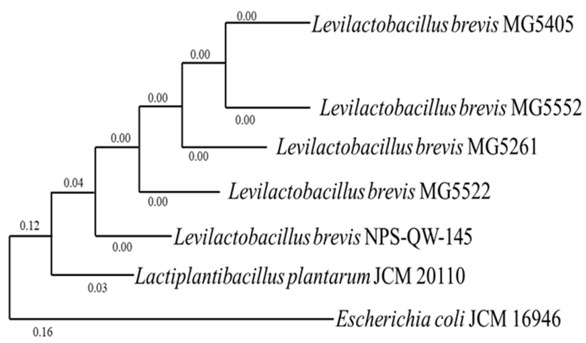
The concentrations of GABA produced by the four selected strains were determined using an amino acid analyzer (Table 1). Strains MG5261, MG5405, MG5522, and MG5552 produced 0.304, 0.260, 0.280, and 0.322 mg/mL GABA, respectively, in MRS medium and 0.624, 0.585, 0.591, and 0.979 mg/mL GABA, respectively, in 1% MSG-containing MRS medium (Table 1). Moreover, GABA production was higher in 1% MSG-containing MRS medium than in MRS medium, indicating that all selected strains exhibited the ability to convert MSG to GABA. L. brevis MG5552 exhibited the highest GABA production under the same conditions, and it also exhibited the highest rate of conversion of glutamate into GABA (data not shown). Previous studies have reported that L. brevis GABA100 produces 27.6 mg/mL GABA, whereas L. brevis DSM 32386 produces 0.262 mg/mL GABA [17,20]. The GABA-producing ability of each strain differs because of the glutamate decarboxylase and other factors. Four strains of L. brevis (MG5552, MG5405, MG5261, and MG5522) were selected for further experiments (Figure 2).

3.4. Effect of CFS of GABA-Producing Strains on Viability of and NO Production in RAW 264.7 Cells

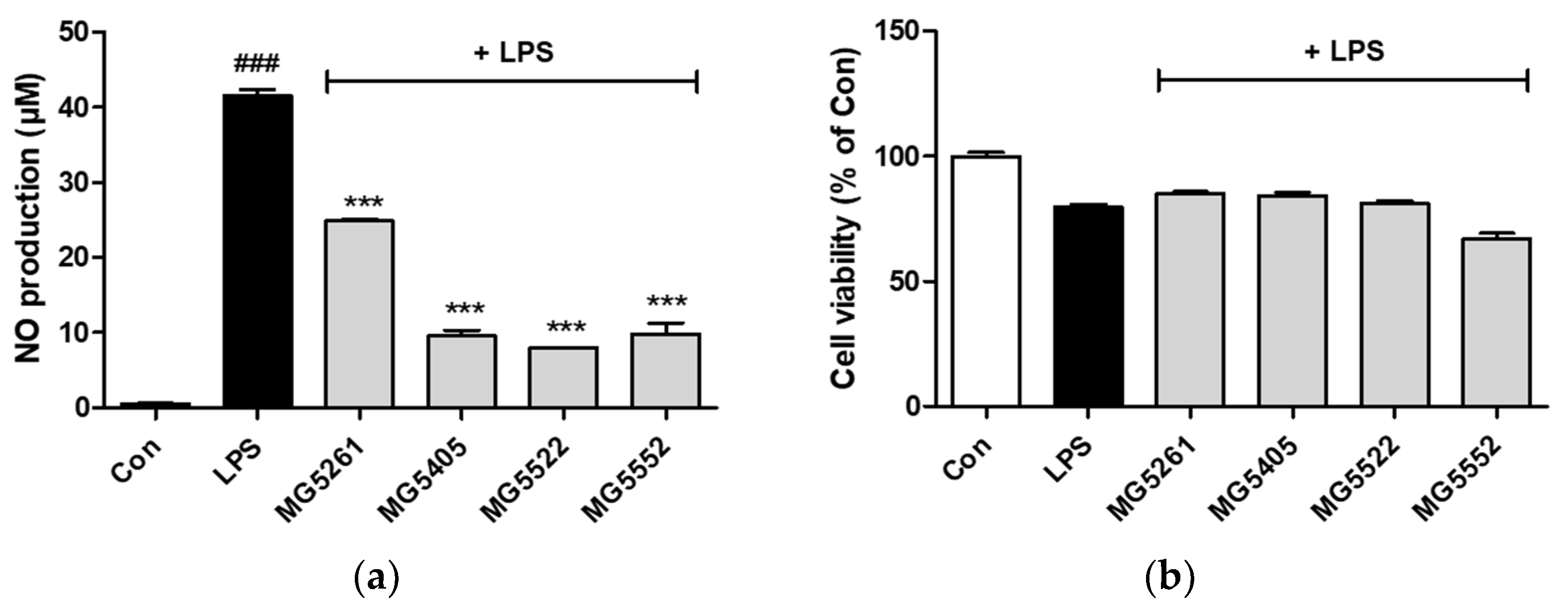
We investigated whether the selected GABA-producing strains were probiotics with anti-inflammatory properties. LPS treatment significantly increased NO production in RAW 264.7 cells compared with that in the control group, whereas CFS treatment significantly reduced NO production (Figure 3a). The NO concentrations after treatment with MG5261, MG5405, MG5522, and MG5552 CFS were 24.91, 9.58, 7.94, and 9.82 μM, respectively, suggesting that the selected strains may have anti-inflammatory effects, which could be attributed to the reduction in NO production. Moreover, the viability of RAW 264.7 cells was determined using the MTT assay. Cell viability after treatment with the CFS of strains MG5261, MG5405, MG5522, and MG5552 was 67.1, 85.1, 84.3%, and 81.2%, respectively (Figure 3b). According to previous studies, probiotics exert anti-inflammatory effects. NO is a key biomolecule that mediates various biological functions, such as inflammation, tumor suppression, and immunomodulation [21]. NO also acts as a biological mediator, like neurotransmitters in the nervous system, and is a signaling molecule involved in the regulation of various physiological mechanisms in the nervous, cardiovascular, and immune systems [22]. Therefore, the inhibition of NO production after treatment with CFS of the selected strains confirms their anti-inflammatory effects. Several studies have reported that GABA-producing probiotics can suppress inflammation by reducing the expression of various factors, such as NO and iNOS [23,24].
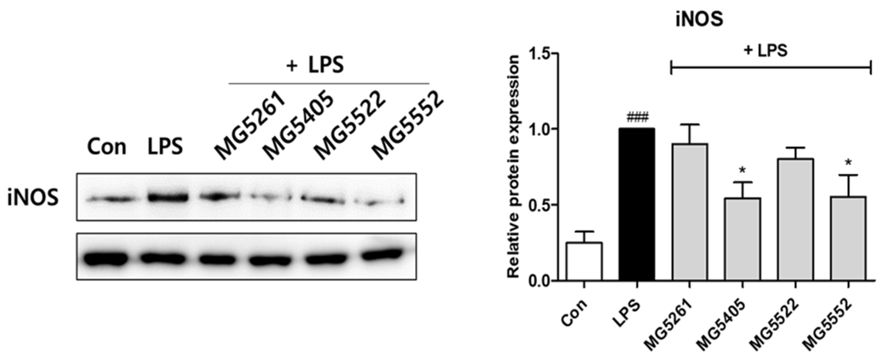
The effect of the CFS of the GABA-producing strains on iNOS protein expression in RAW 264.7 cells was analyzed using Western blotting. iNOS expression was significantly decreased after treatment with the CFS of strains MG5405 and MG5502 (Figure 4). iNOS, an isoform of NOS, is specifically expressed during inflammatory response and is involved in the synthesis of the pro-inflammatory mediator NO [25]. This indicates that the suppression of iNOS expression leads to the suppression of NO generation, which inhibits the inflammatory response induced by LPS. The overexpression of iNOS produces high levels of NO, resulting in tissue damage, septic shock, and other complications during a continuous chronic inflammatory response [26].

3.5. Effect of CFS of GABA-Producing Strains on NF-κB Activation in RAW 264.7 Cells

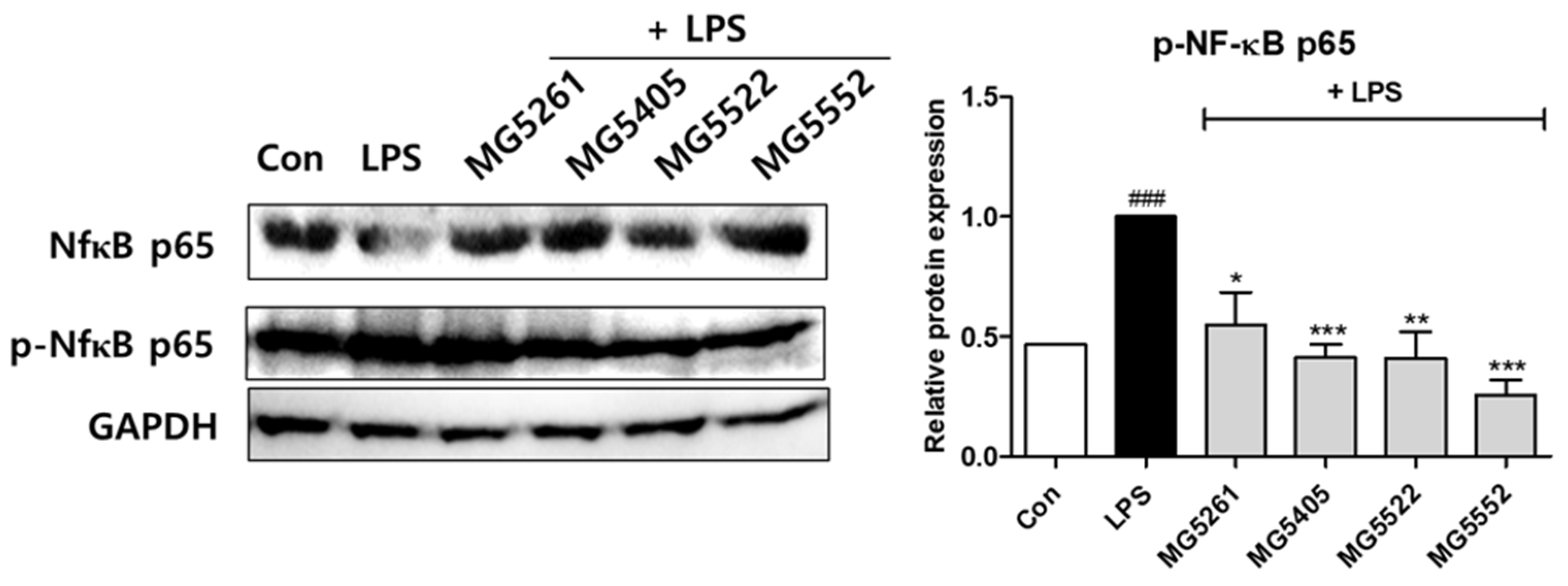
In this study, the inhibition of NF-κB activation was evaluated using Western blotting. As shown in Figure 5, NF-κB p65 expression in RAW 264.7 cells was significantly reduced after stimulation by the CFS of the selected strains, indicating that they inhibited the NF-κB signaling pathway. Moreover, the CFS of all GABA-producing strains inhibited the phosphorylation of NF-κB in RAW 264.7 cells. NF-kB, primarily composed of the subunits p50 and p65, can regulate various aspects of innate and adaptive immunity and is important in regulating immune and inflammatory responses [27]. Furthermore, NF-κB activation induces the production of immunomodulators in macrophages [28]. Free NF-κB translocates to the nucleus, where it activates the expression of inflammatory mediators [27,29]. Thus, the NF-κB signaling pathway participates in the regulation of various inflammatory cytokines, such as interleukins, and plays an important role in both immune and inflammatory reactions [30]. Therefore, the inhibition of the NF-κB signaling pathway could be an effective way to treat chronic inflammatory diseases.

4. Conclusions

We isolated and screened GABA-producing LAB strains from various fermented foods. The GABA-producing strains were screened using TLC, and quantitative analysis of GABA production by these strains was conducted using an amino acid analyzer. The results revealed that the four selected L. brevis strains, namely MG5552, MG5405, MG5261, and MG5522, exhibited the ability to convert glutamate, a GABA precursor, into GABA. Furthermore, the analyses of the anti-inflammatory effects of the selected strains elucidated that almost all selected strains inhibited NO and iNOS production, and NF-kB activity, thus exhibiting anti-inflammatory activity. Therefore, the selected strains have the potential for use in functional foods because of their anti-inflammatory activity and the effects exerted by GABA, such as neurological stability and blood pressure reduction.

Author Contributions

Conceptualization, C.-H.K.; methodology, Y.-L.K., T.H.N., J.-S.K. and J.-Y.P.; formal analysis, Y.-L.K., T.H.N., J.-S.K. and J.-Y.P.; investigation, Y.-L.K., T.H.N. and J.-S.K.; data curation, Y.-L.K., T.H.N., J.-S.K. and J.-Y.P.; writing—original draft preparation, Y.-L.K.; writing—review and editing, Y.-L.K., T.H.N. and C.-H.K.; visualization, Y.-L.K.; supervision, C.-H.K.; project administration, C.-H.K. All authors have read and agreed to the published version of the manuscript.

Funding

This research received no external funding.

Institutional Review Board Statement

Not applicable.

Informed Consent Statement

Not applicable.

Data Availability Statement

All data are presented in the paper.

Conflicts of Interest

The authors declare no conflict of interest.

References

  1. Crittenden, D.L.; Chebib, M.; Jordan, M.J.T. Stabilization of Zwitterions in Solution: GABA Analogues. J. Phys. Chem. A 2005, 109, 4195–4201. [Google Scholar] [CrossRef] [PubMed]
  2. Crittenden, D.L.; Chebib, M.; Jordan, M.J.T. Stabilization of Zwitterions in Solution: γ-Aminobutyric Acid (GABA). J. Phys. Chem. A 2004, 108, 203–211. [Google Scholar] [CrossRef]
  3. Strain, G.W. Nutrition, Brain Function and Behavior. Psychiatr. Clin. N. Am. 1981, 4, 253–268. [Google Scholar] [CrossRef] [PubMed]
  4. Mitsushima, D.; Shwe, T.T.W.; Funabashi, T.; Shinohara, K.; Kimura, F. GABA Release in the Medial Preoptic Area of Cyclic Female Rats. Neuroscience 2002, 113, 109–114. [Google Scholar] [CrossRef]
  5. Dhakal, R.; Bajpai, V.K.; Baek, K.H. Production of gaba (γ-Aminobutyric acid) by microorganisms: A review. Braz J. Microbiol. 2012, 43, 1230–1241. [Google Scholar] [CrossRef] [Green Version]
  6. Masuda, K.; Guo, X.-F.; Uryu, N.; Hagiwara, T.; Watabe, S. Isolation of Marine Yeasts Collected from the Pacific Ocean Showing a High Production of-Aminobutyric Acid. Biosci. Biotechnol. Biochem. 2008, 72, 3265–3272. [Google Scholar] [CrossRef]
  7. Seo, M.-J.; Lee, J.-Y.; Nam, Y.-D.; Lee, S.-Y.; Park, S.-L.; Yi, S.-H.; Lee, M.-H.; Roh, S.W.; Choi, H.-J.; Lim, S.-I. Production of γ-Aminobutyric Acid by Lactobacillus Brevis 340G Isolated from Kimchi and Its Application to Skim Milk. Food Eng. Prog. 2013, 17, 418–423. [Google Scholar] [CrossRef]
  8. Li, H.; Qiu, T.; Huang, G.; Cao, Y. Production of gamma-aminobutyric acid by Lactobacillus brevis NCL912 using fed-batch fermentation. Microb Cell Fact. 2010, 9, 85. [Google Scholar] [CrossRef] [Green Version]
  9. Wallace, T.C.; Guarner, F.; Madsen, K.; Cabana, M.D.; Gibson, G.; Hentges, E.; Sanders, M.E. Human Gut Microbiota and Its Relationship to Health and Disease. Nutr. Rev. 2011, 69, 392–403. [Google Scholar] [CrossRef]
  10. Hwanhlem, N.; Watthanasakphuban, N.; Riebroy, S.; Benjakul, S.; H-Kittikun, A.; Maneerat, S. Probiotic Lactic Acid Bacteria from Kung-Som: Isolation, Screening, Inhibition of Pathogenic Bacteria. Int. J. Food Sci. Technol. 2010, 45, 594–601. [Google Scholar] [CrossRef]
  11. Delcenserie, V.; Martel, D.; Lamoureux, M.; Amiot, J.; Boutin, Y.; Roy, D. Immunomodulatory Effects of Probiotics in the Intestinal Tract. Curr. Issues Mol. Biol. 2008, 10, 37–54. [Google Scholar] [CrossRef] [PubMed] [Green Version]
  12. Soltani, N.; Qiu, H.; Aleksic, M.; Glinka, Y.; Zhao, F.; Liu, R.; Li, Y.; Zhang, N.; Chakrabarti, R.; Ng, T.; et al. GABA Exerts Protective and Regenerative Effects on Islet Beta Cells and Reverses Diabetes. Proc. Natl. Acad. Sci. USA 2011, 108, 11692–11697. [Google Scholar] [CrossRef] [Green Version]
  13. Kook, M.; Cheol, C.S. Production of GABA (Gamma Amino Butyric Acid) by Lactic Acid Bacteria. Food Sci. Anim. Resour. 2013, 33, 377–389. [Google Scholar] [CrossRef]
  14. Kim, M.J.; Kim, K.S. Isolation and Identification of γ-Aminobutyric Acid (GABA)-Producing Lactic Acid Bacteria from Kimchi. J. Korean Soc. Appl. Biol. Chem. 2012, 55, 777–785. [Google Scholar] [CrossRef]
  15. Kanklai, J.; Somwong, T.C.; Rungsirivanich, P.; Thongwai, N. Screening of GABA-Producing Lactic Acid Bacteria from Thai Fermented Foods and Probiotic Potential of Levilactobacillus Brevis F064A for GABA-Fermented Mulberry Juice Production. Microorganisms 2020, 9, 33. [Google Scholar] [CrossRef] [PubMed]
  16. Lin, X.; Chen, X.; Chen, Y.; Jiang, W.; Chen, H. The Effect of Five Probiotic Lactobacilli Strains on the Growth and Biofilm Formation of Streptococcus Mutans. Oral Dis 2015, 21, e128–e134. [Google Scholar] [CrossRef]
  17. Kim, J.Y.; Lee, M.Y.; Ji, G.E.; Lee, Y.S.; Hwang, K.T. Production of Gamma-Aminobutyric Acid in Black Raspberry Juice during Fermentation by Lactobacillus Brevis GABA100. Int. J. Food Microbiol. 2009, 130, 12–16. [Google Scholar] [CrossRef]
  18. Park, K.-B.; Oh, S.-H. Isolation and Characterization of Lactobacillus Buchneri Strains with High γ-Aminobutyric Acid Producing Capacity from Naturally Aged Cheese. Food Sci. Biotechnol. 2006, 15, 86–90. [Google Scholar]
  19. Veno, Y.; Hayakawa, K.; Takahashi, S.; Oda, K. Purification and Characterization of Glutamate Decarboxylase from Lactobacillus Brevis IFO 12005. Biosci. Biotechnol. Biochem. 1997, 61, 1168–1171. [Google Scholar] [CrossRef]
  20. Mancini, A.; Carafa, I.; Franciosi, E.; Nardin, T.; Bottari, B.; Larcher, R.; Tuohy, K.M. In Vitro Probiotic Characterization of High GABA Producing Strain Lactobacilluas Brevis DSM 32386 Isolated from Traditional “Wild” Alpine Cheese. Ann. Microbiol. 2019, 69, 1435–1443. [Google Scholar] [CrossRef]
  21. Shreshtha, S.; Sharma, P.; Kumar, P.; Sharma, R.; Singh, S.P. Nitric Oxide: It’s Role in Immunity. J. Clin. Diagn. Res. 2018, 12, 1–5. [Google Scholar] [CrossRef]
  22. Aktan, F. INOS-Mediated Nitric Oxide Production and Its Regulation. Life Sci. 2004, 75, 639–653. [Google Scholar] [CrossRef] [PubMed]
  23. Bhandage, A.K.; Jin, Z.; Korol, S.V.; Shen, Q.; Pei, Y.; Deng, Q.; Espes, D.; Carlsson, P.O.; Kamali-Moghaddam, M.; Birnir, B. GABA Regulates Release of Inflammatory Cytokines From Peripheral Blood Mononuclear Cells and CD4+ T Cells and Is Immunosuppressive in Type 1 Diabetes. EBioMedicine 2018, 30, 283–294. [Google Scholar] [CrossRef] [PubMed] [Green Version]
  24. Zheng, L.; Lu, X.; Yang, S.; Zou, Y.; Zeng, F.; Xiong, S.; Cao, Y.; Zhou, W. The Anti-Inflammatory Activity of GABA-Enriched Moringa Oleifera Leaves Produced by Fermentation with Lactobacillus Plantarum LK-1. Front. Nutr. 2023, 10, 346. [Google Scholar] [CrossRef] [PubMed]
  25. Han, B.H.; Lee, Y.J.; Yoon, J.J.; Choi, E.S.; Namgung, S.; Jin, X.J.; Jeong, D.H.; Kang, D.G.; Lee, H.S. Hwangryunhaedoktang Exerts Anti-Inflammation on LPS-Induced NO Production by Suppressing MAPK and NF-ΚB Activation in RAW264.7 Macrophages. J. Integr. Med. 2017, 15, 326–336. [Google Scholar] [CrossRef] [PubMed]
  26. Van Thanh, N.; Jang, H.J.; Vinh, L.B.; Linh, K.T.P.; Huong, P.T.T.; Cuong, N.X.; Nam, N.H.; Van Minh, C.; Kim, Y.H.; Yang, S.Y. Chemical Constituents from Vietnamese Mangrove Calophyllum Inophyllum and Their Anti-Inflammatory Effects. Bioorg. Chem. 2019, 88, 102921. [Google Scholar] [CrossRef]
  27. Chalmers, S.A.; Garcia, S.J.; Reynolds, J.A.; Herlitz, L.; Putterman, C. NF-KB Signaling in Myeloid Cells Mediates the Pathogenesis of Immune-Mediated Nephritis. J. Autoimmun. 2019, 98, 33–43. [Google Scholar] [CrossRef]
  28. Kim, H.J.; Kang, C.H.; Jayasooriya, R.G.P.T.; Dilshara, M.G.; Lee, S.; Choi, Y.H.; Seo, Y.T.; Kim, G.Y. Hydrangenol Inhibits Lipopolysaccharide-Induced Nitric Oxide Production in BV2 Microglial Cells by Suppressing the NF-ΚB Pathway and Activating the Nrf2-Mediated HO-1 Pathway. Int. Immunopharmacol. 2016, 35, 61–69. [Google Scholar] [CrossRef]
  29. Niu, X.; Zang, L.; Li, W.; Xiao, X.; Yu, J.J.; Yao, Q.; Zhao, J.; Ye, Z.; Hu, Z.; Li, W. Anti-Inflammatory Effect of Yam Glycoprotein on Lipopolysaccharide-Induced Acute Lung Injury via the NLRP3 and NF-ΚB/TLR4 Signaling Pathway. Int. Immunopharmacol. 2020, 81, 106024. [Google Scholar] [CrossRef]
  30. Luo, J.; Li, X.J.; Lee, G.H.; Huang, J.J.; Whang, W.K.; Zhang, X.D.; Yook, C.S.; Liu, X.Q. Anti-Inflammatory Effects of Two Lupane-Type Triterpenes from Leaves of Acanthopanax Gracilistylus on LPS-Induced RAW264.7 Macrophages. Food Sci. Technol. 2021, 42, e89721. [Google Scholar] [CrossRef]
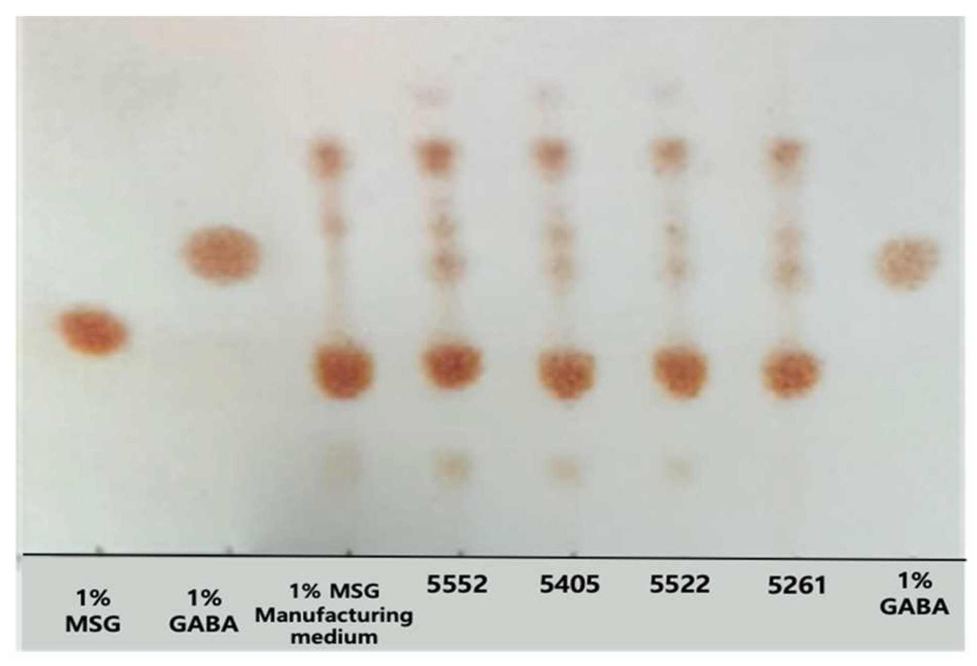
Figure 1. γ-Aminobutyric acid (GABA)-producing capability of different strains in synthetic medium.
Fermentation 09 00612 g001
Figure 2. Phylogenetic relationships of the selected strains with a known Lactic acid bacteria (LAB) strain based on 16S rRNA sequence analysis; primers used: universal primers 27F and 1492R.
Fermentation 09 00612 g002
Figure 3. Effects of CFS of γ-aminobutyric acid (GABA)-producing strains on nitric oxide (NO) production (a) and cytotoxicity (b) in lipopolysaccharide (LPS)-activated RAW264.7 cells. Data indicate significant differences between the mean at p < 0.05 based on t-test. ### p < 0.001 vs. Con and *** p < 0.001 vs. LPS.
Fermentation 09 00612 g003
Figure 4. Effects of CFS of γ-aminobutyric acid (GABA)-producing strains on inducible nitric oxide synthase (iNOS) expression in RAW264.7 cells. Data indicate significant differences between means at p < 0.05 based on t-test. ### p < 0.001 vs. Con and * p < 0.05 vs. LPS.
Fermentation 09 00612 g004
Figure 5. Effect of CFS of γ-aminobutyric acid (GABA)-producing strains on nuclear factor kappa B (NF-κB) activation in RAW264.7 cells. Data indicate significant differences between means at p < 0.05 based on t-test. ### p < 0.001 vs. Con and * p < 0.05, ** p < 0.01, *** p < 0.001 vs. LPS.
Fermentation 09 00612 g005
Table 1. The concentration of γ-aminobutyric acid (GABA) produced by the selected strains.
SpeciesStrainIsolation SourceGABA Concentration (mg/mL)
MRSMRS + 1%MSG
Control --0.000.00
Levilactobacillus brevisMG5261Fermented food0.3040.624
Levilactobacillus brevisMG5405Fermented food0.2600.585
Levilactobacillus brevisMG5522Fermented food0.2810.591
Levilactobacillus brevisMG5552Fermented food0.3220.976
Disclaimer/Publisher’s Note: The statements, opinions and data contained in all publications are solely those of the individual author(s) and contributor(s) and not of MDPI and/or the editor(s). MDPI and/or the editor(s) disclaim responsibility for any injury to people or property resulting from any ideas, methods, instructions or products referred to in the content.