Article

The Effects of a High-Intensity Ultrasound on the Fermentative Activity and Kinetic Growth of Lactobacillus Acidophilus and Lactobacillus Helveticus

by
Norma Angélica Bolívar-Jacobo
1,
Raúl Alberto Reyes-Villagrana
2,*,
Gerardo Pavel Espino-Solís
3,
Ana Luisa Rentería-Monterrubio
1,
Martha María Arévalos-Sánchez
1,
Rogelio Sánchez-Vega
1,
Eduardo Santellano-Estrada
1,
David Chávez-Flores
4 and
América Chávez-Martínez
1,*
1
Facultad de Zootécnica y Ecología, Universidad Autónoma de Chihuahua, Periférico Francisco R. Almada, Km 1, Chihuahua 31453, Mexico
2
Consejo Nacional de Ciencia y Tecnología, Av. Insurgentes Sur 1582, Col. Crédito Constructor, Col. Benito Juárez, Ciudad de Mexico 03940, Mexico
3
Facultad de Medicina y Ciencias Biomédicas, Universidad Autónoma de Chihuahua, Circuito Universitario, Campus ll, Chihuahua 31109, Mexico
4
Facultad de Ciencias Químicas, Universidad Autónoma de Chihuahua, Circuito Universitario, Campus ll, Chihuahua 31125, Mexico
*
Authors to whom correspondence should be addressed.
Fermentation 2023, 9(4), 356; https://doi.org/10.3390/fermentation9040356
Submission received: 7 March 2023 / Revised: 27 March 2023 / Accepted: 28 March 2023 / Published: 4 April 2023
(This article belongs to the Section Fermentation Process Design)

Abstract

:
An ultrasound, a non-thermal technique, can be employed to increase a probiotic’s biomass and its fermentation products. The effects of high-intensity ultrasounds (20%, 30%, and 40% amplitudes for 3 min) on the growth and fermentative profile of Lactobacillus acidophilus and helveticus were studied. The use of an ultrasound decreased the Lag phase and increased the maximum growth potential; however, the effect depended on the amplitude used. For both probiotics, the β-galactosidase activity increased in the treatments with a 20% amplitude—3 min and 30% amplitude—but decreased in the treatment with a 40% amplitude—3 min in comparison to the values found in the control treatment. The two probiotics showed a decrease in the protein concentration when compared with the control treatment. Both probiotics presented the lowest values of proteolysis in the treatments with a 30% amplitude—3 min. Lactic, acetic, and citric acids were the organic acids that were present in the highest concentration and formic acid was not detected in either of the two probiotics. It can be concluded that the ultrasound amplitude has a noticeable influence on the growth and fermentation profiles of both probiotics. The results from this study could be used in subsequences investigations to enhance the postbiotic production of Lactobacillus acidophilus and Lactobacillus helveticus.

1. Introduction

A balanced diet is essential to obtain the necessary nutrients for growth, development, and good health [1]. In this regard, the consumption of functional dairy foods has been increasing due to an increased interest in consuming functional foods as a way to prevent diseases and improve good health [2]. Within functional dairy foods are foods that have prebiotics, probiotics, paraprobiotics, and postbiotics [3].
Probiotics are defined as live microorganisms that, when administered, confer health benefits [3,4]. These have specific recognition structures called molecular patterns, which are associated with microbes through which they carry out their function [5]. These microorganisms have a specific activity and certain characteristics that allow them to carry out their function, such as the ability to resist gastric acids and bile salts, the ability to adhere to epithelial cells, and the ability to possess an antimicrobial activity that favors elimination or minimizes the adherence of pathogens in the gastrointestinal tract [6]. The bioeffects are attributed to the components, metabolic reactions, and metabolites (organic acids, bacteriocins, and H2O2, among others) of the probiotics [7]. Some of these effects are immunomodulatory, cholesterol-lowering, anticancer, antidiabetic, antihypertensive, and hypolipidemic [8]. However, in order to achieve a positive effect on health, the minimum amount of necessary probiotics must be greater than 108 or 109 Log10 CFU/g or ml. In additional their survival during processing and storage has to be assured [9].
In recent years, a new concept, postbiotics, has been introduced within functional foods to those already known (prebiotics and probiotics) [10]. By definition, postbiotics are considered molecules or compounds originating from bacterial cell lysis or those obtained from the fermentative metabolism of probiotics, which have bioactivities [1,11,12,13,14]. Postbiotics are not living microorganisms; therefore, they are considered safer than probiotics. They are non-pathogenic, non-toxic, and they have a greater capacity to resist hydrolysis [1]. Additionally, they easily bind to the sites of action, do not interact with the native microorganisms of the intestine, have greater stability, and are easy to obtain, standardize, transport, and store [6]. Postbiotics are classified in three very specific ways. The first classification corresponds to the place where they come from, which is made up of three subgroups: (A) metabolites obtained from dietary components, (B) those obtained by bacterial biochemical modification of host bioproducts, and (C) those synthesized de novo by bacteria [15]. The second classification is regarding its chemical composition (proteins, carbohydrate lipids, organic acids, vitamins, enzymes, complex molecules, etc.) [16,17]. Additionally, the third classification relates its bioactive potential (immunomodulatory, anti-inflammatory, antimicrobial, antioxidant, antiproliferative, etc.) [8,17,18,19,20].
An ultrasound is used as a non-thermal methodology to obtain postbiotics from probiotics, since it favors bacterial growth and facilitates the exchange of molecules and the obtaining of bioactive compounds [21]. Ultrasounds in general have a working range in the frequency range of 16 kHz to 1 GHz. Currently, ultrasound treatments used in the food industry typically operate from 20 kHz to 1 mHz and can be divided into three subcategories given their acoustic intensities as a function of frequency: low intensity (1 W/cm2 ≤ B < 2 W/cm2) with a frequency range > 1 MHz, intensity medium (2 W/cm2 ≤ M < 10 W/cm2) with a frequency range 100 kHz ≤ f < 1 MHz and high-intensity (10 W/cm2 ≤ A < 1000 W/cm2) with a frequency range 20 kHz ≤ f < 100 kHz [22]. A high-intensity ultrasound (HIU) can modify the biological, physical, and chemical properties of materials; therefore, it has been used be used in food processing [23].
Ultrasonic treatment can induce the formation of temporary pores in the cell membrane, known as “sonoporation”, a term introduced by Lentacker et al. [24], which favors its permeability and allows the exchange of molecules through those pores. Particularly, ultrasonoporation is interpreted as a methodology that entails the effect of acoustic cavitation (the formation of a bubble that contains steam and implodes, generating zones of high temperature and pressure) [25], which is generated by the HIU and applied at the cellular scale to modify the internal structure of biological materials [26]. Ultrasonoporation alters the permeability of the cell plasma membrane through shear stress waves, the Bjerke’s force, and the acoustic radiation generated in acoustic cavitation [27]. The transitory pores formed enable the exit of the compounds [28]. The factors to consider in the use of ultrasound are as follows: the nature of the waves, the time of application, the microorganism, and the temperature [29].
Postbiotics can be added to certain foods and/or used as supplements [29,30]. Some of the most accepted functional foods that are in high demand by the consumer are fermented dairy products [29]. To obtain these products, lactic acid bacteria (LAB) are used [30]. The most used genera are Lactobacillus, Streptococcus, and Leuconostoc [31], and within these, some of the most used bacteria are Lactobacillus acidophilus and Lactobacillus helveticus [32]. L. acidophilus has been used as a probiotic in dairy products, dietary supplements, and fermented milk products, showing positive effects such as the regulation of the intestinal microbiota and the positive regulation of the immune system [33]. Likewise, L. helveticus could be considered a probiotic, as it is used a starter culture in the manufacture of Swiss and Italian cheeses, and recently, its beneficial effects on consumers’ health have been reported, such as its antioxidant, antimicrobial, and antihypertensive capacities. Furthermore, some of the characteristics that stand out in this microorganism are its ability to survive gastrointestinal transit and to adhere to epithelial cells [34,35]. Consequently, the objective of this work was to evaluate the effect of a HIU on the kinetic and growth parameters, as well as the fermentation profiles of L. acidophilus and L. helveticus.

2. Materials and Methods

2.1. Bacterial Culture Preparation

Twelve flasks were prepared with 250 mL of skimmed milk (LALA Light®), to which 2.5 g of L. acidophilus inoculum (LA-5, CHR HANSEN) was added to obtain a 1% w/v culture (8.08 Log10 CFU/mL). The same procedure was performed for L. helveticus (LH-B02, CHR HANSEN). Both microorganisms were donated by Chr Hansen®. Skimmed milk (LALA Light®) was chosen as the growth medium based on the results of a previous study [36].

2.2. Ultrasonic Treatment

After the inoculation of skimmed milk (LALA Light®) with L. acidophilus or L. helveticus, the ultrasonic treatments were applied. Ultrasonication was performed with a 1.3 cm diameter probe ultrasonic processor (20 kHz, GEX750, Sonic, Newtown, CT). Three treatments were performed: 20% amplitude—3 min; 30% amplitude—3 min; and 40% amplitude—3 min; T1, T2, and T3, respectively. The control treatment for each probiotic was the inoculated skimmed milk without application of ultrasound (T0). After ultrasonic treatment, samples were incubated in aerobiosis at a 37 ± 1 °C for 24 h. Treatments were performed using a completely randomized design and in triplicates.

2.3. Kinetic Growth

The zero time of the kinetics was the one after the inoculation of the growth medium and prior to the ultrasound treatment. After that, time one was considered after the ultrasonication treatment, and from there, time samples were taken every two hours for a period of 24 h. Bacterial counts were performed whereby 1 mL was taken from each flask and serial dilutions were made in test tubes with 9 mL of phosphate buffer (J.T. Baker, Edo de México, México). From the 1:5 and 1:6 dilutions, 0.1 mL was plated in duplicates in Man, Rogosa and Sharpe agar (MRS; Difco, Franklin Lakes, NJ, USA) using plate spread technique. Petri dishes were then incubated at 37 ± 1 °C in aerobiosis for 48 h. Results were expressed in Log10 CFU/mL. Bacterial counts were analyzed with the DMFit online software (2009) and the kinetic parameters (yo, Lag, Rate (µmax), and Tmax) were obtained using the trilinear model.

2.4. Cell Membrane Permeability

A LIVE/DEADTM BacLight bacterial viability kit from ThermoFisher Scientific [37] composed of propidium iodide (PI) and SYTO9, which selectively stain cells based on membrane integrity, was used. Samples were acquired on an Attune NxT flow cytometer, with blue, red, violet, and yellow lasers (Thermo Fisher Scientific, Waltham, MA, USA). Fluorescence Minus One (FMO) control was performed to identify fluorescence limits and establish populations and gate strategies. Gates were set according to forward and side scatter characteristics. A total of 100,000 events for each sample was acquired at 500 μL/min. Data were analyzed using FlowJo software V.10.
For the preparation of the samples, 1 mL of each of the treatments was taken and centrifuged at 10,000× g (Eppendorf Centrifuge 5430 R, Hamburg, Germany) for 3 min. The sediments were resuspended with 1 mL of 0.85% NaCl and incubated at room temperature for 60 min and applying a brief shake every 15 min. This procedure was carried out twice. For the positive and negative control, 1 mL of a sample of the bacterial culture was taken and centrifuged at 10,000× g for 3 min. With the sediment, a suspension of live cells and dead cells was prepared. Live cells were resuspended in 1 mL of 0.85% NaCl and dead cells in 1 mL of 70% isopropyl alcohol. Samples were incubated at room temperature for 60 min, mixing the suspensions every 15 min, and then centrifuged at 10,000× g for 3 min. The pellets of the two samples were washed with 1 mL of 0.85% NaCl and centrifuged at 10,000× g for 3 min. Then, these were resuspended in 1 mL of 0.85% NaCl. Subsequently, for cell staining, an aliquot of 977 µL of 0.85% NaCl was taken in a flow cytometry analysis tube, adding 1.5 µL of 3.34 mM SYTO 9 Nucleic Acid Stain (Component A), 1.5 µL of 30 mM Propidium Iodide (Component B) and 10 µL of the bacterial suspension prepared in the previous step. Samples were incubated at room temperature for 15 min in the dark. Finally, 10 μL of the microsphere suspension was added to the stained cell sample, mixed, and analyzed by flow cytometry under the previously described conditions. The standard suspension of microspheres (Component C) was completely resuspended, and then sonicated in a water bath for 10 min. The final volume was 1000 μL to obtain an accurate count of the samples.

2.5. Hydrogen Potential Determination

The pH was measured in a potentiometer (Orion Versa Star, Thermo Scientific, MA, USA) at the zero time, which was after the inoculation of the medium and prior to the ultrasound treatment. Later, the pH was measured after the ultrasonication and from then, measures were taken every 2 h for a period of 24 h, and then the results were graphed.

2.6. β-Galactosidase Activity

The β-galactosidase enzyme activity was determined by the release of o-nitrophenol (ONP) from the hydrolysis of o-nitrophenyl-β-D-galactopyranoside (ONPG) by β-galactosidase [38]. To carry out the reaction, 0.2 mL of sample (cell-free supernatant) was taken and 3 mL of the chromogenic reagent (300 mg of 2-nitrophenyl β-galactopyranoside in 100 mL of 100 mM phosphate-buffered solution at a pH of 7.0) was added. The mixture was incubated at 37 °C for 30 min. After the incubation time had elapsed, the reaction was stopped by adding 2 mL of 0.625 M sodium carbonate (Na2CO3) and the absorbance was measured at 420 nm using a spectrophotometer (UV-1800 Shimadzu, Japan). The amount in moles of ONP resealed by hydrolysis was reported as units (U) based on a standard curve of ONP reagent (Sigma-Aldrich, St. Louis, MO, USA) (y = 0.0203x + 0.0489, R2 = 0.9599). One unit (1U) is defined as the amount of enzyme needed to hydrolyze 1 µmol of o-nitrophenol per minute (1U = 1 µmol of o-NPGal/min/mL or g) under the measurement conditions of the assay (37 °C, pH 7, for 30 min).

2.7. Total Protein Content

The determination of the total protein concentration (TPC) was performed spectrophotometrically according to Bradford (1976) [39]. This technique is based on the colorimetric reaction carried out by proteins with the brilliant blue dye, forming a colored product that enables the estimation of the protein content of the sample by the measure of its absorbance at 595 nm. Bovine serum albumin was used as standard; hence, different concentrations (in a range of 0–14 mg/mL) were prepared and the absorbance was determined to obtain a calibration curve (y = 0.0086x + 0.036, R2 = 0.998). The amounts of protein in the samples were quantified using 0.1 mL of sample (cell-free supernatant) to which 1 mL of Bradford reagent was added; then, the mixture was left to react in the dark for 5 min and later, the absorbance at 595 nm was measured in a spectrophotometer (UV-1800 Shimadzu, Japan). The TPC was reported in mg/mL of BSA.

2.8. Proteolysis

The degree of proteolysis was evaluated according to the method described by Donkor et al. [40]. A total of 150 μL of the sample (cell-free supernatant) was taken and added to 3 mL of the O-phthaldialdehyde (OPA), which was prepared with 25 mL 100 mM of Sodium Tetraborate (Sigma-Aldrich), 2.5 mL of 20% w/v sodium dodecyl sulfate (SDS, Sigma-Aldrich), 40 mg OPA (Sigma-Aldrich) dissolved in 1 mL of methanol, and 100 μL of β-mercaptoethanol (Sigma-Aldrich), which made a volume of 50 mL. The solution was stored in an amber bottle because it was photosensitive and it was used the day it was prepared. The samples were incubated at room temperature for 2 min, and after the incubation time, the absorbance at 220 nm was measured using a spectrophotometer (UV-1800 Shimadzu, Japan).

2.9. Organic Acids Quantification

First, the samples were centrifuged 10,000× g for 10 min (Beckman Coulter Avanti J-26 XPI Centrifuge, Indianapolis, IN, USA) and filtered through a Whatman 0.45 μm membrane filter. Organic acid concentrations were determined by high-performance liquid chromatography (HPLC). For chromatographic separation, a 100 mm Syncronis C18 column (Thermo Fisher Scientific, Waltham, MA, USA) and an acetonitrile/water (60/40) elution mobile phase were used with a flow rate of 1.25 mL/min for a sample volume of 2.0 μL and an absorbance of 254 nm. Organic acids (oxalic, citric, malic, succinic, formic, acetic, and lactic) were identified by their retention times and corresponding area values. Lyophilized organic acid standards (Bio-Rad Organic Acid Standard, Hercules, CA, USA) were rehydrated with 1.0 mL of deionized water. The concentrations of organic acids were expressed in millimolar (mM).

2.10. Statistical Analysis

The analysis of the response variables was carried out with the statistical package Minitab (version 18). To determine significant differences between each of the treatments, for each response variable, a one-way ANOVA and Tukey’s test with p < 0.05 were performed.

3. Results and Discussion

3.1. Kinetic Growth

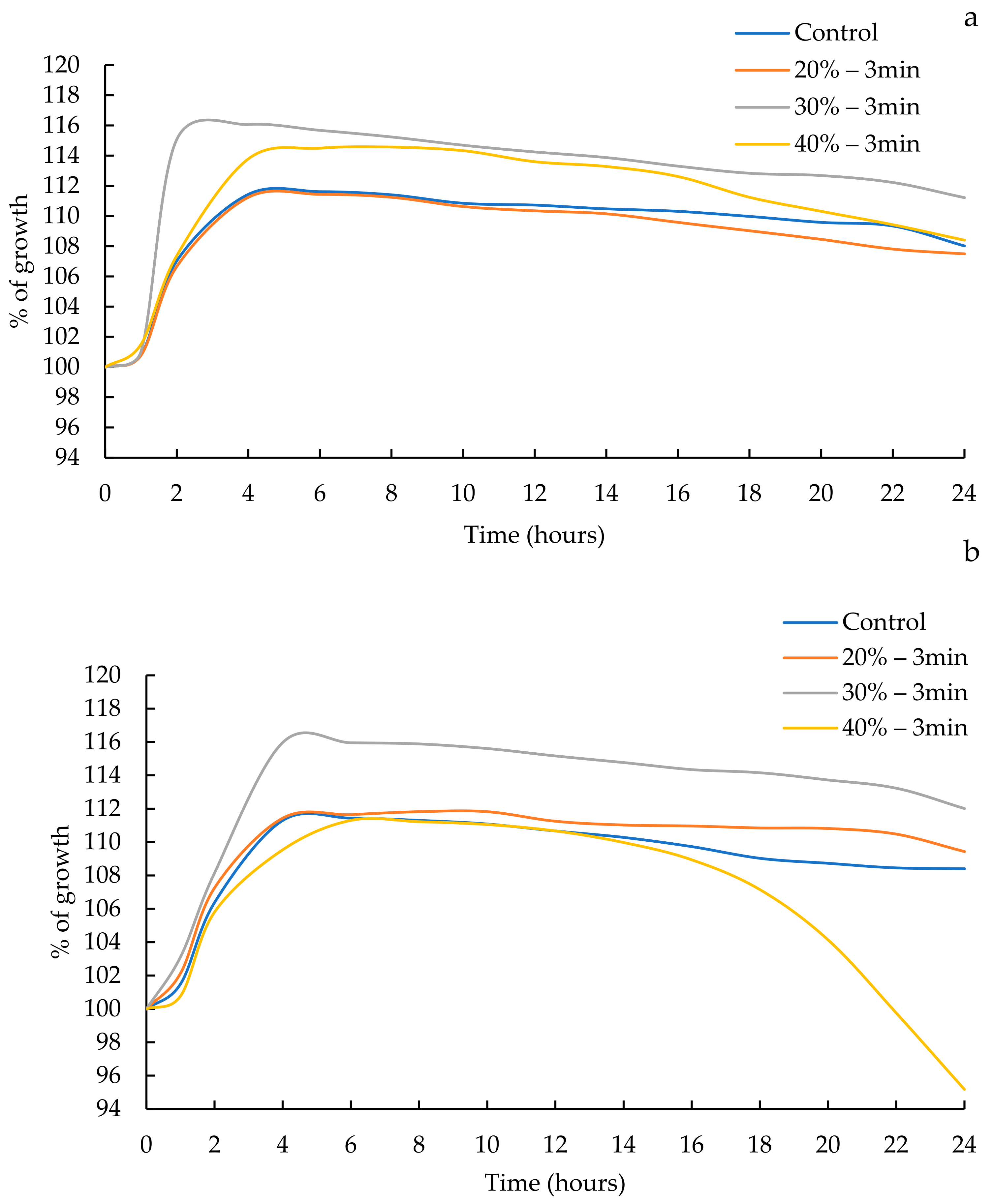
The kinetic growth of L. acidophilus and L. helveticus is shown in Figure 1, and the kinetic parameters are shown in Table 1. The parameter yo, known as the initial point of the sigmoidal growth curve, did not show significant differences (p > 0.05) between treatments. This was expected, since the inoculum was standardized in all the treatments for both strains. The Lag phase showed significant differences (p < 0.05) between the treatments for both L. acidophilus and L. helveticus. For L. acidophilus, the Lag phase was shorter in the treatment with a 40% amplitude (0.748 ± 0.003) (p < 0.05) and larger in the treatment with a 30% amplitude (0.923 ± 0.000) (p < 0.05). For L. helveticus, the Lag phase was shorter in the treatments with a 30% amplitude (0.391 ± 0.035) and a 40% amplitude (0.390 ± 0.046), without statistical differences between them (p > 0.05), and the control treatment showed a larger Lag phase (0.696 ± 0.006) (p < 0.05). The Tmax parameter also showed significant differences between treatments (p < 0.05) for both L. acidophilus and L. helveticus. For L. acidophilus, Tmax was higher in the treatment with a 40% amplitude (2.985 ± 0.031) (p < 0.05) and lower in the treatment with a 20% amplitude (1.998 ± 0.000) (p < 0.05). Regarding L. helveticus, the treatment with a 40% amplitude showed a higher Tmax (4.174 ± 0.006) (p < 0.05) and the treatment with a 20% amplitude presented the lowest value (2.720 ± 0.018) (p < 0.05). On the other hand, in the phase of the maximum growth potential (µmax), significant differences were observed between the treatments (p < 0.05) for both microorganisms. In the case of L. acidophilus, this parameter was higher (p < 0.05) for the treatment with 30% amplitude (1.065 ± 0.000) and lower (p < 0.05) for the treatments with a 20% (0.470 ± 0.014) and 40% amplitude (0.475 ± 0.007), without statistical differences between these treatments. While for L. helveticus, the highest values (p < 0.05) were for the treatments with a 20% amplitude (0.410 ± 0.000) and a 30% amplitude (0.410 ± 0.000) and the lowest value (p < 0.05) for treatment with a 40% amplitude (0.225 ± 0.001). Finally, the regression adjustment coefficient (R2) presented the values between 0.91 and 0.96 for L. acidophilus and between 0.94 and 0.96 for L. helveticus.
Gholamhosseinpour and Hashemi [31] used an ultrasound (30 kHz frequency and 25% amplitude for 5, 10, and 15 min) as pretreatment in reconstituted skimmed milk as a growth medium to carry out fermentation with L. plantarum, observing a decrease in the Lag phase and a greater growth rate in the treatment with a longer ultrasonication time (15 min). Likewise, Dahroud et al. [41] observed a decrease in the Lag phase when using an ultrasound (60% amplitude for 15 s) during the fermentation of L. casei subsp. casei. Similar results, regarding the reduction in the Lag phase, were observed in both probiotics in this study when using an ultrasound, although in the previous study, L. helveticus presented lower values than L. acidophilus. The reduction in this phase was dependent on the ultrasound treatment. Regarding the maximum specific growth rate, Shokri et al. [42] observed that the growth rate of Leu. mesenteroides increased when using an ultrasound (24 kHz, 20% amplitude for 3 and 5 min. and 30% amplitude for 3 and 5 min), and the maximum specific growth rate was found in the treatment with a 30% amplitude—5 min. In addition, Shokri et al. [43] showed that the specific growth rate of Lactobacillus brevis increases when applying a 23 kHz ultrasound compared to the non-sonicated samples, and the highest values were observed in the treatments with a 10 µm amplitude for 5 min and a 15 µm amplitude for 3 min. Likewise, the growth rate of Lactobacillus casei subsp. casei increased when applying an ultrasound (amplitude 60%, time 15 s, and 10 g/L peptone) with respect to the non-sonicated sample [41]. These increases in the growth rates of microorganisms are due to the effect that an ultrasound has on the medium and on the microorganisms, such as the increase in mass transfer via micro-mixing and the promotion of the transport of nutrients from the outside to the inside of the cell [21].
The growth percentage of L. acidophilus and L. helveticus was observed in a range between 11 and 16%. The treatment with a 30% amplitude presented the highest percentages of growth for both probiotics (Figure 2). Similar results were reported by Shokri et al. [42] who found an increase in the growth of Leu. mesenteroides in a range between 12.7 and 35.5% after the application of an ultrasonic treatment with a frequency of 24 kHz (20% and 30% amplitude for 3 and 5 min). Meanwhile, Huang et al. [21] observed a higher percentage of growth of L. paracasei with an increase of 43.5% using a frequency of 28 kHz (pulsed mode of 100 s on time and 10 s off time, and 100 W/L for 30 min after 9 h of fermentation). Likewise, Dahroud et al. [41] applied an ultrasound to improve fermentation and increase the biomass of L. casei subsp. casei, and reported a growth rate of 25% when amplifying the ultrasound with a 60% amplitude for 15 s. On the other hand, a study showed that the survival of Lactobacillus brevis was dependent on the amplitude and the time of the ultrasound application, and samples treated with an ultrasound at an amplitude of 10 µm for 3 and 5 min increased the cell count by 1.09 Log10 CFU/mL [42].

3.2. Cellular Viability or Cell Membrane Permeability

The viability of the probiotics is shown in Figure 3. The control treatment of L. acidophilus presented a lower value of dead cells than L. helveticus, 16.60% vs. 54.60%, respectively. These results show that L. acidophilus is more resistant to the adaptation process and that L. helveticus is more susceptible to adaptation during the fermentation process. Regarding the effect of the ultrasound on L. acidophilus, a shift to the right was observed in the three treatments, indicating an increase in cell size, which was caused by damage to the cell membrane. Regarding L. helveticus, no cell damage was observed for any of the treatments, indicating that the effect of ultrasound on the cell membrane was minimal, and the integrity was preserved.
The highest percentage of viability for L. acidophilus was observed in the treatments with a 20% amplitude (67.30%) and a 30% amplitude (66.70%), while the treatment with a 40% amplitude presented the highest percentage of dead cells (52.20%). L. helveticus showed the same behavior, whereby the highest percentage of living cells was observed in the treatments with a 20% amplitude (40.40%) and a 30% amplitude (37.70%) and the treatment with a 40% amplitude presented the highest percentage of dead cells (71.10%). The increase in the percentage of dead cells of both microorganisms in the treatments with a 40% amplitude can be attributed to the irreversible damage caused by the ultrasound in the membranes of bacterial cells, which is recognized as ultrasonoporation, which generates pores in the membrane and favors its opening, thereby enabling the passage of certain metabolites or substrates that are needed for growth. Other factors that favor the permeability of the membrane are the modification in the chemical bonds and the changes in the structure of the proteins that are part of the structure of the cell membrane. Gholamhosseinpour and Hashemi [31] observed an increase in the permeability of the cell membrane of L. plantarum of 88–94% under ultrasonic treatment (30 kHz; 25% amplitude for 15 min). While Dahroud et al. [41] observed that the permeability of the L. casei membrane did not increase considerably (3% approx.) when applying an ultrasound (24 kHz with duty cycles of 20%, 40%, and 60% and durations of 15, 30, and 45 s). On the other hand, Shokri et al. [42] observed a percentage of permeability of the Leu. mesenteroides membrane between 8.83 and 28.48% when applying an ultrasound (24 kHz; 20%—3 min, 20%—5 min; 30%—3 min; and 30%—5 min). Likewise, [43] evaluated the permeability changes in the cell membrane of Lactobacillus brevis when using a 23 kHz ultrasound treatment, observing that at amplitudes of 10 µm and 15 µm, the cell membrane was damaged, and the percentage of intact cells with an amplitude of 10 µm for 3 min and 5 min were 16% and 19.88%, respectively, and, while using 15 µm for 3 min and 5 min they were 29.56% and 36.25%, respectively.

3.3. pH Kinetics

Figure 4 shows the behavior of the pH kinetics for L. acidophilus and L. helveticus when applying an ultrasound. An initial pH was fixed at 6.24 for both microorganisms. The pH kinetics of L. acidophilus shown significant differences (p < 0.05) in comparison with the control treatment (3.751 ± 0.001) and the rest of the treatments: a 20% amplitude (3.788 ± 0.002), a 30% amplitude (3.798 ± 0.002), and a 40% amplitude (3.791 ± 0.001). For L. helveticus, there was no significant difference (p > 0.05) between the treatments, presenting values between 4.030 ± 0.833 and 3.981 ± 0.824 (Graph 3). The decrease in fermentation times when using an ultrasound has been widely reported [41,43,44,45,46]. Particularly, Potoroko et al. [45] reported a significant decrease in the final pH in mixed kefir starter cultures using an ultrasound (22 ± 1.25 kHz; 60 W/L, 90 W/L, and 120 W/L for 1, 3, and 5 min). Similarly, Dahroud et al. [41] also observed a pH decrease in MRS broth inoculated with L. casei when applying an ultrasound (24 kHz with a 20%, 40%, and 60% amplitude and durations of 15, 30, and 45 s). The decrease in pH values can be associated with the increase in metabolic activities favored by ultrasonic treatment at 20 kHz and the production of acids from the substrates that are fermented by L. acidophilus and L. helveticus. The decrease in pH may be because of sonoporation, which improves the permeability of the cell membrane and increases the cellular uptake of molecules such as lactose or glucose that are subsequently metabolized to acids [47].

3.4. β-Galactosidase Activity

Ultrasound treatments had a significant effect (p < 0.05) on the β-galactosidase activity for L. acidophilus and L. helveticus for all the amplitudes tested (Table 2) in comparison with the control. The highest β-galactosidase activity (p < 0.05) for L. acidophilus was presented by the treatment with a 30% amplitude (452.80 ± 5.01) and the lowest (p < 0.05) was for the treatment with a 40% amplitude (77.43 ± 5.01). A similar behavior was shown by L. helveticus where all the treatments were significantly among themselves (p < 0.05). The highest activity also occurred in the treatment with a 30% amplitude (159.78 ± 1.002) and the lowest in the treatment with a 40% amplitude (70.54 ± 1.002). An increment in the activity of β-galactosidase caused by the application of an ultrasound (20 kHz) can favor fermentative capacity in probiotic microorganisms, as we observed in treatments where a 30% amplitude was applied. Dahroud et al. [41] reported an increase in the speed of the enzymatic reaction through the increase in the substrate–enzyme interaction, which was due to the ultrasound favoring the release of enzymes because of an increase in the permeability of the membrane. Likewise, Shokri et al. [42] showed that an ultrasound at 24 kHz using an amplitude of 30% for 3 and 5 min favored the increase in the Leu. mesenteroides β-galactosidase activity. Gholamhosseinpour and Hashemi [31] observed that pretreatment with an ultrasound (30 kHz frequency; 25% amplitude for 5, 10, and 15 min) increased the activity of β-galactosidase, with the ultrasonication time being one of the parameters that most influenced in the activity. As the ultrasonication time increased, the activity of β-galactosidase increased, presenting its highest activity in the treatment corresponding to 15 min. The parameters used and the results obtained in this study were similar to those reported by Shokri et al. [42]; specifically, the treatment with a 30% amplitude for 3 min presented the highest β-galactosidase activity.

3.5. Total Protein Content

The protein concentration after 24 h of fermentation of L. acidophilus and L. helveticus is observed in Figure 5. The treatments showed significant differences (p < 0.05) among all the treatments in comparison with the control. The lowest concentration (p < 0.05) for L. acidophilus was observed in the treatment with a 20% amplitude (7.36 ± 0.110 mg/mL) and the highest (p < 0.05) when no ultrasound treatment was applied (11.37 ± 0.018 mg/mL). For L. helveticus, the lowest concentration (p < 0.05) was presented by the treatment with a 40% amplitude (2.26 ± 0.183 mg/mL) and the highest (p < 0.05) when no ultrasound treatment was applied (11.18 ± 0.037 mg/mL). A lower protein concentration could be attributed to the effects of an ultrasound in both, the growth medium, and the microorganism. An ultrasound increased cell counts, which implies a higher protein consumption. This also increased the enzyme–substrate reactions, membrane permeability, and enzymes release [47]. However, Dahroud et al. [41] stated that treatment with a 24 kHz frequency ultrasound (20%, 40%, and 60% duty cycles and durations of 15, 30, and 45 s) significantly increased the protein consumption by L. casei spp. casei. Likewise, Huang et al. [21], when using an ultrasound (28 kHz) and L. paracasei, reported a 64.2% increase in the peptide content as a result of an increase in the protein metabolism, which was associated with an increase in extracellular protease activities.

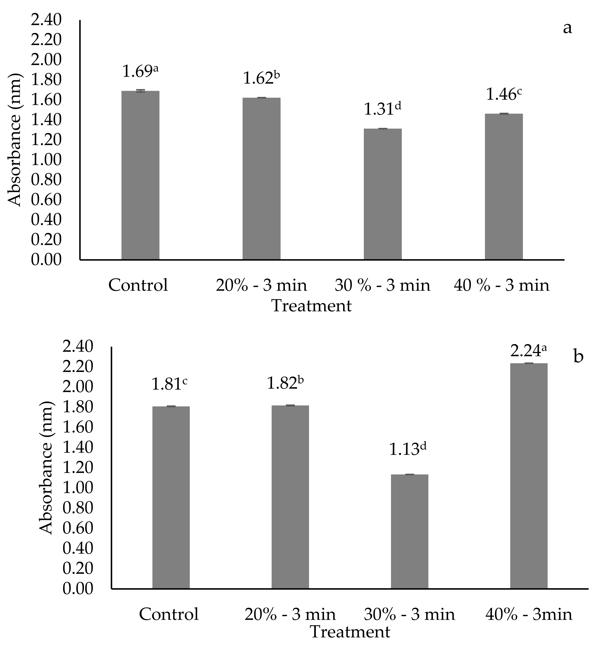
3.6. Proteolysis

The effect on the degree of hydrolysis caused by the US treatments is shown in Figure 6. The treatments showed significant differences (p < 0.05) for both microorganisms in comparison with the control. In relation to L. acidophilus, the control treatment presented the highest degree (p < 0.05) of proteolysis (1.69 ± 0.011) and the treatment with a 30% amplitude presented the lowest degree (p < 0.05) (1.31 ± 0.001). In relation to L. helveticus, as in L. acidophilus, the treatment with a 30% amplitude presented the lowest degree (p < 0.05) of proteolysis (1.13 ± 0.000), and the highest degree of hydrolysis (p < 0.05) was observed in treatment with a 40% amplitude (2.24 ± 0.000). Huang et al. [21] found an increase of approximately 32% in the activity of extracellular proteases of L. paracasei under ultrasound treatment (28 kHz; 100 W/L; 35 min). Lactobacillus spp. strains generally present a set of genes through which adaptation to food matrices and resistance to the conditions of the gastrointestinal tract are facilitated. Milk inoculated with L. helveticus presents a greater proteolytic activity compared to other lactobacilli due to the presence of a powerful proteolytic system [48]. The degree of proteolysis observed by Shokri et al. [42] in a sample subjected to a 23 kHz ultrasound treatment with an amplitude of 10 mm and 5 min had an increase of 36.42% compared to the control, while no significant difference was shown at 3 min. The highest value of proteolysis was presented in the amplitude of 15 mm and 5 min with a value of 47.07% compared to the non-sonicated samples.

3.7. Organic Acids

The effect of an ultrasound on the production of oxalic acid, citric acid, malic acid, succinic acid, acetic acid, and lactic acid after 24 h of fermentation is shown in Table 3. Formic acid was not detected in any of the treatments of the probiotics. Microorganisms can produce a variety of organic acids depending on the growth conditions (growth medium, pH, and temperature) in which the fermentation took place. In this study, lactic, acetic, and citric acids were the organic acids produced in highest concentration by both L. acidophilus and L. helveticus. The highest concentration (p < 0.05) of lactic acid for L. acidophilus occurred in the 40% amplitude treatment (859.40 ± 30.90). In the case of L. helveticus, the 20% amplitude treatment presented the highest concentration (927.51 ± 3.540). The acetic acid concentration for L. acidophilus was higher (p < 0.05) in the control treatment (6.546 ± 0.082) and for L. helveticus, the 20% amplitude treatment (4.953 ± 0.075) presented a higher concentration. The citric acid concentration for L. acidophilus was higher (p < 0.05) in the control treatment (6.115 ± 0.118), and for L. helveticus, the treatments with a 20% amplitude (4.702 ± 0.214), a 30% amplitude (4.537 ± 0.098), and the control treatment (3.786 ± 0.688) presented the highest concentrations without presenting significant differences between them but being significantly different to the treatment with a 40% amplitude (1.051 ± 0.055). For both microorganisms, the concentrations of oxalic and malic acids presented concentrations lower than 1 mM. Dahroud et al. [41] reported lower lactic acid production using an ultrasound (40% amplitude for 30 s) during fermentation with L. casei subsp. casei in an MRS broth medium with 6 g/L peptone. Similarly, Shokri et al. [42], using Leu. Mesenteroides in a lactose-enriched medium, observed a lower production of lactic, acetic, and citric acid when using a 20% amplitude for 5 min for 10 h of incubation. The higher values observed for L. acidophilus and L. helveticus in this study were favored by the effects of ultrasounds and the use of homofermentative bacteria.

4. Conclusions

This investigation addressed the effects of an ultrasound with a frequency of 20 kHz and 20%, 30%, and 40% amplitudes for 3 min, on the growth kinetics and fermentative profile of L. acidophilus and L. helveticus. In general, the ultrasound had an effect on the kinetic parameters, membrane permeability, fermentative profile, acidification, β-galactosidase activity, and proteolysis. Particularly, the observed effects were dependent on the amplitude and the microorganism (probiotic used). The treatment with a 30% amplitude for 3 min was best for both probiotics in terms of increasing the rate and the β-galactosidase activity. Although, L. acidophilus presented more than double the activity of L. helveticus. The cell viability was greater for both probiotics in the treatment with a 20% amplitude for 3 min; however, L. acidophilus was more resistant to the ultrasound process. The results of this study can be used in subsequent investigations that seek to design and optimize the fermentative processes of L. acidophillus and L. helveticus.

Author Contributions

Conceptualization, A.C.-M. and R.A.R.-V.; data curation, E.S.-E.; formal analysis, N.A.B.-J., G.P.E.-S., D.C.-F., M.M.A.-S. and E.S.-E.; funding acquisition, A.C.-M.; investigation, N.A.B.-J. and M.M.A.-S.; methodology, A.C.-M., D.C.-F., M.M.A.-S., G.P.E.-S. and R.A.R.-V.; project administration, A.C.-M.; supervision, A.C.-M., R.A.R.-V., R.S.-V. and A.L.R.-M.; writing—original draft, N.A.B.-J.; writing—review and editing, A.C.-M., A.L.R.-M., R.S.-V., E.S.-E. and R.A.R.-V. All authors have read and agreed to the published version of the manuscript.

Funding

This research received no external funding.

Data Availability Statement

The data presented in this study are available on request from the corresponding author.

Acknowledgments

The authors acknowledge that the Universidad Autónoma de Chihuahua supported this investigation. The Science and Technology National Council of Mexico (CONACYT) provided a graduate study scholarship for Norma Angélica Bolivar-Jacobo.

Conflicts of Interest

The authors declare no conflict of interest.

References

  1. Malashree, L.; Angadi, V.; Yadav, K.S.; Prabha, R. “Postbiotics”—One Step Ahead of Probiotics. Int. J. Curr. Microbiol. Appl. Sci. 2019, 8, 2049–2053. [Google Scholar] [CrossRef]
  2. Taranto, M.; Médici, M.; Font de Valdez, G. Alimentos funcionales probióticos Dras. María Pía Taranto, Marta Médici y Graciela Font de Valdez * Probiotic functional foods. Aliment. Pharmacol. Ther. 2005, 4, 26–34. [Google Scholar]
  3. Guimarães, J.T.; Balthazar, C.F.; Scudino, H.; Pimentel, T.C.; Esmerino, E.A.; Ashokkumar, M.; Freitas, M.Q.; Cruz, A.G. High-intensity ultrasound: A novel technology for the development of probiotic and prebiotic dairy products. Ultrason. Sonochem. 2019, 57, 12–21. [Google Scholar] [CrossRef]
  4. Assimos, D.G. Re: Metabolomic Profiling of Oxalate-Degrading Probiotic Lactobacillus acidophilus and Lactobacillus gasseri. J. Urol. 2020, 203, 247–248. [Google Scholar] [CrossRef]
  5. Santiago-López, L.; Hernández-Mendoza, A.; Garcia, H.S.; Mata-Haro, V.; Vallejo-Cordoba, B.; González-Córdova, A.F. The effects of consuming probiotic-fermented milk on the immune system: A review of scientific evidence. Int. J. Dairy Technol. 2015, 68, 153–165. [Google Scholar] [CrossRef]
  6. Bolivar Jacobo, N.A.; Reyes Villagrana, R.A.; Chávez-Martínez, A. Relación entre probióticos—Postbióticos y sus principales efectos bioactivos. Tecnociencia Chihuah. 2021, 15, 124–139. [Google Scholar] [CrossRef]
  7. Liu, Q.; Yu, Z.; Tian, F.; Zhao, J.; Zhang, H.; Zhai, Q.; Chen, W. Surface components and metabolites of probiotics for regulation of intestinal epithelial barrier. Microb. Cell Fact. 2020, 19, 23. [Google Scholar] [CrossRef] [PubMed] [Green Version]
  8. Rad, A.H.; Hosseini, S.; Moghadaszadeh, M. Postbiotics as Dynamic Biological Molecules and Their Antimicrobial Activity: A Review Literature Review: Postbiotics. Biointerface Res. Appl. Chem. 2021, 10, 317. [Google Scholar]
  9. Rad, A.H.; Aghebati-Maleki, L.; Kafil, H.S.; Gilani, N.; Abbasi, A.; Khani, N. Postbiotics, as dynamic biomolecules, and their promising role in promoting food safety. Biointerface Res. Appl. Chem. 2021, 11, 14529–14544. [Google Scholar] [CrossRef]
  10. Plaza-Diaz, J.; Ruiz-Ojeda, F.J.; Gil-Campos, M.; Gil, A. Mechanisms of Action of Probiotics. Adv. Nutr. 2019, 10, S49–S66. [Google Scholar] [CrossRef] [Green Version]
  11. Koleilat, A. Beyond probiotics the Postbiotics. Gastroenterol. Hepatol. Open Access 2019, 10, 324–326. [Google Scholar] [CrossRef]
  12. Rad, A.H.; Maleki, L.A.; Kafil, H.S.; Zavoshti, H.F.; Abbasi, A. Postbiotics as novel health-promoting ingredients in functional foods. Health Promot. Perspect. 2020, 10, 3–4. [Google Scholar] [CrossRef] [Green Version]
  13. Teame, T.; Wang, A.; Xie, M.; Zhang, Z.; Yang, Y.; Ding, Q.; Gao, C.; Olsen, R.E.; Ran, C.; Zhou, Z. Paraprobiotics and Postbiotics of Probiotic Lactobacilli, Their Positive Effects on the Host and Action Mechanisms: A Review. Front. Nutr. 2020, 7, 570344. [Google Scholar] [CrossRef] [PubMed]
  14. Żółkiewicz, J.; Marzec, A.; Ruszczyński, M.; Feleszko, W. Postbiotics—A step beyond pre-and probiotics. Nutrients 2020, 12, 2189. [Google Scholar] [CrossRef] [PubMed]
  15. Moradi, M.; Kousheh, S.A.; Almasi, H.; Alizadeh, A.; Guimarães, J.T.; Yılmaz, N.; Lotfi, A. Postbiotics produced by lactic acid bacteria: The next frontier in food safety. Compr. Rev. Food Sci. Food Saf. 2020, 19, 3390–3415. [Google Scholar] [CrossRef] [PubMed]
  16. Aguilar-Toalá, J.E.; Garcia-Varela, R.; Garcia, H.S.; Mata-Haro, V.; González-Córdova, A.F.; Vallejo-Cordoba, B.; Hernández-Mendoza, A. Postbiotics: An evolving term within the functional foods field. Trends Food Sci. Technol. 2018, 75, 105–114. [Google Scholar] [CrossRef]
  17. Tomasik, P.; Tomasik, P. Probiotics, non-dairy prebiotics and postbiotics in nutrition. Appl. Sci. 2020, 10, 1740. [Google Scholar] [CrossRef] [Green Version]
  18. Shin, H.S.; Park, S.Y.; Lee, D.K.; Kim, S.A.; An, H.M.; Kim, J.R.; Kim, M.J.; Cha, M.G.; Lee, S.W.; Kim, K.J.; et al. Hypocholesterolemic effect of sonication-killed Bifidobacterium longum isolated from healthy adult Koreans in high cholesterol fed rats. Arch. Pharm. Res. 2010, 33, 1425–1431. [Google Scholar] [CrossRef] [PubMed]
  19. Del Rocio Ruiz-Briseño, M.; Sánchez-Reyes, K.; Alvarez-Zavala, M.; González-Hernández, L.A.; Ramos-Solano, M.; Andrade-Villanueva, J.F. Homeostasis intestinal: Colaboración del sistema inmune con la microbiota. Rev. Médica MD 2018, 9, 337–340. [Google Scholar]
  20. Ghosh, S.; Whitley, C.S.; Haribabu, B.; Jala, V.R. Regulation of Intestinal Barrier Function by Microbial Metabolites. Cell. Mol. Gastroenterol. Hepatol. 2021, 11, 1463–1482. [Google Scholar] [CrossRef]
  21. Huang, G.; Chen, S.; Tang, Y.; Dai, C.; Sun, L.; Ma, H.; He, R. Stimulation of low intensity ultrasound on fermentation of skim milk medium for yield of yoghurt peptides by Lactobacillus paracasei. Ultrason. Sonochem. 2019, 51, 315–324. [Google Scholar] [CrossRef] [PubMed]
  22. Mason, T.J.; Chemat, F.; Ashokkumar, M. Power Ultrasonics for Food Processing; Elsevier Ltd.: Amsterdam, The Netherlands, 2015; ISBN 9781782420361. [Google Scholar] [CrossRef]
  23. Humphrey, V.F. Ultrasound and matter—Physical interactions. Prog. Biophys. Mol. Biol. 2007, 93, 195–211. [Google Scholar] [CrossRef]
  24. Lentacker, I.; De Cock, I.; Deckers, R.; De Smedt, S.C.; Moonen, C.T.W. Understanding ultrasound induced sonoporation: Definitions and underlying mechanisms. Adv. Drug Deliv. Rev. 2014, 72, 49–64. [Google Scholar] [CrossRef] [Green Version]
  25. Lee, J.; Martini, S. Modifying the physical properties of butter using high-intensity ultrasound. J. Dairy Sci. 2019, 102, 1918–1926. [Google Scholar] [CrossRef] [Green Version]
  26. Delalande, A.; Kotopoulis, S.; Postema, M.; Midoux, P.; Pichon, C. Sonoporation: Mechanistic insights and ongoing challenges for gene transfer. Gene 2013, 525, 191–199. [Google Scholar] [CrossRef] [PubMed]
  27. Williams, E.G.; Mann, J.A. Fourier Acoustics: Sound Radiation and Nearfield Acoustical Holography. J. Acoust. Soc. Am. 2000, 108, 1373. [Google Scholar] [CrossRef] [Green Version]
  28. Kooiman, K.; Vos, H.J.; Versluis, M.; De Jong, N. Acoustic behavior of microbubbles and implications for drug delivery. Adv. Drug Deliv. Rev. 2014, 72, 28–48. [Google Scholar] [CrossRef] [PubMed]
  29. de Almada, C.N.; Almada, C.N.; Martinez, R.C.R.; Sant’Ana, A.S. Paraprobiotics: Evidences on Their Ability to Modify Biological Responses, Inactivation Methods and Perspectives on Their Application in Foods. Trends Food Sci. Technol. 2016, 58, 96–114. [Google Scholar] [CrossRef]
  30. Cabeza, E.A. Bacterias ácido-lácticas (BAL): Aplicaciones como cultivos estárter para la industria láctea y cárnica. In Proceedings of the Simposio Regional de Microbiología: Microorganismos Eficientes en el Sector Productivo, Barranquilla, Colombia, 15–16 September 2006; Volume 14, pp. 549–566. [Google Scholar] [CrossRef]
  31. Gholamhosseinpour, A.; Hashemi, S.M.B. Ultrasound pretreatment of fermented milk containing probiotic Lactobacillus plantarum AF1: Carbohydrate metabolism and antioxidant activity. J. Food Process Eng. 2019, 42, e12930. [Google Scholar] [CrossRef] [Green Version]
  32. Gotoh, M.; Sashihara, T.; Ikegami, S.; Yamaji, T.; Kino, K.; Orii, N.; Taketomo, N.; Okubo, K. Efficacy of oral administration of a heat-killed Lactobacillus gasseri OLL2809 on patients of japanese cedar pollinosis with high japanese-cedar pollen-specific IgE. Biosci. Biotechnol. Biochem. 2009, 73, 1971–1977. [Google Scholar] [CrossRef]
  33. Chen, W. Lactic Acid Bacteria Omics and Functional Evaluation; Springer: Berlin/Heidelberg, Germany, 2019; ISBN 9783319600215. [Google Scholar] [CrossRef]
  34. Taverniti, V.; Guglielmetti, S. Health-promoting properties of Lactobacillus helveticus. Front. Microbiol. 2012, 3, 392. [Google Scholar] [CrossRef] [Green Version]
  35. Ianni, F.; Altomare, A.A.; Cenci-Goga, B.T.; Blasi, F.; Grispoldi, L.; Regazzoni, L.; Cossignani, L. Chromatographic characterization and in vitro bioactivity evaluation of lactobacillus helveticus hydrolysates upon fermentation of different substrates. Appl. Sci. 2021, 11, 811. [Google Scholar] [CrossRef]
  36. Bolivar-Jacobo, N.A.; Reyes-Villagrana, R.A.; Rentería-Monterrubio, A.L.; Sánchez-Vega, R.; Santellano-Estrada, E.; Tirado-Gallegos, J.M.; Chávez-Martínez, A. Culture Age, Growth Medium, Ultrasound Amplitude, and Time of Exposure Influence the Kinetic Growth of Lactobacillus acidophilus. Fermentation 2023, 9, 63. [Google Scholar] [CrossRef]
  37. Molecular Probes Inc. LIVE/DEAD® BacLightTM Bacterial Viability and Counting Kit; Product Information; Molecular Probes Inc.: Eugene, OR, USA, 2004; pp. 1–5. [Google Scholar]
  38. Ibrahim, A.H. Enhancement of β-galactosidase activity of lactic acid bacteria in fermented camel milk. Emir. J. Food Agric. 2018, 30, 256–267. [Google Scholar] [CrossRef] [Green Version]
  39. Bradford, M.M. A Rapid and Sensitive Method for the Quantitation of Microgram Quantities of Protein Utilizing the Principle of Protein-Dye Binding. Anal. Biochem. 1976, 72, 248–254. [Google Scholar] [CrossRef]
  40. Donkor, O.N.; Henriksson, A.; Vasiljevic, T.; Shah, N.P. Probiotic Strains as Starter Cultures Improve Angiotensin-Converting Enzyme Inhibitory Activity in Soy Yogurt. J. Food Sci. 2005, 70, m375–m381. [Google Scholar] [CrossRef]
  41. Dahroud, B.D.; Mokarram, R.R.; Khiabani, M.S.; Hamishehkar, H.; Bialvaei, A.Z.; Yousefi, M.; Kafil, H.S. Low Intensity Ultrasound Increases the Fermentation Efficiency of Lactobacillus Casei Subsp.Casei ATTC 39392. Int. J. Biol. Macromol. 2016, 86, 462–467. [Google Scholar] [CrossRef] [PubMed]
  42. Shokri, S.; Shekarforoush, S.S.; Hosseinzadeh, S. Efficacy of Low Intensity Ultrasound on Fermentative Activity Intensification and Growth Kinetic of Leuconostoc Mesenteroides. Chem. Eng. Process.—Process Intensif. 2020, 153, 107955. [Google Scholar] [CrossRef]
  43. Shokri, S.; Terefe, N.S.; Shekarforoush, S.S.; Hosseinzadeh, S. Ultrasound-Assisted Fermentation for Enhancing Metabolic and Probiotic Activities of LactoBacillus Brevis. Chem. Eng. Process.—Process Intensif. 2021, 166, 108470. [Google Scholar] [CrossRef]
  44. Abesinghe, A.M.N.L.; Islam, N.; Vidanarachchi, J.K.; Prakash, S.; Silva, K.F.S.T.; Karim, M.A. Effects of Ultrasound on the Fermentation Profile of Fermented Milk Products Incorporated with Lactic Acid Bacteria. Int. Dairy J. 2019, 90, 1–14. [Google Scholar] [CrossRef] [Green Version]
  45. Potoroko, I.; Kalinina, I.; Botvinnikova, V.; Krasulya, O.; Fatkullin, R.; Bagale, U.; Sonawane, S.H. Ultrasound Effects Based on Simulation of Milk Processing Properties. Ultrason. Sonochem. 2018, 48, 463–472. [Google Scholar] [CrossRef] [PubMed]
  46. Li, Y.; Chen, Z.; Ge, S. Sonoporation: Underlying Mechanisms and Applications in Cellular Regulation. BIO Integr. 2021, 2, 29–36. [Google Scholar] [CrossRef]
  47. Nguyen, T.M.P.; Lee, Y.K.; Zhou, W. Stimulating Fermentative Activities of Bifidobacteria in Milk by Highintensity Ultrasound. Int. Dairy J. 2009, 19, 410–416. [Google Scholar] [CrossRef]
  48. Griffiths, M.W.; Tellez, A.M. Lactobacillus Helveticus: The Proteolytic System. Front. Microbiol. 2013, 4, 30. [Google Scholar] [CrossRef] [PubMed] [Green Version]
Figure 1. Effect of ultrasound (20 kHz) on the growth kinetics of L. acidophilus (a) and L. helveticus (b) at 37 ± 1 °C.
Fermentation 09 00356 g001
Figure 2. Effect of ultrasound (20 kHz) on the growth percentage of L. acidophilus (a) and L. helveticus (b) at 37 ± 1 °C.
Fermentation 09 00356 g002
Figure 3. Flow cytometric analysis of probiotics undergoing ultrasound treatment. (A) L. acidophilus and (B) L. helveticus.
Fermentation 09 00356 g003
Figure 4. Effect of ultrasound (20 kHz) on the pH kinetics of L. acidophilus (a) and L. helveticus (b).
Fermentation 09 00356 g004aFermentation 09 00356 g004b
Figure 5. Effect of ultrasound (20 kHz) on the protein concentration of L. acidophilus (a) and L. helveticus (b). a,b,c,d Different literals in column indicate significant differences between treatments (p < 0.05).
Fermentation 09 00356 g005
Figure 6. Effect of ultrasound (20 kHz) on the proteolysis of L. acidophilus (a) and L. helveticus (b). a,b,c,d Different literals in column indicate significant differences between treatments (p < 0.05).
Fermentation 09 00356 g006
Table 1. Effect of ultrasound on the kinetic growth parameters of L. acidophilus and L. helveticus. (mean ± standard deviation).
TreatmentYoLag (hours)Rate (μmax) (1/Hours)Tmax (Hours)R2
L. acidophilus
Control8.08± 0.000 a0.870 ± 0.017 b0.500 ± 0.014 b2.578 ± 0.024 b0.96
20%—3 min8.08± 0.000 a0.861 ± 0.019 b0.470 ± 0.014 b2.622 ± 0.031 b0.93
30%—3 min8.08± 0.000 a0.923 ± 0.000 a1.065 ± 0.000 a1.998 ± 0.000 c0.93
40%—3 min8.08± 0.000 a0.748 ± 0.003 c0.475 ± 0.007 b2.985 ± 0.031 a0.91
L. helveticus
Control8.08± 0.000 a0.696 ± 0.006 a0.395 ± 0.007 b2.886 ± 0.003 c0.95
20%—3 min8.08± 0.000 a0.573 ± 0.017 b0.410 ± 0.000 a2.720 ± 0.018 d0.96
30%—3 min8.08± 0.000 a0.391 ± 0.035 c0.410 ± 0.000 a3.272 ± 0.026 b0.94
40%—3 min8.08± 0.000 a0.390 ± 0.046 c0.225 ± 0.001 c4.174 ± 0.006 a0.94
Rate (maximum growth potential), Lag (setting constant or adaptation process of the bacteria), yo (initial count), Tmax (time in which the exponential phase ends), and R2 (coefficient of adjustment to the equation). a,b,c,d Different literals in the same column of each microorganism indicate significant differences between treatments (p < 0.05).
Table 2. Effect of ultrasound (20 kHz) on the β-galactosidase (U) of L. acidophilus and L. helveticus.
TreatmentProbiotic
L. acidophilusL. helveticus
Control268.66 ± 0.00 c87.54 ± 0.00 c
20%—3 min318.23 ± 0.00 b137.11 ± 0.00 b
30%—3 min452.8 ± 0.00 a159.78 ± 0.00 a
40%—3 min77.43 ± 0.00 d70.54 ± 0.00 d
a,b,c,d Different letters in the same column mean statistically significant difference among the values of the same parameter, according to the test of Tukey (α < 0.05). ± Standard deviations with respect to the mean values of triplicate runs.
Table 3. Effects of ultrasounds (20 kHz) on the production of organic acids (mM) of L. acidophilus and L. helveticus.
TreatmentOxalic AcidCitric AcidMalic AcidSuccinic AcidAcetic AcidLactic Acid
L. acidophilus
Control0.053 ± 0.013 b6.115 ± 0.118 a0.792 ± 0.020 a1.283 ± 0.075 a6.546 ± 0.082 a716.45 ± 0.970 b
20%—3 min0.102 ± 0.007 a4.297 ± 0.034 b0.153 ± 0.013 a1.126 ± 0.080 a5.773 ± 0.082 b637.20 ± 8.170 b
30%—3 min0.053 ± 0.003 b4.757 ± 0.780 ab0.371 ± 0.257 a1.174 ± 0.019 a5.972 ± 0.028 b681.60 ± 26.30 b
40%—3 min0.045 ± 0.001 b4.206 ± 0.342 b0.547 ± 0.186 a1.610 ± 0.346 a4.145 ± 0.044 c859.40 ± 30.90 a
L. helveticus
Control0.064 ± 0.003 a3.786 ± 0.688 a0.313 ± 0.123 b1.567 ± 0.168 a4.643 ± 0.032 b841.00 ± 15.40 b
20%—3 min0.056 ± 0.001 ab4.702 ± 0.214 a0.587 ± 0.016 ab1.850 ± 0.165 a4.953 ± 0.075 a927.51 ± 3.540 a
30%—3 min0.050 ± 0.004 b4.537 ± 0.098 a0.594 ± 0.053 a1.676 ± 0.073 a4.457 ± 0.008 b824.45 ± 3.690 b
40%—3 min0.013 ± 0.001 c1.051 ± 0.055 bNDND0.074 + 0.104 c15.710 ± 3.250 c
a,b,c Different literals in the same column of each microorganism indicate significant differences between treatments (p < 0.05). ND: not detected.
Disclaimer/Publisher’s Note: The statements, opinions and data contained in all publications are solely those of the individual author(s) and contributor(s) and not of MDPI and/or the editor(s). MDPI and/or the editor(s) disclaim responsibility for any injury to people or property resulting from any ideas, methods, instructions or products referred to in the content.