Article

Improved Extraction Yield, Water Solubility, and Antioxidant Activity of Lentinan from Lentinula edodes via Bacillus subtilis natto Fermentation

1
Research Center of Food Nutrition and Safety, School of Life Sciences, Jianghan University, Wuhan 430056, China
2
Hubei Province Engineering Research Center for Legume Plants, Wuhan 430056, China
3
Hubei Enterprise-College Joint Innovation Center of Healthy Sugar Substitute Product, Wuhan 430056, China
*
Author to whom correspondence should be addressed.
Fermentation 2023, 9(4), 333; https://doi.org/10.3390/fermentation9040333
Submission received: 19 February 2023 / Revised: 20 March 2023 / Accepted: 24 March 2023 / Published: 27 March 2023
(This article belongs to the Special Issue Bioactive Products from Edible and Medicinal Fungi by Fermentation)

Abstract

:
Lentinan has important applications in the food and medicine fields. Fermenting Lentinula edodes with Bacillus subtilis natto increased the lentinan extraction yield by 87.13% and greatly altered the molecular structure and antioxidant activity of lentinan. The uronic acid content in the lentinan molecular structure increased from 2.08% to 4.33%. The fermentation process did not affect the monosaccharide composition of lentinan, comprised of more than 90% glucose residues. Fermentation significantly reduced the molecular weight of lentinan and altered its apparent structure. The water solubility of fermented lentinan was increased by 165.07%, and the antioxidant activity was significantly improved. Fermentation using soybean as a substrate may be beneficial for enhancing the activity of Bacillus subtilis natto and producing lentinan with different molecular weights.

1. Introduction

Lentinula edodes is an edible and medicinal mushroom used as a food ingredient because of its high nutritional value and pleasant flavor. Lentinan extracted from L. edodes has various biological activities, including antitumor, antioxidant, immunomodulatory, antiviral, and anti-inflammatory effects (Yin et al., 2018; Ahn et al., 2017; Ziaja-Sołtys et al., 2020; Wang et al., 2019; Ren et al., 2018) [1,2,3,4,5]. Therefore, many studies have focused on lentinan, including its extraction, purification, structural characterization, modification, and bioactivity. Structural characteristics of polysaccharides, such as their molecular weight (Mw), chemical composition, and branching configuration, largely contribute to their biological activities (Morales et al., 2020; Wang et al., 2018; Li et al., 2016) [6,7,8]. Therefore, different extraction methods can result in different yields, structural characteristics, and bioactivities of polysaccharides (Chen et al., 2018) [9]. For example, gamma irradiation, at a dose of 15 KGy, increased the yield of lentinan obtained via hot-water extraction from 2.01 ± 0.53% to 7.17 ± 0.78%. However, irradiation degraded the microstructures and reduced the molecular weights of the polysaccharides (Akram et al., 2017) [10].
In some studies, the structure of lentinan was modified using physical, chemical, and biological methods to improve its biological activity. Lentinan can be modified by calcium carbonate. The average size of CaCO3 -LNT was slightly larger than unmodified CaCO3, which enhances the expression of MHC-II and CD86 in lymph node dendritic cells (He et al., 2020) [11]. Lentinan was also modified using the nitric acid-sodium selenite method. Lentinan’s surface structure and main elemental components significantly changed after selenizing, increasing its total antioxidant capacity (Ren et al., 2015) [12]. Methods used for polysaccharide modification mainly involve chemical modification rather than physical and microbial modification. However, chemical modification typically requires strong acids, organic solvents, and a strong base, greatly limiting their application in production (Huang et al., 2020) [13]. Biotransformation by microbial fermentation, as a more environmentally friendly, efficient, and suitable mass production method, has received increased attention. Fermentation by Lactobacillus fermentum can change the physicochemical properties, enhance the solubility, and improve the immunomodulatory and prebiotic activities of polysaccharides from longan pulp (Huang et al., 2019) [14]. Additionally, microbial transformation affects the biological functions of isoflavonoids and dietary fiber. Isoflavonoids in Astragalus membranaceus were fermented with Lactiplantibacillus plantarum MG5276; this bioconversion improved the antioxidant activity both in vitro and in vivo (Lee et al., 2022) [15]. Soluble dietary fiber from defatted rice bran obtained through Trichoderma viride fermentation exhibited better water solubility, water-holding capacity, oil-holding capacity and cholesterol absorption capacity (Jia et al., 2019) [16]. Bacillus subtilis natto is a probiotic commonly used in natto fermentation with soybean as the substrate. The content of soluble dietary fiber fermented by B. subtilis natto from millet bran was increased from 2.3% to 13.2%. Meanwhile, the binding and DPPH free radical scavenging capacities were enhanced (Chu et al., 2019) [17].
The study was conducted to analyze the effect of fermentation by B. subtilis natto on the extraction yield, physical and chemical properties, structural characteristics, water solubility, and antioxidant activities of L. edodes under two fermentation conditions, with and without soybean as a substrate. In addition, this study provides a method for the extraction and biotransformation of lentinan from L. edodes.

2. Materials and Methods

2.1. Materials and Reagents

Dried L. edodes were obtained from Fangxian County, Hubei province, China. The dried L. edodes were placed in a laboratory oven at 70 °C for 6 h, resulting in a moisture content ≤ 8%. Dried soybeans were obtained from Jixian Town, Shuangyashan City, Heilongjiang province, China. Bacillus subtilis natto was purchased from the China Center of Industrial Culture Collection (Beijing, China; CICC10263; Country of Origin, Japan); Luria–Bertani liquid medium (Tryptone 10 g/L, Yeast extract 5 g/L, NaCl 10 g/L, pH 7.4); The reagents used in the experiment are listed in Table S1.

2.2. Preparation of Lentinan

The crushed samples were fermented using Bacillus subtilis natto. And then, the fermented and unfermented samples were used to extract lentinan. The details are shown in Figure 1.

2.2.1. Fermentation Method

Bacillus subtilis natto was inoculated into Luria–Bertani liquid medium and incubated at 37 °C for 12 h for sample fermentation. Lentinula edodes and soybean samples were obtained by crushing and sieving (60-mesh sieve, 0.25 mm). The samples were combined in three different proportions: 24 g L. edodes powder (LNT), 12 g L. edodes powder and 12 g soybean powder (LDM), and 24 g soybean powder (DLP). The three samples were sterilized by autoclave sterilization (121 °C for 20 min), cooled at room temperature, mixed with sterile water (1:1, g/mL), and incubated with 5% inoculum above grown in LB medium. The samples were fermented with a static method in a culture bottle with a breathable film at 37 °C for 3 days, with 5 mL sterile water added at 24 and 48 h. Finally, the three fermented samples were vacuum-freeze-dried for lentinan extraction.

2.2.2. Extraction Method of Lentinan

Lentinan from fermented and unfermented samples was extracted by hot water extraction as previously reported by Olawuyi et al. and Zhu et al. with some modifications (Olawuyi et al., 2021; Zhu et al., 2022) [18,19]. The dried samples were extracted with ethanol (w/v = 1:10) for 6 h to remove fat and small molecules. The precipitate was extracted with distilled water (w/v = 1:20) at 95 °C for 6 h. After centrifugation (at 4000× g for 10 min), the supernatant was deproteinated with 3% trichloroacetic acid (v/v = 1:1) overnight at 4 °C. The supernatant was obtained by centrifugation (at 7000× g for 10 min) and then precipitated with the same volume of ethanol overnight at 4 °C. After centrifugation (at 7000× g for 10 min), the precipitate was vacuum freeze-dried for 12 h to obtain the lentinan samples. The extraction yield (%) was calculated using the following equation (Equation (1)).
Yield   ( % ) = weight   of   dried   crudes   polysaccharides ( g ) weight   of   dry   materials   ( g ) × 100 %

2.3. Structural Characteristics of Lentinan

2.3.1. Analysis of Chemical Composition

The total sugar content in the sample was determined using the phenol-sulfuric acid method (Dubios et al., 1956) [20]. A standard curve of glucose content was prepared by using glucose solution as a standard to obtain a linear regression equation: y = 3.3736x + 0.0721, R2 = 0.997. The sample solution was measured according to the linear regression equation.
The uronic acid in the sample was determined using the m-hydroxydiphenyl method (Blumenkrantz et al., 1973) [21]. A standard curve was prepared by using the galacturonic acid solution as a standard to obtain a linear regression equation: y = 3.5057x + 0.0481, R2= 0.998. Then, the sample solution was measured according to the linear regression equation.
The protein content of the sample was measured by the Bradford method (Bradford et al., 1976) [22]. A standard curve was prepared by using the BSA solution as a standard to obtain a linear regression equation: y = 1.2414x + 0.1951, R2 = 0.997. The sample solution was measured according to the linear regression equation.

2.3.2. Analysis of Monosaccharide Composition

The monosaccharide composition of the lentinan samples was measured using high-performance liquid chromatography (U3000, Thermo Fisher Scientific, Waltham, MA, USA) equipped with a ZORBAX EclipseXDB-C18 column (Agilent Technologies, Santa Clara, CA, USA) as described in a previous report with slight modifications (Pei et al., 2022) [23]. The sample (5 mg) was hydrolyzed with 2 mol/L trifluoroacetic acid at 121 °C for 2 h. Then, it was dried under nitrogen flow and add 3 mL of methanol, so repeated the addition of methanol and blow dry 3 times with nitrogen to remove TFA. Subsequently, 5 mL of sterile water was added for vortex mixing. 0.2 mL polysaccharide hydrolysate solution was placed in a 5 mL stoppered test tube, then 0.2 mL 0.5 mol/L NaOH solution and 0.5 mL 0.5 mol/L 1-phenyl-3-methyl-5-pyrazolone solution (PMP) was added and vortexed, after reacting in a 70 °C water bath for 1 h. After the water bath, 0.2 mL 0.5 mol/L HCl was added for neutralization (pH 6–7). Then 1 mL chloroform was added, vortexed mixing and shaken, allowed to stand, and the chloroform phase was discarded and thus extracted thrice. After derivation with PMP, the sample was eluted with acetonitrile and 0.09 mol/L monopotassium phosphate buffer (v/v = 17:83) at 30 °C and a flow rate of 0.8 mL/min. The injection volume of the sample was 10 µL, and the UV detection wavelength was 250 nm. Fucose, rhamnose, glucosamine hydrochloride, galactose amino hydrochloride, arabinose, galactose, glucose, xylose, mannose, ribose, galacturonic acid, and glucuronic acid were used as standards.

2.3.3. Analysis of Molecular Weight

The Mw of the lentinan samples was determined using high-performance gel permeation chromatography equipped with a polymer matrix water-soluble SEC column (8 mm × 300 mm) 3 in series (OHpak SB-803 HQ, Ohpak SB-804 HQ, Ohpak SB-805 HQ) as previously described with minor modifications (Zhu et al., 2022) [19]. Ten kinds of standard dextran with different molecular weights of 1000, 5000, 12,000, 25,000, 50,000, 80,000, 150,000, 270,000, 410,000 and 670,000 were dissolved in 0.05 mol/L NaCl solution to make the control solution with a concentration of 5 mg/mL. The 5 mg of samples were dissolved into l mL 0.05 mol/L NaCl solution, and the sample solution with a 5 mg/mL concentration was prepared. The reference and sample solutions were centrifuged at 7000× g for 10 min and then entered the chromatographic column (0.22 μm). The experimental conditions were as follows: column oven temperature of 40 °C, flow rate of 0.65 mL/min, and injection volume of 30 μL.

2.3.4. Fourier-Transform Infrared Spectra Analyses

Fourier transform infrared spectral analyses of lentinan samples was performed according to a previously reported method with some modifications (Wu et al., 2021) [24]. The powder samples (2 mg) were dried and mixed with KBr pellets for FT-IR analysis. The FTIR spectra were recorded using a Fourier transform infrared spectrophotometer (NICOLET iS50R, Thermo Fisher Scientific, Waltham, MA, USA) in the range of 4000–400 cm−1.

2.3.5. Scanning Electron Microscopy

The morphology of the samples was observed by scanning electron microscopy (FEI, Hillsboro, OR, USA). The lentinan samples were applied to conductive carbon tape and spray-coated with gold; the surface structure of lentinan was observed and photographed at 200×, 2000×, and 5000× magnification using scanning electron microscopy [25].

2.4. Water-Holding Capacity and Water Solubility Capacity

The water holding capacity (WHC) and water solubility capacity (WSC) were determined in triplicate as previously described with minor modifications (Jia et al., 2019; Ahmed et al., 2013) [16,26]. Lentinan (0.050 g) was suspended in 1 mL of distilled water on a vortex mixer, and the mixture was kept at 25 °C for 20 h. After centrifugation (16,000× g for 40 min), the precipitate was dried at 70 °C for 6 h and weighed. The supernatant was precipitated with 3 volumes of anhydrous ethanol overnight at 4 °C. After centrifugation (16,000× g for 10 min), the precipitate was collected, lyophilized, and weighed. The WHC and WSC were calculated as percentages using the following equations (Equations (2) and (3)):
  WHC   ( % )   = Total   sample   weight   after   water   absorption   Total   dry   sample   weight × 100 %
WSC   ( % ) =   Total   carbohydrate   concentration   in   supernatant Total   dry   sample   weight × 100 %

2.5. Antioxidant Activities In Vitro

2.5.1. DPPH Free Radical Scavenging Ability

DPPH free radical scavenging ability was determined with some modifications described by Duan et al. (Duan et al., 2019) [27]. 0.2 mL lentinan solution (0.125, 0.25, 0.50, and 1.0 mg/mL) and 0.2 mL DPPH solution (0.1 mmol/L, formulated with 50.0% ethanol) were mixed. And the mixture was incubated at room temperature for 30 min in the dark. Then the absorbance of the mixture was measured at 517 nm, and ethanol (50.0%) was used as a blank control. The results were expressed as DPPH (%), and the DPPH scavenging rate was calculated as follows (Equation (4)):
  DPPH   scavenging   activity   ( % ) = ( 1   A 2   A 1 A 0 ) × 100
where A0 is the absorbance of the comparison (0.2 mL of distilled water instead of sample), A1 is the absorbance of the sample and 50% ethanol, and A2 is the absorbance of the sample and DPPH.
A trolox calibration curve was prepared for a concentration range of 0–600 μmol/L, and the inhibition percentage obtained for the sample was interpolated to calculate the concentration in trolox equivalents (l μmol/L TE) (Zulueta et al., 2009; Fimbres-Olivarria et al., 2018) [28,29]. The equation of the calibration curve was y = 0.1651x + 3.2732 with a good correlation coefficient (R2 = 0.993).

2.5.2. ABTS+ Free Radical Scavenging Ability

ABTS+ free radical scavenging ability was determined as described by Li et al. (Li et al., 2016) [30]. First, 7.4 mmol/L ABTS+ solution was mixed with 3.8 mmol/L potassium persulphate (v/v = 1:1) at room temperature in the dark for 12 h. Next, 25 μL lentinan solution (0.5, 1.0, 2.0, and 4.0 mg/mL) and 250 μL of ABTS+ working solution were mixed thoroughly and incubated for 15 min in the dark; the absorbance was measured at 734 nm. The ABTS+ free radical scavenging activity was calculated using the following formula (Equation (5)):
  ABTS +   scavenging   activity   ( % ) = ( 1   A 2   A 1 A 0 ) × 100
where A0 is the absorbance of the comparison (25 μL of distilled water instead of the sample), A1 is the absorbance of the sample and phosphate-buffered saline, and A2 is the absorbance of the sample and ABTS+.
The equation of the trolox calibration curve was y = 0.1363x + 3.8306, with a good correlation coefficient (R2 = 0.998).

2.5.3. Ferric-Reducing Antioxidant Power Assay

The ferric-reducing antioxidant power (FRAP) assay was conducted as described by Liu et al. (Liu et al., 2020) [31]. The FRAP working solution was prepared by mixing 300 mmol/L acetate buffer, 10 mmol/L 2,4,6-tri(2-pyridyl)-1,3,5-triazine solution, and 20 mmol/L FeCl3 (10:1:1, v/v/v). The FRAP reagent was incubated at 37 °C in the dark for 5 min and measured at 595 nm as a reagent blank. The lentinan solution (0.1 mL) at various concentrations (0.5, 1.0, 2.0, and 4.0 mg/mL) was added to 0.9 mL FRAP reagent, and the absorbance of the mixture solution was recorded at 595 nm. A standard curve was prepared using different concentrations of FeSO4 with the following equation: y = 1.3658x − 0.1577 (R2 = 0.994). The FRAP of the samples was calculated using the equation listed above and reported as the concentration of the standard solution (μmol/L FeSO4·7H2O).

2.6. Statistical Analyses

Statistical analyses were performed using SPSS (version 25.0; SPSS, Inc., Chicago, IL, USA). A one-way analysis of variance was used to detect significant differences (p < 0.05). Origin 2021 software (OriginLab, Northampton, MA, USA) was used to prepare illustrations.

3. Results and Discussion

3.1. Extraction Yield of Lentinan from Fermented and Unfermented L. edodes

The extraction yields of LNT, LNT-F (fermented sample of L. edodes), LDM, LDM-F (fermented sample of L. edodes mixed with soybean), and DLP-F (fermented sample of soybean) are shown in Table 1. After fermentation, in the LNT and LNT-F samples, the extraction yields of lentinan were increased from 4.74 ± 0.44% to 8.87 ± 1.09%. And in the LDM and LDM-F samples, the yield was increased from 2.33 ± 0.28% to 4.55 ± 0.65%. The fruiting body of L. edodes was degraded, and a large amount of lentinan was released from the cell wall of the fruiting body by extracellular-degrading enzymes from B. subtilis natto (Li et al., 2016) [30]. This may be an important cause of the increase in the extraction yield of lentinan. The result suggested that fermentation was beneficial in improving the extraction yield of lentinan. However, in the DLP-F sample, the polysaccharide yield was 0.96 ± 0.04%. The polysaccharide content in soybean was much lower than that of lentinan. Therefore, soybeans provide nutrients for B. subtilis natto growth during fermentation. The extraction yield was increased by 87.13% and 95.28% without and with soybean as a substrate, respectively. Fermentation using soybean as a substrate may improve the extraction yield of lentinan.

3.2. Physicochemical Characteristics of Fermented and Unfermented Lentinan

3.2.1. Chemical Composition Analysis

The chemical compositions of fermented and unfermented lentinan are shown in Table 1. A previous study showed that L. edodes contains carbohydrates (58–60%), proteins (20–23%), fibers (9–10%), lipids (3–4%), and other active substances, which may reduce the purity of lentinan samples (Sheng et al., 2021) [32]. L. edodes also contain an amount of protein, fibers, and lipids, so it is necessary to eliminate them to avoid affecting the research on the structure and physiological activity of lentinan. Fermentation with B. subtilis natto was performed to remove protein impurities and improve the purity of lentinan samples. After fermentation, the lentinan samples’ total sugar and uronic acid contents were significantly increased, whereas the protein content was significantly decreased. B. subtilis natto can degrade or use carbohydrates and proteins as the energy source for its metabolism during fermentation. In addition, B. subtilis natto secretes extracellular enzymes, such as proteases and cellulases. With the hydrolysis of proteins by protease, the soluble protein content was decreased (Chen et al., 2022) [33].

3.2.2. Monosaccharide Composition Analysis

Fermented and unfermented lentinan contained the same types of monosaccharides (Figure 2) with no significant differences. However, glucose was higher than 90%, making glucose the main monosaccharide (Table 2). These results are similar to those of another study showing that lentinan extracted from L. edodes using boiling water was composed of glucose (92%) and small amounts of galactose (3.9%), and mannose (4.1%) (Jeff et al., 2013) [34].

3.2.3. Molecular Weight Analysis

The molecular weight difference between fermented and unfermented lentinan is shown in Figure 3. The LNT and LNT-F samples showed a similar characteristic peak at an elution time of 30 min, and the relative average molecular weight was reduced from 500.31 to 465.45 KDa (Figure 3A). However, in the LDM and LDM-F samples, the molecular weight of lentinan was significantly reduced from 528.42 to 344.71 and 83.74 KDa (Figure 3B). During B. subtilis natto fermentation, lentinan is degraded to small-molecular lentinan. To degrade or utilize carbohydrates as energy sources for their metabolism, B. subtilis can produce polysaccharide-degrading enzymes to decompose polysaccharides (Wagner et al., 1995) [35]. In fermentation using soybean as a substrate, soybeans may provide more nutrients for B. subtilis natto, which is conducive to the biotransformation of lentinan.

3.2.4. Fourier-Transform Infrared Spectra Analysis

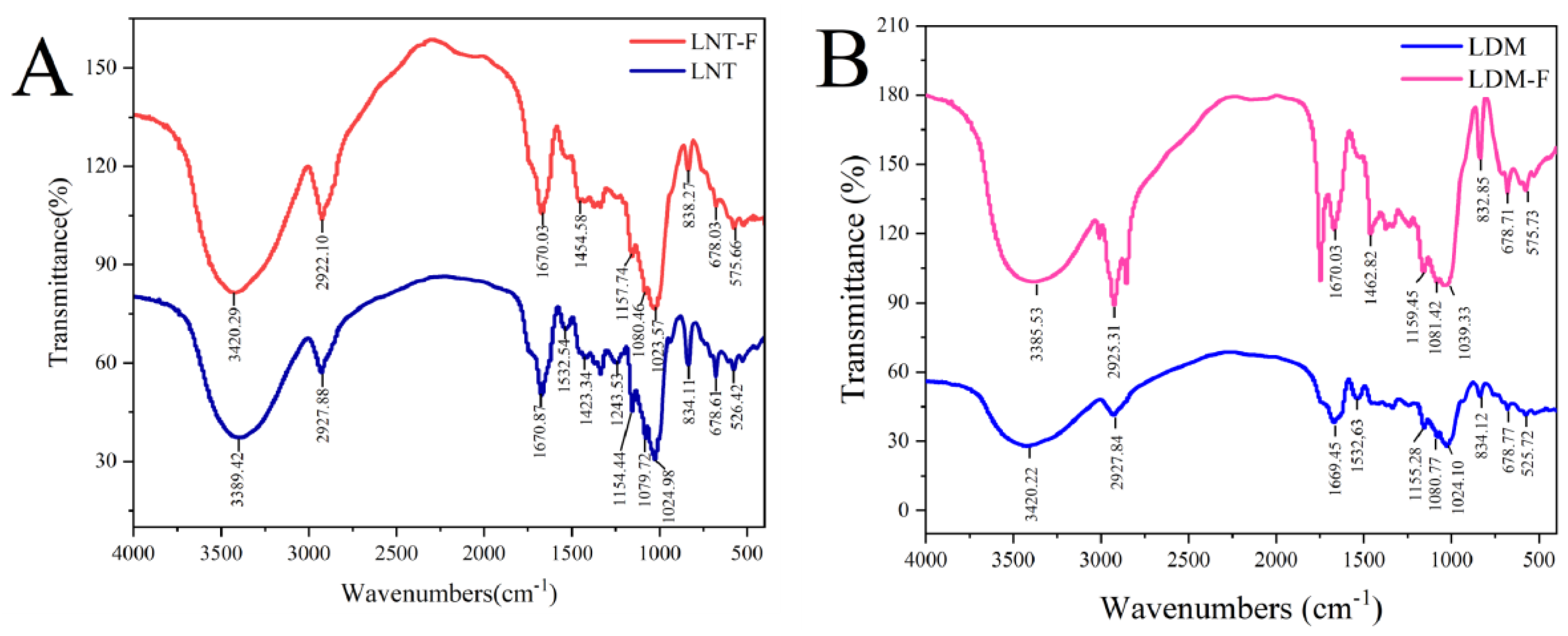
As shown in Figure 4, the lentinan samples have a typical polysaccharide structure but with different relative intensities of absorption bands. The peak in the 3500–3200 cm−1 was attributed to an -OH group, and that at 3000–2900 cm−1 indicated the presence of a C-H group (Xu et al., 2021) [36]. The peaks around 1670 and 1454 cm−1 were attributed to symmetric and asymmetric C=O stretching vibrations, respectively, indicating the presence of polysaccharide uronic acid (Liu et al., 2020) [31]. Three absorption peaks within the range of 1160–1000 cm−1 correspond to the stretching vibrations of the C–O–C and C–O–H link bond vibrations and indicate that pyranose rings were present in the polysaccharides (Liu et al., 2019) [37]. The absorption peaks at around 830 cm−1 suggested that the glycosyl residues of LNT, LNT-F, LDM, and LDM-F were α-type glycosidic linkages (Wang et al., 2018) [38]. Moreover, the skeletal mode of pyranose was responsible for the peak at 678 cm−1 (Jia et al., 2021) [39]. The absorption peak at 1532 cm−1 was attributed to stretching vibrations of C=C and/or C=O groups and asymmetrical stretching vibration of cumulated double bonds (Ren et al., 2018) [5], and the absorption band at 1243 cm−1 corresponded to the stretching vibration of C-O (Sheng et al., 2021) [32].

3.2.5. Scanning Electron Microscopy Analysis

The surface morphology differences between fermented and unfermented lentinan are shown in Figure 5. The surface morphology of lentinan from the LNT sample was rough and loose, with many irregular holes and branches, showing a honeycomb-like porous structure. However, after fermentation, the surface morphology of lentinan from the LNT-F sample exhibited a mixture of smooth lamellae and compact surfaces. The LDM shows a large lamellae structure, whereas the surface morphology of lentinan from the LDM-F sample was loose and had a branching structure. The loose structure confers the hydration and adsorption properties of the sample. As formerly verified by Chen et al., the molecular weight of wheat bran polysaccharides was decreased from 52.02 KDa to 21.19 KDa, and the fermented polysaccharides exhibited higher antioxidant activities after Saccharomyces cerevisiae and Bacillus subtilis fermentation (Chen et al., 2021) [40]. These results indicate that fermentation alters the structure and bioactivity of lentinan.

3.3. WHC and WSC

Water holding capacity is the most popular functional property in food processing (Jeddou et al., 2016) [41]. Polysaccharides with high WHC could be used as a functional ingredient to avoid synaeresis and modify the viscosity and texture of some formulated food (Chu et al., 2019) [17]. Water solubility capacity refers to the extent to which a substance can be dissolved, an important reference indicator for lentinan.
The WHC and WSC of fermented and unfermented lentinan are shown in Table 3. The WHC of the lentinan was significantly reduced after fermentation. A porous surface structure can lead to a higher WHC. The WSCs of lentinan after fermentation without and with soybean as a substrate were increased by 165.07% and 186.71%, respectively. During fermentation, the decrease in the lentinan Mw and increase in the glucuronic acid content may have increased the water solubility of lentinan. Polysaccharides are barely soluble in room temperature water by definition because of their molecular weight and chemical structure. Therefore, breaking lentinan into smaller fractions will increase its water solubility.
Similarly, Huang et al. found that the solubility of unfermented and fermented longan pulp polysaccharide increased by about 86.59%, from 42.50 ± 0.21 to 79.30 ± 0.49 mg/mL. However, the molecular weight was markedly decreased with the fermentation treatment, from 221.63 ± 2.41 KDa to 109.62 ± 10.66 KDa (Huang et al., 2019) [14]. Maybe a larger molecular weight typically leads to lower solubility. The changes in Mw and WSC of lentinan, which is highly water-soluble, have good utilization value in the fields of food and medicine.

3.4. Antioxidant Activities In Vitro

The antioxidant activities of fermented and unfermented lentinan are shown in Figure 6. Compared with that vitamin C, fermented and unfermented lentinan exhibited good DPPH free radical-scavenging abilities. As shown in Figure 6A, 0.125 mg/mL lentinan from the LNT-F (87.95 ± 2.89%) and LDM-F (86.66 ± 1.75%) samples showed higher DPPH free radical scavenging activities compared to those of lentinan from LNT (78.71 ± 1.72%) and LDM (80.39 ± 2.98%) samples. When sample concentration was increased to 1 mg/mL, the DPPH free radical scavenging activities of lentinan from the LNT, LNT-F LDM, and LDM-F samples were 93.95 ± 1.27%, 96.33 ± 2.85%, 88.43 ± 5.59%, and 95.31 ± 1.74%, respectively. The DPPH free radical scavenging activities of the lentinan from LNT, LNT-F, LDM, and LDM-F assayed with TEAC methods were 549.24 ± 6.27 μmol/L TE, 563.62 ± 14.07 μmol/L TE, 515.79 ± 33.86 μmol/L TE, 557.48 ± 10.55 μmol/L TE, respectively. However, these results were not statistically different, as shown in Table 4.
As shown in Figure 6B, compared with vitamin C, fermented and unfermented lentinan exhibited a weaker ABTS+ free radical scavenging ability at concentrations of 0.5–4.0 mg/mL. When the sample concentration was increased to 4.0 mg/mL, the ABTS+ free radical scavenging abilities of lentinan from the LNT, LNT-F, LDM, and LDM-F samples were 34.34 ± 2.05%, 50.47 ± 3.18%, 39.16 ± 1.15%, and 43.56 ± 5.48%, respectively. The ABTS+ free radical scavenging abilities of the lentinan from LNT, LNT-F, LDM, and LDM-F assayed with TEAC methods were 220.06 ± 13.80 μmol/L TE, 352.79 ± 21.35 μmol/L TE, 259.67 ± 9.44 μmol/L TE, 295.91 ± 45.11 μmol/L TE, respectively. The ABTS+ free radical scavenging activities of fermented and unfermented lentinan significantly differed (p < 0.05), with 50% inhibitory concentrations in the order: LNT-F > LDM-F > LDM > LNT (Table 4). Fermentation significantly improved the ABTS+ free radical scavenging activity of lentinan.
As shown in Figure 6C, the FRAP values steadily increased as lentinan solution concentration increased. At a concentration of 0.5 mg/mL, the FRAP values of lentinan from LNT and LNT-F were 31.22 ± 2.70 μmol/L and 47.93 ± 1.65 μmol/L, whereas those of lentinan from LDM and LDM-F were 30.78 ± 2.18 μmol/L and 44.22 ± 1.28 μmol/L. At a concentration of 4 mg/mL, the FRAP values of lentinan from the LNT, LNT-F, LDM, and LDM-F samples were 42.62 ± 0.35, 95.70 ± 2.03, 59.26 ± 2.48, and 77.33 ± 3.26 μmol/L, respectively. Fermentation significantly increased the ferric reducing antioxidant activity of lentinan.
The results indicate that the fermentation process increased the ABTS+ free radical scavenging ability of lentinan. The antioxidant activities of polysaccharides are closely related to their structural characteristics, including their uronic acid content and molecular weights. The polysaccharides obtained by water extraction from Sagittaria sagittifolia L. had a higher uronic acid content (8.62%) than that of polysaccharides obtained by subcritical water extraction (2.75%), inducing stronger antioxidant activity (Gu et al., 2020) [42]. Fermentation is an effective method for biological modification that increases the yield and improves the antioxidant activity of polysaccharides (Mustafa et al., 2022) [43]. Chen et al. showed that Saccharomyces cerevisiae and B. subtilis fermentation increased the yield, reduced the molecular weight, and enhanced the antioxidant activity of polysaccharides from wheat bran (Chen et al., 2021) [40]. We found that the antioxidant activity of lentinan was altered by B. subtilis natto fermentation. This may be because lentinan had a higher purity, higher uronic acid content, and smaller molecular weight after fermentation treatment.

4. Conclusions

This developed an efficient method for extracting and biotransforming lentinan from L. edodes. As a probiotic, lentinan produced by B. subtilis natto fermentation has good application prospects in the food and medicine fields. After fermentation, the lentinan extraction yield was increased by 87.13%, and protein impurities in the lentinan samples were reduced. Biotransformation by B. subtilis natto increased the uronic acid content, reduced the molecular weight, did not change the monosaccharide composition of lentinan, and altered the surface characteristics of lentinan. Both fermented and unfermented lentinan showed infrared spectral characteristics of polysaccharides. The WSC of fermented lentinan was increased by 165.07%. Fermentation significantly increased the ABTS+ free radical scavenging activity and FRAP of lentinan. Fermentation with soybean as a co-substrate may yield smaller molecular weight lentinan, providing a foundation for producing lentinan. In the future, the fermentation conditions of L. edodes could be optimized to improve the yield of polysaccharides and the efficiency of biotransformation.

Supplementary Materials

The following supporting information can be downloaded at: https://www.mdpi.com/article/10.3390/fermentation9040333/s1, Table S1: The reagents used in the experiment.

Author Contributions

Conceptualization, M.X. and Y.Q.; methodology, M.X. and H.L.; software, S.T.; validation, M.X., Y.Q., H.L., S.T., C.C., Y.W. and H.W.; formal analysis, M.X.; investigation, C.C. and Y.W.; resources, H.W.; data curation, M.X.; writing—original draft preparation, M.X.; writing—review and editing, M.X.; visualization, Y.Q.; supervision, H.W.; project administration, H.W.; funding acquisition, H.W. All authors have read and agreed to the published version of the manuscript.

Funding

This research was funded by Wuhan municipal university Industry-University-Research Project, grant number CXY202202.

Institutional Review Board Statement

Not applicable.

Informed Consent Statement

Not applicable.

Data Availability Statement

Not applicable.

Conflicts of Interest

The authors declare no conflict of interest.

References

  1. Yin, C.; Fan, X.; Fan, Z.; Shi, D.; Gao, H. Optimization of Enzymes-Microwave-Ultrasound Assisted Extraction of Lentinus edodes Polysaccharides and Determination of Its Antioxidant Activity. Int. J. Biol. Macromol. 2018, 111, 446–454. [Google Scholar] [CrossRef] [PubMed]
  2. Ahn, H.; Jeon, E.; Kim, J.-C.; Kang, S.G.; Yoon, S.; Ko, H.-J.; Kim, P.-H.; Lee, G.-S. Lentinan from Shiitake Selectively Attenuates AIM2 and Non-Canonical Inflammasome Activation while Inducing pro-Inflammatory Cytokine Production. Sci. Rep. 2017, 7, 1314. [Google Scholar] [CrossRef] [PubMed] [Green Version]
  3. Ziaja-Sołtys, M.; Radzki, W.; Nowak, J.; Topolska, J.; Jabłońska-Ryś, E.; Sławińska, A.; Skrzypczak, K.; Kuczumow, A.; Bogucka-Kocka, A. Processed Fruiting Bodies of Lentinus edodes as a Source of Biologically Active Polysaccharides. Appl. Sci. 2020, 10, 470. [Google Scholar] [CrossRef] [Green Version]
  4. Wang, T.; He, H.; Liu, X.; Liu, C.; Liang, Y.; Mei, Y. Mycelial Polysaccharides of Lentinus edodes (Shiitake Mushroom) in Submerged Culture Exert Immunoenhancing Effect on Macrophage Cells via MAPK Pathway. Int. J. Biol. Macromol. 2019, 130, 745–754. [Google Scholar] [CrossRef]
  5. Ren, G.; Xu, L.; Lu, T.; Yin, J. Structural Characterization and Antiviral Activity of Lentinan from Lentinus edodes Mycelia against Infectious Hematopoietic Necrosis Virus. Int. J. Biol. Macromol. 2018, 115, 1202–1210. [Google Scholar] [CrossRef] [PubMed]
  6. Morales, D.; Rutckeviski, R.; Villalva, M.; Abreu, H.; Soler-Rivas, C.; Santoyo, S.; Iacomini, M.; Smiderle, F.R. Isolation and Comparison of α- and β-D-Glucans from Shiitake Mushrooms (Lentinula edodes) with Different Biological Activities. Carbohydr. Polym. 2020, 229, 115521. [Google Scholar] [CrossRef]
  7. Wang, Y.; Wei, X.; Wang, F.; Xu, J.; Tang, X.; Li, N. Structural Characterization and Antioxidant Activity of Polysaccharide from Ginger. Int. J. Biol. Macromol. 2018, 111, 862–869. [Google Scholar] [CrossRef]
  8. Li, Z.; Nie, K.; Wang, Z.; Luo, D. Quantitative Structure-Activity Relationship Models for the Antioxidant Activity of Polysaccharides. PLoS ONE 2016, 11, e0163536. [Google Scholar] [CrossRef] [Green Version]
  9. Chen, G.; Chen, K.; Zhang, R.; Chen, X.; Hu, P.; Kan, J. Polysaccharides from Bamboo Shoots Processing By-Products: New Insight into Extraction and Characterization. Food Chem. 2018, 245, 1113–1123. [Google Scholar] [CrossRef]
  10. Akram, K.; Shahbaz, H.M.; Kim, G.-R.; Farooq, U.; Kwon, J.-H. Improved Extraction and Quality Characterization of Water-Soluble Polysaccharide from Gamma-Irradiated Lentinus edodes: Extraction of Water-Soluble Polysaccharide. J. Food Sci. 2017, 82, 296–303. [Google Scholar] [CrossRef]
  11. He, J.; Liu, Z.; Jiang, W.; Zhu, T.; Wusiman, A.; Gu, P.; Liu, J.; Wang, D. Immune-Adjuvant Activity of Lentinan-Modified Calcium Carbonate Microparticles on an H5N1 Vaccine. Int. J. Biol. Macromol. 2020, 163, 1384–1392. [Google Scholar] [CrossRef] [PubMed]
  12. Ren, G.; Li, K.; Hu, Y.; Yu, M.; Qu, J.; Xu, X. Optimization of Selenizing Conditions for Seleno-Lentinan and Its Characteristics. Int. J. Biol. Macromol. 2015, 81, 249–258. [Google Scholar] [CrossRef] [PubMed]
  13. Huang, H.; Huang, G. Extraction, Separation, Modification, Structural Characterization, and Antioxidant Activity of Plant Polysaccharides. Chem. Biol. Drug Des. 2020, 96, 1209–1222. [Google Scholar] [CrossRef] [PubMed]
  14. Huang, F.; Hong, R.; Zhang, R.; Yi, Y.; Dong, L.; Liu, L.; Jia, X.; Ma, Y.; Zhang, M. Physicochemical and Biological Properties of Longan Pulp Polysaccharides Modified by Lactobacillus fermentum Fermentation. Int. J. Biol. Macromol. 2019, 125, 232–237. [Google Scholar] [CrossRef] [PubMed]
  15. Lee, J.Y.; Park, H.M.; Kang, C.-H. Antioxidant Effect via Bioconversion of Isoflavonoid in Astragalus membranaceus Fermented by Lactiplantibacillus plantarum MG5276 In Vitro and In Vivo. Fermentation 2022, 8, 34. [Google Scholar] [CrossRef]
  16. Jia, M.; Chen, J.; Liu, X.; Xie, M.; Nie, S.; Chen, Y.; Xie, J.; Yu, Q. Structural Characteristics and Functional Properties of Soluble Dietary Fiber from Defatted Rice Bran Obtained through Trichoderma viride Fermentation. Food Hydrocoll. 2019, 94, 468–474. [Google Scholar] [CrossRef]
  17. Chu, J.; Zhao, H.; Lu, Z.; Lu, F.; Bie, X.; Zhang, C. Improved Physicochemical and Functional Properties of Dietary Fiber from Millet Bran Fermented by Bacillus natto. Food Chem. 2019, 294, 79–86. [Google Scholar] [CrossRef]
  18. Olawuyi, I.F.; Lee, W.Y. Structural Characterization, Functional Properties and Antioxidant Activities of Polysaccharide Extract Obtained from Okra Leaves (Abelmoschus esculentus). Food Chem. 2021, 354, 129437. [Google Scholar] [CrossRef]
  19. Zhu, Y.; Dun, B.; Shi, Z.; Wang, Y.; Wu, L.; Yao, Y. Structural Characterization and Bioactivity Evaluation of Water-Extractable Polysaccharides from Chickpeas (Cicer arietinum L.) Seeds. Front. Nutr. 2022, 9, 946736. [Google Scholar] [CrossRef]
  20. Dubios, M.; Gilles, K.A.; Hamilton, J.K.; Rebers, P.A.; Smith, F. Colorimetric Method for Determination of Sugar and Related Substances. Anal. Chem. 1956, 28, 250–256. [Google Scholar] [CrossRef]
  21. Blumenkrantz, N.; Asboe-Hansen, G. New Method for Quantitative Determination of Uronic Acids. Anal. Biochem. 1973, 54, 484–489. [Google Scholar] [CrossRef] [PubMed]
  22. Bradford, M.M. A Rapid and Sensitive Method for the Quantitation of Microgram Quantities of Protein Utilizing the Principle of Protein-Dye Binding. Anal. Biochem. 1976, 72, 248–254. [Google Scholar] [CrossRef] [PubMed]
  23. Pei, F.; Lv, Y.; Cao, X.; Wang, X.; Ren, Y.; Ge, J. Structural Characteristics and the Antioxidant and Hypoglycemic Activities of a Polysaccharide from Lonicera caerulea L. Pomace. Fermentation 2022, 8, 422. [Google Scholar] [CrossRef]
  24. Wu, J.; Chen, T.; Wan, F.; Wang, J.; Li, X.; Li, W.; Ma, L. Structural Characterization of a Polysaccharide from Lycium barbarum and Its Neuroprotective Effect against β-Amyloid Peptide Neurotoxicity. Int. J. Biol. Macromol. 2021, 176, 352–363. [Google Scholar] [CrossRef] [PubMed]
  25. Su, Y.; Li, L. Structural Characterization and Antioxidant Activity of Polysaccharide from Four Auriculariales. Carbohydr. Polym. 2020, 229, 115407. [Google Scholar] [CrossRef]
  26. Ahmed, Z.; Wang, Y.; Anjum, N.; Ahmad, A.; Khan, S.T. Characterization of Exopolysaccharide Produced by Lactobacillus kefiranofaciens ZW3 Isolated from Tibet Kefir—Part II. Food Hydrocoll. 2013, 30, 343–350. [Google Scholar] [CrossRef]
  27. Duan, G.-L.; Yu, X. Isolation, Purification, Characterization, and Antioxidant Activity of Low-Molecular-Weight Polysaccharides from Sparassis latifolia. Int. J. Biol. Macromol. 2019, 137, 1112–1120. [Google Scholar] [CrossRef] [PubMed]
  28. Zulueta, A.; Esteve, M.J.; Frígola, A. ORAC and TEAC Assays Comparison to Measure the Antioxidant Capacity of Food Products. Food Chem. 2009, 114, 310–316. [Google Scholar] [CrossRef]
  29. Fimbres-Olivarria, D.; Carvajal-Millan, E.; Lopez-Elias, J.A.; Martinez-Robinson, K.G.; Miranda-Baeza, A.; Martinez-Cordova, L.R.; Enriquez-Ocaña, F.; Valdez-Holguin, J.E. Chemical Characterization and Antioxidant Activity of Sulfated Polysaccharides from Navicula sp. Food Hydrocoll. 2018, 75, 229–236. [Google Scholar] [CrossRef]
  30. Li, S.; Shah, N.P. Characterization, Antioxidative and Bifidogenic Effects of Polysaccharides from Pleurotus eryngii after Heat Treatments. Food Chem. 2016, 197, 240–249. [Google Scholar] [CrossRef]
  31. Liu, X.-X.; Liu, H.-M.; Yan, Y.-Y.; Fan, L.-Y.; Yang, J.-N.; Wang, X.-D.; Qin, G.-Y. Structural Characterization and Antioxidant Activity of Polysaccharides Extracted from Jujube Using Subcritical Water. LWT 2020, 117, 108645. [Google Scholar] [CrossRef]
  32. Sheng, K.; Wang, C.; Chen, B.; Kang, M.; Wang, M.; Liu, K.; Wang, M. Recent Advances in Polysaccharides from Lentinus edodes (Berk.): Isolation, Structures and Bioactivities. Food Chem. 2021, 358, 129883. [Google Scholar] [CrossRef] [PubMed]
  33. Chen, X.; Lu, Y.; Zhao, A.; Wu, Y.; Zhang, Y.; Yang, X. Quantitative Analyses for Several Nutrients and Volatile Components during Fermentation of Soybean by Bacillus subtilis natto. Food Chem. 2022, 374, 131725. [Google Scholar] [CrossRef] [PubMed]
  34. Jeff, I.B.; Yuan, X.; Sun, L.; Kassim, R.M.R.; Foday, A.D.; Zhou, Y. Purification and in Vitro Anti-Proliferative Effect of Novel Neutral Polysaccharides from Lentinus edodes. Int. J. Biol. Macromol. 2013, 52, 99–106. [Google Scholar] [CrossRef]
  35. Wagner, J.R.; Gueguen, J. Effects of Dissociation, Deamidation, and Reducing Treatment on Structural and Surface Active Properties of Soy Glycinin. J. Agric. Food Chem. 1995, 43, 1993–2000. [Google Scholar] [CrossRef]
  36. Xu, Y.; Li, Y.; Lu, Y.; Feng, X.; Tian, G.; Liu, Q. Antioxidative and Hepatoprotective Activities of a Novel Polysaccharide (LSAP) from Lepista sordida Mycelia. Food Sci. Hum. Wellness 2021, 10, 536–544. [Google Scholar] [CrossRef]
  37. Liu, X.; Bian, J.; Li, D.; Liu, C.; Xu, S.; Zhang, G.; Zhang, L.; Gao, P. Structural Features, Antioxidant and Acetylcholinesterase Inhibitory Activities of Polysaccharides from Stem of Physalis alkekengi L. Ind. Crops Prod. 2019, 129, 654–661. [Google Scholar] [CrossRef]
  38. Wang, L.; Zhang, B.; Xiao, J.; Huang, Q.; Li, C.; Fu, X. Physicochemical, Functional, and Biological Properties of Water-Soluble Polysaccharides from Rosa roxburghii Tratt Fruit. Food Chem. 2018, 249, 127–135. [Google Scholar] [CrossRef]
  39. Jia, Y.; Wang, Y.; Li, R.; Li, S.; Zhang, M.; He, C.; Chen, H. The Structural Characteristic of Acidic-Hydrolyzed Corn Silk Polysaccharides and Its Protection on the H2O2-Injured Intestinal Epithelial Cells. Food Chem. 2021, 356, 129691. [Google Scholar] [CrossRef]
  40. Chen, Q.; Wang, R.; Wang, Y.; An, X.; Liu, N.; Song, M.; Yang, Y.; Yin, N.; Qi, J. Characterization and Antioxidant Activity of Wheat Bran Polysaccharides Modified by Saccharomyces cerevisiae and Bacillus subtilis Fermentation. J. Cereal Sci. 2021, 97, 103157. [Google Scholar] [CrossRef]
  41. Jeddou, K.B. Structural, Functional, and Antioxidant Properties of Water-Soluble Polysaccharides from Potatoes Peels. Food Chem. 2016, 205, 97–105. [Google Scholar] [CrossRef] [PubMed]
  42. Gu, J.; Zhang, H.; Yao, H.; Zhou, J.; Duan, Y.; Ma, H. Comparison of Characterization, Antioxidant, and Immunological Activities of Three Polysaccharides from Sagittaria sagittifolia L. Carbohydr. Polym. 2020, 235, 115939. [Google Scholar] [CrossRef] [PubMed]
  43. Mustafa, G.; Arshad, M.U.; Saeed, F.; Afzaal, M.; Niaz, B.; Hussain, M.; Raza, M.A.; Nayik, G.A.; Obaid, S.A.; Ansari, M.J.; et al. Comparative Study of Raw and Fermented Oat Bran: Nutritional Composition with Special Reference to Their Structural and Antioxidant Profile. Fermentation 2022, 8, 509. [Google Scholar] [CrossRef]
Figure 1. The preparation process of lentinan from fermented and unfermented Lentinula edodes and soybeans.
Fermentation 09 00333 g001
Figure 2. The HPLC chromatogram of monosaccharide standards and samples.
Fermentation 09 00333 g002
Figure 3. The HPGPC chromatogram of samples. (A) Lentinula edodes powder (LNT) and fermented LNT (LNT-F) samples, (B) soybean powder (LDM) and fermented LDM (LDM-F) samples, (C) calibration standards curve of Mw.
Fermentation 09 00333 g003
Figure 4. Fourier transform infrared spectra of lentinan. (A) Lentinula edodes powder (LNT) and fermented LNT (LNT-F) samples, (B) soybean powder (LDM) and fermented LDM (LDM-F) samples.
Fermentation 09 00333 g004
Figure 5. Scanning electron micrographs of samples. Lentinula edodes powder (LNT) and fermented LNT (LNT-F), soybean powder (LDM) and fermented LDM (LDM-F).
Fermentation 09 00333 g005
Figure 6. Antioxidant activity of fermented and unfermented lentinan: (A) DPPH free radical scavenging ability; (B) ABTS+ free radical scavenging ability; (C) Ferric-reducing antioxidant power (FRAP).
Fermentation 09 00333 g006
Table 1. Extraction yield and chemical composition of fermented and unfermented samples.
Sample NameYield (%)Total Sugars (%)Uronic Acid (%)Total Protein (%)
LNT 4.74 ± 0.44 b54.43 ± 3.44 b2.08 ± 0.54 b6.84 ± 1.21 a
LNT-F8.87 ± 1.09 a65.22 ± 1.29 a4.33 ± 0.21 a2.17 ± 0.81 b
LDM2.33 ± 0.28 b46.37 ± 2.46 b2.66 ± 0.52 b13.07 ± 1.52 a
LDM-F4.55 ± 0.65 a50.77 ± 1.79 a4.38 ± 0.17 a1.63 ± 0.25 b
DLP-F0.96 ± 0.0414.22 ± 0.974.93 ± 0.252.98 ± 0.49
Different letters (a, b) within the row indicate that the interactions between fermented and unfermented samples significantly differed (p ≤ 0.05). Results are expressed as the mean ± standard deviation (n = 3).
Table 2. Monosaccharide composition of lentinan samples.
Sample NameMannose (%)Glucose (%)Galactose (%)
LNT2.5694.692.75
LNT-F1.5494.823.64
LDM4.8691.114.03
LDM-F1.1194.654.24
Table 3. Water-holding capacity (WHC) and water solubility capacity (WSC) of lentinan samples.
Sample NameWHC (%)WSC (%)
LNT551.67 ± 32.50 a15.63 ± 2.13 b
LNT-F76.08 ± 7.17 b41.43 ± 2.40 a
LDM416.49 ± 32.97 b17.61 ± 1.98 b
LDM-F59.12 ± 4.39 b50.49 ± 2.73 a
Different letters (a, b) within the row indicate that the interactions between fermented and unfermented lentinan significantly differed (p ≤ 0.05). Results are expressed as the mean ± standard deviation (n = 3).
Table 4. Fermented and unfermented lentinan 50% inhibitory activity on antioxidant activity.
Sample NameDPPH (mg/mL)ABTS+ (mg/mL)
LNT0.015 ± 0.005 a17.273 ± 8.360 a
LNT-F0.014 ± 0.023 a3.947 ± 0.472 b
LDM0.004 ± 0.006 a10.533 ± 2.438 a
LDM-F0.006 ± 0.007 a6.032 ± 2.359 b
VC0.0300.398
Different letters (a, b) within the row indicate that the interactions between fermented and unfermented lentinan significantly differed (p ≤ 0.05). The results are expressed as the mean ± standard deviation (n = 3).
Disclaimer/Publisher’s Note: The statements, opinions and data contained in all publications are solely those of the individual author(s) and contributor(s) and not of MDPI and/or the editor(s). MDPI and/or the editor(s) disclaim responsibility for any injury to people or property resulting from any ideas, methods, instructions or products referred to in the content.