Review

Butyrate Properties in Immune-Related Diseases: Friend or Foe?

by
Muhammad Anshory
1,2,
Raden Mohamad Rendy Ariezal Effendi
1,3,
Handono Kalim
2,
Reiva Farah Dwiyana
3,
Oki Suwarsa
3,
Tamar E. C. Nijsten
1,
Jan L. Nouwen
4 and
Hok Bing Thio
1,*
1
Department of Dermatology, Erasmus MC, University Medical Centre, 3015 GD Rotterdam, The Netherlands
2
Department of Internal Medicine, Faculty of Medicine, Universitas Brawijaya, Malang 65145, Indonesia
3
Department of Dermatology and Venereology, Faculty of Medicine, Universitas Padjadjaran—Dr. Hasan Sadikin Hospital, Bandung 45363, Indonesia
4
Department of Medical Microbiology and Infectious Diseases, Erasmus MC, University Medical Centre, 3013 GD Rotterdam, The Netherlands
*
Author to whom correspondence should be addressed.
Fermentation 2023, 9(3), 205; https://doi.org/10.3390/fermentation9030205
Submission received: 13 January 2023 / Revised: 7 February 2023 / Accepted: 20 February 2023 / Published: 21 February 2023
(This article belongs to the Special Issue Postbiotics from Production to Their Health-Promoting Aspects)

Abstract

:
Butyrate is a short-chain fatty acid (SCFA) created within the intestinal lumen by bacterial fermentation of largely undigested dietary carbohydrates. Its beneficial effects on cellular energy metabolism and intestinal homeostasis have garnered significant attention among SCFAs. Butyrate also has systemic effects and is known to regulate the immune system. Most of the butyrate and other SCFAs are produced in the human colon, through the fermentation of dietary fiber or resistant starch. However, the modern diet often lacks sufficient intake of fermentable dietary fiber, which can lead to low butyrate levels in the colon. To increase butyrate levels, it is helpful to incorporate fiber sources into meals and drinks that rely on slow bacterial fermentation. Butyrate is well known for its anti-inflammatory properties and has a range of immune system-related properties. As an agonist for GPR41, GPR43, or GPR109A, butyrate may have anti-inflammatory effects through these receptors’ signaling pathways. Butyrate also serves as an epigenetic regulator, responding to environmental or pharmacological changes by inhibiting HDAC, up-regulating miR-7a-5p, and promoting histone butyrylation and autophagy processes. This review discusses the importance of butyrate in regulating immunological homeostasis and the inflammatory response. It also addresses experimental models and human studies investigating the therapeutic potential of butyrate supplementation in immune-related conditions linked to butyrate depletion. Specifically, it covers the role of butyrate in some immune-related diseases such as systemic lupus erythematosus, atopic dermatitis, psoriasis, human immunodeficiency virus, cancer, and several other special conditions.

1. Introduction

Butyrate, a short-chain fatty acid (SCFA), has attracted particular attention because of its advantageous effects on intestinal homeostasis as well as cellular energy metabolism [1,2]. SCFAs are made when microorganisms ferment primarily undigested carbohydrates, especially resistant starch and dietary fermentable fiber, and to a lesser degree, dietary and endogenous proteins. It consists of organic acids, primarily acetate, propionate, and butyrate, in the intestinal lumen [3,4], which are essential in modulating the effects of the gut microbiome on local and systemic immunity, particularly butyrate [4].
Many distinct phyla inside the human colon produce butyrate. The two phyla that produce the most butyrate are Firmicutes and Bacteroides [5]. Butyrate-producing bacteria are prevalent among Gram-positive anaerobic bacteria. Eubacterium rectale/Roseburia spp. from the Clostridium coccoides (or Clostridial cluster XIVa) cluster of Firmicutes and Faecalibacterium prausnitzii from the Clostridium leptum (or Clostridial cluster IV) cluster are two of the most significant groups [6,7]. Numerous in vitro and in vivo studies have indicated that butyrate is essential for managing inflammatory and immunological responses as well as intestinal barrier function [8].
This review aims to discuss the role of butyrate in immune-related disease. It explores how human-source butyrate affects the human body. It also addresses experimental models and human studies investigating the therapeutic potential of butyrate supplementation in immune-related conditions linked to butyrate depletion. Specifically, it covers the role of butyrate in some immune-related diseases such as systemic lupus erythematosus, atopic dermatitis, psoriasis, human immunodeficiency virus, cancer, and several related conditions.

2. Butyrate Properties

2.1. What Is Butyrate?

Butyrate, one of the short-chain fatty acids (SCFA), is produced in the intestines from undigested carbohydrates, proteins, and other substances through bacterial fermentation. These organic acids promote healthy bowel function and reduce the risk of illness through their effects on the intestinal lumen, the colonic musculature, and blood vessels, and through their metabolism by colonocytes. Butyrate specifically helps maintain a normal colonocyte count. In herbivores such as ruminants, SCFAs are spread throughout the body and metabolized in other organs before being taken to the liver via the portal vein [9]. SCFAs, including butyrate, are able to penetrate the blood–brain barrier and enter the brain in humans with average butyrate concentrations of 17.0 pmol/mg of brain tissue [10].
Butyrate has drawn special attention among SCFAs due to its advantageous effects on cellular energy metabolism and intestinal homeostasis [1]. It helps maintain the integrity of the intestinal epithelial barrier by controlling the expression of tight junction proteins and fostering the creation of intestinal mucus [11]. Additionally, butyrate is crucial for digestive health as it provides a significant amount of energy to the colonic mucosa and plays a crucial role in regulating gene expression, inflammation, cell differentiation, and cell death in host cells [7]. These effects of butyrate are summarized in Table 1.

2.2. Internal Butyrate Biosynthesis

Butyrate and other SCFAs are largely produced in the colon intestine of the human body [22]. The fermentation of dietary fiber or resistance starch in the colon is a significant source of butyrate. When fiber is added to the diet, the amount of butyrate produced along the intestinal lumen depends on its chemical structure, solubility, and degree of polymerization. Despite their low fermentability (compared to cellulose and lignin), insoluble fibers have been linked to increased fecal bulk and a faster colonic transit time. Since soluble fibers are easy to ferment, they cause the colon to produce more SCFAs. Higher levels of polymerization produce fibers more resistant to saccharolytic fermentation, which causes fermentation to last longer and expand toward the distal colon. Food companies are interested in incorporating fiber sources into foods and beverages that rely on slow bacterial fermentation to raise distal colonic butyrate concentrations because of the significant role that butyrate plays in the body and the relatively low consumption of fermentable dietary fiber in the modern diet [23].
Gut microbiota play a significant role in dietary fiber fermentation. A metabolically active microbiota makes up the majority of the extraordinarily dense microbial population seen in the human large intestine. This group has a significant impact on health and relies heavily on dietary carbohydrates for energy. Due to their superior degradative enzymes and metabolic skills compared to their hosts, the gut microbiota breaks down the majority of these carbohydrates, as the host is unable to do so [24,25]. The gut microbiota is partly shaped by environmental factors, including diet, and it depends on food metabolites for existence and metabolism. A well-balanced diet, such as one high in fiber, is crucial for maintaining a healthy gut microbiota. In addition, indigestible fibers offer significant rates of butyrogenesis, which fulfill the metabolic needs of epithelial cells and enter the bloodstream to influence other body organs in ways that are immunomodulatory and epigenetic [26,27].
Essentially, butyrate biosynthesis (Figure 1) can occur via (1) direct conversion by butyrate kinase from carbohydrates, (2) indirect conversion (interconversion processes) of organic acids from acetate, succinate, and lactate, and (3) amino acid fermentation from glutamate and lysine [28,29].
The direct conversion from carbohydrate was mediated via a bacterial carbohydrate phosphotransferase system (PTS). The PTS comprises two cytoplasmic energy-coupling proteins (Enzyme I and HPr) and a number of carbohydrate-specific type II enzymes that catalyze simultaneous carbohydrate translocation and phosphorylation. The phosphorylation status of PTS components reflects the availability of carbohydrates and the cellular energy environment. In numerous bacteria, PTS and its related proteins translate this information into signals, which, when transduced via various processes, result in catabolite suppression, inducer regulation, and chemotaxis. These characteristics of PTS equip bacteria with an integrated mechanism that ensures optimum glucose use in challenging conditions [30]. Direct synthesis of butyrate from glucose continues to acetyl-CoA pathway and creates 2 mol of hydrogen/mol of butyrate according to the equation [31].
The acetyl-CoA, glutamate, 4-aminobutyrate, and lysine routes are the other four major recognized processes for butyrate formation (Figure 1). All the pathways lead to a key energy-generating phase where crotonyl-CoA is transformed into butyryl-CoA by the electron-transferring flavoprotein complex (Bcd-Etfαβ). The final conversion to butyrate is performed by various butyryl-CoA transferases using cosubstrates from earlier in the pathways, such as acetoacetate and 4-hydroxybutyrate for the lysine and 4-aminobutyrate pathways, or from external sources, as seen in the case of butyryl-CoA: acetate CoA transferase [32].
Figure 1. Butyrate biosynthesis and related enzymes and byproducts in the process. Diagram shows the biosynthesis of butyrate (in yellow) through several sources such as organic acids (lactate, acetate, succinate; in purple, orange, and pink, respectively), amino acids (L-glutamate, L-lysine; in blue) and carbohydrate fermentation by bacteria (in green) [2,28,29,30,32,33,34,35,36,37,38,39,40].
Fermentation 09 00205 g001

2.3. External Sources of Butyrate

The composition and diversity of the gut microbiota are largely influenced by diet. Various metabolites are produced by the gut microbiota in response to food intake. A diet high in complex indigestible carbohydrates (such as fibers) promotes the production of metabolites from intestinal microbiota, such as butyrate and other SCFA metabolites [26]. Through bacterial fermentation, butyrate is generated from dietary fibers. Butyric acid naturally exists in very small amounts in butter, hard cheeses (e.g., Parmesan cheese), milk (particularly, goat’s and sheep’s milk), yoghurts, cream, and some other fermented foods (including sauerkraut, pickled cucumbers, and fermented soy products) [22]. Butyrate is also indirectly generated from carbohydrates, especially resistant starch. Resistant starch can be found in foods including boiled and cooled potatoes, unripe bananas, legumes, and partially ground seeds. Resistant starch can also be added via manufacturing processes and fortification to morning cereals, tortillas, breads, and maize [41]. Additionally, human breast milk has been investigated as a potential source of butyrate for neonates, assisting them in maintaining their intestinal flora [42,43].

2.4. Butyrate-Producing Bacteria

Butyrate-producing bacteria are a group of bacteria that live in the gut and can break down polysaccharides and produce butyric acid [7]. In the order Clostridiales, most of the organisms that produce butyrate are in clusters I, IV, XIVa, and XVI. According to 16S rRNA gene sequencing, the two most significant groups among them, F. prausnitzii (clostridial cluster IV) and Eubacterium rectale (clostridial cluster XIVa), account for up to 12–14% of the entire gut microbiota in fecal samples from healthy adults [44]. Other typical butyrogenic species are also abundantly dispersed in clusters IV and XIVa, including Butyricicoccus pullicaecorum, Subdoligranulum variabile, Anaerotruncus colihominis, and Papillibacter cinnamivorans from cluster IV; and Butyrivibrio species, Ruminococcus species, Anaerostipes species, Clostridium species, and Ruminococcus species from cluster XIVa [7,24,44]. Other additional possible butyrate-producing bacteria, namely, Actinomycetes, Bacteroidetes, Proteobacteria, and Spirochetes, have also been discovered [7].
The majority of the commensal bacteria that produce butyrate are strictly anaerobes, which makes them hard to culture [7]. The intestinal tract’s normal microecological environment and a balanced microbiota are both maintained by the acidic environment created by butyrate-producing bacteria throughout metabolism and its anaerobic digestion [45]. As a result, butyrate-producing bacteria serve as potential probiotics and are crucial for a number of regular biological processes, including preserving the mucosal barrier, enhancing immunity, and assisting animal digestion and absorption of nutrients [46]. These bacteria are likely to be beneficial in maintaining a healthy gut and restoring disturbed gut homeostasis because a reduction in butyrate-producing bacteria has been seen in patients with various disorders, and the SCFAs produced by these bacteria have positive effects. Consuming probiotics and prebiotics has become a popular method for encouraging butyrate-producing bacteria colonization in humans, which increases the amount of butyrate and enhances its functional activity [47,48].

2.5. Butyrate Effect on Immune System

Butyrate is a well-known anti-inflammatory agent and possesses a number of immune system-related characteristics. As an agonist for GPR41, GPR43, or GPR109A, butyrate may exhibit anti-inflammatory effects via the signaling pathways of these receptors. Butyrate also functions as an epigenetic regulator in response to environmental or pharmacological modification by inhibiting HDAC, up-regulating miR-7a-5p, or inducing histone butyrylation and autophagy processes [49]. Figure 2 shows the effects of butyrate on the modulation of host functions.
Butyrate is known to have an effect on macrophages. It is known that this SCFA inhibits pro-inflammatory cytokines and cells such as neutrophils and M1 macrophages while stimulating anti-inflammatory cells such as T-reg and M2 macrophages [5]. According to a study in antibiotic-pretreated mice, butyrate inhibited histone deacetylase (HDACs), which in turn prevented macrophage activity in vitro. This study demonstrated that, in macrophages, n-butyrate mimics the effects of the well-known HDAC inhibitor, Trichostatin A (TSA). TSA, like n-butyrate, reduced the levels of nitric oxide (NO), interleukin-6 (IL-6), and IL-12p40 that macrophages produced, but it had no effect on the release of TNF-alpha or monocyte chemoattractant protein-1 (MCP-1). The fact that histone H3 acetylation levels were elevated by both n-butyrate and TSA suggests that n-butyrate functions as an HDAC inhibitor [15]. Another in vivo mice study found that butyrate reduced TNF production by inhibiting HDAC-associated NF-κB activity. Butyrate had a stronger effect than propionate on the reduction of NF-κB activation and TNF-α secretion and the rise in PGE2 production [14]. Butyrate is also known to reduce mammalian target of rapamycin (mTOR) activity, which resulted in decreased TNF-α, IL-6, and IL-12 production and increased IL-10 production [16].
Butyrate is also known to affect neutrophils. Butyrate, together with lactate, inhibits the production of pro-inflammatory cytokines TNF-α, IL-6, and IL-12 by neutrophils and monocytes during concanavalin A-induced hepatic inflammation [50]. In a mouse colitis model generated by dextran sulfate sodium, butyrate reduced neutrophil recruitment. Anti-inflammatory cytokines were raised by butyrate, whereas pro-inflammatory cytokines were suppressed. The mechanism may involve the direct suppression of HDAC3 and HDAC9, which results in the inhibition of NF-κB and the activation of PPAR-γ [17]. Butyrate prevented LPS-stimulated neutrophils from producing pro-inflammatory cytokines (TNF-α and CINC-2αβ) and NO both in vitro and ex vivo. Butyrate may affect neutrophils by inhibiting HDAC activity and NF-κB activation [51].
Mast cells, which are prevalent in the mucosa and submucosa of the GI tract, are associated with a variety of illnesses, including allergies [4]. When examining the direct effects of butyrate on mast cells, the jejunal mucosa of pigs treated with butyrate revealed decreased mast cell degranulation and pro-inflammatory cytokine gene expression [52]. These findings confirm a prior study in mast cell culture indicating that the direct action of butyrate on mast cells was mediated by the MAPK (mitogen-activated protein kinases) signaling pathway and the suppression of JNK (jun N-terminal kinase) activation [53].
It has been observed that butyrate administration has a substantial effect on the differentiation, maturation, and overall T lymphocyte-stimulating properties of human monocyte-derived dendritic cells (DC) and macrophages [54]. Butyrate, in in vitro experiments, influenced the development of DC, generated an immunosuppressive impact on DC derived from human monocytes, and suppressed T cell proliferation in the presence of inducers (e.g., LPS, TNF-α). Low and non-toxic-dose butyrate administration decreased the surface marker expression of mature DC (CD80, CD83, CD40, CD45, MHC class II molecules) [55]. SCFAs influence various components of the innate immune response, although there are still some disagreements in the scientific literature [56].
Another role of butyrate in the immune system is through the activation of T-reg. T-reg cells are essential to the development and control of inflammation. T-reg cells exhibit potent anti-inflammatory actions, which are crucial for maintaining immune response balance and preventing both excessive acute inflammation and chronic inflammation [57]. Butyrate has the ability to activate T-reg cells, thereby encouraging them to have an anti-inflammatory function. Abnormal T-reg cell functions are linked to autoimmune disorders [5]. Butyrate also promotes the development of effector T cells such as Th1 and Th17 cells that are mixed with T-reg cells that are IL-10+ anti-inflammatory cells. This action of butyrate is mediated by its HDAC inhibitory effect, which is GPR41 (GPR; orphan G protein-coupled receptor) or GPR43-independent, and the resulting increase in mTOR-S6K activity needed for T-cell differentiation and cytokine production [18].
It is possible to connect the mechanisms governing the process that regulates the way in which the immune system interacts with commensal bacteria on the skin to those occurring in the intestine. Among SCFAs, butyrate inhibits the synthesis of cytokines and inflammatory cells while also promoting the growth of T-regs, the primary cells responsible for numerous physiological processes [58]. Under physiological conditions, commensal skin microorganisms may exert a down-regulating activity and maintain homeostasis to counteract excessive inflammatory reactions [59]. Figure 2 provides a visual overview of the fundamental actions of butyrate on the regulation of host activities.

3. Butyrate as Prevention and Treatment

Butyrate’s importance in the prevention and management of several diseases has already been stated, such as diabetic kidney disease, inflammatory bowel disease, colon cancer, and obesity [12,49,60,61,62,63]. Several factors contribute to this effect such as the source of energy for colonocytes, immunomodulatory function, anti-inflammation, intestinal barrier integrity maintenance, anti-tumor effect, enhanced chemotherapy, and radiotherapy effect [12].
Butyrate has been shown to have potential benefits for metabolic disorders such as diabetes and cardiovascular disease. This is because it can down-regulate the expression of genes involved in intestinal cholesterol biosynthesis, prevent immune cells from infiltrating adipose tissue, prevent and treat diet-induced obesity and insulin resistance, and exhibit anti-atherogenic effects by altering G1-specific cell-cycle proteins [63].

3.1. Overactive Immune Disease

3.1.1. Systemic Lupus Erythematosus

The dysregulated immune response in the intestine of lupus patients may result in microbial dysbiosis, which in turn impairs the integrity of the gut [64,65,66]. Patients with systemic lupus erythematosus (SLE) showed a general decline in alpha-diversity and richness, and a lower F/B ratio. Almost all previous SLE cohorts have reported this imbalance, which seems to be the main sign of SLE dysbiosis, regardless of race, lifestyle, or stage of the disease [67,68,69,70,71,72,73]. He et al. discovered that butyrate treatment increased the diversity and abundance of firmicutes while decreasing the diversity and abundance of Bacteroidetes, Bacteroidia, and Bacteroidales [74].
Several in vitro studies in mice have shown that short-chain fatty acids from the gut microbiota, particularly butyrate from the bacterium Clostridia, encourage the development of T-reg cells to reduce inflammation [75,76,77]. According to Takahashi et al., the butyrate produced by commensal bacteria is a key environmental element that supports the development of T-reg follicular cells in the gut-associated lymphoid tissues (GALT). By altering the immune system in the GALT, commensal bacteria play a crucial role in both the initiation and control of autoimmune arthritis. Given that systemic autoimmune responses are preceded by a commensal bacterial-driven autoimmune response in the GALT, the activation of GALT T-reg follicular cells may represent a new avenue for avoiding autoimmune disease. Future work will also focus on determining whether T-reg follicular cells produced in GALT also prevent the onset of other autoimmune conditions, including systemic lupus erythematosus [78].
Besides inducing T-reg, butyrate has also been shown to enhance macrophage phagocytosis and bacterial killing. In T-reg cells and activated macrophage cultures exposed to butyrate and IL-4, fewer inflammatory cytokines were released than when the macrophages were just given IL-4, showing that butyrate obtained from microorganisms reduces the generation of inflammatory mediators in the gut [79,80].
A high-fiber diet also has an indirect impact on butyrate synthesis, in addition to butyrate supplementation. Panther et al. investigated the possibility that a high-fiber diet could modify the gut microbiota, reduce gut permeability, and lessen the severity of the disease in lupus model mice. It has been demonstrated that in autoimmunity, inadequate soluble dietary fiber absorption either directly causes dysregulation of the immune response or indirectly does so by altering the microbiota [81].

3.1.2. Chronic Inflammatory Skin Diseases

There is evidence that commensal bacteria control inflammation not just within the gastrointestinal but in other external barrier organs such as the skin [82]. Prebiotics, probiotics, synbiotics, and postbiotics are some of the methods that could be utilized to “on-site” treat skin disorders. Among these, postbiotics are the easiest to integrate into products, in contrast to probiotics, which provide obstacles in formulating and packing that must be addressed to guarantee the viability of the microorganisms [83]. The definition of a postbiotic includes inactivated organisms and/or cellular components or metabolic products that are released after bacterial lysis or that are secreted throughout fermentation. It also includes any aspect arising from the metabolic activity of a probiotic or any released particle capable of delivering positive benefits to the host [84]. Butyrate and other microbial byproducts can raise pH levels and restrict other organisms from growing. Furthermore, they can trigger immune cells in the dermis and epidermis as well as immunological mediators generated through keratinocytes [85]. Butyrate also has a eutrophic impact on the skin, reduces epidermal permeability, improves barrier qualities, and suppresses the cutaneous inflammatory response [86]. Considering that butyrate plays such an important role in maintaining healthy skin, it may be utilized as a postbiotic method to prevent or treat various skin conditions, especially for chronic inflammatory skin conditions such as atopic dermatitis (AD) and psoriasis.
Topical butyrate treatment may prove to be a beneficial therapeutic strategy with the ability to “cure” inflammatory skin conditions. Recently, a novel N-(1-carbamoyl-2-phenyl-ethyl) butyramide (FBA) was developed. It can be used for all of butyrate’s known applications, including topical use. FBA is a palatable substance that functions much like genuine butyrate while keeping butyrate’s identical pharmacokinetic characteristics and safety profile [87]. It might be considered as a cutting-edge strategy for use in topical applications in dermatology in the future.
a.
Atopic Dermatitis
There are conflicting reports on the association between atopic eczema and the microbiota in young children, and there is no consensus on what would constitute an “atopy-promoting” or “atopy-preventing” microbiome. According to recent microbiome research, AD in young children is linked to certain intestinal microbiota features. Based on a study in 6-month-old infants by Nylund et al., children with AD had milder symptoms when their total microbiota is more diverse and butyrate-producing bacteria are abundant [88]. These bacteria strengthen the intestinal barrier’s ability by increasing the activation of tight junction proteins while blocking the activation of pro-inflammatory signaling pathways including NF-κB [89]. Furthermore, butyrate regulates the activation and proliferation of T-reg in the colon and boosts the capability of T-regs to limit the growth of effector CD4+ T cells in mice [75]. Therefore, butyrate may be crucial for reducing the symptoms of eczema in young children. Promoting the diversity of the microbiota, particularly butyrate-producing bacteria, can give us new options for managing eczema symptoms. After weaning, a diet high in plant-derived polysaccharides can help sustain microbiota diversity by preventing disturbances in the normal development of the microbiota (such as the use of antibiotics) and butyrate-producing bacteria [90].
According to Trompette et al., an animal model with AD-like skin inflammation has less systemic allergen sensitivity and clinical symptoms when given a diet high in fermentable fiber. This phenomenon is supported by the gut–skin axis, which produces SCFAs, particularly butyrate, which improves skin barrier function by modifying the mitochondrial metabolism of epidermal keratinocytes and increasing the production of essential structural elements. This study shows that dietary fiber and SCFA enhance the integrity of the epidermal barrier, thereby preventing early allergen sensitization and disease onset [91].
b.
Psoriasis
The metabolites of particular gut microbiota populations affect T-reg activity and number (e.g., Bacteroides fragilis, Faecalibacterium prausnitzii, Clostridium cluster IV, and XIVa) [75,92], and several investigations have revealed that psoriasis patients have a lower abundance of protective taxa that produce butyrate in the gut microbiota, which may be related to T-reg defects. These taxa include Akkermansia muciniphila and Faecalibacterium prausnitzii [93,94]. Moreover, it was found that psoriasis patients have lower amounts of the gut microbial genes that encode for the synthesis of butyrate, butyrate kinase, and phosphate butyryl transferase enzymes than matched non-psoriatic controls [95].
In psoriasis, the number and activity of T-regs are impaired, which has detrimental effects on the body’s capacity to regulate the inflammation process [96,97]. T-regs obtained from the serum of patients with psoriasis have been demonstrated to have impaired suppressive function, which was restored by topically applying sodium butyrate to psoriasis lesions. When administered topically, sodium butyrate recovered the increased expression of IL-17 and IL-6 while also restoring the expression of IL-10 and FOXP3 levels. Furthermore, it was shown in the same study that sodium butyrate, despite just being administered topically, was able to decrease systemic inflammatory responses because it was able to stimulate IL-10 and Foxp3 in the spleen and decrease splenomegaly and IL-17 expression [98].
These data suggest that a possible treatment focus for psoriasis disease is the repair of damaged T-reg. As previously mentioned, butyrate replaces the defective T-reg and may be a promising future treatment for psoriasis.

3.2. Immunodeficiency

3.2.1. Human Immunodeficiency Virus (HIV) Patients

It is well known that, in comparison to those who are not infected with HIV-1, untreated HIV-1-infected patients have decreased concentrations of colonic mucosa butyrate-producing bacteria species. R. intestinalis, a prominent butyrate-producing bacterium, was inversely correlated with markers of systemic microbial translocation, immunological activation, and vascular inflammation. Its abundance was decreased to the greatest extent in HIV-1-infected patients who were not receiving treatment [99].
Even though earlier research indicated that butyrate-producing bacteria are significantly reduced in HIV-infected individuals’ stool, relatively few studies have published data directly measuring SCFA levels in stool. HIV-infected people showed a distinctive SCFA profile in their feces compared to HIV-negative individuals, with higher propionate and lower acetate levels. Although some butyrate-producing bacterial genera were underrepresented in HIV-infected people, this did not result in a difference in the baseline butyrate levels in the feces [100]. The same study found that a prebiotic intervention enhanced the abundance of numerous butyrate-producing bacteria in HIV-positive people, which was connected with higher levels of stool butyrate and lower levels of soluble immune activation indicators [100,101].
Despite the potential effect of butyrate, Ortiz et al. showed that butyrate supplementation is insufficient to increase immunological reconstitution, epithelial integrity, or lower microbial translocation in ARV-treated macaques with HIV [102]. This may be the result of a number of factors, such as: (1) other than butyrate, the microorganisms produce several compounds that support the host immune system and epithelial integrity, as well as their competition with pathobionts for intestinal niches. These activities might be required in addition to butyrate’s activity [103,104,105]; (2) although the presence or absence of non-butyrate-producing bacteria directly influences the variability in butyrate levels and saturation thresholds in vitro, the frequencies of butyrate-producing bacteria alone would not influence overall butyrate production [106].

3.2.2. Cancer

Dysbiosis of the gut microbiota is frequently seen in cancer patients, and it affects the microbial metabolites produced, including butyrate-producing bacteria. The gut microbiota has been linked to onset, progress, and cancer therapy responsiveness. It can have a local or systemic impact on cancer cells [107]. In addition to altering metabolite availability and immune system function, microbial metabolites also increase DNA damage and promote cancer growth [108]. Patients who respond to anti-PD-1 immunotherapy have higher compositions of Faecalibacterium prausnitzii and Akkermansia muciniphila, two bacteria that produce SCFA, than non-anti-PD-1 immunotherapy responders [109,110]. Numerous immune cells’ activity, proliferation, and apoptosis are known to be directly regulated by butyrate and have been shown to affect the development of cancer and systemic immunological responses [111].
He et al. evaluated the anticancer activity of microbial metabolites in mice by administering an antibiotic cocktail (ABX), inoculating mice with Mc38 colon cancer cells, and afterwards, administering oxaliplatin chemotherapy [112]. Chemotherapy’s effectiveness was decreased by the ABX treatment, but it was recovered by supplementing the diet with gut microbial metabolites. A large expansion of CD8+ T cells and increased IFN-γ production were linked to the substantial cancer clearance in mice administered with microbial metabolites. The anticancer effect of gut microbial metabolites was eliminated when CD8+ T cells were depleted. It is interesting to note that metabolites directly affected CD8+ T cells’ ability to produce IFN-γ. Metabolite supplementation regenerated most of the metabolites that were decreased in ABX-treated mice, according to metabolomic analyses of colonic contents. Butyrate was the metabolite that most strongly stimulated IFN-γ production among all 63 metabolites that were evaluated for its impacts on CD8+ T cells in vitro. In mice treated with ABX, butyrate matched the actions of microbial metabolites, suggesting its significance in relation to the microbiota-dependent anticancer effects. It is interesting to note that butyrate alone was insufficient to inhibit tumor growth and only had an anticancer impact when combined with chemotherapy. This study offers new potential treatments for these upcoming probiotics and may help to provide insight into the sources of SCFA-producing bacteria, especially species involved in butyrate production that are essential for the anti-tumor response.
Butyrate and phenylbutyrate have been proven to have HDAC inhibitor activity and have been available for oncologic and non-oncologic purposes for many years [113]. Butyrate’s capacity to activate the p21 and BAX genes in healthy cells while de-repressing epigenetically these silenced genes in oncotic cells has a significant role for tumor treatment and prevention due to their effects on apoptosis and cell-cycle arrest [114].
Additionally, HDAC inhibitors can cause autophagic cell death. Through the mitochondria/cytochrome c-mediated route, butyrate activated caspase-3 and apoptosis. Deletion of apoptotic protease activating factor 1 (Apaf-1), the caspase inhibitor Z-VAD-FMK, and overexpression of B cell lymphoma-extra-large (Bcl-XL) result in inhibition of caspase activation [115]. HDAC inhibitors have also been noted to have antimetastatic and antiangiogenic properties. Hypoxia-inducible factor (HIF-1a) and vascular endothelial growth factor (VEGF) were revealed to be two pro-angiogenesis factors whose expression of butyrate was shown to suppress ex vivo, in vivo, and in vitro models [19,20].
Coutzac et al. focused on melanoma and investigated the development of cancer response (metastatic melanoma) by microbial SCFA systemically and anti-CTLA-4 (ipilimumab) inhibition. This work showed how bacterial SCFAs (via systemic route) modulate anti-CTLA-4-stimulated immune responses and their antitumor efficacy. In general, they discovered that high blood levels of butyrate and propionate were related to tolerance of CTLA-4 inhibition and a larger T-reg cell number in melanoma mice models and metastatic melanoma patients [116,117]. Butyrate supplementation in conjunction with CTLA-4 inhibited DC maturation and the formation of both effector and memory tumor-specific CTLs in mice carrying melanoma tumors. Similarly, when baseline PBMC samples from patients with metastatic melanoma were activated with increasing concentrations of butyrate ex vivo, there was an increase in T-reg. Thus, while SCFAs can stimulate the development of powerful CTLs, they can also boost T-reg accumulation and inhibit DC maturation [116,118].

4. Butyrate in Immune-Related Special Condition

4.1. The Relation of Butyrate to the Sleep Cycle, Day and Night Rhythms, and Sleep Hormones

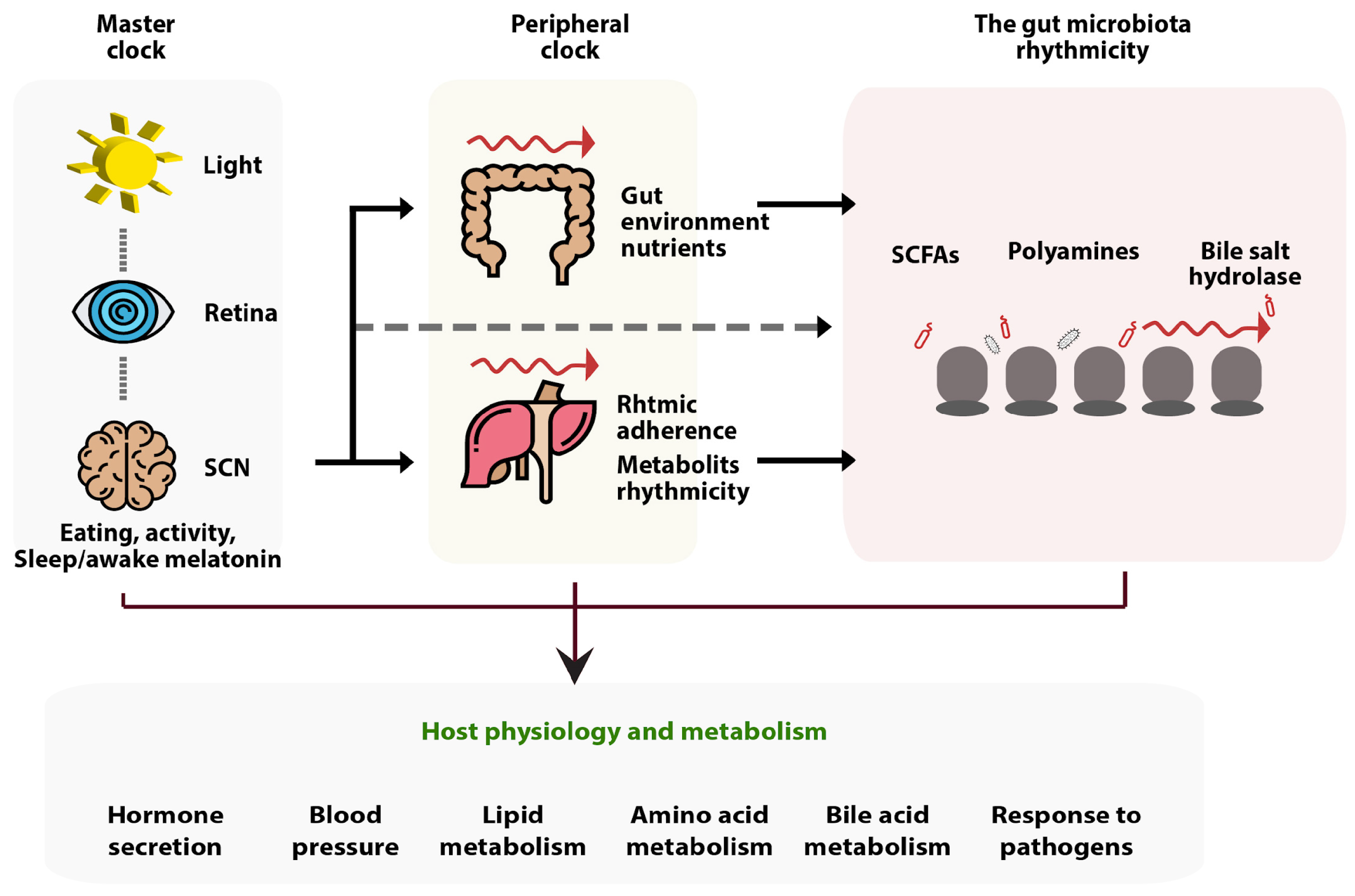
A growing study provides further evidence that the gut microbiota and circadian rhythms interact and communicate in both directions [119]. A central and a peripheral clock constitute the internal clock. The central clock’s primary function is to control each person’s physiological rhythms and patterns. A time generator that is autonomous, has a repeated cycle, and is unique for each person exists in the human body [120]. A central clock that synchronizes peripheral clocks across every part of the body, including the gut, controls circadian rhythms. Light/dark cycles, diet composition, and patterns all affect the microbiota in the gut [121]. Diurnal rhythmicity is crucial for regulating the host–microbiota symbiosis. Oscillations in the microbiota’s metabolome and biogeographical distribution cause the host to enter a state of homeostasis where they are periodically exposed to various bacterial populations, species, functions, and byproducts throughout the course of the day. Some known disruptions to this pattern are antibiotic use or irregular circadian food patterns, which cause a complex disruption of the microbiota’s diurnal rhythm and result in a temporal desynchronization of the circadian liver activities [122].
According to a study of butyrate effects on circadian rhythm, acetate and butyrate both exhibited distinct day–night variations, with high concentrations present throughout the active period. Their results imply that SCFA or lactate produced by the bacteria alters the in vivo phase of host peripheral clocks [123]. The abundance of butyrate and propionate peaks during the light phase, while short-chain fatty acid concentrations (SCFAs) exhibit considerable oscillations. The evidence that butyrate can control Per2 and Bmal1 rhythm patterns in peripheral circadian clocks suggests that the microbiome could influence circadian rhythm through metabolites [124]. The schematic picture of how gut microbiota rhythmicity interacts with the circadian clock is represented in Figure 3.
The composition and function of commensal bacteria as well as the vulnerability of the host to bacterial and viral infection are modulated by circadian rhythms. Although studies of the interactions between circadian rhythms and bacterial or viral infection have been conducted separately, there is still much to understand about the three-way interactions between viruses, bacteria, and circadian rhythms. It is clear that modulating the circadian clock and delivering vaccination at specific times of the day may have significant effects on enhancing host defense against infection and inflammation, to adopt the latest findings demonstrating that adjusting vaccination timing to coincide with the ideal circadian phase could be a potent approach to optimizing vaccine immunogenicity [126]. The rate and strength of the antibody response may vary based on the vaccination’s circadian timing.
According to Langlois et al., a number of vital elements, including vaccine reactogenicity, individual variations in immunological state, and reactivity, may limit potential biological rhythm-dependent responses of people to influenza and other vaccinations [127]. Despite the temptation of suggesting that immunization in the morning would have greater clinical efficacy based on recent studies, further research is required to prove and safely recommend this for general vaccine dosage [128]. Evidence suggests that butyrate has a variety of advantageous impacts on the gastrointestinal. Increasing evidence indicates that it also has effects on the brain. Alterations in butyrate-producing bacteria, for instance, can disrupt the activity of the brain, supporting the idea that a microbiota–gut–brain axis emerges [129]. Szentirmai et al. conducted a study in mice to examine the idea that butyrate could be a signal produced by bacteria that promotes sleep. An increase in non-rapid-eye movement sleep (NREMS) of about 50% was observed in mice following oral gavage delivery of tributyrin, a butyrate pro-drug, for 4 h following the administration. Similarly, intraportal injection of butyrate in mice resulted in rapid and significant elevations in NREMS, which increased by 70% in the first six hours following the butyrate injection. The body temperature significantly decreased after butyrate was administered intraportally as well as orally. Butyrate systemic subcutaneous or intraperitoneal administration had no discernible impact on sleep or body temperature. According to the findings, the sensory mechanism in the liver and/or the wall of the portal vein may be responsible for butyrate’s ability to induce sleep. The gut microbiome may influence sleep by modulating hepatoportal butyrate-sensitive pathways [130].
The timing of studies and the administration of medications must be taken into account in more investigations since circadian rhythms have a substantial impact on host immunity. By addressing the microbiota and its metabolites of interest when they are most vulnerable, we may maximize clinical outcomes by better understanding circadian influences. This would make it possible to utilize drugs at smaller doses, which would lessen their hazardous effects [131].

4.2. Butyrate in Infection

Several defense systems against infectious illnesses are regulated by butyrate. Butyrate and its derivatives may be considered as prospective antibacterial and immune-modulatory medications for treating infectious diseases without the need for antibiotics. “Direct anti-bacterial activities” and “indirect antimicrobial capabilities associated with immunological regulation” are two ways in which butyrate’s effects might be characterized. Butyrate’s synergistic effect with other antimicrobial substances generates a remarkable clearance of bacterial pathogens. Furthermore, butyrate’s indirect antimicrobial effects were validated by in vitro and in vivo experiments. These effects included the promotion of the formation of the defensin and promotion of innate and adaptive immune responses [132]. As demonstrated by the differentiation of macrophages, butyrate imprints innate immune cells via metabolic and epigenetic changes to start the formation of antimicrobial activities [133]. It was discovered that butyrate’s high potential for strengthening immune function derived from both its indirect immune-modulatory effects caused by the inhibition of HDAC and its improvement of T cell impairment regarding commensal bacterial colonization following gut dysbiosis induced by antibiotics [134].
According to Chemudupati et al., butyrate reorganizes the type I interferon (IFN)-mediated innate antiviral immune response. Butyrate inhibits the expression of a large number of the type I IFN-induced antiviral genes, promoting viral infection and replication. Their study demonstrates that metabolites produced by the gut microbiome, including butyrate, can have complex effects on cellular physiology, such as reducing the activity of an innate immune pathway that leads to inflammation and creating a cellular milieu that promotes the development of viruses. Their research also implies that butyrate might be widely utilized as a technique to speed up the growth of virus populations for the research and development of vaccines [135].

5. Disadvantages of Butyrate

It has been suggested that butyrate administration of high-dose sodium butyrate, regardless of how hypertonic the injected fluid is, was able to increase the plasma concentrations of glucose, corticosterone, and adrenocorticotropic hormone (ACTH) [136]. It is possible that sodium butyrate, rather than acting directly as a stressor, sensitizes the response to any stressor, either by handling, injection, or hypertonic saline administration, despite the fact that high doses of sodium butyrate can act as a pharmacological stressor. The two possibilities are now impossible to separate because sodium butyrate supplementation always causes some stress [136].
Kaiko et al. showed that butyrate administration causes autophagy reversion in colonocytes. This may prove why colonocytes preferentially break down butyrate as opposed to other SCFAs such as propionate and acetate, which are also present in high concentrations in the colon. They hypothesized that, despite the Krebs cycle’s ability to employ all three of these SCFAs as substrates, colonocytes mostly use butyrate because its levels must be adjusted to avoid harming stem cells. Propionate and acetate were unable to effectively reduce stem cell proliferation, whereas butyrate could. In contrast to those participating in the metabolism of propionate and acetate, it appears likely that colonocytes up-regulate the enzymes particularly involved in the metabolism of butyrate [137]. However, in other cases, butyrate may worsen the pathophysiology of inflammatory bowel disease (IBD) when the number of colonocytes is significantly decreased by delaying epithelial repair and wound healing. These claims are consistent with existing recommendations that people with irritable bowel syndrome avoid foods that include fermentable oligosaccharides, disaccharides, monosaccharides, and polyols (also known as FODMAP) [138].
Zumbrun et al. found that a high-fiber diet reduced the number of Escherichia spp. in the intestine while increasing intestine butyrate levels in certain mouse models. In these Shiga toxin-producing Escherichia coli (STEC)-infected mice, the one that was fed with the high-fiber diet showed higher levels of colonization, morbidity, and death than mice fed the low-fiber diet. Additionally, Shiga toxin 2 (Stx2) was specifically linked to a higher sensitivity to the high-fiber diet. Overall, they showed that a high-fiber diet can alter Shiga toxin receptor levels, which can worsen the effects of STEC infection [139].
Oral and topical administration of butyrate is particularly challenging due to its terrible taste and odor. Therefore, novel butyrate formulations that are more acceptable and easy to deliver are required [3]. Another useful approach for promoting the widespread use of butyrate against human skin problems could be odorless butyrate releasers [59]. Due to limited evidence to support the effectiveness, safety, or any potential negative effects of topical butyrate application, further research is necessary.

6. Conclusions

Butyrate has attracted special interest due to its favorable benefits for intestinal homeostasis and cellular energy consumption. Butyrate is sourced from foods and supplementation and is also biosynthesized internally through complex carbohydrates and resistance starch fermentation, as well as organic acids and amino acid conversions. This internal process is highly affected by butyrate-producing bacteria, which include Clostridia and several other classes. Butyrate shows many anti-inflammatory properties as well as intestinal and systemic functions. Lower levels of butyrate and/or the bacteria producing it are linked to disease and worse health outcomes, whereas butyrate has demonstrated protective effects. Thus, butyrate is a promising postbiotic production that can potentially prevent and serve as an adjunct to the treatment of several immune-related diseases. Some caveats of butyrate applications could be addressed in future research.
The effectiveness of using postbiotic butyrate is supported by recent data. Unfortunately, butyrate’s undesirable sensorial and physicochemical qualities may be the main cause limiting its application. Butyrate’s application in clinical practice must be supported by products that mitigate its undesirable properties. Due to its limited bioavailability, unpredictable levels in healthy people, lack of reliable clinical evidence proving its effectiveness as a treatment option, and other factors, there are still some challenges for future prospects of butyrate in many cases. To manage infectious diseases without the use of antibiotics, butyrate and butyrate analogs, either separately or combined with other natural compounds, may be evaluated as potentially antibacterial and immunomodulatory.
These actions of butyrate in immune-related diseases have more benefits than disadvantages, but we should always be aware of the consequences of any intervention and decide whether it can complement our treatment as a “friend” or whether we should stay vigilant as bacteria and their by-products are always seen as a “foe”. To further evaluate the potential of butyrate as a prophylactic or therapeutic alternative for inflammatory and immune-mediated conditions, more studies with further experimental models and investigations are needed.

Author Contributions

Conceptualization, H.B.T., M.A. and R.M.R.A.E.; writing—original draft, M.A. and R.M.R.A.E.; writing—review and editing, H.B.T., M.A. and R.M.R.A.E.; visualization, M.A. and R.M.R.A.E.; supervision, H.K., R.F.D., O.S., T.E.C.N., J.L.N. and H.B.T. All authors have read and agreed to the published version of the manuscript.

Funding

This research received no external funding.

Institutional Review Board Statement

Not applicable.

Informed Consent Statement

Not applicable.

Data Availability Statement

Not applicable.

Acknowledgments

We would like to thank the collaborators and the Indonesian Endowment Fund for Education (LPDP) for their support.

Conflicts of Interest

The authors declare no conflict of interest.

References

  1. Guilloteau, P.; Martin, L.; Eeckhaut, V.; Ducatelle, R.; Zabielski, R.; Van Immerseel, F. From the gut to the peripheral tissues: The multiple effects of butyrate. Nutr. Res. Rev. 2010, 23, 366–384. [Google Scholar] [CrossRef] [PubMed] [Green Version]
  2. Liu, H.; Wang, J.; He, T.; Becker, S.; Zhang, G.; Li, D.; Ma, X. Butyrate: A Double-Edged Sword for Health? Adv. Nutr. 2018, 9, 21–29. [Google Scholar] [CrossRef] [Green Version]
  3. Canani, R.B.; Costanzo, M.D.; Leone, L.; Pedata, M.; Meli, R.; Calignano, A. Potential beneficial effects of butyrate in intestinal and extraintestinal diseases. World J. Gastroenterol. 2011, 17, 1519–1528. [Google Scholar] [CrossRef]
  4. Siddiqui, M.T.; Cresci, G.A.M. The Immunomodulatory Functions of Butyrate. J. Inflamm. Res. 2021, 14, 6025–6041. [Google Scholar] [CrossRef]
  5. Chen, J.; Vitetta, L. The Role of Butyrate in Attenuating Pathobiont-Induced Hyperinflammation. Immune. Netw. 2020, 20, e15. [Google Scholar] [CrossRef]
  6. Mokhtari, Z.; Gibson, D.L.; Hekmatdoost, A. Nonalcoholic Fatty Liver Disease, the Gut Microbiome, and Diet. Adv. Nutr. 2017, 8, 240–252. [Google Scholar] [CrossRef] [PubMed] [Green Version]
  7. Louis, P.; Flint, H.J. Diversity, metabolism and microbial ecology of butyrate-producing bacteria from the human large intestine. FEMS Microbiol. Lett. 2009, 294, 1–8. [Google Scholar] [CrossRef] [Green Version]
  8. Tan, J.; McKenzie, C.; Potamitis, M.; Thorburn, A.N.; Mackay, C.R.; Macia, L. The role of short-chain fatty acids in health and disease. Adv. Immunol. 2014, 121, 91–119. [Google Scholar] [PubMed]
  9. Topping, D.L.; Clifton, P.M. Short-chain fatty acids and human colonic function: Roles of resistant starch and nonstarch polysaccharides. Physiol. Rev. 2001, 81, 1031–1064. [Google Scholar] [CrossRef] [PubMed] [Green Version]
  10. Bachmann, C.; Colombo, J.P.; Berüter, J. Short chain fatty acids in plasma and brain: Quantitative determination by gas chromatography. Clin. Chim. Acta 1979, 92, 153–159. [Google Scholar] [CrossRef]
  11. Dalile, B.; Van Oudenhove, L.; Vervliet, B.; Verbeke, K. The role of short-chain fatty acids in microbiota-gut-brain communication. Nat. Rev. Gastroenterol. Hepatol. 2019, 16, 461–478. [Google Scholar] [CrossRef]
  12. Kaźmierczak-Siedlecka, K.; Marano, L.; Merola, E.; Roviello, F.; Połom, K. Sodium butyrate in both prevention and supportive treatment of colorectal cancer. Front. Cell. Infect. Microbiol. 2022, 12, 1023806. [Google Scholar] [CrossRef] [PubMed]
  13. Amiri, P.; Hosseini, S.A.; Ghaffari, S.; Tutunchi, H.; Ghaffari, S.; Mosharkesh, E.; Asghari, S.; Roshanravan, N. Role of Butyrate, a Gut Microbiota Derived Metabolite, in Cardiovascular Diseases: A comprehensive narrative review. Front. Pharmacol. 2021, 12, 837509. [Google Scholar] [CrossRef] [PubMed]
  14. Usami, M.; Kishimoto, K.; Ohata, A.; Miyoshi, M.; Aoyama, M.; Fueda, Y.; Kotani, J. Butyrate and trichostatin A attenuate nuclear factor kappaB activation and tumor necrosis factor alpha secretion and increase prostaglandin E2 secretion in human peripheral blood mononuclear cells. Nutr. Res. 2008, 28, 321–328. [Google Scholar] [CrossRef] [PubMed]
  15. Chang, P.V.; Hao, L.; Offermanns, S.; Medzhitov, R. The microbial metabolite butyrate regulates intestinal macrophage function via histone deacetylase inhibition. Proc. Natl. Acad. Sci USA 2014, 111, 2247–2252. [Google Scholar] [CrossRef] [PubMed] [Green Version]
  16. Wang, Z.; Zhang, X.; Zhu, L.; Yang, X.; He, F.; Wang, T.; Bao, T.; Lu, H.; Wang, H.; Yang, S. Inulin alleviates inflammation of alcoholic liver disease via SCFAs-inducing suppression of M1 and facilitation of M2 macrophages in mice. Int. Immunopharmacol. 2020, 78, 106062. [Google Scholar] [CrossRef] [PubMed]
  17. Simeoli, R.; Mattace Raso, G.; Pirozzi, C.; Lama, A.; Santoro, A.; Russo, R.; Montero-Melendez, T.; Canani, R.B.; Calignano, A.; Perretti, M.; et al. An orally administered butyrate-releasing derivative reduces neutrophil recruitment and inflammation in dextran sulphate sodium-induced murine colitis. Br. J. Pharmacol. 2017, 174, 1484–1496. [Google Scholar] [CrossRef] [Green Version]
  18. Park, J.; Kim, M.; Kang, S.G.; Jannasch, A.H.; Cooper, B.; Patterson, J.; Kim, C.H. Short-chain fatty acids induce both effector and regulatory T cells by suppression of histone deacetylases and regulation of the mTOR–S6K pathway. Mucosal Immunol. 2015, 8, 80–93. [Google Scholar] [CrossRef] [Green Version]
  19. Deroanne, C.F.; Bonjean, K.; Servotte, S.; Devy, L.; Colige, A.; Clausse, N.; Blacher, S.; Verdin, E.; Foidart, J.M.; Nusgens, B.V.; et al. Histone deacetylases inhibitors as anti-angiogenic agents altering vascular endothelial growth factor signaling. Oncogene 2002, 21, 427–436. [Google Scholar] [CrossRef] [Green Version]
  20. Liang, D.; Kong, X.; Sang, N. Effects of histone deacetylase inhibitors on HIF-1. Cell Cycle 2006, 5, 2430–2435. [Google Scholar] [CrossRef] [Green Version]
  21. Coppola, S.; Avagliano, C.; Sacchi, A.; Laneri, S.; Calignano, A.; Voto, L.; Luzzetti, A.; Canani, R.B. Potential Clinical Applications of the Postbiotic Butyrate in Human Skin Diseases. Molecules 2022, 27, 1849. [Google Scholar] [CrossRef]
  22. Pituch, A.; Walkowiak, J.; Banaszkiewicz, A. Butyric acid in functional constipation. Prz. Gastroenterol. 2013, 8, 295–298. [Google Scholar] [CrossRef]
  23. Hamer, H.M.; Jonkers, D.; Venema, K.; Vanhoutvin, S.; Troost, F.J.; Brummer, R.J. Review article: The role of butyrate on colonic function. Aliment. Pharmacol. Ther. 2008, 27, 104–119. [Google Scholar] [CrossRef] [PubMed]
  24. Fu, X.; Liu, Z.; Zhu, C.; Mou, H.; Kong, Q. Nondigestible carbohydrates, butyrate, and butyrate-producing bacteria. Crit. Rev. Food Sci. Nutr. 2019, 59 (Suppl. 1), S130–S152. [Google Scholar] [CrossRef] [PubMed]
  25. Kolenbrander, P.E.; Flint, H.J.; Louis, P.; Scott, K.P.; Duncan, S.H. Commensal bacteria in health and disease. Virulence Mech. Bact. Pathog. 2007, 101–115. [Google Scholar] [CrossRef]
  26. Wilson, A.S.; Koller, K.R.; Ramaboli, M.C.; Nesengani, L.T.; Ocvirk, S.; Chen, C.; Flanagan, C.A.; Sapp, F.R.; Merritt, Z.T.; Bhatti, F.; et al. Diet and the Human Gut Microbiome: An International Review. Dig. Dis. Sci. 2020, 65, 723–740. [Google Scholar] [CrossRef] [PubMed] [Green Version]
  27. Singh, R.K.; Chang, H.W.; Yan, D.; Lee, K.M.; Ucmak, D.; Wong, K.; Abrouk, M.; Farahnik, B.; Nakamura, M.; Zhu, T.H.; et al. Influence of diet on the gut microbiome and implications for human health. J. Transl. Med. 2017, 15, 73. [Google Scholar] [CrossRef] [Green Version]
  28. Esquivel-Elizondo, S.; Ilhan, Z.E.; Garcia-Peña, E.I.; Krajmalnik-Brown, R. Insights into Butyrate Production in a Controlled Fermentation System via Gene Predictions. mSystems 2017, 2, e00051-17. [Google Scholar] [CrossRef] [Green Version]
  29. Louis, P.; Flint, H.J. Formation of propionate and butyrate by the human colonic microbiota. Environ. Microbiol. 2017, 19, 29–41. [Google Scholar] [CrossRef] [Green Version]
  30. Kotrba, P.; Inui, M.; Yukawa, H. Bacterial phosphotransferase system (PTS) in carbohydrate uptake and control of carbon metabolism. J. Biosci. Bioeng. 2001, 92, 502–517. [Google Scholar] [CrossRef]
  31. Hawkes, F.R.; Dinsdale, R.; Hawkes, D.L.; Hussy, I. Sustainable fermentative hydrogen production: Challenges for process optimisation. Int. J. Hydrog. Energy 2002, 27, 1339–1347. [Google Scholar] [CrossRef]
  32. Vital, M.; Howe, A.C.; Tiedje, J.M. Revealing the bacterial butyrate synthesis pathways by analyzing (meta)genomic data. mBio 2014, 5, e00889. [Google Scholar] [CrossRef] [PubMed] [Green Version]
  33. Postma, P.W.; Lengeler, J.W.; Jacobson, G.R. Phosphoenolpyruvate:carbohydrate phosphotransferase systems of bacteria. Microbiol. Rev. 1993, 57, 543–594. [Google Scholar] [CrossRef] [PubMed]
  34. Wang, J.; Zhang, K. Production of mesaconate in Escherichia coli by engineered glutamate mutase pathway. Metab. Eng. 2015, 30, 190–196. [Google Scholar] [CrossRef] [PubMed]
  35. Buckel, W. Energy Conservation in Fermentations of Anaerobic Bacteria. Front. Microbiol. 2021, 12, 703525. [Google Scholar] [CrossRef] [PubMed]
  36. Zhang, J.; Friedrich, P.; Pierik, A.J.; Martins, B.M.; Buckel, W. Substrate-induced radical formation in 4-hydroxybutyryl coenzyme A dehydratase from Clostridium aminobutyricum. Appl. Environ. Microbiol. 2015, 81, 1071–1084. [Google Scholar] [CrossRef] [Green Version]
  37. Brennenstuhl, H.; Didiasova, M.; Assmann, B.; Bertoldi, M.; Molla, G.; Jung-Klawitter, S.; Hübschmann, O.K.; Schröter, J.; Opladen, T.; Tikkanen, R. Succinic Semialdehyde Dehydrogenase Deficiency: In Vitro and In Silico Characterization of a Novel Pathogenic Missense Variant and Analysis of the Mutational Spectrum of ALDH5A1. Int. J. Mol. Sci. 2020, 21, 8578. [Google Scholar] [CrossRef] [PubMed]
  38. Ramachandran, V.S. Encyclopedia of the Human Brain; Col-Mem; Academic Press: Cambridge, MA, USA, 2002. [Google Scholar]
  39. Yang, X.; Schnackenberg, L.K.; Shi, Q.; Salminen, W.F. Hepatic toxicity biomarkers. In Biomarkers in Toxicology; Elsevier: Amsterdam, The Netherlands, 2014; pp. 241–259. [Google Scholar]
  40. Heldt, H.W.; Piechulla, B. Nitrate assimilation is essential for the synthesis of organic matter. Plant Biochem. 2005, 275–308. [Google Scholar] [CrossRef]
  41. Sajilata, M.G.; Singhal, R.S.; Kulkarni, P.R. Resistant Starch-A Review. Compr. Rev. Food Sci. Food Saf. 2006, 5, 1–17. [Google Scholar] [CrossRef]
  42. Jost, T.; Lacroix, C.; Braegger, C.P.; Rochat, F.; Chassard, C. Vertical mother-neonate transfer of maternal gut bacteria via breastfeeding. Environ. Microbiol. 2014, 16, 2891–2904. [Google Scholar] [CrossRef]
  43. Walker, W.A.; Iyengar, R.S. Breast milk, microbiota, and intestinal immune homeostasis. Pediatr. Res. 2015, 77, 220–228. [Google Scholar] [CrossRef] [PubMed] [Green Version]
  44. Walker, A.W.; Duncan, S.H.; Louis, P.; Flint, H.J. Phylogeny, culturing, and metagenomics of the human gut microbiota. Trends Microbiol. 2014, 22, 267–274. [Google Scholar] [CrossRef] [PubMed]
  45. Detman, A.; Mielecki, D.; Chojnacka, A.; Salamon, A.; Blaszczyk, M.K.; Sikora, A. Cell factories converting lactate and acetate to butyrate: Clostridium butyricum and microbial communities from dark fermentation bioreactors. Microb. Cell Fact. 2019, 18, 36. [Google Scholar] [CrossRef] [Green Version]
  46. Zhu, L.B.; Zhang, Y.C.; Huang, H.H.; Lin, J. Prospects for clinical applications of butyrate-producing bacteria. World J. Clin. Pediatr. 2021, 10, 84–92. [Google Scholar] [CrossRef]
  47. Rivière, A.; Selak, M.; Lantin, D.; Leroy, F.; De Vuyst, L. Bifidobacteria and Butyrate-Producing Colon Bacteria: Importance and Strategies for Their Stimulation in the Human Gut. Front. Microbiol. 2016, 7, 979. [Google Scholar] [CrossRef] [Green Version]
  48. Scott, K.P.; Antoine, J.M.; Midtvedt, T.; van Hemert, S. Manipulating the gut microbiota to maintain health and treat disease. Microb. Ecol. Health Dis. 2015, 26, 25877. [Google Scholar] [CrossRef] [PubMed]
  49. Cheng, X.; Zhou, T.; He, Y.; Xie, Y.; Xu, Y.; Huang, W. The role and mechanism of butyrate in the prevention and treatment of diabetic kidney disease. Front. Microbiol. 2022, 13, 961536. [Google Scholar] [CrossRef] [PubMed]
  50. Hazem, S.H.; Hamed, M.F.; Saad, M.A.; Gameil, N.M. Comparison of lactate and beta-hydroxybutyrate in the treatment of concanavalin-A induced hepatitis. Int. Immunopharmacol. 2018, 61, 376–384. [Google Scholar] [CrossRef] [PubMed]
  51. Vinolo, M.A.; Rodrigues, H.G.; Hatanaka, E.; Sato, F.T.; Sampaio, S.C.; Curi, R. Suppressive effect of short-chain fatty acids on production of proinflammatory mediators by neutrophils. J. Nutr. Biochem. 2011, 22, 849–855. [Google Scholar] [CrossRef]
  52. Wang, C.C.; Wu, H.; Lin, F.H.; Gong, R.; Xie, F.; Peng, Y.; Feng, J.; Hu, C.H. Sodium butyrate enhances intestinal integrity, inhibits mast cell activation, inflammatory mediator production and JNK signaling pathway in weaned pigs. Innate Immun. 2018, 24, 40–46. [Google Scholar] [CrossRef]
  53. Diakos, C.; Prieschl, E.E.; Saemann, M.D.; Bohmig, G.A.; Csonga, R.; Sobanov, Y.; Baumruker, T.; Zlabinger, G.J. n-Butyrate inhibits Jun NH(2)-terminal kinase activation and cytokine transcription in mast cells. Biochem. Biophys. Res. Commun. 2006, 349, 863–868. [Google Scholar] [CrossRef] [PubMed]
  54. Millard, A.L.; Mertes, P.M.; Ittelet, D.; Villard, F.; Jeannesson, P.; Bernard, J. Butyrate affects differentiation, maturation and function of human monocyte-derived dendritic cells and macrophages. Clin. Exp. Immunol. 2002, 130, 245–255. [Google Scholar] [CrossRef] [PubMed]
  55. Liu, L.; Li, L.; Min, J.; Wang, J.; Wu, H.; Zeng, Y.; Chen, S.; Chu, Z. Butyrate interferes with the differentiation and function of human monocyte-derived dendritic cells. Cell. Immunol. 2012, 277, 66–73. [Google Scholar] [CrossRef] [PubMed]
  56. Correa-Oliveira, R.; Fachi, J.L.; Vieira, A.; Sato, F.T.; Vinolo, M.A. Regulation of immune cell function by short-chain fatty acids. Clin. Transl. Immunol. 2016, 5, e73. [Google Scholar] [CrossRef]
  57. Rocamora-Reverte, L.; Melzer, F.L.; Wurzner, R.; Weinberger, B. The Complex Role of Regulatory T Cells in Immunity and Aging. Front. Immunol. 2020, 11, 616949. [Google Scholar] [CrossRef] [PubMed]
  58. Meijer, K.; de Vos, P.; Priebe, M.G. Butyrate and other short-chain fatty acids as modulators of immunity: What relevance for health? Curr. Opin. Clin. Nutr. Metab. Care 2010, 13, 715–721. [Google Scholar] [CrossRef]
  59. Schwarz, A.; Bruhs, A.; Schwarz, T. The Short-Chain Fatty Acid Sodium Butyrate Functions as a Regulator of the Skin Immune System. J. Investig. Dermatol. 2017, 137, 855–864. [Google Scholar] [CrossRef] [Green Version]
  60. Lewis, J.D.; Abreu, M.T. Diet as a Trigger or Therapy for Inflammatory Bowel Diseases. Gastroenterology 2017, 152, 398–414.e6. [Google Scholar] [CrossRef]
  61. Zhuang, X.; Li, T.; Li, M.; Huang, S.; Qiu, Y.; Feng, R.; Zhang, S.; Chen, M.; Xiong, L.; Zeng, Z. Systematic Review and Meta-analysis: Short-Chain Fatty Acid Characterization in Patients With Inflammatory Bowel Disease. Inflamm. Bowel Dis. 2019, 25, 1751–1763. [Google Scholar] [CrossRef]
  62. Vernia, P.; Monteleone, G.; Grandinetti, G.; Villotti, G.; Di Giulio, E.; Frieri, G.; Marcheggiano, A.; Pallone, F.; Caprilli, R.; Torsoli, A. Combined oral sodium butyrate and mesalazine treatment compared to oral mesalazine alone in ulcerative colitis: Randomized, double-blind, placebo-controlled pilot study. Dig. Dis. Sci. 2000, 45, 976–981. [Google Scholar] [CrossRef]
  63. Berni Canani, R.; Di Costanzo, M.; Leone, L. The epigenetic effects of butyrate: Potential therapeutic implications for clinical practice. Clin. Epigenetics 2012, 4, 4. [Google Scholar] [CrossRef]
  64. Silverman, G.J.; Azzouz, D.F.; Alekseyenko, A.V. Systemic Lupus Erythematosus and dysbiosis in the microbiome: Cause or effect or both? Curr. Opin. Immunol. 2019, 61, 80–85. [Google Scholar] [CrossRef]
  65. Ma, L.; Morel, L. Loss of gut barrier integrity in lupus. Front. Immunol. 2022, 13, 919792. [Google Scholar] [CrossRef] [PubMed]
  66. Effendi, R.M.R.A.; Anshory, M.; Kalim, H.; Dwiyana, R.F.; Suwarsa, O.; Pardo, L.M.; Nijsten, T.E.C.; Thio, H.B. Akkermansia muciniphila and Faecalibacterium prausnitzii in Immune-Related Diseases. Microorganisms 2022, 10, 2382. [Google Scholar] [CrossRef] [PubMed]
  67. Toumi, E.; Goutorbe, B.; Plauzolles, A.; Bonnet, M.; Mezouar, S.; Militello, M.; Mege, J.L.; Chiche, L.; Halfon, P. Gut microbiota in systemic lupus erythematosus patients and lupus mouse model: A cross species comparative analysis for biomarker discovery. Front. Immunol. 2022, 13, 943241. [Google Scholar] [CrossRef] [PubMed]
  68. Li, Y.; Wang, H.F.; Li, X.; Li, H.X.; Zhang, Q.; Zhou, H.W.; He, Y.; Li, P.; Fu, C.; Zhang, X.H.; et al. Disordered intestinal microbes are associated with the activity of Systemic Lupus Erythematosus. Clin. Sci. 2019, 133, 821–838. [Google Scholar] [CrossRef] [PubMed]
  69. Hevia, A.; Milani, C.; Lopez, P.; Cuervo, A.; Arboleya, S.; Duranti, S.; Turroni, F.; González, S.; Suárez, A.; Gueimonde, M.; et al. Intestinal dysbiosis associated with systemic lupus erythematosus. mBio 2014, 5, e01548-14. [Google Scholar] [CrossRef] [PubMed] [Green Version]
  70. He, Z.; Shao, T.; Li, H.; Xie, Z.; Wen, C. Alterations of the gut microbiome in Chinese patients with systemic lupus erythematosus. Gut Pathog. 2016, 8, 64. [Google Scholar] [CrossRef] [Green Version]
  71. Vieira, J.R.P.; Rezende, A.T.O.; Fernandes, M.R.; da Silva, N.A. Intestinal microbiota and active systemic lupus erythematosus: A systematic review. Adv. Rheumatol. 2021, 61, 42. [Google Scholar] [CrossRef]
  72. Luo, X.M.; Edwards, M.R.; Mu, Q.; Yu, Y.; Vieson, M.D.; Reilly, C.M.; Ahmed, S.A.; Bankole, A.A. Gut Microbiota in Human Systemic Lupus Erythematosus and a Mouse Model of Lupus. Appl. Environ. Microbiol. 2018, 84, e02288-17. [Google Scholar] [CrossRef] [Green Version]
  73. Pan, Q.; Guo, F.; Huang, Y.; Li, A.; Chen, S.; Chen, J.; Liu, H.F.; Pan, Q. Gut Microbiota Dysbiosis in Systemic Lupus Erythematosus: Novel Insights into Mechanisms and Promising Therapeutic Strategies. Front. Immunol. 2021, 12, 799788. [Google Scholar] [CrossRef] [PubMed]
  74. He, H.; Xu, H.; Xu, J.; Zhao, H.; Lin, Q.; Zhou, Y.; Nie, Y. Sodium Butyrate Ameliorates Gut Microbiota Dysbiosis in Lupus-Like Mice. Front. Nutr. 2020, 7, 604283. [Google Scholar] [CrossRef] [PubMed]
  75. Smith, P.M.; Howitt, M.R.; Panikov, N.; Michaud, M.; Gallini, C.A.; Bohlooly, Y.M.; Glickman, J.N.; Garrett, W.S. The microbial metabolites, short-chain fatty acids, regulate colonic Treg cell homeostasis. Science 2013, 341, 569–573. [Google Scholar] [CrossRef] [PubMed] [Green Version]
  76. Arpaia, N.; Campbell, C.; Fan, X.; Dikiy, S.; van der Veeken, J.; deRoos, P.; Liu, H.; Cross, J.R.; Pfeffer, K.; Coffer, P.J.; et al. Metabolites produced by commensal bacteria promote peripheral regulatory T-cell generation. Nature 2013, 504, 451–455. [Google Scholar] [CrossRef] [PubMed] [Green Version]
  77. Furusawa, Y.; Obata, Y.; Fukuda, S.; Endo, T.A.; Nakato, G.; Takahashi, D.; Nakanishi, Y.; Uetake, C.; Kato, K.; Kato, T.; et al. Commensal microbe-derived butyrate induces the differentiation of colonic regulatory T cells. Nature 2013, 504, 446–450. [Google Scholar] [CrossRef]
  78. Takahashi, D.; Hoshina, N.; Kabumoto, Y.; Maeda, Y.; Suzuki, A.; Tanabe, H.; Isobe, J.; Yamada, T.; Muroi, K.; Yanagisawa, Y.; et al. Microbiota-derived butyrate limits the autoimmune response by promoting the differentiation of follicular regulatory T cells. EBioMedicine 2020, 58, 102913. [Google Scholar] [CrossRef]
  79. Fernando, M.R.; Saxena, A.; Reyes, J.L.; McKay, D.M. Butyrate enhances antibacterial effects while suppressing other features of alternative activation in IL-4-induced macrophages. Am. J. Physiol. Gastrointest Liver Physiol. 2016, 310, G822–G831. [Google Scholar] [CrossRef] [Green Version]
  80. Xin, M.L.; Michael, R.E.; Christopher, M.R.; Qinghui, M.; Ahmed, S.A. Diet and Microbes in the Pathogenesis of Lupus. In Lupus; Wahid Ali, K., Ed.; IntechOpen: Rijeka, Croatia, 2017; p. Ch. 8. [Google Scholar] [CrossRef] [Green Version]
  81. Panther, E.J.; Ren, J.; Cabana-Puig, X.; Abdelhamid, L.; Swartwout, B.; Luo, X.M.; Reilly, C.M. The Effect of Dietary Fiber Intake on Systemic Lupus Erythematosus (SLE) Disease in NZB/W Lupus Mice. J. Clin. Cell. Immunol. 2020, 11, 1–8. [Google Scholar] [CrossRef]
  82. Belkaid, Y.; Naik, S. Compartmentalized and systemic control of tissue immunity by commensals. Nat. Immunol. 2013, 14, 646–653. [Google Scholar] [CrossRef] [Green Version]
  83. Callewaert, C.; Knödlseder, N.; Karoglan, A.; Güell, M.; Paetzold, B. Skin microbiome transplantation and manipulation: Current state of the art. Comput. Struct. Biotechnol. J. 2021, 19, 624–631. [Google Scholar] [CrossRef]
  84. Mantziari, A.; Salminen, S.; Szajewska, H.; Malagón-Rojas, J.N. Postbiotics against Pathogens Commonly Involved in Pediatric Infectious Diseases. Microorganisms 2020, 8, 1510. [Google Scholar] [CrossRef] [PubMed]
  85. Chen, Y.E.; Fischbach, M.A.; Belkaid, Y. Skin microbiota-host interactions. Nature 2018, 553, 427–436. [Google Scholar] [CrossRef] [PubMed]
  86. De Pessemier, B.; Grine, L.; Debaere, M.; Maes, A.; Paetzold, B.; Callewaert, C. Gut-Skin Axis: Current Knowledge of the Interrelationship between Microbial Dysbiosis and Skin Conditions. Microorganisms 2021, 9, 353. [Google Scholar] [CrossRef] [PubMed]
  87. Russo, R.; Santarcangelo, C.; Badolati, N.; Sommella, E.; De Filippis, A.; Dacrema, M.; Campiglia, P.; Stornaiuolo, M.; Daglia, M. In vivo bioavailability and in vitro toxicological evaluation of the new butyric acid releaser N-(1-carbamoyl-2-phenyl-ethyl) butyramide. Biomed. Pharmacother. 2021, 137, 111385. [Google Scholar] [CrossRef]
  88. Nylund, L.; Nermes, M.; Isolauri, E.; Salminen, S.; de Vos, W.M.; Satokari, R. Severity of atopic disease inversely correlates with intestinal microbiota diversity and butyrate-producing bacteria. Allergy 2015, 70, 241–244. [Google Scholar] [CrossRef]
  89. Lakhdari, O.; Tap, J.; Béguet-Crespel, F.; Le Roux, K.; de Wouters, T.; Cultrone, A.; Nepelska, M.; Lefèvre, F.; Doré, J.; Blottière, H.M. Identification of NF-κB modulation capabilities within human intestinal commensal bacteria. J. Biomed. Biotechnol. 2011, 2011, 282356. [Google Scholar] [CrossRef] [Green Version]
  90. Flint, H.J.; Scott, K.P.; Louis, P.; Duncan, S.H. The role of the gut microbiota in nutrition and health. Nat. Rev. Gastroenterol. Hepatol. 2012, 9, 577–589. [Google Scholar] [CrossRef]
  91. Trompette, A.; Pernot, J.; Perdijk, O.; Alqahtani, R.A.A.; Domingo, J.S.; Camacho-Muñoz, D.; Wong, N.C.; Kendall, A.C.; Wiederkehr, A.; Nicod, L.P.; et al. Gut-derived short-chain fatty acids modulate skin barrier integrity by promoting keratinocyte metabolism and differentiation. Mucosal Immunol. 2022, 15, 908–926. [Google Scholar] [CrossRef]
  92. Alesa, D.I.; Alshamrani, H.M.; Alzahrani, Y.A.; Alamssi, D.N.; Alzahrani, N.S.; Almohammadi, M.E. The role of gut microbiome in the pathogenesis of psoriasis and the therapeutic effects of probiotics. J. Family Med. Prim. Care. 2019, 8, 3496–3503. [Google Scholar]
  93. Tan, L.; Zhao, S.; Zhu, W.; Wu, L.; Li, J.; Shen, M.; Lei, L.; Chen, X.; Peng, C. The Akkermansia muciniphila is a gut microbiota signature in psoriasis. Exp. Dermatol. 2018, 27, 144–149. [Google Scholar] [CrossRef]
  94. Eppinga, H.; Sperna Weiland, C.J.; Thio, H.B.; van der Woude, C.J.; Nijsten, T.E.C.; Peppelenbosch, M.P.; Konstantinov, S.R. Similar Depletion of Protective Faecalibacterium prausnitzii in Psoriasis and Inflammatory Bowel Disease, but not in Hidradenitis Suppurativa. J. Crohn’s Colitis 2016, 10, 1067–1075. [Google Scholar] [CrossRef] [PubMed] [Green Version]
  95. Shapiro, J.; Cohen, N.A.; Shalev, V.; Uzan, A.; Koren, O.; Maharshak, N. Psoriatic patients have a distinct structural and functional fecal microbiota compared with controls. J. Dermatol. 2019, 46, 595–603. [Google Scholar] [CrossRef] [PubMed]
  96. Owczarczyk-Saczonek, A.; Czerwińska, J.; Placek, W. The role of regulatory T cells and anti-inflammatory cytokines in psoriasis. Acta Derm. Alp Pannonica Adriat 2018, 27, 17–23. [Google Scholar] [CrossRef]
  97. Stockenhuber, K.; Hegazy, A.N.; West, N.R.; Ilott, N.E.; Stockenhuber, A.; Bullers, S.J.; Thornton, E.E.; Arnold, I.C.; Tucci, A.; Waldmann, H.; et al. Foxp3(+) T reg cells control psoriasiform inflammation by restraining an IFN-I-driven CD8(+) T cell response. J. Exp. Med. 2018, 215, 1987–1998. [Google Scholar] [CrossRef]
  98. Schwarz, A.; Philippsen, R.; Schwarz, T. Induction of Regulatory T Cells and Correction of Cytokine Disbalance by Short-Chain Fatty Acids: Implications for Psoriasis Therapy. J. Investig. Dermatol. 2021, 141, 95–104.e2. [Google Scholar] [CrossRef]
  99. Dillon, S.M.; Kibbie, J.; Lee, E.J.; Guo, K.; Santiago, M.L.; Austin, G.L.; Gianella, S.; Landay, A.L.; Donovan, A.M.; Frank, D.N.; et al. Low abundance of colonic butyrate-producing bacteria in HIV infection is associated with microbial translocation and immune activation. AIDS 2017, 31, 511–521. [Google Scholar] [CrossRef]
  100. Serrano-Villar, S.; Vazquez-Castellanos, J.F.; Vallejo, A.; Latorre, A.; Sainz, T.; Ferrando-Martinez, S.; Rojo, D.; Martínez-Botas, J.; del Romero, J.; Madrid, N.; et al. The effects of prebiotics on microbial dysbiosis, butyrate production and immunity in HIV-infected subjects. Mucosal Immunol. 2017, 10, 1279–1293. [Google Scholar] [CrossRef] [Green Version]
  101. Williams, B. Gut Microbiome in HIV Infection: Overcoming Barriers? Dig. Dis. Sci. 2019, 64, 1725–1727. [Google Scholar] [CrossRef] [Green Version]
  102. Ortiz, A.M.; Simpson, J.; Langner, C.A.; Baker, P.J.; Aguilar, C.; Brooks, K.; Flynn, J.K.; Vinton, C.L.; Rahmberg, A.R.; Hickman, H.D.; et al. Butyrate administration is not sufficient to improve immune reconstitution in antiretroviral-treated SIV-infected macaques. Sci. Rep. 2022, 12, 7491. [Google Scholar] [CrossRef]
  103. Lopez, C.A.; Kingsbury, D.D.; Velazquez, E.M.; Baumler, A.J. Collateral damage: Microbiota-derived metabolites and immune function in the antibiotic era. Cell Host Microbe 2014, 16, 156–163. [Google Scholar] [CrossRef] [Green Version]
  104. Koh, A.; Backhed, F. From Association to Causality: The Role of the Gut Microbiota and Its Functional Products on Host Metabolism. Mol. Cell 2020, 78, 584–596. [Google Scholar] [CrossRef] [PubMed]
  105. Allaire, J.M.; Crowley, S.M.; Law, H.T.; Chang, S.Y.; Ko, H.J.; Vallance, B.A. The Intestinal Epithelium: Central Coordinator of Mucosal Immunity. Trends Immunol. 2018, 39, 677–696. [Google Scholar] [CrossRef] [PubMed]
  106. Clark, R.L.; Connors, B.M.; Stevenson, D.M.; Hromada, S.E.; Hamilton, J.J.; Amador-Noguez, D.; Venturelli, O.S. Design of synthetic human gut microbiome assembly and butyrate production. Nat. Commun. 2021, 12, 3254. [Google Scholar] [CrossRef] [PubMed]
  107. Woo, Y.R.; Cho, S.H.; Lee, J.D.; Kim, H.S. The Human Microbiota and Skin Cancer. Int. J. Mol. Sci. 2022, 23, 1813. [Google Scholar] [CrossRef]
  108. González-Sánchez, P.; DeNicola, G.M. The microbiome(s) and cancer: Know thy neighbor(s). J. Pathol. 2021, 254, 332–343. [Google Scholar] [CrossRef]
  109. Gopalakrishnan, V.; Spencer, C.N.; Nezi, L.; Reuben, A.; Andrews, M.C.; Karpinets, T.V.; Prieto, P.A.; Vicente, D.; Hoffman, K.; Wei, S.C.; et al. Gut microbiome modulates response to anti-PD-1 immunotherapy in melanoma patients. Science 2018, 359, 97–103. [Google Scholar] [CrossRef] [Green Version]
  110. Matson, V.; Fessler, J.; Bao, R.; Chongsuwat, T.; Zha, Y.; Alegre, M.L.; Luke, J.J.; Gajewski, T.F. The commensal microbiome is associated with anti-PD-1 efficacy in metastatic melanoma patients. Science 2018, 359, 104–108. [Google Scholar] [CrossRef] [Green Version]
  111. Zitvogel, L.; Ma, Y.; Raoult, D.; Kroemer, G.; Gajewski, T.F. The microbiome in cancer immunotherapy: Diagnostic tools and therapeutic strategies. Science 2018, 359, 1366–1370. [Google Scholar] [CrossRef] [Green Version]
  112. He, Y.; Fu, L.; Li, Y.; Wang, W.; Gong, M.; Zhang, J.; Dong, X.; Huang, J.; Wang, Q.; Mackay, C.R.; et al. Gut microbial metabolites facilitate anticancer therapy efficacy by modulating cytotoxic CD8(+) T cell immunity. Cell Metab. 2021, 33, 988–1000.e7. [Google Scholar] [CrossRef]
  113. Marks, P.A.; Richon, V.M.; Miller, T.; Kelly, W.K. Histone Deacetylase Inhibitors. Adv. Cancer Res. 2004, 91, 137–168. [Google Scholar]
  114. Dashwood, R.H.; Ho, E. Dietary histone deacetylase inhibitors: From cells to mice to man. Semin Cancer Biol. 2007, 17, 363–369. [Google Scholar] [CrossRef] [PubMed] [Green Version]
  115. Shao, Y.; Gao, Z.; Marks, P.A.; Jiang, X. Apoptotic and autophagic cell death induced by histone deacetylase inhibitors. Proc. Natl. Acad. Sci. USA 2004, 101, 18030–18035. [Google Scholar] [CrossRef] [PubMed] [Green Version]
  116. Coutzac, C.; Jouniaux, J.M.; Paci, A.; Schmidt, J.; Mallardo, D.; Seck, A.; Asvatourian, V.; Cassard, L.; Saulnier, P.; Lacroix, L.; et al. Systemic short chain fatty acids limit antitumor effect of CTLA-4 blockade in hosts with cancer. Nat. Commun. 2020, 11, 2168. [Google Scholar] [CrossRef] [PubMed]
  117. Mirzaei, R.; Afaghi, A.; Babakhani, S.; Sohrabi, M.R.; Hosseini-Fard, S.R.; Babolhavaeji, K.; Akbari, S.K.A.; Yousefimashouf, R.; Karampoor, S. Role of microbiota-derived short-chain fatty acids in cancer development and prevention. Biomed. Pharmacother. 2021, 139, 111619. [Google Scholar] [CrossRef] [PubMed]
  118. Rangan, P.; Mondino, A. Microbial short-chain fatty acids: A strategy to tune adoptive T cell therapy. J. Immunother. Cancer 2022, 10, e004147. [Google Scholar] [CrossRef] [PubMed]
  119. Teichman, E.M.; O’Riordan, K.J.; Gahan, C.G.M.; Dinan, T.G.; Cryan, J.F. When Rhythms Meet the Blues: Circadian Interactions with the Microbiota-Gut-Brain Axis. Cell Metabolism 2020, 31, 448–471. [Google Scholar] [CrossRef]
  120. Mehling, A.; Fluhr, J.W. Chronobiology: Biological clocks and rhythms of the skin. Skin Pharmacol. Physiol. 2006, 19, 182–189. [Google Scholar] [CrossRef]
  121. Parkar, S.G.; Kalsbeek, A.; Cheeseman, J.F. Potential Role for the Gut Microbiota in Modulating Host Circadian Rhythms and Metabolic Health. Microorganisms 2019, 7, 41. [Google Scholar] [CrossRef] [Green Version]
  122. Thaiss, C.A.; Levy, M.; Korem, T.; Dohnalová, L.; Shapiro, H.; Jaitin, D.A.; David, E.; Winter, D.R.; Gury-BenAri, M.; Tatirovsky, E.; et al. Microbiota Diurnal Rhythmicity Programs Host Transcriptome Oscillations. Cell 2016, 167, 1495–1510.e12. [Google Scholar] [CrossRef] [Green Version]
  123. Tahara, Y.; Yamazaki, M.; Sukigara, H.; Motohashi, H.; Sasaki, H.; Miyakawa, H.; Haraguchi, A.; Ikeda, Y.; Fukuda, S.; Shibata, S. Gut Microbiota-Derived Short Chain Fatty Acids Induce Circadian Clock Entrainment in Mouse Peripheral Tissue. Sci. Rep. 2018, 8, 1395. [Google Scholar] [CrossRef] [Green Version]
  124. Leone, V.; Gibbons, S.M.; Martinez, K.; Hutchison, A.L.; Huang, E.Y.; Cham, C.M.; Pierre, J.F.; Heneghan, A.F.; Nadimpalli, A.; Hubert, N.; et al. Effects of diurnal variation of gut microbes and high-fat feeding on host circadian clock function and metabolism. Cell Host Microbe. 2015, 17, 681–689. [Google Scholar] [CrossRef] [PubMed] [Green Version]
  125. Wang, H.; Zhang, H.; Su, Y. New Insights into the Diurnal Rhythmicity of Gut Microbiota and Its Crosstalk with Host Circadian Rhythm. Animals 2022, 12, 1677. [Google Scholar] [CrossRef] [PubMed]
  126. Otasowie, C.O.; Tanner, R.; Ray, D.W.; Austyn, J.M.; Coventry, B.J. Chronovaccination: Harnessing circadian rhythms to optimize immunisation strategies. Front. Immunol. 2022, 13, 5588. [Google Scholar] [CrossRef] [PubMed]
  127. Langlois, P.H.; Smolensky, M.H.; Glezen, W.P.; Keitel, W.A. Diurnal Variation in Responses to Influenza Vaccine. Chronobiol. Int. 1995, 12, 28–36. [Google Scholar] [CrossRef]
  128. Lu, D.; Zhao, M.; Chen, M.; Wu, B. Circadian Clock-Controlled Drug Metabolism: Implications for Chronotherapeutics. Drug Metab. Dispos. 2020, 48, 395–406. [Google Scholar] [CrossRef]
  129. Chen, X.; Eslamfam, S.; Fang, L.; Qiao, S.; Ma, X. Maintenance of Gastrointestinal Glucose Homeostasis by the Gut-Brain Axis. Curr. Protein Pept. Sci. 2017, 18, 541–547. [Google Scholar] [CrossRef]
  130. Szentirmai, É.; Millican, N.S.; Massie, A.R.; Kapás, L. Butyrate, a metabolite of intestinal bacteria, enhances sleep. Sci. Rep. 2019, 9, 7035. [Google Scholar] [CrossRef] [Green Version]
  131. Pearson, J.A.; Wong, F.S.; Wen, L. Crosstalk between circadian rhythms and the microbiota. Immunology 2020, 161, 278–290. [Google Scholar] [CrossRef]
  132. Du, K.; Bereswill, S.; Heimesaat, M.M. A literature survey on antimicrobial and immune-modulatory effects of butyrate revealing non-antibiotic approaches to tackle bacterial infections. Eur. J. Microbiol. Immunol. 2021, 11, 1–9. [Google Scholar] [CrossRef]
  133. Schulthess, J.; Pandey, S.; Capitani, M.; Rue-Albrecht, K.C.; Arnold, I.; Franchini, F.; Chomka, A.; Ilott, N.E.; Johnston, D.G.W.; Pires, E.; et al. The Short Chain Fatty Acid Butyrate Imprints an Antimicrobial Program in Macrophages. Immunity 2019, 50, 432–445.e7. [Google Scholar] [CrossRef] [Green Version]
  134. Duan, H.; Yu, L.; Tian, F.; Zhai, Q.; Fan, L.; Chen, W. Antibiotic-induced gut dysbiosis and barrier disruption and the potential protective strategies. Crit. Rev. Food Sci. Nutr. 2022, 62, 1427–1452. [Google Scholar] [CrossRef]
  135. Chemudupati, M.; Kenney, A.D.; Smith, A.C.; Fillinger, R.J.; Zhang, L.; Zani, A.; Liu, S.L.; Anderson, M.Z.; Sharma, A.; Yount, J.S. Butyrate Reprograms Expression of Specific Interferon-Stimulated Genes. J. Virol. 2020, 94, e00326-20. [Google Scholar] [CrossRef] [PubMed]
  136. Gagliano, H.; Delgado-Morales, R.; Sanz-Garcia, A.; Armario, A. High doses of the histone deacetylase inhibitor sodium butyrate trigger a stress-like response. Neuropharmacology 2014, 79, 75–82. [Google Scholar] [CrossRef] [PubMed]
  137. Kaiko, G.E.; Ryu, S.H.; Koues, O.I.; Collins, P.L.; Solnica-Krezel, L.; Pearce, E.J.; Pearce, E.L.; Oltz, E.M.; Stappenbeck, T.S. The Colonic Crypt Protects Stem Cells from Microbiota-Derived Metabolites. Cell 2016, 167, 1137. [Google Scholar] [CrossRef]
  138. Gearry, R.B.; Irving, P.M.; Barrett, J.S.; Nathan, D.M.; Shepherd, S.J.; Gibson, P.R. Reduction of dietary poorly absorbed short-chain carbohydrates (FODMAPs) improves abdominal symptoms in patients with inflammatory bowel disease—A pilot study. J. Crohn’s Colitis 2009, 3, 8–14. [Google Scholar] [CrossRef] [Green Version]
  139. Zumbrun, S.D.; Melton-Celsa, A.R.; Smith, M.A.; Gilbreath, J.J.; Merrell, D.S.; O’Brien, A.D. Dietary choice affects Shiga toxin-producing Escherichia coli (STEC) O157:H7 colonization and disease. Proc. Natl. Acad. Sci. USA 2013, 110, E2126–E2133. [Google Scholar] [CrossRef] [PubMed] [Green Version]
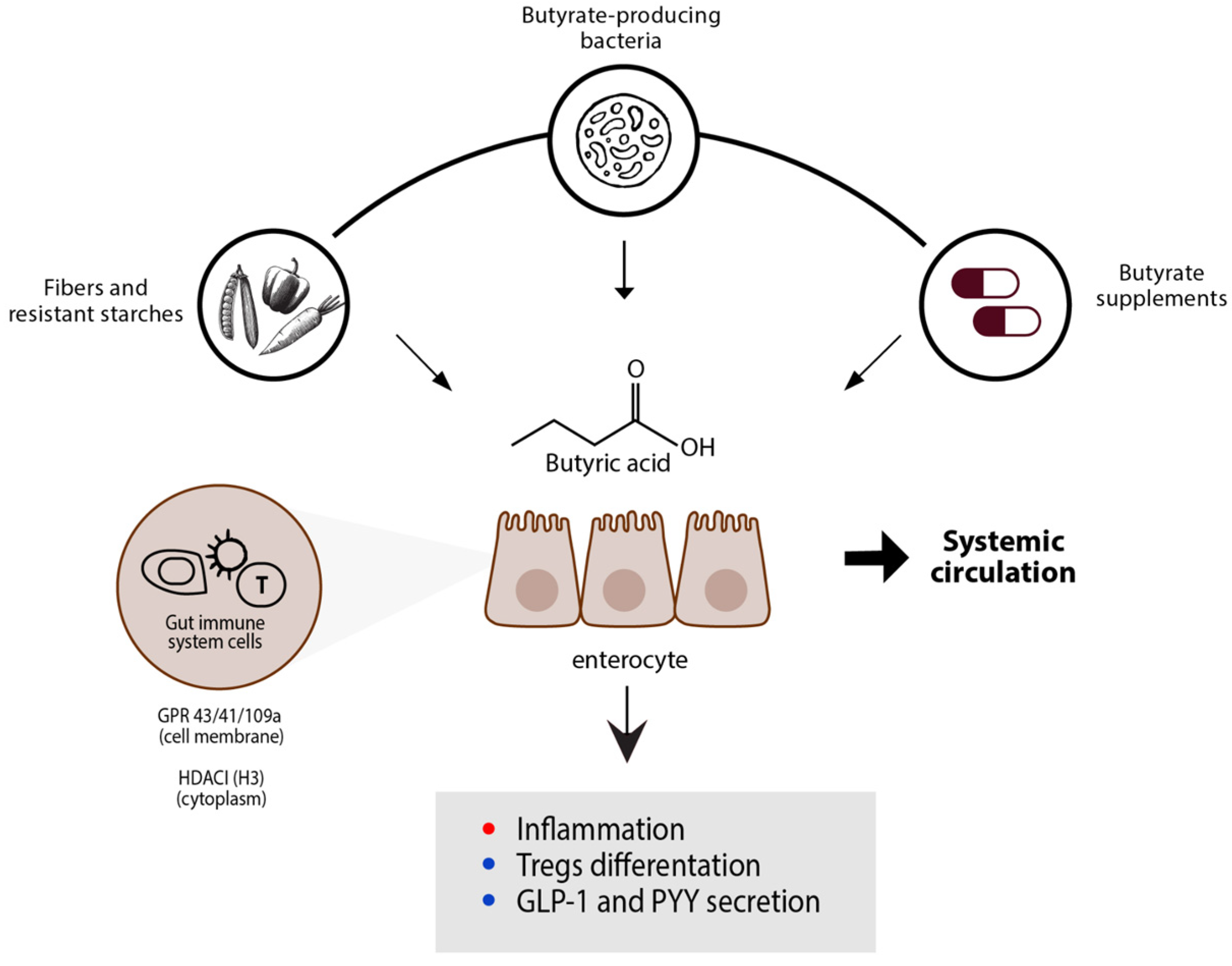
Figure 2. The main modificatory effects that butyrate has on host activities are illustrated schematically. Positive outcomes of butyrate are represented by T-reg differentiation and GPR expression, which result in S. aureus bactericidal activity, whereas negative effects are denoted by inhibition of inflammation. Abbreviations; G-protein coupled receptor (GPR); glucagon-like peptide 1 (GLP-1); histone deacetylase inhibitor (HDACi); peptide YY (PYY); T regulatory cells (T-regs) [21].
Fermentation 09 00205 g002
Figure 3. Schematic diagrams showing that to keep the host’s physiology and metabolism running at a regular rhythm, gut microbiota rhythm collaborates with the host’s circadian clock. Activities, such as habits and sleep/wake cycle, are driven by a master clock in the SCN, which is entrained by environmental stimuli, especially light stimuli, to synchronize with the environment’s regular variations. Abbreviations: suprachiasmatic nucleus (SCN); short-chain fatty acids (SCFA) [125].
Fermentation 09 00205 g003
Table 1. Potential Effects of Butyrate.
Intestinal EffectSystemic EffectImmune System Effect
Ion absorptionIncreases insulin sensitivityInhibits HDAC 3 activity
Improves gut microbial diversityCholesterol synthesisAgonist for GPR41, GPR43, or GPR109A 4
Promotes production of AMPs 1 by intestinal epithelial cellsDecreases atherosclerotic and ischemic lesionsStimulating anti-inflammatory cells like T-reg and M2 macrophages
Intestinal cell proliferation and differentiationIncreases energy expenditureInhibits pro-inflammatory cytokines
Energy substrate for colonic cellsAmmonia scavengerReduces NF-κB 5 activation and TNF-α 6 secretion
Intestinal barrier functionAntioxidant effectsIncreases PGE2 7 production
Regulation of fluid and electrolyte uptakeStimulation of β-oxidation of very long-chain fatty acids and peroxisome proliferationReduces mTOR 8 activity, which results in decreased TNF-α, IL-6 9, IL-12 production and increased IL-10 production
Immune-regulationCFTR 2 functionPromotes the development of effector T cells such as Th1 10 and Th17 cells
Anti-inflammatory effectNeurogenesisInhibits neutrophils and M1 macrophages
Oxidative stressInduces cancer cells apoptosisActivation of PPAR-γ 11
Intestinal motilityAnti-metastatic and antiangiogenic propertiesReduces mast cell degranulation
Visceral perception and rectal complianceS. aureus bactericidal activityStimulates DC 12
Increases digestive secretions
1 AMP: antimicrobial peptides; 2 CFTR: Cystic fibrosis transmembrane conductance regulator; 3 HDAC: histone deacetylase; 4 GPR: G protein-coupled receptor; 5 NF-κB: nuclear factor kappa B; 6 TNFα: tumor necrosis factor alpha; 7 PGE2: prostaglandin E(2); 8 mTOR: mammalian target of rapamycin; 9 IL: interleukin; 10 Th: T helper; 11 PPAR-γ: peroxisome proliferator-activated receptors gamma; 12 DC: dendritic cells [1,3,4,5,12,13,14,15,16,17,18,19,20,21].
Disclaimer/Publisher’s Note: The statements, opinions and data contained in all publications are solely those of the individual author(s) and contributor(s) and not of MDPI and/or the editor(s). MDPI and/or the editor(s) disclaim responsibility for any injury to people or property resulting from any ideas, methods, instructions or products referred to in the content.