Article

Purified Acidic Sophorolipid Biosurfactants in Skincare Applications: An Assessment of Cytotoxic Effects in Comparison with Synthetic Surfactants Using a 3D In Vitro Human Skin Model

1
The Nutrition Innovation Centre for Food and Health (NICHE), School of Biomedical Sciences, Faculty of Life and Health Sciences, Ulster University, Coleraine BT52 1SA, UK
2
Pharmaceutical Science Research Group, Biomedical Science Research Institute, Ulster University, Coleraine BT52 1SA, UK
*
Author to whom correspondence should be addressed.
Fermentation 2023, 9(11), 985; https://doi.org/10.3390/fermentation9110985
Submission received: 11 September 2023 / Revised: 9 November 2023 / Accepted: 15 November 2023 / Published: 18 November 2023
(This article belongs to the Special Issue Production and Application of Bioactive Biosurfactants)

Abstract

:
Acidic sophorolipids (Acidic SL), congeners of sophorolipid biosurfactants, offer a potential alternative to synthetic sodium lauryl ether sulphate (SLES) in skincare applications. However, major challenges associated with the laboratory-based investigations of the cytotoxic effects of Acidic SL have been the utilisation of impure and/or poorly characterised congeners as well as the use of monolayers of skin cells in in vitro assays. While the former limitation makes glycolipids less attractive for use in academic research and skincare applications, the latter does not provide an accurate representation of the in vivo human skin. The present study, therefore, for the first time, assessed the cytotoxic effects of 96% pure Acidic SL on a 3D in vitro skin model in comparison with SLES, with the aim of investigating a natural alternative to synthetic surfactants for potential use in skincare applications. The 3D in vitro skin model was colonised with Staphylococcus epidermidis for 12 h, and afterwards treated with either Acidic SL or SLES at 100 μg mL−1 for a further 12 h. Subsequently, the cytotoxic effects of Acidic SL in comparison with SLES were assessed using a combination of microbiology, molecular biology techniques, immunoassays, and histological analyses. It was demonstrated that Acidic SL had no deleterious effects on the viability of S. epidermidis, tissue morphology, filaggrin expression, and the production of inflammatory cytokines in comparison to SLES. These findings, in conjunction with the possibility to produce Acidic SL from cheaper renewable natural resources, demonstrate that Acidic SL could offer a potential sustainable alternative to synthetic surfactants.

1. Introduction

The development of skincare routines with ingredients that have little or no deleterious effects on the healthy human skin and the skin microbiome, and/or with added functionalities such as skin surface moisturisation, microbiome restoration, and immunomodulation in diseased skin, is highly desirable in the formulation of skincare products [1,2,3]. However, surface active agents including sodium lauryl ether sulphate (SLES), which are a major component of many skincare formulations, are mostly synthesised from less sustainable and poorly degradable petrochemical resources [1,4,5]. In addition, these surfactants are reported to have the potential to cause skin irritation, allergic reaction, and skin microbiome dysbiosis, as well as contributing to ecotoxicity [2,4]. The adverse effects of synthetic surfactants on consumer skin health and the environment have necessitated investigations by the cosmeceutical and biotechnology industries into the production of biologically derived surfactants (biosurfactants) from bacteria and yeasts via microbial fermentation as a substitute to synthetic surfactants in skincare formulations [1,4,6].
Microbial sophorolipids belonging to the glycolipid class of biosurfactants and produced by the yeast Starmerella bombicola have been reported in several studies as promising alternatives to synthetic surfactants [1,4,7,8,9]. The compelling advantages of sophorolipid biosurfactants over synthetic surfactants include enhanced foaming and solubilisation efficacy, low cytotoxicity, and added potential benefits such as wound healing and anticancer effects [7,10,11]. In particular, acidic sophorolipids (Acidic SL), which are a congener of sophorolipids, have been demonstrated to have little or no cytotoxic effects on human keratinocytes and skin dermal fibroblastic cells when assessed in vitro for potential use in skincare applications [7,8,12]. However, a major limitation to the skincare applications of sophorolipid biosurfactants has been the unwillingness to incorporate crude, impure, and/or poorly characterised sophorolipid congeners into skincare products [5,13]. Such a drawback stems from requirements by the EU Cosmetic Legislation (2013/674/EU) for manufacturers to specify the purity, molecular weight, and physio/bio-chemical properties of compounds used in formulations (extensively reviewed elsewhere by Adu and colleague) [5,13]. In addition, most studies on the cytotoxicity assessment of Acidic SL were conducted using 2D in vitro cultures; this allowed for the experimentation on a monolayer of cells only (e.g., keratinocytes and dermal fibroblasts) as opposed to the myriad tissues that make up the complex in vivo human skin [7,8,12,14,15,16]. Although 2D in vitro cell culture models are cheap, easy to use, and have contributed significantly to in vitro studies, they do not always provide an ideal representation of the in vivo human skin and, as such, may not translate into either pre-clinical in vivo animal models or first-in-human clinical trials [14,17,18].
The use of in vivo animal models as a further step on from in vitro 2D cell cultures is widely accepted as the de novo pathway for the generation of pre-clinical data; however, there are significant limitations. These include, but are not limited to, ethical considerations and the intrinsic differences in the anatomical structure, physiological functions, and host microbiota between human and animal models [19,20]. In agreement with the EU directive (Council Directive 76/768/EEC) on a proposed ban on the use of animal models for cosmeceutical ingredients testing, coupled with the limitations associated with the use of 2D in vitro cultures, the use of novel technologies to develop 3D in vitro skin models has become increasingly important [20,21,22,23]. The use of these 3D in vitro skin models allows for an appropriate representation of the complex anatomy and physiological functions of the in vivo human skin while providing an alternative to 2D in vitro skin cultures and the use of animal models in laboratory research [24,25].
At present, 3D skin models such as the LabskinTM full thickness 3D in vitro skin model (LabskinTM), PhenionTM FT LongLife skin model, and EpiskinTM are commercially available for testing the safety and efficacy of drugs and cosmeceutical ingredients [26]. Established among these models is LabskinTM [27,28]. LabskinTM is a full thickness human skin equivalent, specifically developed for studying interactions between the human skin and the skin microbiome [29]. The robust structured architecture of LabskinTM in addition to its well differentiated epidermis, barrier functions, and dry acidic surface makes it an ideal model for high-throughput studies on the effects of cosmetic ingredients on the interaction between the human skin and skin bacteria in vitro; hence, the choice for the present study [24,28].
Three-dimensional in vitro skin models have been utilised in a number of in vitro studies [21,28,30]. Holland and colleagues studied the colonisation of LabskinTM with Staphylococcus aureus and Staphylococcus epidermidis using microarray analysis and bacterial enumeration [19]. There was a ten-fold reduction in colonisation by S. epidermidis compared to S. aureus [19]. The microarray analysis revealed a significant upregulation in gene expression of beta-defensin and tumour necrosis factors compared to the uncolonised and S. aureus-colonised tissues [19]. Lewis and colleagues investigated the processes of in vitro wound healing and skin barrier repair using histological and matrix-assisted laser desorption/ionization-mass spectrometry imaging (MALDI-MSI) analysis, following the creation of artificial wounds in a 3D in vitro skin model [30]. This study allowed for the identification of lipids, such as glycosylceramides, directly involved in skin barrier repair and wound healing in vitro [30]. Nevertheless, to the authors’ knowledge, there is currently no study on the assessment of the effects of purified and chemically characterised sophorolipid biosurfactant congeners on 3D in vitro skin models for potential skincare applications in synthetic surfactants.
The aim of this study was to expand our previous investigations of natural alternatives to synthetically derived surfactants such as SLES for use in skincare applications, from 2D in vitro cell culture models to a pre-clinical 3D in vitro human skin model. To achieve this, we initially colonised the 3D in vitro human skin model with a bacterium representative of the human skin micro-flora (S. epidermidis) and then treated with either SLES or naturally derived non-acetylated acidic sophorolipids. The effect on the skin model of colonisation followed by surfactant treatment was assessed by measuring the bacterial viability of the infective strain, immunological markers of inflammation associated with bacterial infection and surfactant treatments, and direct observation either by H & E staining or IHC.

2. Materials and Methods

2.1. Chemical Characterisation of Acidic SL

Pre-purified non-acetylated acidic sophorolipids purchased from Biosynth Carbosynth, Compton, UK, were chemically characterised using HPLC–MS/ESI [31]. Stock preparations of Acidic SL and SLES were prepared (1 mg mL−1) in sterile phosphate-buffered saline (PBS) (Merck, Gillingham, UK) and stored at −20 °C until required for use.

2.2. 3D In Vitro Skin Model Preparation

The 3D in vitro skin models used in this study, LabskinTM (Innovenn Ltd., York, UK), was supplied after 14 days of seeding, and was delivered in a 12-well cell culture insert as 12 skin constructs, each having a diameter of 1.1 cm (Figure 1). The constructs were partially suspended in LabskinTM maintenance medium (Innovenn Ltd., York, UK) containing 1% (w/v) agarose gel for transportation (Figure 1).
The skin constructs were transferred into a ThinCert™ 12-well cell culture plate (Greiner Bio-One, Frickenhausen, Germany) containing 4.5 mL of pre-warmed maintenance medium and incubated for 48 h at 37 °C in 5% CO2 (v/v) to equilibrate tissue metabolism. For the experiment, the skin constructs were transferred into a 12-well cell culture plate (Sarstedt, Leicester, UK) containing 1 mL of fresh maintenance medium, followed by bacterial inoculation and surfactant treatments, respectively.

2.3. Colonisation of Skin Constructs with Staphylococcus Epidermidis

Staphylococcus epidermidis (DSM 28319), obtained from the German Collection of Microorganisms and Cell Cultures (Leibniz Institute DSMZ, Braunschweig, Germany) as a freeze-dried sample, was revived as per the supplier’s instructions and stored in 25% glycerol (v/v) at −80 °C until required for use.
For the experiment, S. epidermidis was thawed on ice for 20 min (mins), cultured overnight in brain heart infusion broth (BHI) (Oxoid, 3124409, Hampshire, UK) at 37 °C (primary seed culture), followed by the preparation of an exponential phase culture using the primary seed culture at a 1:10 dilution. Inocula prepared from the exponential phase culture were made in pre-warmed Dulbecco’s phosphate-buffered saline (no calcium, no magnesium) (DPBS) (ThermoFisher Scientific, Loughborough, UK). Each construct was seeded at a volume of 10 μL (except for negative control constructs), with a final seeding density of 2.1 × 10⁵ colony-forming unit per square centimetre (CFU cm−2). Negative controls consisted of constructs with 10 μL of sterile DPBS only. Inocula were gently spread on the surface of constructs using an in-house-prepared sterile spreader (Glass Pasteur pipettes) (Scientific Laboratory Supplies Ltd., Lisburn, UK), and the tissues were incubated for 12 h at 37 °C in 5% CO2 (v/v) for skin surface colonisation.

2.4. Surfactant Treatments

The surfactants utilised in this study were SLES and pre-purified/chemically characterised Acidic SL. Post bacterial colonisation and tissue incubation for 12 h, three replicates each of the pre-colonised skin constructs were either untreated (Colonised) or treated with 100 μg mL−1 of Acidic SL and SLES (Colonised + Acidic SL)/(Colonised + SLES) prepared in DPBS. Negative control was treated with 10 μL of sterile DPBS only (Uncolonised). The skin constructs were incubated for further 12 h at 37 °C in 5% CO2 (v/v), and then sampled for bacterial enumeration, tissue processing via formalin fixation and paraffin embedding, RNA extraction, and immunoassays.

2.5. Assessment of Bacterial Viability

The effects of Acidic SL and SLES on S. epidermidis pre-colonised skin constructs surface were assessed via bacteria enumeration using the drop plate method [32]. A total of 1 mL of pre-warmed DPBS was added to sampled 5 mm skin construct punch biopsies in a 1.5 mL Eppendorf tubes, vortex mixed for 2 min, and diluted to 1:10 in a fresh sterile DPBS. Subsequently, 10 μL was plated on BHI agar and incubated for 24 h at 37 °C. The total viable bacteria (colonies) were enumerated and expressed as CFU per millilitre (CFU mL−1). Further assessment for purity consisted of Gram’s staining and phenotypic characterisation via plate streaking of isolated colonies on Mannitol Salt agar (MSA) (Oxoid, BCCB79883, Hampshire, UK) and BrillianceTM E. coli/Coliform agar (Oxoid, 1698837, Hampshire, UK). The bacterial enumeration experiments were performed independently three times, with each having five technical replicates.

2.6. Formalin Fixation, Tissue Processing, and Sectioning

Post 24 h of skin construct treatments (12 h of bacterial inoculation and 12 h of surfactant treatments), a 0.5 cm diameter of the skin construct from each treatment group was fixed in 10% formalin (Merck, Gillingham, UK) for 2 h, and further processed overnight using Leica TP1020 tissue processor (Leica, Nussloch, Germany) as per the manufacturer’s instructions. The sampled tissues were embedded in Blue RibbonTM paraffin wax (Leica, Nussloch, Germany), sectioned at 3 μm using Leica RM2145 rotary microtome (Leica, Nussloch, Germany), and transferred to SuperfrostTM Plus adhesion glass microscope slides (Aquilant Scientific, Newtownards, UK). The tissues were partially dewaxed and left to firmly attach to the glass microscope slides by positioning the slides for drying on an Epredia slide hotplate (ThermoFisher Scientific, Loughborough, UK) at 60 °C for 1 h prior to staining.

2.7. Haematoxylin and Eosin (H & E) Staining

Formalin-fixed, paraffin-embedded, and microtome-sectioned tissue samples were fully deparaffinised in HPLC grade xylene (Merck, Gillingham, UK) three times for a total of 9 min, followed by three washes in HPLC grade ethanol (Merck, Gillingham, UK) (9 min) and a rinse in tap running water for 2 min. The tissues were stained with Mayer’s haematoxylin (Lillie’s Modification) and Eosin Y solution (modified alcohol) (Abcam, Cambridge, UK) as per the manufacturer’s instructions. The samples were mounted with DPX medium (Merck, Gillingham, UK) and viewed using a Digital Sight DS-U2 camera attached to an Eclipse E400 phase contrast microscope (Nikon Europe B. V., Amsterdam, The Netherlands) at 200× magnification. The thickness of the stratum corneum of the imaged tissues was analysed using the ImageJ Software version 1.53 for MacOS (ImageJ, National Institute of Health, Bethesda, MD, USA) [33,34,35].

2.8. Immunohistochemistry (IHC)

For IHC staining, the tissue samples were first processed, paraffin-embedded, sectioned at 3 μm, deparaffinised, and hydrated as detailed above. The antigen retrieval step consisted in heating the tissue samples to 95 °C for 20 min in an in-house-prepared citrate buffer (made with 10 mM anhydrous citric acid and 0.05% in 1000 mL distilled water and adjusted to pH 6.0). The tissues were washed ×3 with phosphate-buffered saline (PBS) (ThermoFisher Scientific, Loughborough, UK), and endogenous peroxidase activity was quenched by incubating the tissues with 100 μL of BLOXALL reagent (Vector Laboratories, Newark, CA, USA) per slide at room temperature (RT) for 10 min. Subsequently, the tissues were incubated for 20 min with 100 μL of normal horse serum, followed by 1 h incubation with 125 μL of prediluted recombinant anti-filaggrin primary antibody (1:500 dilution) (Abcam, ab221155, Cambridge, UK). Next, the tissues were incubated with 100 μL of prediluted biotinylated horse secondary antibody and incubated for 30 min at RT. Tissue staining was performed using Vectastain® Elite® ABC Universal Kit/ImmPACT ® DAP peroxidase substrate (Vector Laboratories, Newark, CA, USA) as per the manufacturer’s instructions. The tissues were counterstained with Mayer’s haematoxylin (Lillie’s Modification) (Abcam, Cambridge, UK) for 9 min, washed in running tap water for 2 min, differentiated in 0.25% acid alcohol for 2 s, and blued for 1 min. The tissue samples were dehydrated, cleared, mounted with DPX medium, and visualised as detailed in Section 2.7. The intensity of IHC staining was analysed using ImageJ Software, and reported as a percentage relative to the uncolonised tissue samples [33].

2.9. Enzyme-Linked Immunosorbent Assay (ELISA)

ELISA was utilised to investigate the effect of surfactants on the production of pro-inflammatory cytokines in the skin construct, using spent culture medium. The spent culture medium was collected into 1.5 mL Eppendorf tubes and stored at −80 °C until required for use. For the ELISA experiments, the spent culture medium was thawed to RT, centrifuged at 1000× g, and supernatants were analysed for Interlukin-8 (IL-8) and interlukin-6 (IL-6) cytokines using commercially available ELISA kits (R&D Systems, Inc., Minneapolis, MN, USA) as per the manufacturer’s instructions. All ELISA experiments were performed independently three times.

2.10. RNA Extraction

Tissue punch biopsies (5 mm; approx. 120 mg), sampled post bacterial colonisation and surfactant treatment, were stored at −80 °C until required for use. For RNA extraction, briefly thawed (on ice) frozen sections of the skin constructs were first homogenised in 1 mL TRIzol™ Reagent (Invitrogen, Paisley, UK) on ice for 20 s using a VDI 12 stand model homogeniser (VWR Scientific, Lutterworth, UK) at setting 5. After homogenisation, total RNA was extracted following the TRIzol™ Reagent manufacturer’s protocol. The total RNA extracted was assessed for integrity using agarose gel electrophoresis and quantified using NanoDrop ND-1000 (ThermoFisher Scientific, Loughborough, UK).

2.11. cDNA Synthesis

The total RNA extracted from the skin constructs was reverse transcribed to generate cDNA samples using a G-STORM GS1 thermal cycler (Gene Technologies Ltd., Somerset, UK) [7]. Unless otherwise stated, all reagents utilised in the cDNA synthesis were sourced from ThermoFisher Scientific, Loughborough, UK. The reagents for the cDNA synthesis consisted of the following reaction mixtures with a total volume of 20 μL: 25 ng of Oligo (dT) 12–18 primer, 10 mM DTT, 0.5 mM dNTP, 50 ng of total RNA, 10 U of SuperScriptTM Reverse Transcriptase II (RT), and 12 μL molecular grade water (UltraPureTM distilled water). The cycling condition for the cDNA synthesis was set as follows: RNA denaturation step of 10 min at 70 °C, primer hybridisation step of 2 min at 42 °C, DNA polymerisation and subsequent cDNA synthesis step at 42 °C for 50 min, and, finally, an RT deactivation step at 70 °C for 15 min. Reverse transcriptase minus (NRT) negative control and No template negative control (NTC) were generated with the supplementation of the respective components.
To confirm that cDNA was successfully synthesised and free from genomic DNA contamination, and to validate the primer sets utilised in this study (Table S1), the synthesised cDNA was used as template DNA for endpoint PCR using a TC 500 Gradient Thermocycler (Techne, UK). The reaction mixture (total volume of 50 μL) for the endpoint PCR reaction consisted of 1 X PCR buffer, 1.5 mM MgCl2, 0.2 mM dNTP mix, 0.5 μM each of forward and reverse primer, 36 μL of molecular grade water (UltraPureTM distilled water), 2.5 U Taq polymerase (all reagents purchased from ThermoFisher Scientific, Loughborough, UK), and 1 μL of template DNA (synthesised cDNA). The cycling conditions for the endpoint PCR were set as follows: an initial denaturation step of 3 min at 94 °C, followed by 31 cycles of denaturation (95 °C for 30 s), a primer annealing step of 60 °C for 30 s, and an extension at 72 °C for 45 s. The final extension was held at 72 °C for 10 min.

2.12. Quantitative PCR (qPCR)

qPCR experiments were performed with synthesised cDNA from the skin constructs’ total RNA, as detailed in a previous study [7], using the primer sets listed on Table S1. All qPCR experiments were performed independently three times. The data generated were analysed using the relative quantification method described by Maussion and colleagues [36]. The fold change, normalised to the reference gene (β-actin) relative to non-surfactant treated control, was calculated as 2−ΔΔCq [36].

2.13. Statistical Analysis

A statistical analysis of all data was carried out using GraphPad Prism version 9.4.1 (458) for MacOS (GraphPad Software, San Diego, CA, USA). Data were analysed via a one-way analysis of variance (ANOVA), followed by Tukey’s multiple comparison test; statistical significance was tested at p ≤ 0.05 levels.

3. Results

3.1. Chemical Characterisation of Acidic SL

The sophorolipids reported in this study were obtained from a commercial source as pre-purified non-acetylated acidic sophorolipid congeners, and they were chemically characterised for congener profile and the relative percentage abundance of congeners present using HPLC–MS/ESI. The HPLC-MS/ESI analysis showed that the relative percentage abundance of congeners corresponding to non-acetylated acidic sophorolipid congeners were 96.41%, whereas the remaining 3.59% of the congeners reported corresponded to the mass of Lactonic SL congeners (Table 1). The predominant peak on the Acidic SL spectrum had ion m/z of 621.3, corresponding to the molecular ion [M+H] + of the Acidic SL congener Acidic, C18:1, which accounted for a relative abundance of 43.81% of the sample (Table 1, Figure 2).

3.2. Effects of Acidic SL and SLES on the Viability of S. epidermidis Colonised on Skin Constructs

The effects of surfactants on the viability of S. epidermidis were assessed by comparing the number of colonies of S. epidermidis on the bacterial-colonised and non-surfactant-treated skin constructs with the constructs colonised with S. epidermidis and treated with surfactants. As expected, no colonies of S. epidermidis were detected on BHI agar for the skin constructs not colonised with S. epidermidis (Figure 3). There was no significant difference in the number of S. epidermidis colonies between the non-surfactant-treated constructs and the constructs treated with 100 μg mL−1 of either Acidic SL or SLES (Figure 3). Moreover, there was no significant difference between the number of S. epidermidis colonies in Acidic SL- and SLES-treated skin constructs (Figure 3). Potential damages to the surface of the skin constructs post bacterial colonisation and surfactant treatment were examined macroscopically and microscopically, and the 3D in vitro skin model was found to be intact.
To determine whether S. epidermidis on BHI agar was free of microbial contaminants, the colonies of S. epidermidis were assessed for purity using Gram’s staining and phenotypic characterisation via plate streaking on selective/differential media. Gram’s staining revealed purple cocci grape-like clusters of approximately 1 μm diameter per cell size (Figure S1). The colonies of S. epidermidis appeared pale pink on MSA; however, on BrillianceTM E. coli/Coliform agar no bacterial growth was observed (Figure S1).

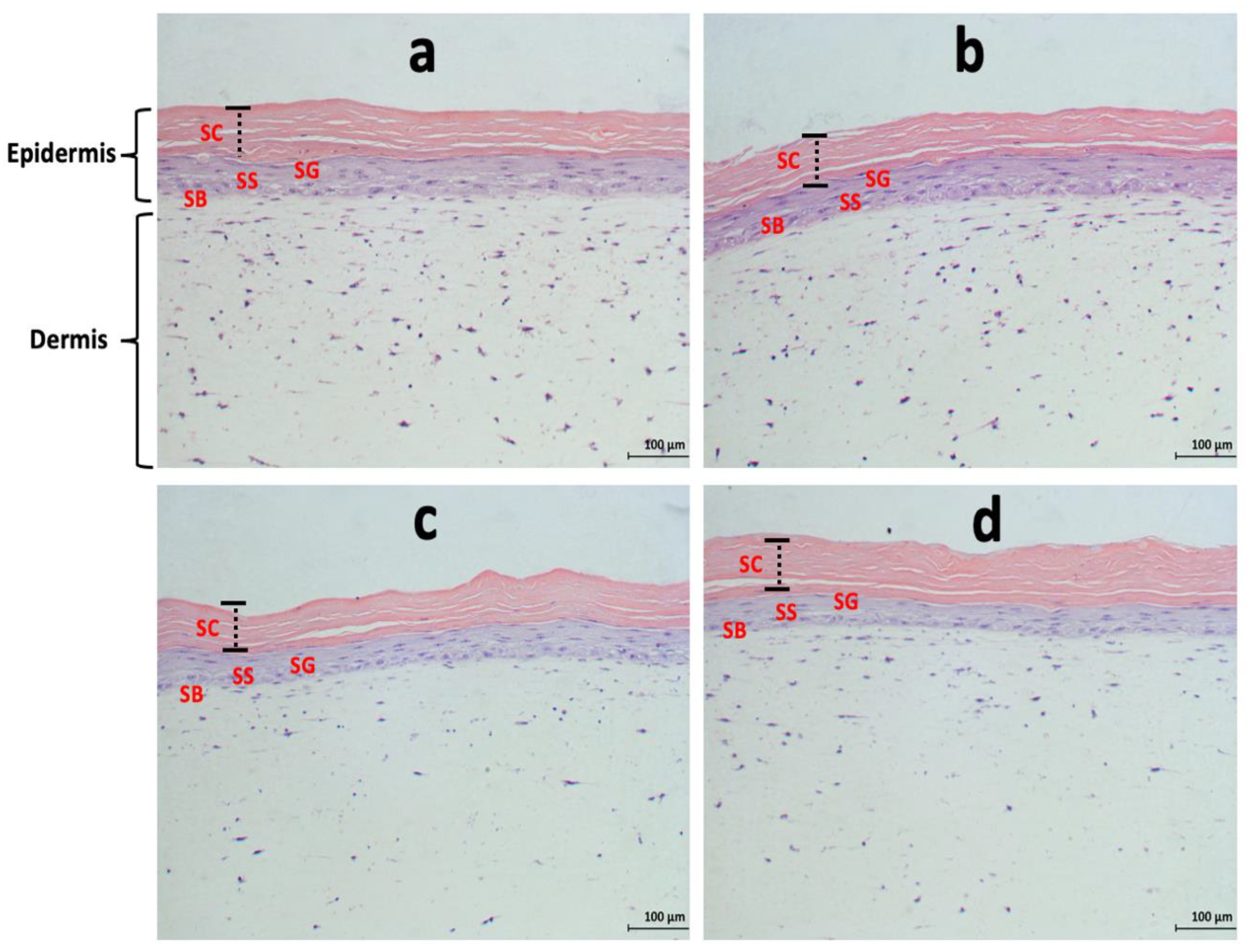
3.3. Effects of Acidic SL and SLES on the Morphology of Pre-Colonised Skin Constructs

Histological analysis revealed a normal skin structure with intact epidermis and evenly distributed fibroblasts in the dermal layer of both the untreated tissues and the tissues treated with 100 μg mL−1 of either Acidic SL or SLES (Figure 4). Ulcerations, tissue erosion, and necrosis were absent in both the untreated and surfactant-treated tissues (Figure 4). Epidermal layers including the stratum corneum (SC), stratum granulosum (SG), stratum spinosum (SS), and stratum basale (SB) were identifiable in all treatment groups (Figure 4). Comparing SLES-treated tissues with those of Acidic SL, there were no observable differences in tissue morphology (Figure 4).
The measured SC thickness of the uncolonised tissues was 66.97 μm (±2.32 μm), S. epidermidis-colonised and non-surfactant-treated tissue was measured at 67.21 μm (±0.59 μm), colonised and Acidic SL-treated tissue was 67.88 μm (±0.60 μm), and that of colonised and SLES-treated tissue was 67.00 μm (±1.53 μm). There was no significant difference in SC thickness between surfactant-treated and untreated tissues (p > 0.9999), or between the Acidic SL- and SLES-treated tissues (p = 0.9924).

3.4. Effects of Acidic SL and SLES on Filaggrin Expression in the Epidermis of Skin Constructs

As previously demonstrated for the H& E staining, the tissue samples under all treatment conditions (uncolonised, colonised, colonised and Acidic SL-treated, or colonised and SLES-treated samples) maintained intact morphology, with each showing distinct skin epidermal and dermal layers (Figure 5). The topmost layer of sectioned skin construct epidermis (SC) under all treatment conditions was stained brown, indicative of FLG immunostaining (Figure 5).
The analysis of FLG immunostaining revealed that there was no significant difference in FLG stain intensity between the non-surfactant-treated tissues and tissues pre-colonised with S. epidermidis and treated with Acidic SL or SLES (Figure 6). Furthermore, there was no significant difference in stain intensity between the Acidic SL- and SLES-treated tissues (Figure 6).

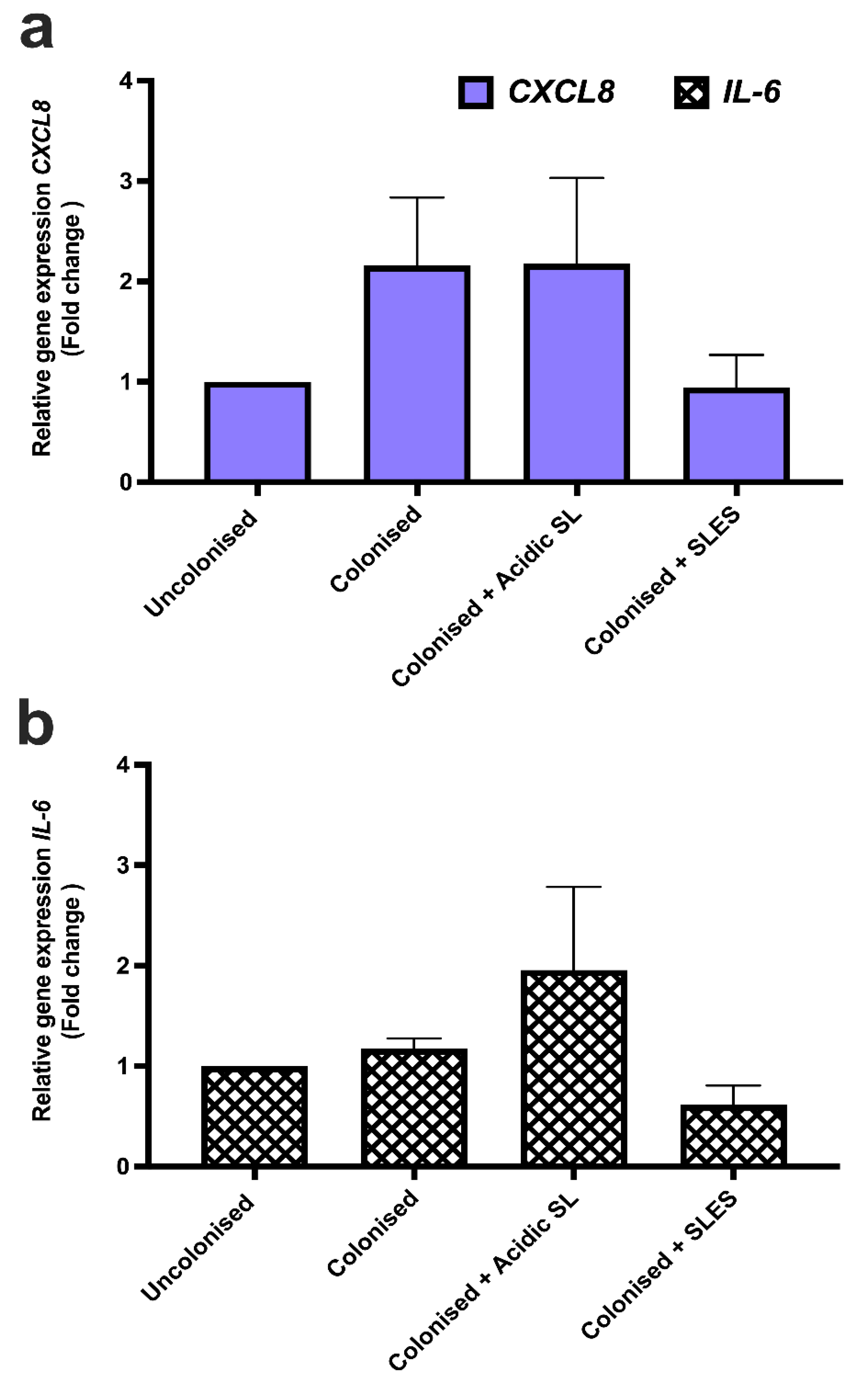
3.5. Effects of Acidic SL and SLES on Pro-Inflammatory Cytokine Production in Skin Constructs

The effects of Acidic SL and SLES on the production of pro-inflammatory cytokines in the skin constructs pre-colonised with S. epidermidis were investigated via ELISA using spent culture medium. There was no significant difference in IL-8 and IL-6 levels between uncolonised and non-surfactant-treated samples, or between the S. epidermidis-colonised and non-surfactant-treated samples (Figure 7a,b). Compared to the control samples, a trend towards decreased IL-8 and IL-6 levels was observed in Acidic SL-treated samples; however, this was not statistically significant (Figure 7a,b). There was no significant difference in IL-8 and IL-6 production between the experimental controls and SLES-treated samples (Figure 7a,b). By directly comparing the levels of IL-8 and IL-6 produced in Acidic SL-treated samples with SLES, no significant differences were observed (Figure 7a,b).

3.6. Effects of Acidic SL and SLES on Pro-Inflammatory Cytokine Gene Expression in LabskinTM

NanoDrop spectrophotometric analysis revealed that none of the samples extracted yielded less than 500 ng μL−1 total RNA and OD 260/280 ratio less than 1.93. The gel electrophoresis analysis showed three non-smearing bands, i.e., 28S rRNA with an amplicon size of 2000 bp, 18S rRNA at 1000 bp, and 5.8S/5 rRNA at 100 bp, indicative of non-degraded RNA (Figure S2). Similarly, cDNA synthesised using total RNA was validated using endpoint PCR and, subsequently, the assessment of primer amplification efficiencies (Figures S3 and S4). The amplification efficiency of β-actin utilised as reference gene was 86.4%, whereas those of CXCL8 and IL-6 (utilised as genes of interest) were 92.3% and 91%, respectively (Figure S4).
The effects of Acidic SL and SLES on the expression of pro-inflammatory cytokine genes were assessed using RT-qPCR. Compared to uncolonised and untreated skin constructs, increased levels of CXCL8 gene expression, as measured by fold change in expression relative to a reference gene (β-actin), was observed in S. epidermidis-colonised untreated and Acidic SL-treated tissues. In both cases, the difference in gene expression was not statistically significant (Figure 8a). S. epidermidis-colonised and SLES-treated constructs showed a reduced level of CXCL8 gene expression compared to constructs colonised and treated with Acidic SL; again, the difference in expression was not significant (Figure 8a). A similar non-significant trend in the expression of IL6 (also measure by fold change in expression relative to the reference gene β-actin) was also observed between each treatment group (Figure 8b).

4. Discussion

The use of robust novel experimental and instrumental techniques which allow for accurate mimicking of the in vivo human system is crucial for the growing skincare industry [24]. Three-dimensional in vitro skin models are currently being used as a replacement for 2D in vitro models, ex vivo, and animal models in skin research, given that 3D in vitro skin models provide a more accurate representation of the anatomy and physiological functions of the in vivo human skin [27,28]. The present study assessed the cytotoxic effects of 96% pure preparations of Acidic SL on LabskinTM full thickness 3D in vitro skin model pre-colonised with S. epidermidis in comparison with SLES. S. epidermidis was utilised in the present study since most skin commensals are Gram-positive bacterial species, and among these, S. epidermidis is one of the most prevalent isolates at the dry niches of the human skin, with the potential to produce antimicrobials (e.g., antibiotics) to inhibit the colonisation of pathogenic skin bacteria [37,38]. Also, the choice of the surfactants and treatment concentration utilised in this study was based on the differential effects of Acidic SL and SLES on the viability and morphology of the 2D in vitro skin cell cultures reported in our previous studies in accordance with the Organisation for Economic Cooperation and Development’s (OECD) guidelines on toxicological studies [7,22,23,31,39]. In these studies, the Acidic SL were less cytotoxic compared with SLES, in that whereas up to 500 μg mL−1 Acidic SL demonstrated no significant cytotoxic effects on cell viability and morphology, the viability of cells was significantly reduced when treated with SLES concentrations above 60 μg mL−1 [7,31]. Hence, the use of these two surfactants in the current study allowed for a further investigation of a sustainable and natural-skin-compatible alternative to SLES for skincare applications using a 3D in vitro skin model.
Bacterial enumeration, histological analysis, and the use of a combination of immunoassays and molecular biology techniques in the present study revealed that at the concentration tested (100 μg mL−1), the purified Acidic SL had no deleterious effects on the viability of S. epidermidis, tissue morphology, FLG expression, and the production of inflammatory cytokines comparable to SLES. To the best of the authors’ knowledge, this is the first time 3D in vitro skin models have been utilised to assess the cytotoxic effects of purified and chemically characterised sophorolipid congeners for potential skincare applications.
The HPLC-MS/ESI analysis of the sophorolipid preparation utilised in this study revealed that Acidic SL congeners comprised 96% of the sample. It is important to understand that Acidic and Lactonic SL congeners can possess completely different bioactivities and that several previous in vitro studies on SL bioactivities utilised preparations possessing a less defined mixture of both Acidic and Lactonic congeners [8,40,41]. Consequently, studies that utilised mixed/impure congeners of sophorolipids resulted in significant interstudy variations as well as contaminant-associated toxicity, which in effect renders sophorolipids less attractive for potential use in skincare and pharmaceutical applications [42,43]. Consistent with the congener profile of the Acidic SL reported in this study, Lydon and colleagues utilised HPLC-MS analysis to characterise Acidic SL for use in the assessment of wound healing and adjuvant antimicrobial properties [12]. The congeners of sophorolipids present in their extracts corresponding to Acidic SL congeners were above 95% [12]. In another study, Callaghan and colleagues utilised HPLC coupled with evaporative light scattering detector (HPLC-ELSD) and HPLC-MS analyses to characterise Lactonic SL [42]. In that study, Lactonic SL congeners comprised 97% of the sample used for testing bioactivity against cancer cell lines [42]. This clearly indicates the importance of utilizing preparations with a high percentage relative abundance of each chemical sub-species of sophorolipid, (Acidic SL or Lactonic SL), to generate data on discernible bioactive effects on both the prokaryotic and eukaryotic cells that are attributable to the respective SL sub-species used [12,42].
The human skin surface in in vivo systems is not sterile, but rather colonised with commensal and mutualistic microorganisms that improve skin health. Hence, the 3D in vitro skin model utilised in the current study was first colonised with a bacterial species dominant within the human skin commensal population, S. epidermidis, and subsequently treated with either Acidic SL and SLES to assess the effects of these surfactants on bacterial viability and interaction with the skin constructs. Neither Acidic SL nor SLES had any significant inhibitory effects on the viability of S. epidermidis. Previous studies on the in vitro interaction between sophorolipids and bacterial cells focused on antimicrobial effects of sophorolipids rather than their compatibility effects on the representatives of the normal human skin microbiota [40,44]. Sophorolipid mixtures with varying sugar head groups were demonstrated to have antimicrobial effects, mainly on Gram-positive bacterial cells such as Streptococcus agalactiae and Bacillus subtilis at concentrations as low as 0.024 mg mL−1; this is in contrast to the results reported in the current study in that no inhibitory effects were observed here [44]. It is, however, worthy of note that the antimicrobial effects of sophorolipids are mainly dependent on their purity, molecular structure, physiochemical properties, properties of bacterial cell membrane, and the mechanism of the sophorolipids’ interaction with the bacterial cell membrane [11,40,45]. Therefore, although all bacterial isolates reported here are Gram-positive, differences in the purity and physiochemical properties of the sophorolipids utilised in the present study compared to the sophorolipid mixtures reported by Shah and colleagues may have contributed to the contrasting dose–response [44]. This notwithstanding, skincare ingredients with non-deleterious effects on the healthy human skin microbiome are highly sought after for various skincare applications [10,11]. Therefore, the non-inhibitory effects of the purified Acidic SL on skin bacteria as demonstrated in the present study, coupled with added functionalities such as foaming and solubilisation properties, makes them ideal candidates for incorporation into skincare products. While a single bacterium does not represent the entirety of the complex human skin microbiome, the preliminary findings that Acidic SL has non-deleterious effects on S. epidermidis pre-colonised on 3D in vitro skin model is an important step towards understanding the effects of sophorolipids on the in vivo human skin microenvironment.
In terms of the effects of Acidic SL on the morphology of the skin model comparative to SLES, no deleterious effects were observed when the tissues were treated with surfactants, stained with H & E, and visualised using phase contrast microscope. Measurement of epidermal thickness is a well-known method for determining the severity of skin inflammation in skin diseases such as atopic dermatitis [34]. Here, neither Acidic SL nor SLES had any effects on the thickness of the measured stratum corneum of the skin constructs. The cytotoxic effects of sophorolipids are hypothesised to be dependent on their level of acetylation [46]. Hence, sophorolipids with high levels of acetylation are proposed to have higher cytotoxic effects, as the acetyl groups on their hydrophilic moiety may have high penetrative effects on cells/tissues [45,46]. In the present study, the Acidic SL utilised were non-acetylated, and this could account for the non-deleterious effects on the 3D in vitro skin model. Another important consideration for the non-deleterious effects of the Acidic SL reported in the current study could be the characteristics of the tissue model system utilised. It has been previously demonstrated that whereas 60 μg mL−1 of SLES significantly reduces the viability of a monolayer of spontaneously transformed human keratinocytes (HaCaT cells), Acidic SL at treatment concentrations up to 500 μg mL−1 have no significant effects on the viability and morphology of HaCaT cells [7,12]. Although the dose effects of Acidic SL utilised in the above studies agree with the present study, there is a contrast in the dose–response of SLES in that no deleterious effects were observed on the skin model with the treatment at 100 μg mL−1 as opposed to the 60 μg mL−1 treatment concentration, which significantly reduced the viability of HaCaT cells [7]. The differences in structural architecture and physiological properties between the monolayer of cells utilised in the studies above and the 3D in vitro skin model reported in the current study may have accounted for this discrepancy [7,12,14]. In particular, monolayers of HaCaT cells lack fibrin matrixes, which influence the robust structural architecture of the 3D in vitro skin models such as the one utilised in the present study [28]. This brings to bear potential future study limitations in transitioning from the use of 2D to 3D in vitro skin models considering the difference in structural architecture of these two separate models and the subsequent differences in response to therapeutic agents. Nevertheless, further studies using a broad range of treatment concentrations and employing additional analytical techniques will be required to observe discernible differences in dose effects between sophorolipid biosurfactants and synthetic surfactants when utilising 3D in vitro skin models.
FLG is a structural protein in the SC of the skin epidermis [47,48]. FLG is fundamental in controlling the shape of keratinocytes, the maintenance of the texture of the epidermis, the promotion of the aggregation of intermediate filaments and the subsequent formation of mechanically resilient corneocyte intracellular matrix, and the control of SC permeability and hydration [49,50,51]. Consequently, a deficiency in filaggrin expression in the stratum corneum of the skin epidermis could lead to impaired skin barrier functions and an exacerbation of non-lesion atopic dermatitis [50,52,53]. Tö Rmä and colleagues demonstrated that the exposure of eleven healthy volunteers to 50 μL of 1% (v/v) SLES for six h significantly reduced the expression of pro-filaggrin in human keratinocytes [54]. In the present study, a non-significant effect of both Acidic SL and SLES on FLG expression was reported. The difference in dose–response in the present study compared to the report by Tö Rmä and colleagues could be explained by the difference in the concentration of SLES utilised (0.01% versus 1% (v/v)) [54]. This notwithstanding, the results reported in the current study suggest that at the tested concentration, Acidic SL may have non-deleterious effects on the architectural structure of the human skin epidermis and on the barrier functions of the human skin comparable to SLES.
Similar to the effects of Acidic SL compared with SLES on tissue morphology and FLG expression in the 3D in vitro skin model, the use of ELISA and qPCR revealed that neither surfactant utilised in the present study had any significant effects on IL-8/CXCL8 and IL-6/IL-6 stimulation compared to control samples. It was interesting to observe a trend drop in IL-8 and IL-6 levels in Acidic SL-treated tissues, indicative of potential immunomodulatory effects; however, this was not statistically significant. Such a trend drop was not observed in SLES-treated tissues or in similarly purified Acidic SL employed in other studies when a monolayer of human keratinocytes was utilised [7]. Further studies focusing on the potential immunomodulatory effects of Acidic SL and associated mechanisms of action in 3D in vitro skin models will be an important future step.
It should be noted that sophorolipids could only be used as a substitute to synthetic surfactants if they are able to demonstrate equal or better performance in skincare formulation at a reasonable market price [55,56]. Therefore, having demonstrated that the Acidic SL utilised in the present study have comparable effects with SLES on the 3D in vitro skin model, in addition to being produced from sustainable natural resources (e.g., industrial waste materials such as vegetal cooking oil waste), and having enhanced solubilisation and foaming functions, Acidic SL could offer a potential natural/sustainable alternative to SLES in skincare applications [5,56].

5. Conclusions

In comparison to the synthetic surfactant SLES, naturally derived Acidic SL demonstrated no deleterious effects on the viability of pre-colonised S. epidermidis, tissue morphology, FLG expression, and the production of pro-inflammatory cytokines in a pre-clinical in vitro 3D skin model. This study demonstrates an early proof of concept on the potential use of purified Acidic SL congeners in skincare applications at concentrations that would not be detrimental to the healthy human skin and bacteria, hence paving the way for further in vitro and in vivo studies on healthy human skin and the skin microbiota using purified sophorolipid biosurfactant congeners. To the best of the authors’ knowledge, this is the first time the cytotoxic effects of purified Acidic SL have been assessed in comparison with synthetic surfactants using a full thickness 3D in vitro skin model. There are, however, specific limitations to the present study, which are worth pointing out. Firstly, despite the simplicity, reliability, and reproducibility of the 3D in vitro model utilised, it currently lacks several important in vivo skin components such as blood vessels, hair, hair follicles, sweat, and sebaceous glands. The incorporation of these complex in vivo skin components into this 3D in vitro model has been deemed unfit for future research due to high production costs and limited technical expertise. Secondly, considering the high cost of developing/acquiring the 3D in vitro skin model, the present study was limited to the use of a single bacterium (S. epidermis) and one surfactant treatment concentration condition (100 μg mL−1). The high monetary cost of the model also limited the number of experimental repeats that could be carried out in the current study and may explain why much of the data was only trending and not significantly significant. The financial cost of generating robust datasets using this model in comparison to using an in vivo animal model must be of significant consideration when choosing a suitable pre-clinical model for the testing for naturally derived compounds such as Acidic SL. Future studies should focus on identifying a broad range of treatment concentrations to conduct dose–response experiments on the bioactivities of sophorolipid biosurfactants when using a 3D in vitro skin model.

Supplementary Materials

The following supporting information can be downloaded at: https://www.mdpi.com/article/10.3390/fermentation9110985/s1, Figure S1: Phenotypic characterisation of sampled S. epidermidis; Figure S2: Gel electrophoretic analysis of total RNA extracted from LabskinTM; Figure S3: Gel electrophoretic analysis of cDNA synthesised from total RNA extracted from LabskinTM; Figure S4: Standard curves and primer efficiencies of primer sets utilised in qPCR analysis; Table S1: Sequence of primer sets and product size of pro-inflammatory markers utilised in endpoint PCR and qPCR analyses.

Author Contributions

S.A.A. conducted experiments and wrote the manuscript. M.S.T. analysed mass spec data and edited the manuscript. P.J.N., R.M. and I.M.B. conceived, designed, and supervised the research. All authors have read and agreed to the published version of the manuscript.

Funding

This work was supported by a Vice Chancellors Research Scholarship awarded to S.A.A. by Ulster University through NICHE. Additional support was obtained from Invest Northern Ireland, U.K., proof of concept grant number 826.

Informed Consent Statement

Not applicable.

Data Availability Statement

The datasets generated during and/or analysed during the current study are available from the corresponding authors on reasonable request.

Acknowledgments

The authors would like to thank the Mass Spectroscopy Unit at Ulster University for their assistance with the chemical analyses of Acidic SL. The authors would also like to thank LabskinTM UK Ltd. for offering SAA training in the appropriate handling of a full thickness 3D in vitro skin model and subsequent histological sectioning and staining.

Conflicts of Interest

The authors declare no conflict of interest.

References

  1. Karnwal, A.; Shrivastava, S.; Al-Tawaha, A.R.M.S.; Kumar, G.; Singh, R.; Kumar, A.; Mohan, A.; Yogita; Malik, T. Microbial Biosurfactant as an Alternate to Chemical Surfactants for Application in Cosmetics Industries in Personal and Skin Care Products: A Critical Review. Biomed Res. Int. 2023, 2023, 2375223. [Google Scholar] [CrossRef] [PubMed]
  2. Ferreira, A.; Vecino, X.; Ferreira, D.; Cruz, J.M.; Moldes, A.B.; Rodrigues, L.R. Novel cosmetic formulations containing a biosurfactant from Lactobacillus paracasei. Colloids Surf. B Biointerfaces 2017, 155, 522–529. [Google Scholar] [CrossRef] [PubMed]
  3. De Almeida, C.V.; Antiga, E.; Lulli, M. Oral and Topical Probiotics and Postbiotics in Skincare and Dermatological Therapy: A Concise Review. Microorganisms 2023, 11, 1420. [Google Scholar] [CrossRef] [PubMed]
  4. Seweryn, A. Interactions between surfactants and the skin—Theory and practice. Adv. Colloid Interface Sci. 2018, 256, 242–255. [Google Scholar] [CrossRef]
  5. Adu, S.A.; Twigg, M.S.; Naughton, P.J.; Marchant, R.; Banat, I.M. Glycolipid Biosurfactants in Skincare Applications: Challenges and Recommendations for Future Exploitation. Molecules 2023, 28, 4463. [Google Scholar] [CrossRef]
  6. Ahmadi-Ashtiani, H.R.; Baldisserotto, A.; Cesa, E.; Manfredini, S.; Zadeh, H.S.; Gorab, M.G.; Khanahmadi, M.; Zakizadeh, S.; Buso, P.; Vertuani, S. Microbial biosurfactants as key multifunctional ingredients for sustainable cosmetics. Cosmetics 2020, 7, 46. [Google Scholar] [CrossRef]
  7. Adu, S.A.; Twigg, M.S.; Naughton, P.J.; Marchant, R.; Banat, I.M. Characterisation of cytotoxicity and immunomodulatory effects of glycolipid biosurfactants on human keratinocytes. Appl. Microbiol. Biotechnol. 2022, 107, 137–152. [Google Scholar] [CrossRef]
  8. Maeng, Y.; Kim, K.T.; Zhou, X.; Jin, L.; Kim, K.S.; Kim, Y.H.; Lee, S.; Park, J.H.; Chen, X.; Kong, M.; et al. A novel microbial technique for producing high-quality sophorolipids from horse oil suitable for cosmetic applications. Microb. Biotechnol. 2018, 11, 917–929. [Google Scholar] [CrossRef]
  9. Vecino, X.; Cruz, J.M.; Moldes, A.B.; Rodrigues, L.R. Biosurfactants in cosmetic formulations: Trends and challenges. Crit. Rev. Biotechnol. 2017, 37, 911–923. [Google Scholar] [CrossRef]
  10. Shu, Q.; Lou, H.; Wei, T.; Liu, X.; Chen, Q. Contributions of Glycolipid Biosurfactants and Glycolipid-Modified Materials to Antimicrobial Strategy: A Review. Pharmaceutics 2021, 13, 227. [Google Scholar] [CrossRef]
  11. Cho, W.Y.; Ng, J.F.; Yap, W.H.; Goh, B.H. Sophorolipids—Bio-Based Antimicrobial Formulating Agents for Applications in Food and Health. Molecules 2022, 27, 5556. [Google Scholar] [CrossRef] [PubMed]
  12. Lydon, H.L.; Baccile, N.; Callaghan, B.; Marchant, R.; Mitchell, C.A.; Banat, I.M. Adjuvant antibiotic activity of acidic sophorolipids with potential for facilitating wound healing. Antimicrob. Agents Chemother. 2017, 61, e02547-16. [Google Scholar] [CrossRef]
  13. Dardouri, M.; Mendes, R.M.; Frenzel, J.; Costa, J.; Ribeiro, I.A.C. Seeking faster, alternative methods for glycolipid biosurfactant characterization and purification. Anal. Bioanal. Chem. 2021, 413, 4311–4320. [Google Scholar] [CrossRef]
  14. Habanjar, O.; Diab-Assaf, M.; Caldefie-Chezet, F.; Delort, L. 3D Cell Culture Systems: Tumor Application, Advantages, and Disadvantages. Int. J. Mol. Sci. 2021, 22, 12200. [Google Scholar] [CrossRef]
  15. Jensen, C.; Teng, Y. Is It Time to Start Transitioning From 2D to 3D Cell Culture? Front. Mol. Biosci. 2020, 7, 33. [Google Scholar] [CrossRef] [PubMed]
  16. Hirata, Y.; Ryu, M.; Oda, Y.; Igarashi, K.; Nagatsuka, A.; Furuta, T.; Sugiura, M. Novel characteristics of sophorolipids, yeast glycolipid biosurfactants, as biodegradable low-foaming surfactants. J. Biosci. Bioeng. 2009, 108, 142–146. [Google Scholar] [CrossRef] [PubMed]
  17. Teimouri, A.; Yeung, P.; Agu, R.; Teimouri, A.; Yeung, P.; Agu, R. 2D vs. 3D Cell Culture Models for In Vitro Topical (Dermatological) Medication Testing. In Cell Culture; IntechOpen Limited: London, UK, 2018. [Google Scholar] [CrossRef]
  18. Kapałczyńska, M.; Kolenda, T.; Przybyła, W.; Zajączkowska, M.; Teresiak, A.; Filas, V.; Ibbs, M.; Bliźniak, R.; Łuczewski, Ł.; Lamperska, K. 2D and 3D cell cultures—A comparison of different types of cancer cell cultures. Arch. Med. Sci. 2018, 14, 910. [Google Scholar] [CrossRef]
  19. Holland, D.B.; Bojar, R.A.; Farrar, M.D.; Holland, K.T. Differential innate immune responses of a living skin equivalent model colonized by Staphylococcus epidermidis or Staphylococcus aureus. FEMS Microbiol. Lett. 2009, 290, 149–155. [Google Scholar] [CrossRef]
  20. Ali, N.; Hosseini, M.; Vainio, S.; Taïeb, A.; Cario-André, M.; Rezvani, H.R. Skin equivalents: Skin from reconstructions as models to study skin development and diseases. Br. J. Dermatol. 2015, 173, 391–403. [Google Scholar] [CrossRef]
  21. Mitchell, C.A.; Donaldson, M.; Francese, S.; Clench, M.R. MALDI MSI analysis of lipid changes in living skin equivalents in response to emollient creams containing palmitoylethanolamide. Methods 2016, 104, 93–100. [Google Scholar] [CrossRef]
  22. Silva, R.J.; Tamburic, S. A State-of-the-Art Review on the Alternatives to Animal Testing for the Safety Assessment of Cosmetics. Cosmetics 2022, 9, 90. [Google Scholar] [CrossRef]
  23. Ma, X.; Wang, F.; Wang, B. Application of an in vitro reconstructed human skin on cosmetics in skin irritation tests. J. Cosmet. Dermatol. 2021, 20, 1933–1941. [Google Scholar] [CrossRef]
  24. Duffy, E.; De Guzman, K.; Wallace, R.; Murphy, R.; Morrin, A. Non-Invasive Assessment of Skin Barrier Properties: Investigating Emerging Tools for In Vitro and In Vivo Applications. Cosmetics 2017, 4, 44. [Google Scholar] [CrossRef]
  25. Rademacher, F.; Simanski, M.; Gläser, R.; Harder, J. Skin microbiota and human 3D skin models. Exp. Dermatol. 2018, 27, 489–494. [Google Scholar] [CrossRef]
  26. De Wever, B.; Petersohn, D.; Mewes, K.R. Overview of human three-dimensional (3D) skin models used for dermal toxicity assessment. Househ. Pers. Care Today 2013, 8, 18–23. [Google Scholar]
  27. Klicks, J.; von Molitor, E.; Ertongur-Fauth, T.; Rudolf, R.; Hafner, M. In vitro skin three-dimensional models and their applications. J. Cell. Biotechnol. 2017, 3, 21–39. [Google Scholar] [CrossRef]
  28. Bojar, R.A. Studying the Human Skin Microbiome Using 3D In Vitro Skin Models. Appl. Vitr. Toxicol. 2015, 1, 165–171. [Google Scholar] [CrossRef]
  29. Larson, P.; Chong, D.; Fleming, E.; Oh, J. Challenges Developing a Human Model System for Skin Microbiome Research HHS Public Access. J. Investig. Dermatol. 2021, 141, 228–231. [Google Scholar] [CrossRef]
  30. Lewis, E.E.L.; Barrett, M.R.T.; Freeman-Parry, L.; Bojar, R.A.; Clench, M.R. Examination of the skin barrier repair/wound healing process using a living skin equivalent model and matrix-assisted laser desorption-ionization-mass spectrometry imaging. Int. J. Cosmet. Sci. 2018, 40, 148–156. [Google Scholar] [CrossRef] [PubMed]
  31. Adu, S.A.; Twigg, M.S.; Naughton, P.J.; Marchant, R.; Banat, I.M. Biosurfactants as Anticancer Agents: Glycolipids Affect Skin Cells in a Differential Manner Dependent on Chemical Structure. Pharmaceutics 2022, 14, 360. [Google Scholar] [CrossRef]
  32. Herigstad, B.; Hamilton, M.; Heersink, J. How to optimize the drop plate method for enumerating bacteria. J. Microbiol. Methods 2001, 44, 121–129. [Google Scholar] [CrossRef]
  33. Schneider, C.A.; Rasband, W.S.; Eliceiri, K.W. NIH Image to ImageJ: 25 years of image analysis. Nat. Methods 2012, 9, 671–675. [Google Scholar] [CrossRef]
  34. Mõcsai, G.; Gáspár, K.; Nagy, G.; Irinyi, B.; Kapitány, A.; Bírõ, T.; Gyimesi, E.; Tõth, B.; Marõdi, L.; Szegedi, A. Severe skin inflammation and filaggrin mutation similarly alter the skin barrier in patients with atopic dermatitis. Br. J. Dermatol. 2014, 170, 617–624. [Google Scholar] [CrossRef]
  35. Therkildsen, P.; Hædersdal, M.; Lock-Andersen, J.; Olivarius, F.D.F.; Poulsen, T.; Wulf, H.C. Epidermal thickness measured by light microscopy: A methodological study. Ski. Res. Technol. 1998, 4, 174–179. [Google Scholar] [CrossRef] [PubMed]
  36. Maussion, G.; Thomas, R.A.; Demirova, I.; Gu, G.; Cai, E.; Chen, C.X.Q.; Abdian, N.; Strauss, T.J.P.; Kelaï, S.; Nauleau-Javaudin, A.; et al. Auto-qPCR; a python-based web app for automated and reproducible analysis of qPCR data. Sci. Rep. 2021, 11, 21293. [Google Scholar] [CrossRef]
  37. Severn, M.M.; Horswill, A.R. Staphylococcus epidermidis and its dual lifestyle in skin health and infection. Nat. Rev. Microbiol. 2023, 21, 97–111. [Google Scholar] [CrossRef] [PubMed]
  38. Brown, M.M.; Horswill, A.R. Staphylococcus epidermidis-Skin friend or foe? PLoS Pathog. 2020, 16, e1009026. [Google Scholar] [CrossRef] [PubMed]
  39. OECD Test No. 439: In Vitro Skin Irritation: Reconstructed Human Epidermis Test Method. Available online: https://www.oecd-ilibrary.org/environment/test-no-439-in-vitro-skin-irritation-reconstructed-human-epidermis-test-method_9789264242845-en (accessed on 23 August 2022).
  40. Sleiman, J.N.; Kohlhoff, S.A.; Roblin, P.M.; Wallner, S.; Gross, R.; Hammerschlag, M.R.; Zenilman, M.E.; Bluth, M.H. Sophorolipids as Antibacterial Agents. Ann. Clin. Lab. 2009, 39, 60–63. [Google Scholar]
  41. Díaz De Rienzo, M.A.; Banat, I.M.; Dolman, B.; Winterburn, J.; Martin, P.J. Sophorolipid biosurfactants: Possible uses as antibacterial and antibiofilm agent. New Biotechnol. 2015, 32, 720–726. [Google Scholar] [CrossRef] [PubMed]
  42. Callaghan, B.; Lydon, H.; Roelants, S.L.K.W.; Van Bogaert, I.N.A.; Marchant, R.; Banat, I.M.; Mitchell, C.A. Lactonic sophorolipids increase tumor burden in Apcmin+/− mice. PLoS ONE 2016, 11, e0156845. [Google Scholar] [CrossRef]
  43. Twigg, M.S.; Baccile, N.; Banat, I.M.; Déziel, E.; Marchant, R.; Roelants, S.; Bogaert, I.N.A. Van Microbial biosurfactant research: Time to improve the rigour in the reporting of synthesis, functional characterization and process development. Microb. Biotechnol. 2021, 14, 147–170. [Google Scholar] [CrossRef] [PubMed]
  44. Shah, V.; Badia, D.; Ratsep, P. Sophorolipids Having Enhanced Antibacterial Activity. Antimicrob. Agents Chemother. 2007, 51, 397. [Google Scholar] [CrossRef]
  45. Urzedo, C.A.; Freitas, Q.; Akemi, V.; Silveira, I.; Pedrine, M.A.; Celligoi, C. Adv Biotech & Micro Antimicrobial Applications of Sophorolipid from Candida bombicola: A Promising Alternative to Conventional Drugs. Adv. Biotechnol. Microbiol. 2018, 9, 555753. [Google Scholar]
  46. Shao, L.; Song, X.; Ma, X.; Li, H.; Qu, Y. Bioactivities of Sophorolipid with Different Structures Against Human Esophageal Cancer Cells. J. Surg. Res. 2012, 173, 286–291. [Google Scholar] [CrossRef]
  47. Boyce, J.A.; Finkelman, F.; Shearer, W.T.; Mcaleer, M.A.; Irvine, A.D. The multifunctional role of filaggrin in allergic skin disease. J. Allergy Clin. Immunol. 2013, 131, 280–291. [Google Scholar] [CrossRef]
  48. Drislane, C.; Irvine, A.D. The role of filaggrin in atopic dermatitis and allergic disease. Ann. Allergy Asthma Immunol. 2019, 124, 36–43. [Google Scholar] [CrossRef] [PubMed]
  49. Pendaries, V.; Malaisse, J.; Pellerin, L.; Le Lamer, M.; Nachat, R.; Kezic, S.; Schmitt, A.M.; Paul, C.; Poumay, Y.; Serre, G.; et al. Knockdown of Filaggrin in a Three-Dimensional Reconstructed Human Epidermis Impairs Keratinocyte Differentiation. J. Investig. Dermatol. 2014, 134, 2938–2946. [Google Scholar] [CrossRef] [PubMed]
  50. Moosbrugger-Martinz, V.; Leprince, C.; Méchin, M.C.; Simon, M.; Blunder, S.; Gruber, R.; Dubrac, S. Revisiting the Roles of Filaggrin in Atopic Dermatitis. Int. J. Mol. Sci. 2022, 23, 5318. [Google Scholar] [CrossRef]
  51. Sandilands, A.; Sutherland, C.; Irvine, A.D.; McLean, W.H.I. Filaggrin in the frontline: Role in skin barrier function and disease. J. Cell Sci. 2009, 122, 1285. [Google Scholar] [CrossRef]
  52. Thyssen, J.P.; Kezic, S.; Hellerup, B.; Denmark, A. Causes of epidermal filaggrin reduction and their role in the pathogenesis of atopic dermatitis. J. Allergy Clin. Immunol. 2014, 134, 792–799. [Google Scholar] [CrossRef] [PubMed]
  53. Levin, J.; Friedlander, S.F.; Del Rosso, J.Q. Atopic Dermatitis and the Stratum Corneum: Part 1: The Role of Filaggrin in the Stratum Corneum Barrier and Atopic Skin. J. Clin. Aesthet. Dermatol. 2013, 6, 16. [Google Scholar] [PubMed]
  54. Tö Rmä, H.; Lindberg, M.; Berne, B. Skin Barrier Disruption by Sodium Lauryl Sulfate-Exposure Alters the Expressions of Involucrin, Transglutaminase 1, Profilaggrin, and Kallikreins during the Repair Phase in Human Skin In Vivo. J. Investig. Dermatol. 2008, 128, 1212–1219. [Google Scholar] [CrossRef] [PubMed]
  55. Marchant, R.; Banat, I.M. Biosurfactants: A sustainable replacement for chemical surfactants? Biotechnol. Lett. 2012, 34, 1597–1605. [Google Scholar] [CrossRef] [PubMed]
  56. Adu, S.A.; Naughton, P.J.; Marchant, R.; Banat, I.M. Microbial Biosurfactants in Cosmetic and Personal Skincare Pharmaceutical Formulations. Pharmaceutics 2020, 14, 360. [Google Scholar] [CrossRef] [PubMed]
Figure 1. Uncolonised 1.1 cm diameter of the LabskinTM 3D in vitro skin model. The skin constructs were seeded in cell culture insert and transported in maintenance medium containing 1% (w/v) agarose gel.
Fermentation 09 00985 g001
Figure 2. ESI-MS of identified peaks resulting from the HPLC separation of pre-purified Acidic SL.
Fermentation 09 00985 g002
Figure 3. Analysis of S. epidermidis viability via the drop plate method. Skin constructs were either uncolonised, colonised with S. epidermidis and not treated with surfactants (Colonised), or colonised and further treated with 100 μg mL−1 of Acidic SL (Colonised + Acidic SL) or SLES (Colonised + SLES). Data are the mean results of triplicate experiments. Error bars represent standard error from the mean. Statistical significance was determined using a one-way ANOVA followed by Tukey’s multiple comparison test, and statistical significance was tested at p ≤ 0.05 levels.
Fermentation 09 00985 g003
Figure 4. H & E staining of (a) tissue samples neither colonised nor treated with surfactants, (b) tissues colonised with S. epidermidis and not treated with surfactants, and (c) tissues colonised with S. epidermidis and further treated with 100 μg mL−1 of Acidic SL or (d) SLES. Post H & E staining, the tissues were imaged using a Digital Sight DS-U2 camera attached to an Eclipse E400 phase contrast microscope at 200× magnification. Epidermal layers such as stratum corneum (SC), stratum granulosum (SG), stratum spinosum (SS), and stratum basale (SB) are identifiable in all treatment groups. Also, the dermal component marked by a spindled-cellular arrangement with oval nuclei (purple dots) remained intact. Three images per well were randomly selected for presentation; the scale bar was set at 100 µm.
Fermentation 09 00985 g004
Figure 5. FLG stain intensity in the SC of skin construct epidermis. IHC staining of FLG in (a) tissues neither colonised nor treated with surfactants, (b) tissues colonised with S. epidermidis and not treated with surfactants, and (c) tissues colonised with S. epidermidis and treated with 100 μg mL−1 of Acidic SL or (d) SLES. The tissues were imaged at 200× magnification. Three images per well were randomly selected for presentation; the scale bar was set at 100 µm.
Fermentation 09 00985 g005
Figure 6. Analysis of FLG stain intensity in the SC of skin construct epidermis. The stain intensity of FLG in the skin epidermis was analysed using ImageJ software. The FLG stain intensity was expressed as a percentage relative to the uncolonised tissue samples. Data are the mean results of triplicate experiments; error bars represent standard error from the mean. Statistical significance was determined using a one-way ANOVA followed by Tukey’s multiple comparison test, and statistical significance was tested at p ≤ 0.05 levels.
Fermentation 09 00985 g006
Figure 7. ELISA analysis of (a) IL-8 and (b) IL-6 production in the skin constructs. The levels of cytokine production were determined via ELISA using spent culture medium of the skin constructs neither colonised nor treated with surfactants (Uncolonised), constructs colonised with S. epidermidis and not treated with surfactants (Colonised), and skin constructs colonised with S. epidermidis and treated with 100 μg mL1 of Acidic SL (Colonised + Acidic SL) or SLES (Colonised + SLES). Data are the mean results of triplicate experiments; error bars represent standard error from the mean. Statistical significance was determined using a one-way ANOVA followed by Tukey’s multiple comparison test, and statistical significance was tested at p ≤ 0.05 levels.
Fermentation 09 00985 g007
Figure 8. qPCR analysis of (a) CXCL8 and (b) and IL-6 expression in the skin construct. Tissues were either uncolonised, colonised with S. epidermidis and not treated with surfactants (Colonised), or colonised and further treated with 100 μg mL−1 of Acidic SL (Colonised + Acidic SL) or SLES (Colonised + SLES). Data are the mean results of triplicate experiments; error bars represent standard error from the mean. Statistical significance was determined using a one-way ANOVA followed by Tukey’s multiple comparison test, and statistical significance was tested at p ≤ 0.05 levels.
Fermentation 09 00985 g008
Table 1. Identification and relative quantification of individual sophorolipid congeners as detected by the HPLC-MS/ESI analysis of the Acidic SL preparation used for skin construct treatment.
Probable StructureObserved m/zPeak Area% Relative Abundance
% Abundance of Acidic SL congeners—96.41
Acidic, C16:0595.3158,650,232.275.74
Acidic, C18:2619.270,540,287.992.55
Acidic, C18:1621.31,210,090,321.4643.81
Acidic, C18:0623.3465,744,019.8416.86
Acidic, R1 = Ac, C16:0637.3350,794,715.7812.70
Acidic, 1Ac, C18:0665.537,939,611.091.37
Acidic, R1 = Ac, C18:1
or
Acidic, R2 = Ac, C18:1
663.3102,670,879.633.72
Acidic, R1 + R2 = Ac, C16:0679.229,300,132.661.06
Acidic, R1 + R2 = Ac, C18:2703.117,417,772.370.63
Acidic, R1 + R2 = Ac, C18:1705.3126,826,959.834.59
Acidic, R1 + R2 = Ac, C18:0707.357,366,159.942.08
Acidic, R1 = Ac, C22:0721.215,293,285.620.55
Acidic, R1 + R2 = Ac, C24:0791.320,615,084.250.75
% Abundance of Lactonic SL congeners—3.59
Lactonic, R1 + R2 = Ac, C18:2
or
Lactonic, R1 + R2 = Ac, C18:2
685.169,269,763.092.51
Lactonic, R1 + R2-Ac, C18:1
or
Lactonic, R1 + R2-Ac, C18:1
or
Lactonic, R1 + R2 = Ac, C18:1
687.129,782,951.001.08
Disclaimer/Publisher’s Note: The statements, opinions and data contained in all publications are solely those of the individual author(s) and contributor(s) and not of MDPI and/or the editor(s). MDPI and/or the editor(s) disclaim responsibility for any injury to people or property resulting from any ideas, methods, instructions or products referred to in the content.

