Article

Metagenomics-Based Analysis of the Effect of Rice Straw Substitution for a Proportion of Whole-Plant Corn Silage on the Rumen Flora Structure and Carbohydrate-Active Enzymes (CAZymes)

College of Animal Science and Technology, Ningxia University, Yinchuan 750021, China
*
Authors to whom correspondence should be addressed.
These authors contributed equally to this work.
Fermentation 2023, 9(11), 954; https://doi.org/10.3390/fermentation9110954
Submission received: 23 October 2023 / Revised: 26 October 2023 / Accepted: 4 November 2023 / Published: 7 November 2023
(This article belongs to the Section Industrial Fermentation)

Abstract

:
The purpose of this study was to investigate the effects of replacing a portion of whole-plant corn silage with straw on the rumen microbial community structure and carbohydrate-active enzyme activity. The experiment employed a single-factor randomized trial design, with eight late-lactation Chinese Holstein dairy cows being randomly divided into two groups of four replicates each. The control group (CS group) was fed a diet consisting of alfalfa silage and a mixture of alfalfa and whole-plant corn silage, while the experimental group (RS group) received a diet in which one-third of the corn silage was replaced with straw while keeping the other components unchanged. The experiment lasted for a total of 21 days, with a pre-feeding period of 14 days and a formal period of 7 days. The rumen fluid collected on day 21 was used for the rumen fermentation parameters and metagenomic analysis. The concentrations of acetic acid, propionic acid, butyric acid, and total volatile fatty acids (TVFA) in the rumen of RS group cows were significantly lower than those in the CS group (p < 0.01). The ratio of acetic acid to propionic acid was significantly higher in the RS group compared to the CS group (p < 0.01). Metagenomic sequencing revealed that at the genus level, compared to the CS group, the abundance of unclassified bacteria, Bacteroides, Alistipes, Butyrivibrio, Chlamydia, Fibrobacter, unclassified Ruminococcaceae, and unclassified Bacteroidetes in the rumen of RS group cows increased, while the abundance of Eubacterium decreased ([LDA > 3.6], p < 0.05). Compared to the CS group, the enzymatic activities of α-L-arabinofuranosidase (EC3.2.1.55), β-xylosidase (EC3.2.1.37), β-glucosidase (EC3.2.1.21), β-glucosylceramidase (EC3.2.1.45), xylanase (EC3.2.1.8), and arabinanase (EC3.2.1.99) in the rumen of RS group cows increased (p < 0.05). According to the correlation analysis, Alistipes, Bacteroides, and Butyrivibrio showed a significant negative correlation with propionic acid (p < 0.05) and a significant positive correlation with the acetic acid-to-propionic acid ratio (p < 0.05). They also showed a significant positive correlation with GH2, GH3, GH20, GH29, GH43, GH78, GH92, CE1, GT4, β-glucosidase (EC3.2.1.21), α-L-arabinofuranosidase (EC 3.2.1.55), β-xylosidase (EC 3.2.1.37), β-glucosylceramidase (EC 3.2.1.45), xylanase (EC 3.2.1.8), and arabinanase (EC 3.2.1.99) (p < 0.05). In summary, straw can not only alter the composition and structure of the rumen microbiota in cows but also affect the relative abundance of CAZymes at different levels within the rumen. Cows may, thus, potentially improve the degradation efficiency of straw diets by increasing the abundance of certain rumen microbiota and enzymes.

1. Introduction

Rice is an annual warm-season grain with the highest grain yield globally, covering an area of over 167 million hectares [1]. Rice straw, as a byproduct of rice production, has abundant production, with a portion being used for animal feed and the rest for straw burning or returning to the fields [2]. The rational development and utilization of rice straw have the potential for significant benefits. Rice straw is considered a low-quality raw material due to its low contents of essential nutrients such as protein, minerals, and vitamins, and the presence of anti-nutritional factors [3]. However, when combined with protein feeds and fiber enzymes, rice straw can meet the nutritional needs of cows. Additionally, due to its low cost, it can contribute to cost savings and increased efficiency [4,5]. Currently, the challenge in utilizing rice straw as a feed resource lies in how to break down its highly polymerized and complex lignocellulosic structure efficiently and rapidly [6]. Therefore, finding efficient ways to utilize lignocellulose is an urgent issue to address in the process of rice straw biotransformation and utilization.
The rumen of cattle harbors a rich variety of microorganisms (bacteria, archaea, protozoa, fungi, and viruses) forming a complex symbiotic network with the host [7]. These microorganisms utilize the nutrients in feed as substrates for their own growth, playing a crucial role in maintaining the host’s physiological functions, immune capabilities, and overall production efficiency [8]. Among them, bacteria and fungi in the rumen are the main microorganisms responsible for cellulose degradation. They secrete enzymes that degrade the lignocellulosic fibers, disrupting the complex lignocellulosic structure to achieve hydrolysis, acid production, and methane generation from lignocellulose [9].
Carbohydrate-active enzymes (CAZymes) are enzymes that primarily target glycosidic bonds and are involved in degrading, synthesizing, or modifying carbohydrates [10]. CAZymes are classified into six categories: glycoside hydrolases (GHs), polysaccharide lyases (PLs), carbohydrate esterases (CEs), glycosyl transferases (GTs), carbohydrate-binding modules (CBMs), and auxiliary activities (AAs). Metagenomics provides a rapid and accurate way to obtain a large amount of biological data and provides rich information for microbial research. It has become an important tool for studying microbial diversity and communities, reflecting the microbial composition and interactions in the samples. Additionally, it allows for the study of metabolic pathways and gene functions at the molecular level [11,12]. Through this technology, advantageous microbial communities that are capable of cellulose degradation, along with various enzymes, functional genes, and metabolic pathways in the rumen can be screened.
In this experiment, dairy cows were used as the subjects. By replacing a portion of whole-plant corn silage with rice straw, the changes in the rumen microbiota of dairy cows were investigated using metagenomics. The aim was to preliminarily analyze and select advantageous microbial communities and enzymes that are effective in degrading rice straw.

2. Materials and Methods

2.1. Animals, Diets, and Experimental Design

A single-factor randomized trial design was used, involving 8 Chinese Holstein dairy cows in their third lactation stage. The cows had an average body weight of 679.8 ± 46.0 kg (mean ± SD) and were producing milk at a rate of 18.1 ± 4.5 kg/d. They were randomly divided into 2 groups, with 4 replicates in each group. The control group (CS group) was fed a diet consisting of alfalfa silage and a mixture of alfalfa and whole-plant corn silage. The experimental group (RS group) received a diet where one-third of the corn silage was replaced with straw while keeping the other components unchanged. The experiment lasted for a total of 21 days, including a pre-feeding period of 14 days and a formal period of 7 days.
The concentrate-to-roughage ratio of the experimental diet was 30:70 (based on the dry matter intake). The control group was fed with corn silage, while the experimental group had rice straw replacing 1/3 of the corn silage. The composition and nutritional level of the diets are shown in Table S1 in the Supplementary Materials, and the nutritional composition of the whole-plant corn silage and straw can be found in Table S2. The experimental animals were allowed to move freely and were fed using a total mixed ration (TMR) feeding method, with free access to water throughout the day. All experimental procedures and animal experiments were conducted in accordance with the guidelines of the relevant ethics committee. This study obtained approval from the Experimental Animal Management and Use Committee of Ningxia University (NXU-371).

2.2. Sample Collection

The collection method for rumen fluid in cows is as follows: first, the cow’s head is fixed in position using a nose clamp, then a metal spring tube is inserted into the rumen through the oral cavity. The rumen fluid naturally flows out through the tube during the cow’s chewing process, and the rumen fluid is then collected. Rumen fluid was collected in this manner on day 21 at different time points: before feeding (0 h) and after feeding (2 h, 6 h, and 12 h) [13]. The collected rumen fluid samples were mixed and divided into 5 mL centrifuge tubes. All samples were quickly placed in liquid nitrogen and transferred to the laboratory for storage at −80 °C. A portion of the samples was used for rumen fermentation parameter determination, while another portion was used for metagenomic sequencing.

2.3. Determination and Methodology of the Indicators of the Rumen Fermentation Parameters

The concentration of NH3-N was determined using the phenol–hypochlorite colorimetric method [14], which is as follows: take 4 milliliters of rumen fluid and centrifuge at 5000× g for 10 min. Collect 2 mL of the supernatant and place it in a 15 mL test tube. Then, add 4 mL of phenol and 5 mL of sodium hypochlorite reagent. After thorough mixing, heat the mixture in a 95 °C water bath for 5 min. After cooling, perform colorimetry at a wavelength of 630 nanometers using a UV spectrophotometer. Prepare an NH4Cl standard solution and plot a standard curve to calculate the concentration of ammonia nitrogen.
The concentration of volatile fatty acids (acetic acid, propionic acid, isobutyric acid, butyric acid, isovaleric acid, and valeric acid) was measured using a gas chromatograph (GC-2030, Shimadzu, Japan; capillary column: 30 m × 0.32 mm × 0.25 μm). The test rumen fluid was transferred to a 5 mL centrifuge tube and centrifuged at 10,000 rpm for 10 min. After centrifugation, 1 mL of the supernatant was transferred to a 1.5 mL centrifuge tube and 0.2 mL of 25% phosphoric acid was added, shaken well, and left to stand for 30 min. It was then centrifuged again at 10,000 rpm for 10 min. The supernatant was then analyzed using a gas chromatograph. A standard curve was constructed using external standards to identify and quantify the VFAs, based on the retention time and peak area of chromatographic peaks. The column temperature program was as follows: initial temperature of 60 °C, increased at a rate of 12.5 °C/min to 190 °C and held for 1 min; injection port temperature of 220 °C; detector temperature of 280 °C; the carrier gas was high-purity nitrogen; injection port pressure of 110 kPa; air flow rate of 200 mL/min; hydrogen flow rate of 32 mL/min; makeup flow rate of 24 mL/min; split injection mode with a split ratio of 30:1; injection volume of 1 μL.

2.4. Experimental Procedures of Metagenomic Sequencing

2.4.1. Sample Testing

(1) The DNA degradation degree and potential contamination were monitored on 1% agarose gels.
(2) DNA concentration was measured using Qubit® dsDNA Assay Kit in Qubit® 2.0 Flurometer (Life Technologies, Carlsbad, CA, USA).
Intact DNA (a clear, complete, or slightly trailing strip at 23 kb with OD values between 1.8 and 2.0 and a DNA content greater than 1 μg) was selected as the sample.

2.4.2. Library Construction

We took 1 μg of qualified DNA samples and used the NEBNext® Ultra™ DNA Library Prep Kit for Illumina (NEB, Ipswich, MA, USA) to generate libraries, adding index codes to determine the sequence origin of each sample. The simplified steps were as follows: qualified DNA samples were randomly fragmented to approximately 350 bp in length, using a Covaris ultrasonicator. The entire library preparation process included end repair, A-tailing, adapter ligation, purification, and PCR amplification.
After library construction, initial quantification using Qubit 2.0 was performed, diluting the library to 2 ng/µL. Subsequently, Agilent 2100 (Santa Clara, CA, USA) was used to check the library’s insert size. Once the insert size matched the expectations, Q-PCR was used to accurately quantify the library’s effective concentration (library effective concentration of >3 nM) to ensure library quality.

2.5. Metagenomic Sequencing

2.5.1. Sequencing Result Pretreatment

(1) The raw data obtained from the Illumina HiSeq sequencing platform using Readfq (V8, https://github.com/cjfields/readfq, accessed on 3 November 2019) were preprocessed to acquire clean data for subsequent analysis. The specific processing steps were as follows: (a) we removed the reads that contained low-quality bases (default quality threshold value of ≤38) above a certain portion (default length of 40 bp); (b) we removed the reads in which the N base reached a certain percentage (default length of 10 bp); (c) we removed the reads that shared an overlap above a certain portion with Adapter (default length of 15 bp).
(2) If there was host contamination in the samples, they would be aligned with a host database using Bowtie2 software (version 2.2.4, http://bowtiebio.sourceforge.net/bowtie2/index.shtml, accessed on 3 November 2019) to filter out reads that may originate from the host. The parameters [15,16] were as follows: --end-to-end, --sensitive, -I 200, and -X 4002.1.2.

2.5.2. Metagenome Assembly

(1) Single sample assembly:
We used the SOAPdenovo software (V2.04, http://soap.genomics.org.cn/soapdenovo.htm, accessed on 3 November 2019) to assemble and analyze the clean data. The parameters from Ref. [17] were employed as follows: -d 1, -M 3, -R, -u, -F, -K 55. After obtaining the scaffolds, we broke them at N connections [17,18] to obtain scaftigs without N. To capture the unused PE reads, we used Bowtie2 software (Bowtie2.2.4) to separately align the clean data of each sample with their respective scaftigs; the parameters from [17] were --end-to-end, --sensitive, -I 200, and -X 400.
(2) Mixed assembly:
All the reads that were not used in the forward step of all samples were combined; then, we used the SOAPdenovo (V2.04)/MEGAHIT (v1.0.4-beta) software for mixed assembly, with the parameters set at the same as for the single assembly. The mixed assembly scaffolds were broken at N connections to obtain scaftigs. Then, we filtered out fragments below 500 bp in length from both the single-sample and mixed assembly-generated scaftigs, which was followed by statistical analysis.

2.5.3. Gene Prediction and Abundance Analysis

(1) We configured the MetaGeneMark (V2.10, http://topaz.gatech.edu/GeneMark/, accessed on 5 November 2019) software with the default parameters and then predicted the ORFs for scaftigs (≥500 bp) from individual samples and the mixed assembly, filtering out those predictions with lengths of less than 100 nt [17,19].
(2) To obtain a non-redundant initial gene catalog, we employed the CD-HIT software (V4.5.8, http://www.bioinformatics.org/cd-hit/, accessed on 5 November 2019) to remove redundancy from the ORF prediction results; the parameter options [20] were -c 0.95, -G 0, -aS 0.9, -g 1, and -d 0.
(3) To obtain the final gene catalog (Unigenes) for subsequent analysis, we utilized Bowtie2 (Bowtie2.2.4) to align the clean data from each sample to the initial gene catalog. This allowed us to calculate the number of reads mapped to genes in each sample. Subsequently, we filtered out genes with a read count of ≤2 in each sample. The following parameter settings: --end-to-end, --sensitive, -I 200, -X 400.
(4) Based on the number of mapped reads and the length of the gene, we performed a statistical analysis of the abundant information of each gene in each sample.
(5) Based on the abundance of information on genes in the gene catalog across various samples, we conducted basic information statistics calculations, core-pan gene analysis, inter-sample correlation analysis, and a Venn diagram analysis of the gene numbers.

2.5.4. Taxonomy Prediction

(1) Using the DIAMOND software (v0.9.9.110, https://github.com/bbuchfink/diamond/, accessed on 8 November 2019), the Unigenes were aligned against sequences extracted from the NCBI NR database (version 2018-01-02, https://www.ncbi.nlm.nih.gov/, accessed on 8 November 2019) for bacteria, fungi, archaea, and viruses. The parameter settings of the software were blastp and -e 1e − 5.
(2) The results with e-values less than or equal to e-value × 10 were selected as the alignments for each sequence. Since each sequence may have multiple alignment results, the LCA algorithm, applied via the MEGAN 4 software for taxonomic classification (https://en.wikipedia.org/wiki/Lowest_common_ancestor, accessed on 5 November 2019), was used to determine the species annotation information for that sequence.
(3) Based on the LCA annotation results and the gene abundance table, abundance information and gene count tables were generated for each sample at 7 taxonomic levels (kingdom, phylum, class, order, family, genus, and species). The abundance of a particular species in a sample was calculated as the sum of the abundance of its genes in that sample. The gene count of a specific species in a sample was determined by counting the number of genes with a non-zero abundance for that species in the sample.
(4) Krona analysis, the exhibition of the generated situation of relative abundance, the exhibition of the abundance cluster heat map, PCA (R ade4 package, version 2.15.3), and NMDS (R vegan package, version 2.15.3) decrease-dimension analysis were based on the abundance table of each taxonomic hierarchy. The difference between groups was tested via Anosim analysis (R vegan package, version 2.15.3). Metastats and LEfSe analysis were used to look for the different species between the groups. The permutation test between groups was used in the Metastats analysis for each taxonomy to obtain the p-value; then, we used the Benjamini and Hochberg false discovery rate to correct the p-value and acquire the q-value [21]. LEfSe analysis was conducted using LEfSe software 1.0.8 (the default LDA score is 3) [22]. Finally, random forest analysis (RandoForest) (R pROC and randomForest packages, version 2.15.3) was used to construct a random forest model. We screened out the important species using MeanDecreaseAccuracy and MeanDecreaseGin, then cross-validated each model (default: 10 times) and plotted the ROC curve.

2.5.5. Common Functional Database Annotations

(1) DIAMOND software (v0.9.9.110, https://github.com/bbuchfink/diamond/, accessed on 9 November 2019) was used to align Unigenes with a functional database. For each sequence, the best blast hit result was selected for further analysis. The parameter settings were blastp and -e 1e − 5. The functional database was the CAZy [23] database (version 201801, http://www.cazy.org/, accessed on 5 November 2019).
(2) Starting from the alignment results, the relative abundances of different functional levels were calculated. The relative abundance of each functional level is equal to the sum of the relative abundances of genes annotated to that functional level.
(3) Based on the functional annotation result and the gene abundance table, the gene number table of each sample in each taxonomy hierarchy was obtained. The gene number of a function in a sample equals the gene number that was annotated to this function when the abundance was non-zero.
(4) Based on the abundance table of each taxonomy hierarchy, the counting of the annotated gene numbers, the exhibition of the general relative abundance situation, the exhibition of the abundance cluster heat map, and the decrease–dimension analysis of PCA and NMDS were conducted, as well as an Anosim analysis of the difference between groups (inside) based on the functional abundance, a comparative analysis of the metabolic pathways, and the Metatat and LEfSe analysis of the functional difference between groups.

2.6. Statistical Analysis

The experimental data were initially analyzed using Excel 2007 statistical software for preliminary statistical analysis. Subsequently, SAS 8.2 statistical analysis software was used for the significance analysis. The data were presented as the mean ± standard error (mean ± SE). A significance level of p < 0.05 was used to determine the statistical significance, while a significance level of p < 0.01 was used to indicate extremely significant differences.

3. Results

3.1. Replacing Part of the Whole-Plant Corn Silage with Rice Straw Changed the Rumen Fermentation

According to Table 1, the concentrations of acetic acid, propionic acid, and butyric acid in the rumen of cows in the RS group were significantly lower than those in the CS group (p < 0.01). The concentrations of valeric acid and isovaleric acid in the RS group were slightly lower compared to the CS group, but the differences were not significant (p > 0.05). The total volatile fatty acid (TVFA) concentration in the RS group was significantly lower than that in the CS group (p < 0.01). However, the acetic acid-to-propionic acid ratio in the rumen of cows in the RS group was significantly higher than that in the CS group (p < 0.01). The concentration of NH3-N in the rumen of cows in the RS group was lower than that in the CS group, but the difference was not significant (p > 0.05).

3.2. Replacing Part of the Whole-Plant Corn Silage with Rice Straw Changed the Structure and Composition of the Ruminal Bacteria

PCA analysis of the species abundance at the phylum level was conducted, and, as shown in Figure 1, samples from the CS group and RS group formed distinct clusters on their own. Moreover, there was a notable distance between the samples of the CS group and RS group, indicating a significant difference in the rumen microbiota structure between the two groups. This suggests that the replacement of a portion of whole-plant corn silage with rice straw had an impact on the composition of the rumen microbial community.
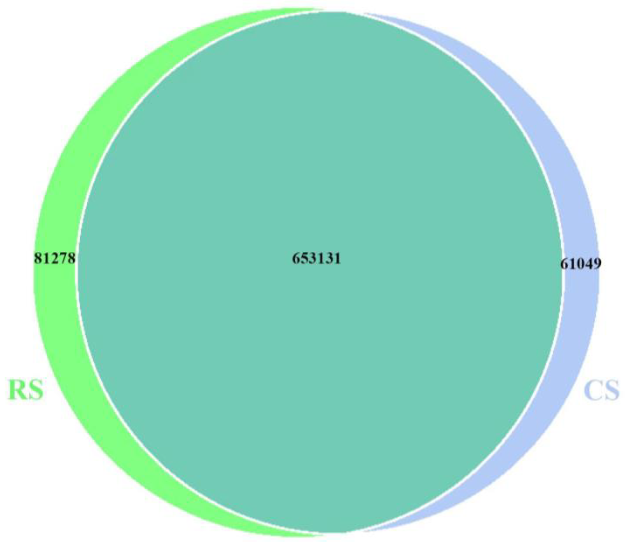
The Venn diagram demonstrates that in the CS group, 714,180 OTUs were identified, with 61,049 OTUs being unique to the CS group. In the RS group, 734,409 OTUs were identified, with 81,278 OTUs being unique to the RS group (Figure 2).
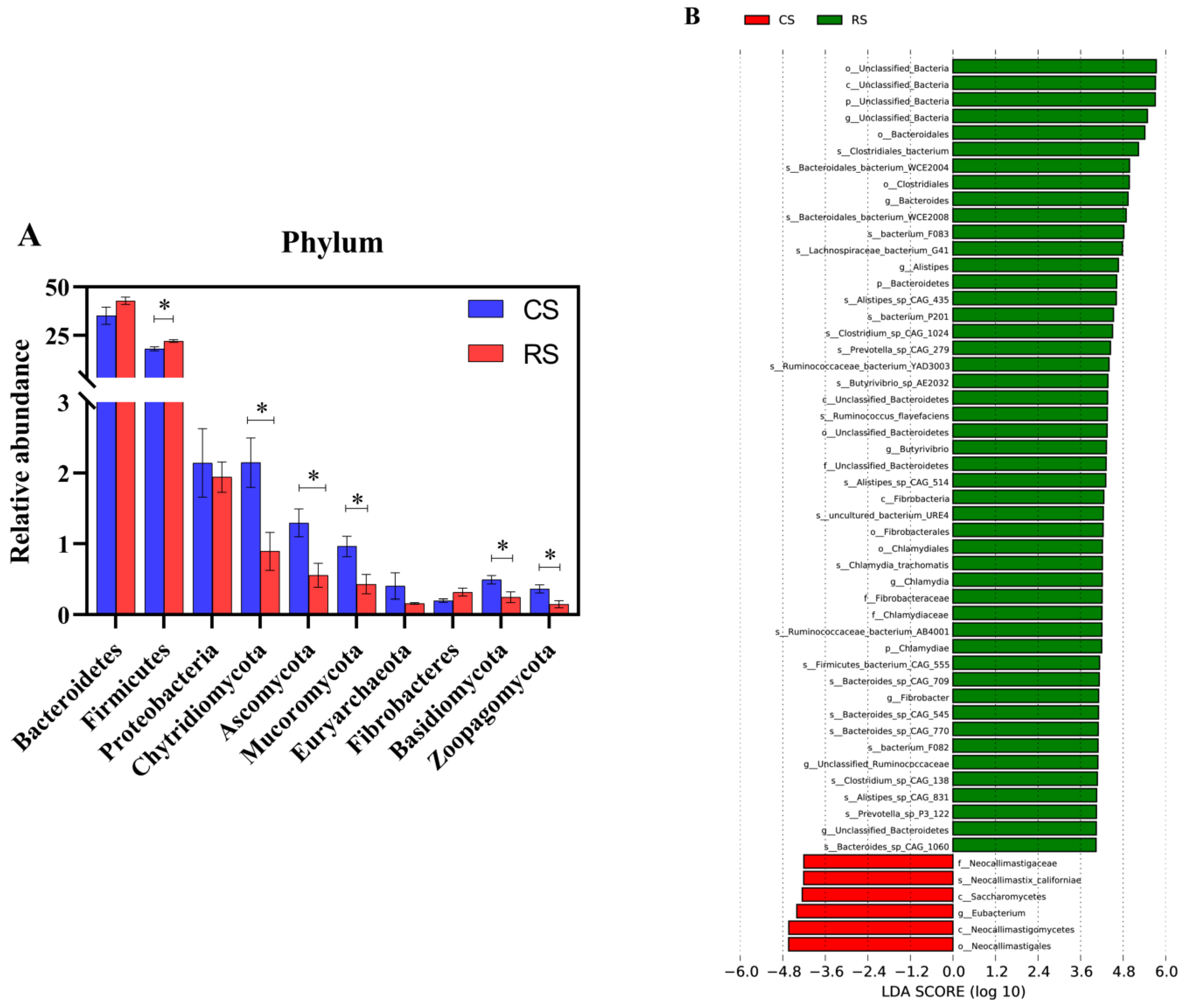
Based on Figure 3A, at the phylum level, Bacteroidetes, Firmicutes, and Proteobacteria were the dominant phyla shared by both the RS and CS groups (relative abundance of >1%). Firmicutes demonstrated significantly higher relative abundance in the RS group compared to the CS group (p < 0.05), while Chytridiomycota, Ascomycota, Mucoromycota, Basidiomycota, and Zoopagomycota exhibited significantly lower relative abundance in the RS group compared to the CS group (p < 0.05). The linear discriminant analysis effect size (LEfSe) was used to identify the microbial composition in the rumen of the RS and CS groups (Figure 3B). The results showed that at the genus level, compared to the CS group, the RS group of cows had an increased abundance of unclassified bacteria, Bacteroides, Alistipes, Butyrivibrio, Chlamydia, Fibrobacter, unclassified Ruminococcaceae, and unclassified Bacteroidetes. On the other hand, the abundance of Eubacterium decreased in the RS group ([LDA > 3.6], p < 0.05).

3.3. Functional Analysis of Bacteria and Fungi in the Rumen of the Rice Straw-Substituted Part of the Whole-Plant Corn Silage

Analysis of the linear discriminant analysis effect size (LEfSe) (Figure 4A) was used to identify significantly different families of CAZymes between the RS group and the CS group. There were 20 families that exhibited significant differences between the two groups ([LDA > 3], p < 0.05). Compared to the CS group, the rumen of cows in the RS group showed an increased abundance of enzyme functionalities in families GH3, GH43, GH78, GH2, GH92, GH20, and GH29, while the families GH77, GH24, GH25, GH73, GH17, GH32, CBM57, CBM20, and CBM48 exhibited decreased enzyme functionalities.
A bar chart of the top 10 abundances at CAZymes level 3 was created (Figure 4B). Compared to the CS group, the rumen of cows in the RS group exhibited a significant increase (p < 0.05) in the abundance of enzyme functionalities such as α-L-arabinofuranosidase (EC3.2.1.55), β-xylosidase (EC3.2.1.37), β-glucosidase (EC3.2.1.21), β-glucosylceramidase (EC3.2.1.45), xylanase (EC3.2.1.8), and arabinanase (EC3.2.1.99).

3.4. Correlation Analysis of Bacteria and Fungi with CAZymes in the Rumen of the Rice Straw Substituted for Part of the Whole-Plant Corn Silage

Significant correlations were analyzed between selected bacterial genera and the rumen fermentation parameters. The analysis revealed (see Figure 5) that Alistips, Bacteroides, and Butyrivibrio were significantly negatively correlated with propionic acid (p < 0.05), while they were significantly positively correlated with the ratio of acetic acid to propionic acid (p < 0.05). Eubacterium was significantly positively correlated with the total volatile fatty acids (TVFA) and valerate (p < 0.05).
Selected significantly different genera and families identified in CAZymes for correlation analysis revealed the following associations (see Figure 6): Alistips, Bacteroides, and Butyrivibrio showed positive correlations with GH2, GH3, GH20, GH29, GH43, GH78, GH92, CE1, and GT4 (p < 0.05). Alistips and Bacteroides demonstrated negative correlations with GH17, GH24, GH25, CBM20, and CBM57 (p < 0.05). Butyrivibrio showed negative correlations with GH24 and CBM57 (p < 0.05). Fibrobacter exhibited a negative correlation with CBM48 (p < 0.05).
Moreover, Alistips, Bacteroides, and Butyrivibrio were positively correlated with β-glucosidase (EC 3.2.1.21), α-L-arabinofuranosidase (EC 3.2.1.55), β-xylosidase (EC 3.2.1.37), β-glucosylceramidase (EC 3.2.1.45), xylanase (EC 3.2.1.8), and arabinanase (EC 3.2.1.99) (p < 0.05). The genus Fibrobacter exhibited negative correlations with α-L-arabinofuranosidase (EC 3.2.1.55), xylanase (EC 3.2.1.8), and arabinanase (EC 3.2.1.99) (p < 0.05).

4. Discussion

Rumen fermentation parameters can not only reflect the development status inside the rumen but also elucidate the fermentation pattern and the extent of feed fermentation within the rumen [24]. Among these parameters, acetic acid, propionic acid, and butyric acid are crucial volatile fatty acids (VFAs) produced during carbohydrate fermentation in the rumen. They can provide up to 70–80% of the energy requirements of ruminant animals. It has been reported that the production of acetic acid is related to fiber digestion [25] and can be used for milk fat synthesis and energy supply [26]. In this experiment, the RS group exhibited a significantly higher ratio of acetic acid to propionic acid compared to the CS group. This may be attributed to the increased abundance of fiber-degrading bacteria in the rumen due to the addition of rice straw, leading to an increase in the proportion of acetic acid in rumen fermentation and promoting a shift in rumen fermentation toward acetate production. Furthermore, through a correlation analysis between rumen microbial communities and rumen fermentation parameters, it was observed that genera such as Bacteroides, Alistipes, and Butyrivibrio were significantly positively correlated with the acetic acid-to-propionic acid ratio and were negatively correlated with propionic acid. These microorganisms are known to be involved in fiber degradation and primarily produce acetic acid [27,28]. This further supports the shift toward acetate-type fermentation in the rumen after the addition of rice straw.
Ruminant animals can digest and utilize fibrous straw-like coarse feed, mainly relying on the rumen microbial flora for physical digestion by bacteria and fungi, as well as the degradation action of various enzymes. This process breaks down and utilizes cellulose substances, providing energy for the organism. Research shows that the dominant bacterial groups in the rumen of ruminant animals are Firmicutes and Bacteroidetes [29]. This experiment also confirmed these findings. Firmicutes, as a fiber-degrading bacterium, plays an important role in cellulose, hemicellulose, starch, and oligosaccharide degradation, producing short-chain fatty acids such as acetic acid, propionic acid, and butyric acid, which are crucial for energy utilization [25,30]. Firmicutes also possess strong plant cell-wall degradation ability [31]. In this study, an increased abundance of Firmicutes was observed, which may be related to the higher crude fiber level in the straw. Fungi can adhere to plant cell walls or penetrate deep into the loosely structured fiber of the plant’s vascular bundle to increase the attachment points for other microorganisms, thereby enhancing the efficiency of cellulose degradation [32]. Research has shown that due to the different plant species in the feed, the composition of lignocellulosic fibers varies, resulting in corresponding changes in the types and proportions of fungi involved in the degradation process [33]. In this study, a decrease in the abundance of the phyla Chytridiomycota, Ascomycota, Mucoromycota, Basidiomycota, and Zoopagomycota was observed in the experimental group. This phenomenon may be due to the corn stalks being opened up during ensiling (microbial fermentation), making it easier for fungi to attach to them. Additionally, the composition of lignocellulosic fiber in straw differs from that of whole-plant ensiled corn and the growth of fungi has a lag period, resulting in variations in the types and proportions of fungi involved in the degradation process.
At the genus level, the RS group exhibited a significantly higher abundance of Bacteroides, Alistipes, Butyrivibrio, and Fibrobacter compared to the CS group. The genus Bacteroides can utilize plant cell walls to provide the host with acetate, butyrate, and succinate [34,35,36], which can further be converted into propionate through the succinate pathway [37]. The genus Alistipes can produce propionate, acetate, or succinate as fermentation end products [38]. Previous studies have indicated that Bacteroides and Alistipes are both associated with oligosaccharide degradation [39,40]. In this study, through the analysis of the correlation between microbial communities and CAZymes, it was found that Bacteroides and Alistipes showed a positive correlation with oligosaccharide-degrading enzyme families (GH20, GH29, GH92). This further confirms the important role of Bacteroides and Alistipes in oligosaccharide degradation. Furthermore, studies have indicated that the abundance of the genus Alistipes is significantly higher in diets with a high forage content [39]. In this study, after the addition of rice straw, the fiber level increased, and there was an increase in the abundance of the genus Alistipes. There are reports indicating that Butyrivibrio is a major hemicellulose-degrading bacterium and is one of the few rumen microorganisms capable of utilizing xylan and pectin [41,42]. It also plays a significant role in carbohydrate degradation and protein breakdown, with its primary products being ethanol, acetic acid, butyric acid, formic acid, and lactic acid, accompanied by the generation of CO2 and H2 [43,44]. In this study, a positive correlation was observed between Butyrivibrio and the hemicellulase enzymes (GH2, GH43, and GH78). This further confirms the crucial role of Butyrivibrio in the degradation of hemicellulose. Research has shown that the abundance of Butyrivibrio increases in high-fiber diets [42]. This aligns with the phenomenon observed in this study, wherein an increase in fiber levels led to an increase in the abundance of Butyrivibrio. Fibrobacter is an important and highly functional fiber-degrading bacterium that secretes cellulases (GH5, GH8, GH9, and GH45) and hemicellulases (GH10, GH26, and GH43). It can utilize cellulose, hemicellulose, and pectin to produce oligomeric xylose and arabinose for other rumen microorganisms to utilize. Additionally, it can generate succinate and convert it into propionate [45]. In this experiment, it was observed that the microbial community structure of fiber-degrading bacteria in the rumen changed, and bacteria with the ability to degrade fiber had a relatively higher abundance in the RS group. Substituting a portion of whole-plant corn silage with rice straw led to changes in the abundance of bacteria and fungi involved in fiber degradation in the rumen. It was found that fiber-related anaerobic fungi and bacteria in the rumen exhibited a negative correlation [46]. This phenomenon was also observed in the current experiment, where it was noted that in the RS group, there was an increase in the abundance of bacterial genera with fiber-degrading capabilities, while the abundance of fungal phyla with fiber-degrading capabilities decreased. This phenomenon may be attributed to competitive interactions between bacteria and fungi in the rumen, but the specific mechanisms behind this process require further investigation. In summary, different bacteria in the rumen collaborate to assist the host in digesting feed. With the addition of rice straw, there is an increase in the abundance of bacterial genera capable of fiber degradation, aiding the host in breaking down rice straw. Therefore, it can be concluded that Bacteroides, Alistipes, Butyrivibrio, and Fibrobacter play a positive role in the degradation of rice straw.
The replacement of dietary elements has a significant impact on the abundance of CAZyme genes in the rumen of cows. We observed that starch-degrading enzymes (GH77), lysozymes (GH24 and GH25), and hemicellulose-degrading enzymes (GH73, GH17, and GH32) were more abundant in the CS group, while cellulases (GH3), hemicellulose-degrading enzymes (GH43, GH78, and GH2), and oligosaccharide-degrading enzymes (GH92, GH20, and GH29) were more abundant in the RS group. This is consistent with previous research findings [47,48], indicating that high-fiber diets contain more carbohydrate-active enzymes that degrade complex carbohydrates. Research has shown that rice straw has a higher content of non-digestible fiber compared to whole-plant corn silage [49]. Arabinoxylan and arabinan are the major components of hemicellulose in the plant cell walls of rice straw [50,51,52]. In this study, the RS group had a higher abundance of the enzymes responsible for the degradation of arabinoxylan and arabinan compared to the CS group. These enzymes include α-L-arabinofuranosidase (EC 3.2.1.55), xylanase (EC 3.2.1.8), β-xylosidase (EC 3.2.1.37), and arabinanase (EC 3.2.1.99). The main chain of arabinoxylan is composed of β-D-xylopyranosyl residues linked by (1,4)-glycosidic bonds, and it possesses various structural side chains [53]. α-L-arabinofuranosidase (EC 3.2.1.55) is a debranching enzyme that removes arabinose substituents from arabinoxylan and promotes hemicellulose hydrolysis [54]. Xylanase (EC 3.2.1.8) acts on the main chain of xylan, hydrolyzing the internal β-1,4-D-xylosidic bonds in the xylan, producing xylobiose, xylotriose, and larger xylooligosaccharides, which are further attacked by β-xylosidase (EC 3.2.1.37) to produce xylose [55,56,57]. However, the β-xylosidases (EC 3.2.1.37) secreted by the GH43 family only catalyze the hydrolysis reaction [58]. Arabinan, composed of α-1,5-linked L-arabinofuranose residues, is a polysaccharide in plant cell walls modified by α-1,3- and α-1,2-linked arabinofuranosyl residues. Arabinanase (EC 3.2.1.99) catalyzes the hydrolysis of the α-1,5-linked L-arabinofuranosyl residues in arabinan, releasing arabinooligosaccharides and L-arabinose [59,60]. Moreover, the CAZymes (carbohydrate-active enzymes) results indicate an increase in the abundance of β-glucosidase (EC 3.2.1.21). As a crucial enzyme for cellulose degradation, it collaborates with endo-β-1,4-glucanase (EC 3.2.1.4) and cellobiose hydrolase (EC 3.2.1.91) to hydrolyze cellulose [61,62]. β-glucosidase facilitates the cleavage of β-1,4-glycosidic bonds within the cellobiose, breaking it down into two glucose molecules [63].

5. Conclusions

Straw can alter the composition of the rumen microbial community in cows. At the phylum level, it significantly increases the relative abundance of Firmicutes. At the genus level, there is a significant increase in the relative abundance of Bacteroides, Alistipes, Butyrivibrio, and Fibrobacter. Additionally, straw can alter the relative abundance of CAZymes (carbohydrate-active enzymes) at different hierarchical levels within the cow’s rumen. This alteration may enhance the degradation efficiency of straw by increasing the relative abundance of enzymes such as α-L-arabinofuranosidase (EC 3.2.1.55), xylanase (EC 3.2.1.8), β-xylosidase (EC 3.2.1.37), and arabinanase (EC 3.2.1.99).

Supplementary Materials

The following supporting information can be downloaded at: https://www.mdpi.com/article/10.3390/fermentation9110954/s1, Table S1: Composition and nutrient levels of experimental diets (DM basis), Table S2: the nutritional composition of whole-plant corn silage and straw.

Author Contributions

X.X. designed the experiments and revised the manuscript. Y.M., W.Y., Y.C., W.R. and S.Y. conducted the experiments. L.Z. provided the experimental reagents and the equipment. Y.M. analyzed the data and wrote the manuscript. All authors have read and agreed to the published version of the manuscript.

Funding

This study was funded by the National Natural Science Foundation of China (31660675).

Institutional Review Board Statement

All experimental procedures and animal experiments were conducted in accordance with the guidelines of the relevant ethics committee. This study obtained approval from the Experimental Animal Management and Use Committee of Ningxia University (NXU-371).

Informed Consent Statement

Not applicable.

Data Availability Statement

Data will be made available upon request to the corresponding author.

Conflicts of Interest

The authors declare no conflict of interest. The funders had no role in the design of the study; in the collection, analyses, or interpretation of data; in the writing of the manuscript; or in the decision to publish the results. or property resulting from any ideas, methods, instructions, or products referred to in the content.

References

  1. Akay, H. Grain and Straw Yield of Paddy Cultivars and Feed Quality Traits of Paddy Straw. Gesunde Pflanz. 2022, 74, 549–560. [Google Scholar] [CrossRef]
  2. Suretno, N.D.; Adriyani, F.Y.; Hevrizen, R. Content and potential of rice straw as a mineral source of zinc in ruminant feed. Iop Conf. Ser. Earth Environ. Sci. 2021, 653, 12022. [Google Scholar] [CrossRef]
  3. Peripolli, V.; Barcellos, J.O.J.; Prates, Ê.R.; McManus, C.; Silva, L.P.D.; Stella, L.A.; Costa Junior, J.B.G.; Lopes, R.B. Nutritional value of baled rice straw for ruminant feed. Rev. Bras. Zootec. 2016, 45, 392–399. [Google Scholar] [CrossRef]
  4. Hoerbe, J.B.; Sessim, A.G.; Pereira, G.R.; Brutti, D.D.; Oliveira, T.E.; Barcellos, J.O.J. Cow-calf intensification through the feeding of rice straw. Livest. Sci. 2020, 242, 104296. [Google Scholar] [CrossRef]
  5. Peripolli, V.; Barcellos, J.O.J.; Prates, Ê.R.; McManus, C.; Stella, L.A.; Camargo, C.M.; Costa Jr, J.B.G.; Bayer, C. Additives on in vitro ruminal fermentation characteristics of rice straw. Rev. Bras. Zootec. 2017, 46, 240–250. [Google Scholar] [CrossRef]
  6. Ma, Y.; Chen, X.; Khan, M.Z.; Xiao, J.; Cao, Z. A Combination of Novel Microecological Agents and Molasses Role in Digestibility and Fermentation of Rice Straw by Facilitating the Ruminal Microbial Colonization. Front. Microbiol. 2022, 13, 948049. [Google Scholar] [CrossRef]
  7. Huws, S.A.; Creevey, C.J.; Oyama, L.B.; Mizrahi, I.; Denman, S.E.; Popova, M.; Muñoz-Tamayo, R.; Forano, E.; Waters, S.M.; Hess, M. Addressing global ruminant agricultural challenges through understanding the rumen microbiome: Past, present, and future. Front. Microbiol. 2018, 9, 2161. [Google Scholar] [CrossRef]
  8. Lobo, R.R.; Faciola, A.P. Ruminal Phages—A Review. Front. Microbiol. 2021, 12, 763416. [Google Scholar] [CrossRef]
  9. Liang, J.; Nabi, M.; Zhang, P.; Zhang, G.; Cai, Y.; Wang, Q.; Zhou, Z.; Ding, Y. Promising biological conversion of lignocellulosic biomass to renewable energy with rumen microorganisms: A comprehensive review. Renew. Sustain. Energy Rev. 2020, 134, 110335. [Google Scholar] [CrossRef]
  10. Plouhinec, L.; Neugnot, V.; Lafond, M.; Berrin, J. Carbohydrate-active enzymes in animal feed. Biotechnol. Adv. 2023, 65, 108145. [Google Scholar] [CrossRef]
  11. Nam, N.N.; Do, H.; Loan, T.K.; Lee, N.Y. Metagenomics: An Effective Approach for Exploring Microbial Diversity and Functions. Foods 2023, 12, 2140. [Google Scholar] [CrossRef] [PubMed]
  12. Tringe, S.G.; Rubin, E.M. Metagenomics: DNA sequencing of environmental samples. Nat. Rev. Genet. 2005, 6, 805–814. [Google Scholar] [CrossRef] [PubMed]
  13. Van Gastelen, S.; Antunes-Fernandes, E.C.; Hettinga, K.A.; Klop, G.; Alferink, S.; Hendriks, W.H.; Dijkstra, J. Enteric methane production, rumen volatile fatty acid concentrations, and milk fatty acid composition in lactating Holstein-Friesian cows fed grass silage-or corn silage-based diets. J. Dairy Sci. 2015, 98, 1915–1927. [Google Scholar] [CrossRef] [PubMed]
  14. Weatherburn, M.W. Phenol-hypochlorite reaction for determination of ammonia. Anal. Chem. 1967, 39, 971–974. [Google Scholar] [CrossRef]
  15. Karlsson, F.H.; Tremaroli, V.; Nookaew, I.; Bergström, G.; Behre, C.J.; Fagerberg, B.; Nielsen, J.; Bäckhed, F. Gut metagenome in European women with normal, impaired and diabetic glucose control. Nature 2013, 498, 99–103. [Google Scholar] [CrossRef]
  16. Karlsson, F.H.; Fåk, F.; Nookaew, I.; Tremaroli, V.; Fagerberg, B.; Petranovic, D.; Bäckhed, F.; Nielsen, J. Symptomatic atherosclerosis is associated with an altered gut metagenome. Nat. Commun. 2012, 3, 1245. [Google Scholar] [CrossRef]
  17. Qin, N.; Yang, F.; Li, A.; Prifti, E.; Chen, Y.; Shao, L.; Guo, J.; Le Chatelier, E.; Yao, J.; Wu, L.; et al. Alterations of the human gut microbiome in liver cirrhosis. Nature 2014, 513, 59–64. [Google Scholar] [CrossRef]
  18. Mende, D.R.; Waller, A.S.; Sunagawa, S.; Jarvelin, A.I.; Chan, M.M.; Arumugam, M.; Raes, J.; Bork, P. Assessment of metagenomic assembly using simulated next generation sequencing data. PLoS ONE 2012, 7, e31386. [Google Scholar] [CrossRef]
  19. Nielsen, H.B.; Almeida, M.; Juncker, A.S.; Rasmussen, S.; Li, J.; Sunagawa, S.; Plichta, D.R.; Gautier, L.; Pedersen, A.G.; Le Chatelier, E.; et al. Identification and assembly of genomes and genetic elements in complex metagenomic samples without using reference genomes. Nat. Biotechnol. 2014, 32, 822–828. [Google Scholar] [CrossRef]
  20. Sunagawa, S.; Coelho, L.P.; Chaffron, S.; Kultima, J.R.; Labadie, K.; Salazar, G.; Djahanschiri, B.; Zeller, G.; Mende, D.R.; Alberti, A.; et al. Ocean plankton. Structure and function of the global ocean microbiome. Science 2015, 348, 1261359. [Google Scholar] [CrossRef]
  21. White, J.R.; Nagarajan, N.; Pop, M. Statistical methods for detecting differentially abundant features in clinical metagenomic samples. PLoS Comput. Biol. 2009, 5, e1000352. [Google Scholar] [CrossRef] [PubMed]
  22. Segata, N.; Izard, J.; Waldron, L.; Gevers, D.; Miropolsky, L.; Garrett, W.S.; Huttenhower, C. Metagenomic biomarker discovery and explanation. Genome Biol. 2011, 12, R60. [Google Scholar] [CrossRef] [PubMed]
  23. Cantarel, B.L.; Coutinho, P.M.; Rancurel, C.; Bernard, T.; Lombard, V.; Henrissat, B. The Carbohydrate-Active EnZymes database (CAZy): An expert resource for Glycogenomics. Nucleic Acids Res. 2009, 37, D233–D238. [Google Scholar] [CrossRef]
  24. Liu, H.; Zhou, J.; Degen, A.; Liu, H.; Cao, X.; Hao, L.; Shang, Z.; Ran, T.; Long, R. A comparison of average daily gain, apparent digestibilities, energy balance, rumen fermentation parameters, and serum metabolites between yaks (Bos grunniens) and Qaidam cattle (Bos taurus) consuming diets differing in energy level. Anim. Nutr. 2023, 12, 77–86. [Google Scholar] [CrossRef]
  25. Ahmad, A.A.; Yang, C.; Zhang, J.; Kalwar, Q.; Liang, Z.; Li, C.; Du, M.; Yan, P.; Long, R.; Han, J.; et al. Effects of Dietary Energy Levels on Rumen Fermentation, Microbial Diversity, and Feed Efficiency of Yaks (Bos grunniens). Front. Microbiol. 2020, 11, 625. [Google Scholar] [CrossRef] [PubMed]
  26. Zhao, H.; Lv, F.; Liu, G.; Pang, X.; Han, X.; Wang, X. Effects of starters with different NDF/starch ratio on rumen fermentation parameters and rumen microorganisms in lambs. Front. Vet. Sci. 2023, 10, 1064774. [Google Scholar] [CrossRef] [PubMed]
  27. Cheng, C.; Yin, Y.; Bian, G. Effects of whole maize high-grain diet feeding on colonic fermentation and bacterial community in weaned lambs. Front. Microbiol. 2022, 13, 1018284. [Google Scholar] [CrossRef]
  28. Xiao, J.X.; Alugongo, G.M.; Chung, R.; Dong, S.Z.; Li, S.L.; Yoon, I.; Wu, Z.H.; Cao, Z.J. Effects of Saccharomyces cerevisiae fermentation products on dairy calves: Ruminal fermentation, gastrointestinal morphology, and microbial community. J. Dairy Sci. 2016, 99, 5401–5412. [Google Scholar] [CrossRef]
  29. Li, F.; Li, C.; Chen, Y.; Liu, J.; Zhang, C.; Irving, B.; Fitzsimmons, C.; Plastow, G.; Guan, L.L. Host genetics influence the rumen microbiota and heritable rumen microbial features associate with feed efficiency in cattle. Microbiome 2019, 7, 92. [Google Scholar] [CrossRef]
  30. Jumpertz, R.; Le, D.S.; Turnbaugh, P.J.; Trinidad, C.; Bogardus, C.; Gordon, J.I.; Krakoff, J. Energy-balance studies reveal associations between gut microbes, caloric load, and nutrient absorption in humans. Am. J. Clin. Nutr. 2011, 94, 58–65. [Google Scholar] [CrossRef]
  31. Pope, P.B.; Denman, S.E.; Jones, M.; Tringe, S.G.; Barry, K.; Malfatti, S.A.; McHardy, A.C.; Cheng, J.F.; Hugenholtz, P.; McSweeney, C.S.; et al. Adaptation to herbivory by the Tammar wallaby includes bacterial and glycoside hydrolase profiles different from other herbivores. Proc. Natl. Acad. Sci. USA 2010, 107, 14793–14798. [Google Scholar] [CrossRef] [PubMed]
  32. Akin, D.E.; Borneman, W.S. Role of Rumen Fungi in Fiber Degradation. J. Dairy Sci. 1990, 73, 3023–3032. [Google Scholar] [CrossRef] [PubMed]
  33. Pitta, D.W.; Pinchak, W.E.; Dowd, S.E.; Osterstock, J.; Gontcharova, V.; Youn, E.; Dorton, K.; Yoon, I.; Min, B.R.; Fulford, J.D.; et al. Rumen Bacterial Diversity Dynamics Associated with Changing from Bermudagrass Hay to Grazed Winter Wheat Diets. Microb. Ecol. 2010, 59, 511–522. [Google Scholar] [CrossRef] [PubMed]
  34. Solden, L.M.; Hoyt, D.W.; Collins, W.B.; Plank, J.E.; Daly, R.A.; Hildebrand, E.; Beavers, T.J.; Wolfe, R.; Nicora, C.D.; Purvine, S.O.; et al. New roles in hemicellulosic sugar fermentation for the uncultivated Bacteroidetes family BS11. ISME J. 2017, 11, 691–703. [Google Scholar] [CrossRef]
  35. Louis, P.; Hold, G.L.; Flint, H.J. The gut microbiota, bacterial metabolites and colorectal cancer. Nat. Rev. Microbiol. 2014, 12, 661–672. [Google Scholar] [CrossRef]
  36. Liang, J.; Zhang, H.; Zhang, P.; Zhang, G.; Cai, Y.; Wang, Q.; Zhou, Z.; Ding, Y.; Zubair, M. Effect of substrate load on anaerobic fermentation of rice straw with rumen liquid as inoculum: Hydrolysis and acidogenesis efficiency, enzymatic activities and rumen bacterial community structure. Waste Manag. 2021, 124, 235–243. [Google Scholar] [CrossRef]
  37. Koh, A.; De Vadder, F.; Kovatcheva-Datchary, P.; Bäckhed, F. From Dietary Fiber to Host Physiology: Short-Chain Fatty Acids as Key Bacterial Metabolites. Cell 2016, 165, 1332–1345. [Google Scholar] [CrossRef]
  38. Holman, D.B.; Gzyl, K.E. A meta-analysis of the bovine gastrointestinal tract microbiota. FEMS Microbiol. Ecol. 2019, 95, fiz072. [Google Scholar] [CrossRef]
  39. Wang, L.; Zhang, G.; Xu, H.; Xin, H.; Zhang, Y. Metagenomic Analyses of Microbial and Carbohydrate-Active Enzymes in the Rumen of Holstein Cows Fed Different Forage-to-Concentrate Ratios. Front. Microbiol. 2019, 10, 649. [Google Scholar] [CrossRef]
  40. Mu, Y.Y.; Qi, W.P.; Zhang, T.; Zhang, J.Y.; Mao, S.Y. Gene function adjustment for carbohydrate metabolism and enrichment of rumen microbiota with antibiotic resistance genes during subacute rumen acidosis induced by a high-grain diet in lactating dairy cows. J. Dairy Sci. 2021, 104, 2087–2105. [Google Scholar] [CrossRef]
  41. Díaz Carrasco, J.M.; Cabral, C.; Redondo, L.M.; Pin Viso, N.D.; Colombatto, D.; Farber, M.D.; Fernández Miyakawa, M.E. Impact of Chestnut and Quebracho Tannins on Rumen Microbiota of Bovines. Biomed. Res. Int. 2017, 2017, 9610810. [Google Scholar] [CrossRef] [PubMed]
  42. Forster, R.J.; Gong, J.; Teather, R.M. Group-specific 16S rRNA hybridization probes for determinative and community structure studies of Butyrivibrio fibrisolvens in the rumen. Appl. Environ. Microb. 1997, 63, 1256–1260. [Google Scholar] [CrossRef] [PubMed]
  43. Mao, S.Y.; Huo, W.J.; Zhu, W.Y. Microbiome–metabolome analysis reveals unhealthy alterations in the composition and metabolism of ruminal microbiota with increasing dietary grain in a goat model. Environ. Microbiol. 2016, 18, 525–541. [Google Scholar] [CrossRef] [PubMed]
  44. Fernando, S.C.; Purvis, H.T.; Najar, F.Z.; Sukharnikov, L.O.; Krehbiel, C.R.; Nagaraja, T.G.; Roe, B.A.; DeSilva, U. Rumen Microbial Population Dynamics during Adaptation to a High-Grain Diet. Appl. Environ. Microb. 2010, 76, 7482–7490. [Google Scholar] [CrossRef] [PubMed]
  45. Neumann, A.P.; Suen, G. The Phylogenomic Diversity of Herbivore-Associated Fibrobacter spp. Is Correlated to Lignocellulose-Degrading Potential. Msphere 2018, 3, 10–1128. [Google Scholar] [CrossRef]
  46. Azad, E.; Fehr, K.B.; Derakhshani, H.; Forster, R.; Acharya, S.; Khafipour, E.; McGeough, E.; McAllister, T.A. Interrelationships of Fiber-Associated Anaerobic Fungi and Bacterial Communities in the Rumen of Bloated Cattle Grazing Alfalfa. Microorganisms 2020, 8, 1543. [Google Scholar] [CrossRef]
  47. Shen, J.; Zheng, L.; Chen, X.; Han, X.; Cao, Y.; Yao, J. Metagenomic Analyses of Microbial and Carbohydrate-Active Enzymes in the Rumen of Dairy Goats Fed Different Rumen Degradable Starch. Front. Microbiol. 2020, 11, 1003. [Google Scholar] [CrossRef]
  48. Bohra, V.; Dafale, N.A.; Purohit, H.J. Understanding the alteration in rumen microbiome and CAZymes profile with diet and host through comparative metagenomic approach. Arch. Microbiol. 2019, 201, 1385–1397. [Google Scholar] [CrossRef]
  49. Nazli, M.H.; Halim, R.A.; Abdullah, A.M.; Hussin, G.; Samsudin, A.A. Potential of feeding beef cattle with whole corn crop silage and rice straw in Malaysia. Trop. Anim. Health Prod. 2018, 50, 1119–1124. [Google Scholar] [CrossRef]
  50. Puițel, A.C.; Suditu, G.D.; Danu, M.; Ailiesei, G.; Nechita, M.T. An Experimental Study on the Hot Alkali Extraction of Xylan-Based Hemicelluloses from Wheat Straw and Corn Stalks and Optimization Methods. Polymers 2022, 14, 1662. [Google Scholar] [CrossRef]
  51. Wilkens, C.; Andersen, S.; Dumon, C.; Berrin, J.G.; Svensson, B. GH62 arabinofuranosidases: Structure, function and applications. Biotechnol. Adv. 2017, 35, 792–804. [Google Scholar] [CrossRef]
  52. Chen, C.; Zhao, X.; Wang, X.; Wang, B.; Li, H.; Feng, J.; Wu, A. Mutagenesis of UDP-xylose epimerase and xylan arabinosyl-transferase decreases arabinose content and improves saccharification of rice straw. Plant Biotechnol. J. 2021, 19, 863–865. [Google Scholar] [CrossRef] [PubMed]
  53. Zhang, X.; Chen, T.; Lim, J.; Gu, F.; Fang, F.; Cheng, L.; Campanella, O.H.; Hamaker, B.R. Acid gelation of soluble laccase-crosslinked corn bran arabinoxylan and possible gel formation mechanism. Food Hydrocoll. 2019, 92, 1–9. [Google Scholar] [CrossRef]
  54. Wu, X.; Zhang, S.; Zhao, S.; Dai, L.; Huang, S.; Liu, X.; Yu, J.; Wang, L. Functional Specificity of Three α-Arabinofuranosidases from Different Glycoside Hydrolase Families in Aspergillus niger An76. J. Agric. Food Chem. 2022, 70, 5039–5048. [Google Scholar] [CrossRef] [PubMed]
  55. Liu, Y.; Huang, L.; Zheng, D.; Xu, Z.; Li, Y.; Shao, S.; Zhang, Y.; Ge, X.; Lu, F. Biochemical characterization of a novel GH43 family β-xylosidase from Bacillus pumilus. Food Chem. 2019, 295, 653–661. [Google Scholar] [CrossRef]
  56. Terrasan, C.R.F.; Guisan, J.M.; Carmona, E.C. Xylanase and β-xylosidase from Penicillium janczewskii: Purification, characterization and hydrolysis of substrates. Electron. J. Biotechnol. 2016, 23, 54–62. [Google Scholar] [CrossRef]
  57. Fanchini Terrasan, C.R.; Trobo-Maseda, L.; Moreno-Pérez, S.; Carmona, E.C.; Pessela, B.C.; Guisan, J.M. Co-immobilization and stabilization of xylanase, β-xylosidase and α-l-arabinofuranosidase from Penicillium janczewskii for arabinoxylan hydrolysis. Process Biochem. 2016, 51, 614–623. [Google Scholar] [CrossRef]
  58. Smaali, I.; Rémond, C.; O Donohue, M.J. Expression in Escherichia coli and characterization of β-xylosidases GH39 and GH-43 from Bacillus halodurans C-125. Appl. Microbiol. Biotechnol. 2006, 73, 582–590. [Google Scholar] [CrossRef]
  59. Santos, C.R.; Polo, C.C.; Costa, M.C.M.F.; Nascimento, A.F.Z.; Meza, A.N.; Cota, J.; Hoffmam, Z.B.; Honorato, R.V.; Oliveira, P.S.L.; Goldman, G.H.; et al. Mechanistic Strategies for Catalysis Adopted by Evolutionary Distinct Family 43 Arabinanases. J. Biol. Chem. 2014, 289, 7362–7373. [Google Scholar] [CrossRef]
  60. Inácio, J.M.; de Sá-Nogueira, I. Characterization of abn2 (yxiA), Encoding a Bacillus subtilis GH43 Arabinanase, Abn2, and Its Role in Arabino-Polysaccharide Degradation. J. Bacteriol. 2008, 190, 4272–4280. [Google Scholar] [CrossRef]
  61. Sorensen, A.; Lubeck, P.S.; Lubeck, M.; Teller, P.J.; Ahring, B.K. β-glucosidases from a new Aspergillus species can substitute commercial β-glucosidases for saccharification of lignocellulosic biomass. Can. J. Microbiol. 2011, 57, 638–650. [Google Scholar] [CrossRef] [PubMed]
  62. Volkov, P.V.; Rozhkova, A.M.; Zorov, I.N.; Sinitsyn, A.P. Cloning, purification and study of recombinant GH3 family β-glucosidase from Penicillium verruculosum. Biochimie 2020, 168, 231–240. [Google Scholar] [CrossRef] [PubMed]
  63. Tiwari, P.; Misra, B.N.; Sangwan, N.S. β-Glucosidases from the fungus trichoderma: An efficient cellulase machinery in biotechnological applications. Biomed. Res. Int. 2013, 2013, 203735. [Google Scholar] [CrossRef] [PubMed]
Figure 1. The effect of partial rice straw substitution for whole-plant corn silage on the diversity of rumen-associated bacterial communities in dairy cows (n = 4): PCA analysis of rumen microorganisms.
Fermentation 09 00954 g001
Figure 2. The effect of partial rice straw substitution for whole-plant corn silage on the diversity of the rumen-associated bacterial communities in dairy cows (n = 4): Venn diagram of the OTUs in the ruminal microbiota.
Fermentation 09 00954 g002
Figure 3. Rice straw replaced part of the proportion of the whole-plant corn silage and changed the relative abundances of microbiota in the rumen: (A) relative abundances of the top 10 bacterial communities at the phylum level, * p < 0.05; (B) histogram of the LDA effect values for marker species. The ordinate represents the taxonomic units with significant differences between groups, and the abscissa represents the logarithmic score values of the LDA analysis of each taxonomic unit in a bar chart.
Fermentation 09 00954 g003
Figure 4. Rice straw replaced part of the whole-plant corn silage and changed the relative abundances of the CAZymes in the rumen: (A) histogram of the LDA effect values for the CAZymes. The ordinate represents the CAZyme family, with significant differences between groups, and the abscissa represents the logarithmic score values of the LDA analysis of each taxonomic unit in a bar chart. (B) relative abundances of the top 10 enzymes at the EC level; * p < 0.05.
Fermentation 09 00954 g004
Figure 5. Correlation of bacteria and fungi in the rumen with ruminal fermentation parameters in whole-corn silage partially replaced with rice straw.
Fermentation 09 00954 g005
Figure 6. Correlation analysis of bacteria and fungi with CAZymes in the rumen of rice straw substituted for a proportion of the whole-plant corn silage.
Fermentation 09 00954 g006
Table 1. Changes in the rumen fermentation parameters in dairy cows under silage conditions with rice straw partially replacing the whole-plant corn feed.
ItemCSRS
NH3-N (mg/dL)11.67 ± 0.2611.08 ± 0.15
Acetate (mmol/L)72.28 ± 2.18 A67.47 + 1.4 B
Propionate (mmol/L)19.38 ± 0.44 A16.32 ± 0.54 B
Isobutyrate (mmol/L)0.74 ± 0.090.78 ± 0.12
Butyrate (mmol/L)10.86 ± 0.55 A7.85 ± 0.55 B
Isovalerate (mmol/L)1.08 ± 0.051.01 ± 0.06
Valerate (mmol/L)0.97 ± 0.170.71 ± 0.07
TVFA (mmol/L)105.31 ± 2.52 A94.14 ± 2.46 B
Acetate/propionate3.73 ± 0.18 B4.13 ± 0.09 A
Note: Peer shoulder labels with different capital letters indicate highly significant differences (p < 0.01), and the same or no letter shoulder labels indicate non-significant differences (p ≥ 0.05).
Disclaimer/Publisher’s Note: The statements, opinions and data contained in all publications are solely those of the individual author(s) and contributor(s) and not of MDPI and/or the editor(s). MDPI and/or the editor(s) disclaim responsibility for any injury to people or property resulting from any ideas, methods, instructions or products referred to in the content.