Article

Using Deep Ocean Water in the Fermentation of Antrodia cinnamomea to Boost Magnesium Ion Bioabsorption and Anti-Inflammatory Effects in the Brain of an Alzheimer’s Disease Rat Model

1
Department of Life Science, National Taitung University, Taitung 95092, Taiwan
2
C.N.S. Biotechnology Corp., Taichung 403030, Taiwan
*
Author to whom correspondence should be addressed.
Fermentation 2023, 9(10), 893; https://doi.org/10.3390/fermentation9100893
Submission received: 17 August 2023 / Revised: 21 September 2023 / Accepted: 25 September 2023 / Published: 4 October 2023
(This article belongs to the Special Issue Bioactivity Change in Fermented Foods)

Abstract

:
Alzheimer’s disease (AD) is a neurodegenerative disorder characterized by the deposition of amyloid β-peptide (Aβ) and subsequent oxidative inflammatory response, leading to brain damage and memory loss. This study explores the potential of Antrodia cinnamomea (AC), a Taiwan-native fungus known for its anti-inflammatory and antioxidant properties. The metabolites of AC, including dehydroeburicoic acid (DEA), 4-acetylantroquinonol B (4-AAQB), dehydrosulphurenic acid (DSA), and polysaccharides, were of particular interest. In the experiment, deep ocean water (DOW) was used to facilitate the solid-state fermentation of Antrodia cinnamomea NTTU 206 (D-AC), aiming to enhance its functional components. The impact of D-AC on the modulation of AD-related risk factors and the augmentation of cognitive abilities was subsequently evaluated in an AD rat model. This model was established via consecutive infusions of Aβ40 into the brain over a 28-day period. The administration of D-AC resulted in remarkable improvements in the rats’ reference memory, spatial probe test, and working memory. Notably, it restored the hippocampal magnesium levels by upregulating the expression of the magnesium transporter MAGT1. Concurrently, D-AC significantly downregulated the expressions of β-secretase 1 (BACE1) and the phosphorylated tau protein (p-tau), which were both implicated in AD progression. Additionally, it mitigated inflammatory responses, as suggested by the decreased levels of tumor necrosis factor α (TNF-α), interleukin-1β (IL-1β), and interleukin-6 (IL-6) in the hippocampus and cerebral cortex. Ultimately, the ability of D-AC to restore the brain magnesium levels, attenuate inflammatory responses, and reduce hippocampal Aβ40 deposition led to significant improvements in the cognitive decline of AD rats. D-AC demonstrated a comparable efficacy with its counterpart, AC fruiting bodies (F-AC group), despite their componential differences. This study underscores the potential of D-AC, enriched through fermentation, as a novel dietary strategy for Alzheimer’s disease prevention.

1. Introduction

Alzheimer’s disease (Alzheimer’s disease, AD) is a neurodegenerative disease of the brain. It is a progressive mental decline that occurs in middle or late age. Its pathological features are specific nerve cell degeneration, neuritic spots, and nerve fiber tangles [1]. Hippocampal neurons and the cerebral cortex suffer the most damage [2,3,4]. Amyloid β-peptide (Aβ) is associated with brain lesions in AD patients [5]. The APP precursor protein is cleaved by BACE to form Aβ, which is deposited in the cerebral cortex and hippocampus. Aβ in turn leads to neuronal damage producing neurofibrillary tangles and senile plaques. The accumulation of Aβ in parenchyma and blood vessels leads to the mobilization of microglia and promotes inflammation. This in turn induces nitric oxide (NO), reactive oxygen species (ROS), pro-inflammatory cytokines (TNFα, IL-1β, and IL-6), and prostaglandin (PGE2). These cascading oxidative-inflammatory reactions promote neuronal death and lead to the impairment of memory and learning [6].
Antrodia cinnamomea (AC) is a unique fungus in Taiwan. Previous studies have shown that Antrodia cinnamomea extract (ACE) has a wide range of biological activities, including anti-cancer, antioxidant, hepatoprotective, anti-hyperlipidemic, and anti-inflammatory activities [7,8,9]. Since the wild A. cinnamomea fruiting body only grows on Taiwan’s native camphor trees (Cinnamomum kanehirae), it is very precious. Its price is more than ten times that of the mycelium product; therefore, it causes a serious problem since Cinnamomum kanehirae tree conservation is difficult. The use of solid substrates to cultivate A. cinnamomea does not require the use of Cinnamomum kanehirae wood components, which helps to reduce the illegal harvesting of conservation Cinnamomum kanehirae trees. The biotechnology industry is also actively developing mycelium fermentation technology in anticipation of replacing the use of Antrodia cinnamomea fruiting bodies [9].
Past studies have pointed out that both the fruiting body and liquid mycelium of Antrodia cinnamomea can inhibit Aβ40-induced PC12 cytotoxicity and improve learning and memory impairment in rats, but the fruiting body has a better effect [10]. The mycelium of A. cinnamomea in this study contains high concentrations of 4-acetylantroquinonol B (4-AAQB), dehydrosulphurenic acid (DSA), dehydroeburicoic acid (DEA), and polysaccharides. Since they have anti-inflammatory and anti-oxidative effects [11,12,13,14], they may be used as the functional ingredients of Antrodia cinnamomea in preventing AD.
Deep ocean water (DOW) has the characteristics of low temperature, high nutrient salt, being clear and clean, and few pathogenic bacteria because light cannot penetrate. DOW contains a variety of trace elements. Recent studies have pointed out that DOW has many health benefits, including preventing cardiovascular disease [15], lowering blood pressure [16], improving obesity [17], etc. Studies have found that Cordyceps cicadae mycelium can increase the content of functional ingredients by absorbing mineral elements in DOW and enhancing the effect of suppressing Aβ40-induced cytotoxicity [18].
This study primarily focuses on the cultivation of Antrodia cinnamomea using DOW, with an emphasis on examining whether this cultivation method can enhance various functional components (4-AAQB, DEA, and DSA) of the fungus. Additionally, the research delves into the efficacy of DOW-cultivated A. cinnamomea in improving memory and learning capabilities relevant to Alzheimer’s Disease. The study also investigates the bio-absorption of magnesium of DOW in the brain. It questions whether this absorbed magnesium influences the brain’s uptake of magnesium ions and subsequently affects the improvement of Alzheimer’s symptoms. By integrating the use of DOW as a cultivation medium and innovating the associated techniques, the study aims to augment the functional components of Antrodia cinnamomea and its therapeutic effects on memory and learning deficits in Alzheimer’s Disease animal models.

2. Materials and Methods

2.1. Chemicals

Various reagents, recombinant proteins, antibodies, and analytical kits were used in this study. The amyloid β protein fragment 1–40 (A1075) came from Sigma-Aldrich chemie GmbH; the 95% ethanol from ECHO Chemical Co., Ltd.; and both the potato dextrose agar (PDA) and broth (PDB) from Difco Laboratories Inc. (Detroit, MI, USA). Acetonitrile, methanol, and NaOH were also purchased from Merck KGaA Millipore (Darmstadt, Germany), magnesium chloride anhydrous was purchased from Thermo Fisher Scientific Inc. (Waltham, MA, USA), and terrell (Isoflurane, USP) liquid for inhalation was purchased from Piramal Critical Care Inc. (Bethlehem, PA, USA). For the ELISA assay, the mouse TNF-α protein (50349-MNAE), mouse IL-6 protein (50349-MNAE), and mouse IL-1β protein (50349-MNAE) were purchased from SinoBiological Inc. (North Wales, PA, USA); the rabbit anti-mouse TNF-α polyclonal antibody (AB2148P) was purchased from EMD Millipore Corporation (TemecμLa, CA, USA); the mouse anti-rat IL-6 monoclonal antibody (sc-57315) and mouse anti-human IL-1β monoclonal antibody (sc-32294) were purchased from Santa Cruz Biotechnology, Inc. (Dallas, TX, USA); and the goat anti-rabbit IgG, (H + L), and peroxidase conjugated anti-body (31460) was purchased from Pierce Biotechnology (Rockford, IL, USA). For the analytical kits, the pierce BCA protein assay kit (LSG23225) was purchased from Thermo Fisher Scientific Inc. (Rockford, IL, USA); the magnesium assay kit was purchased from Dialab Trade GmbH (Neudorf, Ausria); the rat Aβ 40 (I-ER0754), BACE (ER0756), and RAGE (ER1853) analytical kits were purchased from Wuhan Fine Biotech Co., Ltd. (Wuchan, China); the MAGT1 assay kit (MBS2890531-96) was purchased from Mybiosource Inc. (San Diego, CF, USA); the rat sAPPα assay kit (I-JP27419) was purchased from Immuno Biological Laboratories Inc. (Minneapolis, MN, USA); and the polymer detection system (RE7140-K) was purchased from Leica Biosystems Nussloch GmbH (Chicago, IL, USA).

2.2. The Source of DOW

The Eastern Taiwan Deep Sea Water Innovation and Research Center (Taitung, Taiwan) provided concentrated deep ocean water (DOW), harvested from the Pacific Ocean’s depths of 670 m near Eastern Taiwan. The DOW concentrate contained various minerals and trace elements. The concentration of sodium (Na) was 1790 mg/mL, potassium (K) was 814 mg/mL, calcium (Ca) was 19.1 mg/mL, and magnesium (Mg) was 101,000 mg/mL. The concentration of selenium (Se) and copper (Cu) were both less than 0.01 mg/mL, while molybdenum (Mo) was 0.56 mg/mL, strontium (Sr) was 0.2 mg/mL, iron (Fe) was 4.37 mg/mL, and zinc (Zn) was 0.13 mg/mL. The concentration of chloride (Cl) was 301,000 mg/mL, sulfate (SO42−) was 18,500 mg/mL, and bromide (Br) was 8380 mg/mL. The test data were provided by Eastern Deep Ocean Water Innovation and R&D Center of Ministry of Economic Affairs (Taitung, Taiwan).

2.3. Plate Culture Method of Antrodia cinnamomea Mycelium

The strain of Antrodia cinnamomea NTTU 206 used in this study was isolated from the wild fruiting bodies of A. cinnamomea located in Eastern Taiwan. Chosen as the target strain for this research due to its faster growth rate and ability to produce high concentrations of 4-acetylantroquinonol B (4-AAQB), dehydroeburicoic acid (DEA), and dehydrosulphurenic acid (DSA), this strain exemplifies the potent medicinal qualities of this species. Antrodia cinnamomea NTTU 206 was cultured on a potato dextrose agar (PDA) plate at 30 °C incubator for 10 days. The mycelia on the PDA plate was used as the inoculum for the seed culture in liquid fermentation.

2.4. Seed Culture of Liquid Fermentation for Antrodia cinnamomea Mycelium

Two cylindrical colonies of A. cinnamomea mycelia, each with a radius of 0.7 cm, were inoculated into 100 mL of liquid potato dextrose broth (24 g/L). The culture was incubated at 30 °C with a shaking speed of 150 rpm in a dark incubator for one week. After fermentation, the seed culture medium containing mycelial pellets was used as the inoculation substance for solid-state fermentation.

2.5. Solid-State Fermentation of Antrodia cinnamomea Mycelium

Red Quinoa (5 g) and barley (30 g) were used as the culture substrate, soaked in a glass bottle for one day after sterilization, and then added with 5 mL of the mycelial liquid containing mycelial pellets from the above liquid culture and cultured at 27 °C with a relative humidity of 70% and in a dark room for 90 days, then dried and analyzed for the active component content via HPLC.

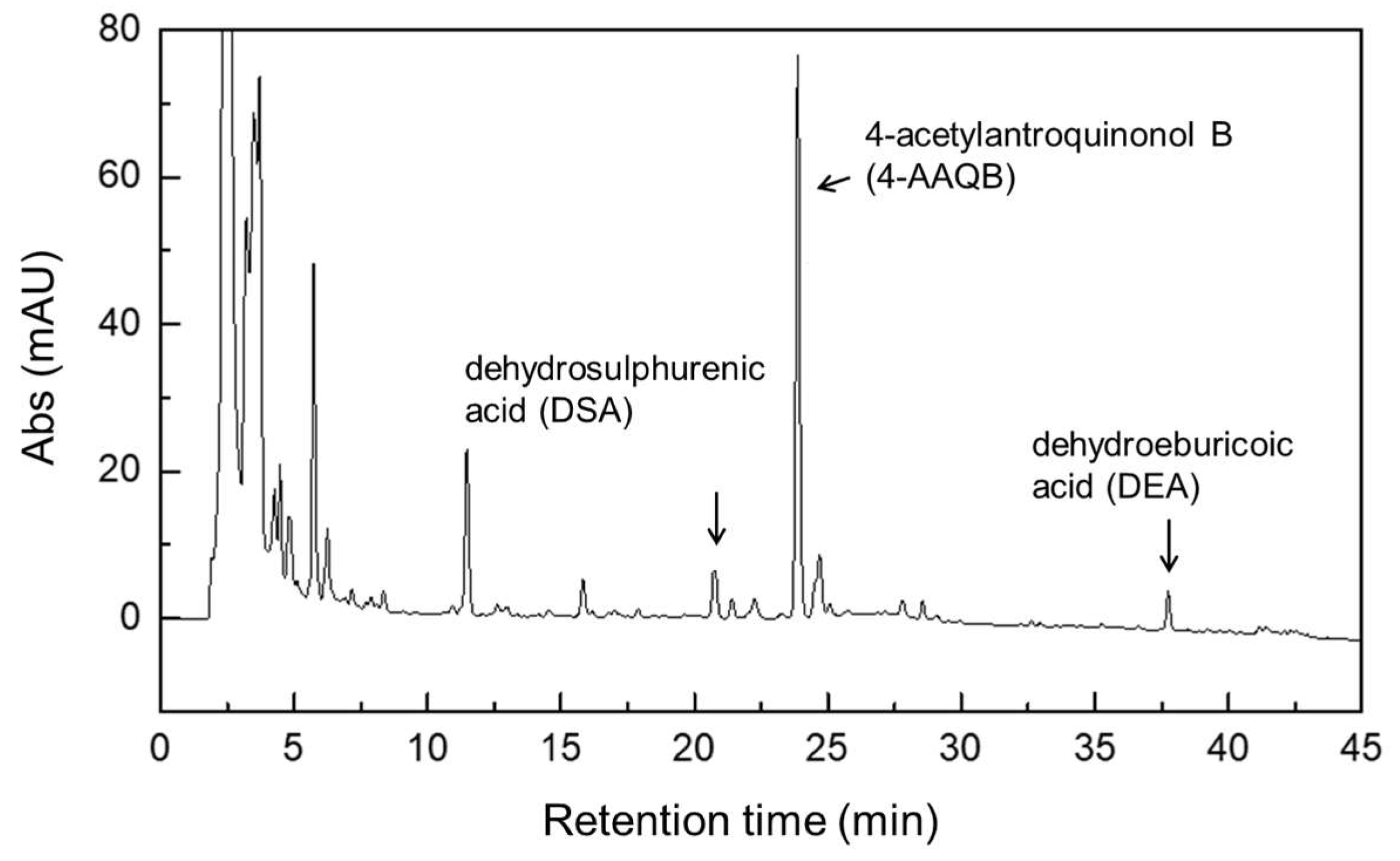
2.6. Extraction and Analysis Method of Active Components of Antrodia cinnamomea Mycelium

The analysis method was based on the methods of Jiang and Geethangili and modified [19,20]. The solid-state fermentation product was placed in a 60 °C oven and dried for 24 h. Then, it was ground into powder. It was extracted with 10 times the volume of methanol in a 60 °C water bath at 150 rpm for 2 h. The extract was filtered through a 0.45 μm filter membrane for the HPLC analysis. The HPLC analysis was performed using a J’sphere ODS-M80/S-4 μm/8 nm/250 × 4.6 mm l.D. column. The mobile phase consisted of solvent A (acetonitrile) and solvent B (ultrapure water + 0.1% formic acid). The gradient elution was as follows: 45% A and 55% B for 0–40 min, 100% A and 0% B for 40–45 min, and 45% A and 55% B for 45–55 min. The flow rate was 1 mL/min and the UV wavelength was 254 nm. The sample injection volume was 20 μL. For the quantitative analysis, purified 4-AAQB, DEA, and DSA, each with a purity of over 99%, were used as the standards. The calibration curves for each of the identified compounds were prepared using known concentrations of 4-AAQB, DEA, and DSA. The yield of each compound in the extract was calculated based on the area under the curve (AUC) in the HPLC chromatogram.

2.7. Magnesium Content Determination Method

The magnesium assay kit (Dialab Trade GmbH, Neudorf, Austria) was used. An amount of 1 μL of homogenate and 100 μL of color reagent were mixed and incubated at room temperature for 5 min, allowing xylidyl blue to chelate the magnesium ion in an alkaline environment and form a water-soluble purple-red compound. The absorbance at 520 nm was measured after the reaction.

2.8. β-1,3-Glucan Analysis Method

β-1,3-glucan is a polysaccharide and its extraction method was modified from Lin and Chen [21]. An amount of 0.05 g of Antrodia cinnamomea mycelium powder was extracted with 0.5 mL of ultrapure water at 95 °C for 1 h, shaking for 3 s every 10 min. The extract was centrifuged at 10,000× g for 20 min and the supernatant was collected. An amount of 0.1 mL of 20% TCA was added and incubated at 4 °C for 30 min to precipitate the protein, then centrifuged at 10,000× g for 10 min before the supernatant was collected. An amount of 1.5 mL of 95% ethanol was added and incubated at 4 °C for 12 h to precipitate the polysaccharide, then centrifuged at 6000× g for 15 min before the supernatant was discarded. The precipitate was dried at 55 °C and became the polysaccharide extract. The β-1,3-glucan analysis used aniline blue, a fluorescent dye that reacts specifically with β-1,3-D-glucan. The dried polysaccharide extract was dissolved in 0.5 mL of glycine-NaOH buffer (pH 9.5). An amount of 60 μL of polysaccharide solution and 210 μL of 0.033% aniline blue (in pH 9.5 glycine-NaOH buffer) were mixed and incubated at 55 °C in the dark for 30 min, then at room temperature in the dark for another 30 min. The absorbance was measured using an HPLC fluorescence detector (excitation wavelength 400 nm, absorption wavelength 460 nm) and the β-1,3-glucan content was calculated by referring to the standard curve, using curdlan as the standard [22].

2.9. Animal Experiment Grouping and Dose

Six-week-old male SD rats were purchased from Lesco Biotechnology Co., Ltd. (Taipei, Taiwan), with seven per group and a total of 56 rats raised at a temperature of 23 ± 1 °C, a light cycle from 8:00 to 20:00 and ad libitum access to food and water. The rats were pre-fed until they reached a weight of 300–350 g and then grouped and implanted with osmotic pumps. The dose conversion was based on the US FDA announcement, taking a 60 kg adult as the reference and multiplying by 6.25 times the recommended intake per kilogram of body weight per day for humans as the dose for rats, as well as by referring to Wang et al., and viewing it from a product development-oriented perspective [10], the daily dose of Antrodia cinnamomea mycelium product required for adults was set at 1.6 g. The UM 1 X group and the DM 1 X group both required a daily dose of 1.6 g for adults, while the daily dose per kilogram of rat required was calculated as 0.198 g. The DM 3 X group required a daily dose of 4.8 g for adults, while the daily dose per kilogram of rat required was calculated as 0.594 g. The F-E group required a daily dose of 0.08 g for adults, while the daily dose per kilogram of rat required was calculated as 0.014 g. The DM-E 3 X group required a daily dose of 0.18 g for adults, while the daily dose per kilogram of rat required was calculated as 0.024 g. The DM-W 3 X group required a daily dose of 2.88 g for adults, while the daily dose per kilogram of rat required was calculated as 0.374 g.

2.10. Surgical Operation for Establishing Alzheimer’s Disease Model Rats

A micro-osmotic pump, tube, and infusion needle, all sourced from RWD Life Science Inc. (San Diego, CA, USA), were utilized to induce Alzheimer’s-like symptoms in rats. The infusion rate was set at 0.125 μL/h. The Aβ40 solution was prepared by dissolving the peptide in 35% acetonitrile and 0.1% trifluoroacetic acid. Rats were anesthetized using animal gas anesthesia and subsequently positioned using a stereotaxic instrument. The left skull was exposed and drilled (relative to the bregma; 0.8 mm posterior, 1.4 mm lateral) according to the atlas of Paxinos and Watson [23] and using a stereotaxic frame (Narishige, Tokyo, Japan). The infusion kit was inserted to a depth of 4.0 mm below the skull surface and secured with biological adhesive pads. The micro-osmotic pump, filled with 95 μL of the Aβ40 solution, was then subcutaneously implanted behind the neck of the rats. It released 4.9–5.5 nmol of Aβ40 into the brain for 28 days. The rats were returned to their cages and began experimental feeding the day post-surgery [10].

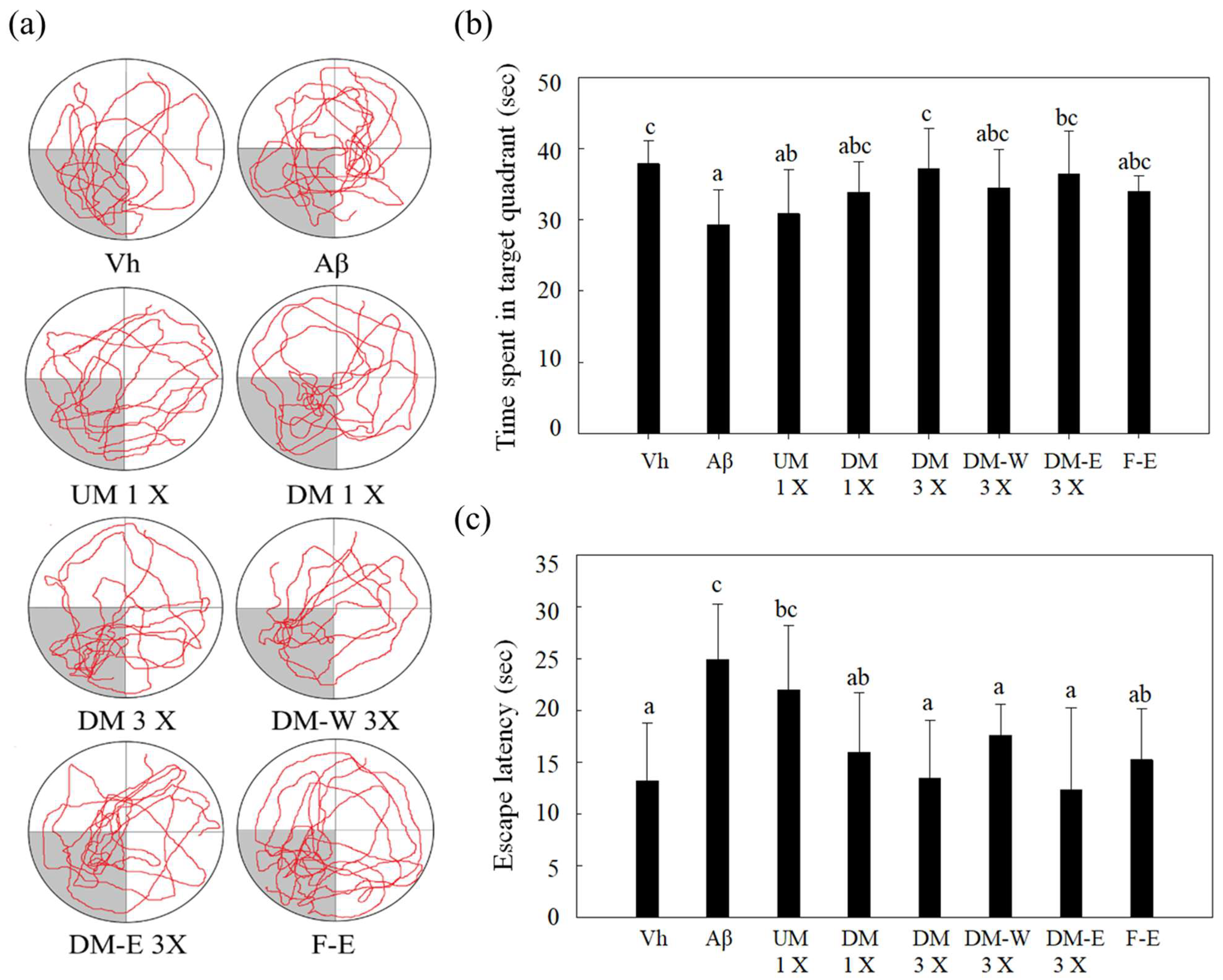
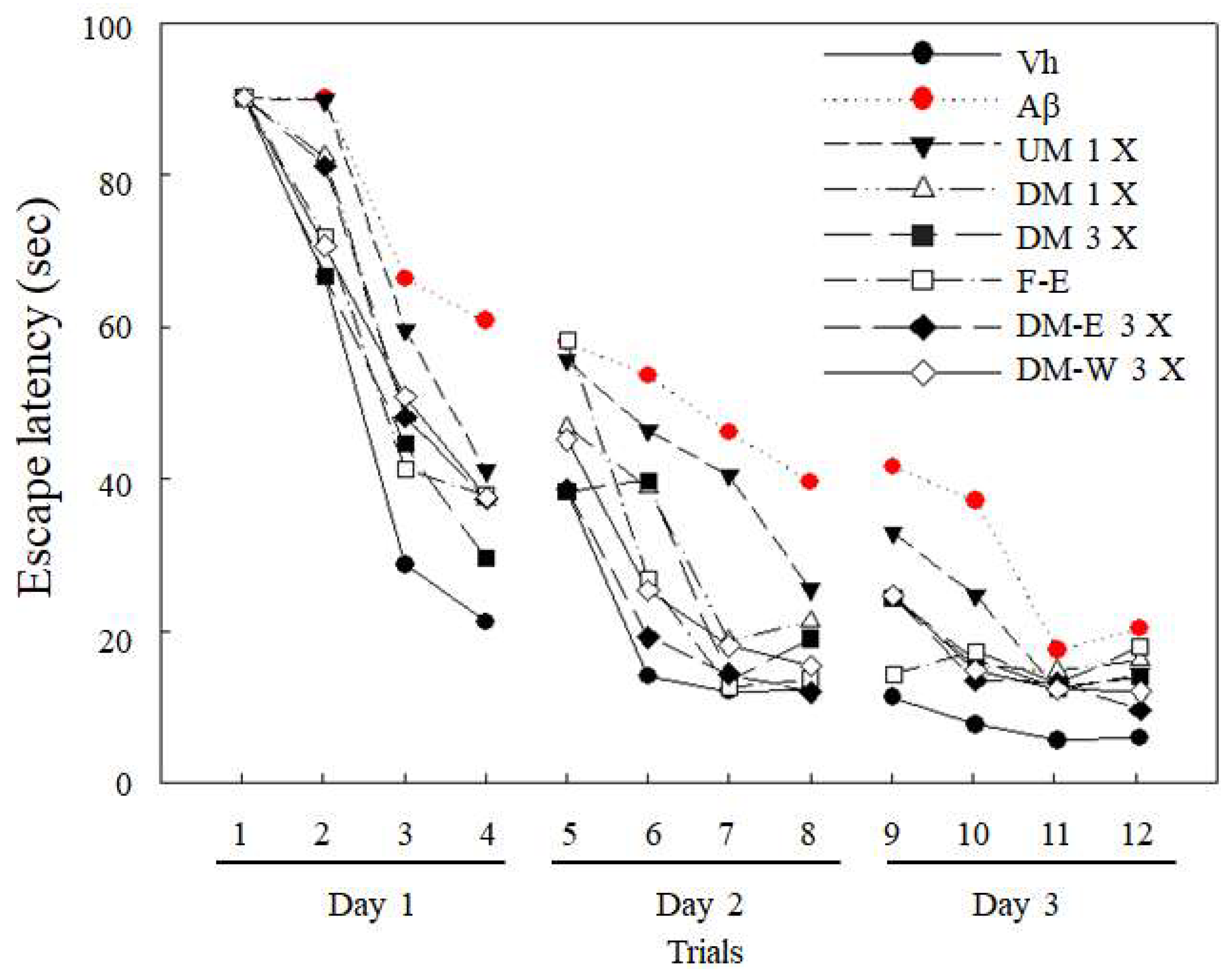
2.11. Water Maze Test

The water maze memory–learning ability test was performed on day 24 after surgery, including a reference memory task, spatial probe trace, and working memory task. The diameter of the swimming pool was 168 cm, the water level height was 40 cm, and the swimming pool contained a movable rest platform (escape platform). The diameter of the platform was 10 cm and the height was 35 cm. The swimming pool was divided into four quadrants (I, II, III, and IV) and five entry points were set up. The rest platform was placed in the center of quadrant III. During the test, a camera was installed directly above the center of the swimming pool to record the swimming path of the experimental animals [10].
The reference memory task was performed from day 24 to day 26 after surgery. The rest platform was fixed in quadrant III. The rats entered four entry points randomly in sequence with their heads facing outwards. They were trained four times a day for 90 s each time; if they found the rest platform within 90 s, they were allowed to rest for 30 s, then returned to the cage for 30 s before proceeding to the next test; if they did not find the rest platform within 90 s, they were placed on the rest platform and rested for 30 s, then removed from the swimming pool for 30 s before proceeding to the next training session. A total of 12 tests were performed. The reference memory task can be used to observe the long-term memory ability of rats [10].
Spatial probe test was performed immediately after the reference memory task on day 26. The rest platform was removed from the swimming pool and the rats entered from entry point I in quadrant I. They swam for 90 s, while recording how long they stayed in quadrant III where the rest platform was originally placed during the reference memory task and their entire swimming path.
The working memory task was performed on day 27 after surgery. The rest platform was placed in a different quadrant (quadrant IV). They were trained five times, entering five entry points randomly in sequence with their heads facing outwards. Each time for 90 s; if they found the rest platform within 90 s, they were allowed to rest for 15 s, then removed from the swimming pool for 60 s before proceeding to the next test. If they did not find the rest platform within 90 s, they were placed on the rest platform and rested for 15 s, then removed from the swimming pool for 60 s before proceeding to the next training session. The test results were averaged and the differences between groups were statistically analyzed. The first training session was cognitive training and was not included in the calculation. The working memory task can be used to observe the short-term memory ability of rats [10].

2.12. Animal Sacrifice and Brain Tissue Processing

After fasting for 12 h, the animals were sacrificed via carbon dioxide asphyxiation and their brain tissue was collected, rapidly frozen in liquid nitrogen, and then cut 2–3 mm from the right ventricle and immersed in 10% formalin for paraffin embedding and slicing (3–5 μm/slice) for subsequent tissue immunostaining. The remaining brain tissue was divided into the cortex and hippocampus regions according to the Groswisky method [24], and then the cortex was divided into the left and right hemispheres. The hippocampus and the right cortex were added with 700 μL of PBS buffer (0.276% Na2HPO4·12H2O, 0.035% NaH2PO4·2H2O, and 0.9% NaCl) for homogenization. After homogenization, it was centrifuged at 15,000× g for 30 min at 4 °C and the supernatant was stored at −80 °C for the subsequent protein analysis and ion determination in the brain tissue.

2.13. Protein Content Analysis

The commercially available BCA protein assay kit (23225, Thermo Fisher Scientific, Rockford, IL, USA) was used. The protein reduced divalent copper ions (Cu2+) to produce Cu+ was used, which specifically colored BCA to quantify it. The BCA protein assay kit contained reagent A (BCA), reagent B (CuSO4), and standard bovine serum albumin (BSA). Reagent A and reagent B were mixed evenly at a ratio of 50:1. An amount of 10 μL of standard solution or tissue homogenate was mixed with 200 μL of BCA reagent and incubated at 37 °C for 30 min. The absorbance at 562 nm was read, a standard curve was made with the standard product, and the protein concentration of the sample was calculated by fitting the absorbance into the standard curve.

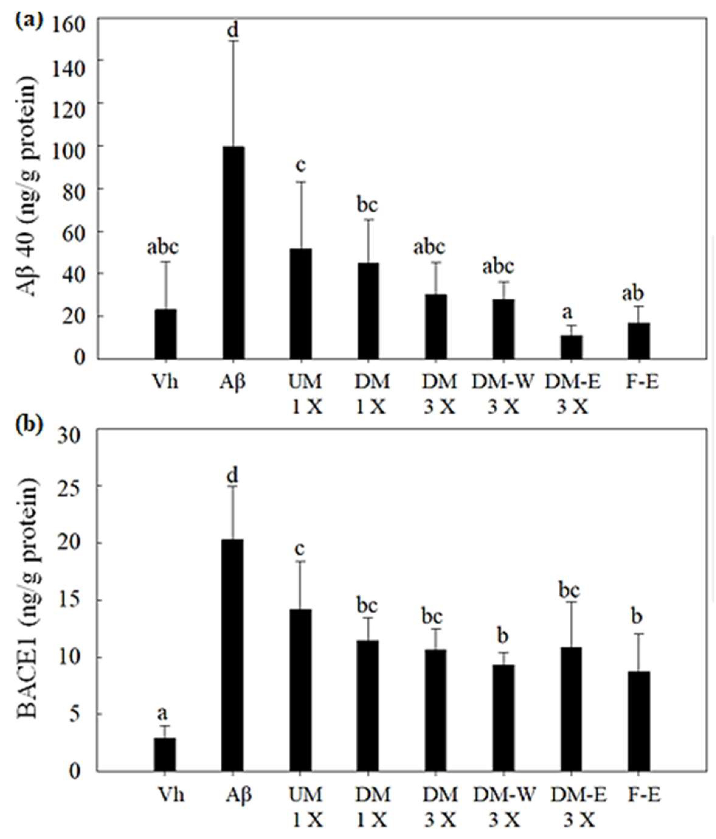
2.14. ELISA Analysis of Target Protein

First, 100 μL of protein standard or tissue homogenate was added to a 96-well plate and reacted at 37 °C for 60 min, then removed. An amount of 100 μL of primary antibody was added and reacted at 37 °C for 60 min, then removed and washed three times. An amount of 100 μL of secondary antibody (pierce goat anti-rabbit IgG, (H + L), peroxidase conjugate anti-body, pierce Biotechnology, 1:50,000; pierce goat anti-mouse IgG, (H + L), peroxidase conjugated anti-body, Pierce Biotechnology, 1:50,000) was added and reacted at 37 °C for 60 min, then removed and washed six times. An amount of 100 μL of color reagent 3,3’5,5’-tetramethylbenzidine (TMB) was added and reacted at 37 °C for 15 min, then 50 μL of 2 N sulfuric acid was added to stop the reaction. The absorbance at 450 nm was measured using a full-wavelength spectrophotometer.

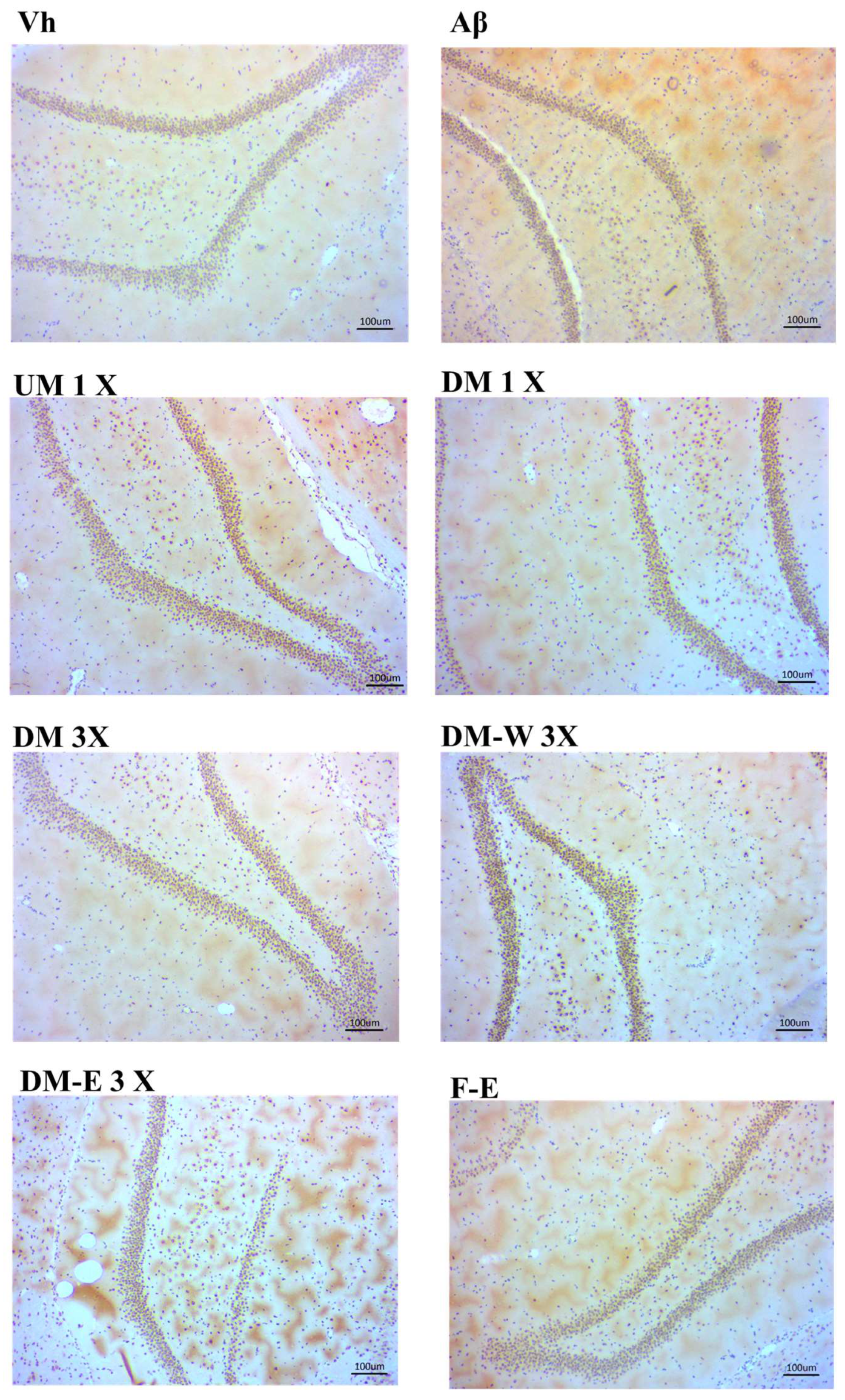
2.15. Immunohistochemical Stain of RAGE in Hippocampus

The brain tissue was fixed in 10% formalin at pH 7.4. The brains were blocked, and serial 35 μm thick frozen sections cut on a sledge microtome were collected sequentially and without interruption into the wells. The brain sections were processed for immunohistochemical staining with the RAGE antibody according to our previous study [25].

2.16. Statistical Analysis

All data are presented as mean ± SD. The data were analyzed via one-way ANOVA using SPSS 20.0, followed by Duncan’s multiple range test for the comparison of differences between groups. p < 0.05 indicates significant difference.

3. Results

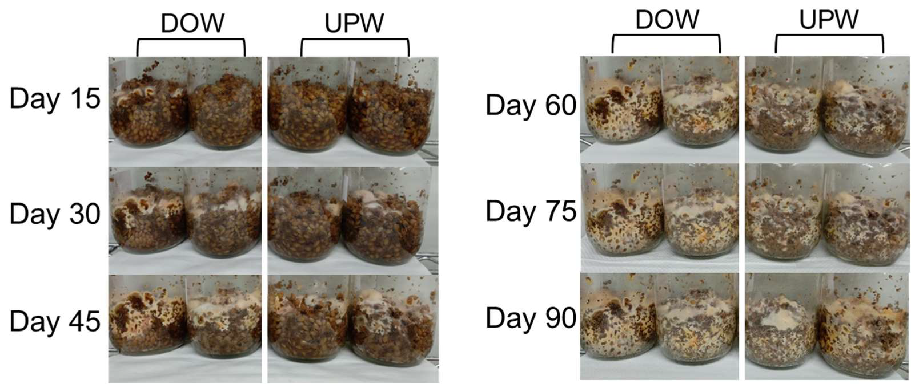
3.1. Growth Variations of A. cinnamomea NTTU 206 Solid-State Fermented Mycelia Cultured in DOW and UPW

This study initially investigates the growth variations of A. cinnamomea NTTU 206 mycelium cultured in DOW and UPW, respectively. The results of Figure 1 indicate that in media supplemented with DOW, A. cinnamomea NTTU 206 mycelium demonstrated more robust growth characteristics. Notably, during the day 60–70 cultivation period, the group cultured with DOW exhibited a faster growth rate and higher growth density compared to the UPW group, which showed a slower growth trajectory. Importantly, by day 60–70, the mycelium in the DOW group had fully colonized the entire solid medium, while the UPW group required as long as 90 days to achieve the same level of colonization. These findings suggest that DOW effectively enhances the growth of A. cinnamomea NTTU 206 in solid-state fermentation, which could potentially further stimulate the production of its secondary metabolites and bioactive compounds. This study further employed the HPLC analysis to determine the concentration of key functional components in the A. cinnamomea NTTU 206 solid-state fermentation product. Figure 2 presents the HPLC chromatograms of three major functional components: 4-AAQB, DSA, and DEA.

3.2. Effects of Adding DOW and MgCl2 on the Functional Components Produced by Fermentation of A. cinnamomea NTTU 206

The effect of adding DOW and MgCl2 on the fermentation of A. cinnamomea NTTU 206 strain to produce functional components was investigated via the plate culture method. The results are shown in Table 1. The production of 4-AAQB in the MgCl2 group was 0.36 mg/plate, which was significantly higher than that in the UPW and DOW groups (p < 0.05). The production of DSA and DEA in the DOW group was significantly higher than that in the UPW and MgCl2 groups (p < 0.05), as shown in the 82.57 mg/plate and 98.96 mg/plate, respectively. The functional components produced by solid-state fermentation culture are shown in Table 2. The production amounts of 4-AAQB, DSA, and DEA in the DOW group were significantly higher than those in the UPW and MgCl2 groups (p < 0.05) as 719 mg/kg, 7184 mg/kg and 2874 mg/kg, respectively. However, although the production of β-1,3-glucan tended to increase in the DOW group, there was no significant difference among the three groups (p > 0.05). The results show that DOW can significantly increase the production of 4-AAQB, DSA, and DEA, but its main functional components do not come from MgCl2 in ocean water, but may be from other minerals and nutrients.

3.3. Effects of A. cinnamomea NTTU 206 Solid-State Fermented Mycelia on Brain-Infused Aβ40 Rats on Memory Test and Spatial Learning Ability

In this study, the Aβ40 solution was continuously infused into the brains of rats to induce the Alzheimer’s disease model of memory impairment. Furthermore, we explored the effect of the DOW-cultured A. cinnamomea NTTU 206 solid-state fermentation mycelium on the memory and learning ability of Alzheimer’s disease rats. Firstly, the memory ability was evaluated with the water maze. The reference memory test is to set a platform in the third quadrant of the swimming pool and record the time spent by the rats searching for the escape platform as a way to evaluate the learning and memory ability of the rats. The results are shown in Figure 3. On the first day, there was no significant difference (p > 0.05) in the time spent by the experimental animals searching for the escape platform in each group. The results of the memory test on the second day showed that the Aβ group took longer to search for the escape platform than the Vh group, and there was a significant difference (p < 0.05). Although the search time of other groups tended to decrease, there was no significant difference with the Aβ group (p > 0.05). The results of the swimming test on the third day showed that the UM 1 X, DM 1 X, and DM-E 3 X groups could significantly reduce the time spent searching for the escape platform (p < 0.05).
After the reference memory test, the escape platform was immediately removed for a spatial detection test, and the swimming path and time were recorded. This test helps to identify the spatial detection ability of the experimental animals in the water maze and can present the true state of the memory and learning performance. After the escape platform is removed, the time and path of the experimental animals wandering in the quadrant of the original escape platform can be regarded as one of the memory learning indicators of the spatial detection test. The results are shown in Figure 4. The swimming path of the Vh group was concentrated in the quadrant of the original escape platform. The Aβ group searched without a target, and their swimming paths were relatively scattered, while the swimming paths of the other groups fed with test substances were more concentrated in the target quadrant. Swimming time in the target quadrant was significantly higher in the Vh, DM 3 X, and DM-E 3 X groups than in the Aβ group (p < 0.05), increasing by 29%, 26.8%, and 24%, respectively. The UM 1 X, DM 1 X, DM-W 3 X, and F-E groups increased by 5.4%, 15.6%, 16.1%, and 17.6%, respectively. These results proved that feeding DM 3 X and DM-E 3 X can significantly improve the spatial learning and memory abilities of AD rats.
On the 25th day after the operation, the working memory test was carried out. The escape platform was placed in different quadrants (the fourth quadrant), and the rats were tested from five different entry points to test the short-term memory ability of the rats. As shown in Figure 4, the total time spent searching for rest platforms in the Vh, DM 1 X, DM 3 X, DM-W 3 X, DM-E 3 X, and F-E groups was significantly lower than that in the Aβ group (p < 0.05). They decreased by 47.1%, 36%, 45.8%, 38.9%, 50%, and 29%, respectively. The UM 1 X group cultivated in ultrapure water only decreased by 11.6%, which proved that DM 1 X, DM 3 X, DM-W 3 X, DM-E 3 X, and F-E can significantly improve the short-term memory ability of AD rats.

3.4. Effects of A. cinnamomea NTTU 206 Solid-State Fermentation Mycelia on the Expression of Aβ40 and BACE1 Proteins in the Hippocampus of Rats with Brain Infusion of Aβ40

The Aβ production pathway involves β- and γ-secretases. APP is cleaved by β-secretase to produce the soluble sAPPβ peptide and β-CTF, and then the remaining fragments are cleaved by γ-secretase to produce AICD and Aβ [26]. In the AD brains, BACE activity and protein expression are significantly increased [27]. Therefore, this study explored the effect of feeding the DOW-cultured A. cinnamomea solid-state fermentation products on the expression of Aβ40 and BACE1 in the hippocampus of AD rats. As shown in Figure 5, the expression of Aβ40 in the Aβ group was significantly higher than that in the Vh group due to the continuous infusion of Aβ40 (p < 0.05), indicating that there was an obvious deposition of Aβ40 in the hippocampus. Feeding the test substances UM 1 X, DM 1 X, DM 3 X, DM-W 3 X, DM-E 3 X, and F-E all significantly reduced the deposition of Aβ40 (p < 0.05). Compared with the Aβ group, they were reduced by 48%, 55%, 69.6%, 71.4%, 88.8%, and 82.6%, respectively. As shown in Figure 5, the expression of BACE1 in the Aβ group was significantly higher than that in the Vh group (p < 0.05). However, feeding the test substances UM 1 X, DM 1 X, DM 3 X, DM-W 3 X, DM-E 3 X, and F-E significantly decreased the expression of BACE1 (p < 0.05). Compared with the Aβ group, the reductions were 30.08%, 43.56%, 47.48%, 54.07%, 46.58%, and 56.91%, respectively.

3.5. Effects of A. cinnamomea NTTU 206 Solid-State Fermented Mycelia on the Expression of sAPPα, p-tau, and RAGE Proteins in the Hippocampus of Rats with Brain Infusion of Aβ40

APP has two cleavage pathways: the non-amyloid-like pathway and the amyloid-like pathway. The non-amyloidogenic pathway is that, after APP is cleaved by α-secretase, sAPPα and α-CTF will be produced. sAPPα can stabilize the resting membrane potential of cells, help synapse formation, and protect nerves [26]. As shown in Figure 6, the expression of sAPPα in the Aβ group was only slightly decreased compared with the Vh group, but there was no significant difference (p > 0.05). The feeding test substance group also had a tendency to increase the expression of sAPPα compared with the Aβ group, but there was no significant difference (p > 0.05).
The tau protein is a highly soluble microtubule-associated protein (MAP) whose main function is to maintain the stability of axons and microtubules. The hyperphosphorylated tau protein, a major component of neurofibrillary tangles (NFTs), is associated with neurological degeneration and cognitive decline [28]. As shown in Figure 6, the expression of p-tau in the Aβ group was significantly higher than that in the Vh group (p < 0.05). The DM 1 X, DM 3 X, DM-E 3 X, and F-E groups could significantly reduce the expression of p-tau enhanced by the Aβ40 infusion (p < 0.05). However, the effect of the UM 1 X and DM-W 3 X groups was not significant (p > 0.05). It was shown that A. cinnamomea NTTU 206 cultured with DOW can improve the occurrence of neurotangles by inhibiting the p-tau protein.
RAGE binds to intracellular Aβ, which results in a neurotoxic or pro-inflammatory response [29,30]. As shown in Figure 6, each test group significantly reduced the expression of RAGE compared with the Aβ group (p < 0.05). This indicates that after administering the test substance, the expression of RAGE in the brain was significantly reduced, which may reduce the binding of RAGE to Aβ and, in turn, reduce its inflammatory response. To validate these molecular-level interactions, immunohistochemistry (IHC) staining was performed as an additional analytical method. The IHC results corroborated our initial findings, further substantiating the observed downregulation of RAGE expression in the test groups (Figure 7). RAGE is prominently expressed around the hippocampal regions, a crucial area for memory and learning. This study confirmed that rats infused with Aβ40 displayed a significant accumulation of the RAGE protein in their hippocampus, indicating that Aβ40 induces RAGE expression. However, A. cinnamomea NTTU 206 fermentation products, which are rich in anti-inflammatory components, showed a significant reduction in RAGE expression in the test groups. The A. cinnamomea NTTU 206 products cultured with DOW demonstrated noticeable anti-inflammatory effects, exhibiting a level of RAGE inhibition comparable to that achieved by the extracts from Antrodia cinnamomea fruiting bodies. These findings further underline the potential efficacy of DOW-cultivated Antrodia cinnamomea mycelia as an anti-inflammatory agent.

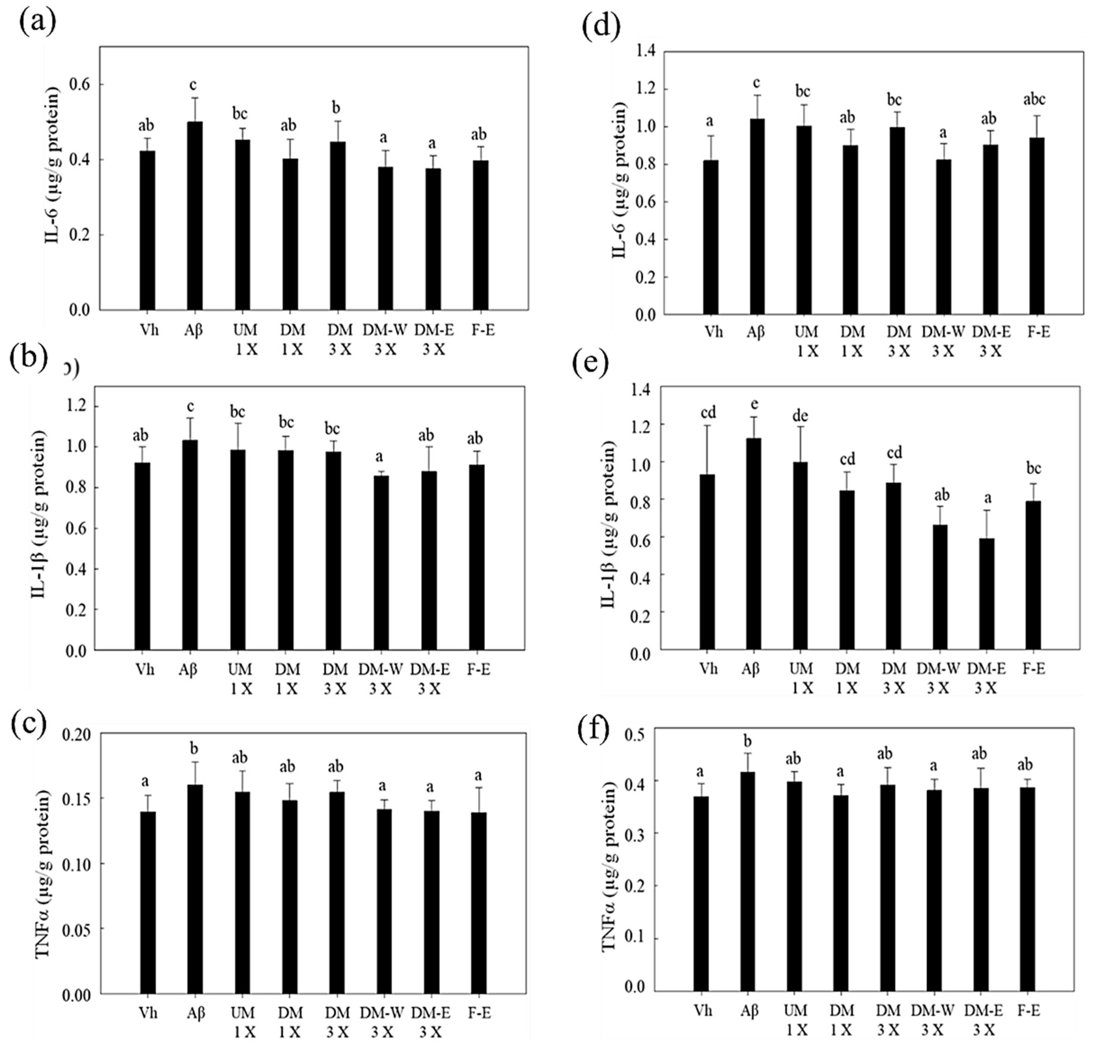
3.6. Effects of A. cinnamomea NTTU 206 Solid-State Fermentation Mycelium on the Expression of IL-6, IL-1β, TNF-α Proteins in the Hippocampus of Rats with Brain Infusion of Aβ40

The levels of pro-inflammatory cytokines such as IL-1β, IL-6, IL-10, TNF-α, and TGF-β were significantly elevated in the brain tissue and CSF of AD patients [31,32,33]. As shown in Figure 8a–c, Aβ40 induced a significant increase in the expression levels of the IL-6, IL-1β, and TNF-α proteins in the brain tissue (p < 0.05), indicating that the infusion of Aβ40 can induce inflammation in the hippocampus of rats. The mycelia of A. cinnamomea cultured in ultrapure water (UM 1 X group) had the effect of reducing the expression of the IL-6, IL-1β, and TNF-α proteins, but could not achieve a significant reduction (p > 0.05). The mycelia of A. cinnamomea cultured in deep ocean water (DM 1 X group) could significantly reduce the expression levels of the IL-6, IL-1β, and TNF-α proteins (p < 0.05). It shows that DOW may increase the functional compounds of A. cinnamomea or compound organic minerals to enhance the anti-inflammatory effect. Regarding the anti-inflammatory evaluation of the extracts, the water extracts (DM-W 3X group) significantly decreased the expression levels of the IL-6, IL-1β, and TNF-α proteins (p < 0.05). The ethanol extract (DM-E 3X group) significantly decreased the expression of the IL-6 and IL-1β proteins (p < 0.05). It is shown that both the water extract and the alcohol extract of Antrodia cinnamomea mycelium have functional components that inhibit the inflammatory response induced by Aβ in the hippocampus.

3.7. Effects of A. cinnamomea NTTU 206 Solid-State Fermentation Mycelia on the Expressions of IL-6, IL-1β, and TNF-α Proteins in the Cerebral Cortex of Rats with Brain Infusion of Aβ40

Figure 8d–f shows that the protein expressions of IL-6, IL-1β, and TNF-α were significantly increased in the Aβ group compared to the Vh group (p < 0.05). This indicates that Aβ40 infusion can cause inflammation in the cerebral cortex of rats. Among the groups that were fed with test substances, the UM 1 X group did not significantly reduce the expression levels of the IL-6, IL-1β, and TNF-α proteins (p > 0.05). However, the DM 1 X group could significantly reduce the expression of the IL-6 protein (p < 0.05). Both the water extract (DM-W 3 X) and the alcohol extract (DM-E 3 X group) could significantly reduce the expression of the IL-6, IL-1β, and TNF-α proteins (p < 0.05). This suggests that the water-soluble polysaccharides and 4-AAQB, DEA, and DSA in the mycelium of A. cinnamomea may be the functional components that inhibit the inflammatory response induced by Aβ in the cerebral cortex. F-E, the wild A. cinnamomea fruiting body extracts, can significantly reduce the expression levels of the IL-6, IL-1β, and TNF-α proteins in the cerebral cortex (p < 0.05). Therefore, it can be observed that the mycelium cultured with DOW can achieve the same effect as the wild A. cinnamomea fruiting body extract.

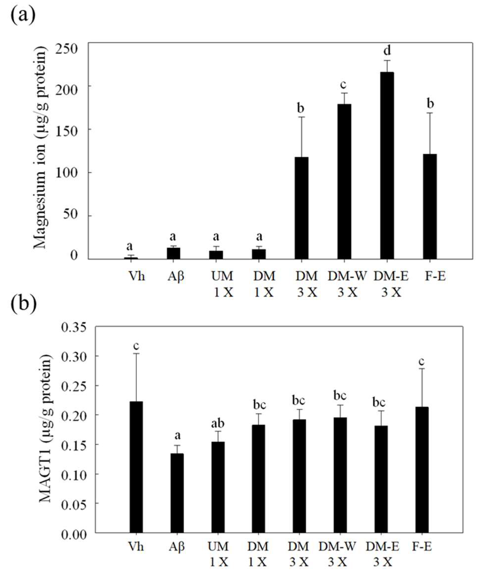
3.8. Effect of A. cinnamomea NTTU 206 Solid-State Fermented Mycelium on Magnesium Uptake in the Hippocampus of Rats with Brain Infusion of Aβ40
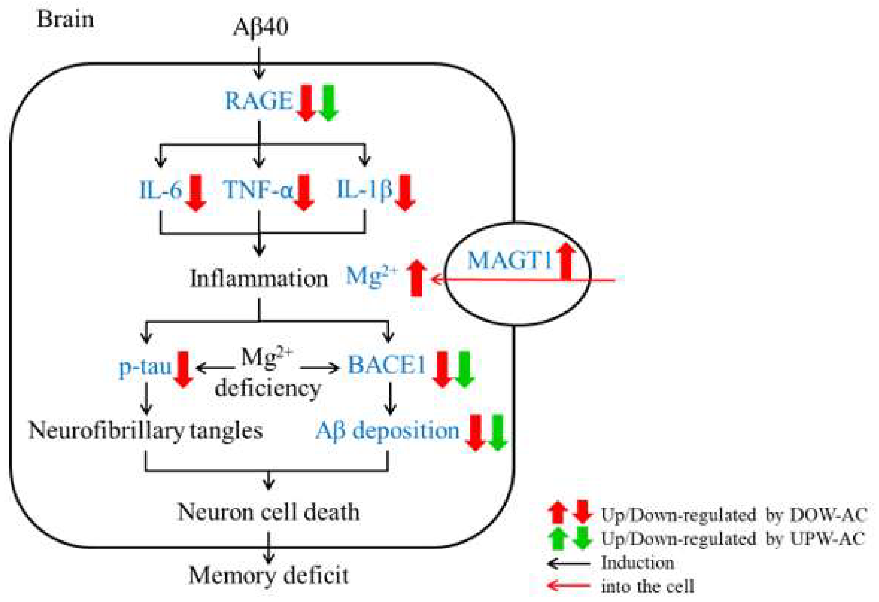
Previous studies have demonstrated that Cordyceps cicadae can chelate and absorb magnesium ions from deep ocean water and convert them into organic forms to increase bioabsorption rates in the brain. Magnesium ions in the brain can reduce the expression of phosphorylated tau proteins, thereby improving AD [34]. Therefore, this study explores the effects of the DOW-cultured mycelium of A. cinnamomea on the concentration of magnesium ions in the hippocampus of AD rats. As shown in Figure 9, the DM 3 X, DM-W 3 X, DM-E 3 X, and F-E groups all significantly increased the concentration of magnesium ions in the hippocampal tissue (p < 0.05).
The MAGT1 protein transports magnesium ions from the outside of the cell membrane to the inside, increasing the magnesium ion content in the cell. Magnesium ions can increase the permeability of the blood–brain barrier and promote the clearance of Aβ [35]. As shown in Figure 9, the expression of the MAGT1 protein in the Aβ group was significantly lower than that in the Vh group (p < 0.05), indicating that the infusion of Aβ40 may increase the uptake of magnesium ions in the rat hippocampus. Mycelia cultured with UPW (UM 1X group) did not significantly increase the expression of the MAGT1 protein (p > 0.05). However, the DM 1 X, DM 3 X, DM-W 3 X, DM-E 3 X, and F-E groups could significantly increase the expression of the MAGT1 protein (p < 0.05). The mycelia of A. cinnamomea cultured via DOW and its extracts (DM 1 X, DM 3 X, DM-W 3 X, and DM-E 3 X groups) can significantly increase the expression of MAGT1 and reduce the effect of Aβ production. F-E does not contain DOW magnesium ions but still increases MAGT1 expression.

4. Discussion

DOW is rich in ions required for various physiological functions. During the fermentation process with the added DOW, the ions of DOW accumulate in the fermentation product, thereby improving its functional composition and endowing the minerals of DOW [36]. Previous studies in our laboratory have shown that DOW can effectively increase the production of total triterpenoids and total polysaccharides by 8% and 462%, respectively, in the fermented mycelium of A. cinnamomea [8]. In the present study, we discovered that the use of DOW significantly enhances the generation of A. cinnamomea mycelia compared to ultrapure water. Although there was no substantial increase in the production of β-1,3-glucan, DOW markedly elevated the concentration of three key anti-inflammatory constituents. Specifically, in terms of 4-AAQB production, DOW led to a 1.16-fold enhancement relative to ultrapure water and a 1.12-fold enhancement compared to magnesium chloride. For DSA production, cultivation in DOW improved the yield by 2.18-fold over ultrapure water and further increased it by 1.37-fold over magnesium chloride. Concerning DEA, the inclusion of magnesium chloride led to a 1.74-fold increase relative to ultrapure water, whereas substituting it with DOW led to an additional 1.34-fold improvement. These findings suggest that while magnesium chloride in DOW indeed serves as a pivotal element in boosting the fermentation and bioactive component yield of A. cinnamomea, other nutrients and minerals in DOW may also offer supplementary beneficial effects. As a result, the target bioactive components produced in DOW surpassed those generated using magnesium chloride alone. Therefore, DOW holds considerable promise for application in the fermentative production of A. cinnamomea mycelia. Past studies have confirmed that magnesium ions absorbed by Cordyceps cicadae contribute to the uptake efficiency of magnesium ions in the brain. Adding DOW-cultured Cordyceps cicadae can effectively supplement the magnesium ions needed by the human body and inhibit inflammation, thereby preventing the development of AD [34]. Consequently, this study suggests that the Antrodia cinnamomea mycelium cultured in DOW may also hold potential for improving AD, potentially through the enhanced absorption of magnesium.
Aβ formation is one of the pathological hallmarks of AD. APP is cleaved by TACE and BACE to form Aβ40 [37,38,39,40,41]. Past studies have compared the effects of mycelia and the fruiting bodies of A. cinnamomea on improving the memory and learning ability of AD rats. The results indicated that the fruiting bodies had a better effect than mycelia [10]. However, this study found for the first time that the mycelium of A. cinnamomea cultured via DOW can significantly improve the memory and learning ability of AD compared to the mycelium cultured via UPW. This improvement effect can reach a level similar to the fruiting body extract. This is an important breakthrough for the development of A. cinnamommea mycelium as a functional product for the prevention of Alzheimer’s disease.
In this study, Aβ40 was injected into the brain of rats for 28 consecutive days to induce Alzheimer’s disease rats with memory and learning impairment. Therefore, memory-learning ability is an important behavioral test to assess improvement in AD. In various water maze tests, including reference memory tests for long-term memory, working memory tests for short-term memory, and probe tests for spatial detection, A. cinnamomea mycelium cultured with DOW has a better effect than mycelium cultured in UPW. Importantly, mycelium cultured with DOW was similar to the fruiting bodies in improving the memory–learning ability. Both the ethanol extract and the water extract of mycelia can improve the memory and learning ability of AD rats. It shows that it must contain functional ingredients that can improve AD. According to the analysis results, the main functional component of the water extract is β-glucan, and the functional components of the ethanol extract include 4-AAQB, DEA, and DSA. These components may be the main functional components of mycelium against Aβ40-induced brain damage and memory impairment.
APP is cleaved by BACE and the remaining fragment is cleaved by γ-secretase to generate Aβ [26,42]. Neuropathology occurs when the tau protein is defective and no longer normally stabilizes microtubules [43]. However, APP is cleaved by α-secretase to form sAPPα and α-CTF. sAPPα can stabilize the resting membrane potential of neuron, help synapse formation, and protect the nerves [26]. Therefore, the inhibition of BACE and p-tau can slow down the formation and deposition of Aβ and inhibit the development of AD. The cleavage of APP towards the formation of sAPPα contributes to neuronal protection. In this study, feeding the A. cinnamomea NTTU 206 solid-state fermentation mycelia, supplemented with UPW and DOW for the fermentation culture, can reduce the expression of the Aβ40, BACE1, and p-tau proteins in the hippocampus of rats. The alcohol extract of mycelium can reduce the expression of Aβ40 and p-tau, while the water extract can reduce the expression of BACE1. In this study, the mycelium of A. cinnamomea cultured via DOW also tended to increase the protein expression of sAPPα. These results showed that A. cinnamomea mycelium can reduce the deposition of Aβ40 mainly by inhibiting the expression of AD-related risk factors and then improving the subsequent oxidative inflammation. Although the mycelium of A. cinnamomea cultivated via DOW has many different active ingredients from the fruiting body, it has similar effects. These reasons may come from the minerals of DOW absorbed by the mycelium of A. cinnamomea and the active ingredients (4-AAQB, DEA, and DSA) that were increased after the cultivation with DOW.
Proinflammatory cytokines, such as IL-1β, IL-6, IL-10, and TNF-α, and transforming growth factor-β (TGF-β) are significantly elevated in the tissues and cerebrospinal fluid of AD patients [20,31,32,33]. Inflammatory factors in neurons increase Aβ production and activate microglia-induced inflammation [44]. Intracellular Aβ binds to RAGE, resulting in neurotoxic or pro-inflammatory responses [29,45,46]. Past studies have pointed out that feeding mice with the methanol extract of A. cinnamomea solid-state fermentation mycelium significantly reduces the mRNA expression of IL-6 and iNOS and reduces the expression of TNF-α, NF-κB, and IL-6 [47]. In in vitro cell experiments, 4-AAQB can significantly inhibit the protein expression of iNOS and COX-2 and has good antioxidant and anti-inflammatory activities [14].
4AAQB can significantly reduce the levels of TNF-α and IL-6 in the mouse serum, showing that 4AAQB has good anti-inflammatory activity [48]. Polysaccharides from A. cinnamomea can achieve anti-inflammatory effects by inhibiting NF-κB activation [12]. In this study, feeding the mycelium of A. cinnamomea can reduce the expression of the IL-6, IL-1β, TNF-α, and RAGE proteins in the hippocampus, and the mycelium cultured in DOW has a better anti-inflammatory effect than that cultured in UPW. The expression levels of the IL-6 and IL1β proteins in the hippocampus can be inhibited by the ethanol extract and the water extract. Especially, the water extract has a more significant effect on the inhibition of TNF-α protein expression. The above shows that 4-AAQB, DEA, and DSA in mycelium may have the effect of inhibiting the Aβ40-induced inflammatory response. Inhibiting the inflammatory response can reduce the deposition of Aβ in the hippocampal tissue, thereby inhibiting the neuronal damage in the brain.
In AD animal models, feeding magnesium-containing substances can effectively supplement the lack of magnesium ions in the brain, thereby improving the effect of AD [34]. This study shows that the magnesium ions absorbed by A. cinnamomea mycelia during the fermentation process can help to supplement the lack of magnesium in the hippocampus. The main reason is to increase the expression of the MAGT1 protein on the cell membrane of the hippocampus and increase the uptake of extracellular magnesium ions into the cell. Previous studies have also pointed out that the MAGT1 protein transports magnesium ions from the outside of the cell membrane to the inside of the cell, increasing the content of magnesium ions in the cell. Magnesium ions can increase the permeability of the blood–brain barrier and promote the clearance of Aβ [35]. This study showed that the expression of the MAGT1 protein in the hippocampus of AD rats could be increased by feeding the A. cinnamomea mycelia cultured in DOW, but not the mycelia cultured in UPW. This may also be one of the main reasons why A. cinnamomea mycelia cultivated via DOW is better than that cultivated via UPW in improving AD.
The A. cinnamomea strains used in this study were isolated from the wild fruiting bodies of A. cinnamomea. After adding DOW for the solid-state culture of A. cinnamomea, the mycelium product not only has higher concentrations of 4-AAQB, DEA, DSA, and β-1,3 glucan, but also organic DOW minerals. Therefore, A. cinnamomea mycelium cultured with DOW is different from the traditional mycelium cultured with UPW in the composition of functional components. In the experiment of AD model animals in this study, the mycelium of A. cinnamomea cultured via DOW can achieve the same effect on improving Alzheimer’s disease as in the fruiting body in various evaluation tests, including the memory and learning ability in the water maze (reference memory test, working memory test, and probe test), the inhibition of brain risk factors (p-tau, Aβ40 deposition, and BACE), and inflammatory factors (IL-6, IL-1β, TNF-α, and RAGE).
This study discovered, for the first time, that mycelia of A. cinnamomea cultured via DOW can increase the magnesium ion content and MAGT1 expression in the hippocampus of rats. A. cinnamomea mycelia cultured in DOW completed three regulations in the AD animal model (Figure 10): (1) increasing the protein expression of MAGT1 to supplement the magnesium ion concentration in the hippocampus of AD rats; (2) inhibiting the expression of the p-tau protein to reduce neurofibrillary tangles; and (3) inhibiting the expression of the RAGE protein to inhibit the microglia release of pro-inflammatory factors. The mycelium product of A. cinnamomea cultured in DOW can prevent AD development by inhibiting a series of chain reactions, thereby reducing the expression of BACE and reducing the deposition and formation of Aβ. This study confirmed that the mycelium and the fruiting bodies of A. cinnamomea cultured via DOW have the same effect on improving AD. However, their commercial price differs by 10 times. The mycelium of A. cinnamomea developed in this research can not only reduce the cost, but also help prevent the illegal logging of camphor trees in forests. Furthermore, the mycelium of A. cinnamomea cultured in DOW did not cause toxicity to the liver and kidney in this test and can be used as a health food for preventing Alzheimer’s disease.

Author Contributions

T.-Y.X.: Experiments on fermentation, functional compound analysis, and animal test; D.-Y.T.: experiments on fermentation, functional compound analysis; C.-L.L.: experiments, experiment design, funding application, experiment discussion, paper writing, and submission. All authors have read and agreed to the published version of the manuscript.

Funding

This research was funded by Ministry of Economic Affairs, Taiwan (MOST 110-2320-B-143-001).

Institutional Review Board Statement

The study was conducted according to the guidelines of the Declaration of Helsinki and approved by the Institutional Animal Care and Use Committee (IACUC) of the National Taitung University.

Informed Consent Statement

Not applicable.

Data Availability Statement

All data included in this study are available upon request by contacting the corresponding authors.

Conflicts of Interest

The authors declare no conflict of interest.

References

  1. McKhann, G.; Drachman, D.; Folstein, M.; Katzman, R.; Price, D.; Stadlan, E.M. Clinical diagnosis of Alzheimer’s disease: Report of the NINCDS-ADRDA Work Group under the auspices of Department of Health and Human Services Task Force on Alzheimer’s Disease. Neurology 1984, 34, 939–944. [Google Scholar] [CrossRef]
  2. Simic, G. Pathological tau proteins in argyrophilic grain disease. Lancet Neurol. 2002, 1, 276. [Google Scholar] [CrossRef] [PubMed]
  3. Pearson, R.C.; Esiri, M.M.; Hiorns, R.W.; Wilcock, G.K.; Powell, T.P. Anatomical correlates of the distribution of the pathological changes in the neocortex in Alzheimer disease. Proc. Natl. Acad. Sci. USA 1985, 82, 4531–4534. [Google Scholar] [CrossRef] [PubMed]
  4. Uylings, H.B.; de Brabander, J.M. Neuronal changes in normal human aging and Alzheimer’s disease. Brain Cogn. 2002, 49, 268–276. [Google Scholar] [CrossRef] [PubMed]
  5. Zheng, H.; Koo, E.H. Biology and pathophysiology of the amyloid precursor protein. Mol. Neurodegener. 2011, 6, 27. [Google Scholar] [CrossRef]
  6. Lee, D.C.; Rizer, J.; Hunt, J.B.; Selenica, M.L.; Gordon, M.N.; Morgan, D. Review: Experimental manipulations of microglia in mouse models of Alzheimer’s pathology: Activation reduces amyloid but hastens tau pathology. Neuropathol. Appl. Neurobiol. 2013, 39, 69–85. [Google Scholar] [CrossRef]
  7. Shi, Y.; Yang, S.; Lee, D.Y.; Lee, C. Increasing anti-Abeta-induced neurotoxicity ability of Antrodia camphorata-fermented product with deep ocean water supplementary. J. Sci. Food Agric. 2016, 96, 4690–4701. [Google Scholar] [CrossRef]
  8. Wang, L.C.; Kuo, I.U.; Tsai, T.Y.; Lee, C.L. Antrodia camphorata-fermented product cultured in deep ocean water has more liver protection against thioacetamide-induced fibrosis. Appl. Microbiol. Biotechnol. 2013, 97, 9955–9967. [Google Scholar] [CrossRef]
  9. Li, H.X.; Wang, J.J.; Lu, C.L.; Gao, Y.J.; Gao, L.; Yang, Z.Q. Review of Bioactivity, Isolation, and Identification of Active Compounds from Antrodia cinnamomea. Bioengineering 2022, 9, 494. [Google Scholar] [CrossRef]
  10. Wang, L.C.; Wang, S.E.; Wang, J.J.; Tsai, T.Y.; Lin, C.H.; Pan, T.M.; Lee, C.L. In vitro and in vivo comparisons of the effects of the fruiting body and mycelium of Antrodia camphorata against amyloid beta-protein-induced neurotoxicity and memory impairment. Appl. Microbiol. Biotechnol. 2012, 94, 1505–1519. [Google Scholar] [CrossRef]
  11. Cao, Y.N.; Yue, S.S.; Wang, A.Y.; Xu, L.; Hu, Y.T.; Qiao, X.; Wu, T.Y.; Ye, M.; Wu, Y.C.; Qi, R. Antrodia cinnamomea and its compound dehydroeburicoic acid attenuate nonalcoholic fatty liver disease by upregulating ALDH2 activity. J. Ethnopharmacol. 2022, 292, 115146. [Google Scholar] [CrossRef] [PubMed]
  12. Lu, M.K.; Lee, M.H.; Chao, C.H.; Hsu, Y.C. Physiochemical changes and mechanisms of anti-inflammation effect of sulfated polysaccharides from ammonium sulfate feeding of Antrodia cinnamomea. Int. J. Biol. Macromol. 2020, 148, 715–721. [Google Scholar] [CrossRef] [PubMed]
  13. Liu, Y.; Yang, A.; Qu, Y.; Wang, Z.; Zhang, Y.; Liu, Y.; Wang, N.; Teng, L.; Wang, D. Ameliorative effects of Antrodia cinnamomea polysaccharides against cyclophosphamide-induced immunosuppression related to Nrf2/HO-1 signaling in BALB/c mice. Int. J. Biol. Macromol. 2018, 116, 8–15. [Google Scholar] [CrossRef]
  14. Wang, H.C.; Chu, F.H.; Chien, S.C.; Liao, J.W.; Hsieh, H.W.; Li, W.H.; Lin, C.C.; Shaw, J.F.; Kuo, Y.H.; Wang, S.Y. Establishment of the metabolite profile for an Antrodia cinnamomea health food product and investigation of its chemoprevention activity. J. Agric. Food Chem. 2013, 61, 8556–8564. [Google Scholar] [CrossRef] [PubMed]
  15. Lee, C.Y.; Lee, C.L. Comparison of the Improvement Effect of Deep Ocean Water with Different Mineral Composition on the High Fat Diet-Induced Blood Lipid and Nonalcoholic Fatty Liver Disease in a Mouse Model. Nutrients 2021, 13, 1732. [Google Scholar] [CrossRef]
  16. Sheu, M.J.; Chou, P.Y.; Lin, W.H.; Pan, C.H.; Chien, Y.C.; Chung, Y.L.; Liu, F.C.; Wu, C.H. Deep sea water modulates blood pressure and exhibits hypolipidemic effects via the AMPK-ACC pathway: An in vivo study. Mar. Drugs 2013, 11, 2183–2202. [Google Scholar] [CrossRef]
  17. Chang, W.T.; Lu, T.Y.; Cheng, M.C.; Lu, H.C.; Wu, M.F.; Hsu, C.L. Deep Sea Water Improves Abnormalities in Lipid Metabolism through Lipolysis and Fatty Acid Oxidation in High-Fat Diet-Induced Obese Rats. Mar. Drugs 2017, 15, 386. [Google Scholar] [CrossRef]
  18. Chang, C.Y.; Yang, P.X.; Yu, T.L.; Lee, C.L. Cordyceps cicadae NTTU 868 Mycelia Fermented with Deep Ocean Water Minerals Prevents D-Galactose-Induced Memory Deficits by Inhibiting Oxidative Inflammatory Factors and Aging-Related Risk Factors. Nutrients 2023, 15, 1968. [Google Scholar] [CrossRef]
  19. Geethangili, M.; Tzeng, Y.M. Review of Pharmacological Effects of Antrodia camphorata and Its Bioactive Compounds. Evid. Based Complement. Alternat. Med. 2011, 2011, 212641. [Google Scholar] [CrossRef]
  20. Jiang, H.; Hampel, H.; Prvulovic, D.; Wallin, A.; Blennow, K.; Li, R.; Shen, Y. Elevated CSF levels of TACE activity and soluble TNF receptors in subjects with mild cognitive impairment and patients with Alzheimer’s disease. Mol. Neurodegener. 2011, 6, 69. [Google Scholar] [CrossRef]
  21. Lin, E.S.; Chen, Y.H. Factors affecting mycelial biomass and exopolysaccharide production in submerged cultivation of Antrodia cinnamomea using complex media. Bioresour. Technol. 2007, 98, 2511–2517. [Google Scholar] [CrossRef] [PubMed]
  22. Shedletzky, E.; Unger, C.; Delmer, D.P. A microtiter-based fluorescence assay for (1,3)-beta-glucan synthases. Anal. Biochem. 1997, 249, 88–93. [Google Scholar] [CrossRef]
  23. Paxinos, G.; Watson, C. The Rat Brain in Stereotaxic Coordinates: The New Coronal Set; Elsevier Academic: London, UK, 2005. [Google Scholar]
  24. Yamaguchi, Y.; Miyashita, H.; Tsunekawa, H.; Mouri, A.; Kim, H.C.; Saito, K.; Matsuno, T.; Kawashima, S.; Nabeshima, T. Effects of a novel cognitive enhancer, spiro[imidazo-[1,2-a]pyridine-3,2-indan]-2(3H)-one (ZSET1446), on learning impairments induced by amyloid-beta1-40 in the rat. J. Pharmacol. Exp. Ther. 2006, 317, 1079–1087. [Google Scholar] [CrossRef]
  25. Lee, C.L.; Kuo, T.F.; Wang, J.J.; Pan, T.M. Red mold rice ameliorates impairment of memory and learning ability in intracerebroventricular amyloid beta-infused rat by repressing amyloid beta accumulation. J. Neurosci. Res. 2007, 85, 3171–3182. [Google Scholar] [CrossRef]
  26. Thinakaran, G.; Koo, E.H. Amyloid precursor protein trafficking, processing, and function. J. Biol. Chem. 2008, 283, 29615–29619. [Google Scholar] [CrossRef] [PubMed]
  27. Fukumoto, H.; Cheung, B.S.; Hyman, B.T.; Irizarry, M.C. Beta-secretase protein and activity are increased in the neocortex in Alzheimer disease. Arch. Neurol. 2002, 59, 1381–1389. [Google Scholar] [CrossRef]
  28. Goedert, M.; Spillantini, M.G. A century of Alzheimer’s disease. Science 2006, 314, 777–781. [Google Scholar] [CrossRef] [PubMed]
  29. Arancio, O.; Zhang, H.P.; Chen, X.; Lin, C.; Trinchese, F.; Puzzo, D.; Liu, S.; Hegde, A.; Yan, S.F.; Stern, A.; et al. RAGE potentiates Abeta-induced perturbation of neuronal function in transgenic mice. EMBO J. 2004, 23, 4096–4105. [Google Scholar] [CrossRef] [PubMed]
  30. Deane, R.; Du Yan, S.; Submamaryan, R.K.; LaRue, B.; Jovanovic, S.; Hogg, E.; Welch, D.; Manness, L.; Lin, C.; Yu, J.; et al. RAGE mediates amyloid-beta peptide transport across the blood-brain barrier and accumulation in brain. Nat. Med. 2003, 9, 907–913. [Google Scholar] [CrossRef]
  31. Tarkowski, E.; Issa, R.; Sjogren, M.; Wallin, A.; Blennow, K.; Tarkowski, A.; Kumar, P. Increased intrathecal levels of the angiogenic factors VEGF and TGF-beta in Alzheimer’s disease and vascular dementia. Neurobiol. Aging 2002, 23, 237–243. [Google Scholar] [CrossRef]
  32. Blum-Degen, D.; Muller, T.; Kuhn, W.; Gerlach, M.; Przuntek, H.; Riederer, P. Interleukin-1 beta and interleukin-6 are elevated in the cerebrospinal fluid of Alzheimer’s and de novo Parkinson’s disease patients. Neurosci. Lett. 1995, 202, 17–20. [Google Scholar] [CrossRef] [PubMed]
  33. Mrak, R.E.; Griffin, W.S. Potential inflammatory biomarkers in Alzheimer’s disease. J. Alzheimers Dis. 2005, 8, 369–375. [Google Scholar] [CrossRef]
  34. Wu, Y.Z.; Lee, C.L. Cordyceps cicadae NTTU 868 Mycelium with The Addition of Bioavailable Forms of Magnesium from Deep Ocean Water Prevents the Aβ40 and Streptozotocin-induced Memory Deficit via Suppressing Alzheimer’s Disease Risk Factors and Increasing Magnesium Uptake of Brain. Fermentation 2021, 7, 39. [Google Scholar]
  35. Zhu, D.; Su, Y.; Fu, B.; Xu, H. Magnesium Reduces Blood-Brain Barrier Permeability and Regulates Amyloid-beta Transcytosis. Mol. Neurobiol. 2018, 55, 7118–7131. [Google Scholar] [CrossRef]
  36. Lee, C.L. The advantages of deep ocean water for the development of functional fermentation food. Appl. Microbiol. Biotechnol. 2015, 99, 2523–2531. [Google Scholar] [CrossRef]
  37. Matsumoto, A.; Itoh, K.; Seki, T.; Motozaki, K.; Matsuyama, S. Human brain carboxypeptidase B, which cleaves beta-amyloid peptides in vitro, is expressed in the endoplasmic reticulum of neurons. Eur. J. Neurosci. 2001, 13, 1653–1657. [Google Scholar] [CrossRef]
  38. Eckman, E.A.; Watson, M.; Marlow, L.; Sambamurti, K.; Eckman, C.B. Alzheimer’s disease beta-amyloid peptide is increased in mice deficient in endothelin-converting enzyme. J. Biol. Chem. 2003, 278, 2081–2084. [Google Scholar] [CrossRef] [PubMed]
  39. Ayyalasomayajula, N.; Suresh, C. Mechanistic comparison of current pharmacological treatments and novel phytochemicals to target amyloid peptides in Alzheimer’s and neurodegenerative diseases. Nutr. Neurosci. 2018, 21, 682–694. [Google Scholar] [CrossRef] [PubMed]
  40. De Strooper, B.; Iwatsubo, T.; Wolfe, M.S. Presenilins and gamma-secretase: Structure, function, and role in Alzheimer Disease. Cold Spring Harb. Perspect. Med. 2012, 2, a006304. [Google Scholar] [CrossRef]
  41. Golde, T.E.; Estus, S.; Younkin, L.H.; Selkoe, D.J.; Younkin, S.G. Processing of the amyloid protein precursor to potentially amyloidogenic derivatives. Science 1992, 255, 728–730. [Google Scholar] [CrossRef]
  42. Vetrivel, K.S.; Meckler, X.; Chen, Y.; Nguyen, P.D.; Seidah, N.G.; Vassar, R.; Wong, P.C.; Fukata, M.; Kounnas, M.Z.; Thinakaran, G. Alzheimer disease Abeta production in the absence of S-palmitoylation-dependent targeting of BACE1 to lipid rafts. J. Biol. Chem. 2009, 284, 3793–3803. [Google Scholar] [CrossRef] [PubMed]
  43. Pant, M.K.; Amin, N.D.; Amin, N.; Pant, H.C. Phosphorylation Activity in the Alzheimer’s Disease and Normal Brain is Modulated by Microtubule-Associated Protein, Tau in Vitro. J. Alzheimers Dis. 1999, 1, 169–182. [Google Scholar] [CrossRef]
  44. Meraz-Rios, M.A.; Toral-Rios, D.; Franco-Bocanegra, D.; Villeda-Hernandez, J.; Campos-Pena, V. Inflammatory process in Alzheimer’s Disease. Front. Integr. Neurosci. 2013, 7, 59. [Google Scholar] [CrossRef] [PubMed]
  45. Deane, R.; Wu, Z.; Zlokovic, B.V. RAGE (yin) versus LRP (yang) balance regulates alzheimer amyloid beta-peptide clearance through transport across the blood-brain barrier. Stroke 2004, 35, 2628–2631. [Google Scholar] [CrossRef] [PubMed]
  46. Raucci, A.; Cugusi, S.; Antonelli, A.; Barabino, S.M.; Monti, L.; Bierhaus, A.; Reiss, K.; Saftig, P.; Bianchi, M.E. A soluble form of the receptor for advanced glycation endproducts (RAGE) is produced by proteolytic cleavage of the membrane-bound form by the sheddase a disintegrin and metalloprotease 10 (ADAM10). FASEB J. 2008, 22, 3716–3727. [Google Scholar] [CrossRef] [PubMed]
  47. Tsai, T.C.; Tung, Y.T.; Kuo, Y.H.; Liao, J.W.; Tsai, H.C.; Chong, K.Y.; Chen, H.L.; Chen, C.M. Anti-inflammatory effects of Antrodia camphorata, a herbal medicine, in a mouse skin ischemia model. J. Ethnopharmacol. 2015, 159, 113–121. [Google Scholar] [CrossRef]
  48. Chang, C.H.; Hsu, C.C.; Lee, A.S.; Wang, S.W.; Lin, K.T.; Chang, W.L.; Peng, H.C.; Huang, W.C.; Chung, C.H. 4-Acetylantroquinonol B inhibits lipopolysaccharide-induced cytokine release and alleviates sepsis through of MAPK and NFkappaB suppression. BMC Complement. Altern. Med. 2018, 18, 108. [Google Scholar] [CrossRef]
Figure 1. Growth variations of A. cinnamomea NTTU 206 solid-state fermented mycelia cultured in DOW and UPW.
Fermentation 09 00893 g001
Figure 2. HPLC chromatograms of three major functional components 4-AAQB, DSA, and DEA of A. cinnamomea NTTU 206 solid-state fermented mycelia.
Fermentation 09 00893 g002
Figure 3. The effect of Antrodia cinnamomea NTTU 206 solid-state fermentation product on the reference memory task of rats infused with Aβ40 was investigated. Rats were divided into two groups and injected with either vehicle solution (Vh group) or Aβ40 solution (Aβ group) in the brain, without receiving any test materials. The other Aβ40-infused rats received different doses and forms of Antrodia cinnamomea (AC) products, including UPW-AC-mycelium (0.198 g/kg/day, UM 1 X group), DOW-AC-mycelium (0.198 g/kg/day, DM 1 X group), DOW-AC-mycelium (0.597 g/kg/day, DM 3 X group), DOW-AC-mycelium-water extract (0.374 g/kg/day, DM-W 3 X group), DOW-AC-mycelium-ethanol extract (0.024 g/kg/day, DM-E 3 X group), and AC-fruiting body-ethanol extract (0.014 g/kg/day, F-E group). Data are presented as mean ± SD (n = 7). Mean values within each column with different letters are significantly different (p < 0.05).
Fermentation 09 00893 g003
Figure 4. Effect of Antrodia cinnamomea NTTU 206 solid-state fermentation product on memory and learning ability of rats infused with Aβ40 in spatial probe traces and working memory test. (a) Swimming path, (b) Time spent in the target quadrant of probe test, (c) Escape latency of working memory test. Rats were divided into two groups and injected with either vehicle solution (Vh group) or Aβ40 solution (Aβ group) in the brain, without receiving any test materials. The other Aβ40-infused rats received different doses and forms of Antrodia cinnamomea (AC) products, including UPW-AC-mycelium (0.198 g/kg/day, UM 1 X group), DOW-AC-mycelium (0.198 g/kg/day, DM 1 X group), DOW-AC-mycelium (0.597 g/kg/day, DM 3 X group), DOW-AC-mycelium-water extract (0.374 g/kg/day, DM-W 3 X group), DOW-AC-mycelium-ethanol extract (0.024 g/kg/day, DM-E 3 X group), and AC-fruiting body-ethanol extract (0.014 g/kg/day, F-E group). Data are presented as mean ± SD (n = 7). Mean values within each column with different letters are significantly different (p < 0.05).
Fermentation 09 00893 g004
Figure 5. Effect of Antrodia cinnamomea NTTU 206 solid-state fermentation product on Aβ40 (a) and BACE (b) protein expression of rats with AD induced via Aβ40 i.c.v infusion in the hippocampus. Rats were divided into two groups and injected with either vehicle solution (Vh group) or Aβ40 solution (Aβ group) in the brain, without receiving any test materials. The other Aβ40-infused rats received different doses and forms of Antrodia cinnamomea (AC) products, including UPW-AC-mycelium (0.198 g/kg/day, UM 1 X group), DOW-AC-mycelium (0.198 g/kg/day, DM 1 X group), DOW-AC-mycelium (0.597 g/kg/day, DM 3 X group), DOW-AC-mycelium-water extract (0.374 g/kg/day, DM-W 3 X group), DOW-AC-mycelium-ethanol extract (0.024 g/kg/day, DM-E 3 X group), and AC-fruiting body-ethanol extract (0.014 g/kg/day, F-E group). Data are presented as mean ± SD (n = 7). Mean values within each column with different letters are significantly different (p < 0.05).
Fermentation 09 00893 g005
Figure 6. The effect of Antrodia cinnamomea NTTU 206 solid-state fermentation product on the sAPPα (a), p-tau (b) and RAGE (c) protein expressions in the hippocampus of rats with AD induced via Aβ40 infusion. Rats were divided into two groups and injected with either vehicle solution (Vh group) or Aβ40 solution (Aβ group) in the brain, without receiving any test materials. The other Aβ40-infused rats received different doses and forms of Antrodia cinnamomea (AC) products, including UPW-AC-mycelium (0.198 g/kg/day, UM 1 X group), DOW-AC-mycelium (0.198 g/kg/day, DM 1 X group), DOW-AC-mycelium (0.597 g/kg/day, DM 3 X group), DOW-AC-mycelium-water extract (0.374 g/kg/day, DM-W 3 X group), DOW-AC-mycelium-ethanol extract (0.024 g/kg/day, DM-E 3 X group), and AC-fruiting body-ethanol extract (0.014 g/kg/day, F-E group). Data are presented as mean ± SD (n = 7). Mean values within each column with different letters are significantly different (p < 0.05).
Fermentation 09 00893 g006
Figure 7. Immunohistochemical analysis of RAGE in the hippocampus of Aβ40-Induced AD Rat treated with Antrodia cinnamomea NTTU 206 solid-state fermentation product. Rats were divided into two groups and injected with either vehicle solution (Vh group) or Aβ40 solution (Aβ group) in the brain, without receiving any test materials. The other Aβ40-infused rats received different doses and forms of Antrodia cinnamomea (AC) products, including UPW-AC-mycelium (0.198 g/kg/day, UM 1 X group), DOW-AC-mycelium (0.198 g/kg/day, DM 1 X group), DOW-AC-mycelium (0.597 g/kg/day, DM 3 X group), DOW-AC-mycelium-water extract (0.374 g/kg/day, DM-W 3 X group), DOW-AC-mycelium-ethanol extract (0.024 g/kg/day, DM-E 3 X group), and AC-fruiting body-ethanol extract (0.014 g/kg/day, F-E group). Data are presented as mean ± SD (n = 7). Mean values within each column with different letters are significantly different (p < 0.05).
Fermentation 09 00893 g007
Figure 8. Effect of Antrodia cinnamomea NTTU 206 solid-state fermentation product on IL-6, IL-1β, and TNF-α protein expression of rats infused with Aβ40 in cortex (ac) and hippocampus (df). Rats were divided into two groups and injected with either vehicle solution (Vh group) or Aβ40 solution (Aβ group) in the brain, without receiving any test materials. The other Aβ40-infused rats received different doses and forms of Antrodia cinnamomea (AC) products, including UPW-AC-mycelium (0.198 g/kg/day, UM 1 X group), DOW-AC-mycelium (0.198 g/kg/day, DM 1 X group), DOW-AC-mycelium (0.597 g/kg/day, DM 3 X group), DOW-AC-mycelium-water extract (0.374 g/kg/day, DM-W 3 X group), DOW-AC-mycelium-ethanol extract (0.024 g/kg/day, DM-E 3 X group), and AC-fruiting body-ethanol extract (0.014 g/kg/day, F-E group). Data are presented as mean ± SD (n = 7). Mean values within each column with different letters are significantly different (p < 0.05).
Fermentation 09 00893 g008
Figure 9. The effect of Antrodia cinnamomea NTTU 206 solid-state fermentation product on the magnesium ion content (a) and MAGT1 protein expression (b) in the hippocampus of rats infused with Aβ40. Rats were divided into two groups and injected with either vehicle solution (Vh group) or Aβ40 solution (Aβ group) in the brain, without receiving any test materials. The other Aβ40-infused rats received different doses and forms of Antrodia cinnamomea (AC) products, including UPW-AC-mycelium (0.198 g/kg/day, UM 1 X group), DOW-AC-mycelium (0.198 g/kg/day, DM 1 X group), DOW-AC-mycelium (0.597 g/kg/day, DM 3 X group), DOW-AC-mycelium-water extract (0.374 g/kg/day, DM-W 3 X group), DOW-AC-mycelium-ethanol extract (0.024 g/kg/day, DM-E 3 X group), and AC-fruiting body-ethanol extract (0.014 g/kg/day, F-E group). Data are presented as mean ± SD (n = 7). Mean values within each column with different letters are significantly different (p < 0.05).
Fermentation 09 00893 g009
Figure 10. Mechanisms of various Antrodia cinnamomea NTTU 206 solid-state fermentation product in the regulation of the formation of Aβ and pro-inflammatory response in Aβ40-induced Alzheimer’s disease rat.
Fermentation 09 00893 g010
Table 1. Effects of DOW and MgCl2 on the functional components produced by the fermentation of Antrodia cinnamomea NTTU 206 under plate culture.
Culture Water4-AAQB (mg/Plate)DSA (mg/Plate)DEA (mg/Plate)
UPW0.27 ± 0.03 b48.38 ± 0.81 a44.17 ± 2.14 a
DOW0.16 ± 0.03 a82.57 ± 4.99 c98.96 ± 13.36 c
MgCl20.36 ± 0.06 c68.55 ± 3.93 b68.20 ± 8.20 b
Data are presented as the means ± SD (n = 3). Mean values within each column with different superscript letters are significantly different (p < 0.05). UPW: ultrapure water, DOW: deep ocean water, MgCl2: magnesium chloride.
Table 2. Effects of DOW and MgCl2 on the functional components produced by the fermentation of Antrodia cinnamomea NTTU 206 under solid-state fermentation.
Culture Water4-AAQB (mg/kg)DSA (mg/kg)DEA (mg/kg)β-1,3-Glucan@@@(mg/kg)
UPW621 ± 54 a3294 ± 1525 a1231 ± 624 a5495 ± 1215 a
DOW719 ± 114 a7184 ± 1071 b2874 ± 631 b5874 ± 501 a
MgCl2642 ± 24 a5260 ± 150 ab2137 ± 213 ab5388 ± 517 a
Data are presented as the means ± SD (n = 3). Mean values within each column with different superscript letters are significantly different (p < 0.05). UPW: ultrapure water, DOW: deep ocean water, MgCl2: magnesium chloride.
Disclaimer/Publisher’s Note: The statements, opinions and data contained in all publications are solely those of the individual author(s) and contributor(s) and not of MDPI and/or the editor(s). MDPI and/or the editor(s) disclaim responsibility for any injury to people or property resulting from any ideas, methods, instructions or products referred to in the content.

