Article

Design of 5′-UTR to Enhance Keratinase Activity in Bacillus subtilis

1
Key Laboratory of Industrial Biotechnology, Ministry of Education, School of Biotechnology, Jiangnan University, 1800 Lihu Road, Wuxi 214122, China
2
Science Center for Future Foods, Jiangnan University, 1800 Lihu Road, Wuxi 214122, China
*
Authors to whom correspondence should be addressed.
Fermentation 2022, 8(9), 426; https://doi.org/10.3390/fermentation8090426
Submission received: 13 August 2022 / Revised: 18 August 2022 / Accepted: 19 August 2022 / Published: 27 August 2022
(This article belongs to the Special Issue Applied Microorganisms and Industrial/Food Enzymes)

Abstract

:
Keratinase is an important industrial enzyme, but its application performance is limited by its low activity. A rational design of 5′-UTRs that increases translation efficiency is an important approach to enhance protein expression. Herein, we optimized the 5′-UTR of the recombinant keratinase KerZ1 expression element to enhance its secretory activity in Bacillus subtilis WB600 through Spacer design, RBS screening, and sequence simplification. First, the A/U content in Spacer was increased by the site-directed saturation mutation of G/C bases, and the activity of keratinase secreted by mutant strain B. subtilis WB600-SP was 7.94 times higher than that of KerZ1. Subsequently, the keratinase activity secreted by the mutant strain B. subtilis WB600-SP-R was further increased to 13.45 times that of KerZ1 based on the prediction of RBS translation efficiency and the multi-site saturation mutation screening. Finally, the keratinase activity secreted by the mutant strain B. subtilis WB600-SP-R-D reached 204.44 KU mL−1 by reducing the length of the 5′ end of the 5′-UTR, which was 19.70 times that of KerZ1. In a 5 L fermenter, the keratinase activity secreted by B. subtilis WB600-SP-R-D after 25 h fermentation was 797.05 KU mL−1, which indicated its high production intensity. Overall, the strategy of this study and the obtained keratinase mutants will provide a good reference for the expression regulation of keratinase and other industrial enzymes.

1. Introduction

Keratin is an insoluble protein waste widely distributed in the epidermis and its appendages of animals, such as hair, feathers, nails, claws, carapace, horns and beaks [1,2,3]. Due to the richness of cysteine and glycine, and the cross-linked disulfide bonds formed between cysteine residues, keratin has a dense structure and low solubility in water [1]. The current chemical and physical methods for extracting keratin inevitably involve processes such as high temperature, microwave, and strong acid or alkali, which will not only cause product damage but also huge energy consumption and environmental burden [4].
Keratinase is a hydrolase with the ability to specifically degrade keratin and is considered to be an important biological enzyme that can improve existing processes in many industries such as tanning, cosmetics, detergents and feed [5,6,7,8,9,10]. Keratinases are mainly secreted by bacteria, fungi and actinomycetes found in soil, water or various sources rich in keratin substances [11,12,13]. The native keratinase gene has been extensively recombinantly expressed in conventional engineered strains [14,15]. Compared with the higher misfolding rate of Escherichia coli and the long-term fermentation of Pichia, Bacillus subtilis has the advantages of a short cycle and strong secretion capacity [16,17,18,19,20]. Strategies such as promoter optimization, signal peptide screening and pro-peptide engineering have all been used to regulate the expression of keratinase in B. subtilis [15,17,18]. However, the lower activity and expression capacity remain challenges for keratinase toward application.
The 5′-untranslated region (5′-UTR) of prokaryotic mRNAs contains the initiation codon, the Shine–Dalgarno (SD) sequence and the translational enhancer sequence, which play an important role in mRNA stability and translation initiation [21,22,23]. Since translation initiation is the rate-limiting step in gene expression, 5′-UTRs are often engineered to regulate protein expression levels in various biotechnological applications, including biosensor development, metabolic engineering and gene circuits [24,25,26,27]. Salis et al. established the “RBS calculator” to aid in the design of RBS sequences, which can increase the target translation initiation rate by 100,000-fold in E. coli [28]. Josh et al. constructed a random 5′-UTR library and trained a convolutional neural network on activity data obtained from the library in high-throughput parallel growth experiments to accurately predict Saccharomyces cerevisiae 5′-UTR elements with potentially high expression capacity [29]. In addition, although it is difficult to form a complete system, the modification of other structures of the mRNA 5′-UTR beyond the ribosome binding site is still widely reported. Xiao et al. developed a portable 5’-UTR sequence for enhancing the protein export of the industrial strain Bacillus licheniformis DW2. The optimized SD sequence is presented in single-stranded form on the hairpin loop for better ribosome recognition and recruitment. By optimizing the free energy of folding, this 5′-element can effectively enhance the expression of eGFP by about 50-fold [30].
In a previous study, we have successfully expressed the recombinant keratinase KerZ1 in B. subtilis WB600 with the P43 promoter [18]. Herein, we will optimize the 5′-UTR of the recombinant keratinase KerZ1 expression element to enhance its activity in B. subtilis WB600 by Spacer design, RBS screening and sequence simplification. This study will be a paradigm for enhancing protein expression by designing 5′-UTRs in B. subtilis.

2. Materials and Methods

2.1. Gene, Plasmids and Strains

In previous studies, we have expressed the keratinase gene from Bacillus licheniformis in B. subtilis WB600 to obtain the recombinant keratinase KerZ1 [18]. Strains, plasmids and primers used in this study are listed in Table 1. All plasmids were derived from the backbone vector pP43NMK-Ker. E. coli JM109 was used for plasmid cloning and enrichment. All mutants achieved secretory expression in B. subtilis WB600. In site-directed saturation mutagenesis, the entire plasmid was amplified using mutant primers containing the degenerate base N using plasmid pP43NMK-ker as a template. Subsequently, the circularization of all linearized plasmids was done using Gibson assembly. All plasmids were chemically transformed into competent cells of E. coli JM109 and B. subtilis WB600.

2.2. Medium and Culture Conditions

Escherichia coli JM109 was cultured in Luria–Bertani medium (yeast powder 5, peptone 10, sodium chloride 10) g L−1 supplemented with 100 μg mL−1 ampicillin at 37 °C for 12 h. B. subtilis WB600 cells carrying recombinant plasmids were cultured in fermentation medium (glucose 30 g L−1, yeast extract 5.72 g L−1, soybean meal 40 g L−1, Na2HPO4·12H2O 3 g L−1, KH2PO4 1.5 g L−1, MgSO4·7H2O 0.3 g L−1) supplemented with 50 μg mL−1 kanamycin at 37 °C for 24 h. For site-directed saturation mutagenesis, 96-well plates were used for the culturing and screening of recombinant strains. Transformants of B. subtilis WB600 were picked into 96-well plates containing 1 mL of fermentation medium and cultured with shaking at 37 °C for 24 h.

2.3. Keratinase Activity

A reaction system containing 150 μL of 50 mM Gly/NaOH buffer (pH 9.0), 100 μL of 2.5% soluble keratin (CAS RN: 69430-36-0) and 50 μL of appropriately diluted enzyme was incubated at 60 °C for 20 min. Then, 200 μL of 0.5 mol L−1 trichloroacetic acid (TCA) was added to stop the reaction, and the system was centrifuged at 12000× g for 2 min. 200 μL of the supernatant was added to 1 mL of 4% Na2CO3, followed by 200 μL of Folin–Ciocalteu reagent, and the chromogenic system was mixed and incubated at 50 °C for 10 min. Keratinase activity was calculated from absorbance at 660 nm and a tyrosine standard curve. All experiments were repeated three times, and the control group was mixed with trichloroacetic acid before adding the enzyme solution.

2.4. Analysis of 5′-UTR Secondary Structure

The translation initiation efficiency after the mutation of the RBS sequence in the Bacillus subtilis recombinant keratinase KerZ1 expression system was predicted using RBS Calculator v2.0 (https://salislab.net/software/forward, accessed on 4 March 2022) [28]. The 5′-UTR sequence of the keratinase expression element was uploaded to the online server mfold (http://www.unafold.org/mfold/applications/rna-folding-form.php, accessed on 20 June 2022) for secondary structure prediction and Gibbs free energy calculation [31].

2.5. Fermentation Performance Validation in Fermenter

Culture validation was performed using a 5 L fermenter (T&J Bio-engineering Co., Ltd., Shanghai, China) containing 3.0 L fermentation medium. The initial fermentation temperature was set to 37 °C, and the pH of the system was maintained at 7 by automatically pumping in ammonia. During fermentation, the aeration rate was set at 0.5 vvm and the dissolved oxygen (DO) was maintained at 20−30% by correlating the DO with the stirring speed. Glucose with a concentration of 720 g L−1 was replenished in two stages at 40 mL h−1 and 30 mL h−1.

3. Results and Discussion

3.1. Replacement of Spacer Sequence C/G to A/T

The Spacer sequence is located between the RBS and the target gene. Studies have shown that the activation of translation by A/U-rich Spacer sequences is independent of SD sequences, initiation codons and prior cistron translation [32]. A/U-rich sequences may improve translation efficiency by enhancing interaction with ribosomal protein S1 [33]. To improve the translation efficiency of keratinase, we performed saturation mutations on the C/G bases in the Spacer sequence of the 5′-UTR element of keratinase KerZ1 (Figure 1a).
High-throughput screening was performed for the Spacer sequence by measuring the keratinase activity secreted by the mutant strains. After preliminary screening, eight Spacer sequence mutant strains with improved keratinase activity in 96-well plate fermentation were obtained, namely 1-A1, 1-E1, 1-E2, 1-H4, 2-A10, 2-F12, 3-B8 and 3-F3. Subsequently, rescreening was performed at the shake flask level. The activity assay showed that the keratinase activity secreted by the mutant strains obtained after preliminary screening was significantly higher than that of the original strain (KerZ1, 10380 U mL−1). Among them, the keratinase activities secreted by strains 1-H4 and 3-B8 were significantly higher than others, reaching 59,845 and 82,435 U mL−1, which were 5.76 and 7.94 times that of KerZ1, respectively (Figure 1b).
Sequencing showed that the C/G bases in the Spacer sequence of the screened strains were multi-mutated to A/T bases, and the corresponding mRNA bases were A/U bases (Table 2). Similar results were shown in a study by Ilya A. et al.; A/U-rich enhancers derived from highly expressed phoP genes increased monocistronic mRNA expression nearly five-fold [32]. However, in the mutant strain 3-B8 with the highest keratinase activity, the −3 site of the Spacer sequence was mutated from C to T, and the −1 site was mutated from C to G, which was not a complete A/U substitution. Perhaps the presence of G bases in the Spacer sequence may also improve the expression efficiency of the protein, which remains to be verified in the future. Finally, we named 3-B8 as B. subtilis WB600-SP and carried out the next transformation on this basis.

3.2. Screening of Ribosome Binding Sites (RBS)

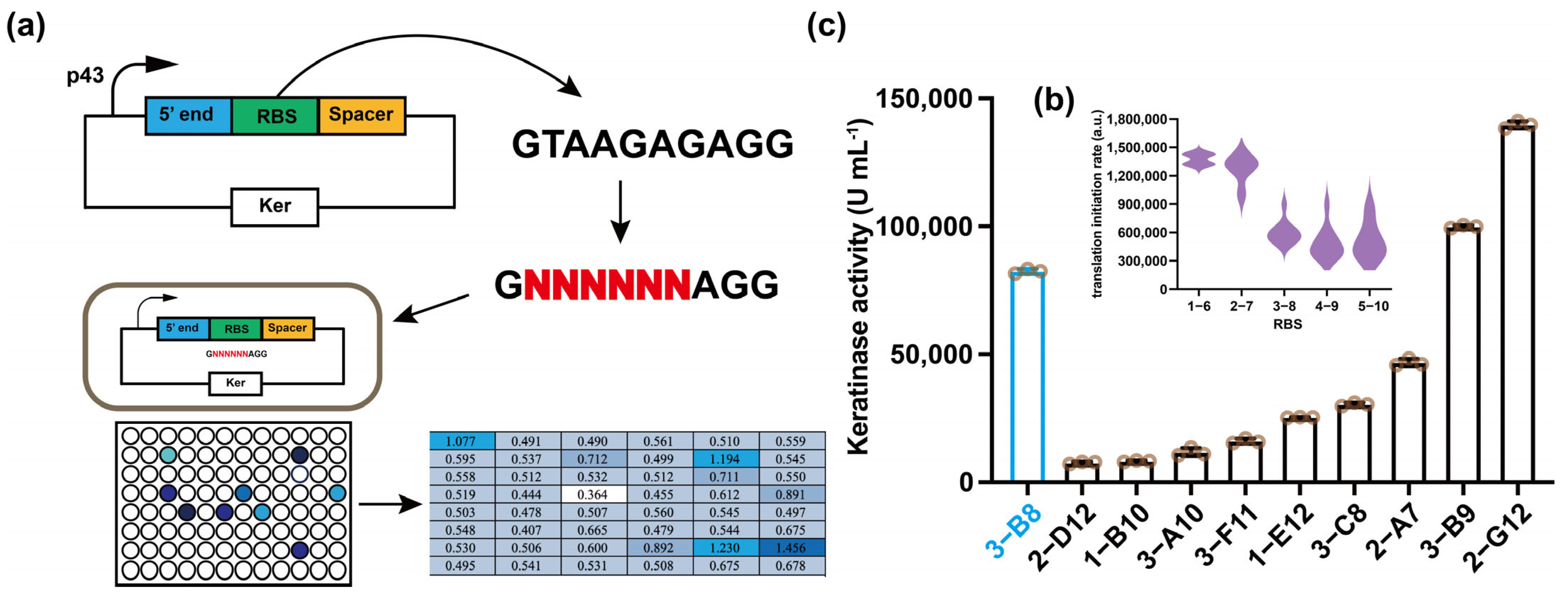
RBS sequence is the core of 5 ‘UTR, usually composed of 4–9 nucleotides and rich in a and G bases [34]. In prokaryotes, the RBS sequence also has the SD sequence, which can complementarily pair with the 3′ end of ribosomal 16S rRNA to facilitate ribosome binding to mRNA [35]. The easier the ribosome binds to RBS, the more stable the complex formed and the higher the translation initiation efficiency. To screen for RBS sequences with higher translation initiation efficiency, we performed saturation mutations on the original RBS sequences (Figure 2a). Based on the original RBS sequence (GTAAGAGAGG) of plasmid p43NMK, the 6-base group gradually moved closer to the translation initiation site (NNNNNNGAGG, GNNNNNNAGG until GTAANNNNNN) to predict the effect of RBS sequence mutation on the translation initiation rate [28]. The results show that the generation of mutations closer to the translation initiation site has less effect on the translation initiation rate. Positions 2–7 of RBS have the highest possible translation initiation efficiency when predicted for mutations (Figure 2b). Therefore, primers were designed to perform saturation mutation on positions 2–6 of RBS, that is, the −13 to −18 region of the total sequence, to obtain a strain with enhanced keratinase secreting activity.
The theoretical number of transformants were selected three times for screening to achieve 95% coverage. The primary screening in the 96-well plate showed that the keratinase activity secreted by nine strains was higher than that of the control strain B. subtilis WB600-SP, namely 1-B10, 1-E12, 2-A7, 2-D12, 2-G12, 3-A10, 3-B9, 3-C8, 3-F11. After re-screening in the shake flask, only the keratinase activities secreted by 2-G12 and 3-B9 were significantly higher than those of the control, reaching 139,650 and 99,860 U mL−1, which were 13.45 and 9.62 times that of the original strain (KerZ1, 10380 U mL−1), respectively (Figure 2c). Although the translation initiation efficiency prediction results were different from the actual expression, the translation initiation efficiency of the 2-G11 and 3-B9 strains with increased activity in the shake flask was significantly higher than that of the original strain (Table 3), which indicates that it is effective to screen RBS sequences based on the prediction of translation initiation efficiency. We named 2-G12 as B. subtilis WB600-SP-R.

3.3. Simplification of the 5′ End Sequence

There is a 9 bp single chain sequence at the 5′-end of the 5′-UTR stem-loop structure, which has no additional function other than carrying the initiation site for transcription. Studies have shown that the simplification of the single-stranded sequence is helpful to weaken the influence of the downstream expressed gene sequence on the 5′-UTR region and enhance the protein expression [30]. In addition, simplifying the single-chain sequence can adjust the proportion of each base in the 5′-UTR region [36]. To this end, we used the 5′-UTR region of KerZ1 as a template to adjust the base ratio and prevent additional stem-loop structures by deleting a/T or C/G bases one by one. At the same time, for the change of stem ring structure caused by the introduction of mutation, a base mutation was introduced to maintain the original stem ring structure, as shown in Table 4.
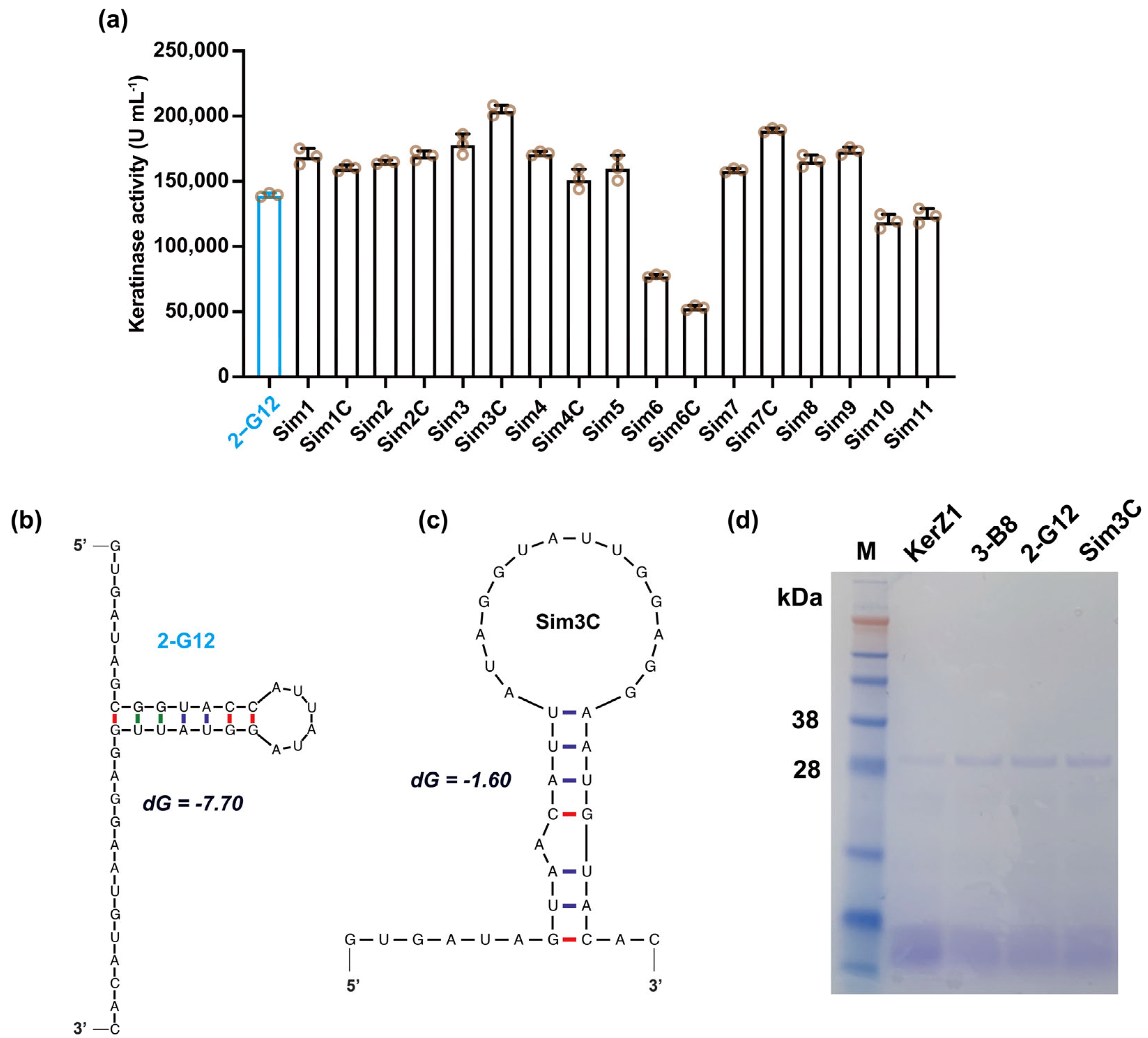
A keratinase activity test showed that the activity of mutant Sim3C reached 204.44 KU mL−1 (Figure 3a), which was 19.70 times that of keratinase KerZ1 (10,380 U mL−1), and was named B. subtilis WB600-SP-R-D. SDS-PAGE was used to verify the keratinases secreted by the optimal mutant strains obtained under different strategies (Figure 3d). The results showed that the expression of keratinase increased gradually. In addition, we also obtained several mutants with increased keratinase activity to varying degrees. Using mfold to predict the secondary structure of mRNA, we found that 5′ deletions altered the mRNA secondary structure (Figure 3b,c). The SD sequence is located on the stem-loop to form a single chain, which is more conducive to its recognition and to the recruitment of ribosomes. In addition, the Gibbs free energy of Sim3C (−1.60) was lower than that of 2-G12 (−7.70), indicating that Sim3C mRNA is more easily bound to ribosomes and has a higher translation initiation rate.

3.4. Fermentation Performance of B. subtilis WB600-SP-R-D

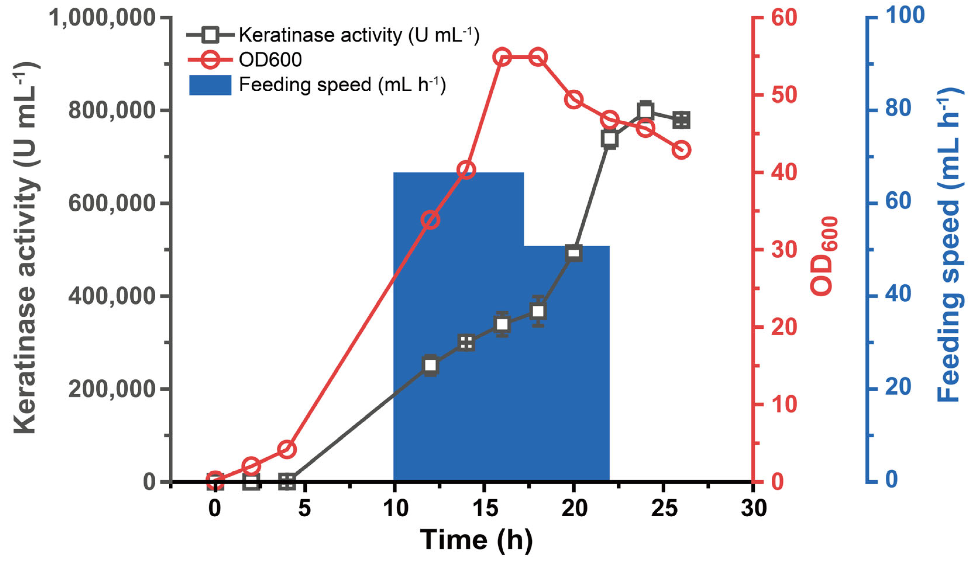
Exploring the enzyme-producing properties of mutant strains in larger systems is not only the basis for the development of industrial strains but also the verification of production strength. To this end, we fermented the mutant strain B. subtilis WB600-SP-R-D in a 5 L fermenter to test its ability and strength to secrete keratinase. Previous studies have shown that excess glucose causes recombinant Bacillus subtilis to produce large amounts of lactic acid as a by-product, which inhibits bacterial growth [37]. While it is possible to automatically pump ammonia to adjust the pH of the environment, this reduces the ability of cells to produce keratinase [15,38]. Therefore, 720 g L−1 of glucose was continuously fed for 10–22 h to keep the glucose concentration at 1–30 g L−1 during fermentation. The feeding was stopped after the 22nd hour, so that the glucose was almost exhausted at the end of the fermentation, which was more conducive to the subsequent separation of keratinase. After 27 h of fermentation, the mutant strain B. subtilis WB600-SP-R-D reached the peak activity of 797.05 KU mL−1 at 25th hour of fermentation, which was 76.79 times that of the original strain (Figure 4). Therefore, the mutant B. subtilis WB600-SP-R-D after 5′-UTR optimization showed excellent and stable enzyme production ability.

4. Conclusions

Keratinase is a promising keratin treatment, but the low activity is still the resistance to use. In this study, we obtained several mutant strains with increased keratinase secretion through the modification of 5′-UTR. A/U base substitution in Spacer region, RBS sequence optimization and the deletion of a single-stranded sequence at the 5′ end made the keratinase activity secreted by mutant B. subtilis WB600-SP-R-D reach 204.44 KU mL−1, which was 19.70 times that of keratinase KerZ1 (10,380 U mL−1). In addition, B. subtilis WB600-SP-R-D had the highest activity in the 5 L fermenter at 797.05 KU mL−1, showing excellent enzyme production capacity and stability. In conclusion, the rational design of 5′-UTR can significantly improve the expression activity of keratinase in B. subtilis, which will be a typical example of the regulation of recombinant protein expression.

Author Contributions

Conceptualization, J.F. and Z.P.; methodology, J.F., G.Z. (Guanyu Zhou) and Z.P.; validation, J.F., G.Z. (Guanyu Zhou) and X.J.; formal analysis, J.F., G.Z. and Z.P. (Guanyu Zhou); data curation, J.F., G.Z. (Guanyu Zhou) and Z.P.; writing—original draft preparation, J.F. and Z.P.; writing—review and editing, Z.P., G.Z. (Guoqiang Zhang) and J.Z.; supervision, Z.P., G.Z. (Guoqiang Zhang) and J.Z. All authors have read and agreed to the published version of the manuscript.

Funding

This work was supported by the National Key Research and Development Program of China (2021YFC2104000) and a grant from the Key Technologies R & D Program of Jiangsu Province (BE2021624).

Informed Consent Statement

Not applicable.

Data Availability Statement

Not applicable.

Conflicts of Interest

The authors declare no conflict of interest.

References

  1. Wang, B.; Yang, W.; McKittrick, J.; Meyers, M.A. Keratin: Structure, mechanical properties, occurrence in biological organisms, and efforts at bioinspiration. Prog. Mater. Sci. 2016, 76, 229–318. [Google Scholar] [CrossRef]
  2. Meyers, M.A.; Chen, P.-Y.; Lin, A.Y.-M.; Seki, Y. Biological materials: Structure and mechanical properties. Prog. Mater. Sci. 2008, 53, 1–206. [Google Scholar] [CrossRef]
  3. Donato, R.K.; Mija, A. Keratin Associations with Synthetic, Biosynthetic and Natural Polymers: An Extensive Review. Polymers 2019, 12, 32. [Google Scholar] [CrossRef] [PubMed]
  4. Callegaro, K.; Brandelli, A.; Daroit, D.J. Beyond plucking: Feathers bioprocessing into valuable protein hydrolysates. Waste Manag. 2019, 95, 399–415. [Google Scholar] [CrossRef]
  5. Brandelli, A.; Daroit, D.J.; Riffel, A. Biochemical features of microbial keratinases and their production and applications. Appl. Microbiol. Biotechnol. 2010, 85, 1735–1750. [Google Scholar] [CrossRef]
  6. Fang, Z.; Yong, Y.-C.; Zhang, J.; Du, G.; Chen, J. Keratinolytic protease: A green biocatalyst for leather industry. Appl. Microbiol. Biotechnol. 2017, 101, 7771–7779. [Google Scholar] [CrossRef]
  7. Zhang, Z.; Li, D.; Zhang, X. Enzymatic decolorization of melanoidins from molasses wastewater by immobilized keratinase. Bioresour. Technol. 2019, 280, 165–172. [Google Scholar] [CrossRef]
  8. Peng, Z.; Mao, X.; Mu, W.; Du, G.; Chen, J.; Zhang, J. Modifying the Substrate Specificity of Keratinase for Industrial Dehairing to Replace Lime-Sulfide. ACS Sustain. Chem. Eng. 2022, 10, 6863–6870. [Google Scholar] [CrossRef]
  9. Gupta, R.; Sharma, R.; Beg, Q.K. Revisiting microbial keratinases: Next generation proteases for sustainable biotechnology. Crit. Rev. Biotechnol. 2013, 33, 216–228. [Google Scholar] [CrossRef]
  10. Verma, A.; Singh, H.; Anwar, S.; Chattopadhyay, A.; Tiwari, K.K.; Kaur, S.; Dhilon, G.S. Microbial keratinases: Industrial enzymes with waste management potential. Crit. Rev. Biotechnol. 2017, 37, 476–491. [Google Scholar] [CrossRef]
  11. Qiu, J.; Wilkens, C.; Barrett, K.; Meyer, A.S. Microbial enzymes catalyzing keratin degradation: Classification, structure, function. Biotechnol. Adv. 2020, 44, 107607. [Google Scholar] [CrossRef] [PubMed]
  12. Su, C.; Gong, J.-S.; Qin, J.; Li, H.; Li, H.; Xu, Z.-H.; Shi, J.-S. The tale of a versatile enzyme: Molecular insights into keratinase for its industrial dissemination. Biotechnol. Adv. 2020, 45, 107655. [Google Scholar] [CrossRef] [PubMed]
  13. Daroit, D.J.; Brandelli, A. A current assessment on the production of bacterial keratinases. Crit. Rev. Biotechnol. 2014, 34, 372–384. [Google Scholar] [CrossRef]
  14. Prakash, P.; Jayalakshmi, S.K.; Sreeramulu, K. Purification and characterization of extreme alkaline, thermostable keratinase, and keratin disulfide reductase produced by Bacillus halodurans PPKS-2. Appl. Microbiol. Biotechnol. 2010, 87, 625–633. [Google Scholar] [CrossRef] [PubMed]
  15. Peng, Z.; Zhang, J.; Song, Y.; Guo, R.; Du, G.; Chen, J. Engineered pro-peptide enhances the catalytic activity of keratinase to improve the conversion ability of feather waste. Biotechnol. Bioeng. 2021, 118, 2559–2571. [Google Scholar] [CrossRef] [PubMed]
  16. Hu, H.; Gao, J.; He, J.; Yu, B.; Zheng, P.; Huang, Z.; Mao, X.; Yu, J.; Han, G.; Chen, D. Codon Optimization Significantly Improves the Expression Level of a Keratinase Gene in Pichia pastoris. PLoS ONE 2013, 8, e58393. [Google Scholar] [CrossRef] [PubMed]
  17. Su, C.; Gong, J.-S.; Sun, Y.-X.; Qin, J.; Zhai, S.; Li, H.; Li, H.; Lu, Z.-M.; Xu, Z.-H.; Shi, J.-S. Combining Pro-peptide Engineering and Multisite Saturation Mutagenesis To Improve the Catalytic Potential of Keratinase. ACS Synth. Biol. 2019, 8, 425–433. [Google Scholar] [CrossRef]
  18. Peng, Z.; Mao, X.; Zhang, J.; Du, G.; Chen, J. Biotransformation of keratin waste to amino acids and active peptides based on cell-free catalysis. Biotechnol. Biofuels 2020, 13, 61. [Google Scholar] [CrossRef]
  19. Fang, Z.; Zhang, J.; Liu, B.; Jiang, L.; Du, G.; Chen, J. Cloning, heterologous expression and characterization of two keratinases from Stenotrophomonas maltophilia BBE11-1. Process Biochem. 2014, 49, 647–654. [Google Scholar] [CrossRef]
  20. Cui, W.; Han, L.; Suo, F.; Liu, Z.; Zhou, L.; Zhou, Z. Exploitation of Bacillus subtilis as a robust workhorse for production of heterologous proteins and beyond. World J. Microbiol. Biotechnol. 2018, 34, 145. [Google Scholar] [CrossRef]
  21. Gingold, H.; Pilpel, Y. Determinants of translation efficiency and accuracy. Mol. Syst. Biol. 2011, 7, 481. [Google Scholar] [CrossRef] [PubMed]
  22. Espah Borujeni, A.; Salis, H.M. Translation Initiation is Controlled by RNA Folding Kinetics via a Ribosome Drafting Mechanism. J. Am. Chem. Soc. 2016, 138, 7016–7023. [Google Scholar] [CrossRef] [PubMed]
  23. Andreeva, I.; Belardinelli, R.; Rodnina, M.V. Translation initiation in bacterial polysomes through ribosome loading on a standby site on a highly translated mRNA. Proc. Natl. Acad. Sci. USA 2018, 115, 4411–4416. [Google Scholar] [CrossRef] [PubMed]
  24. Yu, Q.; Li, Y.; Ma, A.; Liu, W.; Wang, H.; Zhuang, G. An efficient design strategy for a whole-cell biosensor based on engineered ribosome binding sequences. Anal. Bioanal. Chem. 2011, 401, 2891–2898. [Google Scholar] [CrossRef] [PubMed]
  25. Zhou, S.; Ding, R.; Chen, J.; Du, G.; Li, H.; Zhou, J. Obtaining a Panel of Cascade Promoter-5′-UTR Complexes in Escherichia coli. ACS Synth. Biol. 2017, 6, 1065–1075. [Google Scholar] [CrossRef]
  26. Zhang, D.; Zhou, C.Y.; Busby, K.N.; Alexander, S.C.; Devaraj, N.K. Light-Activated Control of Translation by Enzymatic Covalent mRNA Labeling. Angew. Chem. Int. Ed. Engl. 2018, 57, 2822–2826. [Google Scholar] [CrossRef]
  27. Viegas, S.C.; Apura, P.; Martinez-García, E.; de Lorenzo, V.; Arraiano, C.M. Modulating Heterologous Gene Expression with Portable mRNA-Stabilizing 5′-UTR Sequences. ACS Synth. Biol. 2018, 7, 2177–2188. [Google Scholar] [CrossRef]
  28. Salis, H.M.; Mirsky, E.A.; Voigt, C.A. Automated design of synthetic ribosome binding sites to control protein expression. Nat. Biotechnol. 2009, 27, 946–950. [Google Scholar] [CrossRef]
  29. Cuperus, J.T.; Groves, B.; Kuchina, A.; Rosenberg, A.B.; Jojic, N.; Fields, S.; Seelig, G. Deep learning of the regulatory grammar of yeast 5′ untranslated regions from 500,000 random sequences. Genome Res. 2017, 27, 2015–2024. [Google Scholar] [CrossRef]
  30. Xiao, J.; Peng, B.; Su, Z.; Liu, A.; Hu, Y.; Nomura, C.T.; Chen, S.; Wang, Q. Facilitating Protein Expression with Portable 5′-UTR Secondary Structures in Bacillus licheniformis. ACS Synth. Biol. 2020, 9, 1051–1058. [Google Scholar] [CrossRef]
  31. Zuker, M. Mfold web server for nucleic acid folding and hybridization prediction. Nucleic Acids Res. 2003, 31, 3406–3415. [Google Scholar] [CrossRef]
  32. Osterman, I.A.; Evfratov, S.A.; Sergiev, P.V.; Dontsova, O.A. Comparison of mRNA features affecting translation initiation and reinitiation. Nucleic Acids Res. 2013, 41, 474–486. [Google Scholar] [CrossRef]
  33. Lee, H.-M.; Ren, J.; Kim, W.Y.; Vo, P.N.L.; Eyun, S.-I.; Na, D. Introduction of an AU-rich Element into the 5’ UTR of mRNAs Enhances Protein Expression in Escherichia coli by S1 Protein and Hfq Protein. Biotechnol. Bioprocess Eng. 2021, 26, 749–757. [Google Scholar] [CrossRef]
  34. Zelcbuch, L.; Antonovsky, N.; Bar-Even, A.; Levin-Karp, A.; Barenholz, U.; Dayagi, M.; Liebermeister, W.; Flamholz, A.; Noor, E.; Amram, S.; et al. Spanning high-dimensional expression space using ribosome-binding site combinatorics. Nucleic Acids Res. 2013, 41, e98. [Google Scholar] [CrossRef] [PubMed]
  35. Amin, M.R.; Yurovsky, A.; Chen, Y.; Skiena, S.; Futcher, B. Re-annotation of 12,495 prokaryotic 16S rRNA 3’ ends and analysis of Shine-Dalgarno and anti-Shine-Dalgarno sequences. PLoS ONE 2018, 13, e0202767. [Google Scholar] [CrossRef] [PubMed]
  36. Komarova, E.S.; Chervontseva, Z.S.; Osterman, I.A.; Evfratov, S.A.; Rubtsova, M.P.; Zatsepin, T.S.; Semashko, T.A.; Kostryukova, E.S.; Bogdanov, A.A.; Gelfand, M.S.; et al. Influence of the spacer region between the Shine–Dalgarno box and the start codon for fine-tuning of the translation efficiency in Escherichia coli. Microb. Biotechnol. 2020, 13, 1254–1261. [Google Scholar] [CrossRef] [PubMed]
  37. Liu, Y.; Liu, L.; Shin, H.-D.; Chen, R.R.; Li, J.; Du, G.; Chen, J. Pathway engineering of Bacillus subtilis for microbial production of N-acetylglucosamine. Metab. Eng. 2013, 19, 107–115. [Google Scholar] [CrossRef] [PubMed]
  38. Gu, Y.; Lv, X.; Liu, Y.; Li, J.; Du, G.; Chen, J.; Rodrigo, L.-A.; Liu, L. Synthetic redesign of central carbon and redox metabolism for high yield production of N-acetylglucosamine in Bacillus subtilis. Metab. Eng. 2019, 51, 59–69. [Google Scholar] [CrossRef]
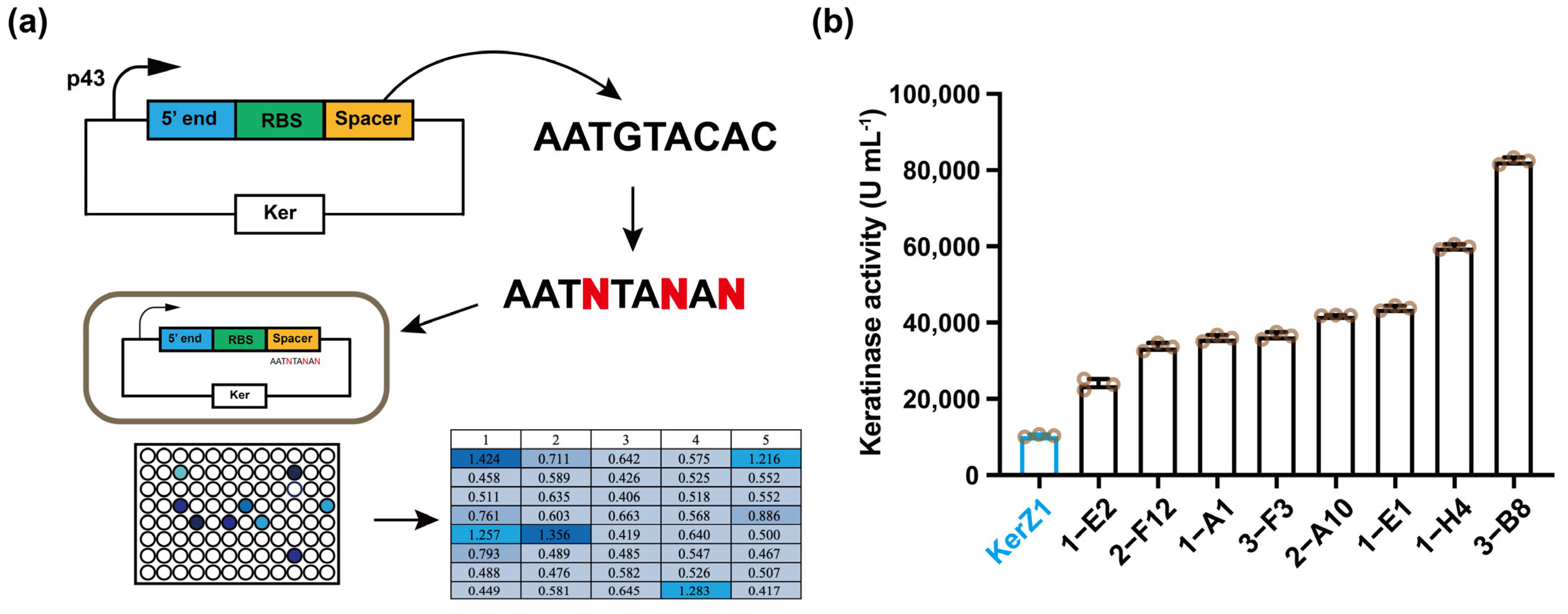
Figure 1. Base substitution of Spacer to enhance keratinase activity. (a) Schematic diagram of base substitution design of Spacer and screening process of mutant strains. (b) Fermentation in shake flasks to rescreen Spacer mutant strains. KerZ1 was secreted by the original strain B. subtilis WB600 pP43NMK-Ker.
Fermentation 08 00426 g001
Figure 2. Base optimization of RBS to increase translation initiation rate to enhance keratinase activity. (a) Schematic diagram of base optimization design of RBS and screening process of mutant strains. (b) Predicted transcription initiation rates corresponding to mutations in different regions of the RBS sequence. (c) Fermentation in shake flasks to rescreen RBS mutant strains.
Fermentation 08 00426 g002
Figure 3. Deletion of the 5′ end of the 5′ UTR to enhance keratinase activity. (a) Keratinase activity of mutant strains with deletions at the 5′ end of the 5′ UTR. (b) mRNA secondary structure of the original sequence at the 5′ end of the 5′ UTR. (c) mRNA secondary structure of mutant Sim3C at the 5′ end of the 5′ UTR. (d) SDS-PAGE verified the keratinase secreted by the optimal mutant strains obtained under different strategies.
Fermentation 08 00426 g003
Figure 4. Fermentation performance of mutant strain B. subtilis WB600-SP-R-D. Fed-batch fermentation of strain B. subtilis WB600-SP-R-D in a 5 L fermenter. The glucose concentration was maintained within 1–30 g L−1 by adding 720 g L−1 glucose. Then the feed was stopped after the 22nd hour, and the glucose was almost exhausted by the end of the fermentation.
Fermentation 08 00426 g004
Table 1. List of strains, plasmids and primers used in this study.
NameSequence (5′-3′)
Strains
JM109Escherichia coli
WB600Bacillus subtilis 168 derivate, missing nprE aprE epr bpr mpr nprB
WB600-KerBacillus subtilis WB600 contains plasmid pP43NMK-Ker
Plasmids
pP43NMKAmpr, Kmr, E. coliB. subtilis shuttle vector
pP43NMK-KerpP43NMK derivate with B. licheniformis Ker gene under the control of the promoter P43
Primers
Spacer
Spacer-3N-FTTATAGGTAAGAGAGGAATNTANANATGATGAGGAAAAAGAGTTTTTGGCTTGG
Spacer-3N-RATTCCTCTCTTACCTATAATGGTACCGCTAT
RBS
RBS-6N-FTAGCGGTACCATTATAGGNNNNNNAGGAATGTATAGATGATGAGGAAAAAGAGTTTTTG
RBS-6N-RCCTATAATGGTACCGCTATCACTTTATATTTTACATAATCG
5′ end
Sim1-FTAAAATATAAAGTGATAGCGTACCATTATAGGTATTGGAGGAATGTACAC
Sim2-FTAAAATATAAAGTGATAGGTACCATTATAGGTATTGGAGGAATGTACAC
Sim3-FTAAAATATAAAGTGATAGTACCATTATAGGTATTGGAGGAATGTACAC
Sim4-FATTATGTAAAATATAAAGTATAGTACCATTATAGGTATTGGAGGAATGTACAC
Sim5-FATTATGTAAAATATAAATATAGTACCATTATAGGTATTGGAGGAATGTACAC
Sim6-FATGTAAAATATAAAGTGATGCGGTACCATTATAGGTATTGGAGGAATGTACAC
Sim7-FATGTAAAATATAAAGTGAGCGGTACCATTATAGGTATTGGAGGAATGTACAC
Sim1C-FTAAAATATAAAGTGATAGCGTAACATTATAGGTATTGGAGGAATGTACAC
Sim2C-FTAAAATATAAAGTGATAGGTAACATTATAGGTATTGGAGGAATGTACAC
Sim3C-FTAAAATATAAAGTGATAGTAACATTATAGGTATTGGAGGAATGTACAC
Sim4C-FATTATGTAAAATATAAAGTATAGTAACATTATAGGTATTGGAGGAATGTACAC
Sim6C-FATGTAAAATATAAAGTGATGCGGTAACATTATAGGTATTGGAGGAATGTACAC
Sim7C-FATGTAAAATATAAAGTGAGCGGTAACATTATAGGTATTGGAGGAATGTACAC
Sim8-FATGTAAAATATAAAGTGGCGGTACCATTATAGGTATTGGAGGA
Sim9-FGATTATGTAAAATATAAAGGGCGGTACCATTATAGGTATTGGAGGA
Sim10-FATTATGTAAAATATAAACGCGGTACCATTATAGGTATTGGAGGA
Sim11-FATTATGTAAAATATAAAGTACCATTATAGGTATTGGAGGAATGTACAC
Sim123-RTATCACTTTATATTTTACATAATCGCGCGCTTTTTTTC
Sim4591011-RTTTATATTTTACATAATCGCGCGCTTTTTTTCACG
Sim678-RCACTTTATATTTTACATAATCGCGCGCTTTTTTTC
Table 2. Spacer base mutants and their corresponding keratinase activities.
StrainsSpacer (5′-3′)Activity (U mL−1)
KerZ1AATGTACAC10,380
1-A1
1-E1
1-E2
1-H4
2-A10
2-F12
3-B8
3-F3
AATTTACAT
AATTTACAT
AATTTACAC
AATTTATAA
AATTTACAT
AATTTACAA
AATGTATAG
AATTTACAT
35,935
43,800
23,745
59,845
41,915
33,615
82,435
36,565
Table 3. RBS base mutants and their corresponding keratinase activities.
StrainsRBS (5′-3′)Activity (U mL−1)Translation Initiation Rate (au)
SPGTAAGAGAGG82,43572.08
1-B10
1-E12
2-A7
2-D12
2-G12
3-A10
3-B8
3-C8
3-F11
GCTGCACAGG
GCTTGCGAGG
GGGAAGTAGG
GATGGTAAGG
GTATTGGAGG
GAAAGACAGG
GGACGGAAGG
GTGTTGCAGG
GGGGGCTAGG
7695
25,350
45,740
7510
139,650
11,060
99,860
30,395
15,710
11.63
26.81
362.40
37.82
85.58
23.71
127.08
29.45
121.96
Table 4. Mutant sequences for simplified design of the 5′-UTR region.
Mutants5′-UTR Sequence (5′-3′)
2-G12GTGATAGCGGTACCATTATAGGTATTGGAGGAATGTACAC
Sim1GTGATAGC--GTACCATTATAGGTATTGGAGGAATGTACAC
Sim1CGTGATAGC--GTAACATTATAGGTATTGGAGGAATGTACAC
Sim2GTGATAG----GTACCATTATAGGTATTGGAGGAATGTACAC
Sim2CGTGATAG----GTAACATTATAGGTATTGGAGGAATGTACAC
Sim3GTGATA------GTACCATTATAGGTATTGGAGGAATGTACAC
Sim3CGTGATA------GTAACATTATAGGTATTGGAGGAATGTACAC
Sim4GT--ATA------GTACCATTATAGGTATTGGAGGAATGTACAC
Sim4CGT--ATA------GTAACATTATAGGTATTGGAGGAATGTACAC
Sim5--T--ATA------GTACCATTATAGGTATTGGAGGAATGTACAC
Sim6GTGAT--GCGGTACCATTATAGGTATTGGAGGAATGTACAC
Sim6CGTGAT--GCGGTAACATTATAGGTATTGGAGGAATGTACAC
Sim7GTGA----GCGGTACCATTATAGGTATTGGAGGAATGTACAC
Sim7CGTGA----GCGGTAACATTATAGGTATTGGAGGAATGTACAC
Sim8GTG------GCGGTACCATTATAGGTATTGGAGGAATGTACAC
Sim9G--G------GCGGTACCATTATAGGTATTGGAGGAATGTACAC
Sim10------------GCGGTACCATTATAGGTATTGGAGGAATGTACAC
Sim11------------------GTACCATTATAGGTATTGGAGGAATGTACAC
Note: -- is the deleted base; the underlined base is the modified base to maintain the stem-loop structure.
Publisher’s Note: MDPI stays neutral with regard to jurisdictional claims in published maps and institutional affiliations.