Article

Cultivation of Lactic Acid Bacteria and Evaluation of the Antimicrobial Potential of Partially Purified Bacteriocin-like Inhibitory Substances against Cariogenic and Food Pathogens

by
Amanda Romana Santos da Silva
1,
Pamela Oliveira de Souza de Azevedo
1,
Attilio Converti
2,* and
Ricardo Pinheiro de Souza Oliveira
1
1
Department of Biochemical-Pharmaceutical Technology, Faculty of Pharmaceutical Sciences, University of São Paulo, Av. Prof. Lineu Prestes, 580, Bl 16, São Paulo 05508-900, Brazil
2
Department of Civil, Chemical and Environmental Engineering, University of Genoa, Pole of Chemical Engineering, Via Opera Pia 15, 16145 Genoa, Italy
*
Author to whom correspondence should be addressed.
Fermentation 2022, 8(8), 400; https://doi.org/10.3390/fermentation8080400
Submission received: 28 July 2022 / Revised: 10 August 2022 / Accepted: 15 August 2022 / Published: 18 August 2022

Abstract

:
One of the major challenges in the pharmaceutical industry is the search for new antimicrobial compounds that can replace antibiotics. Lactic acid bacteria (LAB) can produce bacteriocin-like inhibitory substances (BLIS) that have a bacteriostatic or bactericidal effect against different bacterial genera, including those responsible for dental caries. Among the pathological processes of microbial etiology, the dental caries stands out, whose main pathogenic agent is the species Streptococcus mutans, present in about 80–90% of the oral cavity. In this context, this study aimed to produce and semi-purify BLIS from Lactobacillus plantarum ST16 Pa, Bifidobacterium lactis BL 04, Lactococcus lactis CECT-4434 and Lactobacillus lactis 27 as well as to assess their antimicrobial potential against important dental caries causing pathogens like S. mutans UA159, Listeria innocua 2711, Carnobacterium maltaromaticum CECT 4020, Staphylococcus aureus CECT 239, and Escherichia coli ATCC 25922. While BLIS from L. plantarum ST16 Pa and L. lactis CECT-4434 were able to inhibit the growth only of S. mutans UA159, that which was produced by B. lactis BL 04 did so against all bioindicator strains; therefore, this suggests that its application could be important in the control of cariogenic microorganisms.

1. Introduction

The human oral cavity is inhabited by hundreds of bacterial species that relate to each other and the host in a complex way, such as through competitive, cooperative, and parasitic relations, which can lead to oral diseases [1,2]. Oral diseases are one of the world’s main problems that affect child and youth populations. The Global Burden of Disease estimated that oral diseases affect about 3.5 billion people worldwide, the most common being dental caries. Globally, it is estimated that 2.3 billion people suffer from caries in permanent teeth and more than 530 million children suffer from caries in deciduous teeth [3].
Streptococcus mutans is considered the main agent of caries, as it has peculiar properties such as the ability to colonize the dental surface through the adhesion and production of extracellular polysaccharides, which enables the formation of a thick biofilm. In addition, it has the ability to survive in acidic mediums and accumulate intracellular polysaccharides from the fermentation of glucose and other carbohydrates [4]. A carious injury begins with the establishment of a specific bacterial population, which is able to demineralize the enamel under a specifically modified environment in the oral cavity [5]. Different changes appear in the oral ecosystem leading to the proliferation of bacterial biofilm, composed especially by S. mutans cells.
Another important microorganism is Staphylococcus aureus, which can be found in the larynx, oral and nasal cavity, or in contaminated foods [6]. S. aureus is also the main vector of bacterial endocarditis because, using dental caries as a gateway to the blood system, it can reach the heart and produce a series of toxins, which can give children more risk and even be fatal [7].
The oral cavity is a dynamic ecosystem with permanent characteristics and a peculiar heterogeneous microbiota, whose imbalance leads to the beginning of major oral diseases such as periodontitis and dental caries [8]. Conventional treatment of these diseases involves the removal of bacterial plaque by mechanical means and therapy with antimicrobial drugs that may have limited efficacy due to resistance of microorganisms to antibiotics [9]. According to the World Health Organization (WHO) and the United Nations Food and Agriculture Organization (FAO), probiotics are “live microorganisms which when administered in adequate amounts confer a health benefit on the host” [10]. Traditionally, the main area of study of probiotics concerns the gastrointestinal tract, with the objective of preventing and/or treating diseases. Even though probiotics are widely used for the treatment of intestinal diseases, they have not yet been applied to treat oral diseases due to the scarcity of dental studies. Studies have already shown that some probiotics can increase the proportion of beneficial bacteria in the mouth, contributing to the prevention and therapy of oral diseases [11,12,13]. To be effective in the limitation or prevention of dental caries, probiotics must (a) adhere to and colonize dental surfaces by integrating themselves into the bacterial community constituting dental biofilm; (b) compete with cariogenic and pathogenic bacteria, antagonizing them, and inhibiting and/or delaying their proliferation; (c) metabolize sugars with low acid production to minimize tooth enamel erosion; (d) hinder the organization of extracellular matrix responsible for biofilm formation; (e) limit the production of cytotoxic products by pathogenic bacteria; and (f) beneficially alter the parameters influencing tooth plate formation (e.g., salivary components, buffering power, etc.) [11,12,13,14,15].
During the last decade, several researchers have demonstrated the beneficial use of probiotics in the oral cavity. For instance, bacteria belonging to the genus Bifidobacterium showed encouraging results in the control of ligature-induced periodontitis in mice by modulating the host response [16]. In humans, the ingestion of B. animalis subsp. lactis led to decreased IL-1β levels in the gingival crevicular fluid [17] and was successfully used as a complement to mechanical treatment of periodontitis [18]. Some Lactobacillus strains exhibited potential for dental caries prevention, mainly due to their possible inhibitor activity against cariogenic streptococci and the fact that they are considered safe for oral administration in humans [8].
Bacteriocins are low molecular weight peptides that have inhibitory activity against bacteria, protozoa, fungi, and viruses. Although bacteriocins are produced by several microorganisms, those produced by lactic acid bacteria (LABs) have been receiving greater attention because this group of bacteria is generally safe for human consumption [19,20]. The antimicrobial activity of bacteriocins varies according to the bacteria that produce them and the environment in which they grow, and may have a bacteriostatic or bactericidal effect. Although bacteriocins produced by probiotics are generally recognized as safe (GRAS), nisin is the only approved for use as a food preservative, whose activity analysis parameters, concentration in each type of food, and issues related to safety and commercial use by the food industry are very well defined by WHO/FAO [21].
The relevance of this work is justified in the high rates of caries on a world scale and the therapeutic potential of probiotics that showed promising results in major oral diseases such as periodontitis and dental caries [22,23]. However, doubts still remain about the best probiotic strains for each oral disease [24], as well as their dosage and frequency of administration. Studies dealing with the prevention of oral pathologies are mostly directed to diet control, salivation stimuli, mechanical removal of dental plaque, and remineralization of non-cavitated surfaces in which the process has already begun. Therefore, it is ignored by the main oral health promotion programs one of the most important characteristics of oral microbiota—that is, the relationships among the different bacterial communities, especially the competitive ones [1].
In the context of the search for effective bacteriocins against cariogenic and food pathogens, the aim of this study was the identification of bacteriocin-producing LABs with the potential to contribute to the development of alternative methods for the control of oral biofilm and health promotion in the oral cavity.

2. Materials and Methods

2.1. Bacterial Strains

Different species of bacteriocin-producing LABs were used in this study, namely Lactobacillus plantarum ST16 Pa, Pediococcus pentosaceus ATCC 33316, Lactococcus lactis CECT-4434, Bifidobacterium lactis BL 04 (Danisco, Sassenage, France), Lactobacillus sakei subsp. sakei 2a, and Lactobacillus lactis 27 (S.T. Dupont, Paris, France), while Streptococcus thermophilus TA040 (S.T. Dupont, Paris, France) was used as a control.
As for the bioindicator strains, Streptococcus mutans UA159 (ATCC 700610) and Escherichia coli ATCC 25922 were purchased from the American Type Culture Collection, Carnobacterium maltaromaticum CECT 4020 and Staphylococcus aureus CECT 239 from Spanish Type Culture Collection (CECT), while Listeria innocua CLIST 2711 was kindly provided by the Instituto Oswaldo Cruz (Rio de Janeiro, Brazil).

2.2. Culture Conditions

P. pentosaceus ATCC 33316, L. plantarum ST16 Pa, L. lactis CECT-4434, B. lactis BL 04, L. sakei 2a, and L. lactis 27 were grown anaerobically in De Man, Rogosa, and Sharpe (MRS) medium (Difco Laboratories, Detroit, MI, USA) at 30 °C and pH 6.5 ± 0.2 for 16 h under 150 rpm orbital agitation (TE-424, Technical, Piracicaba, Brazil). For a purity check, cells from each culture were seeded in Petri plates containing MRS medium plus 2% agar and incubated overnight at 37 °C.
All bioindicator strains (1.0 mL), previously cryopreserved at −70 °C in the presence of 20% glycerol (w/v), were aerobically reactivated in 5.0 mL of Tryptic Soy Broth (TSB) (Difco Laboratories, Detroit, MI, USA) at 37 °C for 16 h without agitation. For purity check, cells from each culture were seeded in Petri plates containing TSB plus 2% agar and incubated overnight at 37 °C.

2.3. Co-Cultures

To detect possible synergistic effects, bacteriocin-producing strains (1.0 mL) were cultivated in co-cultures with each other and/or with S. thermophilus TA040 in 250 mL Erlenmeyer flasks containing 100 mL of MRS broth supplemented with 1% (w/v) L-cysteine and 0.04% (w/v) sodium thioglyolate (Difco).

2.4. Kinetic Study of Lactic Acid Bacteria Growth

The growth kinetics of LABs (L. plantarum 16 PA, L. lactis CECT4434, B. lactis BL04, and L. lactis 27) was investigated along 52 h cultures using the pour plate methodology. For this purpose, the initial concentration of each LAB was adjusted to an optical density at 600 nm (OD600 nm) of 0.8–0.9, which corresponded to 107–108 colony forming units (CFU) per mL. LABs were grown in sterile tubes containing 50 mL of MRS broth (Difco) at 37 °C, from which 100 µL samples were collected every 2 h. Afterwards, serial dilution (1:10) was performed using sterile distilled water, and all dilutions were seeded on the surface of 1% (w/v) MRS agar in triplicate. After plate incubation at 37 °C for 24 h, colonies were counted, and the results expressed in CFU/mL.

2.5. Acidification Kinetics

The acidification profile was monitored for 24 h by the CINAC system (Cinétique d’Acidification) software (Ysebaert, Frépillon, France). Both monocultures and co-cultures were kept in a water bath without agitation at 37 °C in 250 mL Schott flasks (Schott Duran, Laborglas, São Paulo, Brazil) containing 200 mL of MRS medium (for LABs producing bacteriocin-like inhibitory substances (BLIS)) or M17 medium (for S. thermophilus). After the assessment of the acidification profile, the cultures were centrifuged at 4470× g and 4 °C for 15 min to obtain the cell-free supernatant (CFS) for later determination of bacteriocin antimicrobial activity.

2.6. Determination of Antimicrobial Activity by the Critical Dilution Technique

To prevent the action of organic acids, the CFSs had their pH adjusted to 6.0–6.5 by addition of 1.0 M NaOH. They were then heated at 80 °C for 10 min to inactivate proteases, and sterilized by filtration in membranes with 0.20 µm pore diameter (Millipore, Billerica, MA, USA).
The antimicrobial activity of each BLIS was evaluated against the bioindicator strains L. innocua CLIST 2711, C. maltaromaticum CECT 4020, S. aureus CECT 239 and E. coli ATCC 25922, and the results were expressed in arbitrary units per milliliter (AU/mL). After serial dilution of CFSs (1:2, 1:4 and 1:8, v/v) in 25 mM sodium phosphate buffer, pH 7.0, 10 µL of each dilution were applied on the surface of plates containing TSB agar (0.75%, w/v) previously inoculated with the selected bioindicator strain (~106 CFU/mL) and later incubated at 37 °C for 24 h. The first dilution in which there was no inhibition halo formation (n) was considered in the equation:
AU / mL   = D n · 1000 P
where D is the dilution factor and P the volume of supernatant placed on the TSB agar medium.

2.7. Determination of Antimicrobial Activity against Cariogenic Microorganism

To evaluate the potential anticariogenic effect of BLIS produced by LABs at different stages of bacterial growth, BLIS activity was also determined as ability to inhibit the growth of the S. mutans UA159 strain by the micro-dilution method using a microplate reader [25,26]. The assays were performed on sterile 96-well plate (TPP, Trasadingen, Switzerland) containing M17 (Difco) medium supplemented with 1.0% (w/v) lactose (100 µL), an aliquot of overnight culture of this pathogen (50 µL), and BLIS solution (50 µL), after pH correction to 6.0–6.5 with 1.0 M NaOH, thermal treatment for 10 min at 80 °C, and filter sterilization (0.22 µm). The resulting suspension (200 µL) was well mixed, and the microplate was incubated inside a microplate reader (BioTek, Winooski, VT, USA) at 37 °C. S. mutans growth was determined automatically every hour for 24 h on the microplate reader at OD600 nm. BLIS activity was determined in triplicate.

2.8. Effect of Proteases, Solvents, Salts and Detergents on BLIS

To investigate the eventual protein nature of antimicrobial compounds, the CFSs were treated with 1.0 mg/mL tripsin, pepsin, or papain (Sigma-Aldrich, Saint Louis, MO, USA) for 2 h at 37 °C. A 10 µL aliquot of BLIS solution prepared as described in the Section 2.6 was poured in a Petri dish containing L. innocua 2711 as a bio-indicator strain. The same volume of 1.0 mg/mL enzyme solution was poured in the Petri dish in such a way that it would just partially cover BLIS [27]. The proteinaceous nature of BLIS was determined by the absence of inhibition halo against the bio-indicator strain.
To check BLIS stability, CFSs were treated in 1.0-mL conical tubes at 30 °C for 2 h with different organic solvents (acetonitrile and isopropanol), salts (sodium chloride and ammonium sulfate), and detergents (Triton X-100, ethylenediaminetetraacetic acid (EDTA), Tween 20, Tween 80, sodium dodecyl sulfate (SDS)) at a final concentration of 1.0% (w/v). In addition, BLIS was submitted to heat treatment at different temperatures, namely 30, 50, 70, and 90 °C for 1 h, or 120 °C for 15 min. After that, the BLIS stability was checked by the agar diffusion method against L. innocua 2711 [27].

2.9. BLIS Concentration by Precipitation with Ammonium Sulfate

After verification of protein nature of BLIS, they were concentrated by the salting-out technique. Precipitation was performed using 10 mL of each CFS by adding ammonium sulfate at six different concentrations (10, 20, 30, 40, 50, and 60% w/v). The antimicrobial activity of BLIS was then assessed to identify the optimal ammonium sulfate concentration to recover them. Afterwards, the tubes were vigorously mixed for 1 min and then incubated at 10 °C and 100 rpm for 1 h. After sample centrifugation at 4470× g and 4 °C for 30 min, each precipitate was resuspended with 10% (v/v) of the initial volume (20 mL) of 25 mM ammonium acetate solution (pH 6.5) (Labsynth Produtos para Laboratórios, Diadema, Brazil) [25], and sterilized by filtration through membranes with 0.22 µm pore diameter [27]. The antimicrobial activity of all concentrated BLIS was determined by the agar diffusion method.

3. Results and Discussion

3.1. Antimicrobial Activity of BLIS by the Critical Dilution Technique

The results of the antimicrobial activity of cell-free supernatants (CFSs) determined by the critical dilution technique showed that, among the BLIS-producing bacteria under investigation, L. plantarum ST16 PA and especially B. lactis BL 04 had the largest inhibitory effects against L. innocua 2711 (Table 1). Antimicrobials produced by L. plantarum ST16 Pa also showed efficiency in inhibiting different species of the genera Enterococcus, Lactobacillus, Pseudomonas, Streptococcus, Staphylococcus, and Listeria [19]. In a similar study, B. lactis CFS was able to inhibit both L. monocytogenes and E. coli ATCC 25922 growth, thus demonstrating its antimicrobial potential against both Gram-positive and Gram-negative bacteria [28].
When compared to the other BLIS-producing LABs, L. lactis 27 exhibited the highest antimicrobial activity against S. aureus CECT 239, contrary to L. lactis CECT-4434 that showed no activity against this pathogen. P. pentosaceus ATCC 33316 and L. sakei subsp. sakei 2a, although described in the literature as bacteriocin producers [29,30], did not exert any antimicrobial activity against the bioindicator strains tested in this study.

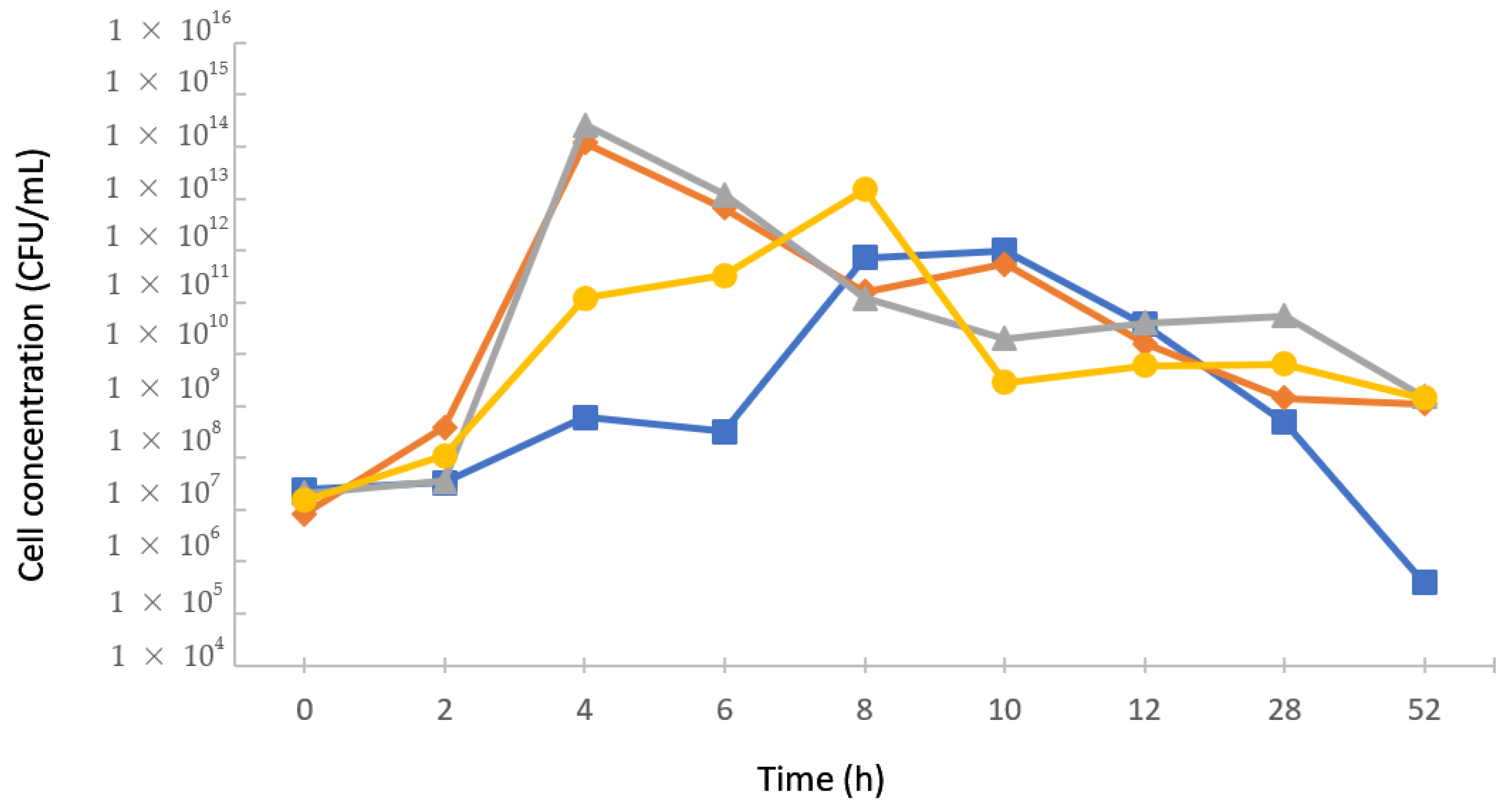
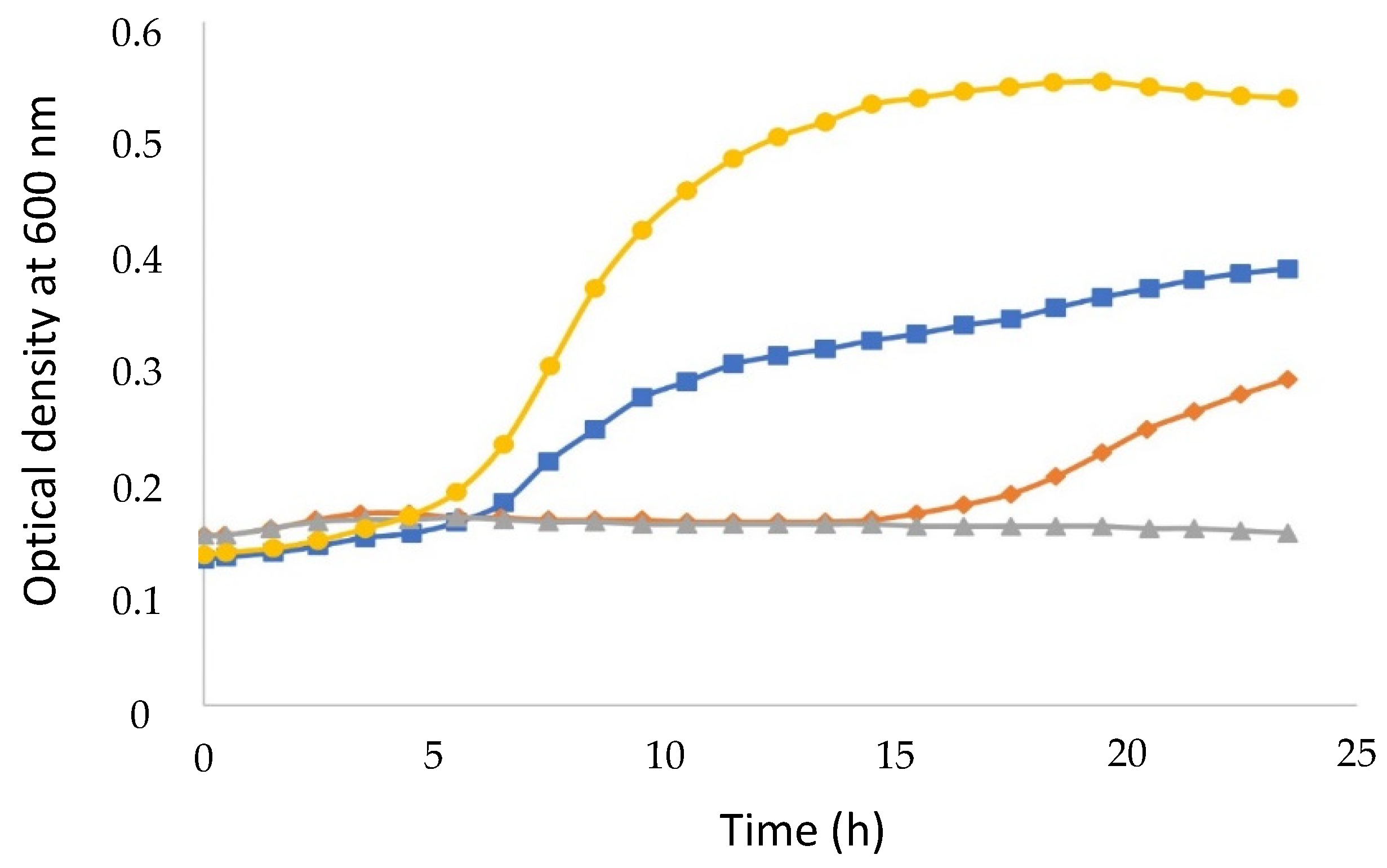
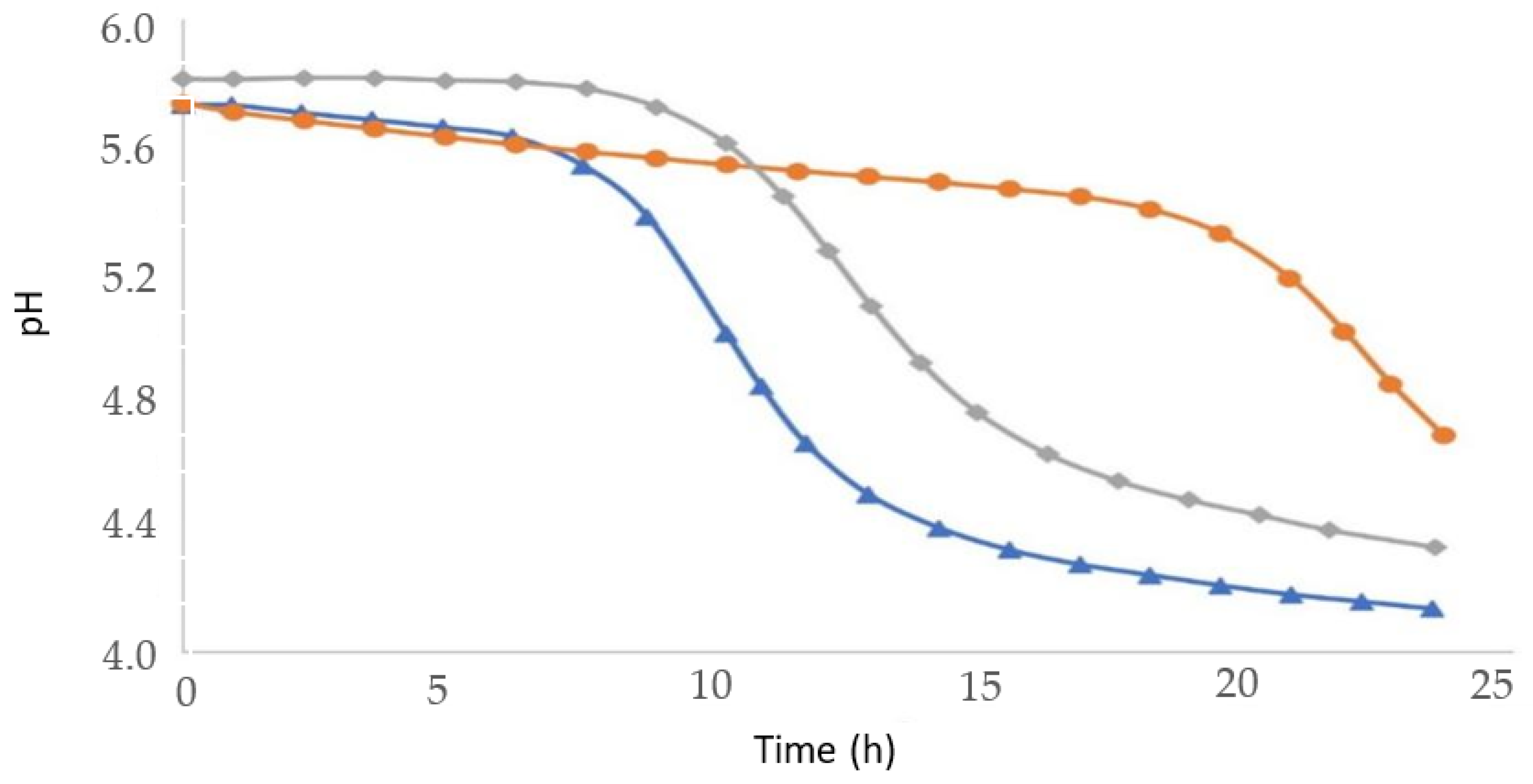
3.2. Kinetics of Lactic Bacteria Growth in Monocultures

The selected LABs showed quick initial growth, with start of the exponential phase after only 2 h of cultivation. The only exception to this was L. plantarum ST16 PA, which displayed a 6 h lag phase. Therefore, this strain suffered a reduction (4 log units) in cell concentration after 52 h of cultivation (4.0 ± 0.0 × 10⁵ CFU/mL), compared to the beginning of the exponential phase (3.2 ± 0.0 × 10⁹ CFU/mL), which was much larger than those observed for L. lactis CECT 4434, L. lactis 27 (1 log unit), and B. lactis BL04 (2 log units) (Figure 1).

3.3. Antimicrobial Activity against Cariogenic Microorganism

To evaluate the antimicrobial activity against the cariogenic S. mutans UA159 strain by the micro-dilution method, only the BLIS-producing LABs that were successful against non-cariogenic bioindicator strains were selected. Although all the selected LABs demonstrated antimicrobial activity against S. mutans UA159, L. plantarum ST16 Pa and L. lactis CECT-4434 ensured the most significant and promising results (Table 2).
Figure 2 shows that the addition of CFS containing the B. lactis BL 04 BLIS delayed the growth of S. mutans UA159 by approximately 13 h compared to its cultivation without CFS, and that this effect was directly proportional to BLIS concentration. However, the capability of the bioindicator strain to grow, although more slowly, even without any CFS dilution suggests that the BLIS effect was likely bacteriostatic rather than bactericidal. In addition, the effectiveness of this effect is demonstrated by the fact that it occurred even in the presence of the most diluted CFS (CFS2), leading to a reduction of no less than 40% of the final OD600 nm compared to the culture without BLIS.
These results corroborate with the observations of a study in which the administration of B. animalis subs. lactis BB-12 and L. rhamnosus GG allowed to control S. mutans development, thus reducing oral plaque and gingival inflammation in healthy young adults [17]. Similarly, Bhalla et al. [31] observed a significant reduction in S. mutans counts in saliva after 1 h and 7 days of B. animalis subsp. lactis BB-12 administration, confirming the anticariogenic power of this LAB.
As Figure 3 shows, the addition of CFS from L. plantarum ST16 PA culture, either as such or diluted, had an even more pronounced effect on S. mutans growth. In particular, while the most diluted CFS (CFS2) delayed in just a few hours the onset of the exponential phase compared to the run without BLIS, the least diluted one (CFS1) caused a delay as long as 17 h, and the undiluted one completely suppressed growth.
As a result, the reduction of OD600 nm at the end of cultures (i.e., of the final concentration of L. plantarum ST16 Pa cells) was directly proportional to BLIS content in CFS. Similar results were reported against different species belonging to the genera Enterobacter, Enterococcus, Lactobacillus, Pseudomonas, Staphylococcus, and Streptococcus [32].
Figure 4 and Figure 5 illustrate the effects on the growth of the same pathogen of CFSs from L. lactis CECT-4434 and L. lactis 27 cultures, respectively. As expected by the fact that the two strains belong to the same species, the two antimicrobials showed similarity in their peculiar inhibition profile, which was characterized by no significant delay in the onset of the exponential phase, reduction in OD600 nm at the end of cultures proportional to BLIS content in CFS, and total growth suppression in the presence of undiluted CFSs.
In the study by Sudhir et al. [33], L. acidophilus, when administered in children for 30 days using curd as a vehicle, promoted significant reduction in S. mutans counts in saliva. The joint administration of L. acidophilus LA5 and B. lactis BB12 also significantly reduced the growth of S. mutans by 7 and 30 days after the ingestion of these probiotics by children [34]. However, after 6 months of discontinuation of probiotic administration, S. mutans counts returned to the level observed at the beginning of the study, which means that there was no effective colonization of the oral cavity by these probiotics and that they should be constantly administered.
Comparing the results of this section, one can infer that the almost complete suppression of S. mutans UA159 growth evident in Figure 2, Figure 3 and Figure 4 may have been the result of a bactericidal effect of BLIS produced by L. plantarum ST16 Pa, L. lactis CECT-4434, and L. lactis 27 cultures, unlike the bacteriostatic one evidenced in Figure 1 for B. lactis BL 04 BLIS. In addition, the absence of any delay of the exponential phase of the pathogen growth in Figure 3 and Figure 4 suggests that the bactericidal action of L. lactis CECT-4434 and L. lactis 27 BLIS may have taken longer than those of BLIS produced by L. plantarum ST16 Pa and B. lactis BL 04 BLIS. However, any attempts to relate these different inhibition profiles with possible action mechanisms would require additional research effort.

3.4. Preliminary Characterization of BLIS

To investigate the chemical nature of BLIS, each CFS that exhibited antimicrobial activity in previous tests was treated separately with 1.0 mg/mL papain, pepsin and trypsin, and its antimicrobial activity was again tested against L. innocua 2711. The results showed that all enzyme treatments resulted in loss of antimicrobial activity, indicating the protein nature of all BLIS and suggesting that they could be class II bacteriocins [32].
To evaluate the stability of the protein compounds responsible for antimicrobial activity, the CFSs of B. lactis BL 04 and L. lactis 27 underwent treatments with different organic solvents (acetonitrile and isopropanol), salts (sodium chloride and ammonium sulfate), and detergents (Triton 100-X, EDTA, Tween 20, Tween 20 and SDS) and tested for their inhibitory effect against L. innocua 2711 (Table 3).
The inhibition halos generated by B. lactis BL 04 and L. lactis 27 CFSs after solvent treatment showed diameters (9.57–11.29 and 9.27–9.41 mm, respectively) close to those obtained using water as a control (10.04 and 8.85 mm, respectively), demonstrating that solvents did not influence the inhibitory effect of their respective BLIS. On the other hand, the B. lactis BL 04 BLIS suffered a small reduction in its antimicrobial effect after ammonium sulfate treatment (7.81 mm), while this same salt enhanced the effect of the L. lactis 27 one (14.35 mm). These results suggest that, despite the small decrease in antimicrobial activity observed in the former case, salting-out with ammonium sulfate could generally be a viable methodology as a first step for BLIS concentration and partial purification without significant losses of their activities.
Pathogenic bacteria such as E. coli and members of the Listeria genus are known to be important vehicles of diseases transmitted by contaminated foods [35]. The CFS antimicrobial activity observed in this study corroborates the results of a recent study in which the L. lactis GH1 CFS proved to be able to inhibit the growth of L. monocytogenes ATCC 15313 (1.55 ± 0.14 mm) [36] as the likely result of the production of organic acids, with consequent pH reduction, hydrogen peroxide, bacteriocins, or BLIS [37]. The stability of L. lactis 27 BLIS in organic solvents observed in this study is in line with the complete retention of L. lactis strains’ antimicrobial activity reported in mixture with 10% (v/v) acetone, benzene, chloroform, dimethylsulfoxide, ethanol, ethyl acetate, and methanol [38]. On the other hand, the small loss of antimicrobial activity of B. lactis BL 04 CFS caused by the ammonium sulfate addition can be attributed to some change in protein three-dimensional structure of BLIS, which commonly leads to a loss of functionality and the formation of potentially immunogenic aggregates.

3.5. BLIS Production in Co-Cultures

The results of monocultures showed that B. lactis BL 04 is the LAB, among those considered in this study, with the broadest spectrum of action against the tested bioindicator strains, including L. innocua 2711. Therefore, it was grown in co-culture with S. thermophilus TA040, which previously displayed synergism in co-culture with L. rhamnosus by significantly reducing the acidification time [39]. The results of the antimicrobial activity of the CFS of this co-culture against L. innocua 271 are gathered in Table 4 and compared to those of the respective monocultures.
The CFS from S. thermophilus TA040 monoculture showed low antimicrobial activity when using MRS broth supplemented with 1.0% (w/v) L-cysteine and 0.04% (w/v) sodium thioglycolate (1600 AU/mL), and even null antimicrobial activity when using the M17 medium, thus corroborating the absence of reports in the literature on antimicrobial activity of this strain. On the contrary, the antimicrobial activity of CFS from B. lactis BL 04 monoculture was remarkable (6400 AU/mL) and coincided with the one determined using the critical dilution technique. When this microorganism was cultivated in co-culture with S. thermophilus TA040, the CFS antimicrobial activity doubled (12,800 AU/mL) compared to its monoculture, proving the previously supposed synergism [39].
It is known that the acidification ability of probiotic strains is one of the most significant technological characteristics of starter cultures, particularly in the manufacture of milk-based products, because the pH reduction inhibits the growth of a large number of pathogenic microorganisms and/or microorganisms responsible for deterioration of fermented products, thus improving their storage properties [36].
As illustrated in Figure 6, the B. lactis BL 04 and S. thermophilus TA040 co-culture shortened the acidification time (i.e., the time to lower the pH from 5.8 to 4.3) by approximately 8 h compared to the B. lactis BL 04 monoculture (24 h), while S. thermophilus TA040 alone could not achieve this goal even at the end of cultivation.

3.6. BLIS Concentration and Partial Purification by Salting out with Ammonium Sulfate

Table 5 shows the results of the antimicrobial activity of the CFSs from L. lactis 27 and B. lactis BL 04 monocultures after BLIS concentration with ammonium sulfate at different concentrations (10, 20, 30, 40, 50, 60% w/v).
In both cases the strongest antimicrobial activity was obtained using the largest dosage of ammonium sulfate (60%, w/v), but that of B. lactis BL 04 BLIS (102,400 AU/mL) was nothing less than 15 times higher than that of L. lactis 27 BLIS. These results suggest that the BLIS produced by this bifidobacterium was insensitive to possible changes induced by the salting out process in its protein three-dimensional structure and may be successfully exploited to hinder the development of pathogens.

4. Conclusions

The results of this study showed that, among the selected lactic acid bacteria (LABs), Lactobacillus plantarum ST16 Pa, Bifidobacterium lactis BL 04, Lactococcus lactis CECT-4434, and Lactobacillus lactis 27 were able to produce bacteriocin-like substances (BLIS), unlike Pediococcus pentosaceus ATCC 33316 and Lactobacillus sakei 2a. Among them, B. lactis BL 04 was the LAB with the largest spectrum of antimicrobial activity, being able to inhibit the growth of Listeria innocua 2711, Carnobacterium maltaromaticum CECT 4020, Staphylococcus aureus CECT 239, and Escherichia coli ATCC 25922. On the other hand, L. plantarum ST16 Pa and L. lactis CECT-4434 showed the greatest antimicrobial effect against the cariogenic pathogen Streptococcus mutans UA159. B. lactis BL 04 antimicrobial activity was two times higher than those of the other LABs, and the acidification time was significantly reduced when this bacterium was cultivated in co-culture with Streptococcus thermophilus TA040, which suggests a possible synergistic effect between these two microorganisms.
The antimicrobial activities of L. lactis 27 and B. lactis BL 04 BLIS after ammonium sulfate precipitation increased proportionally to the concentration of this salt up to 60% w/v. However, the activity of latter (102,400 AU/mL) was 15 times higher than the one of former, suggesting that the B. lactis BL 04 BLIS may have been insensitive to possible three-dimensional changes induced by the salting-out process. The results of this study also indicate the potential application of B. lactis BL 04 BLIS, produced either in monoculture or in co-culture with S. thermophilus TA040, to prevent the development of pathogens.

Author Contributions

Conceptualization, A.R.S.d.S. and R.P.d.S.O.; methodology, A.R.S.d.S. and P.O.d.S.d.A.; validation, P.O.d.S.d.A., R.P.d.S.O. and A.C.; formal analysis, P.O.d.S.d.A., R.P.d.S.O. and A.C.; investigation, A.R.S.d.S.; resources, R.P.d.S.O.; data curation, P.O.d.S.d.A. and A.C.; writing—original draft preparation, A.R.S.d.S.; writing—review and editing, P.O.d.S.d.A., R.P.d.S.O. and A.C.; visualization, P.O.d.S.d.A. and A.C.; supervision, R.P.d.S.O.; project administration, R.P.d.S.O.; funding acquisition, R.P.d.S.O. All authors have read and agreed to the published version of the manuscript.

Funding

The authors are grateful for the financial support of São Paulo Research Foundation (FAPESP) [grant 2018/25511-1] and the National Council for Scientific and Technological Development (CNPq) [grants 870258/1997-4; 312923/2020-1; 408783/2021-4].

Institutional Review Board Statement

Not applicable.

Informed Consent Statement

Not applicable.

Data Availability Statement

The data presented in this study are available on request from the corresponding author.

Conflicts of Interest

The authors declare no conflict of interest.

References

  1. Belda-Ferre, P.; Alcaraz, L.D.; Cabrera-Rubio, R.; Romero, H.; Simón-Soro, A.; Pignatelli, M.; Mira, A. The oral metagenome in health and disease. ISME J. 2012, 6, 46–56. [Google Scholar] [CrossRef] [PubMed]
  2. Marsh, P.D.; Zaura, E. Dental biofilm: Ecological interactions in health and disease. J. Clin. Periodontol. 2017, 44, S12–S22. [Google Scholar] [CrossRef] [PubMed]
  3. GBD 2017 Disease and Injury Incidence and Prevalence Collaborators. Global, regional, and national incidence, prevalence, and years lived with disability for 354 diseases and injuries for 195 countries and territories, 1990–2017: A systematic analysis for the Global Burden of Disease Study 2017. Lancet 2018, 392, 1789–1858. [Google Scholar] [CrossRef]
  4. Gálvez, A.; Abriouel, H.; López, R.L.; Ben Omar, N. Bacteriocin-based strategies for food biopreservation. Int. J. Food Microbiol. 2007, 120, 51–70. [Google Scholar] [CrossRef] [PubMed]
  5. Lata, S.; Varghese, N.O.; Varughese, J.M. Remineralization potential of fluoride and amorphous calcium phosphate-casein phospho peptide on enamel lesions: An in vitro comparative evaluation. J. Conserv. Dent. 2010, 13, 42–46. [Google Scholar] [CrossRef]
  6. Rubaba, M.; Muhamma, H.; Amin, N.S.; Deog-Hwan, O. Biosensors for rapid and sensitive detection of Staphylococcus aureus in food. Biosens. Bioelectron. 2018, 105, 49–57. [Google Scholar] [CrossRef]
  7. Mealey, B.L. Influence of periodontal infections on systemic health. Periodontol. 2000 1999, 21, 197–209. [Google Scholar] [CrossRef]
  8. Willis, J.R.; Gabaldón, T. The human oral microbiome in health and disease: From sequences to ecosystems. Microorganisms 2020, 8, 308. [Google Scholar] [CrossRef]
  9. Haque, M.; Sartelli, M.; Haque, S.Z. Dental infection and resistance-global health consequences. Dent. J. 2019, 7, 22. [Google Scholar] [CrossRef]
  10. Food and Agriculture Organization (FAO) of the United Nations Food Safety and Quality: Probiotics. Available online: http://www.fao.org/food/food-safety-quality/a-z-index/probiotics/en/ (accessed on 24 July 2022).
  11. Sabatini, S.; Lauritano, D.; Candotto, V.; Silvestre, F.J.; Nardi, G.M. Oral probiotics in the management of gingivitis in diabetic patients: A double blinded randomized controlled study. J. Biol. Regul. Homeost. Agents. 2017, 31, 197–202. [Google Scholar]
  12. Tekce, M.; Ince, G.; Gursoy, H.; Dirikan Ipci, S.; Cakar, G.; Kadir, T.; Yilmaz, S. Clinical and microbiological effects of probiotic lozenges in the treatment of chronic periodontitis: A 1-year follow-up study. J. Clin. Periodontol. 2015, 42, 363–372. [Google Scholar] [CrossRef] [PubMed]
  13. Keller, M.K.; Brandsborg, E.; Holmstrom, K.; Twetman, S. Effect of tablets containing probiotic candidate strains on gingival inflammation and composition of the salivary microbiome: A randomised controlled trial. Benef. Microbes. 2018, 9, 487–494. [Google Scholar] [CrossRef] [PubMed]
  14. Nadkerny, P.V.; Ravishankar, P.L.; Pramod, V.; Agarwal, L.A.; Bhandari, S. A comparative evaluation of the efficacy of probiotic and chlorhexidine mouthrinses on clinical inflammatory parameters of gingivitis: A randomized controlled clinical study. J. Indian Soc. Periodontol. 2015, 19, 633–639. [Google Scholar] [CrossRef] [PubMed]
  15. Grusovin, M.G.; Bossini, S.; Calza, S.; Cappa, V.; Garzetti, G.; Scotti, E.; Gherlone, E.F.; Mensi, M. Clinical efficacy of Lactobacillus reuteri-containing lozenges in the supportive therapy of generalized periodontitis stage III and IV, grade C: 1-year results of a double-blind randomized placebo-controlled pilot study. Clin. Oral Investig. 2020, 24, 2015–2024. [Google Scholar] [CrossRef] [PubMed]
  16. Oliveira, L.F.; Salvador, S.L.; Silva, P.H.; Furlaneto, F.A.; Figueiredo, L.; Casarin, R.; Ervolino, E.; Palioto, D.B.; Souza, S.L.S.; Taba, M., Jr.; et al. Benefits of Bifidobacterium animalis subsp. lactis probiotic in experimental periodontitis. J. Periodontol. 2017, 88, 197–208. [Google Scholar] [CrossRef]
  17. Toiviainen, A.; Jalasvuori, H.; Lahti, E.; Gursoy, U.; Salminen, S.; Fontana, M.; Flannagan, S.; Eckert, G.; Kakaras, A.; Paster, B.; et al. Impact of orally administered lozenges with Lactobacillus rhamnosus GG and Bifidobacterium animalis subsp. lactis BB-12 on the number of salivary mutans streptococci, amount of plaque, gingival inflammation and the oral microbiome in healthy adults. Clin. Oral Investig. 2015, 19, 77–83. [Google Scholar] [CrossRef]
  18. Invernici, M.M.; Salvador, S.L.; Silva, P.H.F.; Soares, M.S.M.; Casarin, R.; Palioto, D.B.; Souza, S.L.S.; Taba, M., Jr.; Navaes, A.B., Jr.; Furlaneto, F.A.C.; et al. Effects of Bifidobacterium probiotic on the treatment of chronic periodontitis: A randomized clinical trial. J. Clin. Periodontol. 2018, 45, 1198–1210. [Google Scholar] [CrossRef]
  19. Sabo, S.S.; Vitolo, M.; Domínguez González, J.M.; Oliveira, R.P.S. Overview of Lactobacillus plantarum as a promising bacteriocins producer among lactic acid bacteria. Food Res. Int. 2014, 64, 527–536. [Google Scholar] [CrossRef]
  20. Balciunas, E.M.; Al Arni, S.; Converti, A.; Leblanc, J.G.; Oliveira, R.P.S. Production of bacteriocin-like inhibitory substances (BLIS) by Bifidobacterium lactis using whey as a substrate. Int. J. Dairy Technol. 2016, 69, 236–242. [Google Scholar] [CrossRef]
  21. Porto, M.C.W.; Kuniyoshi, T.M.; Azevedo, P.O.S.; Vitolo, M.; Oliveira, R.P.S. Pediococcus spp.: An important genus of lactic acid bacteria and pediocin producers. Biotechnol. Adv. 2017, 35, 365–374. [Google Scholar] [CrossRef] [PubMed]
  22. Seminario-Amez, M.; Lopez-Lopez, J.; Estrugo-Devesa, A.; Ayuso-Montero, R.; Jane-Salas, E. Probiotics and oral health: A systematic review. Med. Oral Patol. Oral Cir. Bucal. 2017, 22, e282–e288. [Google Scholar] [CrossRef] [PubMed]
  23. Sivamaruthi, B.S.; Kesika, P.; Chaiyasut, C. A review of the role of probiotic supplementation in dental caries. Probiotics Antimicrob. Proteins 2020, 12, 1300–1309. [Google Scholar] [CrossRef] [PubMed]
  24. Veiga, P.; Suez, J.; Derrien, M.; Elinav, E. Moving from probiotics to precision probiotics. Nat. Microbiol. 2020, 5, 878–880. [Google Scholar] [CrossRef]
  25. Piazentin, A.C.M.; Mendonça, C.M.N.; Vallejo, M.; Mussato, S.I.; Oliveira, R.P.S. Bacteriocin-like inhibitory substances production by Enterococcus faecium 135 in co-culture with Ligilactobacillus salivarius and Limosilactobacillus reuteri. Braz. J. Microbiol. 2022, 53, 131–141. [Google Scholar] [CrossRef] [PubMed]
  26. Pereira, W.A.; Piazentin, A.C.M.; Oliveira, R.C.; Mendonça, C.M.N.; Tabata, Y.A.; Mendes, M.A.; Fock, R.A.; Makiyama, E.N.; Corrêa, B.; Vallejo, M.; et al. Bacteriocinogenic probiotic bacteria isolated from an aquatic environment inhibit the growth of food and fish pathogens. Sci. Rep. 2022, 12, 5530. [Google Scholar] [CrossRef] [PubMed]
  27. Todorov, S.D.; Dicks, L.M.T. Screening for bacteriocin-producing lactic acid bacteria from boza, a traditional cereal beverage from Bulgaria. Process. Biochem. 2006, 41, 11–19. [Google Scholar] [CrossRef]
  28. Martinez, F.A.C.; Domínguez, J.M.; Converti, A.; Oliveira, R.P.S. Production of bacteriocin-like inhibitory substance by Bifidobacterium lactis in skim milk supplemented with additives. J. Dairy Res. 2015, 82, 350–355. [Google Scholar] [CrossRef]
  29. Carvalho, K.G.; Bambirra, F.H.S.; Nicoli, J.R.; Oliveira, J.S.; Santos, A.M.C.; Bemquerer, M.P.; Miranda, A.; Franco, B.D.G.M. Characterization of multiple antilisterial peptides produced by sakacin P-producing Lactobacillus sakei subsp. sakei 2a. Arch. Microbiol. 2018, 200, 635–644. [Google Scholar] [CrossRef]
  30. Maria, P.; Sofia, A. Pediocins: The bacteriocins of pediococci. Sources, production, properties and applications. Microb. Cell Fact. 2009, 8, 3. [Google Scholar] [CrossRef]
  31. Bhalla, M.; Ingle, N.A.; Kaur, N.; Yadav, P. Mutans streptococci estimation in saliva before and after consumption of probiotic curd among school children. J. Int. Soc. Prev. Community Dent. 2015, 5, 31–34. [Google Scholar] [CrossRef]
  32. Todorov, S.D.; Prévost, H.; Lebois, M.; Dousset, X.; LeBlanc, J.G.; Franco, B.D.G.M. Bacteriocinogenic Lactobacillus plantarum ST16Pa isolated from papaya (Carica papaya)-From isolation to application: Characterization of a bacteriocin. Food Res. Int. 2011, 44, 1351–1363. [Google Scholar] [CrossRef]
  33. Sudhir, R.; Praveen, P.; Anantharaj, A.; Venkataraghavan, K. Assessment of the effect of probiotic curd consumption on salivary pH and Streptococcus mutans counts. Niger. Med. J. 2012, 53, 135–139. [Google Scholar] [CrossRef]
  34. Ashwin, D.; Ke, V.; Taranath, M.; Ramagoni, N.K.; Nara, A.; Sarpangala, M. Effect of probiotic containing ice-cream on salivary mutans streptococci (SMS) levels in children of 6–12 years of age: A randomized controlled double blind study with six-months follow up. J. Clin. Diagn. Res. 2015, 9, ZC06–ZC09. [Google Scholar] [CrossRef] [PubMed]
  35. Alegbeleye, O.O.; Singleton, I.; Sant’Ana, A.S. Sources and contamination routes of microbial pathogens to fresh produce during field cultivation: A review. Food Microbiol. 2018, 73, 177–208. [Google Scholar] [CrossRef] [PubMed]
  36. Jawan, R.; Abbasiliasi, S.; Mustafa, S.; Kapri, M.R.; Halim, M.; Ariff, A.B. In vitro evaluation of potential probiotic strain Lactococcus lactis Gh1 and its bacteriocin-like inhibitory substances for potential use in the food industry. Probiotics Antimicrob. Proteins 2021, 13, 422–440. [Google Scholar] [CrossRef]
  37. Nishant, T.; Sathish Kumar, D.; Arun Kumar, R.; Hima Bindu, K.; Raviteja, Y. Bacteriocin producing probiotic lactic acid bacteria. J. Microb. Biochem. Technol. 2011, 3, 121–124. [Google Scholar] [CrossRef]
  38. Barman, S.; Ghosh, R.; Mandal, D.C. Production optimization of broad spectrum bacteriocin of three strains of Lactococcus lactis isolated from homemade buttermilk. Ann. Agrar. Sci. 2018, 16, 286–296. [Google Scholar] [CrossRef]
  39. Oliveira, R.P.S.; Perego, P.; Nogueira, M.; Oliveira, D.; Converti, A. Effect of inulin on the growth and metabolism of a probiotic strain of Lactobacillus rhamnosus in co-culture with Streptococcus thermophilus. LWT-Food Sci. Technol. 2012, 47, 358–363. [Google Scholar] [CrossRef]
Figure 1. Cell concentration, expressed in colony forming units per milliliter (CFU/mL), of the lactic acid bacteria under investigation along 52-h cultivations. Lactobacillus plantarum STP16 A (), Lactococcus lactis CECT-4434 (), Bifidobacterium lactis BL4 (), Lactococcus lactis 27 ().
Fermentation 08 00400 g001
Figure 2. Streptococcus mutans UA159 growth curves in the absence () or in the presence of cell-free supernatant (CFS) from Bifidobacterium lactis BL 04 culture. CFS = CFS as such (), CFS1 = 1:1/2 (v/v) diluted CFS (), CFS2 = 1:1 (v/v) diluted CFS ().
Fermentation 08 00400 g002
Figure 3. Streptococcus mutans UA159 growth curves in the absence () or in the presence of cell-free supernatant (CFS) from Lactobacillus plantarum ST16 Pa culture. CFS = CFS as such (), CFS1 = 1:1/2 (v/v) diluted CFS (), CFS2 = 1:1 (v/v) diluted CFS ().
Fermentation 08 00400 g003
Figure 4. Streptococcus mutans UA159 growth curves in the absence () or in the presence of cell-free supernatant (CFS) from Lactococcus lactis CECT-4434 culture. CFS = CFS as such (), CFS1 = 1:1/2 (v/v) diluted CFS (), CFS2 = 1:1 (v/v) diluted CFS ().
Fermentation 08 00400 g004
Figure 5. Streptococcus mutans UA159 growth curves in the absence () or in the presence of cell-free supernatant (CFS) from Lactobacillus lactis 27 culture. CFS = CFS as such (), CFS1 = 1:1/2 (v/v) diluted CFS (), CFS2 = 1:1 (v/v) diluted CFS ().
Fermentation 08 00400 g005
Figure 6. Acidification profiles of monocultures of Streptococcus thermophilus TA040 () and Bifidobacterium lactis BL 04 UA159 () as well as of their co-culture ().
Fermentation 08 00400 g006
Table 1. Antimicrobial activity, expressed in AU/mL, of bacteriocin-like inhibitory substances (BLIS) produced by the lactic acid bacteria (LAB) under investigation against different bioindicator strains.
BLIS-Producing LABBioindicator Strain
Listeria innocua
2711
Carnobacterium maltaromaticum
CECT 4020
Staphylococcus aureus
CECT 239
Escherichia coli ATCC 25922
Lactobacillus plantarum ST16 Pa3200400800- 1
Pediococcus pentosaceus ATCC 33316----
Lactococcus lactis CECT-4434800---
Bifidobacterium lactis BL 046400800800400
Lactobacillus sakei 2a----
Lactobacillus lactis 2716008003200-
1 No antimicrobial activity.
Table 2. Antimicrobial activity of cell-free supernatants (CFS) from cultures of lactic acid bateria (LABs) able to produce bacteriocin-like inhibitory substances (BLIS) against Streptococcus mutans UA159. Results were expressed by optical density (OD600 nm).
BLIS-Producing LABCFS 1CFS1 2CFS2 3
Lactobacillus plantarum ST16 Pa1.430.940.54
Lactococcus lactis CECT-44341.460.730.27
Bifidobacterium lactis BL 040.80.240.12
Lactobacillus lactis 271.290.640.32
1 CFS = cell free supernatant; 2 CFS1 = 1:1/2 (v/v) diluted cell free supernatant; 3 CFS2 = 1:1 (v/v) diluted cell free supernatant.
Table 3. Results of stability tests carried out on Bifidobacterium lactis BL 04 and Lactobacillus lactis 27 cell-free supernatants (CFSs) after treatment in different organic solvents, salts and detergents. Values refer to the post-treatment ability of CFSs to inhibit the growth of the pathogenic strain Listeria innocua 2711 expressed as diameter of inhibition halos (mm).
Treatment AgentBifidobacterium lactis BL 04Lactobacillus lactis 27
Acetonitrile9.57 ± 0.099.41 ± 0.09
Isopropanol11.29 ± 0.149.27 ± 0.08
Sodium chloride11.45 ± 0.1415.35 ± 0.13
Ammonium sulfate7.81 ± 0.0814.35 ± 0.12
Triton 100-X15.07 ± 0.1314.12 ± 0.12
EDTA11.65 ± 0.1115.92 ± 0.14
Tween 2011.18 ± 0.1013.54 ± 0.12
Tween 809.83 ± 0.0912.35 ± 0.11
SDS10.80 ± 0.108.23 ± 0.08
Water (control)10.04 ± 0.108.85 ± 0.09
Table 4. Antimicrobial activity (AU/mL) against Listeria innocua 2711 of the cell-free supernatants from monocultures and co-culture of Bifidobacterium lactis BL 04 and Streptococcus thermophilus TA040.
CultureAntimicrobial Activity (AU/mL)
Bifidobacterium lactis BL 046400
Streptococcus thermophilus TA0401600
Bifidobacterium lactis BL 04 +
Streptococcus thermophilus TA040
12,800
Table 5. Antimicrobial activity (AU/mL) against Listeria innocua 2711 of the cell-free supernatants from Bifidobacterium lactis BL 04 and Lactobacillus lactis 27 monocultures after precipitation with ammonium sulfate at different concentrations (% w/v).
Ammonium Sulfate Concentration (% w/v)Bifidobacterium lactis BL 04Lactobacillus lactis 27
103200800
2064003200
3012,8003200
4025,6003200
5051,2006400
60102,4006400
Publisher’s Note: MDPI stays neutral with regard to jurisdictional claims in published maps and institutional affiliations.

