Article

Improved Spectrophotometric Method for Determination of High-Range Volatile Fatty Acids in Mixed Acid Fermentation of Organic Residues

by
Natthiporn Aramrueang
1,*,
Passanun Lomwongsopon
2,
Sasiprapa Boonsong
1 and
Papassorn Kingklao
1
1
Department of Biotechnology, Faculty of Science, Mahidol University, Bangkok 10400, Thailand
2
Section of Bioscience and Engineering, Department of Chemistry and Bioscience, Aalborg University, Fredrik Bajers Vej 7H, 9220 Aalborg, Denmark
*
Author to whom correspondence should be addressed.
Fermentation 2022, 8(5), 202; https://doi.org/10.3390/fermentation8050202
Submission received: 2 April 2022 / Revised: 15 April 2022 / Accepted: 27 April 2022 / Published: 29 April 2022
(This article belongs to the Special Issue Waste Fermentation and Volatile Fatty Acids)

Abstract

:
Volatile fatty acids (VFAs) are the important intermediates indicating the stability and performance of fermentation process. This study developed the spectrophotometric method for determining high-range VFA concentration in mixed-acid fermentation samples. The performance was compared with the gas chromatography (GC) technique. The calibration curves of the modified method showed linearity over a wide and high concentration range of 250–5000 mg/L for individual C2–C6 VFAs in both linear and branched chains. In order to evaluate the modified method for VFA determination in complex fermentation matrices, fermentation samples produced from acidogenic fermentation of plant materials were spiked with acetic (500–1500 mg/L) and butyric acids (1000 mg/L). The accuracy and precision of the modified method for VFA determination were in the range of 94.68–106.50% and 2.35–9.26%, respectively, comparable to the GC method (94.42–99.13% and 0.17–1.93%). The developed method was applicable to measuring all C2–C6 compounds and VFA concentrations in the fermentation samples and had an acceptable accuracy and precision. The proposed method is analytically reliable and offers significant advantages in the rapid determination of VFAs in mixed acid fermentation of organic residues.

Graphical Abstract

1. Introduction

Mixed-culture fermentations are recognized as a biorefinery platform to produce higher-value products from organic residues. They have offered a number of advantages over conventional pure culture fermentation; for example, cross-feeding mechanisms during syntrophic growth could overcome thermodynamically infeasible processes and open a new window for bioproduction [1]. One of the essential metabolites in mixed-culture fermentation is volatile fatty acids (VFAs) which generally refer to low-molecular-weight carboxylates consisting of two to six carbon atoms (C2–C6): acetic acid, propionic acid, n- or iso-butyric acid, n- or iso-valeric acid, and caproic acid [2]. VFA is an intermediate product of several fermentation processes and a major product in acidogenic fermentation that has become the significant biorefinery platform for upgrading organic wastes to several products such as biofuels [3], biodegradable plastics [4,5], and biochemicals for many manufacturing industries, e.g., food, dyes, paints, pharmaceuticals, pesticides, perfumes, and textiles [2,6]. VFA has gained much attention for its variety of applications and the high volume of market potential [7].
A variety of VFA species is found in different mixed-culture fermentation processes. During anaerobic digestion, mixed consortia of hydrolytic, acidogenic, acetogenic, and methanogenic bacteria work synergistically to convert organic matter to biomethane. All forms of VFA could be found in digestate, but acetic acid, propionic acid, and butyric acid generally are present in relatively higher concentrations than other types [8,9]. Acidogenic fermentation, sometimes called dark fermentation, produces VFAs as final products, and the profile of acid types depends on several parameters, e.g., organic loading rate (OLR), hydraulic retention time (HRT), substrate types, temperature, and pH [10,11]. Propionic acid was reported to be in the majority when the feedstock has a high level of biodegradability, such as molasses, lignocellulose hydrolysate, and crude glycerol [12]. Butyric acid was reported to be high in acidogenic fermentation with higher pH of 6.5, probably due to chain elongation of shorter chain acids (acetic acid) [13] or with bioaugmentation of Clostridium species [14]. The acidogenic phase of acetone-ethanol-butanol (ABE) fermentation also produces acetic acid and butyric acid as intermediate steps prior to re-assimilating those acids to produce ABE in the solventogenic phase [15].
Vinegar and bioextract are also produced from acidogenic fermentation of fruit and vegetable waste taking advantage of co-metabolisms of several fungi, yeasts (e.g., Saccharomyces), and bacteria (e.g., Acetobacterium and Clostridium) [16], in which acetic acid, butyric acid, and propionic acid are commonly found [17,18]. Agricultural biomass, especially plant materials, is not only a carbon source for the fermentation process, but also contains antioxidants, polyphenolics, flavonoids, and other bioactive compounds, offering additional biological activity [19,20,21]. Among biomass reported, mangosteen peel (Garcinia mangostana), spent coffee ground, and galangal (Alpinia galanga) have shown antimicrobial, antifungal, and pesticide properties [22,23,24,25,26]. Regarding the fermentation process, the amount and type of VFAs produced during fermentation is essential and relevant to the performance and stability of the process. Therefore, the rapid recognition of the VFA level provides a significant diagnosis of the process’s success or failure.
Several methods for the measurement of VFA have been published. Gas chromatography (GC) and high-performance liquid chromatography (HPLC) are analytical methods based on the separation and determination of individual VFA concentrations. However, the limitations of these methods are the requirement of a skilled operator, high cost, and intensive maintenance, which reduce their advantage for routine monitoring purposes [27]. Many studies determined VFA content by a conventional titration method, which is cheaper and less complicated for a routine measurement [28,29]. However, losing a fraction of VFAs during boiling and back titration (only 80% of VFAs incorporated) reduced its accuracy [30]. Furthermore, the method is less precise when applied to real fermentation samples as it is a complex matrix containing high concentrations of interfering components [31]. This method depends on the calculation that involves the correction factors, which are primarily influenced by many ion species, e.g., HCO3, HS, NH4+, H2PO4 [32,33]. In addition, the titration method has insufficient sensitivity to a detect low concentration of VFAs [27].
Spectroscopic techniques have been widely used to identify organic compounds, including VFAs. Near- and Mid-Infrared Spectroscopy have been developed for online measurement of VFAs and was reported to accurately and non-destructively measure individual VFAs [34,35]. They are beneficial for real-time monitoring in industrial-scale fermenters or anaerobic digesters. 1H Nuclear Magnetic Resonance (1H-NMR) spectroscopy has also been used to quantify VFAs in the rumen. However, 1H-NMR is not suitable for complicated mixtures because the overlapping chemical shift hinders the analysis of target VFAs [36]. Another method reported for VFA analysis is Mass Spectroscopy (MS), e.g., Secondary Electrospray Ionization High-Resolution Mass Spectrometry (SESI-HRMS) [37] and MS coupled with chromatography [38]. These advanced spectroscopic techniques provide great analytical competence, and they are extensively used in the quantification and qualification of target compounds in unknown complex matrices. Still, the drawbacks are high maintenance costs and the need for a specialized operator and software.
Colorimetric and spectrophotometric methods are quick and inexpensive for VFA determination, making them suitable for routine monitoring and control. The spectrophotometric method for the determination of VFA published by Montgomery [39] was based on the ferric hydroxamate method for determining carboxylic esters. The modified Montgomery methods had been developed to improve the accuracy and precision [32,40]. However, the assays were validated solely on acetic acid determination. Previous spectrophotometric methodologies have not yet been validated by longer chain VFA measurement, and there was no report on the effect of linear and branched-chain VFAs on accuracy and precision. In addition, limited information was reported on the assessment of spectrophotometric methods on real fermentation samples which contain interferences. These factors restrain the method from practical use in monitoring and controlling the fermentation process.
The objectives of this study were to develop the spectrophotometric method for VFA determination and to evaluate the accuracy and precision of the developed method with all VFA compounds in mixed-acid fermentation samples. Comparisons have been made regarding the accuracy and precision of the GC method. The developed method reduces chemical usage and detects a higher limit of VFA concentration. The results of this study could extend the application of the spectrophotometric assay for a rapid, inexpensive, and reliable method for monitoring VFA levels during a mixed-acid fermentation process.

2. Materials and Methods

2.1. Reagents

Individual VFA standards (≥99%) and samples, including acetic acid, propionic acid, butyric acid, iso-butyric acid, valeric acid, iso-valeric acid, and caproic acid, were obtained from Sigma-Aldrich (Madrid, Spain). The developed spectrophotometric method used analytical-grade ethylene glycol (Carlo Erba, Sabadell, Spain), hydroxylamine hydrochloride (NH2OH·HCl) (Ajax Finechem, Sydney, NSW, Australia), sodium hydroxide (NaOH) (EMSURE®, Darmstadt, Germany), sulfuric acid (H2SO4) (QRëCTM, Auckland, New Zealand), hydrochloric acid (HCl) (EMSURE®, Darmstadt, Germany), and ferric chloride (FeCl3) (Merck, Darmstadt, Germany).

2.2. Fermentation Process

Mixed-acid fermentation of mangosteen peel (MP) (Garcinia mangostana), spent coffee ground (SCG), and galangal rhizome (GR) (Alpinia galanga) was fermented separately under anaerobic conditions by a mixed microbial consortium. The mixed microbial consortium was received initially from a local fermentation of organic residues (fruit and vegetable waste) (Nakhon Ratchasima). Before this experiment, the consortium was subcultured and used in-house in sequential batches for fruit and vegetable waste fermentation. The initial organic loads were 100 g wet basis (WB)/L of plant biomass, and 100 g dried basis (DB)/L of supplemented sucrose as a carbon source. The fermentation process was conducted in a 5-L reactor working volume at 37 °C. For each biomass feedstock, a fermentation process was carried out with three replicate sampling to validate the accuracy of the developed spectrophotometric method. The VFA concentration for each reactor was monitored regularly, and they reached a steady state before the 90-d fermentation period.

2.3. Sample Preparation

The fermentate samples were centrifuged at 10,000× g for 15 min to clarify and avoid interference from insoluble substances. All VFA standards, VFA samples, and fermentation samples were filtrated with a 0.22-µm nylon filter prior to VFA analysis by the developed method and GC analysis.

2.4. Bacteria Community Analysis by 16S rRNA Gene Sequencing

The microbial consortium used for the mixed acid fermentation process was collected from an earlier fermentation batch and stored in the storage medium (DNA/RNA Shield Fecal Collection Tube R1101, PANGEA LaboratoryTM, Zymo Research, Irvine, CA, USA) at 4 °C. Microbial DNA was extracted with a ZymoBIOMICS®-96 MagBead DNA Kit (Zymo Research, Irvine, CA, USA) and removed polymerase chain reaction (PCR) inhibitors, according to manufacturer’s instructions. PCR was performed in a real-time PCR machine with the custom-designed primers by Zymo Research (Quick-16STM Primer Set V3-V4, Zymo Research, Irvine, CA, USA).

2.5. Determination of VFAs by the Modified Spectrophotometric Method

The spectrophotometric method developed in this study was modified from the Montgomery method [39]. It is based on the well-known ferric hydroxamate reaction. VFAs are organic chemicals consisting of two to six carbon atoms with the carboxylic group (–COOH). The carboxylic group can react with alcohol under the acid condition to generate the derivative ester, in which the –OH (hydroxyl) group is replaced by an –O–alkyl (alkoxy) group. Ester can then react with hydroxylamine in a nucleophilic reaction in an alkaline medium, and then the pH is decreased to produce hydroxamic acid [41]. Hydroxamic acid can form a stable complex with a transition metal, particularly iron (III) ion, that generates the color and can be detected by UV-Vis spectrophotometric analysis (Figure 1).
This study aimed to improve the assay performance for measuring high-molecular-weight VFAs. The specific modifications were increasing esterification time, varying chemical concentration and the ratio of reagents, reducing reaction volume (previous methods: 15 mL), and increasing a higher detection range (previous methods: 100–1400 mg/L). Moreover, we separated the single-step formation of the iron-hydroxamic acid complex into two steps to improve the formation rate of hydroxamic acid in acidic pH before forming the complex with iron.
In this modified method, the calibration curve for each VFA was prepared at 250, 500, 1000, 2000, 3000, 4000, and 5000 mg/L by diluting individual VFA standards (≥99%) in distilled water. The other analytical-grade VFA samples used for validating the methods were prepared for three replicates at 2000 mg/L. For the reaction assay, 0.4 mL of sample was mixed with 0.4 mL of concentrate ethylene glycol and 0.1 mL of 90 g/L H2SO4 in a screw-cap test tube. The mixture was heated at 100 °C for 10 min. After cooling down to room temperature, the mixture was added and mixed vigorously in a separate step with 0.5 mL of 18 g/L NH2OH·HCl and 0.5 mL of 75 g/L NaOH, followed by 0.5 mL of 140 g/L HCl, and 2 mL of 3.5 g/L FeCl3. The reaction assay was analyzed by UV-Visible spectrophotometer at 513 nm.

2.6. Determination of VFAs by Gas Chromatograph

Similar to the modified spectrophotometric method, the individual VFA standards were prepared at 250, 500, 1000, 2000, 3000, and 5000 mg/L in five replicates to quantify VFA concentration in the samples. Each VFA standard was injected separately to identify peaks and retention times. The analysis was performed on a GC-2014 gas chromatograph (GC-2014, Shimadzu, Japan) equipped with an automatic sample injector and a flame ionization detector (GC-FID). The analytes were separated on a polar capillary column of fused silica with nitroterephthalic acid modified polyethylene glycol (DB-FFAP, Agilent 25 m × 0.32 mm ID × 0.50 μm). Helium (99.999%) was used as carrier gas at 42 cm/sec. The oven temperature was fixed constant at 162 °C for a total analysis time of 6 min. The injection volume of the sample was 0.2 μL and was performed in 50:1 split mode at 250 °C. The detector temperature was maintained at 250 °C with nitrogen (99.999%) makeup gas at 40 mL/min.

2.7. Assays Validation

Fermentation samples were withdrawn from SCG, GR, and MP reactors and diluted in distilled water. Spiked fermentation samples were prepared by adding certified VFA standards (≥99%) (acetic acid or butyric acid) into the fermentation samples with the target analytes at 500, 750, 1000, 1500 mg/L of acetic acid, and 1000 mg/L of butyric acid. Blank samples were diluted fermentation samples before spiking. The modified spectrophotometric method was validated for its precision and accuracy compared with the GC methods. The method precision was evaluated by calculating the relative standard deviation (RSD), while the accuracy was comparing the measured and prepared concentrations.

2.8. Statistical Analysis

The statistical analysis was conducted using SPSS Statistics 25 software. The descriptive statistics (means and standard deviations) of the total amount of VFA were analyzed using One-Way ANOVA with a 95% confidence interval (statistically significant at the alpha level of 0.05). Statistical differences were compared using Duncan Post Hoc testing.

3. Results and Discussion

3.1. Figures of Merit of the Modified Spectrophotometric Methods

The calibration curves of the developed spectrophotometric method were examined and compared with the GC method. The calibration curves were plotted over a wide and high concentration range from 250 to 5000 mg/L for individual C2–C6 VFAs. The linearity and precision of the calibration curves for the developed method and GC for VFA determination are presented in Table 1. The linear work ranges (250–5000 mg/L) were established for all C2–C6 VFAs in both methods, showing the correlation coefficient (R2) ranges of 0.9872–0.9993 for the proposed method and 0.9992–0.9998 for the GC method, respectively. The precisions of inter-day determination of VFA standards were determined. The relative standard deviations (RSD) were found from 7–12% for the proposed method (n = 3), and 8–15% for GC method (n = 5). The GC chromatograms of C2–C6 VFA standards showed a good peak separation at different retention times, as shown in Figure 2. The corresponding calibration curves were plotted between the theoretical concentrations and the peak area for the GC method, and between the concentrations against the absorbance for the modified spectrophotometric method, as shown in Figure 3. The results showed that high-molecular-weight VFAs have a higher voltage signal based on the GC peak area, whereas they showed a lower absorbance value, especially for the branched-chain VFAs. The results indicated the difference in detection sensitivity of C2–C6 VFAs.

3.2. Determination of C2–C6 Volatile Fatty Acids

The accuracy and precision of the developed method were investigated by different C2–C6 VFAs in both linear and branched chains (n- and iso-forms) at the concentration of 2000 mg/L. In this study, the analytical results from GC were used as a reference for comparing with the modified method.
For the developed method, the measured C2–C6 VFA concentrations were 1910–2102 mg/L, with an accuracy of 95.51–105.11% and a precision of 1.67–5.58%. The measured VFA concentrations ranged from 1941–2013 mg/L for the GC method, with relatively higher accuracy of 97.03–100.65% and a precision of 0.55–3.23%, as shown in Table 2. From the statistical analysis, the measured concentrations for each C2–C6 VFA were not significantly different (p-value > 0.05) between the modified and GC methods, as shown in Figure 4.
In this work, the spectrophotometric method was modified and examined for detecting high-molecular-weight and branched-chain VFAs. In previous developments, only acetic acid was the only VFA compound used as a reference for method evaluation [32,39,40]. The results showed that the modified method accurately measured propionic acid, butyric acid, valeric acid, and caproic acid in linear and branched chains at a high concentration range comparable to the GC method. However, the branched-chain VFAs (iso-butyric and iso-valeric acids) had lower absorbance values than the linear forms (n-butyric acid and n-valeric acid). A previous study from Thompson (1950) found that iso-butyric acid and iso-valeric acid had a slow rate of hydroxamic acid formation; therefore, they had a less intense final color [42]. Using this assay to determine branched-chain acids might need a longer time for the reaction to complete. Based on the ferric hydroxamate reaction, the modified assay developed in this study showed comparable results for determining all types of VFA with the analytical GC method.

3.3. Application of the Developed Methods to Fermentation Samples

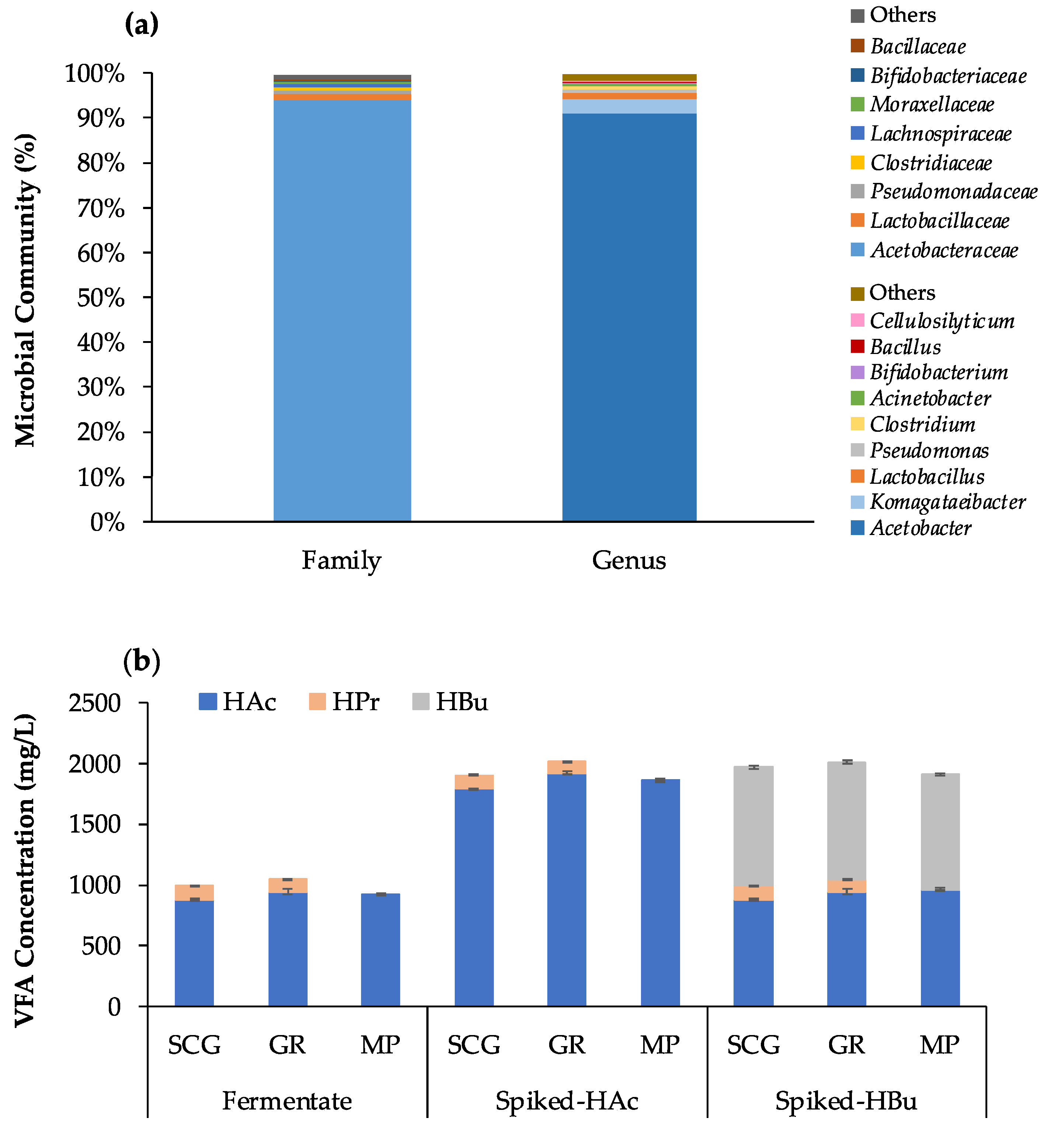
The applicability of the developed method was investigated in mixed-acid fermentation samples with different matrices from plant materials (SCG, GR, and MP). From 16S rRNA gene sequence analysis of the microbial consortium used for fermentation, two bacteria phyla were identified with relative abundances above 1.0%, whereas Proteobacteria (95.4%) and Firmicutes (4.0%) were the most abundant phyla accounting for 99.4% of the total bacteria population. Two bacteria families with relative abundances above 1.0% were identified in this study, whereas Acetobacteraceae was the most dominant family accounting for 93.9% of the total bacteria community, followed by Lactobacillaceae (1.5%) (Figure 5a). Three bacteria genera were found with relative abundances of more than 1.0%. The most abundant genus was Acetobacter accounting for 93.9% of the total population, followed by Komagataeibacter (3.1%) and Lactobacillus (1.5%).
According to the dominant genus Acetobacter, the main VFA product in the fermentation samples was acetic acid with a small amount of propionic acid. The VFA profile of the initial fermentation samples and the spiked samples representing mixed-VFAs are shown in Figure 5b. Fermentation samples were spiked with acetic acid and butyric acid to evaluate the performance of the developed method for VFA determination in real fermentation matrices. Acetic acid, propionic acid, and butyric acid were commonly found in acidogenic fermentation [10,18,19]. The variety of VFAs produced depends on biomass feedstock, microbial inoculum, and operational conditions [11,18,19].
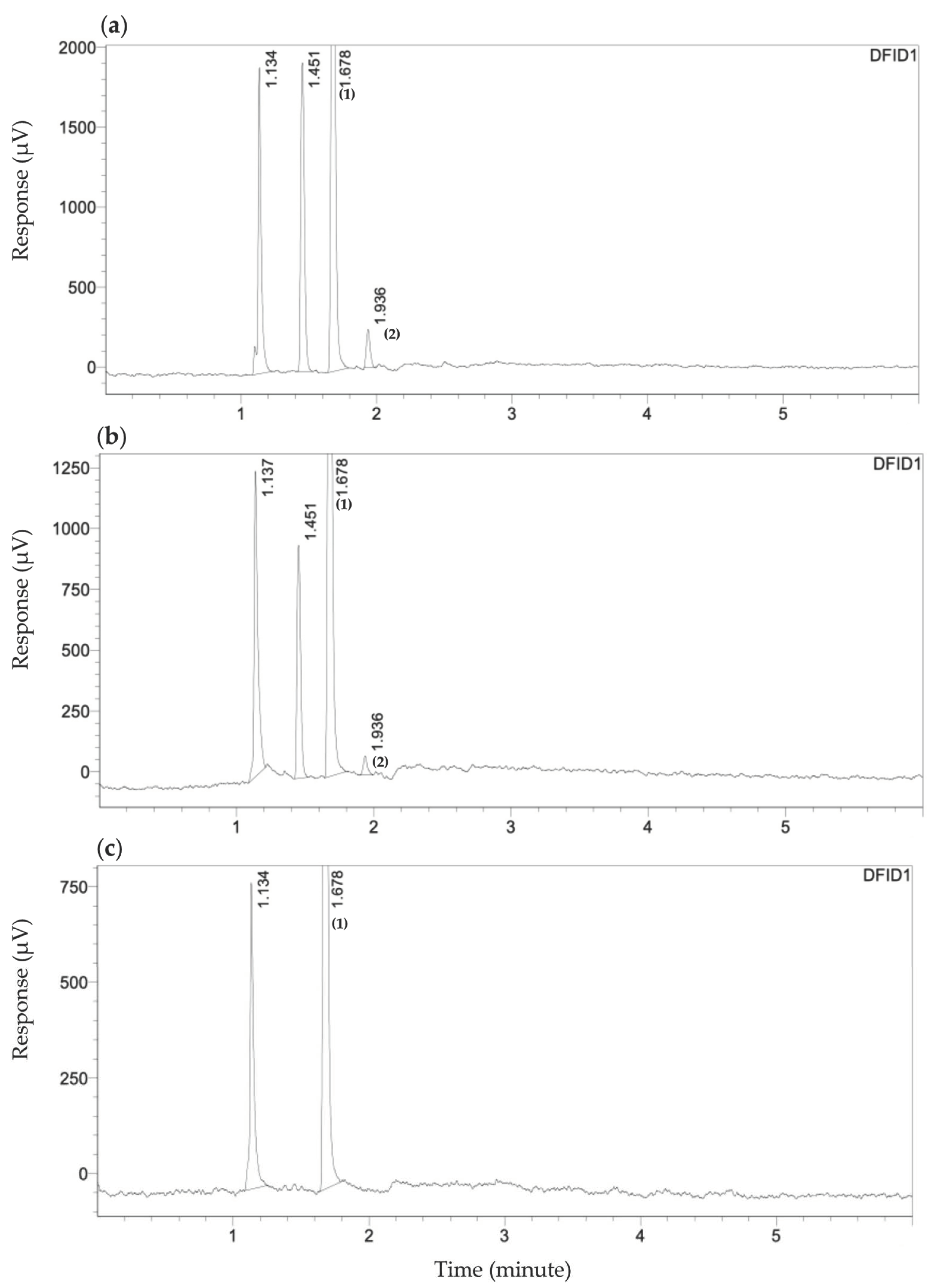
The GC chromatograms of the initial fermentation samples showing volatile compounds (VFAs and alcohols) were presented in Figure 6. The samples from acidogenic fermentation contained acetic acid as a major VFA component with a small amount of propionic acid and ethanol.
Different VFA concentrations were spiked into the samples with the added concentrations of 500, 750, 1000, and 1500 mg/L for acetic acid and 1000 mg/L for butyric acid. For the modified spectrophotometric method, the measured VFA concentrations of the native fermentation samples were 923–1047 and 923–2547 mg/L after spiking. The selected concentration ranges represented acidogenic fermentation which the VFA concentrations are typically high.
The performance of the developed method was determined based on the theoretical VFA concentrations added and compared to the GC method, as shown in Table 3. The accuracy and precision of the modified method applied to the spiked fermentation samples were 94.68–106.50% and 2.35–9.26%, respectively. In comparison, the accuracy and precision of the GC method were 94.42–99.13% and 0.17–1.93%, respectively.
In this experiment, some native fermentation samples (SCG1 and GR1) containing mixed acids with a small amount of propionic acid (in the range of 100–124 mg/L), the accuracy of the developed method on the total VFA determination in native SCG1 and GR1 samples were in the range 102.86–106.37%, which is slightly higher than that of the analytical GC method. However, the results from the two methods were not significantly different (p-value > 0.05). In comparison, the accuracy of native MP1 containing only acetic acid was slightly better than that of SCG1 and GR1 (102.08%). The accuracy on the fermentation samples and the spiked samples (500–1500 mg/L VFA added) were in a good accuracy and precision range.
Among 18 fermentation samples tested, in most samples, the VFA results measured by the modified method were not significantly different (p-value > 0.05) from those measured by GC, as shown in Figure 7. The spectrophotometric results from SCG samples tended to have a higher measured value than the GC method. This might be attributed to the interfering components in the complex plant-derived matrix since SCG contains a high level of polysaccharides and phenolic compounds. These compounds extracted during the fermentation process showed a high absorbance value at the wavelengths close to the wavelength used in the developed method (513 nm) [43]. Thus, these compounds could interfere with the measurement.
The original Montgomery’s study validated the application of the method on samples from crude sewage sludge, digested sewage sludge, and effluent from bacon factory by spiking 200–600 mg/L acetic acid. The accuracy can be calculated at 95.0–120.0%. The spectrophotometric method developed in this study showed extended advantages on different fermentation matrices with the recovered analysis of spiked acetic and butyric acid at higher concentrations (500–1500 mg/L) and slightly better accuracy (94.7–106.5%). A developed method by Chatterjee et al. [32] was tested on anaerobic digestate samples containing VFAs concentration in the range of 394–1662 mg/L. The accuracy of the modified spectrophotometric method compared to VFA concentration measured by GC was in the range of 99.18–116.22%. The modified spectrophotometric method showed an improvement over the distillation method, which obtained only 66.2–76.1% recovery with respect to results measured by the modified method.
The modified method accurately determined both small and large VFA compounds, represented by acetic acid and butyric acid. These VFAs are commonly found in acidogenic fermentation and anaerobic digestion [9,17,18]. The sensitivity of individual VFA compounds toward a measuring technique is different; for example, the peak area or voltage signal detected by GC. Similarly, the different sensitivity in spectroscopic absorption of each VFA compound affects its concentrations read. For mixed-type VFA fermentation, the ratio of major VFA components in the samples should be quantified or known to prepare the calibration curve accordingly. Thus, the modified method can be used for routine monitoring of VFA levels.

3.4. The Improvement of the Modified Spectrophotometric Method

The comparison of this proposed method and the previous developments, in terms of VFA type, detection range, the total volume of the reaction, precision, and accuracy, is presented in Table 4. This study validated the precision and accuracy of the determination of all C2–C6 VFAs, including linear and branched chains, while the previous studies were validated solely with acetic acid. In this study, the accuracy of acetic acid determination was 96.98–105.88%, comparable to previous studies (82.10–115.35%) [32,40]. The standard curves developed in this study were in the range of 250–5000 (R2 = 0.9872–0.9993), compared to 10–1200 (R2 was not reported) and 100–1400 mg/L (R2 = 0.9920) from the previous studies of Siedlecka et al. [40] and Chatterjee et al. [32], respectively. As mentioned previously in the methods, a very high concentration of chemicals resulted in intense color production and high absorbance. According to Beer–Lambert’s law, the absorbance higher than a specific value (normally around one) will have a non-linear relationship with sample concentration, caused by the shading effect of chromophore molecules to each other or so-called inhomogeneity in the optical medium [44]. After the hydroxamic reaction, Chatterjee et al. [32] reported the need for a dilution step to reduce the color before measuring absorbance (final volume after dilution is 49.7 mL), in which the final absorbance was still high (1.8 at an acetic concentration of 1400 mg/L). This study modified the method by reducing reaction concentration and adjusting the chemical ratio in each step to obtain ready-to-measure final color. As a result, the developed method in this study can increase the detectable concentration up to 5000 mg/L with satisfactory linearity, which could benefit the measurement of high VFA concentration in fermentation samples. Reducing the total volume of the reaction to 4.3 mL per sample asserts the advantages of this developed assay more than previously proposed methods. This developed assay also consumed around 15 min to get the concentration result. The spectrophotometric method developed in this study is rapid, simple, and accurate for VFA determination and thus suitable for routine analysis of a large number of samples.
Among simple techniques for determining total VFA concentration, a titration method has been used for monitoring VFAs in fermentation process. The titration method was firstly proposed by DiLallo and Albertson as two-point back titration [45]. The method requires adding standard acid and base and includes boiling to remove carbonic acid and CO2, which are prone to gross inaccuracy. Boiling, in particular, could result in losing a fraction of VFAs, and the back-titration (increase pH from 4.0 to 7.0) incorporates only 80% of VFAs in the measurement [30]. Although several titration methods were developed after that, such as 5 and 8 pH point titration [46], they are not very precise when applied to real fermentation samples with a complex matrix containing high concentrations of interfering components [31]. Therefore, the spectrophotometric method would offer a better recovery, be more specific to VFAs, and be less sensitive to interferences.
Similar to the traditional titration method, the limitation of the spectrophotometric method is on separating individual VFAs. Thus, it can only determine the total concentration of VFAs present in the samples. Identifying VFA type and measuring individual VFA concentration requires an analytical method with the separation techniques, such as GC and high-performance liquid chromatography (HPLC). An analytical method with the separation techniques is needed to acquire information on particular VFA types to reflect the stages of fermentation or biological processes. In addition, the chromatographic methods provide a high resolution, low limits of detection (LOD), a wide linear dynamic range (LDR), and good reproducibility [27]. However, the limitations of these methods are the requirement of a skilled operator, high cost, and intensive maintenance, which reduce their advantage for routine monitoring purposes [27]. Therefore, a cheap and straightforward VFA determination method with acceptable accuracy is essential for monitoring a known fermentation process.
For a spectrophotometric method, VFA concentration is determined based on the absorbance value of all VFA types combined. With a known ratio of major VFAs, an adjusted calibration curve or mixed-acid standard can be employed to improve the measurement’s accuracy. In other words, the accuracy would be off by the differences in absorbance sensitivity of the VFAs underestimated. In this experiment, using the fermentation samples containing mixed acids with a small amount of propionic acid, the accuracy of the developed method on the total VFA determination in SCG1 and GR1 samples was in the range of 102.86–106.37%, which is slightly higher than that of the analytical GC method. However, the results from the two methods were not significantly different (p-value > 0.05). Further related research would include improving the absorbance sensitivity of individual VFAs to be more comparable, the characterization of interferences and correction, and the potential applications to other fermentation processes.
In this study, the spectrophotometric method was developed for rapid and straightforward monitoring VFA in the mixed-acid fermentation of organic residues, typically containing a high level of VFAs (acetic, propionic, and butyric acids) [16,17,18]. All C2–C6 VFAs and real fermentation samples proved the method’s accuracy and precision. VFAs or short-chain fatty acids (SCFA) can also be found in other fermentation matrices for example, fermented food by Lactobacillus [47]. VFAs are often produced by fermentation of lactose, lipolysis, and degradation of amino acids, which greatly influence the taste of cheese or fermented milk products [48,49]. This situation illustrates the benefit of this modified spectrophotometric method for monitoring VFA-related metabolic reactions in various bioprocesses. However, since this method is based on the esterification of VFAs, it is likely to be interfered with by traces of dissolved esters in fermentation matrices [39], thus affecting the analysis accuracy. In wine fermentation, for instance, 2-phenylethyl acetate and isoamyl acetate, the two acetate esters produced by yeast in genera Hanseniaspora and Pichia [50], might cause a false positive. In fermented dairy products, the analysis of VFAs might be affected by the composition variables, e.g., cheese salt and fat contents [48]. Other fatty acids might also be esterified and interfere with the analysis. Moreover, colored and colloidal matrices generally interfere with the spectrophotometric method. Further effort should be given to verify this developed method on real fermentation samples with different compositions to evaluate its application.
Through the simple spectrophotometric method developed for detecting a high range of VFA concentration, after clarification, fermented liquid can be directly used for analysis. However, the sample with low VFAs, such as effluent from a healthy anaerobic digester, might need to be up-concentrated to be detectable by this method. Up-concentration of VFAs has been performed by solvent extraction using, for example, n-octanol, ionic liquid (trihexyl(tetradecyl)phosphonium chloride), dimethyl carbonate, dichloromethane, and methyl-tert-butyl-ether [38,51,52]. In future work, solvent extraction coupled with this developed spectrophotometric method could be investigated to extend the limit of quantification for application in low VFAs samples. However, the effect of solvent type on the accuracy and precision of the method needs to be verified.

4. Conclusions

Determining total VFA concentration is essential for monitoring the performance and stability of the fermentation process. The modified spectrophotometric method was developed for determining a high-ranged concentration of C2–C6 VFAs in mixed acid fermentation samples. A detection limit was improved for a high concentration range of 250–5000 mg/L. Using VFAs prepared in distilled water, the proposed method had an excellent accuracy of 95.51–105.11%, with a precision range of 1.67–5.58%. For the spiked fermentation samples, the accuracy and precision of the modified method were in the range of 94.68–106.50% and 2.35–9.26%, respectively, compared to the GC (94.42–99.13% and 0.17–1.93%). The developed method is applicable to measure all VFA types in acidogenic fermentation samples with a complex matrix. The developed spectrophotometric method offers a reliable and accurate measurement of VFAs. The developed method has advantages over others in lessened time, cost, and chemical usage allowing a practical application in the fermentation process. The limitation of the spectrophotometric method is on separating individual VFAs. Therefore, it only determines the concentration of total VFAs present in the samples. Further related research would include improving the absorbance sensitivity of individual VFAs, the characterization of interferences and correction, and the potential application to other fermentation processes.

Author Contributions

Conceptualization, N.A.; methodology, N.A. and P.L.; investigation, N.A., P.L., S.B. and P.K.; validation, S.B. and P.K.; formal analysis, N.A.; resources, N.A.; writing—original draft preparation, N.A; writing—review and editing, N.A. and P.L.; supervision, N.A. All authors have read and agreed to the published version of the manuscript.

Funding

The research was funded by Mahidol University.

Institutional Review Board Statement

Not applicable.

Informed Consent Statement

Not applicable.

Data Availability Statement

Not applicable.

Acknowledgments

This research project was supported by Mahidol University, and the CIF and CNI Grant, Faculty of Science, Mahidol University. The authors thank Chatsupat Saengcheewin and Napat Aroonpai for helping with data and statistical analyses, and Ruihong Zhang and Ke Wang from the Department of Biological and Agricultural Engineering, University of California, Davis, USA for providing support on research facility.

Conflicts of Interest

The authors declare no conflict of interest.

References

  1. Fritts, R.K.; McCully, A.L.; McKinlay, J.B. Extracellular metabolism sets the table for microbial cross-feeding. Microbiol. Mol. Biol. Rev. 2021, 85, e00135-20. [Google Scholar] [CrossRef] [PubMed]
  2. Lee, W.S.; Chua, A.S.M.; Yeoh, H.K.; Ngoh, G.C. A review of the production and applications of waste-derived volatile fatty acids. Chem. Eng. J. 2014, 235, 83–99. [Google Scholar] [CrossRef]
  3. Fei, Q.; Chang, H.N.; Shang, L.; Kim, N.; Kang, J. The effect of volatile fatty acids as a sole carbon source on lipid accumulation by Cryptococcus albidus for biodiesel production. Bioresour. Technol. 2011, 102, 2695–2701. [Google Scholar] [CrossRef] [PubMed]
  4. Munir, S.; Jamil, N. Polyhydroxyalkanoates (PHA) production in bacterial co-culture using glucose and volatile fatty acids as carbon source. J. Basic Microbiol. 2018, 58, 247–254. [Google Scholar] [CrossRef]
  5. Wang, K.; Zhang, R. Production of Polyhydroxyalkanoates (PHA) by Haloferax mediterranei from Food Waste Derived Nutrients for Biodegradable Plastic Applications. J. Microbiol. Biotechnol. 2020, 31, 338–347. [Google Scholar] [CrossRef]
  6. Wainaina, S.; Lukitawesa; Kumar Awasthi, M.; Taherzadeh, M.J. Bioengineering of anaerobic digestion for volatile fatty acids, hydrogen or methane production: A critical review. Bioengineered 2019, 10, 437–458. [Google Scholar] [CrossRef] [Green Version]
  7. Kim, N.J.; Lim, S.J.; Chang, H.N. Volatile Fatty Acid Platform: Concept and Application; Wiley: Hoboken, NJ, USA, 2018; Volume 1, pp. 173–190. [Google Scholar]
  8. Van Aarle, I.M.; Perimenis, A.; Lima-Ramos, J.; De Hults, E.; George, I.F.; Gerin, P.A. Mixed inoculum origin and lignocellulosic substrate type both influence the production of volatile fatty acids during acidogenic fermentation. Biochem. Eng. J. 2015, 103, 242–249. [Google Scholar] [CrossRef]
  9. Aramrueang, N.; Zhang, R.; Liu, X. Application of biochar and alkalis for recovery of sour anaerobic digesters. J. Environ. Manag. 2022, 307, 114538. [Google Scholar] [CrossRef]
  10. Sukphun, P.; Sittijunda, S.; Reungsang, A. Volatile Fatty Acid Production from Organic Waste with the Emphasis on Membrane-Based Recovery. Fermentation 2021, 7, 159. [Google Scholar] [CrossRef]
  11. Aramrueang, N.; Rapport, J.; Zhang, R. Effects of hydraulic retention time and organic loading rate on performance and stability of anaerobic digestion of Spirulina platensis. Biosyst. Eng. 2016, 147, 174–182. [Google Scholar] [CrossRef]
  12. Chen, Y.; Zhang, X.; Chen, Y. Propionic acid-rich fermentation (PARF) production from organic wastes: A review. Bioresour. Technol. 2021, 339, 125569. [Google Scholar] [CrossRef] [PubMed]
  13. Yu, P.; Tu, W.; Wu, M.; Zhang, Z.; Wang, H. Pilot-scale fermentation of urban food waste for volatile fatty acids production: The importance of pH. Bioresour. Technol. 2021, 332, 125116. [Google Scholar] [CrossRef] [PubMed]
  14. Atasoy, M.; Cetecioglu, Z. Butyric acid dominant volatile fatty acids production: Bio-augmentation of mixed culture fermentation by Clostridium butyricum. J. Environ. Chem. Eng. 2020, 8, 104496. [Google Scholar] [CrossRef]
  15. Pratto, B.; Chandgude, V.; de Sousa Junior, R.; Cruz, A.J.G.; Bankar, S. Biobutanol production from sugarcane straw: Defining optimal biomass loading for improved ABE fermentation. Ind. Crops Prod. 2020, 148, 112265. [Google Scholar] [CrossRef]
  16. Panda, S.K.; Mishra, S.S.; Kayitesi, E.; Ray, R.C. Microbial-processing of fruit and vegetable wastes for production of vital enzymes and organic acids: Biotechnology and scopes. Environ. Res. 2016, 146, 161–172. [Google Scholar] [CrossRef]
  17. Tripetchkul, S.; Kusuwanwichid, S.; Koonsrisuk, S.; Akeprathumchai, S. Utilization of wastewater originated from naturally fermented virgin coconut oil manufacturing process for bioextract production: Physico-chemical and microbial evolution. Bioresour. Technol. 2010, 101, 6345–6353. [Google Scholar] [CrossRef] [PubMed]
  18. Jayakrishnan, U.; Deka, D.; Das, G. Enhancing the volatile fatty acid production from agro-industrial waste streams through sludge pretreatment. Environ. Sci. Water Res. Technol. 2019, 5, 334–345. [Google Scholar] [CrossRef]
  19. Wijngaard, H.H.; Rößle, C.; Brunton, N. A survey of Irish fruit and vegetable waste and by-products as a source of polyphenolic antioxidants. Food Chem. 2009, 116, 202–207. [Google Scholar] [CrossRef]
  20. Aramrueang, N.; Asavasanti, S.; Khanunthong, A. Chapter 1—Leafy Vegetables. Academic Press: Cambridge, MA, USA, 2019; pp. 245–272. [Google Scholar]
  21. Aramrueang, N.; Zicari, S.M.; Zhang, R. Characterization and compositional analysis of agricultural crops and residues for ethanol production in California. Biomass Bioenergy 2017, 105, 288–297. [Google Scholar] [CrossRef]
  22. Ye, H.; Wang, Q.; Zhu, F.; Feng, G.; Yan, C.; Zhang, J. Antifungal Activity of Alpha-Mangostin against Colletotrichum gloeosporioides In Vitro and In Vivo. Molecules 2020, 25, 5335. [Google Scholar] [CrossRef]
  23. Silva, C.S.; Gabriel, C.; Cerqueira, F.; Manso, M.C.; Vinha, A. Coffee Industrial Waste as a Natural Source of Bioactive Compounds with Antibacterial and Antifungal Activities; Formatex Research Center: Badajoz, Spain, 2015; pp. 131–136. [Google Scholar]
  24. Zhou, Y.-Q.; Liu, H.; He, M.-X.; Wang, R.; Zeng, Q.-Q.; Wang, Y.; Ye, W.-C.; Zhang, Q.-W. A Review of the Botany, Phytochemical, and Pharmacological Properties of Galangal, 1st ed.; Elsevier: Amsterdam, The Netherlands, 2018; Volume 7, pp. 351–396. [Google Scholar]
  25. Chouni, A.; Paul, S. A review on phytochemical and pharmacological potential of Alpinia galanga. Pharmacogn. J. 2018, 10, 9–15. [Google Scholar] [CrossRef] [Green Version]
  26. Wang, B.; Liu, F.; Li, Q.; Xu, S.; Zhao, X.; Xue, P.; Feng, X. Antifungal activity of zedoary turmeric oil against Phytophthora capsici through damaging cell membrane. Pestic. Biochem. Physiol. 2019, 159, 59–67. [Google Scholar] [CrossRef] [PubMed]
  27. Fernández, R.; Dinsdale, R.M.; Guwy, A.J.; Premier, G.C. Critical analysis of methods for the measurement of volatile fatty acids. Crit. Rev. Environ. Sci. Technol. 2016, 46, 209–234. [Google Scholar] [CrossRef]
  28. Mu, Z.-X.; He, C.-S.; Jiang, J.-K.; Zhang, J.; Yang, H.-Y.; Mu, Y. A modified two-point titration method for the determination of volatile fatty acids in anaerobic systems. Chemosphere 2018, 204, 251–256. [Google Scholar] [CrossRef]
  29. Vikas, O.; Mridul, U. Bioconversion of papaya peel waste into vinegar using Acetobacter aceti. Int. J. Sci. Res. 2014, 3, 409–411. [Google Scholar]
  30. Lahav, O.; Morgan, B. Titration methodologies for monitoring of anaerobic digestion in developing countries—A review. J. Chem. Technol. Biotechnol. 2004, 79, 1331–1341. [Google Scholar] [CrossRef] [Green Version]
  31. Sun, H.; Guo, J.; Wu, S.; Liu, F.; Dong, R. Development and validation of a simplified titration method for monitoring volatile fatty acids in anaerobic digestion. Waste Manag. 2017, 67, 43–50. [Google Scholar] [CrossRef]
  32. Chatterjee, B.; Radhakrishnan, L.; Mazumder, D. New approach for determination of volatile fatty acid in anaerobic digester sample. Environ. Eng. Sci. 2018, 35, 333–351. [Google Scholar] [CrossRef]
  33. Lützhøft, H.-C.H.; Boe, K.; Fang, C.; Angelidaki, I. Comparison of VFA titration procedures used for monitoring the biogas process. Water Res. 2014, 54, 262–272. [Google Scholar] [CrossRef]
  34. Li, X.-Y.; Feng, Y.; Duan, J.-L.; Feng, L.-J.; Wang, Q.; Ma, J.-Y.; Liu, W.-Z.; Yuan, X.-Z. Model-based mid-infrared spectroscopy for on-line monitoring of volatile fatty acids in the anaerobic digester. Environ. Res. 2022, 206, 112607. [Google Scholar] [CrossRef]
  35. Stockl, A.; Lichti, F. Near-infrared spectroscopy (NIRS) for a real time monitoring of the biogas process. Bioresour. Technol. 2018, 247, 1249–1252. [Google Scholar] [CrossRef] [PubMed]
  36. Wang, M.; Wang, H.; Zheng, H.; Uhrin, D.; Dewhurst, R.J.; Roehe, R. Comparison of HPLC and NMR for quantification of the main volatile fatty acids in rumen digesta. Sci. Rep. 2021, 11, 1–15. [Google Scholar] [CrossRef] [PubMed]
  37. Lee, J.H.; Zhu, J. Optimizing secondary electrospray ionization high-resolution mass spectrometry (SESI-HRMS) for the analysis of volatile fatty acids from gut microbiome. Metabolites 2020, 10, 351. [Google Scholar] [CrossRef]
  38. Ghidotti, M.; Fabbri, D.; Torri, C.; Piccinini, S. Determination of volatile fatty acids in digestate by solvent extraction with dimethyl carbonate and gas chromatography-mass spectrometry. Anal. Chim. Acta 2018, 1034, 92–101. [Google Scholar] [CrossRef] [PubMed]
  39. Montgomery, H.; Dymock, J.F.; Thom, N. The rapid colorimetric determination of organic acids and their salts in sewage-sludge liquor. Analyst 1962, 87, 949–955. [Google Scholar] [CrossRef]
  40. Siedlecka, E.M.; Kumirska, J.; Ossowski, T.; Glamowski, P.; Gołębiowski, M.; Gajdus, J.; Kaczyński, Z.; Stepnowski, P. Determination of volatile fatty acids in environmental aqueous samples. Pol. J. Environ. Stud. 2008, 17, 351–356. [Google Scholar]
  41. Silva, M.A.; Correa, R.A.; Maria, G.d.O.; Antoniosi Filho, N.R. A new spectrophotometric method for determination of biodiesel content in biodiesel/diesel blends. Fuel 2015, 143, 16–20. [Google Scholar] [CrossRef] [Green Version]
  42. Thompson, A.R. A colorimetric method for the determination of esters. Aust. J. Chem. 1950, 3, 128–135. [Google Scholar] [CrossRef]
  43. Ballesteros, L.F.; Cerqueira, M.A.; Teixeira, J.A.; Mussatto, S.I. Characterization of polysaccharides extracted from spent coffee grounds by alkali pretreatment. Carbohydr. Polym. 2015, 127, 347–354. [Google Scholar] [CrossRef] [Green Version]
  44. Tolbin, A.Y.; Pushkarev, V.E.; Tomilova, L.G.; Zefirov, N.S. Threshold concentration in the nonlinear absorbance law. Phys. Chem. Chem. Phys. 2017, 19, 12953–12958. [Google Scholar] [CrossRef]
  45. DiLallo, R.; Albertson, O.E. Volatile acids by direct titration. J. Water Pollut. Control Fed. 1961, 33, 356–365. [Google Scholar]
  46. Vannecke, T.; Lampens, D.; Ekama, G.; Volcke, E. Evaluation of the 5 and 8 pH point titration methods for monitoring anaerobic digesters treating solid waste. Environ. Technol. 2015, 36, 861–869. [Google Scholar] [CrossRef] [PubMed]
  47. Hati, S.; Patel, M.; Mishra, B.K.; Das, S. Short-chain fatty acid and vitamin production potentials of Lactobacillus isolated from fermented foods of Khasi Tribes, Meghalaya, India. Ann. Microbiol. 2019, 69, 1191–1199. [Google Scholar] [CrossRef]
  48. Innocente, N.; Moret, S.; Corradini, C.; Conte, L. A rapid method for the quantitative determination of short-chain free volatile fatty acids from cheese. J. Agric. Food Chem. 2000, 48, 3321–3323. [Google Scholar] [CrossRef]
  49. Nakae, T.; Elliott, J. Production of volatile fatty acids by some lactic acid bacteria. II. Selective formation of volatile fattyacids by degradation of amino acids. J. Dairy Sci. 1965, 48, 293–299. [Google Scholar] [CrossRef]
  50. Rojas, V.; Gil, J.V.; Piñaga, F.; Manzanares, P. Studies on acetate ester production by non-Saccharomyces wine yeasts. Int. J. Food Microbiol. 2001, 70, 283–289. [Google Scholar] [CrossRef]
  51. Mkhize, N.T.; Msagati, T.A.; Mamba, B.B.; Momba, M. Determination of volatile fatty acids in wastewater by solvent extraction and gas chromatography. Phys. Chem. Earth 2014, 67, 86–92. [Google Scholar] [CrossRef]
  52. Reyhanitash, E.; Zaalberg, B.; Kersten, S.R.; Schuur, B. Extraction of volatile fatty acids from fermented wastewater. Sep. Purif. Technol. 2016, 161, 61–68. [Google Scholar] [CrossRef]
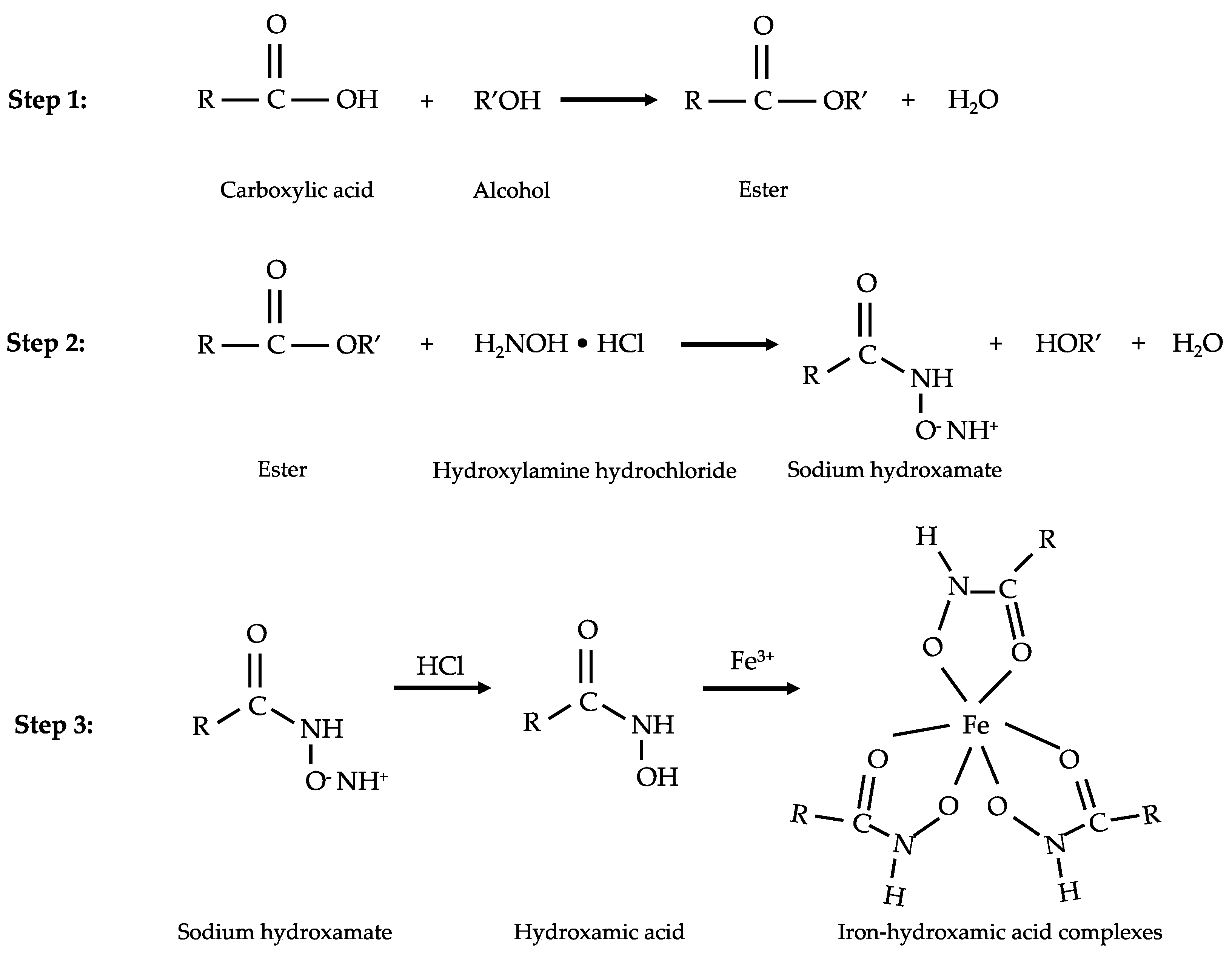
Figure 1. Reaction steps for VFA determination by the modified spectrophotometric method.
Fermentation 08 00202 g001
Figure 2. GC chromatograms of the standard VFAs: (1) acetic acid, (2) propionic acid, (3) iso-butyric acid, (4) butyric acid, (5) iso-valeric acid, (6) valeric acid, and (7) caproic acid at the concentrations of (a) 250 mg/L; (b) 2000 mg/L and (c) 5000 mg/L.
Fermentation 08 00202 g002
Figure 3. Calibration curves plotted between (a) the theoretical concentrations of C2–C6 VFAs versus the peak area for GC; (b) the theoretical concentrations of C2–C6 VFAs versus the absorbance at 513 nm for the developed method. Acetic acid (HAc), propionic acid (HPr), butyric acid (HBu), iso-butyric acid (Iso-HBu), valeric acid (HVa), iso-valeric acid (Iso-HVa), and caproic acid (HCa). Error bars represent the standard deviations for GC (n = 5), and the developed method (n = 3).
Fermentation 08 00202 g003
Figure 4. Comparison of VFA determination between theoretically added concentration (black) and the VFA determination by the developed method (grey) and GC (white). Acetic acid (HAc), propionic acid (HPr), butyric acid (HBu), iso-butyric acid (Iso-HBu), valeric acid (HVa), iso-valeric acid (Iso-HVa), and caproic acid (HCa). Error bars represent the standard deviations of the developed method and GC (n = 3) with statistical analysis. Statistical differences between each measurement were represented by letters (a–c). Measurements with the same letter are not significantly different (p-value > 0.05).
Fermentation 08 00202 g004
Figure 5. Relative abundance and composition of the microbial community at (a) Family; (b) Genus levels and the corresponding VFAs profile of the fermentation samples (fermentate) from spent coffee ground (SCG), galangal rhizome (GR), and mangosteen peel (MP), and the spiked samples with 1000 mg/L of acetic acid (spiked-HAc) and with 1000 mg/L of butyric acid (spiked-HBu). Error bars represent the standard deviations for the developed method and GC (n = 3).
Fermentation 08 00202 g005
Figure 6. GC chromatograms of the fermentation samples from (a) spent coffee ground (SCG); (b) galangal rhizome (GR) and (c) mangosteen peel (MP). Peak identifications are (1) acetic acid and (2) propionic acid.
Fermentation 08 00202 g006
Figure 7. VFA determination using the modified spectrophotometric (SP) method and GC for (a) spent coffee ground (SCG); (b) galangal root (GR) and (c) mangosteen peel (MP). Error bars represent the standard deviations the developed method and GC (n = 3). Statistical differences between each measurement were represented by letters (a–h). Measurements with the same letter are not significantly different (p-value > 0.05).
Fermentation 08 00202 g007
Table 1. Linearity and precision of the calibration curves for the developed spectrophotometric method and GC for VFA determination.
Acetic
Acid
Propionic
Acid
Butyric
Acid
Isobutyric
Acid
Valeric
Acid
Isovaleric
Acid
Caproic
Acid
Calibration curves of the developed method
Linear range (mg/L)250–5000250–5000250–5000250–5000250–5000250–5000250–5000
Slope ± SD0.1854 ± 0.00470.1227 ± 0.00170.0924 ± 0.00240.0526 ± 0.00250.0712 ± 0.00370.0294 ± 0.00110.0654 ± 0.0034
Intercept ± SD0.0114 ± 0.01710.0124 ± 0.00890.0042 ± 0.00730.0066 ± 0.00510.0089 ± 0.00740.0032 ± 0.00220.0174 ± 0.0032
Determination coefficient (R2) 0.99840.99850.99480.99890.99930.99920.9872
Precision (%) (n = 3)10.998.106.9412.149.3411.459.70
Calibration curves of GC method
Retention time (min)1.6771.9232.2862.0152.9472.4963.927
Linear range (mg/L)250–5000250–5000250–5000250–5000250–5000250–5000250–5000
Slope ± SD10,793 ± 79217,119 ± 151120,801 ± 154321,426 ± 137721,821 ± 118622,669 ± 252018,922 ± 1390
Intercept ± SD −937 ± 614−1035 ± 1049−957 ± 1705−1567 ± 930−1430 ± 1012−1158 ± 937−245 ± 1480
Determination coefficient (R2) 0.99930.99920.99950.99940.99970.99980.9997
Precision (%) (n = 5)11.2210.899.638.009.1715.0014.70
Table 2. Analytical results of VFA determination, the accuracy (%), and precision (% relative standard deviation; RSD) of the developed method (n = 3) and GC (n = 3).
AcidAdded 1
(mg/L)
SpectrophotometricGC
Measured 2
(mg/L)
Accuracy
(%)
Precision
(% RSD)
Measured 2 (mg/L)Accuracy
(%)
Precision
(% RSD)
Acetic20002019 ± 91100.934.491989 ± 3499.461.72
Propionic20002102 ± 110105.115.242013 ± 30100.651.49
Butyric20002032 ± 39101.621.921977 ± 4798.852.36
Isobutyric20002023 ± 38101.141.881941 ± 1897.030.92
Valeric20002033 ± 114101.665.581991 ± 1199.550.55
Isovaleric20002034 ± 34101.701.671986 ± 4599.292.25
Caproic20001910 ± 7795.514.031967 ± 6398.353.23
1 added concentration and 2 measured concentration.
Table 3. Analytical results of VFA determination in fermentation samples, and the accuracy (%) and precision (% relative standard deviation; RSD) of the developed method (n = 3) and GC (n = 3).
SampleInitial VFAs (mg/L)Added
Acetic Acid (mg/L)
Added
Butyric Acid (mg/L)
Estimated VFAs (mg/L)SpectrophotometricGC
Total Found (mg/L)Accuracy (%)Precision (% RSD)Total Found (mg/L)Accuracy (%)Precision (% RSD)
SCG 1992 ± 16--9921056 ± 85106.378.06992 ± 16100.001.58
SCG 2992 ± 16500-14921563 ± 99104.716.321436 ± 1296.230.80
SCG 3992 ± 16750-17421844 ± 104105.855.641659 ± 2195.231.24
SCG 4992 ± 161000-19922026 ± 93101.714.571903 ± 395.500.17
SCG 5992 ± 161500-24922558 ± 163102.646.352353 ± 4594.421.93
SCG 6992 ± 16-100019922120 ± 150106.437.081967 ± 2498.750.59
GR 11047 ± 21--10471077 ± 47102.864.371047 ± 21100.002.03
GR 21047 ± 21500-15471506 ± 11297.397.451488 ± 596.230.32
GR 31047 ± 21750-17971747 ± 16297.259.261736 ± 596.610.26
GR 41047 ± 211000-20472013 ± 6398.373.132018 ± 1398.600.66
GR 51047 ± 211500-25472528 ± 11599.254.562419 ± 1694.980.66
GR 61047 ± 21-100020471972 ± 5896.342.952010 ± 1398.220.57
MP 1923 ± 8--923942 ± 27102.082.84923 ± 8100.000.90
MP 2923 ± 8500-14231408 ± 3898.942.721410 ± 1299.130.82
MP 3923 ± 8750-16731618 ± 9296.725.681624 ± 1897.071.14
MP 4923 ± 81000-19232048 ± 48106.502.351861 ± 1496.780.75
MP 5923 ± 81500-24232294 ± 6594.682.832314 ± 2195.500.91
MP 6923 ± 8-100019231965 ± 47102.172.381906 ± 1999.131.01
Table 4. Comparison between the proposed spectrophotometric method and previous developments.
AcidConcentration Range of Standard Curve (mg/L)Total Volume of Reaction (mL)Accuracy
(%)
Precision
(% RSD)
References
Acetic-Caproic250–50004.395.51–105.111.67–5.58This study
Acetic250–50004.3100.934.49This study
Acetic10–120014.782.10–104.21.3–14.0[40]
Acetic100–140049.795.76–115.351.38–27.63[32]
Publisher’s Note: MDPI stays neutral with regard to jurisdictional claims in published maps and institutional affiliations.