Review

Novel Developments on Stimuli-Responsive Probiotic Encapsulates: From Smart Hydrogels to Nanostructured Platforms

by
Andres J. Garcia-Brand
1,†,
Valentina Quezada
1,†,
Cristina Gonzalez-Melo
1,
Angie D. Bolaños-Barbosa
2,
Juan C. Cruz
1,* and
Luis H. Reyes
2,*
1
Department of Biomedical Engineering, Universidad de los Andes, Bogotá 111711, Colombia
2
Product and Process Design Group (GDPP), Department of Chemical and Food Engineering, Universidad de los Andes, Bogotá 111711, Colombia
*
Authors to whom correspondence should be addressed.
These authors contributed equally to this work.
Fermentation 2022, 8(3), 117; https://doi.org/10.3390/fermentation8030117
Submission received: 11 February 2022 / Revised: 3 March 2022 / Accepted: 3 March 2022 / Published: 8 March 2022

Abstract

:
Biomaterials engineering and biotechnology have advanced significantly towards probiotic encapsulation with encouraging results in assuring sufficient bioactivity. However, some major challenges remain to be addressed, and these include maintaining stability in different compartments of the gastrointestinal tract (GIT), favoring adhesion only at the site of action, and increasing residence times. An alternative to addressing such challenges is to manufacture encapsulates with stimuli-responsive polymers, such that controlled release is achievable by incorporating moieties that respond to chemical and physical stimuli present along the GIT. This review highlights, therefore, such emerging delivery matrices going from a comprehensive description of addressable stimuli in each GIT compartment to novel synthesis and functionalization techniques to currently employed materials used for probiotic’s encapsulation and achieving multi-modal delivery and multi-stimuli responses. Next, we explored the routes for encapsulates design to enhance their performance in terms of degradation kinetics, adsorption, and mucus and gut microbiome interactions. Finally, we present the clinical perspectives of implementing novel probiotics and the challenges to assure scalability and cost-effectiveness, prerequisites for an eventual niche market penetration.

1. Introduction

The term “probiotic” has been complimented since its first appearance in the 1960s. It was initially defined as a substance secreted by microorganisms that has beneficial effects on the human body [1,2]. Then, in 1980, some specific characteristics were added to this definition. Additional claims stated that probiotics are “strains that have a beneficial impact, non-toxic, non-allergic, and nonpathogenic, available in large quantities as viable cells, suitable for the environment of the gut, and storable as well as stable” [1]. The Food and Agriculture Organization (FAO) and the World Health Organization (WHO) defined probiotics as “live microorganisms that, when administered in adequate amounts, confer a health benefit on the host” [1,2,3,4]. Additionally, both organizations have classified products containing live organisms into four categories: (a) live or active cultures; (b) probiotics in food or supplements without a health claim; (c) probiotics in food or supplements with a specific health claim; and (d) probiotic drugs [2].
Probiotic strains generally belong to the following genera: Lactobacillus, Bifidobacterium, Saccharomyces, Pediococcus, Streptococcus, and Leuconostoc [1]. Lactobacillus and Bifidobacterium are the most common strains. Different aspects need to be considered when selecting probiotic strains, and these include stomach pH and bile tolerance, adherence to epithelial surfaces, capacity for immunostimulation, antagonistic activity against pathogens, and antimutagenic and anticarcinogenic properties [5]. Probiotics have been successfully employed in manufacturing a wide variety of fermented products for daily consumption, including yogurt, kefir, sour pickles, milk, miso soup, and several soft cheeses [6]. The average probiotic consumption for a single person varies from 107 to 109 CFU/mg/day, whereas the significant benefit probiotic content in food must be of the order of 106 CFU/g [1,4]. Major health benefits have been attributed to these microorganisms, which nowadays can be used for the prevention and treatment of ailments such as liver disorders, cardiovascular diseases, dental caries, gastrointestinal inflammation, diarrhea, diabetes, obesity, and irritable bowel syndrome [1,3,6,7].
Probiotics can produce essential metabolites, including enzymes, vitamins, amino acids, peptides, exopolysaccharides, antioxidants, and anti-inflammatory agents. For example, some Bifidobacterium strains can produce B6 vitamin, while L. reuteri can produce cobalamin [8]. Some studies performed on pediatric patients who suffered from some kind of food allergy showed, with moderate certainty, that consumption of probiotics such as L. rhamnosius GG, LC705, L. casei LOCK 0900 and LOCK 0908, L. paracasei LOCK 0919, B. breve Bbi99, Propionibacterium freudenreichii ssp, or Shermanii JS could alleviate the symptoms caused by bovine lactose intolerance. This is because probiotics are thought to induce the production of β-galactosidase and lactase enzymes, helping to metabolize lactose quite effectively [8,9].
However, it is important to keep in mind that first, the strain must reach its site of action, usually the gut, and thus survive the physiological stress met during its ingestion, i.e., acid stomach and gut pH and the presence of biliary salts. Furthermore, its ingestion must not lead to any major risks for the host, maintaining its characteristics and remaining stable during the manufacturing process where it is usually incorporated into a delivery matrix [6]. A few strains can maintain viability after 1 h at a pH of 1, and most of them lose viability after 3 h at a pH of 3. The human stomach pH varies from 1 when fasting to 4.5 after eating. The process can take more than 3 h [10].
Different strategies exist to protect probiotics and their viability by edible carriers such as cheese, drinks, and bread [11,12]. Also, polymeric matrices have attracted significant attention for encapsulation, protection, and probiotic release [13]. Probiotics have evolved in sophistication from the first to the fourth generation. First-generation probiotics are either fresh or lyophilized cells without any coating. This has led to a low survival rate between 7% and 30%. Second-generation probiotics are incorporated into polymeric capsules or tablets with fillers. Usually, these strains show higher survival rates, but low performance due to rapid metabolite degradation. Third-generation probiotics are those encapsulated in natural, semi-synthetic, or synthetic polymers. The microcapsules are designed to be consumed gradually, which helps maintain the metabolites’ activity. Their structure can be a 3D matrix, a crosslinked construct, or have external coating. Fourth-generation probiotics are those incorporated into biofilms, which improve protection when transiting the gastrointestinal tract [14,15].
Encapsulation techniques are widely used for varied applications in the food industry, including masking and design of flavors, colors, and odors, improving the shelf life of products, protecting some components against nutritional loss, and regulating undesirable oxidative reactions. With probiotics, encapsulation provides protection from media effects and enhanced viability, and allows controlled dosing and handling of cells [16]. One of the biggest challenges when encapsulating is selecting effective and safe materials for the capsules’ manufacturing, an efficient release system, and proper production techniques (e.g., extrusion, emulsion, spray-drying, etc.). A much more comprehensive discussion of different methods used in the design of encapsulation microgels is given by McClements [13]. Additionally, it is vital to consider economic, regulatory, and consumption factors to assure scalability and successful market penetration [13,16].
Materials used for capsule manufacturing have diverse origins. The most common ones are derivates from cellulose, proteins, polysaccharides, carrageenan, gelatin, pectin, and alginate [16]. Materials are chosen depending on the characteristic physicochemical and structural features of the capsules, and generally, polysaccharides and proteins are selected due to their versatility. In this regard, natural polymers such as alginate, xanthan gum with divalent cations, casein gels, or gelatins have been chosen due to their ease of crosslinking, and the possibility of combining them to achieve different levels of mechanical resistance. Chitosan, lysine, or whey protein are used for the external coating of structures [15]. Some of the most important physicochemical properties of the materials to consider for a rational design are solubility, gelation mechanisms, degradability, and electric properties [13]. There are different methods to perform encapsulation processes such as injection (e.g., extrusion, atomization, and microfluidic), template techniques (e.g., emulsions), biopolymer phase separation, precipitation, reduction, drying, and more recently, biofilm formation to promote colonization and enhance the permanence of probiotics in the host intestinal mucosa [13,14].
The gastrointestinal tract comprises the mouth, esophagus, stomach, gut, and colon. Its microbiota concentration varies over the tract due to changes in pH and the presence of bile and enzymes. For example, in the stomach, such concentration is low (101 bacteria/g), increasing through the duodenum (103 bacteria/g), the jejunum (104 bacteria/g), and the ileum (107 bacteria/g). The largest concentration of microorganisms is found in the gut and colon, rounding 1011 to 1012 bacteria/g [17]. Such microbiota present a great phylogenetic diversity, allowing the required metabolic performance. The present microbiota are mainly composed of Bacteroidetes, Firmicutes, and Proteobacteria, and to a lesser extent by Actinobacteria, Clostridium, Enterobacter, Verrumicrobia, bacteriophages, viruses, and several Aspergillus, Candida, Cryptococcus, and Penicillium fungi genres [7,18,19].
The digestion and nutrient absorption processes are carried out by the small intestine, where there is an intestinal barrier composed of a mucosae layer and a cell component (intestinal epithelium and underlying lamina propria) that acts as a physical barrier to the microorganisms present in the gut [3]. Mucosae is composed of an outer loosely adhering layer and a dense inner layer. This last one is the first effective defense mechanism because of its high density, preventing most bacteria from penetrating and adhering [17]. The intestinal epithelium creates a separation between the gut lumen and the lamina propria, and comprises enterocytes, goblet, Paneth, and endoenterocrine cells. In contrast, the lamina propria is formed by dendritic cells, macrophages, and plasma cells that can engulf pathogens and eliminate apoptotic cells and waste [3].
The microbiota existing along the intestine have an immunological vigilance function that allows the detection of pathogens and stimulates the immune system to respond adequately. The pathogen control mechanism comprises four major steps: first, production of bacteriocin and other inhibitors; second, the competitive exclusion by the binding sites; third, stimulation of the immune response; and last, the inhibition of virulent genes or expression of proteins in pathogens [7]. Intestinal homeostasis occurs when the immune system establishes an equilibrium between commensal, mutualistic, and opportunistic bacteria. This happens when the microbiota communicates effectively with the immune system through a healthy intestinal barrier [3].
In this review, we describe the current polymeric delivery systems that could be implemented to fabricate novel stimuli-responsive encapsulates of probiotics that take advantage of the innate stimulus of the GIT while preserving the biological activity of the transported microorganisms. Contrary to previous reviews [20,21,22,23] in the field of probiotic encapsulates, here we intend to combine current and emerging polymeric delivery systems with stimuli responsiveness typically employed in the fields of tissue engineering and targeted delivery of pharmacological compounds. We exemplify how frequently used techniques for encapsulation can be extrapolated to rationally fabricate novel 3D matrices capable of responding actively to their surroundings and overcoming current issues, and taking advantage of the harsh conditions of the GIT to improve their functionalities. In addition, through this review, we point out pivotal points to set a straightforward approach to rational design potential stimuli-responsive encapsulates to reach clinical applicability. Hence, the scope of this review is focused only on the engineering of probiotics delivery to improve the GIT’s health.

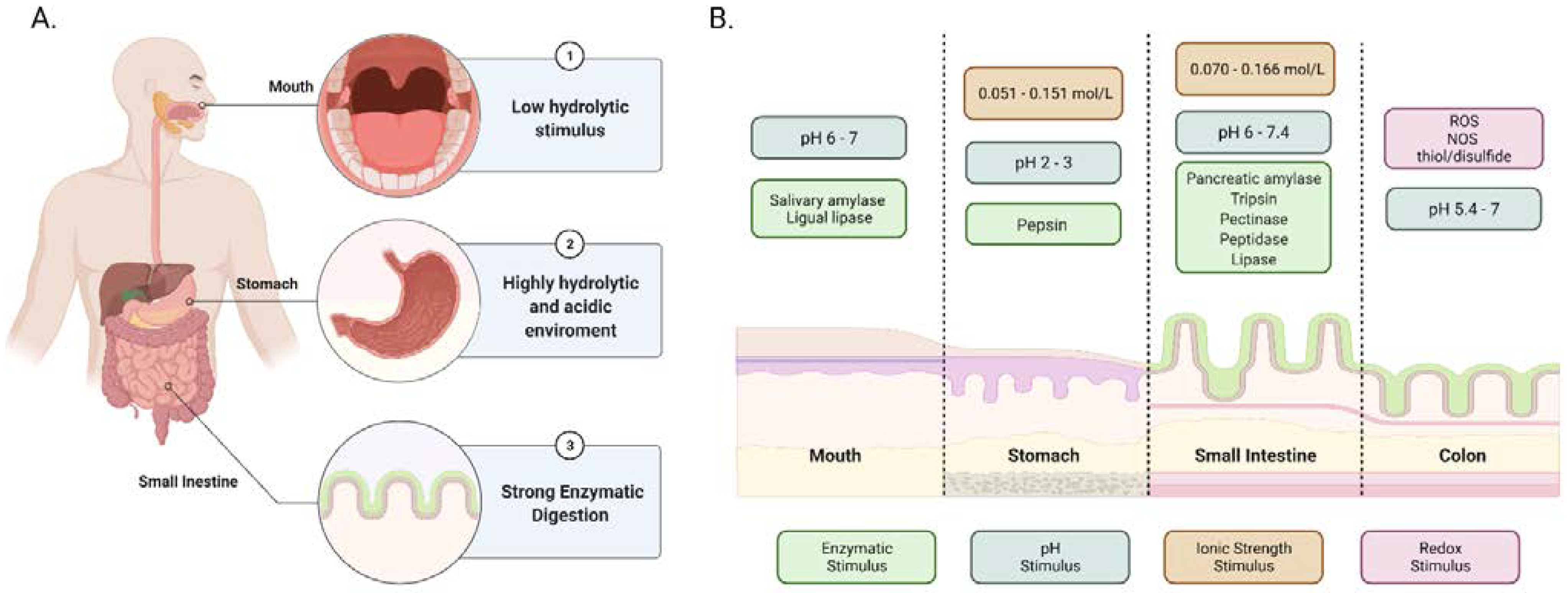
2. Gastrointestinal Tract (GIT) Stimuli as Tool for Design

Besides biomaterials formulation based on polymer science serving as a helpful tool for novel probiotic encapsulation, the need for a complete understanding of the environment for oral administration is a field of intensive research as it is key to assuring optimal performance of encapsulates in the GIT. Additionally, GIT conditions set a baseline to guide the required physicochemical properties for designing novel encapsulates. While in some cases, the harsh conditions of the GIT and its innate reactive stimuli appeared as a major obstacle to reaching the site of action of several encapsulates (Figure 1), novel engineering approaches have considered responses to such stimuli for the development of polymeric networks programmed to undergo volume changes, structural transitions, partial degradation, and structural network rearrangements [24]. Thus, the proper understating of the ionic strength, redox potential, pH, and enzymatic stimuli [25,26] present in the GIT is essential to rationally design multi-structured matrices capable of active response to the conditions of each GIT component while protecting the probiotic cargoes [27]. The addressable stimuli for developing biomaterials showing such dynamic responses are discussed below.

2.1. Enzymatic Degradation and pH Changes

Table 1 synthesized the function and characteristics of essential enzymes present throughout the GIT, but details will be explained below. The chemical digestion starts in the oral cavity and consists of ptyalin (or alpha-amylase) and lingual lipase. The bolus is then swallowed into the esophagus and transits along it to reach the stomach [28]. Pepsin is the principal enzyme involved in protein digestion (i.e., hydrolysis of proteins). These enzymes are active in the acidic medium of the stomach by the secreted hydrochloric acid (HCl) and bile coming from the liver. Oxyntic and pyloric glands are two types of glands in the gastric mucosa that help during the chemical digestion process taking place in the stomach. The oxyntic glands contain parietal cells that secrete HCl at a pH of 0.8 to eliminate pathogenic microorganisms and denature proteins for further enzymatic degradation by pepsin. The G-cells in the pyloric glands secrete the hormone gastrin, which stimulates the secretion of HCl by parietal cells [28].
The pancreas produces several digestive enzymes (i.e., pancreatic amylase, pancreatic lipase, trypsinogen, chymotrypsinogen, procarboxypeptidase, and proelastase) that reach the stomach to sections away from the acidic environment and the small intestine where they work optimally at more basic pH values (pH 6 to 7) due to the presence of bicarbonate secreted by the pancreas [28].
The small intestine comprises the duodenum, the jejunum, and the ileum. Pancreatic amylase is released into the duodenum to further digest incoming starch molecules. The acinar cells (exocrine cells of the pancreas) produce and transport the digestive enzymes. These cells have insulin receptors, where insulin binds to assure a normal acinar cell function, regulating the pancreatic amylase secretion and starch digestion [29].
The surface area available for absorption of peptides and proteins in the large intestine is reduced due to a lack of villi, microvilli, and crypts. Here, the considerable amount of bacteria present facilitates the digestion of residual food into caloric substances for subsequent absorption [30].

2.2. Ionic Strength and Redox Potential

Among the commonly found chemical stimuli present in the GIT, ionic strength and reduction–oxidation (redox) potency provide crucial cues for in situ extracellular matrix changes as a function of the systemic response of mammalian body functions [24]. Thus, the existence of monovalent and bivalent ions as well as reactive oxygen species (ROS) and nitrogen–oxygen species (NOS) are produced under different ionic strength and redox conditions, and therefore represent an addressable route to ionize polymeric structures [36].
The ionic strength of a solution or media is comprehended as the measure of the concentration of electrically charged species in a solution [37], which changes with the different fluids present throughout the GIT and can be advantageous for the release of on-cargo matrices commonly employed in oral drug release. Moreover, they can be exploited for controlling the response of polymeric biomaterials (e.g., swelling, dissolution, and degradation) [38,39,40,41,42], which, in turn, is critical for the survival of the probiotics [43,44,45]. Moreover, marked changes in the ionic potential of GIT fluids allow the control disaggregation of foods and ingestible encapsulates by destabilization of polypeptide bonds and polymeric structural rearrangements.
Typical ionic strength values vary from 0.051 to 0.151 mol/L in the gastric fluid of the stomach and between 0.070–0.166 mol/L in the intestinal fluid [46]. However, as ionic strength is defined by the reactive species at equilibrium present in each GIT compartment, the state of ionic power is also regulated by the food intake and location within the GIT [41].
The redox potency is used to describe a system’s overall reducing or oxidizing capacity through the presence of chemically reactive species [41]. Its relevance in biological science stems from its role in shaping the structure and function of microbial communities, as demonstrated by the precise communication in the gut microbiome and epithelial cells in the mucosa layers [47,48]. In particular, the redox balance in the GIT is mainly mediated by the presence of ROS, NOS, thiol/disulfide redox systems (i.e., glutathione/glutathione disulfide (GSH/GSSG), cysteine/cystine (Cys/CySS), and reduced and oxidized thioredoxin (Trx/TrxSS) redox couples), which are predominant molecules that interfere in preserving tissue redox homeostasis, metabolic functions, and cellular integrity. For instance, maintaining mucus fluidity and absorption of nutrients and protecting against chemical-induced oxidant injury at the luminal surface is largely achieved by dynamically adjusting the thiol–disulfide status [36].

3. Polymeric Platforms and Delivery Systems of Probiotics

The selection of optimal material for probiotic encapsulation and the appropriate processing route are key parameters to ensure an efficient delivery strategy, where early degradation by GIT stimuli and harsh conditions are largely avoided. Nevertheless, selecting the materials for superior performance in complex physiological environments such as the GIT is a task made challenging not only by the obstacles to be overcome to reach the target site but also by the need to maintain high biological activities and positive responses to the changing surroundings [49,50]. The fast-growing notion that encapsulates can be composed of active biomaterials should be driven by matching the material’s properties with expected responses through the GIT. Particularly, polymers exhibit versatile molecular moieties that have been widely exploited to fabricate chemical and physical delivery platforms with properties that can be finely tuned by adjusting interchain interactions [51]. Chemical polymeric scaffolds are formed by covalent bonds between adjacent chains, while the physical ones are maintained together by charged polyvalent surfactants or ion interactions [24].
Moreover, their versatile processability schemes facilitate obtaining different morphologies, functionalities, and the possibility to form composites with nanostructured materials in search of an enhanced response when subjected to a stimulus [52,53,54]. Typical polymeric encapsulates comprise microparticles, microspheres, microcapsules, hydrogels, and, more recently, nanocomposite 3D matrices. However, despite the success in preparing encapsulation systems capable of maintaining biological activity, more concerted stimuli responsiveness and higher resistance to harsh environments remain a significant constraint for moving to clinical setups [52]. Accordingly, the following section will discuss the most used materials and processing schemes for probiotic encapsulation in conjunction with alternatives to take advantage of their resultant properties to overcome the different challenges of each GIT compartment.

3.1. Polymeric Materials in Probiotic Encapsulation

In protected-delivery technologies, a suitable polymeric material should be able to preserve its core from adverse environmental conditions (e.g., reduce the acid-induced degradation of probiotics by gastric fluids in the stomach), exhibit inertness with the encapsulated materials, promote a controlled release of the encapsulate, achieve higher encapsulation efficiencies of bioactive compounds on a per mass basis and, ultimately, favor high levels of absorption into the targeted organs (i.e., the overall efficacy of the compounds) [27,55,56]. All the selected materials must also be biodegradable and biocompatible since they will be in direct contact with various types of cells [27,57].
Among the natural polymers, alginate, a heteropolysaccharide, has been applied successfully as a pH-sensitive material for the encapsulation of probiotic bacteria [58]. Alginate, extracted from algae, is composed of two monosaccharide units: α-L-guluronic acid and β-D-mannuronic acid, linked together by a β (1–4) glycosidic bond. Due to its toxicity, inexpensiveness, ease of processing, and biocompatibility, calcium alginate has been extensively employed in the encapsulation of probiotics [55,59,60,61,62,63]. Yet, calcium alginate encapsulates are chemically susceptible to disintegration in the presence of excess monovalent ions, Ca2+-chelating agents such as phosphate and citrate, and harsh chemical conditions (e.g., low pH) [64,65,66]. To increase the stability of alginate and decrease the loss of encapsulated material, alginate is usually coated with polycationic polymers such as chitosan and poly-L-lysine [27,57,66,67].
Chitosan is a very abundant polysaccharide obtained from chitin and is composed of (1,4)-linked 2-amino-deoxy-b-d-glucan [68]. It also shows high biocompatibility and biodegradability under physiological conditions. For these reasons, it has enabled several encapsulation applications in the food and pharmaceutical industries, including liposome coating, chitosan–alginate coating, controlled delivery of small molecules and biologicals, and the release of bioactive metabolites (e.g., essential oils, probiotics, vitamins, antioxidants, and flavors). The unique cationic character of chitosan allows forming multi-layer systems with anionic alginate for probiotic encapsulation, which can bring efficient protection to cargoes, reduced porosity, stability at various pH ranges, and reduced leakage of the encapsulated probiotic. In experiments where the gastric environment was simulated, chitosan coating was more efficient than poly-l-lysine and alginate coatings in protecting probiotics, which represents a possible route to overcome the challenges of oral delivery [68]. Furthermore, chitosan coating with drying processes can prolong the long-term storage of some probiotics at different temperatures [69].
Another frequently studied material for encapsulation is gelatin, a commercially available denatured protein obtained by the hydrolysis of collagen from the skin and bones of bovines or fish. This protein might be positively or negatively charged, depending on whether an acidic or alkaline method was used for its extraction, which can be exploited to design multifunctional controlled release systems [70]. For example, gelatin-coated alginate capsules and microspheres can protect labile drugs from the stomach’s acidic environment, enabling their release in the target intestinal area, whose environment has a basic pH [71]. Other studies have reported that the addition of fish gelatin significantly raised polymer matrix density and improved the physical integrity of alginate capsules because of a more stable and ordered 3D structure. Some other aspects, such as the survival rate of microorganisms during the GIT’s passage, are enhanced compared to non-encapsulated cells [72].
Poly-l-lysine is a cationic, non-ribosomal, non-toxic, biocompatible, biodegradable, and antimicrobial homopolymer produced by modified strains of Streptomyces albulus. Lysine is frequently used as a preservative and food additive [73], and studied as an alginate bead coating. The marked antimicrobial activity of most cationic polymers poses a major challenge because they tend to inhibit the growth of some microorganism strains, depending on pH and incubation times. That is the case not only of lysine but also of chitosan and polyethyleneimines [74]. However, different in vitro studies that have implemented mixed manufacturing techniques, such as freeze-frying, have shown that poly-l-lysine coatings for alginate capsules are well-suited to maintain cells’ growth and proliferation at low pH values and viability for a storage period of up to 16 weeks at 4 °C [22].
The materials mentioned above correspond to the most popular and studied polymers for survival enhancement and protection of probiotics in hostile environments, such as those found in the compartments of the GIT. Many other materials might be suitable for producing capsules and coatings for this application, including polyethyleneimines, poly(2-dimethyl(aminoethyl)methacrylate), dextran, pectin, Arabic gum, starch, sodium caseinate, polyvinyl alcohol, polyethylene glycol, polyacrylic acid, and succinylated or acylated carrageenan [22,73,75,76].

3.2. Hydrogels

Hydrogels have drawn particular attention among encapsulation alternatives for probiotics, given their ease of processing, the wide range of materials available for their fabrication, and their capability to form three-dimensional networks that protect the probiotic’s integrity [77]. Remarkably, the inertness of hydrogels in environments with high water activity makes them suitable to entrap molecules and microorganisms, and ensure their integrity and viability in physiological environments [78]. However, one of the main concerns about implementing hydrogels is the proper tuning of mechanical performance, along with optimal porosity to enable microorganism survival while maintaining sufficient cell entrapment and a considerable degree of swelling [79]. This has been addressed by chemically modified polymers through different routes, including the addition of ionizable functional groups, functionalized backbones, and combined polymeric blending [50,80,81].
Hydrogels can be classified mainly according to their (i) composition (homo or copolymers), (ii) network size (macrogels, microgels, nanogels), (iii) electrical charge (non-ionic, cationic, anionic, amphoteric or zwitterionic), and (iv) crosslinking method (physical or chemical) [24,82]. Moreover, recent studies (Table 2) have demonstrated the fabrication of chemically crosslinked platforms oriented toward the encapsulation of probiotics into hydrogel beads crosslinked with the aid of glutaraldehyde, Genipin, calcium chloride (CaCl2), and ferrous sulfate (FeSO4) [38,83,84,85]. Alginate, gelatin, and chitosan are the three main polymers of choice for hydrogel synthesis as they have proven to be effective in protecting probiotic cells from harsh environmental conditions [82,86].
One of the most intensive research areas is the development of strategies for tuning degradation rates and matrix porosities to improve their performance in protecting encapsulated living organisms [87]. This has been achieved by the supramolecular design of monomeric structures and by controlling polymerization reactions with carefully applied light and thermal stimuli [88,89]. Depending on the polymerization scheme selected (i.e., bulk polymerization, solution polymerization, suspension polymerization, emulsion polymerization, and graft polymerization), macroscopic and microscopic properties such as porosity and polymeric mesh size might change significantly [90]. Notably, the capacity of engineering the tortuosity and interconnectivity of hydrogels’ mesh has been reported as critical for the smart release of the encapsulated cargoes [91]. Further control over such a process can be achieved by chemical modifications with hygroscopic polymers such as polyethyleneglycol, which leads to the enhanced mucoadhesiveness of the encapsulates by both physical entanglement and hydrogen bonding with the base encapsulating polymer [92,93]. Therefore, the following section will discuss the fast-growing area of stimuli-responsive hydrogels, emphasizing how they can be activated, de-activated, or re-activated for a particular delivery purpose [94].

3.3. Microencapsulates

Microparticles, microcapsules, and microspheres usually made of food-grade polymers, such as alginate, chitosan, carboxymethyl cellulose, cellulose acetate phthalate, xanthan gum, starch, carrageenan, gelatin, and pectin [59,110], have demonstrated to be protective barriers of high performance against the GIT’s environmental conditions [111,112,113,114,115]. These microencapsulates’ dimensions usually range between 1 and 1000 µm [25]. According to recent studies, an effective microencapsulation system should maintain the stability of the probiotics during storage, protect them from the harsh conditions of the upper GIT, release them in the colon, and finally, promote their ability to colonize the mucosal surfaces [25,116,117,118].
Microparticles typically consist of a core composed of one to several ingredients surrounded by a wall or barrier of uniform or non-uniform thickness, either single-layered or multi-layered. The design of microencapsulated ingredients requires knowledge of (1) the core, (2) the materials for encapsulation, (3) the interactions between the core, matrix, and the environment, (4) the stability of the microencapsulated ingredients under storage conditions and when incorporated into food matrices, and (5) the mechanisms that control the release of the core [56,59,110]. Matrix degradation, and consequently, the release of its contents, can be controlled to occur at different times. Larger particles generally release encapsulated compounds more slowly and over more extended periods, while particle size reduction favors adhesiveness and therefore prolonged GI transit time, leading to a higher drug bioavailability [56,59,110,119].
Typical technologies employed for probiotic encapsulation include emulsification [120,121,122], emulsification and enzymatic gelation [123,124,125,126], atomization (e.g., spray drying [121,122,127], spray freeze drying [121,122,127,128,129]), coating and agglomeration [122,130,131,132], and extrusion [133,134]. Several recent microencapsulation studies of probiotics are summarized in Table 3.

3.4. Nanostructured Platforms

One of the leading strategies to improve microencapsulates’ tolerance to different GIT environments and ensure their efficient delivery is physical and chemical blending with stimuli-responsive and high mechanical performance nanomaterials [144,145,146]. This approach allows superior control over probiotic delivery due to the unique properties attainable by forming nanocomposites and the possibility of providing multimodal delivery platforms (i.e., including more than one encapsulated component) where survival of probiotics is increased substantially [147,148]. Accordingly, several nanostructured materials have been explored in the fabrication of next-generation delivery platforms, including polymeric and iron oxide-based nanoparticles, nanosheets (e.g., graphene oxide—GO and phyllosilicate clays), nanoliposomes, micelles, and nanoparticles derived from naturally occurring polymers such as nanocellulose and starch nanocrystals [149,150,151].
The dispersion of nanoparticles (NPs) into polymeric arrangements (to form nanocomposites) is attractive mainly due to their intrinsic capacity to fill out pores, therefore avoiding the diffusion of molecules such as hydrogen ions, bile salts, or digestive enzymes that may lead to the undesirable degradation of the encapsulated probiotics [152]. For instance, magnesium oxide (MgO) NPs have been incorporated into alginate–gelatin microgels, which resulted in a more stable encapsulation of probiotics when compared to unmodified microgels. The MgO NPs help neutralize the hydrogen ions present in the gastric fluids, which diminishes the acid-induced degradation of probiotics and maintains a neutral pH inside the microgels [27]. Alternatively, the use of NPs can help improve some physicochemical properties of the encapsulates, such as hardness, compressibility, cohesiveness, and adhesiveness. This is the case of chitosan NPs, which have been reported to enhance the mucoadhesive properties of hydrogels [153]. Through this approach, the interaction with the intestinal mucus is improved by the electrostatic interaction and physical entanglements of the chitosan-containing matrices facilitated by the positive charge of chitosan.
Another approach suggests that combining polymeric platforms with nanocrystals derived from polysaccharides (e.g., cellulose and starch) enhances mechanical stability and shelling properties [154], and increases the surface area for target delivery [155]. For instance, when cellulose nanocrystals (NCs) are combined with alginate during the microencapsulation process, the dissolution time increases while porosity is reduced significantly [156]. Moreover, ionic interaction between the material and the nanocellulose filler reduces the infiltration of gastric fluids, preventing the degradation of probiotics [157]. When starch NCs are implemented as fill-in alginate-based delivery platforms, thicker barrier protection against gastric and intestinal juices provides a stable mechanism to decrease probiotics mortality [149].
Remarkably, novel developments in the fabrication of nanocarbon-based materials (e.g., reduced graphene oxide, graphene quantum dots, graphene nanoribbons), silica-based nanocarriers, and inorganic nanoparticles have enabled the fabrication of emerging nanocomposites with improved mechanical strengths, high drug loading, and reduced toxicity [144,158,159]. These features have been reported to not only favor the controlled release of on-cargo molecules but also increase the survival of encapsulated living organisms (e.g., probiotics and mammalian cells) [160,161]. Similarly, clay mineral silicates have gained popularity given their unique cationic exchange properties that can be exploited to fabricate water barriers due to hydrogen bonding [162,163]. Kim and colleagues reported superior shape integrity for bentonite/alginate-based encapsulates during gastric fluid exposure and appropriate disintegration in the intestinal area upon oral administration in mice [158]. Relevant nanocomposites for probiotic encapsulation are listed in Table 4. Other recent reports suggest that incorporating nanostructured materials into delivery platforms makes it possible to accompany the encapsulation of probiotics with other therapeutic and bioactive molecules, thereby providing a route for multimodal controlled release [80,164].

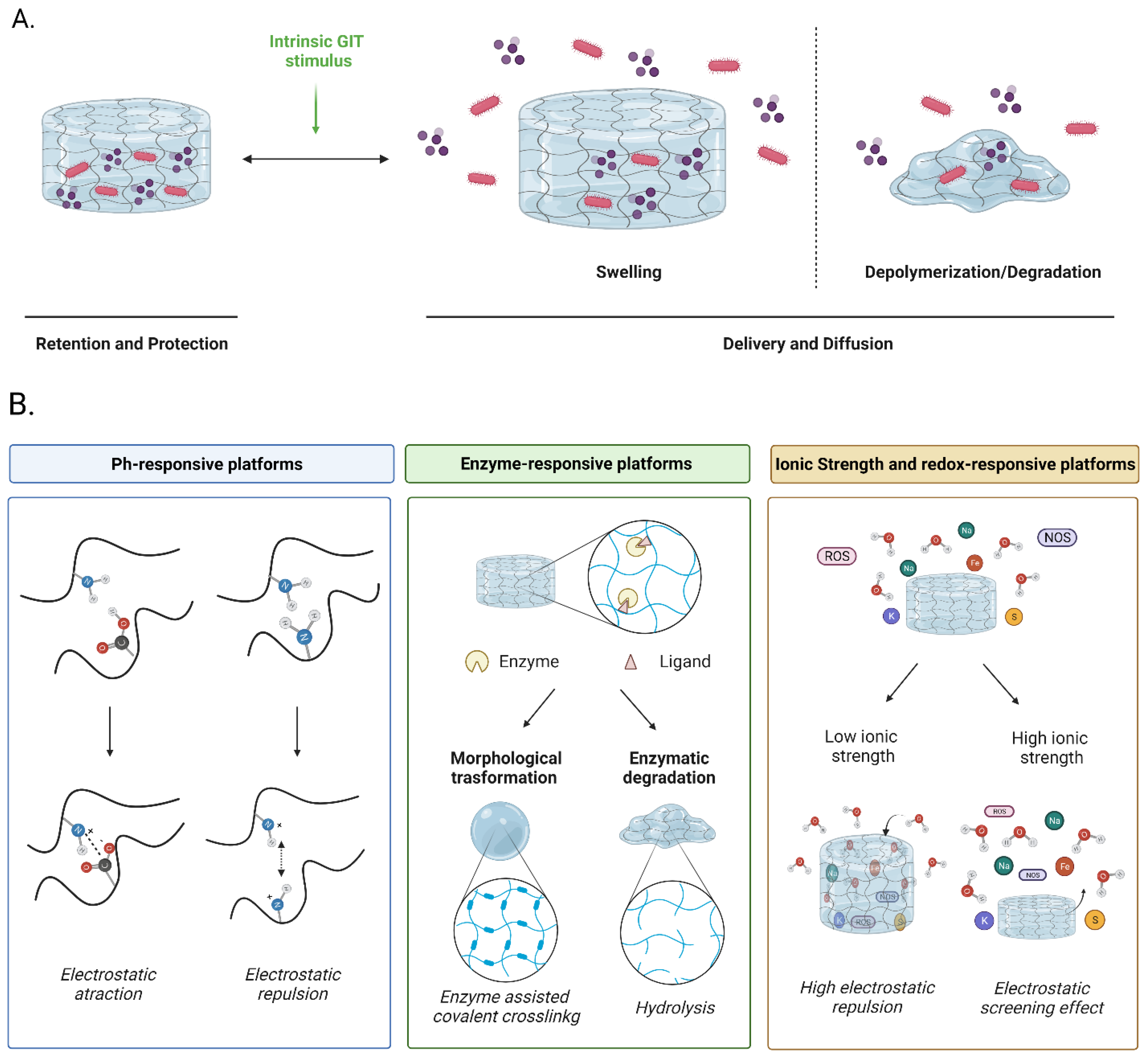
4. Improving Functionalities of Polymeric Matrices for Probiotic Encapsulation

Although processing schemes and properties of the materials are vital parameters for controlling their response, the rational design of polymeric multi-responsive platforms capable of taking advantage of innate GIT stimuli is a groundbreaking alternative to overcome current issues. Accordingly, given the predominant role of singular stimuli along GIT sections, multi-layer platforms of micro and nanostructured matrices or supramolecular encapsulates appear well-suited to actively respond to environments with particular physicochemical conditions. In this regard, one of the significant challenges is the precise control of interactions between polymeric components such that the obtained 3D constructs only respond to a set of stimuli of interest. This section is dedicated to correlating the engineering of polymer responses with the innate stimuli of the GIT (discussed in Section 2) through processing schemes explored in Section 3 (Figure 2).

4.1. pH-Responsive Matrices

As pH changes through the GIT are evident (Section 2.1), the use of ionizable polymers that undergo physical and chemical changes is a current alternative to take advantage of the presence of amines, carboxylic acids, hydroxyls, sulfonic acids, and imines [165]. This approach relies on the chemical reactivity of the pendant functional groups in the backbone of polymeric structures to provide responsiveness and cargo release through careful control of swelling–deswelling processes resulting from electrostatic repulsion (i.e., ionic strength). Thus, pH-responsive moieties in polymers offer an alternative to control the probiotic’s release based on desired pH conditions at the delivery region of the GIT.
pH-responsive polymers can be classified as (a) cationic or (b) anionic, based on their capability to donate or accept electrons from the medium [166]. While cationic polymers (e.g., chitosan, poly(ethylenimine), poly(2-dimethyl(aminoethyl) methacrylate), and poly(l-lysine)) are characterized by the presence of ionizable basic groups at low pH (pH < pKa), anionic polymers (e.g., alginate, hyaluronic acid, polymethyl methacrylate) are characterized by acidic functional groups ionizable at higher pH (pH > pKa) [82,167]. The differential ionizable capabilities of cationic and anionic polymers are key features that can be exploited to improve the response of novel multi-layer encapsulates. The high-water solubility of anionic polymers at acidic pH and low-water solubility at neutral makes them suitable to overcome the oral cavity pH and gradually dissolve in the stomach. In contrast, cationic polymers are suitable for overcoming oral and stomach pH environments providing stability when they reach the upper small intestine and colon, as they swell in basic and neutral environments. These features lead to two possible alternatives to fabricate probiotics encapsulates: (a) the single use of anionic polymers and (b) the combined use of cationic and anionic polymers in multi-structured approaches that encompass the cleavage of sensitive polymers, as shown in Table 5. In this regard, multi-structured platforms based on microencapsulates fabricated with cationic colloidal micelles and chemically crosslinked polyremes combined with nanofunctional fillers dispersed in anionic hydrogels seem to be a promising alternative to entrap probiotics successfully [168].
Moreover, anionic (or polyacidic) matrices for small intestine and colon delivery of probiotics have been reported to respond actively to changes in osmotic pressure differences for cargo release [168]. The involved osmotic force induces swelling and promotes pore opening, and as a result, the facilitated release of probiotics through the formed meshed network. Concerning this, several studies have reported that by tuning the core resistance for small intestine delivery and rationally incorporating surface functional groups, the survival of the probiotic could be improved by more than 70% [104,169].
With the appropriate selection of encapsulation schemes and materials (i.e., ensuring survival in the stomach environment and rapid release in the small intestine), it is possible to synthesize encapsulates with superior resistance to acidic environments and rapid depolymerization in neutral environments [170]. Moreover, by combining polycationic co-polymers and polypeptides, the resulting matrices exhibit reduced permeability, and improved stability in physiological media has been demonstrated to be an efficient alternative for the fabrication of pH-responsive materials [104]. As shown by Gately et al., polymers such as shellac can be employed as sources of ionizable groups to tune degradation rates as a function of the pH while simultaneously taking advantage of the high chemical stability and natural bioadhesiveness of this polymer [171].
Alternatively, recent reports indicate that pH-responsive drug encapsulates exploit environmental pH as stimuli for releasing molecules upon cleavage of acid-sensitive bonds [104]. Such an approach has also been implemented successfully in encapsulating metabolizable sugars and salts, which have been demonstrated to improve probiotics’ organoleptic properties and prolong their shelf life [172,173]. Moreover, this approach largely addresses the limited availability of the encapsulated nutrients at the site of action due to unspecific release and fluid infiltration along the transit through the GIT [174].

4.2. Enzyme-Responsive Matrices

The materials that respond to enzymes must contain substrate mimics or recognition elements that can be identified by enzymes (i.e., specific amino acid sequences), and the substrates must be accessible by enzymes [181]. Conversely, the action of the enzymes can induce either chemical or physical modifications in the matrices that lead to their degradation and, in turn, the release of encapsulated molecules [24].
The presence of different enzymes along the GIT can help release the probiotics at the site of action by the cleavage of specific moieties on the matrices [24]. Pathophysiological conditions such as inflammatory diseases of the GIT (e.g., ulcerative colitis and inflammatory bowel disease) can be potentially targeted by probiotics as they have been reported to upregulate matrix metalloproteinases (MMP) and esterases, which can degrade certain polymeric materials [31].
From a design point of view, the enzyme-responsive material must contain substrate mimics recognition motifs for effective active site binding or substrates capable of interacting with the enzyme molecules without imparting detrimental conformational changes (i.e., significant secondary and tertiary structural changes) [181]. Although to our knowledge, this approach has not been implemented for probiotic encapsulation and delivery purposes, it might be suitable for this task, given the possibility of incorporating well-known ester molecules (e.g., triglycerol monostearate and ascorbyl palmitate) into the polymeric matrices, which are recognizable by several promiscuous enzymes [154,182]. Additionally, exogenous enzymes can be potentially crosslinked with polymeric matrices such that their release upon degradation at the site of action accelerates probiotics and other cargo delivery even further.
Some natural polymers like pectin can be degraded enzymatically without any chemical modification. Zhu and colleagues developed a pectin-based system for colon-specific delivery assisted by the pectinase enzymes resident therein [33]. The enzyme degradation kinetics of pectin has been enhanced by blending it with other polymers such as ethylcellulose, hydroxypropylmethylcellulose, chitosan, poly(lysine), and polyacrylate derivatives containing quaternary ammonium groups to take advantage of the enzymatic repertoire of the bacterial flora in the presence of co-encapsulated enzyme co-factors to potentiate their action [183]. Lai and colleagues exposed guar gum to a simulated colonic fluid containing β-mannanase and demonstrated in situ pore generation by ionic interactions and the subsequent cargo release [184]. Other materials used for colon targeting include amylose combined with ethylcellulose, galactomannan, inulin, and dextran [185]. In particular, dextran has been reported to excel in targeted colon delivery applications due to its relatively high chemical stability throughout the GIT, and its breakdown by dextranases thought to be exclusively present in the colon microflora [186].

4.3. Ionic Strength and Redox-Responsive Matrices

Ion-responsive matrices react to the ionic composition of the surrounding medium, where such composition determines the magnitude and range of electrostatic interactions resulting from ion binding and electrostatic screening effects [187]. By altering electrostatic interactions between the polymer chains, it is possible to change their 3D network organization radically.
Ionic strength has proven to have a marked effect on charged poly(N-isopropyl acrylamide) (PolyNIPAM) hydrogels, altering their swelling/shrinking behavior [37,184]. In this regard, it has been observed that a strongly charged hydrogel is likely to swell at low ionic strengths due to the strong interchain electrostatic repulsion but shrinks at high ionic strengths due to electrostatic screening effects [187]. Therefore, once a critical ionic strength level is reached, the hydrogel undergoes a conformational change from a swollen to a compact state, resulting in a regime of impeded diffusion [41,188].
The swelling and shrinking characteristics imparted by ionic strength or pH changes may also alter the electrostatic interactions between a substance and the encapsulating 3D polymeric matrix. Additionally, several ion-responsive matrices are also pH-responsive. An example of a pH/ionic strength and temperature-sensitive hydrogel has been reported for insulin delivery [189]. In this case, the release profile was evaluated in vitro, showing a negligible release in simulated gastric fluid and sustained release in simulated intestinal fluid, making it suitable for the intended site of action. The charge-inducing effect of the medium can explain the observed release mechanism of this type of matrices. For instance, under conditions where the encapsulated substance and the polymers have opposite charges, there will be an electrostatic attraction between them, leading to retention within the hydrogel matrix. In contrast, under conditions where the encapsulated substance and the polymers have identical charges (or one of them is uncharged), there will be an electrostatic repulsion (or no electrostatic attraction), leading to release from the hydrogel matrix [187].
Redox-responsive polymeric matrices can respond to biological stimuli generated by the presence of oxidants or reducing agents in the medium or to changes in the redox conditions [53]. In this case, matrices are equipped with chemical groups such as disulfide bonds, organometallic compounds, viologens, or tetrathiafulvalene, widely used to induce redox responsiveness.
Recent studies highlight that enzymatically generated ROS have been shown to function as second messengers in many signal transduction pathways via the transient oxidative activity on sensor proteins bearing thiol groups sensitive to oxidant molecules. Examples of redox-sensitive proteins include tyrosine phosphatases that regulate MAPK pathways, the focal adhesion kinase, and several components responsible for NF-kB activation [190].
A pH/redox-dual responsive nanoemulsion-embedded composite hydrogel could be a promising candidate for efficient oral delivery and controlled intestinal release of magnesium and other ions. The hydrogel is synthesized by crosslinking a biocompatible, pH-responsive pseudo-peptide, poly(L-lysine isophthalamide), and the redox-sensitive molecule L-cystine dimethyl ester dihydrochloride. As reported by Huang et al. [191], these hydrogels exhibit a compact structure at acidic gastric pH but become highly swollen or degraded under neutral pH and reduce conditions of the intestinal environment. The authors highlight that the ion release profiles indicated that the nanoemulsion-embedded composite hydrogel could retain and protect magnesium ions in the simulated gastric fluid buffer at pH 1.2 but efficiently release them in the simulated intestinal fluid buffer at pH 6.8 in the presence of 1,4-dithiothreitol as a reducing agent.
Likewise, Liu et al. introduced a redox/pH dual stimulus-responsive cellulose hydrogel that was prepared by incorporating enamine and disulfide bonds in the same system at physiological pH. The cellulose hydrogel was obtained by mixing aqueous solutions of cellulose acetoacetate and cystamine dihydrochloride at room temperature. The cellulose hydrogel showed reversible sol–gel transitions in response to pH and redox potential changes [192].
Xiao et al. [77] developed a redox-responsiveness hydrogel to encapsulate the probiotic microorganism L. rhamnosus with the final goal of treating bacterial enteritis. This hydrogel was prepared by the auto-crosslinking of thiolated hyaluronic acid. Upon exposure to H2S, which is excreted by intestinal pathogens, the hydrogel degrades locally and rapidly releases the cargoes, thereby competing with source pathogens for binding to the host. Further endurance experiments indicated that encapsulation of L. rhamnosus led to a significant increase in viability that was maintained even after transit throughout the GIT. Compared with free cells, encapsulated L. rhamnosus showed a superior therapeutic effect for Salmonella-induced enteritis and negligible toxicity in vivo. Taken together, their results demonstrated the feasibility of implementing redox-responsive hydrogels for the encapsulation and targeted delivery of orally administered therapeutic probiotics by taking advantage of altered intestinal conditions observed during the invasion of pathogens.
However, despite the recent advances in the field, ionic strength and redox-responsive polymeric matrices are still in their infancy, and therefore much work will be needed to move to an eventual clinical translation.

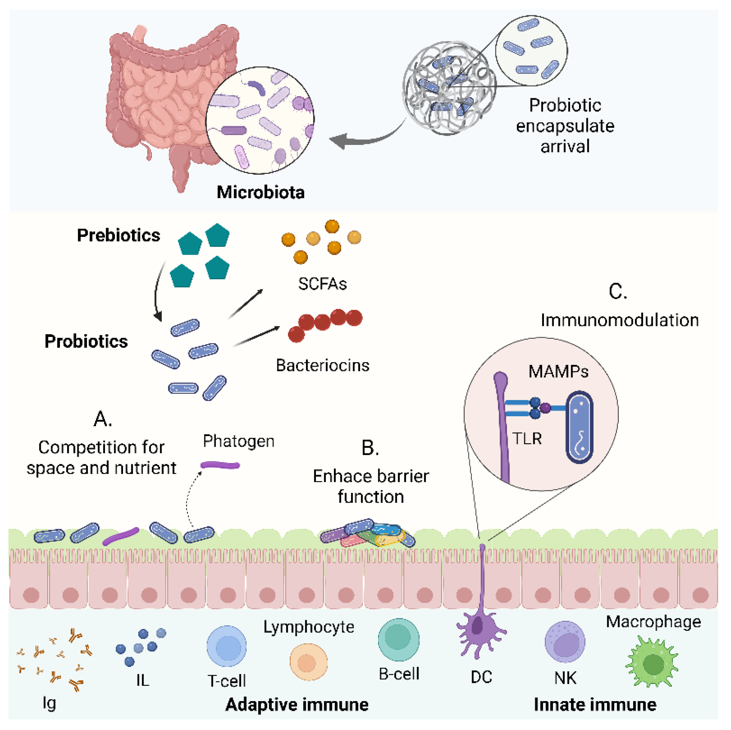
5. Physiological Barriers to Overcome

Although the stimuli-responsiveness of probiotic encapsulates is a vital attribute to accomplish therapeutic action in the intestine, successful colonization of the GIT is a pivotal aspect for conferring sufficient host-interaction [26]. Therefore, the design of delivery vehicles must take into consideration the interaction details of the encapsulated microorganisms (Figure 3) with the barrier receptors to promote their adsorption. Probiotics are commonly rejected during this process, as they are sensed as invaders by the gut microbiome. This colonization resistance is one of the most critical issues to overcome for the long-term success of probiotic therapies. This has been addressed by tuning the degradation kinetics of the delivery matrix for maintaining adhesion to the intestinal mucosa for more extended periods. In this section, we discuss key aspects of probiotics colonization to the GIT and provide a set of recommendations for the rational design of polymeric matrices for probiotics encapsulation.

5.1. Composition of the Gut Microbiome

The gut microbiome involves a complex ecosystem of microorganisms (i.e., bacteria, viruses, protozoa, and fungi) and their collective genetic material [193]. When this ecosystem exists in a state of mutually beneficial symbiosis, the microbiome is considered healthy and beneficial for the host [194]. Some of the primary functions of gut microbiota include nutrient metabolism, xenobiotic and drug metabolism, maintenance of structural integrity of the gut mucosal barrier, immunomodulation, and protection against pathogens [195].
The concentration of microbiota gradually increases throughout the GIT, starting with a small number of microorganisms in the stomach and finalizing with a high one in the colon [17]. This can be explained by the low pH and the presence of bile or pancreatic enzymes in the stomach and proximal duodenum, where bacteria fail to survive or proliferate [17].
Most gut microorganisms are strictly anaerobic and belong to the phyla Firmicutes, Bacteriodetes, and Proteobacteria [17,193]. Other gut bacteria with a minor presence in the healthy gut (usually below 1%) belong to the phyla Actinobacteria, Verrumicrobia, Acidobacteria, or Fusobacteria [17].
Human microbiome composition varies according to the location in the GIT. Predominant bacterial genera in the oral cavity include Firmicutes, Bacteroidetes, Proteobacteria, Actinobacteria, Spirochaetes, and Fusobacteria [196,197]. However, the predominant ones in saliva are Gemella, Veillonella, Neisseria, Fusobacterium, Streptococcus, Prevotella, Pseudomonas, and Actinomyces [196,198]. The most abundant bacteria in the human esophagus belong to the phylum Firmicutes and the genus Streptococcus [17,196]. In this, bacterial communities are dominated either by Streptococcus, Prevotella, and Veillonella, or Haemophilus and Rothia [17,196,199]. The low pH of the stomach is not an obstacle to finding a diverse microbiota. The genera commonly found in the corpus and antrum include Bacillales incertae seidis, Streptococcaceae, Enterobacteriaceae, Leptotrichiaceae, Veillonellaceae, and Pseudomonadaceae [17,196,200,201]. The small intestine exhibits unique microbial compositional profiles as opposed to the previous compartments. Duodenum, jejunum, and ileum are dominated by Proteobacteria and Lactobacillales [196,202]. Duodenum harbors similar genera as the stomach (i.e., Bacillales incertae sedis, Streptococcaceae, Enterobacteriaceae, Leptorichiaeceae, Veillonellaceae, and Pseudomonadaceae) [200], jejunum and ileum most common communities consist of streptococci, lactobacilli, Gammaproteobacteria, the Enterococcus group, and the Bacteroides group [17,77,199]. The terminal ileum exhibits a composition closer to that of the colon, which typically includes Clostridiaceae, Lachnospiraceae, Peptostreptococcaceae, Ruminococcaceae, Enterobacteriaceae, and Bacteroidaceae [17,196,200]. Lastly, the colon is mostly colonized by bacterial phyla such as Bacteroidetes, Firmicutes, Verrucomicrobia, Proteobacteria, and Actinobacteria [196,201]. However, the intestine is also colonized by archaea, bacteriophages, viruses, unicellular eukaryotes, and fungi. The human gut harbors several fungi, including the genera Aspergillus, Candida, Cryptococcus, and Penicillium, which account for about 0.2–0.3% of the total gut microbiota [17].

5.2. Colonization Resistance

The gut microbiome provides resistance against colonization by exogenous microorganisms [203]. Although the involved mechanisms have not been fully elucidated, some of the identified underlying processes include secretion of antimicrobial products, nutrient competition, support of gut barrier integrity, and bacteriophage deployment [203,204].
The mechanisms of colonization have been classified into direct and indirect. In the first case, the resistance refers strictly to exogenous microbial colonization [26], mainly through killing and competition for resources. This set of mechanisms tends to act between more closely related species [204]. Killing or growth suppression can be mediated by bacteriocins—short, toxic peptides produced in the ribosomes of specific bacterial species—which can disturb processes involving RNA and DNA and also mediate pore formation in the cell membrane of microorganisms [26,204,205,206,207,208]. Other antibacterial factors, like bacteriophages, can play an important role in excluding specific gut bacteria, thereby contributing to colonization resistance by lysing and transferring genetic information [204].
Also, metabolic byproducts such as short-chain fatty acids (SCFAs) (e.g., acetic, propionic, and butyric acid) can have an inhibitory effect on the growth of bacteria [204]. SCFAs are mainly produced by bacteria through fermentation of nondigestible carbohydrates [204] and can impair bacterial growth by changing intracellular pH and altering metabolic routes [203]. Bile acids, which are amphipathic and cholesterol-derived molecules secreted into the small intestine [204], also exhibit antimicrobial properties [203]. Deoxycholic acid, a secondary bile acid, is bactericidal to many bacteria by membrane disruption and subsequent leakage of the cellular content [203,209,210,211,212]. Finally, nutrient and space competition occur more easily in bacterial strains that belong to the same species. Microbiota nutrients absorption and physical competition for adhesion sites (often glycan structures) could prevent the proliferation of pathogens [203,204].
Alternatively, indirect colonization resistance usually stimulates the innate or adaptive immune system. However, non-immune defenses might also be involved [204]. In this regard, the first non-immune barriers are the mucus layers in the small intestine, cecum, and colon. The gut barrier consists of the inner and outer mucus layers, the epithelial barrier, and its immune barrier. The inner mucus layer is very robust and firmly attached to the epithelium, forming a physical barrier for bacteria. This prevents direct interaction of microbial pathogens with the epithelial layer and a potential inflammatory response [203]. As described by Pickard et al., the mucus is decorated by various glycans that serve as nutrient sources or adhesion receptors for microbes. These can be cleaved by bacterial enzymes, and the resulting free sugars (e.g., fucose) can suppress pathogen or pathobiont virulence. The host can also oxidize sugars via reactive nitrogen species produced by inducible nitric oxide synthase [204].
In concern to the immune response, the innate immunity is briefly stimulated by microbe-associated molecular patterns produced by bacteria and viruses, via toll-like receptors (TLRs) and the protein MyD88, on either epithelial cells or dendritic cells. ILC3 and Th17 cells can be activated to produce the interleukin IL-22, which has been reported to promote the secretion of antimicrobial peptides such as Reg3g from epithelial cells. Also, by the acquired immunity, B cells produce IgA and IgG antibodies, targeting symbionts or pathogens in the intestinal lumen and preventing possible systemic infections [204].

5.3. Mucosal Adhesion in Probiotics Colonization

After overcoming the harsh conditions of the GIT, the benefits of probiotics largely depend on successful mucosa colonization [26]. Thus, enhanced interaction between encapsulated probiotics at the small intestine and colon level needs to be considered with careful attention to engineer systems with the capability of increasing the bioaccessibility of the delivered microorganisms and extending their residence times [213,214]. In this regard, a rational design of the encapsulates surface biochemistry is critical for an appropriate material–intestinal surface interaction to enhance the adhesion of mucosal and intestinal epithelial cells (IECs). Such design must be based on the structural features of the mucosal layer that is mainly composed of polymerized glycoproteins, also known as mucins, and specific cell surfaces ligands [215]. However, the microorganism binding to the mucosa and the IECs is a complex process that comprises several irreversible and reversible stages. Initially, probiotics bind to the mucosa by the interplay of non-specific physical interactions and forces, e.g., Van der Waals attraction and electrostatic and hydrophobic interactions [213]. The irreversible stages involve molecular specific reactions mediated by mucus-binding proteins (MBPs) [216,217], fibronectin-binding proteins (FBPs) [218], surface-layer proteins (SLPs) [219], and non-protein molecules present on the probiotics’ surface, such as teichoic acids (TA) [220] and exopolysaccharides (EPS) [221]. However, intestinal probiotic adhesion and further colonization can be compromised by competitive exclusion where intestinal bacteria compete for adhesion sites [222].
Over the years, several attempts have been made to increase the adhesion of probiotics to negatively charged intestinal mucus by coating the encapsulates with positively charged polymers such as chitosan and different types of nanomaterials [93,223,224]. In fact, strengthening mucus/polymer interactions makes it possible to form transient gels due to intermolecular entanglements between ionizable monomers and mucins. Such gels represent a clear opportunity as platforms to increase the residence time of the encapsulate carriers [93]. Similarly, hydrogen bonding, hydrophobic interaction potential, electrostatic interactions, and lectin/sugar recognition are reported to be key parameters in the design of more robust mucoadhesives [93,225,226]. Furthermore, as polymer/mucus binding at the GIT mainly proceeds by hydrogen bonding, water molecules can interact with mucus strongly, limiting polymer interactions. Thus, incorporating hygroscopic materials such as polyethylene glycol (PEG) in encapsulates has increased mucoadhesion [92]. Also, this approach has been reported to open tight epithelial junctions to improve the mucus/polymer contact area [153].

5.4. Prebiotics Co-Immobilization as an Alternative to Improve Probiotics Colonization

Prebiotics are non-viable substrates that serve as nutrients for beneficial microorganisms, including probiotic and indigenous strains [227]. An exogenous probiotic strain must compete for nutrients and space with microorganisms in the indigenous microbiome. Therefore, prebiotics have been considered alternative nutritional substrates that could potentially lower strain competition and favor colonization of probiotics [228,229]. Prebiotics such as fructooligosaccharides (FOS) [230], xylooligosaccharides (XOS) [231], galactooligosaccharides (GOS) [229], and inulin [232] have demonstrated their potential for helping overcome colonization resistance of probiotics [26]. However, detailed studies on the impact of prebiotics are rather scarce and therefore represent an opportunity to engineer next-generation probiotics.

6. Future Directions for Probiotic Encapsulation and Clinical Practice

From a clinical perspective, the delivery of probiotics has shown promising results in clinical trials to prevent, mitigate and treat specific diseases such as traveler’s diarrhea, antibiotic-associated diarrhea, acute diarrhea in children, intestine bowel syndrome, inflammatory bowel disease, and atopic dermatitis [233]. One of the most attractive features of probiotics implementation is their possible extrapolation from the food industry to specific disease treatment without incurring further scrutiny by governmental agencies (e.g., Food and Drug Administration—FDA) since probiotics are considered functional foods and not pharmacological compounds [234,235]. However, relevant clinical data have revealed ambiguous results and a lack of data on possible side effects, recommendations on proper probiotic selection for single disease treatment, and their exact mechanism of action [236]. For this purpose, 1831 clinical trials have been conducted to better understand probiotics’ interaction with the human body, as reported in the ClinicalTrials.gov database (accessed 18 January 2022).
Moreover, besides the fast-growing research on probiotics encapsulation using polymers, the translation of such technologies remains limited, as evidenced by just one clinical trial found in the ClinicalTrials.gov database (accessed 18 January 2022). Despite this limited number of studies at the clinical level, several studies conducted in vitro report on promising probiotics encapsulates capable of overcoming the GIT’s harsh conditions, as demonstrated by more than 13,000 patents awarded in this field (lens.org, accessed 19 January 2022). Nevertheless, as for the clinical trials, preclinical models are also scarce, and only 32 research articles have been published to date, including the term “in vivo” (PubMed.gov, accessed 19 January 2022). This trend is exacerbated even further when the term “stimuli-responsive” is included in the patent search algorithm, as only 802 patents were available (lens.org, accessed 19 January 2022).
The limited translation of current and emerging probiotics encapsulation technologies strongly indicates that there is still a long way to go before stimuli-responsive matrices reach clinical implementation. In this regard, one of the most important obstacles to overcome is to design and manufacture cost-effective encapsulation processes, as they largely define scalability and the requirements of approved food additives [104]. Conversely, scaling up routes should be considered from the beginning of the developments such that the economic output and long-term sustainability is assured. Moreover, to facilitate complying with numerous regulatory requirements for clinical research and, ultimately, the claim substantiation and market access [237], the developments should consider stimuli-responsive materials already approved for human use.

7. Conclusions

Recent technological advancements in encapsulation facilitate the formulation of probiotics’ site-selective delivery with high cell viability and prolonged stabilities and residence times. Among such advancements are stimuli-responsive polymeric matrices, which offer an avenue for the precise delivery of probiotics by taking advantage of changes in physicochemical conditions throughout the GIT. These include changes in pH, ionic strength, redox potential, and even mechanical stress. Among the developed matrices, hydrogels and the nanostructured platforms stand out for their ease of modification and processing, versatility to facilitate multimodal delivery, and the possibility to finely tune multi-stimuli responsiveness to address multiple components of the GIT. The most popular and studied matrices involve alginate and its derivatives coated with chitosan or gelatin, as they have proven to tolerate the harsh conditions of the GIT and maintain superior probiotics viability.
Thus far, most smart platforms for probiotics delivery take advantage of pH-responsiveness for engineering site-selective delivery vehicles. Emerging materials have considered other stimuli and, most recently, the action of enzymes either native to the GIT or added exogenously to the matrix to favor selective degradation and release at the site of the action. Besides protection of the probiotic as it transits through the GIT, sufficient host interaction is only achievable upon successful colonization of the delivered strains, determined by possible competition with indigenous microorganisms. One way to overcome this limitation is by administering prebiotics, which are nutritional substrates that favor the proliferation of beneficial microorganisms. Additionally, polymers with specific moieties can interact selectively with mucus to form transient gels that might extend the residence time. Despite the progress over the past few years, the rational design of encapsulates for targeted probiotic delivery is still in its infancy. Therefore, much work needs to be invested in elucidating the routes for developing materials that involve simple manufacturing schemes that can be scaled up to address the increasingly higher demand for these products. Also, this might help accelerate the defining of conditions for relevant pre-clinical and clinical tests.

Author Contributions

Conceptualization, J.C.C. and L.H.R.; methodology, A.J.G.-B., V.Q., C.G.-M. and A.D.B.-B.; software, A.J.G.-B. and V.Q.; validation, A.J.G.-B. and V.Q.; formal analysis, A.J.G.-B. and V.Q.; investigation, A.J.G.-B., V.Q., C.G.-M. and A.D.B.-B.; resources, J.C.C. and L.H.R.; data curation, A.J.G.-B., V.Q., C.G.-M. and A.D.B.-B.; writing—original draft preparation, A.J.G.-B., V.Q., C.G.-M. and A.D.B.-B.; writing—review and editing, J.C.C. and L.H.R.; visualization, A.J.G.-B. and V.Q.; supervision, J.C.C. and L.H.R.; project administration, J.C.C. and L.H.R.; funding acquisition, J.C.C. and L.H.R. All authors have read and agreed to the published version of the manuscript.

Funding

This research was funded by the Colombian Ministry of Science, Technology, and Innovation (Minciencias), Grant ID 120484467244.

Institutional Review Board Statement

Not applicable.

Informed Consent Statement

Not applicable.

Data Availability Statement

Not applicable.

Conflicts of Interest

The authors declare no conflict of interest.

References

  1. Ranjha, M.M.A.N.; Shafique, B.; Batool, M.; Kowalczewski, P.; Shehzad, Q.; Usman, M.; Manzoor, M.F.; Zahra, S.M.; Yaqub, S.; Aadil, R.M. Nutritional and Health Potential of Probiotics: A Review. Appl. Sci. 2021, 11, 11204. [Google Scholar] [CrossRef]
  2. Quigley, E.M. Prebiotics and Probiotics in Digestive Health. Clin. Gastroenterol. Hepatol. 2018, 17, 333–344. [Google Scholar] [CrossRef]
  3. La Fata, G.; Weber, P.; Mohajeri, M.H. Probiotics and the Gut Immune System: Indirect Regulation. Probiot. Antimicrob. Proteins 2018, 10, 11–21. [Google Scholar] [CrossRef] [PubMed]
  4. Oberoi, K.; Tolun, A.; Altintas, Z.; Sharma, S. Effect of Alginate-Microencapsulated Hydrogels on the Survival of Lactobacillus rhamnosus under Simulated Gastrointestinal Conditions. Foods 2021, 10, 1999. [Google Scholar] [CrossRef]
  5. Mattila-Sandholm, T.; Myllärinen, P.; Crittenden, R.; Mogensen, G.; Fondén, R.; Saarela, M. Technological challenges for future probiotic foods. Int. Dairy J. 2002, 12, 173–182. [Google Scholar] [CrossRef]
  6. Butel, M.-J. Probiotics, gut microbiota and health. Med. Mal. Infect. 2014, 44, 1–8. [Google Scholar] [CrossRef] [PubMed]
  7. Vitetta, L.; Briskey, D.; Alford, H.; Hall, S.; Coulson, S. Probiotics, prebiotics and the gastrointestinal tract in health and disease. Inflammopharmacology 2014, 22, 135–154. [Google Scholar] [CrossRef]
  8. Chugh, B.; Kamal-Eldin, A. Bioactive compounds produced by probiotics in food products. Curr. Opin. Food Sci. 2020, 32, 76–82. [Google Scholar] [CrossRef]
  9. Tan-Lim, C.S.C.; Esteban-Ipac, N.A.R. Probiotics as treatment for food allergies among pediatric patients: A meta-analysis. World Allergy Organ. J. 2018, 11, 25. [Google Scholar] [CrossRef]
  10. Maragkoudakis, P.A.; Zoumpopoulou, G.; Miaris, C.; Kalantzopoulos, G.; Pot, B.; Tsakalidou, E. Probiotic potential of Lactobacillus strains isolated from dairy products. Int. Dairy J. 2006, 16, 189–199. [Google Scholar] [CrossRef]
  11. Rolim, F.R.; Neto, O.C.F.; Oliveira, M.E.G.; Oliveira, C.J.; Queiroga, R.C. Cheeses as food matrixes for probiotics: In vitro and in vivo tests. Trends Food Sci. Technol. 2020, 100, 138–154. [Google Scholar] [CrossRef]
  12. Soares, M.B.; Martinez, R.C.; Pereira, E.P.; Balthazar, C.F.; Cruz, A.G.; Ranadheera, C.S.; Sant’Ana, A.S. The resistance of Bacillus, Bifidobacterium, and Lactobacillus strains with claimed probiotic properties in different food matrices exposed to simulated gastrointestinal tract conditions. Food Res. Int. 2019, 125, 108542. [Google Scholar] [CrossRef] [PubMed]
  13. McClements, D.J. Designing biopolymer microgels to encapsulate, protect and deliver bioactive components: Physicochemical aspects. Adv. Colloid Interface Sci. 2016, 240, 31–59. [Google Scholar] [CrossRef] [PubMed] [Green Version]
  14. Salas-Jara, M.J.; Ilabaca, A.; Vega, M.; García, A. Biofilm Forming Lactobacillus: New Challenges for the Development of Probiotics. Microorganisms 2016, 4, 35. [Google Scholar] [CrossRef] [PubMed]
  15. Trush, E.; Poluektova, E.A.; Beniashvilli, A.G.; Shifrin, O.S.; Poluektov, Y.M.; Ivashkin, V.T. The Evolution of Human Probiotics: Challenges and Prospects. Probiot. Antimicrob. Proteins 2020, 12, 1291–1299. [Google Scholar] [CrossRef] [PubMed]
  16. Huq, T.; Khan, A.; Khan, R.A.; Riedl, B.; Lacroix, M. Encapsulation of Probiotic Bacteria in Biopolymeric System. Crit. Rev. Food Sci. Nutr. 2013, 53, 909–916. [Google Scholar] [CrossRef] [PubMed] [Green Version]
  17. Dieterich, W.; Schink, M.; Zopf, Y. Microbiota in the Gastrointestinal Tract. Med. Sci. 2018, 6, 116. [Google Scholar] [CrossRef] [PubMed] [Green Version]
  18. Fanaro, S.; Chierici, R.; Guerrini, P.; Vigi, V. Intestinal microflora in early infancy: Composition and development. Acta Paediatr. 2007, 92, 48–55. [Google Scholar] [CrossRef]
  19. Tannock, G.W. Molecular assessment of intestinal microflora. Am. J. Clin. Nutr. 2001, 73, 410s–414s. [Google Scholar] [CrossRef] [Green Version]
  20. Baral, K.C.; Bajracharya, R.; Lee, S.H.; Han, H.-K. Advancements in the Pharmaceutical Applications of Probiotics: Dosage Forms and Formulation Technology. Int. J. Nanomed. 2021, 16, 7535–7556. [Google Scholar] [CrossRef]
  21. Gao, Y.; Wang, X.; Xue, C.; Wei, Z. Latest developments in food-grade delivery systems for probiotics: A systematic review. Crit. Rev. Food Sci. Nutr. 2021, 1–18. [Google Scholar] [CrossRef] [PubMed]
  22. Asgari, S.; Pourjavadi, A.; Licht, T.R.; Boisen, A.; Ajalloueian, F. Polymeric carriers for enhanced delivery of probiotics. Adv. Drug Deliv. Rev. 2020, 161–162, 1–21. [Google Scholar] [CrossRef] [PubMed]
  23. Yoha, K.S.; Nida, S.; Dutta, S.; Moses, J.A.; Anandharamakrishnan, C. Targeted Delivery of Probiotics: Perspectives on Research and Commercialization. Probiot. Antimicrob. Proteins 2021, 14, 15–48. [Google Scholar] [CrossRef] [PubMed]
  24. El-Husseiny, H.M.; Mady, E.A.; Hamabe, L.; Abugomaa, A.; Shimada, K.; Yoshida, T.; Tanaka, T.; Yokoi, A.; Elbadawy, M.; Tanaka, R. Smart/stimuli-responsive hydrogels: Cutting-edge platforms for tissue engineering and other biomedical applications. Mater. Today Bio 2021, 13, 100186. [Google Scholar] [CrossRef] [PubMed]
  25. Yao, M.; Xie, J.; Du, H.; McClements, D.J.; Xiao, H.; Li, L. Progress in microencapsulation of probiotics: A review. Compr. Rev. Food Sci. Food Saf. 2020, 19, 857–874. [Google Scholar] [CrossRef] [Green Version]
  26. Han, S.; Lu, Y.; Xie, J.; Fei, Y.; Zheng, G.; Wang, Z.; Liu, J.; Lv, L.; Ling, Z.; Berglund, B.; et al. Probiotic Gastrointestinal Transit and Colonization After Oral Administration: A Long Journey. Front. Cell. Infect. Microbiol. 2021, 11, 609722. [Google Scholar] [CrossRef]
  27. Yao, M.; Li, B.; Ye, H.; Huang, W.; Luo, Q.; Xiao, H.; McClements, D.J.; Li, L. Enhanced viability of probiotics (Pediococcus pentosaceus Li05) by encapsulation in microgels doped with inorganic nanoparticles. Food Hydrocoll. 2018, 83, 246–252. [Google Scholar] [CrossRef]
  28. Patricia, J.J.; Dhamoon, A.S. Physiology, Digestion, StatPearls. September 2021. Available online: https://www.ncbi.nlm.nih.gov/books/NBK544242/ (accessed on 24 January 2022).
  29. Gachons, C.P.D.; Breslin, P.A.S. Salivary Amylase: Digestion and Metabolic Syndrome. Curr. Diabetes Rep. 2016, 16, 102. [Google Scholar] [CrossRef]
  30. Patel, G.; Misra, A. Oral Delivery of Proteins and Peptides: Concepts and Applications. In Challenges in Delivery of Therapeutic Genomics and Proteomics; Elsevier: Amsterdam, The Netherlands, 2011; pp. 481–529. [Google Scholar] [CrossRef]
  31. Bertoni, S.; Machness, A.; Tiboni, M.; Bártolo, R.; Santos, H.A. Reactive oxygen species responsive nanoplatforms as smart drug delivery systems for gastrointestinal tract targeting. Biopolymers 2019, 111, e23336. [Google Scholar] [CrossRef]
  32. Shubakov, A.; Mikhailova, E.; Prosheva, V.; Belyy, V. Swelling and Degradation of Calcium-Pectic Gel Particles Made of Pectins of Silene vulgaris and Lemna minor Callus Cultures at Different Concentrations of Pectinase in an Artificial Colon Environment. Int. J. Biomed. 2018, 8, 60–64. [Google Scholar] [CrossRef]
  33. Zhu, W.; Han, C.; Dong, Y.; Jian, B. Enzyme-responsive mechanism based on multi-walled carbon nanotubes and pectin complex tablets for oral colon-specific drug delivery system. J. Radioanal. Nucl. Chem. Artic. 2019, 320, 503–512. [Google Scholar] [CrossRef]
  34. Kim, Y.S.; Erickson, R.H. Role of Peptidases of the Human Small Intestine in Protein Digestion. Gastroenterology 1985, 88, 1071–1073. [Google Scholar] [CrossRef]
  35. Pirahanchi, Y.; Sharma, S. Biochemistry, Lipase, StatPearls, July 2021. Available online: https://www.ncbi.nlm.nih.gov/books/NBK537346/ (accessed on 24 January 2022).
  36. Circu, M.L.; Aw, T.Y. Redox biology of the intestine. Free Radic. Res. 2011, 45, 1245–1266. [Google Scholar] [CrossRef] [Green Version]
  37. Arnaut, L. Elementary reactions in solution. In Chemical Kinetics; Elsevier: Amsterdam, The Netherlands, 2021; pp. 263–293. [Google Scholar] [CrossRef]
  38. Qi, X.; Simsek, S.; Chen, B.; Rao, J. Alginate-based double-network hydrogel improves the viability of encapsulated probiotics during simulated sequential gastrointestinal digestion: Effect of biopolymer type and concentrations. Int. J. Biol. Macromol. 2020, 165, 1675–1685. [Google Scholar] [CrossRef] [PubMed]
  39. Yang, L.; Han, Z.; Chen, C.; Li, Z.; Yu, S.; Qu, Y.; Zeng, R. Novel probiotic-bound oxidized Bletilla striata polysaccharide-chitosan composite hydrogel. Mater. Sci. Eng. C 2020, 117, 111265. [Google Scholar] [CrossRef] [PubMed]
  40. Liu, H.; Cai, Z.; Wang, F.; Hong, L.; Deng, L.; Zhong, J.; Wang, Z.; Cui, W. Colon-Targeted Adhesive Hydrogel Microsphere for Regulation of Gut Immunity and Flora. Adv. Sci. 2021, 8, 2101619. [Google Scholar] [CrossRef]
  41. Bearat, H.; Vernon, B. Environmentally responsive injectable materials. In Injectable Biomaterials; Elsevier: Amsterdam, The Netherlands, 2011; pp. 263–297. [Google Scholar] [CrossRef]
  42. Alkekhia, D.; Hammond, P.T.; Shukla, A. Layer-by-Layer Biomaterials for Drug Delivery. Annu. Rev. Biomed. Eng. 2020, 22, 1–24. [Google Scholar] [CrossRef]
  43. Pei, J.; Xiong, L.; Bao, P.; Chu, M.; Yan, P.; Guo, X. Secondary Structural Transformation of Bovine Lactoferricin Affects Its Antibacterial Activity. Probiot. Antimicrob. Proteins 2020, 13, 873–884. [Google Scholar] [CrossRef]
  44. Rattanaburi, P.; Charoenrat, N.; Pongtharangkul, T.; Suphantharika, M.; Wongkongkatep, J. Hydroxypropyl methylcellulose enhances the stability of o/w Pickering emulsions stabilized with chitosan and the whole cells of Lactococcus lactis IO-1. Food Res. Int. 2018, 116, 559–565. [Google Scholar] [CrossRef]
  45. Taniguchi, M.; Nambu, M.; Katakura, Y.; Yamasaki-Yashiki, S. Adhesion mechanisms of Bifidobacterium animalis subsp. lactis JCM 10602 to dietary fiber. Biosci. Microbiota Food Health 2021, 40, 59–64. [Google Scholar] [CrossRef]
  46. Lindahl, A.; Ungell, A.-L.; Knutson, L.; Lennernäs, H. Characterization of Fluids from the Stomach and Proximal Jejunum in Men and Women. Pharm. Res. 1997, 14, 497–502. [Google Scholar] [CrossRef] [PubMed]
  47. Million, M.; Raoult, D. Linking gut redox to human microbiome. Hum. Microbiome J. 2018, 10, 27–32. [Google Scholar] [CrossRef]
  48. Vincent, S.G.T.; Jennerjahn, T.; Ramasamy, K. Environmental variables and factors regulating microbial structure and functions. In Microbial Communities in Coastal Sediments; Elsevier: Amsterdam, The Netherlands, 2021; pp. 79–117. [Google Scholar] [CrossRef]
  49. Kwiecień, I.; Kwiecień, M. Application of Polysaccharide-Based Hydrogels as Probiotic Delivery Systems. Gels 2018, 4, 47. [Google Scholar] [CrossRef] [PubMed] [Green Version]
  50. Abad, I.; Conesa, C.; Sánchez, L. Development of Encapsulation Strategies and Composite Edible Films to Maintain Lactoferrin Bioactivity: A Review. Materials 2021, 14, 7358. [Google Scholar] [CrossRef]
  51. Gheorghita, R.; Anchidin-Norocel, L.; Filip, R.; Dimian, M.; Covasa, M. Applications of Biopolymers for Drugs and Probiotics Delivery. Polymers 2021, 13, 2729. [Google Scholar] [CrossRef] [PubMed]
  52. Saboktakin, M.R.; Tabatabaei, R.M. Supramolecular hydrogels as drug delivery systems. Int. J. Biol. Macromol. 2015, 75, 426–436. [Google Scholar] [CrossRef]
  53. Municoy, S.; Álvarez Echazú, M.I.; Antezana, P.E.; Galdopórpora, J.M.; Olivetti, C.; Mebert, A.M.; Foglia, M.L.; Tuttolomondo, M.V.; Alvarez, G.S.; Hardy, J.G.; et al. Stimuli-Responsive Materials for Tissue Engineering and Drug Delivery. Int. J. Mol. Sci. 2020, 21, 4724. [Google Scholar] [CrossRef]
  54. Pinteala, M.; Abadie, M.J.M.; Rusu, R.D. Smart Supra-and Macro-Molecular Tools for Biomedical Applications. Materials 2020, 13, 3343. [Google Scholar] [CrossRef]
  55. Safi, M.A.E. Materials and Techniques for Microencapsulation of Probiotics. Biosci. Biotechnol. Res. Commun. 2021, 14, 922–928. [Google Scholar] [CrossRef]
  56. da Silva, B.V.; Barreira, J.C.; Oliveira, M.B.P. Natural phytochemicals and probiotics as bioactive ingredients for functional foods: Extraction, biochemistry and protected-delivery technologies. Trends Food Sci. Technol. 2016, 50, 144–158. [Google Scholar] [CrossRef] [Green Version]
  57. Survival of Lactobacillus Acidophilus as Probiotic Bacteria using Chitosan Nanoparticles. Int. J. Eng. 2017, 30, 456–463. [CrossRef]
  58. Allan-Wojtas, P.; Hansen, L.T.; Paulson, A. Microstructural studies of probiotic bacteria-loaded alginate microcapsules using standard electron microscopy techniques and anhydrous fixation. LWT 2008, 41, 101–108. [Google Scholar] [CrossRef]
  59. Shori, A.B. Microencapsulation Improved Probiotics Survival During Gastric Transit. HAYATI J. Biosci. 2017, 24, 1–5. [Google Scholar] [CrossRef]
  60. Hansen, L.T.; Allan-Wojtas, P.; Jin, Y.-L.; Paulson, A. Survival of Ca-alginate microencapsulated Bifidobacterium spp. in milk and simulated gastrointestinal conditions. Food Microbiol. 2002, 19, 35–45. [Google Scholar] [CrossRef]
  61. Etchepare, M.D.A.; Barin, J.S.; Cichoski, A.J.; Jacob-Lopes, E.; Wagner, R.; Fries, L.L.M.; De Menezes, C.R. Microencapsulation of probiotics using sodium alginate. Ciência Rural 2015, 45, 1319–1326. [Google Scholar] [CrossRef] [Green Version]
  62. Mathews, S. Microencapsulation of Probiotics by Calcium Alginate and Gelatin and Evaluation of its Survival in Simulated Human Gastro-Intestinal Condition. Int. J. Curr. Microbiol. Appl. Sci. 2017, 6, 2080–2087. [Google Scholar] [CrossRef] [Green Version]
  63. Khosravi Zanjani, M.A.; Tarzi, B.G.; Sharifan, A.; Mohammadi, N. Microencapsulation of probiotics by calcium alginate-gelatinized starch with chitosan coating and evaluation of survival in simulated human gastro-intestinal condition. Iran. J. Pharm. Res. 2014, 13, 843–852. [Google Scholar] [CrossRef]
  64. Krasaekoopt, W.; Bhandari, B.; Deeth, H. The influence of coating materials on some properties of alginate beads and survivability of microencapsulated probiotic bacteria. Int. Dairy J. 2004, 14, 737–743. [Google Scholar] [CrossRef]
  65. Shu, X.; Zhu, K. The release behavior of brilliant blue from calcium–alginate gel beads coated by chitosan: The preparation method effect. Eur. J. Pharm. Biopharm. 2002, 53, 193–201. [Google Scholar] [CrossRef]
  66. Lee, J.S.; Cha, D.S.; Park, H.J. Survival of Freeze-Dried Lactobacillus bulgaricus KFRI 673 in Chitosan-Coated Calcium Alginate Microparticles. J. Agric. Food Chem. 2004, 52, 7300–7305. [Google Scholar] [CrossRef]
  67. Koo, S.M.; Cho, Y.H.; Huh, C.S.; Baek, Y.J.; Park, J.Y. Improvement of the Stability of Lactobacillus casei YIT 9018 by Microencapsulation Using Alginate and Chitosan. J. Microbiol. Biotechnol. 2001, 11, 376–383. [Google Scholar]
  68. Călinoiu, L.-F.; Ştefănescu, B.E.; Pop, I.D.; Muntean, L.; Vodnar, D.C. Chitosan Coating Applications in Probiotic Microencapsulation. Coatings 2019, 9, 194. [Google Scholar] [CrossRef] [Green Version]
  69. Santos, M.A.S.; Machado, M.T.C. Coated alginate–chitosan particles to improve the stability of probiotic yeast. Int. J. Food Sci. Technol. 2020, 56, 2122–2131. [Google Scholar] [CrossRef]
  70. Albadran, H.A.; Monteagudo-Mera, A.; Khutoryanskiy, V.V.; Charalampopoulos, D. Development of chitosan-coated agar-gelatin particles for probiotic delivery and targeted release in the gastrointestinal tract. Appl. Microbiol. Biotechnol. 2020, 104, 5749–5757. [Google Scholar] [CrossRef]
  71. Annan, N.; Borza, A.; Hansen, L.T. Encapsulation in alginate-coated gelatin microspheres improves survival of the probiotic Bifidobacterium adolescentis 15703T during exposure to simulated gastro-intestinal conditions. Food Res. Int. 2008, 41, 184–193. [Google Scholar] [CrossRef]
  72. Liu, J.; Liu, F.; Ren, T.; Wang, J.; Yang, M.; Yao, Y.; Chen, H. Fabrication of fish gelatin/sodium alginate double network gels for encapsulation of probiotics. J. Sci. Food Agric. 2021, 101, 4398–4408. [Google Scholar] [CrossRef]
  73. Ramos, P.E.; Cerqueira, M.A.; Teixeira, J.A.; Vicente, A.A. Physiological protection of probiotic microcapsules by coatings. Crit. Rev. Food Sci. Nutr. 2018, 58, 1864–1877. [Google Scholar] [CrossRef]
  74. Hlaing, S.P.; Kim, J.; Lee, J.; Kwak, D.; Kim, H.; Yoo, J.-W. Enhanced Viability of Probiotics Against Gastric Acid by One-Step Coating Process with Poly-L-Lysine: In Vitro and In Vivo Evaluation. Pharmaceutics 2020, 12, 662. [Google Scholar] [CrossRef]
  75. Misra, S.; Pandey, P.; Mishra, H.N. Novel approaches for co-encapsulation of probiotic bacteria with bioactive compounds, their health benefits and functional food product development: A review. Trends Food Sci. Technol. 2021, 109, 340–351. [Google Scholar] [CrossRef]
  76. Zhu, Y.; Wang, Z.; Bai, L.; Deng, J.; Zhou, Q. Biomaterial-based encapsulated probiotics for biomedical applications: Current status and future perspectives. Mater. Des. 2021, 210, 110018. [Google Scholar] [CrossRef]
  77. Xiao, Y.; Lu, C.; Liu, Y.; Kong, L.; Bai, H.; Mu, H.; Li, Z.; Geng, H.; Duan, J. Encapsulation of Lactobacillus rhamnosus in Hyaluronic Acid-Based Hydrogel for Pathogen-Targeted Delivery to Ameliorate Enteritis. ACS Appl. Mater. Interfaces 2020, 12, 36967–36977. [Google Scholar] [CrossRef] [PubMed]
  78. Mosqueda, I.S.; Bousquets, A.L.; Montiel-Sosa, J.F.; Corona, L.; Álvarez, Z.G.; Gochi, L.C. Encapsulation of Lactobacillus plantarum ATCC 8014 and Pediococcus acidilactici ATCC 8042 in a freeze-dried alginate-gum arabic system and its in vitro testing under gastrointestinal conditions. J. Microencapsul. 2019, 36, 591–602. [Google Scholar] [CrossRef] [PubMed]
  79. Li, R.; Zhang, Y.; Polk, D.B.; Tomasula, P.M.; Yan, F.; Liu, L. Preserving viability of Lactobacillus rhamnosus GG in vitro and in vivo by a new encapsulation system. J. Control. Release 2016, 230, 79–87. [Google Scholar] [CrossRef] [PubMed] [Green Version]
  80. Su, J.; Cai, Y.; Zhi, Z.; Guo, Q.; Mao, L.; Gao, Y.; Yuan, F.; Van der Meeren, P. Assembly of propylene glycol alginate/β-lactoglobulin composite hydrogels induced by ethanol for co-delivery of probiotics and curcumin. Carbohydr. Polym. 2020, 254, 117446. [Google Scholar] [CrossRef]
  81. Dafe, A.; Etemadi, H.; Dilmaghani, A.; Mahdavinia, G.R. Investigation of pectin/starch hydrogel as a carrier for oral delivery of probiotic bacteria. Int. J. Biol. Macromol. 2017, 97, 536–543. [Google Scholar] [CrossRef]
  82. Sharpe, L.A.; Daily, A.M.; Horava, S.D.; A Peppas, N. Therapeutic applications of hydrogels in oral drug delivery. Expert Opin. Drug Deliv. 2014, 11, 901–915. [Google Scholar] [CrossRef] [Green Version]
  83. Patarroyo, J.L.; Florez-Rojas, J.S.; Pradilla, D.; Valderrama-Rincón, J.D.; Cruz, J.C.; Reyes, L.H. Formulation and Characterization of Gelatin-Based Hydrogels for the Encapsulation of Kluyveromyces lactis—Applications in Packed-Bed Reactors and Probiotics Delivery in Humans. Polymers 2020, 12, 1287. [Google Scholar] [CrossRef]
  84. Ghibaudo, F.; Gerbino, E.; Orto, V.C.D.; Gomez-Zavaglia, A. Pectin-iron capsules: Novel system to stabilise and deliver lactic acid bacteria. J. Funct. Foods 2017, 39, 299–305. [Google Scholar] [CrossRef]
  85. Falco, C.Y.; Falkman, P.; Risbo, J.; Cárdenas, M.; Medronho, B. Chitosan-dextran sulfate hydrogels as a potential carrier for probiotics. Carbohydr. Polym. 2017, 172, 175–183. [Google Scholar] [CrossRef]
  86. Patarroyo, J.; Fonseca, E.; Cifuentes, J.; Salcedo, F.; Cruz, J.; Reyes, L. Gelatin-Graphene Oxide Nanocomposite Hydrogels for Kluyveromyces lactis Encapsulation: Potential Applications in Probiotics and Bioreactor Packings. Biomolecules 2021, 11, 922. [Google Scholar] [CrossRef]
  87. Badeau, B.A.; Deforest, C.A. Programming Stimuli-Responsive Behavior into Biomaterials. Annu. Rev. Biomed. Eng. 2019, 21, 241–265. [Google Scholar] [CrossRef] [PubMed]
  88. Dutta, S.; Samanta, P.; Dhara, D. Temperature, pH and redox responsive cellulose based hydrogels for protein delivery. Int. J. Biol. Macromol. 2016, 87, 92–100. [Google Scholar] [CrossRef] [PubMed]
  89. Noirbent, G.; Dumur, F. Photoinitiators of polymerization with reduced environmental impact: Nature as an unlimited and renewable source of dyes. Eur. Polym. J. 2020, 142, 110109. [Google Scholar] [CrossRef]
  90. Ahmed, E.M. Hydrogel: Preparation, characterization, and applications: A review. J. Adv. Res. 2015, 6, 105–121. [Google Scholar] [CrossRef] [Green Version]
  91. Samal, S.K.; Dash, M.; Dubruel, P.; Van Vlierberghe, S. Smart polymer hydrogels: Properties, synthesis and applications. In Smart Polymers and their Applications, 1st ed.; Aguilar, M.R., Román, J.S., Eds.; Woodhead Publishing Limited: Cambridge, UK, 2014; pp. 237–270. ISBN 978-085-709-702-6. [Google Scholar]
  92. Liu, L.; Yao, W.; Rao, Y.; Lu, X.; Gao, J. pH-Responsive carriers for oral drug delivery: Challenges and opportunities of current platforms. Drug Deliv. 2017, 24, 569–581. [Google Scholar] [CrossRef] [Green Version]
  93. Peppas, N.A.; Huang, Y. Nanoscale technology of mucoadhesive interactions. Adv. Drug Deliv. Rev. 2004, 56, 1675–1687. [Google Scholar] [CrossRef]
  94. Tang, Y.; Heaysman, C.L.; Willis, S.; Lewis, A.L. Physical hydrogels with self-assembled nanostructures as drug delivery systems. Expert Opin. Drug Deliv. 2011, 8, 1141–1159. [Google Scholar] [CrossRef]
  95. Sun, Q.; Wicker, L. Hydrogel Encapsulation of Lactobacillus casei by Block Charge Modified Pectin and Improved Gastric and Storage Stability. Foods 2021, 10, 1337. [Google Scholar] [CrossRef]
  96. Yeung, T.W.; Üçok, E.F.; Tiani, K.A.; McClements, D.J.; Sela, D.A. Microencapsulation in Alginate and Chitosan Microgels to Enhance Viability of Bifidobacterium longum for Oral Delivery. Front. Microbiol. 2016, 7, 494. [Google Scholar] [CrossRef] [Green Version]
  97. Sohail, A.; Turner, M.; Coombes, A.G.; E Bostrom, T.; Bhandari, B. Survivability of probiotics encapsulated in alginate gel microbeads using a novel impinging aerosols method. Int. J. Food Microbiol. 2011, 145, 162–168. [Google Scholar] [CrossRef]
  98. Yeung, T.W.; Arroyo-Maya, I.J.; McClements, D.J.; Sela, D.A. Microencapsulation of probiotics in hydrogel particles: Enhancing Lactococcus lactis subsp. cremoris LM0230 viability using calcium alginate beads. Food Funct. 2015, 7, 1797–1804. [Google Scholar] [CrossRef] [PubMed]
  99. Li, Y.; Feng, C.; Li, J.; Mu, Y.; Liu, Y.; Kong, M.; Cheng, X.; Chen, X. Construction of multilayer alginate hydrogel beads for oral delivery of probiotics cells. Int. J. Biol. Macromol. 2017, 105, 924–930. [Google Scholar] [CrossRef] [PubMed]
  100. Hu, X.; Liu, C.; Zhang, H.; Hossen, A.; Sameen, D.E.; Dai, J.; Qin, W.; Liu, Y.; Li, S. In vitro digestion of sodium alginate/pectin co-encapsulated Lactobacillus bulgaricus and its application in yogurt bilayer beads. Int. J. Biol. Macromol. 2021, 193, 1050–1058. [Google Scholar] [CrossRef] [PubMed]
  101. Kahieshesfandiari, M.; Nami, Y.; Lornezhad, G.; Kiani, A.; Javanmard, A.; Jaymand, M.; Haghshenas, B. Herbal hydrogel-based encapsulated Enterococcus faecium ABRIINW.N7 improves the resistance of red hybrid tilapia against Streptococcus iniae. J. Appl. Microbiol. 2021, 131, 2516–2527. [Google Scholar] [CrossRef]
  102. Ming, Z.; Han, L.; Bao, M.; Zhu, H.; Qiang, S.; Xue, S.; Liu, W. Living Bacterial Hydrogels for Accelerated Infected Wound Healing. Adv. Sci. 2021, 8, 2102545. [Google Scholar] [CrossRef]
  103. Riaz, T.; Iqbal, M.W.; Saeed, M.; Yasmin, I.; Hassanin, H.A.M.; Mahmood, S.; Rehman, A. In vitro survival of Bifidobacterium bifidum microencapsulated in zein-coated alginate hydrogel microbeads. J. Microencapsul. 2019, 36, 192–203. [Google Scholar] [CrossRef]
  104. Črnivec, I.G.O.; Neresyan, T.; Gatina, Y.; Bučar, V.K.; Skrt, M.; Dogša, I.; Matijašić, B.B.; Kulikova, I.; Lodygin, A.; Ulrih, N.P. Polysaccharide Hydrogels for the Protection of Dairy-Related Microorganisms in Adverse Environmental Conditions. Molecules 2021, 26, 7484. [Google Scholar] [CrossRef]
  105. Mirmazloum, I.; Ladányi, M.; Omran, M.; Papp, V.; Ronkainen, V.-P.; Pónya, Z.; Papp, I.; Némedi, E.; Kiss, A. Co-encapsulation of probiotic Lactobacillus acidophilus and Reishi medicinal mushroom (Ganoderma lingzhi) extract in moist calcium alginate beads. Int. J. Biol. Macromol. 2021, 192, 461–470. [Google Scholar] [CrossRef]
  106. Afzaal, M.; Khan, A.U.; Saeed, F.; Arshad, M.S.; Khan, M.A.; Saeed, M.; Maan, A.A.; Khan, M.K.; Ismail, Z.; Ahmed, A.; et al. Survival and stability of free and encapsulated probiotic bacteria under simulated gastrointestinal conditions and in ice cream. Food Sci. Nutr. 2020, 8, 1649–1656. [Google Scholar] [CrossRef] [Green Version]
  107. Tarifa, M.C.; Piqueras, C.M.; Genovese, D.B.; Brugnoni, L.I. Microencapsulation of Lactobacillus casei and Lactobacillus rhamnosus in pectin and pectin-inulin microgel particles: Effect on bacterial survival under storage conditions. Int. J. Biol. Macromol. 2021, 179, 457–465. [Google Scholar] [CrossRef]
  108. Praepanitchai, O.-A.; Noomhorm, A.; Anal, A.K. Survival and Behavior of Encapsulated Probiotics (Lactobacillus plantarum) in Calcium-Alginate-Soy Protein Isolate-Based Hydrogel Beads in Different Processing Conditions (pH and Temperature) and in Pasteurized Mango Juice. BioMed Res. Int. 2019, 2019, 9768152. [Google Scholar] [CrossRef] [PubMed] [Green Version]
  109. Cook, M.T.; Tzortzis, G.; Charalampopoulos, D.; Khutoryanskiy, V.V. Microencapsulation of a synbiotic into PLGA/alginate multiparticulate gels. Int. J. Pharm. 2014, 466, 400–408. [Google Scholar] [CrossRef] [PubMed] [Green Version]
  110. Sultana, K.; Godward, G.; Reynolds, N.; Arumugaswamy, R.; Peiris, P.; Kailasapathy, K. Encapsulation of probiotic bacteria with alginate–starch and evaluation of survival in simulated gastrointestinal conditions and in yoghurt. Int. J. Food Microbiol. 2000, 62, 47–55. [Google Scholar] [CrossRef]
  111. Holkem, A.T.; Favaro-Trindade, C.S. Potential of solid lipid microparticles covered by the protein-polysaccharide complex for protection of probiotics and proanthocyanidin-rich cinnamon extract. Food Res. Int. 2020, 136, 109520. [Google Scholar] [CrossRef] [PubMed]
  112. González-Ferrero, C.; Irache, J.; González-Navarro, C. Soybean protein-based microparticles for oral delivery of probiotics with improved stability during storage and gut resistance. Food Chem. 2018, 239, 879–888. [Google Scholar] [CrossRef]
  113. Harshitha, K.; Kulkarni, P.; Vaghela, R.; Varma, V.K.; Deshpande, D.; Hani, U. Probiotic and Prebiotic-probiotic PEC Microparticles for Sustaining and Enhancing Intestinal Probiotic Growth. Curr. Drug Deliv. 2015, 12, 299–307. [Google Scholar] [CrossRef]
  114. Haffner, F.B.; Girardon, M.; Fontanay, S.; Canilho, N.; Duval, R.E.; Mierzwa, M.; Etienne, M.; Diab, R.; Pasc, A. Core–shell alginate@silica microparticles encapsulating probiotics. J. Mater. Chem. B 2016, 4, 7929–7935. [Google Scholar] [CrossRef]
  115. Di Natale, C.; Lagreca, E.; Panzetta, V.; Gallo, M.; Passannanti, F.; Vitale, M.; Fusco, S.; Vecchione, R.; Nigro, R.; Netti, P. Morphological and Rheological Guided Design for the Microencapsulation Process of Lactobacillus paracasei CBA L74 in Calcium Alginate Microspheres. Front. Bioeng. Biotechnol. 2021, 9, 660691. [Google Scholar] [CrossRef]
  116. Anselmo, A.C.; McHugh, K.J.; Webster, J.; Langer, R.; Jaklenec, A. Layer-by-Layer Encapsulation of Probiotics for Delivery to the Microbiome. Adv. Mater. 2016, 28, 9486–9490. [Google Scholar] [CrossRef] [Green Version]
  117. Fakhrullin, R.F.; Lvov, Y.M. “Face-Lifting” and “Make-Up” for Microorganisms: Layer-by-Layer Polyelectrolyte Nanocoating. ACS Nano 2012, 6, 4557–4564. [Google Scholar] [CrossRef]
  118. Tripathi, P.; Beaussart, A.; Alsteens, D.; Dupres, V.; Claes, I.; von Ossowski, I.E.; De Vos, W.M.; Palva, A.; Lebeer, S.; Vanderleyden, J.; et al. Adhesion and Nanomechanics of Pili from the Probiotic Lactobacillus rhamnosus GG. ACS Nano 2013, 7, 3685–3697. [Google Scholar] [CrossRef] [PubMed]
  119. Pothakamury, U.R.; Barbosa-Cánovas, G.V. Fundamental aspects of controlled release in foods. Trends Food Sci. Technol. 1995, 6, 397–406. [Google Scholar] [CrossRef]
  120. Chen, M.-J.; Chen, K.-N. Applications of Probiotic Encapsulation in Dairy Products. In Encapsulation and Controlled Release Technologies in Food Systems; Blackwell Publishing: Ames, IA, USA, 2007; pp. 83–112. [Google Scholar] [CrossRef]
  121. Kailasapathy, K. Encapsulation technologies for functional foods and nutraceutical product development. CAB Rev. Perspect. Agric. Veter. Sci. Nutr. Nat. Resour. 2009, 4. [Google Scholar] [CrossRef]
  122. de Vos, P.; Faas, M.M.; Spasojevic, M.; Sikkema, J. Encapsulation for preservation of functionality and targeted delivery of bioactive food components. Int. Dairy J. 2010, 20, 292–302. [Google Scholar] [CrossRef]
  123. Picot, A.; Lacroix, C. Encapsulation of bifidobacteria in whey protein-based microcapsules and survival in simulated gastrointestinal conditions and in yoghurt. Int. Dairy J. 2004, 14, 505–515. [Google Scholar] [CrossRef]
  124. Heidebach, T.; Först, P.; Kulozik, U. Microencapsulation of probiotic cells by means of rennet-gelation of milk proteins. Food Hydrocoll. 2009, 23, 1670–1677. [Google Scholar] [CrossRef]
  125. Heidebach, T.; Först, P.; Kulozik, U. Transglutaminase-induced caseinate gelation for the microencapsulation of probiotic cells. Int. Dairy J. 2009, 19, 77–84. [Google Scholar] [CrossRef]
  126. Li, X.Y.; Chen, X.G.; Cha, D.S.; Park, H.J.; Liu, C. Microencapsulation of a probiotic bacteria with alginate–gelatin and its properties. J. Microencapsul. 2008, 26, 315–324. [Google Scholar] [CrossRef]
  127. Semyonov, D.; Ramon, O.; Kaplun, Z.; Levin-Brener, L.; Gurevich, N.; Shimoni, E. Microencapsulation of Lactobacillus paracasei by spray freeze drying. Food Res. Int. 2010, 43, 193–202. [Google Scholar] [CrossRef]
  128. Wang, Z.; Finlay, W.; Peppler, M.; Sweeney, L. Powder formation by atmospheric spray-freeze-drying. Powder Technol. 2006, 170, 45–52. [Google Scholar] [CrossRef]
  129. Zuidam, N.J.; Shimoni, E. Overview of Microencapsulates for Use in Food Products or Processes and Methods to Make Them. In Encapsulation Technologies for Active Food Ingredients and Food Processing; Zuidam, N.J., Nedovic, V., Eds.; Springer: New York, NY, USA, 2010; Volume 1, pp. 3–29. [Google Scholar] [CrossRef]
  130. Champagne, C.; Fustier, P. Microencapsulation for delivery of probiotics and other ingredients in functional dairy products. In Functional Dairy Products; Elsevier: Amsterdam, The Netherlands, 2007; pp. 404–426. [Google Scholar] [CrossRef]
  131. Liu, H.; Cui, S.W.; Chen, M.; Li, Y.; Liang, R.; Xu, F.; Zhong, F. Protective approaches and mechanisms of microencapsulation to the survival of probiotic bacteria during processing, storage and gastrointestinal digestion: A review. Crit. Rev. Food Sci. Nutr. 2019, 59, 2863–2878. [Google Scholar] [CrossRef] [PubMed]
  132. Zaghari, L.; Basiri, A.; Rahimi, S. Preparation and characterization of double-coated probiotic bacteria via a fluid-bed process: A case study on Lactobacillus reuteri. Int. J. Food Eng. 2020, 16. [Google Scholar] [CrossRef]
  133. Krasaekoopt, W.; Bhandari, B.; Deeth, H. Evaluation of encapsulation techniques of probiotics for yoghurt. Int. Dairy J. 2003, 13, 3–13. [Google Scholar] [CrossRef]
  134. Lee, Y.; Ji, Y.R.; Lee, S.; Choi, M.-J.; Cho, Y. Microencapsulation of Probiotic Lactobacillus acidophilus KBL409 by Extrusion Technology to Enhance Survival under Simulated Intestinal and Freeze-Drying Conditions. J. Microbiol. Biotechnol. 2019, 29, 721–730. [Google Scholar] [CrossRef] [Green Version]
  135. Kanmani, P.; Kumar, R.S.; Yuvaraj, N.; Paari, K.; Pattukumar, V.; Arul, V. Effect of cryopreservation and microencapsulation of lactic acid bacterium Enterococcus faecium MC13 for long-term storage. Biochem. Eng. J. 2011, 58-59, 140–147. [Google Scholar] [CrossRef]
  136. Cook, M.T.; Tzortzis, G.; Charalampopoulos, D.; Khutoryanskiy, V.V. Production and Evaluation of Dry Alginate-Chitosan Microcapsules as an Enteric Delivery Vehicle for Probiotic Bacteria. Biomacromolecules 2011, 12, 2834–2840. [Google Scholar] [CrossRef]
  137. Chávarri, M.; Marañón, I.; Ares, R.; Ibáñez, F.C.; Marzo, F.; Villarán, M.D.C. Microencapsulation of a probiotic and prebiotic in alginate-chitosan capsules improves survival in simulated gastro-intestinal conditions. Int. J. Food Microbiol. 2010, 142, 185–189. [Google Scholar] [CrossRef]
  138. Li, X.Y.; Chen, X.G.; Sun, Z.W.; Park, H.J.; Cha, D.-S. Preparation of alginate/chitosan/carboxymethyl chitosan complex microcapsules and application in Lactobacillus casei ATCC 393. Carbohydr. Polym. 2011, 83, 1479–1485. [Google Scholar] [CrossRef]
  139. Zaeim, D.; Sarabi-Jamab, M.; Ghorani, B.; Kadkhodaee, R.; Tromp, R. Electrospray-assisted drying of live probiotics in acacia gum microparticles matrix. Carbohydr. Polym. 2018, 183, 183–191. [Google Scholar] [CrossRef]
  140. Ivanovska, T.P.; Mladenovska, K.; Zhivikj, Z.; Pavlova, M.J.; Gjurovski, I.; Ristoski, T.; Petrushevska-Tozi, L. Synbiotic loaded chitosan-Ca-alginate microparticles reduces inflammation in the TNBS model of rat colitis. Int. J. Pharm. 2017, 527, 126–134. [Google Scholar] [CrossRef]
  141. Hadzieva, J.; Mladenovska, K.; Crcarevska, M.S.; Dodov, M.G.; Dimchevska, S.; Geškovski, N.; Grozdanov, A.; Popovski, E.; Petruševski, G.; Chachorovska, M.; et al. Lactobacillus casei loaded Soy Protein-Alginate Microparticles prepared by Spray-Drying. Food Technol. Biotechnol. 2017, 55, 173–186. [Google Scholar] [CrossRef] [PubMed]
  142. Moghanjougi, Z.M.; Bari, M.R.; Khaledabad, M.A.; Amiri, S.; Almasi, H. Microencapsulation of Lactobacillus acidophilus LA-5 and Bifidobacterium animalis BB-12 in pectin and sodium alginate: A comparative study on viability, stability, and structure. Food Sci. Nutr. 2021, 9, 5103–5111. [Google Scholar] [CrossRef] [PubMed]
  143. Coghetto, C.C.; Brinques, G.B.; Siqueira, N.; Pletsch, J.; Soares, R.M.; Ayub, M.A.Z. Electrospraying microencapsulation of Lactobacillus plantarum enhances cell viability under refrigeration storage and simulated gastric and intestinal fluids. J. Funct. Foods 2016, 24, 316–326. [Google Scholar] [CrossRef]
  144. Reddy, S.; He, L.; Ramakrishana, S.; Luo, H. Graphene nanomaterials for regulating stem cell fate in neurogenesis and their biocompatibility. Curr. Opin. Biomed. Eng. 2019, 10, 69–78. [Google Scholar] [CrossRef]
  145. Shi, H.; Liu, W.; Xie, Y.; Yang, M.; Liu, C.; Zhang, F.; Wang, S.; Liang, L.; Pi, K. Synthesis of carboxymethyl chitosan-functionalized graphene nanomaterial for anticorrosive reinforcement of waterborne epoxy coating. Carbohydr. Polym. 2020, 252, 117249. [Google Scholar] [CrossRef]
  146. Gholamali, I.; Yadollahi, M. Bio-nanocomposite Polymer Hydrogels Containing Nanoparticles for Drug Delivery: A Review. Regen. Eng. Transl. Med. 2021, 7, 129–146. [Google Scholar] [CrossRef]
  147. Lattuada, E.; Leo, M.; Caprara, D.; Salvatori, L.; Stoppacciaro, A.; Sciortino, F.; Filetici, P. DNA-GEL, Novel Nanomaterial for Biomedical Applications and Delivery of Bioactive Molecules. Front. Pharmacol. 2020, 11. [Google Scholar] [CrossRef]
  148. Rabiee, N.; Bagherzadeh, M.; Ghadiri, A.M.; Fatahi, Y.; Baheiraei, N.; Safarkhani, M.; Aldhaher, A.; Dinarvand, R. Bio-multifunctional noncovalent porphyrin functionalized carbon-based nanocomposite. Sci. Rep. 2021, 11, 6604. [Google Scholar] [CrossRef]
  149. Thangrongthong, S.; Puttarat, N.; Ladda, B.; Itthisoponkul, T.; Pinket, W.; Kasemwong, K.; Taweechotipatr, M. Microencapsulation of probiotic Lactobacillus brevis ST-69 producing GABA using alginate supplemented with nanocrystalline starch. Food Sci. Biotechnol. 2020, 29, 1475–1482. [Google Scholar] [CrossRef]
  150. Yuan, L.; Wei, H.; Yang, X.-Y.; Geng, W.; Peterson, B.W.; van der Mei, H.C.; Busscher, H.J. Escherichia coli Colonization of Intestinal Epithelial Layers In Vitro in the Presence of Encapsulated Bifidobacterium breve for Its Protection against Gastrointestinal Fluids and Antibiotics. ACS Appl. Mater. Interfaces 2021, 13, 15973–15982. [Google Scholar] [CrossRef]
  151. Duarte, I.F.B.; Mergulhão, N.L.O.N.; Silva, V.D.C.; de Bulhões, L.C.G.; Júnior, I.D.B.; Silva, A.C. Natural Probiotics and Nanomaterials: A New Functional Food. In Prebiotics and Probiotics—From Food to Health; IntechOpen: London, UK, 2021. [Google Scholar] [CrossRef]
  152. Yu, H.; Park, J.-Y.; Kwon, C.W.; Hong, S.-C.; Park, K.-M.; Chang, P.-S. An Overview of Nanotechnology in Food Science: Preparative Methods, Practical Applications, and Safety. J. Chem. 2018, 2018, 5427978. [Google Scholar] [CrossRef]
  153. Razavi, S.; Janfaza, S.; Tasnim, N.; Gibson, D.L.; Hoorfar, M. Nanomaterial-based encapsulation for controlled gastrointestinal delivery of viable probiotic bacteria. Nanoscale Adv. 2021, 3, 2699–2709. [Google Scholar] [CrossRef]
  154. Mettu, S.; Hathi, Z.; Athukoralalage, S.; Priya, A.; Lam, T.N.; Ong, K.L.; Choudhury, N.R.; Dutta, N.K.; Curvello, R.; Garnier, G.; et al. Perspective on Constructing Cellulose-Hydrogel-Based Gut-Like Bioreactors for Growth and Delivery of Multiple-Strain Probiotic Bacteria. J. Agric. Food Chem. 2021, 69, 4946–4959. [Google Scholar] [CrossRef] [PubMed]
  155. Hasan, N.; Rahman, L.; Kim, S.-H.; Cao, J.; Arjuna, A.; Lallo, S.; Jhun, B.H.; Yoo, J.-W. Recent advances of nanocellulose in drug delivery systems. J. Pharm. Investig. 2020, 50, 553–572. [Google Scholar] [CrossRef]
  156. Park, M.; Lee, D.; Hyun, J. Nanocellulose-alginate hydrogel for cell encapsulation. Carbohydr. Polym. 2015, 116, 223–228. [Google Scholar] [CrossRef]
  157. Huq, T.; Fraschini, C.; Khan, A.; Riedl, B.; Bouchard, J.; Lacroix, M. Alginate based nanocomposite for microencapsulation of probiotic: Effect of cellulose nanocrystal (CNC) and lecithin. Carbohydr. Polym. 2017, 168, 61–69. [Google Scholar] [CrossRef]
  158. Liu, Y.; Lyu, Y.; Hu, Y.; An, J.; Chen, R.; Chen, M.; Du, J.; Hou, C. Novel Graphene Oxide Nanohybrid Doped Methacrylic Acid Hydrogels for Enhanced Swelling Capability and Cationic Adsorbability. Polymers 2021, 13, 1112. [Google Scholar] [CrossRef]
  159. Phan, L.; Vo, T.; Hoang, T.; Cho, S. Graphene Integrated Hydrogels Based Biomaterials in Photothermal Biomedicine. Nanomaterials 2021, 11, 906. [Google Scholar] [CrossRef]
  160. Wang, X.; Guo, W.; Li, L.; Yu, F.; Li, J.; Liu, L.; Fang, B.; Xia, L. Photothermally triggered biomimetic drug delivery of Teriparatide via reduced graphene oxide loaded chitosan hydrogel for osteoporotic bone regeneration. Chem. Eng. J. 2020, 413, 127413. [Google Scholar] [CrossRef]
  161. Lu, Y.-J.; Lan, Y.-H.; Chuang, C.-C.; Lu, W.-T.; Chan, L.-Y.; Hsu, P.-W.; Chen, J.-P. Injectable Thermo-Sensitive Chitosan Hydrogel Containing CPT-11-Loaded EGFR-Targeted Graphene Oxide and SLP2 shRNA for Localized Drug/Gene Delivery in Glioblastoma Therapy. Int. J. Mol. Sci. 2020, 21, 7111. [Google Scholar] [CrossRef]
  162. Kim, J.; Hlaing, S.P.; Lee, J.; Saparbayeva, A.; Kim, S.; Hwang, D.S.; Lee, E.H.; Yoon, I.-S.; Yun, H.; Kim, M.-S.; et al. Exfoliated bentonite/alginate nanocomposite hydrogel enhances intestinal delivery of probiotics by resistance to gastric pH and on-demand disintegration. Carbohydr. Polym. 2021, 272, 118462. [Google Scholar] [CrossRef]
  163. Batista, D.P.C.; de Oliveira, I.N.; Ribeiro, A.R.B.; Fonseca, E.J.S.; Santos-Magalhães, N.S.; de Sena-Filho, J.G.; Teodoro, A.V.; Grillo, L.A.M.; de Almeida, R.S.; Dornelas, C.B. Encapsulation and release of Beauveria bassiana from alginate–bentonite nanocomposite. RSC Adv. 2017, 7, 26468–26477. [Google Scholar] [CrossRef] [Green Version]
  164. Nascimento, D.M.; Nunes, Y.L.; Figueirêdo, M.C.B.; de Azeredo, H.M.C.; Aouada, F.A.; Feitosa, J.P.A.; Rosa, M.F.; Dufresne, A. Nanocellulose nanocomposite hydrogels: Technological and environmental issues. Green Chem. 2018, 20, 2428–2448. [Google Scholar] [CrossRef] [Green Version]
  165. Zhu, Y.; Chen, F. pH-Responsive Drug-Delivery Systems. Chem. Asian J. 2014, 10, 284–305. [Google Scholar] [CrossRef]
  166. Lavanya, K.; Chandran, S.V.; Balagangadharan, K.; Selvamurugan, N. Temperature- and pH-responsive chitosan-based injectable hydrogels for bone tissue engineering. Mater. Sci. Eng. C 2020, 111, 110862. [Google Scholar] [CrossRef]
  167. Andrade, F.; Roca-Melendres, M.M.; Durán-Lara, E.F.; Rafael, D.; Schwartz, S., Jr. Stimuli-Responsive Hydrogels for Cancer Treatment: The Role of pH, Light, Ionic Strength and Magnetic Field. Cancers 2021, 13, 1164. [Google Scholar] [CrossRef]
  168. Bazban-Shotorbani, S.; Hasani-Sadrabadi, M.M.; Karkhaneh, A.; Serpooshan, V.; Jacob, K.I.; Moshaverinia, A.; Mahmoudi, M. Revisiting structure-property relationship of pH-responsive polymers for drug delivery applications. J. Control. Release 2017, 253, 46–63. [Google Scholar] [CrossRef]
  169. Mei, L.; He, F.; Zhou, R.-Q.; Wu, C.-D.; Liang, R.; Xie, R.; Ju, X.-J.; Wang, W.; Chu, L.-Y. Novel Intestinal-Targeted Ca-Alginate-Based Carrier for pH-Responsive Protection and Release of Lactic Acid Bacteria. ACS Appl. Mater. Interfaces 2014, 6, 5962–5970. [Google Scholar] [CrossRef]
  170. Singh, I.; Singh, I.; Kumar, P.; Kumar, P.; Pillay, V.; Pillay, V.; Singh, I.; Singh, I.; Kumar, P.; Kumar, P.; et al. Site-specific delivery of polymeric encapsulated microorganisms: A patent evaluation of US20170165201A1. Expert Opin. Ther. Patents 2018, 28, 703–708. [Google Scholar] [CrossRef]
  171. Gately, N.M.; Kennedy, J.E. The Development of a Melt-Extruded Shellac Carrier for the Targeted Delivery of Probiotics to the Colon. Pharmaceutics 2017, 9, 38. [Google Scholar] [CrossRef]
  172. Stratford, M.; Steels, H.; Novodvorska, M.; Archer, D.B.; Avery, S.V. Extreme Osmotolerance and Halotolerance in Food-Relevant Yeasts and the Role of Glycerol-Dependent Cell Individuality. Front. Microbiol. 2019, 9, 3238. [Google Scholar] [CrossRef]
  173. Shen, T.-Y.; Qin, H.-L.; Gao, Z.-G.; Fan, X.-B.; Hang, X.-M.; Jiang, Y.-Q. Influences of enteral nutrition combined with probiotics on gut microflora and barrier function of rats with abdominal infection. World J. Gastroenterol. 2006, 12, 4352–4358. [Google Scholar] [CrossRef]
  174. Ma, J.; Wang, W.; Sun, C.; Gu, L.; Liu, Z.; Yu, W.; Chen, L.; Jiang, Z.; Hou, J. Effects of environmental stresses on the physiological characteristics, adhesion ability and pathogen adhesion inhibition of Lactobacillus plantarum KLDS 1.0328. Process Biochem. 2020, 92, 426–436. [Google Scholar] [CrossRef]
  175. Singh, P.; Medronho, B.; Alves, L.; Da Silva, G.J.; Miguel, M.; Lindman, B. Development of carboxymethyl cellulose-chitosan hybrid micro- and macroparticles for encapsulation of probiotic bacteria. Carbohydr. Polym. 2017, 175, 87–95. [Google Scholar] [CrossRef]
  176. Enck, K.; Banks, S.; Yadav, H.; Welker, M.E.; Opara, E.C. Development of a Novel Oral Delivery Vehicle for Probiotics. Curr. Pharm. Des. 2020, 26, 3134–3140. [Google Scholar] [CrossRef]
  177. Zhang, H.; Yang, C.; Zhou, W.; Luan, Q.; Li, W.; Deng, Q.; Dong, X.; Tang, H.; Huang, F. A pH-Responsive Gel Macrosphere Based on Sodium Alginate and Cellulose Nanofiber for Potential Intestinal Delivery of Probiotics. ACS Sustain. Chem. Eng. 2018, 6, 13924–13931. [Google Scholar] [CrossRef]
  178. Wang, M.; Zang, Y.; Hong, K.; Zhao, X.; Yu, C.; Liu, D.; An, Z.; Wang, L.; Yue, W.; Nie, G. Preparation of pH-sensitive carboxymethyl cellulose/chitosan/alginate hydrogel beads with reticulated shell structure to deliver Bacillus subtilis natto. Int. J. Biol. Macromol. 2021, 192, 684–691. [Google Scholar] [CrossRef]
  179. Zheng, H.; Gao, M.; Ren, Y.; Lou, R.; Xie, H.; Yu, W.; Liu, X.; Ma, X. An improved pH-responsive carrier based on EDTA-Ca-alginate for oral delivery of Lactobacillus rhamnosus ATCC 53103. Carbohydr. Polym. 2016, 155, 329–335. [Google Scholar] [CrossRef]
  180. Ben Thomas, M.; Vaidyanathan, M.; Radhakrishnan, K.; Raichur, A.M. Enhanced viability of probiotic Saccharomyces boulardii encapsulated by layer-by-layer approach in pH responsive chitosan–dextran sulfate polyelectrolytes. J. Food Eng. 2014, 136, 1–8. [Google Scholar] [CrossRef]
  181. Chandrawati, R. Enzyme-responsive polymer hydrogels for therapeutic delivery. Exp. Biol. Med. 2016, 241, 972–979. [Google Scholar] [CrossRef]
  182. Wang, Y.; Heng, C.; Zhou, X.; Cao, G.; Jiang, L.; Wang, J.; Li, K.; Wang, D.; Zhan, X. Supplemental Bacillus subtilis DSM 29784 and enzymes, alone or in combination, as alternatives for antibiotics to improve growth performance, digestive enzyme activity, anti-oxidative status, immune response and the intestinal barrier of broiler chickens. Br. J. Nutr. 2020, 125, 494–507. [Google Scholar] [CrossRef]
  183. Liu, L.; Fishman, M.L.; Kost, J.; Hicks, K.B. Pectin-based systems for colon-specific drug delivery via oral route. Biomaterials 2003, 24, 3333–3343. [Google Scholar] [CrossRef]
  184. Lai, H.; Lin, K.; Zhang, W.; Zhang, Z.; Jie, L.; Wu, Y.; He, Q. Development of pH- and enzyme-controlled, colon-targeted, pulsed delivery system of a poorly water-soluble drug: Preparation and in vitro evaluation. Drug Dev. Ind. Pharm. 2010, 36, 81–92. [Google Scholar] [CrossRef]
  185. Pinto, J.F. Site-specific drug delivery systems within the gastro-intestinal tract: From the mouth to the colon. Int. J. Pharm. 2010, 395, 44–52. [Google Scholar] [CrossRef]
  186. Shrivastava, S.; Shrivastava, A.; Sinha, S. Dextran carrier macromolecules for colon-specific delivery of 5-aminosalicylic acid. Indian J. Pharm. Sci. 2013, 75, 277–283. [Google Scholar] [CrossRef] [Green Version]
  187. Yang, Z.; Chen, L.; McClements, D.J.; Qiu, C.; Li, C.; Zhang, Z.; Miao, M.; Tian, Y.; Zhu, K.; Jin, Z. Stimulus-responsive hydrogels in food science: A review. Food Hydrocoll. 2021, 124, 107218. [Google Scholar] [CrossRef]
  188. Li, H.; Yew, Y.K. Simulation of soft smart hydrogels responsive to pH stimulus: Ionic strength effect and case studies. Mater. Sci. Eng. C 2009, 29, 2261–2269. [Google Scholar] [CrossRef]
  189. Rasool, N.; Yasin, T.; Heng, J.Y.; Akhter, Z. Synthesis and characterization of novel pH-, ionic strength and temperature- sensitive hydrogel for insulin delivery. Polymer 2010, 51, 1687–1693. [Google Scholar] [CrossRef]
  190. Neish, A.S. Redox signaling mediated by the gut microbiota. Free Radic. Res. 2013, 47, 950–957. [Google Scholar] [CrossRef] [Green Version]
  191. Huang, Y.; Wang, Z.; Zhang, G.; Ren, J.; Yu, L.; Liu, X.; Yang, Y.; Ravindran, A.; Wong, C.; Chen, R. A pH/redox-dual responsive, nanoemulsion-embedded hydrogel for efficient oral delivery and controlled intestinal release of magnesium ions. J. Mater. Chem. B 2021, 9, 1888–1895. [Google Scholar] [CrossRef]
  192. Liu, H.; Rong, L.; Wang, B.; Xie, R.; Sui, X.; Xu, H.; Zhang, L.; Zhong, Y.; Mao, Z. Facile fabrication of redox/pH dual stimuli responsive cellulose hydrogel. Carbohydr. Polym. 2017, 176, 299–306. [Google Scholar] [CrossRef]
  193. Cresci, G.A.; Izzo, K. Gut Microbiome. In Adult Short Bowel Syndrome; Academic Press: Cambridge, MA, USA, 2019; pp. 45–54. ISBN 9780128143308. [Google Scholar]
  194. Durack, J.; Lynch, S.V. The gut microbiome: Relationships with disease and opportunities for therapy. J. Exp. Med. 2019, 216, 20–40. [Google Scholar] [CrossRef] [Green Version]
  195. Jandhyala, S.M.; Talukdar, R.; Subramanyam, C.; Vuyyuru, H.; Sasikala, M.; Nageshwar Reddy, D. Role of the normal gut microbiota. World J. Gastroenterol. 2015, 21, 8787–8803. [Google Scholar] [CrossRef]
  196. Ruan, W.; Engevik, M.; Spinler, J.K.; Versalovic, J. Healthy Human Gastrointestinal Microbiome: Composition and Function After a Decade of Exploration. Am. J. Dig. Dis. 2020, 65, 695–705. [Google Scholar] [CrossRef] [Green Version]
  197. Dewhirst, F.E.; Chen, T.; Izard, J.; Paster, B.J.; Tanner, A.C.R.; Yu, W.-H.; Lakshmanan, A.; Wade, W.G. The Human Oral Microbiome. J. Bacteriol. 2010, 192, 5002–5017. [Google Scholar] [CrossRef] [Green Version]
  198. Quigley, E.M.M. Gut microbiome as a clinical tool in gastrointestinal disease management: Are we there yet? Nat. Rev. Gastroenterol. Hepatol. 2017, 14, 315–320. [Google Scholar] [CrossRef]
  199. Okubo, C.K. Biochemical studies of CELO virus: An oncogenic avian adenovirus. Experientia 1983, 39, 303–304. [Google Scholar] [CrossRef]
  200. Vasapolli, R.; Schütte, K.; Schulz, C.; Vital, M.; Schomburg, D.; Pieper, D.H.; Vilchez-Vargas, R.; Malfertheiner, P. Analysis of Transcriptionally Active Bacteria Throughout the Gastrointestinal Tract of Healthy Individuals. Gastroenterology 2019, 157, 1081–1092. [Google Scholar] [CrossRef] [Green Version]
  201. Hillman, E.T.; Lu, H.; Yao, T.; Nakatsu, C.H. Microbial Ecology along the Gastrointestinal Tract. Microbes Environ. 2017, 32, 300–313. [Google Scholar] [CrossRef] [Green Version]
  202. Gu, S.; Chen, D.; Zhang, J.-N.; Lv, X.; Wang, K.; Duan, L.-P.; Nie, Y.; Wu, X.-L. Bacterial Community Mapping of the Mouse Gastrointestinal Tract. PLoS ONE 2013, 8, e74957. [Google Scholar] [CrossRef] [Green Version]
  203. Ducarmon, Q.R.; Zwittink, R.D.; Hornung, B.V.H.; Van Schaik, W.; Young, V.B.; Kuijper, E.J. Gut Microbiota and Colonization Resistance against Bacterial Enteric Infection. Microbiol. Mol. Biol. Rev. 2019, 83, e00007-19. [Google Scholar] [CrossRef] [PubMed]
  204. Pickard, J.M.; Zeng, M.Y.; Caruso, R.; Núñez, G. Gut microbiota: Role in pathogen colonization, immune responses, and inflammatory disease. Immunol. Rev. 2017, 279, 70–89. [Google Scholar] [CrossRef] [PubMed]
  205. Cotter, P.D.; Ross, R.; Hill, C. Bacteriocins—A viable alternative to antibiotics? Nat. Rev. Genet. 2012, 11, 95–105. [Google Scholar] [CrossRef] [PubMed]
  206. Mukhopadhyay, J.; Sineva, E.; Knight, J.; Levy, R.M.; Ebright, R.H. Antibacterial Peptide Microcin J25 Inhibits Transcription by Binding within and Obstructing the RNA Polymerase Secondary Channel. Mol. Cell 2004, 14, 739–751. [Google Scholar] [CrossRef] [Green Version]
  207. Destoumieux-Garzón, D.; Péduzzi, J.; Thomas, X.; Djediat, C.; Rebuffat, S. Parasitism of Iron-siderophore Receptors of Escherichia Coli by the Siderophore-peptide Microcin E492m and its Unmodified Counterpart. BioMetals 2006, 19, 181–191. [Google Scholar] [CrossRef]
  208. Parks, W.M.; Bottrill, A.R.; Pierrat, O.A.; Durrant, M.C.; Maxwell, A. The action of the bacterial toxin, microcin B17, on DNA gyrase. Biochimie 2007, 89, 500–507. [Google Scholar] [CrossRef]
  209. Thanissery, R.; Winston, J.A.; Theriot, C.M. Inhibition of spore germination, growth, and toxin activity of clinically relevant C. difficile strains by gut microbiota derived secondary bile acids. Anaerobe 2017, 45, 86–100. [Google Scholar] [CrossRef]
  210. Watanabe, M.; Fukiya, S.; Yokota, A. Comprehensive evaluation of the bactericidal activities of free bile acids in the large intestine of humans and rodents. J. Lipid Res. 2017, 58, 1143–1152. [Google Scholar] [CrossRef] [Green Version]
  211. Kurdi, P.; Kawanishi, K.; Mizutani, K.; Yokota, A. Mechanism of Growth Inhibition by Free Bile Acids in Lactobacilli and Bifidobacteria. J. Bacteriol. 2006, 188, 1979–1986. [Google Scholar] [CrossRef] [Green Version]
  212. Sannasiddappa, T.H.; Lund, P.A.; Clarke, S.R.; Sannasiddappa, T. In Vitro Antibacterial Activity of Unconjugated and Conjugated Bile Salts on Staphylococcus aureus. Front. Microbiol. 2017, 8, 1581. [Google Scholar] [CrossRef] [Green Version]
  213. Chua, J.C.L.; Hale, J.D.F.; Silcock, P.; Bremer, P.J. Bacterial survival and adhesion for formulating new oral probiotic foods. Crit. Rev. Food Sci. Nutr. 2019, 60, 2926–2937. [Google Scholar] [CrossRef] [PubMed]
  214. Van Tassell, M.L.; Miller, M.J. Lactobacillus Adhesion to Mucus. Nutrients 2011, 3, 613–636. [Google Scholar] [CrossRef] [PubMed] [Green Version]
  215. Brito, M.B.; Diaz, J.P.; Muñoz-Quezada, S.; Llorente, C.G.; Gil, A. Probiotic Mechanisms of Action. Ann. Nutr. Metab. 2012, 61, 160–174. [Google Scholar] [CrossRef] [PubMed]
  216. Mercier-Bonin, M.; Chapot-Chartier, M.-P. Surface Proteins of Lactococcus lactis: Bacterial Resources for Muco-adhesion in the Gastrointestinal Tract. Front. Microbiol. 2017, 8, 2247. [Google Scholar] [CrossRef] [PubMed]
  217. Juge, N. Microbial adhesins to gastrointestinal mucus. Trends Microbiol. 2012, 20, 30–39. [Google Scholar] [CrossRef] [PubMed]
  218. Schwarz-Linek, U.; Höök, M.; Potts, J.R. Fibronectin-binding proteins of Gram-positive cocci. Microbes Infect. 2006, 8, 2291–2298. [Google Scholar] [CrossRef] [PubMed]
  219. Saára, M.; Sleytr, U.B. S-Layer Proteins. J. Bacteriol. 2000, 182, 859–868. [Google Scholar] [CrossRef] [Green Version]
  220. Wu, X.; Han, J.; Gong, G.; Koffas, M.A.G.; Zha, J. Wall teichoic acids: Physiology and applications. FEMS Microbiol. Rev. 2020, 45, fuaa064. [Google Scholar] [CrossRef]
  221. Zhou, Y.; Cui, Y.; Qu, X. Exopolysaccharides of lactic acid bacteria: Structure, bioactivity and associations: A review. Carbohydr. Polym. 2018, 207, 317–332. [Google Scholar] [CrossRef]
  222. Kos, B.; Šušković, J.; Vuković, S.; Šimpraga, M.; Frece, J.; Matošić, S. Adhesion and aggregation ability of probiotic strainLactobacillus acidophilusM92. J. Appl. Microbiol. 2003, 94, 981–987. [Google Scholar] [CrossRef] [Green Version]
  223. Chen, S.; Cao, Y.; Ferguson, L.R.; Shu, Q.; Garg, S. Evaluation of mucoadhesive coatings of chitosan and thiolated chitosan for the colonic delivery of microencapsulated probiotic bacteria. J. Microencapsul. 2012, 30, 103–115. [Google Scholar] [CrossRef] [PubMed]
  224. Chen, M.-C.; Mi, F.-L.; Liao, Z.-X.; Hsiao, C.-W.; Sonaje, K.; Chung, M.-F.; Hsu, L.-W.; Sung, H.-W. Recent advances in chitosan-based nanoparticles for oral delivery of macromolecules. Adv. Drug Deliv. Rev. 2013, 65, 865–879. [Google Scholar] [CrossRef] [PubMed]
  225. Davis, A.M.; Teague, S.J. Hydrogen Bonding, Hydrophobic Interactions, and Failure of the Rigid Receptor Hypothesis. Angew. Chem. Int. Ed. 1999, 38, 736–749. [Google Scholar] [CrossRef]
  226. Shi, L.; Caldwell, K.D. Mucin Adsorption to Hydrophobic Surfaces. J. Colloid Interface Sci. 2000, 224, 372–381. [Google Scholar] [CrossRef]
  227. Gibson, G.R.; Hutkins, R.; Sanders, M.E.; Prescott, S.L.; Reimer, R.A.; Salminen, S.J.; Scott, K.; Stanton, C.; Swanson, K.S.; Cani, P.D.; et al. Expert consensus document: The International Scientific Association for Probiotics and Prebiotics (ISAPP) consensus statement on the definition and scope of prebiotics. Nat. Rev. Gastroenterol. Hepatol. 2017, 14, 491–502. [Google Scholar] [CrossRef] [Green Version]
  228. Walter, J.; Maldonado-Gómez, M.X.; Martínez, I. To engraft or not to engraft: An ecological framework for gut microbiome modulation with live microbes. Curr. Opin. Biotechnol. 2017, 49, 129–139. [Google Scholar] [CrossRef]
  229. Ma, C.; Wasti, S.; Huang, S.; Zhang, Z.; Mishra, R.; Jiang, S.; You, Z.; Wu, Y.; Chang, H.; Wang, Y.; et al. The gut microbiome stability is altered by probiotic ingestion and improved by the continuous supplementation of galactooligosaccharide. Gut Microbes 2020, 12, 1785252. [Google Scholar] [CrossRef]
  230. Mao, B.; Gu, J.; Li, D.; Cui, S.; Zhao, J.; Zhang, H.; Chen, W. Effects of Different Doses of Fructooligosaccharides (FOS) on the Composition of Mice Fecal Microbiota, Especially the Bifidobacterium Composition. Nutrients 2018, 10, 1105. [Google Scholar] [CrossRef] [Green Version]
  231. Hansen, C.H.F.; Larsen, C.S.; Petersson, H.O.; Zachariassen, L.F.; Vegge, A.; Lauridsen, C.; Kot, W.; Krych, A.; Nielsen, D.S.; Hansen, A.K. Targeting gut microbiota and barrier function with prebiotics to alleviate autoimmune manifestations in NOD mice. Diabetologia 2019, 62, 1689–1700. [Google Scholar] [CrossRef] [Green Version]
  232. Vandeputte, D.; Falony, G.; Vieira-Silva, S.; Wang, J.; Sailer, M.; Theis, S.; Verbeke, K.; Raes, J. Prebiotic inulin-type fructans induce specific changes in the human gut microbiota. Gut 2017, 66, 1968–1974. [Google Scholar] [CrossRef]
  233. Islam, S.U. Clinical Uses of Probiotics. Medicine 2016, 95, e2658. [Google Scholar] [CrossRef] [PubMed]
  234. Doron, S.; Snydman, D. Risk and Safety of Probiotics. Clin. Infect. Dis. 2015, 60, S129–S134. [Google Scholar] [CrossRef] [PubMed] [Green Version]
  235. FDA. Generally Recognized as Safe (GRAS). Available online: https://www.fda.gov/food/food-ingredients-packaging/generally-recognized-safe-gras (accessed on 25 January 2022).
  236. Dronkers, T.M.; Ouwehand, A.C.; Rijkers, G.T. Global analysis of clinical trials with probiotics. Heliyon 2020, 6, e04467. [Google Scholar] [CrossRef]
  237. Degnan, F.H. Clinical studies involving probiotics: When FDA’s investigational new drug rubric applies-and when it may not. Gut Microbes 2012, 3, 485–489. [Google Scholar] [CrossRef] [PubMed] [Green Version]
Figure 1. Main GIT components (A) and intrinsic exploitable stimuli (B) are considered when fabricating novel stimuli-responsive platforms for probiotics encapsulation and targeted delivery. Colored boxes correspond to enzymatic (green), pH (blue), ionic strength (orange), and redox (purple) stimuli.
Fermentation 08 00117 g001
Figure 2. Schematic of stimuli-responsive matrices to approachable GIT stimulus. (A) General stimuli-driven conformational changes of polymeric platforms. (B) Mechanism of physicochemical changes of stimuli-responsive platforms by intrinsic pH changes, enzymatic action, ionic strength, and potential redox changes.
Fermentation 08 00117 g002
Figure 3. Beneficial interactions of encapsulated probiotics with gut microbiota can be classified according to three different mechanisms: (A) Nutrient and space competition with other microorganisms present in the site of action (i.e., pathogens), (B) strengthening of the mucosal barriers, and (C) modulating and stimulating immunological responses of the host. Probiotics also can secrete metabolites as SCFAs or bactericidal molecules (Created with BioRender.com accessed on 1 February 2022).
Fermentation 08 00117 g003
Table 1. Function and characteristics of the enzymes present in different components of the GIT.
GI Tract PartEnzymeFunction and CharacteristicsRef.
MouthSalivary amylase It digests starch into maltose and maltotriose by working at an optimum pH of 6.7 to 7. This cleavage decreases the glucose-polymer chain length and the viscosity of starch.[28,29]
Lingual lipaseIt catalyzes the hydrolysis of fatty acids. It hydrolyzes the ester bonds in the triglycerides to form diacylglycerols and monoacylglycerols.[28,30]
StomachPepsinIt breaks down the internal peptide bonds of proteins at an optimal pH from 2 to 3.[28]
Small intestinePancreatic amylaseIt digests starch in the duodenal portion into maltose and maltotriose.[29]
TrypsinIt is an endopeptidase that hydrolyzes the internal peptide bonds of proteins. It converts chymotrypsinogen, procarboxypeptidase, and proelastase to their active forms.[28,31]
PectinaseIt causes the degradation of pectin chains, a polysaccharide found in the cell wall of plants.[32,33]
PeptidaseIt plays a role in protein digestion before absorption.[34]
LipaseIt is produced in the pancreas and oversees fat digestion. It breaks down triglycerides into free fatty acids and glycerol. [35]
Table 2. Common delivery systems used in probiotic encapsulation with hydrogels.
MaterialProcessing MethodCrosslinking AgentDelivery SystemEncapsulated
Probiotics Strain
Ref.
Thiolated hyaluronic acidSelf-crosslinking-MacrogelsL. rhamnosus[77]
Pectin methylesteraseIonotropic gelationCaCl2MacrogelsL. casei[95]
Type A GelatinCold gelationGlutaraldehydeMacrogelsK. lactis[83]
Chitosan-coated alginateDual aerosols
Ionotropic gelation
CaCl2Core/shell beadsL. rhamnosus
L. acidophilus
B. longum
[96,97]
Calcium-alginateIonotropic gelationCaCl2MicrogelsL. lactis[98]
AlginateIonic crosslinkingCalcium ion (Ca2+)Multilayer beadsB. breve[99]
Pectin/GlucoseIonotropic gelationCaCl2Freeze-dried gelsL. rhamnosus[79]
Alginate/low methoxyl pectin
Alginate/κ-carrageenan
Ionotropic gelationCaCl2Double layer beadsL. rhamnosus[38]
Pectin/IronIonotropic gelationFeSO4Freeze-dried beadsL. plantarum[84]
Gelatin/Sodium AlginateIonotropic gelationCaCl2Gels
Microbeads
B. longum
L. bulgaricus
[72,100]
Alginate/Basil seed mucilageIonotropic gelationCaCl2Hydrogel beadsE. faecium[101]
methacrylate-modified gelatin
methacrylate-modified hyaluronic acid
Light-irradiation crosslinkingLAPMicrobeads immobilized in a hydrogel matrixL.reuteri[102]
Zein-coated alginateIonotropic gelationCaCl2Core-shell bedsB. bifidum[103]
Alginate/xanthan gum
Alginate/gum acacia
Alginate/sodium caseinate
Alginate/chitosan
Alginate/starch Alginate/carrageenan
Ionic crosslinkingCaCl2Core-shell bedsL. rhamnosus[4]
Calcium AlginateVibrating nozzle encapsulator
Ionotropic gelation
CaCl2Single-layer beadsL. plantarum
L.rhamnosus
L. lactis
L. acidophilus
L. casei
[104,105,106]
Pectin/Inulin
Pectin
Water/Oil blending
Ionotropic gelation
CaCl2Single-layer beadsL. casei
L. rhamnosus
[107]
Calcium-alginate-soy protein isolateIonotropic gelationCaCl2Single-layer beadsL. plantarum[108]
PLGA/alginateSolvent Evaporation-Microbeads immobilized in a hydrogel matrixB. breve[109]
Chitosan/Dextran Sulfate (DXS)GelationGenipinBulky Hydrogel
Single-layer beads
L. rhamnosus[85]
Table 3. Recent studies in probiotic encapsulation employing microencapsulates.
Delivery SystemMaterialProcessing MethodEncapsulated Probiotics StrainRef.
MicrocapsulesAlginate/chitosanFreeze drying
External gelation
E. faecium
B. breve
[135,136]
MicrospheresAlginate/chitosanExtrusionL. gasseri
B. bifidum
[137]
MicrocapsulesAlginate/chitosan/carboxymethyl celluloseExtrusionL. casei[138]
MicroparticlesAcacia gumElectrosprayL. plantarum[139]
MicroparticlesChitosan/Calcium/alginateSpray dryingL. casei[140]
MicroparticlesSoy protein isolate/AlginateSpray dryingL. casei[141]
MicrocapsulesPectin/Sodium alginateEmulsionL. acidophilus
B. animalis
[142]
MicrocapsulesSodium alginate
Sodium alginate-citric pectin
ElectrosprayL. plantarum[143]
Table 4. Recent studies in probiotic encapsulation employing nanostructured platforms.
Platform
Material
NanomaterialEncapsulated Probiotics StrainAttractive PropertiesRef.
Propylene glycol alginateβ-lactoglobulin (β-lg) NanoparticlesL. rhamnosusHigh trapping efficiency for bioactive molecules.[80]
Alginate/lecithinCellulose Nanocrystals (CNC)L. rhamnosusImproved compression strength, decreased swelling in the gastric fluid, and increased cell viability than unmodified alginate encapsulates.[157]
AlginateBentoniteB. bassiana
L. rhamnosus
Reduced permeability, release kinetics, and pore size.
Delayed gastric fluid penetration.
[162,163]
GelatinGraphene Oxide (GO)K. lactisHigher mechanical stability and integrity.
Tunable degradation rates by varying GO composition.
[86]
Table 5. Relevant pH-responsive matrices for improving probiotics delivery.
MaterialProcessing MethodConfigurationEncapsulated ProbioticsFunctionalityRef.
Carboxymethyl Cellulose/ChitosanDrop-wise addition
Nozzle-spray
Macroparticles
Microparticles
L. rhamnosusReduced swelling at pH 2.4 and remarkable swelling at pH 7.4. [175]
Oxidized AlginateIonotropic gelationSingle-layer beadsL. caseiHighly resistant to the acidic environment by polymeric rearrangement and sensitive degradation in neutral-basic pH.[176]
Alginate (SA) and TEMPO-oxidized cellulose nanofiberIonotropic gelationMacrospheresL. plantarumControlled swelling in the intestinal fluid and core compact in the gastric fluid.[177]
Carboxymethyl cellulose/chitosan/alginateIonotropic gelationMultilayer beadsB. subtilispH-dependent degradability rates.[178]
EDTA-Ca- AlginateEmulsificationMicrospheresL. rhamnosuspH-driven swelling and disassembly mediated by calcium (Ca2+) release. [179]
Chitosan and dextran sulfateLayer-by-layer techniqueMicrocapsulesS. boulardiipH-dependent electrostatic interactions between raw materials enable an almost impermeable protective matrix at low pH and the appearance of pores at neutral pH. [180]
Ca-alginate/protamineCo-extrusionMicrocapsuleL. caseiBlockage of diffusion channels at acidic pH and easy degradation at a neutral pH environment.[169]
Publisher’s Note: MDPI stays neutral with regard to jurisdictional claims in published maps and institutional affiliations.

