Article

Heterologous Expression of Thermotolerant α-Glucosidase in Bacillus subtilis 168 and Improving Its Thermal Stability by Constructing Cyclized Proteins

The Key Laboratory of Industrial Biotechnology of Ministry of Education, School of Biotechnology, Jiangnan University, Wuxi 214122, China
*
Authors to whom correspondence should be addressed.
Fermentation 2022, 8(10), 498; https://doi.org/10.3390/fermentation8100498
Submission received: 2 September 2022 / Revised: 25 September 2022 / Accepted: 26 September 2022 / Published: 29 September 2022
(This article belongs to the Special Issue Applied Microorganisms and Industrial/Food Enzymes)

Abstract

:
α-glucosidase is an essential enzyme for the production of isomaltooligosaccharides (IMOs). Allowing α-glucosidase to operate at higher temperatures (above 60 °C) has many advantages, including reducing the viscosity of the reaction solution, enhancing the catalytic reaction rate, and achieving continuous production of IMOs. In the present study, the thermal stability of α-glucosidase was significantly improved by constructing cyclized proteins. We screened a thermotolerant α-glucosidase (AGL) with high transglycosylation activity from Thermoanaerobacter ethanolicus JW200 and heterologously expressed it in Bacillus subtilis 168. After forming the cyclized α-glucosidase by different isopeptide bonds (SpyTag/SpyCatcher, SnoopTag/SnoopCatcher, SdyTag/SdyCatcher, RIAD/RIDD), we determined the enzymatic properties of cyclized AGL. The optimal temperature of all cyclized AGL was increased by 5 °C, and their thermal stability was generally improved, with SpyTag-AGL-SpyCatcher having a 1.74-fold increase compared to the wild-type. The results of molecular dynamics simulations showed that the RMSF values of cyclized AGL decreased, indicating that the rigidity of the cyclized protein increased. This study provides an efficient method for improving the thermal stability of α-glucosidase.

1. Introduction

Isomaltooligosaccharides (IMOs) are functional oligosaccharides consisting of 2 to 10 glucosyl saccharide units linked by α-1, 6-glycosidic bonds [1]. The prebiotic component of IMOs mainly include isomaltose, panose and isomaltotriose [2]. IMOs are also known as “Bifidus factors” due to their ability to effectively promote the growth and multiplication of Bifidobacterium in the human body [3]. As a common prebiotic with water-soluble dietary fiber function, low caloric value [4], and anti-cavity properties [5], IMOs are widely used as food additives and feed ingredients in the food industries [6] and animal husbandry [7].
IMOs are mainly produced in three ways (Figure 1): 1. Utilizing the retrosynthesis of glucoamylase, glucose is synthesized into oligosaccharides such as isomaltose and maltose in high glucose concentration solutions [8]. However, the method of producing IMOs has the disadvantages of low yield, complex products, and long production cycles, which makes it difficult to be applied industrially. 2. Sucrose and maltose are converted by dextransucrase, and the maltose in the mixture reacts with the glucose produced by sucrose hydrolysis to produce IMOs [9]. 3. IMOs are industrially produced mainly by α-glucosidase catalyzed maltose. The method consists of three main steps: (1) Starch is liquefied by heat-resistant α-amylase to produce oligosaccharides and dextrins. (2) Further saccharification of oligosaccharides and dextrins by glycosylases occurs, such as β-amylase and pullulanase. (3) α-glucosidase (EC 3.2.1.20) transfers a glucosyl residue from the donor substrate to the 6-OH group of the non-reducing glucose unit and produces IMOs [10]. The research and application of amylase and pullulanase have been well established. With the increasing requirements of industrial production, α-glucosidase has become a key enzyme for the industrial production of IMOs.
Besides hydrolysis activity, some α-glucosidases also have transglycosylation activity [11]. The transglycosylation reaction mediated by α-glucosidase consists of two sequential steps [12]: 1. Nucleophilic residues (Asp or Glu) attack the glycoside and form a covalent bond with the split glycosidic bond. 2. Another (acid/base) catalytic residue (Asp or Glu) mediates the transfer of the glycosyl portion to the receptor molecule (the glycoside substrate), completing the glycosylation and eventually forming IMOs. Most industrially applied α-glucosidases are derived from Aspergillus niger, generally more stable below 50 °C. It quickly becomes inactivated when the temperature is above 60 °C [13]. However, it is beneficial for reducing the viscosity of the reaction solution, improving the catalytic efficiency, and increasing the production of IMOs by adequately increasing the temperature of the transglycosylation reaction [14]. Poor thermal stability has become a limiting factor for the commercial application of α-glucosidase. Therefore, the screening and modification of α-glucosidase with good thermal stability are of great significance for the efficient production of IMOs [15,16].
Currently, the thermal stability of α-glucosidase is mainly improved by two methods. The most common method is to obtain heat-resistant α-glucosidase by cloning the α-glucosidase of thermophilic microorganisms. Hung et al. isolated a strain of Geobacillus sp. expressing α-glucosidase from an extreme environment [17]. Zhang et al. heterologously expressed α-glucosidase (GSJ) from this source in Escherichia coli (E. coli) [14]. The optimal temperature of the recombinant α-glucosidase was 65 °C, and the t1/2 (Half-life) was 84 h at 60 °C. Another method to improve the thermal stability of α-glucosidase is site-directed mutagenesis. Zhou et al. mutated four sites (Leu152, Asn208, Lys285, and Thr430) in T. tengcongensis MB4-derived α-glucosidase (TtGluA). As a result, the T50 (the temperature when the enzyme activity is reduced by half) of mutant K285P was increased by 10.5 °C [18].
Isopeptide bond is the irreversible covalent bond formed spontaneously by the side chains of lysine (Lys) and asparagine/aspartate (Asn/Asp) residues, which can effectively mediate the cyclization of proteins [19,20,21]. The Tag/Catcher system consist of two short polypeptide tags; the Catcher is able to specifically bind the Tag peptide and catalyze the formation of an isopeptide bond between two amino acid side chains. The thermal stability of the protein can be effectively enhanced by the formation of cyclized proteins. SpyTag/SpyCatcher [22] and SnoopTag/SnoopCatcher [21] have been widely used to construct cyclized proteins, while SdyTag/SdyCatcher [23] was less used. Chen et al. [24] used both site-directed mutagenesis and cyclization of isopeptide bonds to improve the thermal stability of trehalose synthase (Tres), and the final results showed that the t1/2 of all four cyclized Tres increased by 2–3 times at 55 °C, and the T50 increased by 7.5–15.5 °C. Further, the improved thermal stability of cyclized Tres was much better than that of the fixed-point mutation. Wang et al. [25] used SpyTag/SpyCatcher to mediate the cyclization of lichenase, and the cyclized protein showed significantly higher activity than the linear protein after heat treatment. The same strategy was also applied to the construction of cyclized phytase [26] and firefly luciferase [27].
This research aims to construct α-glucosidases with heat resistance and potential industrial applications. We heterologous expressed α-glucosidase from Thermoanaerobacter ethanolicus JW200 [28] and Geobacillus sp. Strain HTA-462. Three traditional isopeptide bonds (SpyTag/SpyCatcher, SnoopTag/SnooopCatcher, SdyTag/SdyCatcher) and a novel pair of short peptide tags [29] (RIAD/RIDD), which were considered to be effective in forming cyclized proteins and thus improving the thermal stability of the enzyme, were applied in the construction of cyclized α-glucosidase. Furthermore, the enzymatic properties of cyclized α-glucosidase were compared with wild-type α-glucosidase, and molecular dynamics simulations were performed to analyze the reasons for the improved thermal stability of cyclized α-glucosidase. Finally, we successfully constructed cyclized α-glucoside with significantly improved thermal stability.

2. Materials and Methods

2.1. Materials

The strains and plasmids used to construct the recombinant α-glucosidase are shown in Table S1. E. coli JM109 was used for gene cloning and Bacillus subtilis 168 was used for gene expression. PrimeStar and restriction endonucleases EcoR I, Hind III, Nde I, and Mlu I, were purchased from Nanjing Novozymes (Nanjing, China). LB medium (peptone 10 g/L yeast powder 5 g/L NaCl 10 g/L) was used to culture the organisms. Maltose, kanamycin, and glucopyranoside were purchased from Shanghai Biotech (Shanghai, China). The Ni-NTA purification column used for protein purification was purchased from Thousand Pure Co. (Wuxi, China). The IMOs standards for HPLC were purchased from Shanghai Yuanye Biotechnology Co. (Shanghai, China).

2.2. Methods

2.2.1. Heterologous Expression of α-Glucosidase in B. subtilis 168

The nucleotide sequence of α-glucosidase from Thermoanaerobacter ethanolicus JW200 (agl, EF635970.1) and Geobacillus sp. strain HTA-462 (gsj, AB15481) were obtained by NCBI online search (https://www.ncbi.nlm.nih.gov, accessed on 5 July 2022), and the gene fragment with EcoR I and Hind III digestion sites was synthesized by Anznta Biotechnology (Suzhou, China). Meanwhile, 6×Histag was connected to the N-terminal terminus of recombinant α-glucosidase for protein purification. The EcoR I and Hind III digested gene fragments were ligated with pMA5 plasmid and transferred into E. coli JM109 for cloning, colony PCR was performed for initial verification of plasmids. The verified positive transformants were picked and inoculated into a 10 mL LB medium containing 0.1 mg/mL kanamycin, incubated overnight for 12 h, and then the recombinant plasmids were extracted and sent to GENEWIZ Biotechnology (Suzhou, China) for sequencing. The recombinant plasmid was transferred into B. subtilis 168 for heterologous expression of α-glucosidase according to the method of You et al. [30]. Recombinant α-glucosidases are called AGL and GSJ, which are encoded by the agl and gsj genes, respectively.

2.2.2. Expression Analysis and Purification of Recombinant α-Glucosidase

The recombinant strains were inoculated into a 50 mL LB medium and cultured at 37 °C, 200 rpm for 20 h. The cells in the culture medium were harvested by centrifuging at 8000× g for 5 min, and the supernatant was discarded. The cells were washed twice with 10 mL of phosphate-buffered saline (PBS) buffer (pH 7.4) and resuspended with 5 mL buffer. Cells were disrupted by sonication (power was set to 90 W) at 4 °C for 20 min, and the mixture solution was centrifuged at 12,000× g for 20 min to remove cell debris. The supernatant was loaded onto Ni-NTA resin pre-equilibrated with 50 mM PBS buffer (pH 7.4) containing 500 mM NaCl; the targeted proteins were eluted with 50 mM PBS buffer (pH 7.4) containing 500 mM NaCl and 200 mM imidazole. The expression of recombinant α-glucosidase was analyzed by SDS-polyacrylamide gel electrophoresis (SDS-PAGE), and the concentration was determined by the Bradford method.

2.2.3. Determination of Recombinant α-Glucosidase Activity

α-glucosidase was obtained and purified according to Section 2.2.3, and purified α-glucosidase was used for the determination of enzyme activity. The enzymatic activity of recombinant α-glucosidase was determined by the pNPG method [31]. The reaction mixture (200 µL) contained 20 mM pNPG, 50mM PBS buffer (pH 7.0), and 50 uL of α-glucosidase. The reaction was performed at 60 °C for 20 min and terminated by adding 600 µL 1.0 M Na2CO3. The amount of pNP was characterized by the absorbance value at 410 nm. One unit (1 U) of enzyme activity was defined as the amount of the enzyme producing 1 µmol pNP per min, and specific activity was defined as units per mg protein.

2.2.4. Determination of Transglycosylation Activity

The mixture (1 mL) containing 50 mM PBS buffer (pH 7.0), 300 mg maltose, and 50 µL enzyme solution was incubated at 60 °C for different times. The content of transglycosylation products during the reaction were detected by HPLC (Agilent 1260, Santa Clara, CA, USA) on an XBridge NH2-column (4.6 mm×250 mm, 3.5 µm). The mobile phase consisted of 70% (v/v) acetonitrile and 30% (v/v) water; a RID detector (Agilent Co., Santa Clara, CA, USA) was used for detection. The temperature of the column and detector is kept at 40 °C.

2.2.5. Construction of the Cyclized α-Glucosidase

The nucleotide sequences of SpyTag/SpyCatcher, SnoopTag/SnoopCatcher, SdyTag/SnoopCatcher, and RIDD/RIAD with linker (3×GGGS) were synthesized by Anznta Biotechnology (Suzhou, China). RIAD, SpyTag, SnoopTag, and SdyTag were connected to the N-terminus of α-glucosidase, while RIDD, SpyCatcher, SnoopCatcher, and SdyCatcher were connected to the C-terminus. With the expression of recombinant vectors in B. subtilis 168, four cyclized α-glucosidase variants (SpyTag-AGL-SpyCatcher, SnoopTag-AGL-SnoopCatcher, SdyTag-AGL-SdyCatcher, and RIAD-AGL-RIDD) were obtained. Using SpyTag-AGL-SpyCatcher as an example, to verify the formation site of the isopeptide bond, we mutated the key site (K11) of SpyTag-AGL-SpyCatcher, and the primers used for mutation are shown in Table S2. If the mutated SpyTag-AGL-SpyCatcher is changed to a linear protein, the mutated site is proved to be the site for the formation of the isopeptide bond. The changes in molecular weight of the cyclized proteins were analyzed by SDS-PAGE and combined with the 3D structure of cyclized AGL to determine whether cyclization occurred.

2.2.6. Characterization of the Enzymatic Properties

The optimal temperature of recombinant α-glucosidase was determined by measuring the enzyme activity at different reaction temperatures for 20 min at pH 7.0.
The thermal stability of recombinant α-glucosidase was measured by incubating the purified enzyme at 65 °C for 42 h in a substrate-free PBS buffer (pH 7.0), and samples were taken every 6 h to determine the residual enzyme activity.
The optimal pH of recombinant α-glucosidase was determined by measuring the enzyme activity at different pH in different buffers (acetate buffer, pH 4.0–6.0; sodium phosphate buffer, pH 6.0–8.0; glycine-NaOH buffer, pH 8.0–10.0) at 60 °C.
The pH stability of recombinant α-glucosidase was determined by incubating the purified enzyme in various buffers (acetate buffer, pH 5.0–6.0; sodium phosphate buffer, pH 7.0–8.0; glycine-NaOH buffer, pH 9.0) at 4 °C for 24 h and the residual enzyme activity was determined at 60 °C.
Kinetic parameters were determined by measuring the enzymatic activity at pH 7.0, 65°C, with various concentrations (10–50 mM) of pNPG as substrate. After the reaction, the experimental data were analyzed using GraphPad Prism 8.0 to determine the Vmax, Kcat, and Km values.

2.2.7. Molecular Dynamics Simulation of Cyclized α-Glucosidase

Due to the lack of suitable crystal templates, the amino acid sequence (Table S3) of wild-type and cyclized α-glucosidase were submitted to AlphaFold 2 software to obtain the three-dimensional structure of cyclized α-glucosidase. The 3D structure of cyclized α-glucosidase was analyzed by pymol, and Gromacs (http://www.gromacs.org/, accessed on 5 July 2022) simulated the molecular dynamics of recombinant α-glucosidase at 338K. Specifically, the thermal fluctuations of wild-type and cyclized AGL were analyzed using an OPLS-AA force field. The enzyme was surrounded by H2O containing 0.15 M NaCl with pH 7.0 in a dodecahedron box, and the distance between the protein and the edge of the box was set to 1.2 nm. The water molecules were described by the simple point charge (SPC) explicit solvent model. Due to the lack of isopeptide bond parameters in the classical force field, we modified the force field by defining the non-standard residues of LYT, ASQ and ADN manually based on LYS, ASP and ASN. According to the method of Chen et al. [24], we modified the aminoacids.rtp and aminoacids.hdb files and updated the specbond.dat file. The indexes of “peptide tags“, “linkers“ and “AGL“ were generated by the command “gmx make_ndx“ (Gromacs). The stability of the system is assessed by the root mean square deviation (RMSD). Root mean square fluctuations (RMSF) were used to assess the stability of protein residues.

3. Result and Discussion

3.1. Heterologous Expression of Thermotolerant α-Glucosidase in B. Subtilis 168

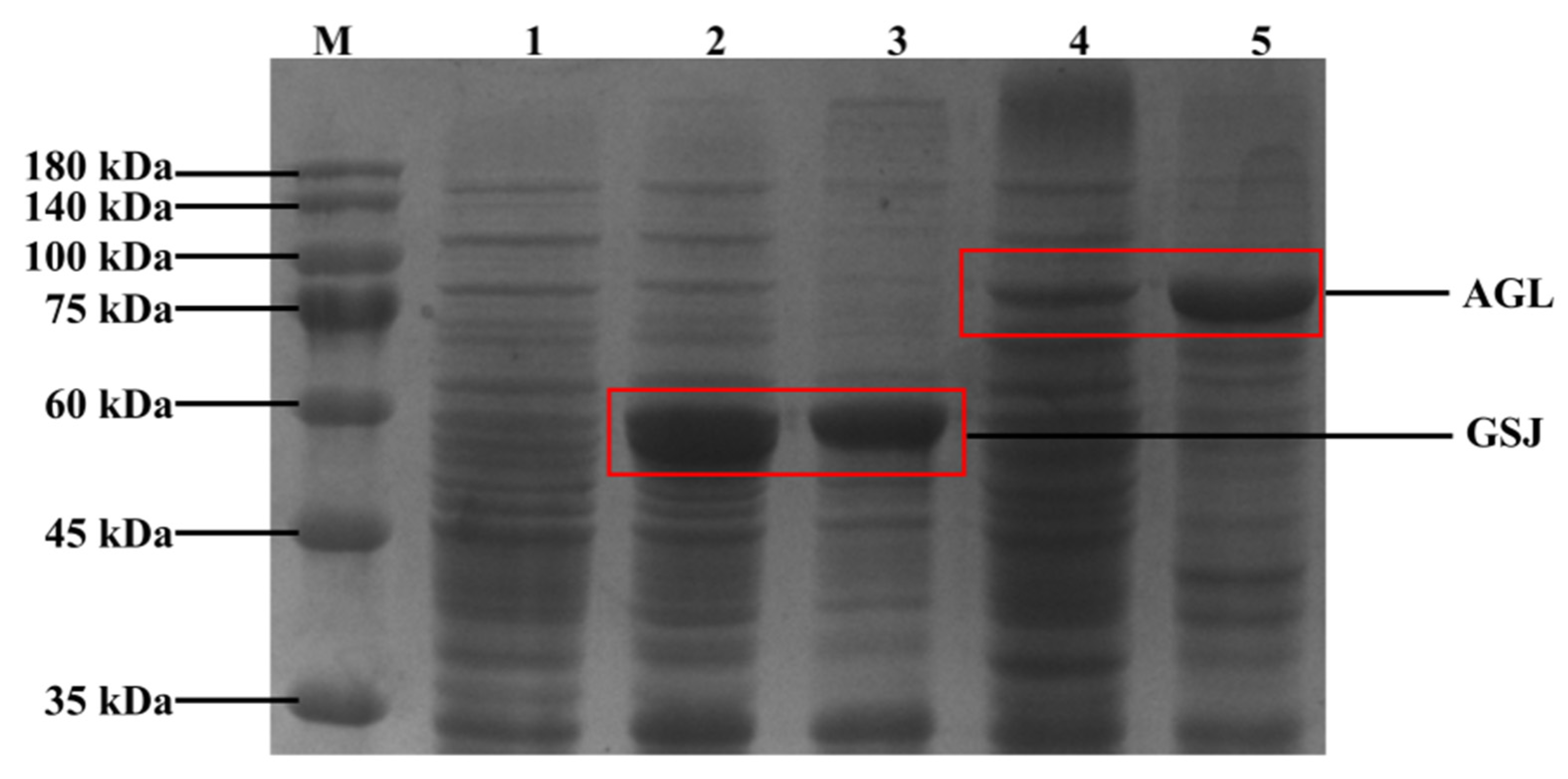
As a critical enzyme for the production of IMOs, the poor thermal stability of α-glucosidase is a significant factor limiting its application. Therefore, we searched the BRENDA (https://www.brenda-enzymes.org//, accessed on 5 July 2022) enzyme database and selected α-glucosidase with good heat resistance and industrial application potential. In the present study, we constructed recombinant plasmids by amplifying codon-optimized gene fragments and ligating them into the pMA5 vector. The recombinant strains B. Subtilis 168/pMA5-gsj and B. Subtilis 168/pMA5-agl were obtained after transferring the recombinant plasmids into B. subtilis 168. The results of SDS-PAGE (Figure 2) indicated that both gsj (code a 59 kDa protein) and agl (code a 96 kDa protein) genes could be expressed normally in B. subtilis 168. The enzyme activity of recombinant α-glucosidase was determined by the pNPG method. The results showed that the enzyme activity of GSJ was about 3.7 U·mL−1, and AGL was about 2.5 U·mL−1.

3.2. Transglucosylation Activity of Recombinant α-Glucosidase

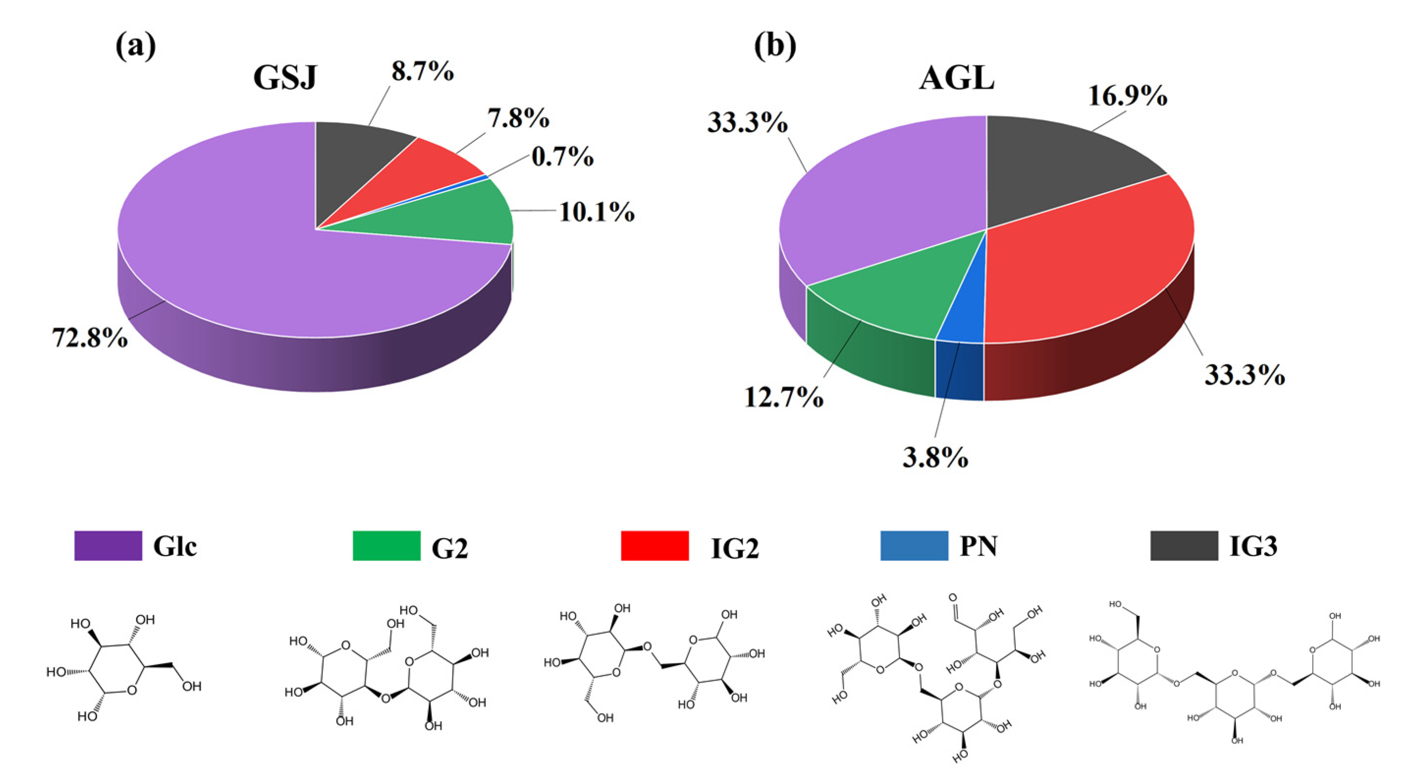
α-glucosidase has both transglycosylation and hydrolysis activities. At high concentrations of maltose (G2) solution, α-glucosidase initially synthesizes panose (PN) and maltotriose (G3). With the increasing amount of glucose (Glc) delivered from the glycosylation step, α-glucosidase starts to synthesize isomaltose (IG2) and isomaltotriose (IG3) [7]. The transglycosylation activity of α-glucosidase is essential for the production of IMOs. Therefore, to determine the transglycosylation activity of recombinant α-glucosidase, we added an appropriate amount of recombinant α-glucosidase to a high concentration of maltose solution and detected the changes of each component during the reaction by HPLC. As shown in Figure 3a, after 12 h of transglycosylation reaction, only 17.2% of maltose was converted to IMOs in the mixture containing GSJ, while 72.8% of maltose was hydrolyzed to glucose. In the AGL mixture, 54% of maltose was converted to IMOs, and 33.3% was hydrolyzed to glucose (Figure 3b). The results show that the hydrolysis activity of GSJ is much higher than the transglycosylation activity, which implies that GSJ is unsuitable for producing IMOs. In contrast, AGL has a high transglycosylation activity and can be applied to the production of IMOs. The α-glucosidase derived from Aspergillus niger is widely used in producing α-glucosidase due to its good transglycosylation activity. In addition, the α-glucosidase from Aspergillus awamori and Aspergillus carbonarious also showed transglycosylation activity [32]. In suitable conditions, Aspergillus niger-derived α-glucosidase can convert about 55% of maltose to IMOs [33]; this is similar to the transglycosylation activity of the recombinant α-glucosidase in this study, which also indicates the potential of recombinant α-glucosidase to be used for the production of IMOs.

3.3. Construction of Cyclized AGL to Improve the Thermal Stability of Recombinant α-Glucosidase

The cyclic peptide is a type of naturally occurring polypeptide molecule with a circular structure [34]. Due to its good stability, genetic engineering, protein engineering, other methods have developed a series of molecular cyclization techniques, such as protein trans-splicing (PTS) [35], expressed protein ligation (EPL) mediated by intrinsic peptide [36], and sortagging mediated by Sortase [37]. Although these cyclization methods can improve the stability of proteins, achieving the above linkage methods requires complex reaction conditions or the involvement of specific catalysts, and the reaction efficiency is low [38]. Isopeptide bond has been widely used due to its specificity, stability, and rapid reaction. We used three traditional isopeptide bonds (SpyTag/SpyCatcher, SnoopTag/SnoopCatcher, SdyTag/SnoopCatcher) and a novel short peptide tag (RIAD/RIDD) to form cyclized AGL. According to the method in Section 2.2.5 and Figure S1, we constructed recombinant plasmids and expressed them in B. subtilis 168. As shown in Figure 4, the results of SDS-PAGE indicated that the recombinant proteins RIAD-AGL-RIDD (~103 kDa), SpyTag-AGL-SpyCatcher (~103 kDa), SnoopTag-AGL-SnoopCatcher (~104 kDa), and SdyTag-AGL-SdyCatcher (~103 kDa) were successfully expressed, and the molecular weights were increased. To verify whether the isopeptide bond was formed, we mutated the key site of SpyTag-AGL-SpyCatcher. It was found that the band was located between cyclized AGL and wild-type AGL, indicating that AGL was successfully cyclized by isopeptide bonds. After purifying the cyclized protein, we assayed the enzyme activity of cyclized AGL (Table 1). We found that the activity of SpyTag-AGL-SpyCatcher, SnoopTag-AGL-SnoopCatcher, and SdyTag-AGL-SdyCatcher had no significant change. In contrast, the enzyme activity of RIAD-AGL-RIDD decreased by 56.5%. Therefore, we performed further enzymatic property characterization of SpyTag-AGL-SpyCatcher, SnoopTag-AGL-SnoopCatcher, and SdyTag-AGL-SdyCatcher.

3.4. Optimal Temperature and Thermal Stability of Cyclized AGL

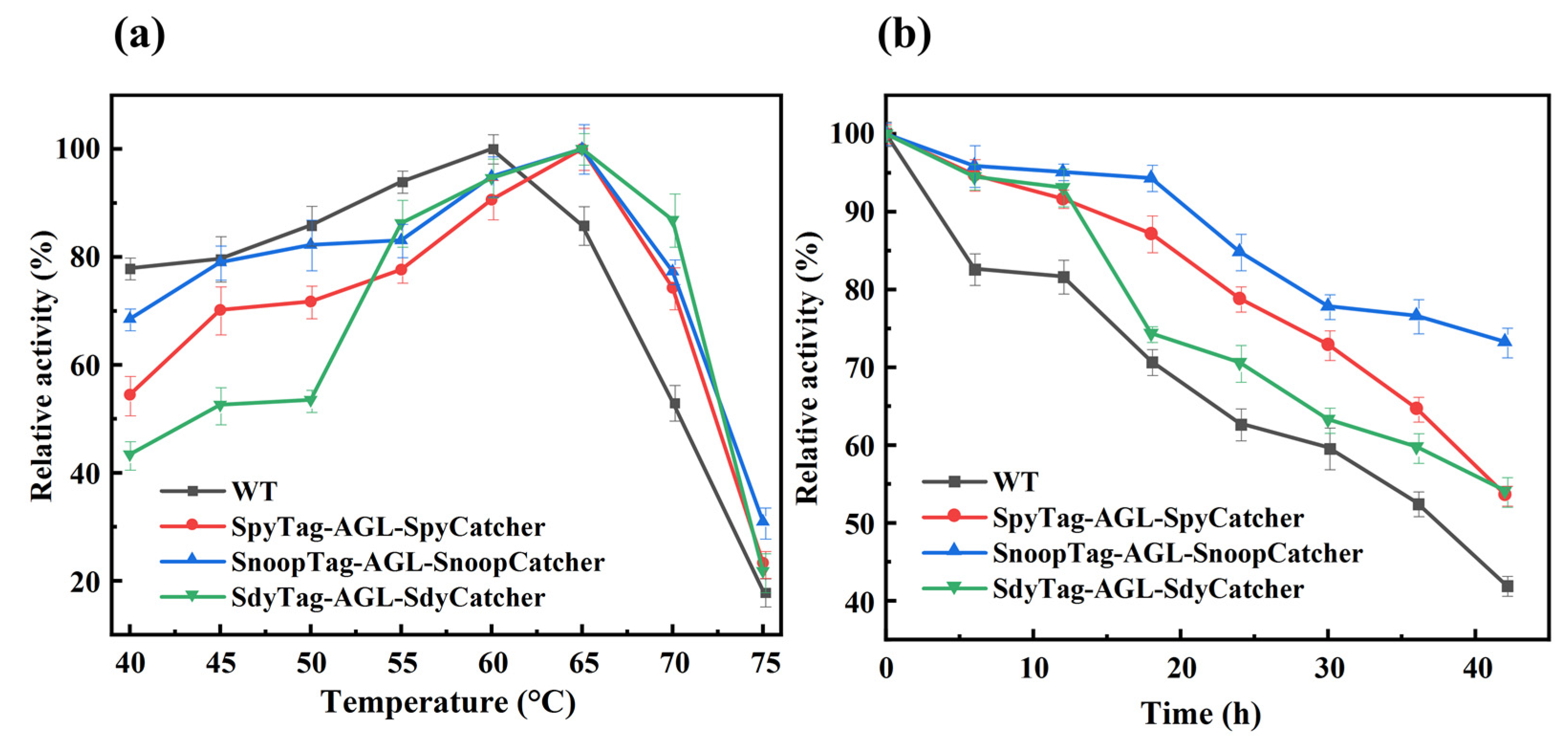
To visually investigate the changes in enzyme activity at different temperatures, the optimal temperature of wild-type and cyclized AGL was characterized by the relative enzyme activity at different temperatures (40–75 °C). As shown in Figure 5a, the optimal temperature of all cyclized AGL was increased by 5 °C compared to the wild-type AGL. In addition, the relative enzyme activity of all cyclized AGL was higher than wild-type AGL at temperatures above 60 °C.
For the thermal stability experiments, we incubated the recombinant α-glucosidase at 65 °C for 42 h and measured the enzyme activity. The results are shown in Figure 5b. Compared to wild-type AGL, all cyclized AGL thermal stability was improved. After incubation at 65 °C for 42 h, the residual enzyme activities of SpyTag-AGL-SpyCatcher and SdyTag-AGL-SdyCatcher were 53.7% and 54.2%, respectively, while the wild-type AGL was 42.1%. In contrast, the thermal stability of SnoopTag-AGL-SnoopCatcher was significantly improved, and the residual enzyme activity after 42 h remained 73.2%, which was 1.74-fold higher than the wild type.
This result was similar to previous studies on cyclized proteins’ optimal temperature and thermal stability. While the cyclized protein increased thermal stability, the optimal temperature was also increased. Chen et al. [24] found that the optimal temperature of cyclized Tres increased by 5–10 °C after using various isopeptide bonds. Wang et al. [25] used SpyCatcher/SpyTag to cause spontaneous cyclization of lichenase, and the optimal temperature of cyclized lichenase was increased by 5 °C. Likewise, Si et al. [27] constructed cyclized luciferase by SpyCatcher/SpyTag, which eventually increased the optimal temperature of luciferase by 5 °C.

3.5. Optimal pH and pH Stability of Cyclized AGL

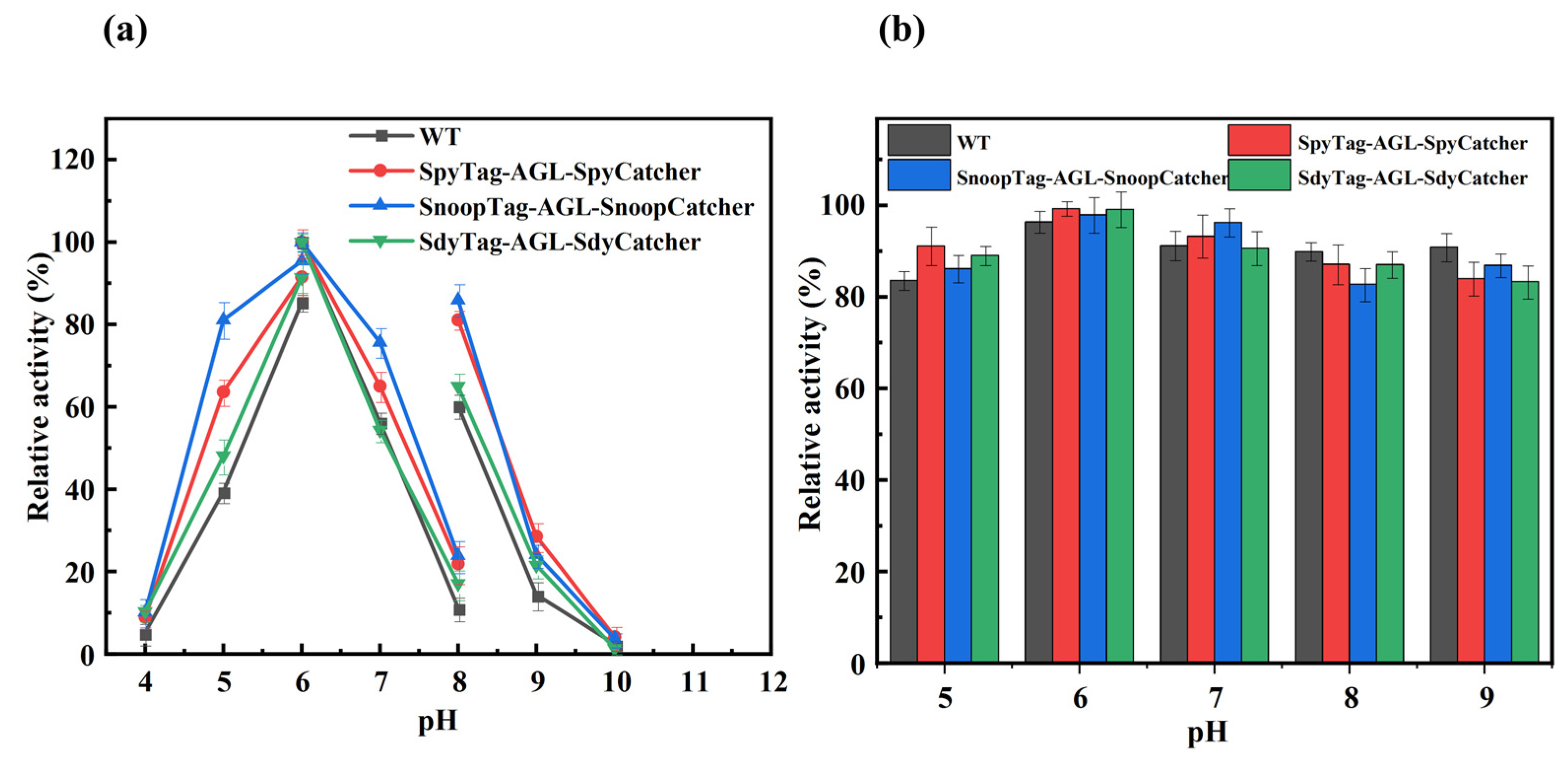
We determined the enzyme activity of recombinant α-glucosidase at different pH according to method Section 2.2.5. As shown in Figure 6a, the optimal pH value was 6.0 for wild-type AGL and cyclized AGL, and the relative activity of all cyclized AGL was slightly higher than that of wild-type AGL at pH values in the range of 4.0–10.0. The residual enzyme activities of wild-type AGL and cyclized AGL were similar after 24 h incubation in different pH buffers (Figure 6b).

3.6. Kinetic Analysis of Recombinant α-Glucosidase

The kinetic parameters of recombinant α-glucosidase were determined at pH 7.0 and 65 °C with pNPG as the substrate. The Km, Vmax and Kcat values of cyclized AGL were similar to the wild-type AGL (Table 2), which indicated that the cyclization reaction does not affect the catalytic efficiency of recombinant α-glucosidase. However, there is no consistent conclusion on whether the insertion of isopeptide bonds affects the catalytic efficiency of the protein. Previous studies have shown no effect of cyclization on the Km values of luciferase [27] and β-lactamase [26], while the kinetic parameters of xylanase [39] and lichenase [25] showed significant differences in polysaccharide before and after cyclization.

3.7. Molecular Dynamics Simulation Analysis of Cyclized AGL

The three-dimensional models of cyclized AGL were simulated to explore the molecular mechanism of increased thermal stability of cyclized AGL. Furthermore, MD simulation was performed at 338 K by GROMACS. Due to the lack of a suitable crystal structure as a template, we conducted homology modeling by ALPHA-FOLD to obtain the 3D structure of cyclized AGL. As shown in Figure 7, all three isopeptide bonds successfully cyclized the recombinant protein, and cyclization had no effect on the active sites of AGL compared to wild-type AGL (Figure S2). In addition, there was no significant change in the distance between the terminals in the wild-type and cyclized versions (Figure S3), indicating that the isopeptide bond did not distort the enzyme. The structural alignment revealed that cyclization did not cause conformational changes in AGL (Figure S4).
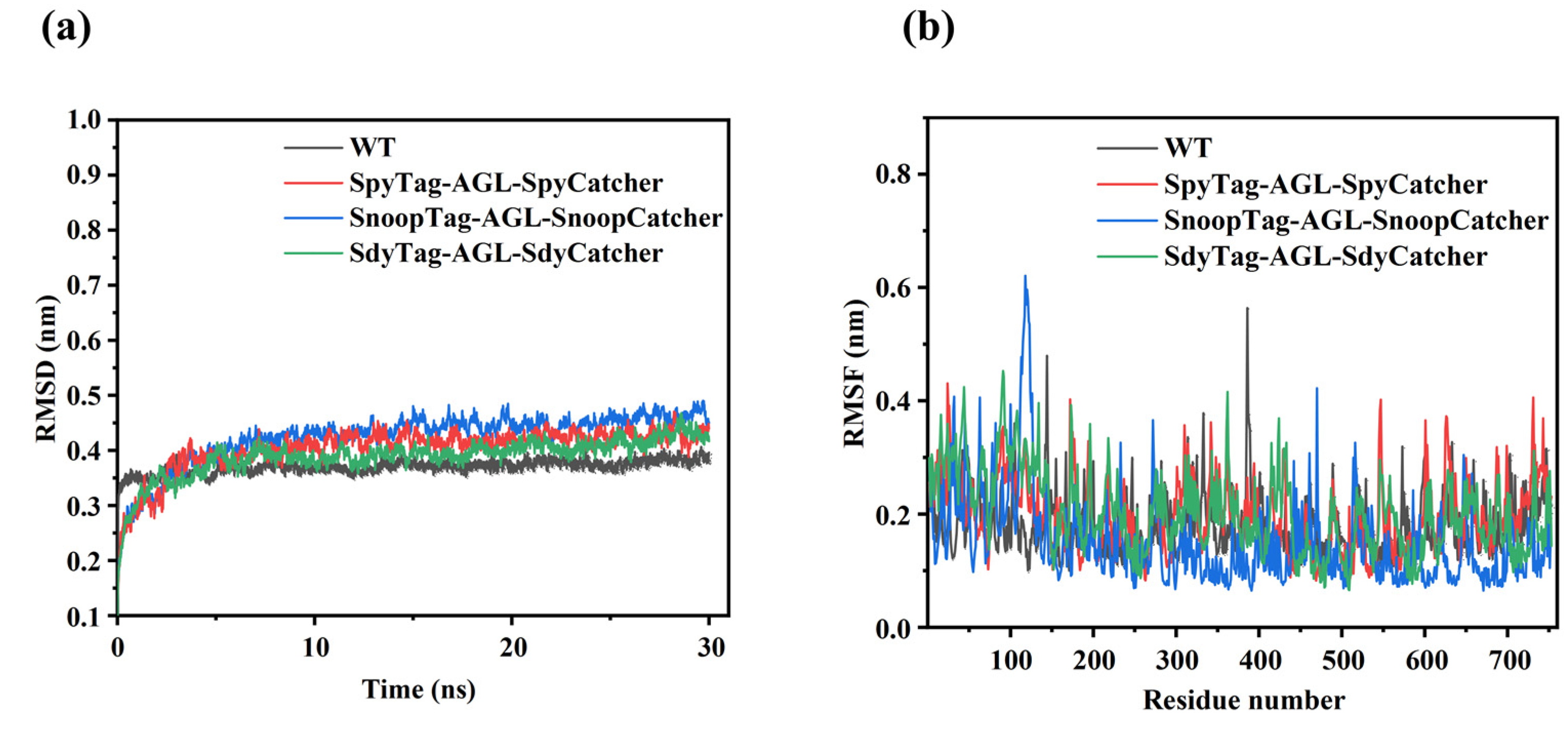
In the present study, we analyzed the dynamic behavior of the cyclized AGL systems by performing 30-ns MD simulations, and the RMSD value characterized the stability of the systems. As shown in Figure 8a, the WT system fluctuate between 0–0.4 nm until it reaches equilibrium at 5 ns. SpyTag-AGL-SpyCatcher, SnoopTag-AGL-SpyCatcher, and SdyTag-AGL-SdyCatcher systems fluctuate from 0 to 0.48 nm and maintain equilibrium between 5 and 30 ns. SnoopTag-AGL-SpyCatcher system has the highest RMSD value (0.48 nm), while the four systems were maintaining equilibrium. These results indicated that the four systems remained stable between 5 and 30 ns, and the 5 to 30 ns simulation trajectories could be used for further analysis, including Rg and RMSF.
As shown in Figure 8b, the fluctuations of most residues were similar in the four systems. The WT system’s RMSF values were higher than the cyclized AGL system at residues 150–200, 320–350, 380–400, 560–590, and 700–740. This result shows that the rigidity of the cyclized AGL increased in these regions, contributing to the increased thermal stability of the cyclized AGL. Moreover, the RMSF values of the SnoopTag-AGL-SnoopCatcher system are lower than WT, SpyTag-AGL-SpyCatcher, and SdyTag-AGL-SdyCatcher systems at residues 300–400, 600–700, which explained the better thermal stability of SnoopTag-AGL-SnoopCatcher. In summary, the results of MD simulations are consistent with the experimental data.

4. Conclusions

In the present study, we heterologously expressed α-glucosidase with industrial application potential in B. subtilis 168. based on the molecular cyclization of isopeptide bonds, we constructed cyclized proteins to improve the thermal stability of recombinant α-glucosidase. Among them, SpyTag/SpyCatcher can significantly improve the thermal stability of the recombinant protein. MD simulations showed that the rigidity of the cyclized proteins was increased, indicating that improving the thermal stability of α-glucosidase by isopeptide bonds is an effective method. Due to its good thermal stability and high transglycosylation activity, cyclized AGL can be used to produce IMOs continuously. Therefore, in future studies, we will try to immobilize the enzyme with different materials and determine the optimal conditions for immobilization. The immobilized enzyme is used in the continuous production of IMOs. In addition, we will select suitable mutation sites for targeted mutagenesis by rational design to further improve the thermal stability and transglycosylation activity of recombinant α-glucosidase.

Supplementary Materials

The following supporting information can be downloaded at: https://www.mdpi.com/article/10.3390/fermentation8100498/s1, Figure S1: Construction of recombinant plasmids for cyclized AGL; Figure S2: Three-dimensional structure of wild-type AGL; Figure S3: The distance between the terminals; Figure S4: Structural alignment of cyclized AGL with wild-type; Table S1: strains and plasmids used in this study; Table S2: Primers used to construct the cyclized proteins; Table S3: Amino acid sequence of cyclized protein.

Author Contributions

Conceptualization, Z.R. and X.Z.; methodology, Z.W.; software, H.Z.; validation, Q.W., M.F. and R.L.; formal analysis, M.X.; investigation, M.H.; resources, M.H.; data curation, Z.W.; writing—original draft preparation, Z.W.; writing—review and editing, Z.W.; visualization, M.H.; supervision, X.Z.; project administration, X.Z.; funding acquisition, Z.R. All authors have read and agreed to the published version of the manuscript.

Funding

This research was funded by National Key Research and Development Program of China, (No. 2021YFC2100900), National Natural Science Foundation of China (No. 32171471, No. 32071470), Key Research and Development Project of Shandong Province, China (2019JZZY020605), the Project Funded by the Priority Academic Program Development of Jiangsu Higher Education Institutions, Top-notch Academic Programs Project of Jiangsu Higher Education Institutions.

Institutional Review Board Statement

Not applicable.

Informed Consent Statement

Not applicable.

Data Availability Statement

The data that support the findings of this study are available from the corresponding author upon reasonable request.

Conflicts of Interest

The authors declare no conflict of interest.

References

  1. Huang, Z.; Li, Z.; Su, Y.; Zhu, Y.; Zeng, W.; Chen, G.; Liang, Z. Continuous Production of Isomalto-oligosaccharides by Thermo-inactivated Cells of Aspergillus niger J2 with Coarse Perlite as an Immobilizing Material. Appl. Biochem. Biotechnol. 2018, 185, 1088–1099. [Google Scholar] [CrossRef] [PubMed]
  2. Shi, Q.; Hou, Y.; Juvonen, M.; Tuomainen, P.; Kajala, I.; Shukla, S.; Goyal, A.; Maaheimo, H.; Katina, K.; Tenkanen, M. Optimization of Isomaltooligosaccharide Size Distribution by Acceptor Reaction of Weissella confusa Dextransucrase and Characterization of Novel alpha-(1 -> 2)-Branched Isomaltooligosaccharides. J. Agric. Food Chem. 2016, 64, 3276–3286. [Google Scholar] [CrossRef] [PubMed]
  3. Ojha, S.; Mishra, S.; Chand, S. Production of isomalto-oligosaccharides by cell bound alpha-glucosidase of Microbacterium sp. Lwt-Food Sci. Technol. 2015, 60, 486–494. [Google Scholar] [CrossRef]
  4. Soto-Maldonado, C.; Concha-Olmos, J.; Caceres-Escobar, G.; Menses-Gomez, P. Sensory evaluation and glycaemic index of a food developed with flour from whole (pulp and peel) overripe banana (Musa cavendishii) discards. Lwt-Food Sci. Technol. 2018, 92, 569–575. [Google Scholar] [CrossRef]
  5. Wu, Q.; Pi, X.e.; Liu, W.; Chen, H.; Yin, Y.; Yu, H.D.; Wang, X.; Zhu, L. Fermentation properties of isomaltooligosaccharides are affected by human fecal enterotypes. Anaerobe 2017, 48, 206–214. [Google Scholar] [CrossRef]
  6. Madsen, L.R., II; Stanley, S.; Swann, P.; Oswald, J. A Survey of Commercially Available Isomaltooligosaccharide-Based Food Ingredients. J. Food Sci. 2017, 82, 401–408. [Google Scholar] [CrossRef]
  7. Gu, X.L.; Li, H.; Song, Z.H.; Ding, Y.N.; He, X.; Fan, Z.Y. Effects of isomaltooligosaccharide and Bacillus supplementation on sow performance, serum metabolites, and serum and placental oxidative status. Anim. Reprod. Sci. 2019, 207, 52–60. [Google Scholar] [CrossRef]
  8. Nakakuki, T. Present status and future of functional oligosaccharide development in Japan. Pure Appl. Chem. 2002, 74, 1245–1251. [Google Scholar] [CrossRef]
  9. Bivolarski, V.; Vasileva, T.; Bozov, P.; Iliev, I. INFLUENCE OF DIFFERENT ACCEPTORS ON SYNTHESIS OF GLUCOOLIGOSACCHARIDES BY PURIFIED DEXTRANSUCRASE FROM LEUCONOSTOC MESENTEROIDES URE 13. Comptes Rendus De L Acad. Bulg. Des. Sci. 2013, 66, 1405–1412. [Google Scholar]
  10. Sorndech, W.; Sagnelli, D.; Blennow, A.; Tongta, S. Combination of amylase and transferase catalysis to improve IMO compositions and productivity. Lwt-Food Sci. Technol. 2017, 79, 479–486. [Google Scholar] [CrossRef]
  11. Rudeekulthamrong, P.; Sawasdee, K.; Kaulpiboon, J. Production of long-chain isomaltooligosaccharides from maltotriose using the thermostable amylomaltase and transglucosidase enzymes. Biotechnol. Bioprocess Eng. 2013, 18, 778–786. [Google Scholar] [CrossRef]
  12. Ma, M.; Okuyama, M.; Sato, M.; Tagami, T.; Klahan, P.; Kumagai, Y.; Mori, H.; Kimura, A. Effects of mutation of Asn694 in Aspergillus niger alpha-glucosidase on hydrolysis and transglucosylation. Appl. Microbiol. Biotechnol. 2017, 101, 6399–6408. [Google Scholar] [CrossRef] [PubMed]
  13. Chen, D.-L.; Tong, X.; Chen, S.-W.; Chen, S.; Wu, D.; Fang, S.-G.; Wu, J.; Chen, J. Heterologous Expression and Biochemical Characterization of alpha-Glucosidase from Aspergillus niger by Pichia pastroris. J. Agric. Food Chem. 2010, 58, 4819–4824. [Google Scholar] [CrossRef] [PubMed]
  14. Zhang, F.; Wang, W.; Bah, F.B.M.; Song, C.; Zhou, Y.; Ji, L.; Yuan, Y. Heterologous Expression of a Thermostable alpha-Glucosidase from Geobacillus sp. Strain HTA-462 by Escherichia coli and Its Potential Application for Isomaltose-Oligosaccharide Synthesis. Molecules 2019, 24, 1413. [Google Scholar] [CrossRef] [PubMed]
  15. Zhou, C.; Xue, Y.; Ma, Y. Evaluation and directed evolution for thermostability improvement of a GH 13 thermostable alpha-glucosidase from Thermus thermophilus TC11. BMC Biotechnol. 2015, 15, 97. [Google Scholar] [CrossRef] [PubMed]
  16. Park, J.-E.; Park, S.R.; Woo, J.Y.; Hwang, H.S.; Cha, J.; Lee, H. Enzymatic Properties of a Thermostable alpha-Glucosidase from Acidothermophilic Crenarchaeon Sulfolobus tokodaii Strain 7. J. Microbiol. Biotechnol. 2013, 23, 56–63. [Google Scholar] [CrossRef] [PubMed]
  17. Hung, V.S.; Hatada, Y.; Goda, S.; Lu, J.; Hidaka, Y.; Li, Z.J.; Akita, M.; Ohta, Y.; Watanabe, K.; Matsui, H.; et al. alpha-Glucosidase from a strain of deep-sea Geobacillus: A potential enzyme for the biosynthesis of complex carbohydrates. Appl. Microbiol. Biotechnol. 2005, 68, 757–765. [Google Scholar] [CrossRef]
  18. Zhou, C.; Xue, Y.; Ma, Y. Enhancing the thermostability of alpha-glucosidase from Thermoanaerobacter tengcongensis MB4 by single proline substitution. J. Biosci. Bioeng. 2010, 110, 12–17. [Google Scholar] [CrossRef]
  19. Zakeri, B.; Howarth, M. Spontaneous Intermolecular Amide Bond Formation between Side Chains for Irreversible Peptide Targeting. J. Am. Chem. Soc. 2010, 132. [Google Scholar] [CrossRef]
  20. Zakeri, B.; Fierer, J.O.; Celik, E.; Chittock, E.C.; Schwarz-Linek, U.; Moy, V.T.; Howarth, M. Peptide tag forming a rapid covalent bond to a protein, through engineering a bacterial adhesin. Proc. Natl. Acad. Sci. USA 2012, 109, E690–E697. [Google Scholar] [CrossRef]
  21. Veggiani, G.; Nakamura, T.; Brenner, M.D.; Gayet, R.V.; Yan, J.; Robinson, C.V.; Howarth, M. Programmable polyproteams built using twin peptide superglues. Proc. Natl. Acad. Sci. USA 2016, 113, 1202–1207. [Google Scholar] [CrossRef] [Green Version]
  22. Fierer, J.O.; Veggiani, G.; Howarth, M. SpyLigase peptide-peptide ligation polymerizes affibodies to enhance magnetic cancer cell capture. Proc. Natl. Acad. Sci. USA 2014, 111, E1176–E1181. [Google Scholar] [CrossRef]
  23. Wong, J.X.; Gonzalez-Miro, M.; Sutherland-Smith, A.J.; Rehm, B.H.A. Covalent Functionalization of Bioengineered Polyhydroxyalkanoate Spheres Directed by Specific Protein-Protein Interactions. Front. Bioeng. Biotechnol. 2020, 8, 44. [Google Scholar] [CrossRef] [PubMed]
  24. Chen, Y.; Zhao, Y.; Zhou, X.; Liu, N.; Ming, D.; Zhu, L.; Jiang, L. Improving the thermostability of trehalose synthase from Thermomonospora curvata by covalent cyclization using peptide tags and investigation of the underlying molecular mechanism. Int. J. Biol. Macromol. 2021, 168, 13–21. [Google Scholar] [CrossRef] [PubMed]
  25. Wang, J.; Wang, Y.; Wang, X.; Zhang, D.; Wu, S.; Zhang, G. Enhanced thermal stability of lichenase from Bacillus subtilis 168 by SpyTag/SpyCatcher-mediated spontaneous cyclization. Biotechnol. Biofuels 2016, 9, 79. [Google Scholar] [CrossRef]
  26. Schoene, C.; Bennett, S.P.; Howarth, M. SpyRing interrogation: Analyzing how enzyme resilience can be achieved with phytase and distinct cyclization chemistries. Sci. Rep. 2016, 6, 21151. [Google Scholar] [CrossRef] [PubMed]
  27. Si, M.; Xu, Q.; Jiang, L.; Huang, H. SpyTag/SpyCatcher Cyclization Enhances the Thermostability of Firefly Luciferase. PLoS ONE 2016, 11, e0162318. [Google Scholar] [CrossRef]
  28. Wang, Y.-H.; Jiang, Y.; Duan, Z.-Y.; Shao, W.-L.; Li, H.-Z. Expression and characterization of an alpha-glucosidase from Thermoanaerobacter ethanolicus JW200 with potential for industrial application. Biologia 2009, 64, 1053–1057. [Google Scholar] [CrossRef]
  29. Kang, W.; Ma, T.; Liu, M.; Qu, J.; Liu, Z.; Zhang, H.; Shi, B.; Fu, S.; Ma, J.; Lai, L.T.F.; et al. Modular enzyme assembly for enhanced cascade biocatalysis and metabolic flux. Nat. Commun. 2019, 10, 4248. [Google Scholar] [CrossRef]
  30. You, J.; Yang, C.; Pan, X.; Hu, M.; Du, Y.; Osire, T.; Yang, T.; Rao, Z.J.B.T. Metabolic engineering of Bacillus subtilis for enhancing riboflavin production by alleviating dissolved oxygen limitation. Bioresour. Technol. 2021, 333, 125228. [Google Scholar] [CrossRef]
  31. Jung, J.H.; Seo, D.H.; Holden, J.F.; Kim, H.S.; Baik, M.Y.; Park, C.S. Broad substrate specificity of a hyperthermophilic alpha-glucosidase from Pyrobaculum arsenaticum. Food Sci. Biotechnol. 2016, 25, 1665–1669. [Google Scholar] [CrossRef] [PubMed]
  32. Anindyawati, T.; Ann, Y.-G.; Ito, K.; Iizuka, M.; Minamiura, N. Two kinds of novel α-glucosidases from Aspergillus awamori KT-11: Their purifications, properties and specificities. J. Ferment. Bioeng. 1998, 85, 465–469. [Google Scholar] [CrossRef]
  33. Shimba, N.; Shinagawa, M.; Hoshino, W.; Yamaguchi, H.; Yamada, N.; Suzuki, E.-i. Monitoring the hydrolysis and transglycosylation activity of alpha-glucosidase from Aspergillus niger by nuclear magnetic resonance spectroscopy and mass spectrometry. Anal. Biochem. 2009, 393, 23–28. [Google Scholar] [CrossRef] [PubMed]
  34. Cascales, L.; Craik, D.J. Naturally occurring circular proteins: Distribution, biosynthesis and evolution. Org. Biomol. Chem. 2010, 8, 5035–5047. [Google Scholar] [CrossRef]
  35. Boecker, J.K.; Friedel, K.; Matern, J.C.J.; Bachmann, A.-L.; Mootz, H.D. Generation of a Genetically Encoded, Photoactivatable Intein for the Controlled Production of Cyclic Peptides. Angew. Chem. Int. Ed. 2015, 54, 2116–2120. [Google Scholar] [CrossRef]
  36. De Rosa, L.; Cortajarena, A.L.; Romanelli, A.; Regan, L.; D’Andrea, L.D. Site-specific protein double labeling by expressed protein ligation: Applications to repeat proteins. Org. Biomol. Chem. 2012, 10, 273–280. [Google Scholar] [CrossRef]
  37. Agwa, A.J.; Craik, D.J.; Schroeder, C.I. Cyclizing Disulfide-Rich Peptides Using Sortase A. Methods Mol. Biol. (Clifton N.J.) 2019, 2012, 29–41. [Google Scholar] [CrossRef]
  38. Minato, Y.; Ueda, T.; Machiyama, A.; Shimada, I.; Iwai, H. Segmental isotopic labeling of a 140 kDa dimeric multi-domain protein CheA from Escherichia coli by expressed protein ligation and protein trans-splicing. J. Biomol. NMR 2012, 53, 191–207. [Google Scholar] [CrossRef]
  39. Lin, Y.; Jin, W.; Wang, J.; Cai, Z.; Wu, S.; Zhang, G. A novel method for simultaneous purification and immobilization of a xylanase-lichenase chimera via SpyTag/SpyCatcher spontaneous reaction. Enzym. Microb. Technol. 2018, 115, 29–36. [Google Scholar] [CrossRef]
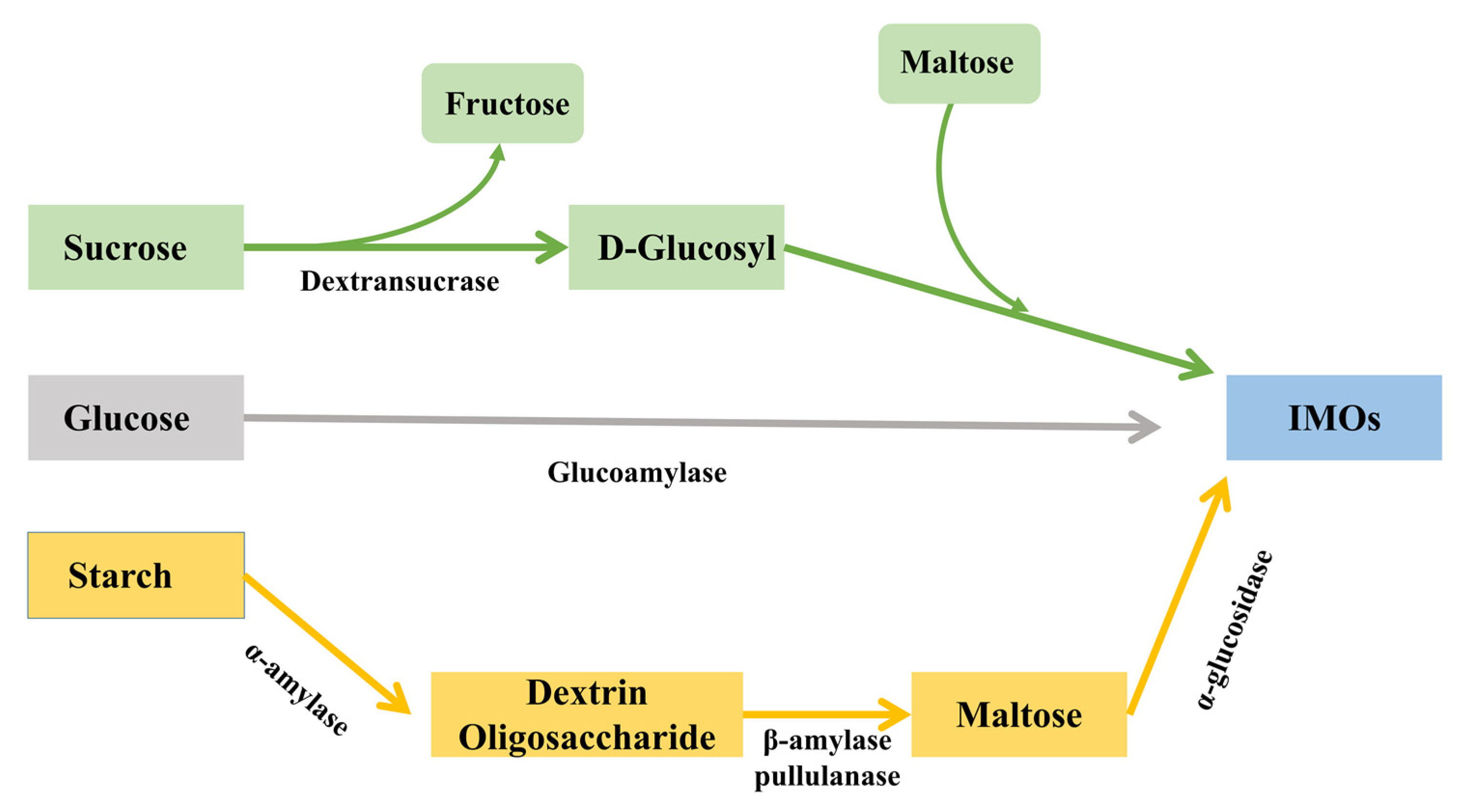
Figure 1. Three main ways for the production of IMOs: Utilizing the retrosynthesis of glucoamylase (grey routine), sucrose and maltose are converted by dextransucrase (green routine), maltose catalyzed by α-glucosidase (yellow routine).
Fermentation 08 00498 g001
Figure 2. SDS-PAGE analysis of recombinant α-glucosidase. Lane M, protein marker; Lane 1, Control (B. Subtilis 168/pMA5 crude enzyme); Lane 2, B. Subtilis 168/pMA5-gsj crude enzyme; Lane 3, B. Subtilis 168/pMA5-gsj broken cell precipitate; Lane 4, B. Subtilis 168/pMA5-agl crude enzyme; Lane 5 B. Subtilis 168/pMA5-agl broken cell precipitate.
Fermentation 08 00498 g002
Figure 3. Analysis of transglycosylation products. The transglycosylation products of maltose by GSJ (a) and AGL (b).
Fermentation 08 00498 g003
Figure 4. SDS-PAGE analysis of cyclized AGL. Lane M, protein marker; Lane 1, Control (wild type AGL); Lane 2, RIAD-AGL-RIDD; Lane 3, SpyTag-AGL-SpyCatcher; Lane 4, SnoopTag-AGL-SnoopCatcher; Lane 5, SdyTag-AGL-SdyCatcher; Lane 6, linear AGL.
Fermentation 08 00498 g004
Figure 5. Effects of temperature on the activity and stability of wild-type and cyclized AGL: (a) optimal temperature, (b) thermal stability at 65 °C. All assays were performed in triplicate, and the standard deviations of biological replicates are indicated by error bars.
Fermentation 08 00498 g005
Figure 6. Effects of pH on the activity and stability of wild-type and cyclized AGL: (a) optimal pH, (b) pH stability. All assays were performed in triplicate, and the standard deviations of biological replicates are indicated by error bars.
Fermentation 08 00498 g006
Figure 7. 3D structure of cyclized AGL (the pink spheres highlight the enzyme active sites). (a) SpyTag-AGL-SpyCatcher, (b) SnoopTag-AGL-SnoopCatcher, (c) SdyTag-AGL-SdyCatcher.
Fermentation 08 00498 g007
Figure 8. MD simulations of wild-type AGL and cyclized AGL. (a) RMSD values, (b) RMSF values.
Fermentation 08 00498 g008
Table 1. Specific enzyme activities of wild-type and cyclized AGL.
EnzymeSpecific Enzyme Activity (U·mg−1)Relative Enzyme Activity (100%)
WT27.91 ± 0.5100.0 ± 1.7
RIAD-AGL-RIDD12.01 ± 0.743.5 ± 2.5
SpyTag-AGL-SpyCatcher26.43 ± 0.394.7 ± 1.1
SnoopTag-AGL-SnoopCatcher28.55 ± 0.6102.3 ± 2.1
SdyTag-AGL-SdyCatcher27.18 ± 0.397.4 ± 1.1
All assays are performed in triplicate and the standard deviation of biological replicates is expressed as a numerical error.
Table 2. Kinetic parameters of wild-type AGL and cyclized AGL.
EnzymeKm (mM)Vmax (U mg−1)Kcat (S−1)
WT1.83 ± 0.0636.42 ± 0.12282.31±2.1
SpyTag-AGL-SpyCatcher1.86 ± 0.0335.94 ± 0.07276.38±7.2
SnoopTag-AGL-SnoopCatcher1.82 ± 0.0536.12 ± 0.15278.92±6.8
SdyTag-AGL-SdyCatcher1.86 ± 0.0636.31 ± 0.19281.59±6.5
All assays are performed in triplicate and the standard deviation of biological replicates is expressed as a numerical error.
Publisher’s Note: MDPI stays neutral with regard to jurisdictional claims in published maps and institutional affiliations.