Article

Static Magnetic Field-Assisted Fermentation of Ginkgo biloba Leaves by Lacticaseibacillus paracasei: Process Optimization for Total Flavonoids, Nutritional Components, and Hypoglycemic Activity Analysis

School of Biomedicine and Pharmacy, Jiangsu Agri-animal Husbandry Vocational College, No.8, Fenghuang East Road, Taizhou 225300, China
*
Author to whom correspondence should be addressed.
Fermentation 2026, 12(9), 439; https://doi.org/10.3390/fermentation12090439
Submission received: 6 August 2026 / Revised: 12 September 2026 / Accepted: 15 September 2026 / Published: 17 September 2026
(This article belongs to the Section Probiotic Strains and Fermentation)

Abstract

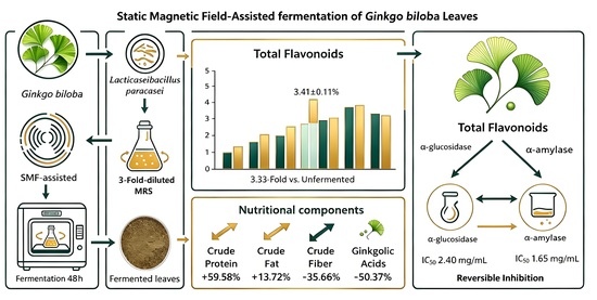
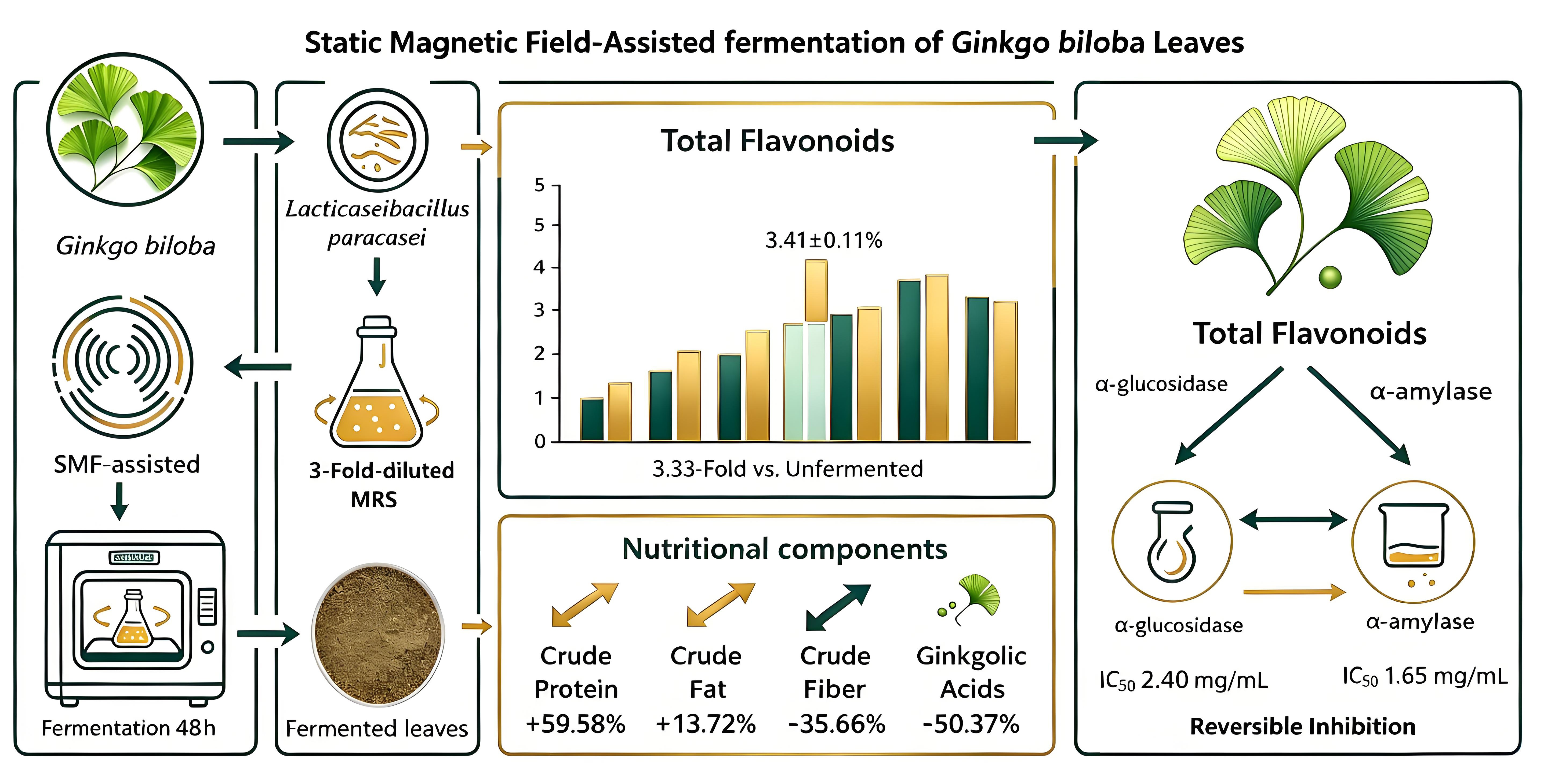
A static magnetic field (SMF)-assisted liquid-state fermentation strategy using Lacticaseibacillus paracasei (L. paracasei) is proposed to improve the comprehensive utilization value of Ginkgo biloba leaves. Taking total flavonoid extraction yield as the objective, Box-Behnken response surface methodology was adopted to optimize the fermentation process. Meanwhile, the nutritional components and hypoglycemic activity of Ginkgo biloba leaves in vitro before and after fermentation were compared and analyzed. The optimal fermentation conditions were determined as follows: 3-fold-diluted De Man, Rogosa, and Sharpe (MRS) medium, an SMF intensity of 4.2 mT, an SMF duration of 2.9 h, and an inoculum size of 6.5%. Under these conditions, the total flavonoid extraction yield reached (3.41 ± 0.11)%, which was 3.33 times that of unfermented raw material. Analysis of the nutritional components revealed that after SMF-assisted fermentation, crude protein and crude fat contents increased by 59.58% and 13.72%, while ash and crude fiber contents decreased by 24.84% and 35.66%, respectively. SMF application also significantly affected the crude fiber content (p < 0.05). Meanwhile, the contents of phytic acid, tannins, phenolic acids, and ginkgolic acids in the SMF-assisted group declined by 29.56%, 43.10%, 23.42%, and 50.37%, respectively, and the SMF further promoted the degradation of anti-nutritional factors. Hypoglycemic activity analysis indicated that fermentation markedly enhanced the inhibitory effect of Ginkgo biloba leaf-derived flavonoids against α-glucosidase and α-amylase, and that SMF stimulation may also contribute to strengthening this inhibitory activity. This study demonstrates that SMF-coupled probiotic fermentation is an effective and environmentally friendly approach for processing Ginkgo biloba leaves, providing experimental support for high-value utilization of plant-based resources through physical-field-assisted microbial fermentation.

Graphical Abstract

1. Introduction

Ginkgo biloba L., commonly referred to as ginkgo, is a plant cultivated worldwide. Its leaves, fruits, and seeds are all recognized as edible and medicinal resources. Notably, Ginkgo biloba leaves are rich in bioactive compounds such as flavonoids and terpene lactones, which exert antioxidant, anti-inflammatory, hypoglycemic, and hypolipidemic properties, making them widely exploited in food, pharmaceuticals, as well as for animal feed [1]. In unprocessed Ginkgo biloba leaves, most flavonoids occur in bound forms, which hinders their extraction and results in low recovery yield [2]. Furthermore, Ginkgo biloba leaves contain anti-nutritional substances such as phytic acid, tannins, and ginkgolic acids. These compounds lower the bioavailability of nutrients and can also pose food safety risks, thereby limiting the full exploitation of Ginkgo biloba leaf resources.
Probiotic fermentation is an efficient green technique for enrichment of bioactive compounds and elimination of anti-nutritional factors in plants [3,4]; it has attracted extensive research interest within the field of plant-based fermentation. Physical field-assisted fermentation technologies have also experienced rapid development in recent years, with static magnetic field (SMF) treatments being the most prevalent [5,6]. Yao et al. [7] described how an SMF of suitable intensity can accelerate microbial respiratory metabolism, regulate bacterial membrane permeability and metabolic pathways, and upregulate the activities of key metabolic enzymes, ultimately improving the nutritional and functional attributes of fermented materials. Existing studies on fermentation of Ginkgo biloba leaves have predominantly focused on single microbial fermentation and have not applied physical fields. For example, Jian et al. [8] fermented Ginkgo biloba leaves with Aspergillus niger for 96 h and obtained a carboxymethyl cellulase activity of 9.50 U. They found that the post-fermentation crude protein content increased from 11.20% to 23.49%, alongside elevated concentrations of total flavonoids and terpene lactones. Moreover, Li et al. [9] utilized Bacillus licheniformis for Ginkgo biloba leaf fermentation and observed significant accumulations of flavonoids, total amino acids, crude protein, and flavor metabolites. The resulting fermented leaves also showed much greater immunomodulatory activity, nutritional value, and palatability. However, there have yet to be systematic investigations into SMF-assisted fermentation of Ginkgo biloba leaves.
As a representative probiotic lactic acid bacterium, Lacticaseibacillus paracasei (L. paracasei) secretes various hydrolases and synthesizes organic acids throughout the fermentation process. These metabolites break down the dense structure of plant cell walls and weaken the intermolecular forces that bind flavonoids to plant matrices, thereby facilitating the liberation of flavonoids during extraction and ameliorating the nutritional quality of substrates [10,11]. With this background in mind, we conducted SMF-assisted liquid-state fermentation of Ginkgo biloba leaves using L. paracasei, and applied Box-Behnken response surface methodology to optimize the fermentation towards maximization of the total flavonoid extraction yield. The resulting changes in basic nutrient content, anti-nutritional factors, and α-glucosidase and α-amylase inhibitory activity were then evaluated in vitro. This work establishes an optimized SMF-probiotic coupled fermentation method for Ginkgo biloba leaves, providing an experimental basis for greener and more profitable processing technology.

2. Materials and Methods

2.1. Materials and Reagents

Ginkgo biloba leaves were provided by Taizhou Xiyang Food Co., Ltd. (Taizhou, Jiangsu, China). L. paracasei LP-33 was purchased from the Shanghai Biobank Biotechnology Center (Shanghai, China). Rutin standard (HPLC ≥ 98%), p-nitrophenyl-β-D-galactopyranoside (PNPG, purity ≥ 98%), α-glucosidase (79 U/mg), acarbose, and α-amylase (3700 U/g) were obtained from Solarbio Science & Technology Co., Ltd. (Beijing, China). Ginkgolic acid (HPLC ≥ 98%) and 3,5-dinitrosalicylic acid (HPLC ≥ 99%) were sourced from Shanghai Yuanye Bio-Technology Co., Ltd. (Shanghai, China). Phosphate-buffered saline (PBS×1, pH 7.4) was supplied by Wuhan Servicebio Technology Co., Ltd. (Wuhan, China). Sodium hydroxide (NaOH), aluminum nitrate (Al(NO3)3), sodium nitrite (NaNO2), anhydrous methanol, and other analytical reagents were purchased from Sinopharm Chemical Reagent Co., Ltd. (Shanghai, China). De Man, Rogosa, and Sharpe (MRS) medium was obtained from Qingdao Hope Bio-Technology Co., Ltd. (Qingdao, China).

2.2. Preparation of L. paracasei Seed Culture

A single colony of activated L. paracasei was picked and then inoculated into 150 mL of liquid MRS medium. The culture was incubated at 37 °C and shaken at 200 rpm for 12 h. Subsequently, the culture was diluted with sterilized water to a concentration of 1.0 × 10⁸ CFU/mL in order to prepare the seed culture.

2.3. Preparation of Fermented Ginkgo biloba Leaves

10 g of Ginkgo biloba leaf powder (sieved through a 60-mesh) was weighed and added to 200 mL of MRS medium at an optimal dilution, and the mixture was sterilized at 121 °C for 20 min, followed by cooling to room temperature. Next, an optimal proportion of the prepared L. paracasei seed culture was inoculated and incubated at 37 °C, shaken at 200 rpm for 12 h, and then placed in a magnetic thermostatic incubator (MFEI-LH1, Indust, Wuxi, China) and treated with the SMF at an optimal intensity and duration. Thereafter, the culture was held in incubation again until the total fermentation time reached 48 h. After fermentation, the pH value and total plate count (TPC) of the fermentation broth were determined. The solid Ginkgo biloba leaves were collected via centrifugation and freeze-dried at −80 °C to obtain dried powder, defining this treatment as the SMF-assisted group. The uninoculated, non-SMF-assisted treatment was represented as the unfermented control group, while the inoculated fermentation without SMF assistance was the conventional fermentation group.

2.3.1. Determination of pH

A pH meter (PB-10, Sartorius, Germany) was calibrated, and its electrode was immersed in the fermentation broth. The pH value was recorded once the reading became stable.

2.3.2. Determination of TPC

0.5 mL of fermentation broth was collected and diluted through a 10-fold serial dilution method. 100 μL of diluted bacterial solution was evenly spread on a solid MRS medium and incubated at 37 °C for 48 h; the colony count was determined via: TPC (CFU/mL) = Number of colonies on the plate × dilution factor × 10, where 10 is the conversion coefficient from the plated volume of 100 μL to 1 mL.

2.3.3. Extraction and Determination of Total Flavonoid Content

2.0 g of freeze-dried fermented Ginkgo biloba leaf powder was weighed and placed into a round-bottom flask, then mixed with 40 mL of anhydrous methanol. Extraction was performed through the reflux method in a water bath at 70 °C for 1 h. The filtrate was collected and diluted to 100 mL, followed by determination of the total flavonoid content via a modified version of the NaNO2-Al(NO3)3-NaOH chromogenic method [12].
A series of rutin standard solutions (0, 0.02, 0.04, 0.06, 0.08, 0.10 mg/mL) were prepared using anhydrous methanol as the solvent. For color development, 10.0 mL of each standard was mixed sequentially with 1.0 mL 5% (w/w) NaNO2, 1.0 mL 10% (w/w) Al(NO3)3, and 8.0 mL 4% (w/w) NaOH, with a 6 min standing interval following each reagent addition. The mixture was diluted to 25.0 mL with methanol, then shaken and equilibrated for 15 min. Absorbance at 510 nm was measured against a methanol blank; the linear curve established between rutin mass concentration (x) and absorbance (y) followed the regression equation y = 4.56x − 0.0007, with a coefficient of determination R2 = 0.9997. A 10.0 mL aliquot of Ginkgo biloba leaf extract was treated identically, and its absorbance was used to calculate flavonoid concentration using this regression equation. The total flavonoid extraction yield was calculated as follows:
W ( % ) =   C × V × N M   × 100
where W is the extraction yield (%), C is the flavonoid concentration in the sample solution (mg/mL), V is the sample volume (mL), N is the dilution factor, and M is the mass of dried Ginkgo biloba leaves (mg).

2.4. Single-Factor Experiment

The fermentation temperature was fixed at 37 °C, and the inoculum size was 5%. First, the effect of different MRS medium dilution factors (undiluted, 3-fold, 5-fold, and 7-fold) on total flavonoid extraction yield was investigated, with the unfermented group serving as a blank control. Based on the optimal dilution factor of MRS medium, the basic conditions were set to an SMF intensity of 2 mT, an SMF duration of 2 h, and an inoculum size of 5%. Subsequently, the effects of SMF intensity (2, 4, 6, 8, and 10 mT), SMF duration (1, 2, 3, 4, and 5 h), and inoculum size (1%, 3%, 6%, 9%, and 12%) on the total flavonoid extraction yield were studied. Each experiment was repeated three times, and the conventional fermentation group without SMF assistance was used as a control.

2.5. Response Surface Optimization Experiment

On the basis of the single-factor experiments, SMF intensity (A), SMF duration (B), and inoculum size (C) were selected as key factors, and the total flavonoid extraction yield (Y) was taken as the response value. Design-Expert 11.0 software (Stat-Ease Inc., Minneapolis, MN, USA) was leveraged to perform a three-factor and three-level Box-Behnken experimental design for optimizing the fermentation process.

2.6. Nutritional Components Analysis of Ginkgo biloba Leaves

2.6.1. Determination of Basic Nutritional Components

Ginkgo biloba leaf samples were prepared using the optimal fermentation process described above. The contents of crude protein, moisture, crude fat, and ash were determined according to AOAC standard methods [13]. The crude fiber content was measured in accordance with GB/T 6434-2022 (National Standard of China, 2022) [14]. Total carbohydrate content was calculated by the following formula: Total carbohydrate (%) = 100% − crude protein (%) − crude fat (%) − ash (%) − crude fiber (%).

2.6.2. Determination of Anti-Nutritional Factors

Phytic acid content was determined in accordance with GB 5009.153-2016 (National Standard of China, 2016) [15]. The contents of tannin and phenolic acids were measured using the Folin-Ciocalteu method described by Makkar et al. [16]. Ginkgolic acids were extracted via methanol reflux according to the method presented in Section 2.3.3, and measured via ultra-performance liquid chromatography (UPLC) coupled with mass spectrometry (ACQUITY H-Class Plus, Waters, Milford, MA, USA). The mobile phase consisted of methanol (A) and 0.1% formic acid aqueous solution (B). A UPLC BEH C18 column (2.1 mm × 100 mm, 1.7 μm) was adopted with gradient elution: 0–1 min, 10% A; 1–9 min, 40% A; 9–11 min, 60% A; 11–12 min, 95% A; 12–15 min, 5% A. The flow rate was set to 0.4 mL/min, the column temperature to 30 °C, and the injection volume to 2 μL. The electrospray ionization voltage was 2500 V, and the detection was performed in negative ion mode. The mass scanning range was 50–1200 m/z; the desolvation gas flow rate was 800 L/h, and the desolvation temperature was 450 °C.

2.7. Determination of Hypoglycemic Activity In Vitro

2.7.1. Determination of α-Glucosidase Inhibitory Activity

100 μL of total flavonoid extracts with different concentrations (0.5, 1, 2.5, 5, 7.5, and 10 mg/mL) were mixed with 100 μL of α-glucosidase solution (1 U/mL) and incubated at 37 °C for 10 min. Then, 100 μL of 5.0 mmol/L PNPG solution was added to the reaction for another 30 min. 200 μL of 1 mol/L sodium carbonate solution was then introduced to terminate the reaction, and the absorbance was measured at 405 nm. Acarbose was used as a positive control. The inhibitory rate of α-glucosidase was calculated as follows: Inhibitory rate (%) = [1 − (A1 − A2)/(A3 − A4)] × 100%, where A1 is the absorbance of the sample, A2 is the background absorbance of the sample (PBS replacing the enzyme solution), A3 is the absorbance of the blank control (PBS replacing the sample solution), and A4 is the background absorbance of the blank control (PBS replacing the enzyme solution).

2.7.2. Determination of α-Amylase Inhibitory Activity

Equal volumes of total flavonoids extract and 100 μL of α-amylase solution (1 U/mL) were mixed and incubated at 37 °C for 10 min. Then 100 μL of 1% starch solution was added and reacted at 37 °C for 10 min. Afterward, 200 μL of 0.63% 3,5-dinitrosalicylic acid solution was introduced, and the mixture was heated in boiling water for 5 min. After cooling to room temperature, 500 μL of distilled water was introduced and mixed uniformly; the absorbance was then measured at 540 nm. The calculation method for the inhibition rate was consistent with that of α-glucosidase.

2.7.3. Reversible/Irreversible Inhibition Identification

Total flavonoids extracted from SMF-assisted fermented Ginkgo biloba leaves were used as the test sample. A series of α-glucosidase solutions (0.2, 0.4, 0.6, 0.8, 1.0 U/mL) and total flavonoids at different concentrations (0, 0.1, 0.5, 1.0 mg/mL) were prepared. Experiments were performed following the aforementioned protocols, and absorbance readings were recorded at 30 s intervals during the reaction. The initial reaction rate (V) was plotted against enzyme concentration for linear fitting. The same experimental protocol was applied for α-amylase.

2.8. Data Analysis

All experiments were conducted in triplicate, and the results were expressed as mean ± standard deviation. SPSS 22.0 software (SPSS Inc., Chicago, IL, USA) was used for one-way analysis of variance (ANOVA), and Duncan’s multiple range test was employed for comparison. A probability value of p < 0.05 was considered statistically significant.

3. Results and Discussion

3.1. Single-Factor Experimental Analysis

3.1.1. The Effect of MRS Medium Dilution Factor on Total Flavonoid Extraction Yield

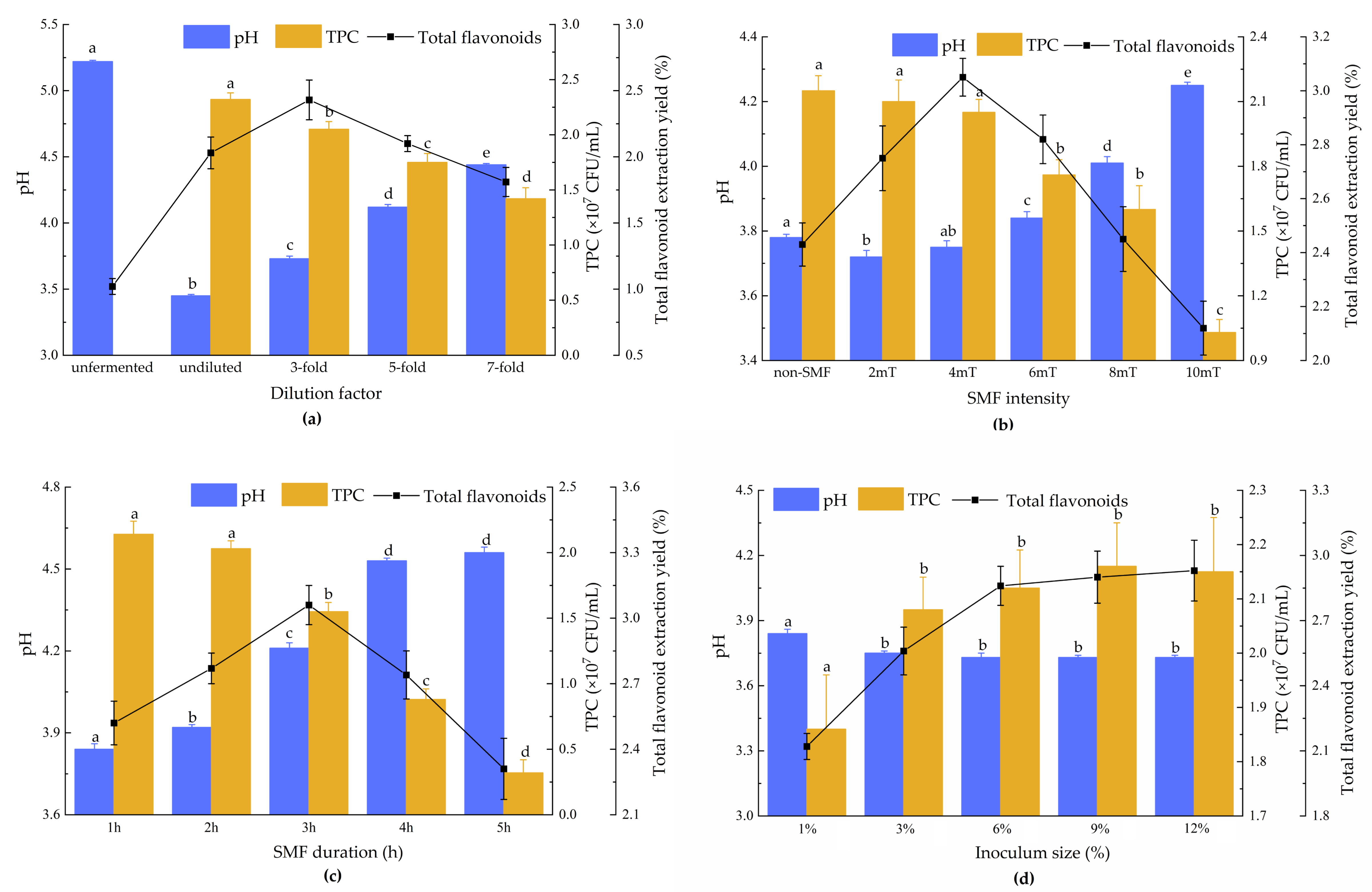
As presented in Figure 1a, the extraction yield of total flavonoids from Ginkgo biloba leaves fermented by L. paracasei significantly exceeds that of the unfermented group (p < 0.05). With the increase in dilution factor, the extraction yield of total flavonoids varied significantly (p < 0.05), exhibiting an initial rise followed by a decline. The maximum total flavonoid extraction yield of 2.43% was achieved at a 3-fold dilution; this was due to the continuous dilution of the medium, which led to a gradual shortage of nutrients. Then, the stress of starvation enhanced the capacity of L. paracasei to degrade plant cell walls via enzymatic hydrolysis, facilitating the release of bound flavonoids during extraction [17]. However, further dilution to 5-fold and 7-fold resulted in excessively low nutrient concentrations that restricted the growth and reproduction of bacterial cells, reduced the total viable count, and consequently limited the efficiency of enzymatic hydrolysis. Accordingly, 3-fold-diluted MRS medium was selected as the optimal choice for fermenting Ginkgo biloba leaves with L. paracasei.

3.1.2. The Effect of SMF Intensity on Total Flavonoid Extraction Yield

Fermentation results assisted by different SMF intensities are showcased in Figure 1b. Compared to the fermented group without SMF assistance, the total flavonoid extraction yields from Ginkgo biloba leaves fermented with SMFs of 2–4 mT were significantly higher (p < 0.05). Low-intensity SMF is known to stimulate the metabolic activity of lactobacilli, elevate fermentation-related enzyme activities, and strengthen metabolic capacity [18]. The maximum total flavonoid extraction yield of 3.05% was obtained at a field strength of 4 mT. Notably, the TPC decreased sharply at 10 mT, where the high-strength SMF impaired the membrane-barrier function of L. paracasei, triggering intracellular ion leakage and osmotic imbalance and ultimately leading to cell lysis and death [19,20]. The resulting restriction of enzymatic hydrolysis during fermentation led to a significant reduction in the total flavonoid extraction yield (p < 0.05) [21]. Thus, a 4 mT SMF intensity was optimal for assisting L. paracasei fermentation of Ginkgo biloba leaves for flavonoid extraction.

3.1.3. The Effect of SMF Duration on Total Flavonoid Extraction Yield

As depicted in Figure 1c, fermentation assisted by an SMF for an appropriate duration effectively facilitated the extraction and release of flavonoids from Ginkgo biloba leaves, with the maximum extraction yield reaching 3.06% at 3 h. Kthiri et al. [22] have reported that prolonged SMF exposure induces excessive stress in microbial cells and disrupts cell membrane integrity, leading to cell death. This finding is consistent with how the TPC of L. paracasei continuously decreased and the pH value rose as the SMF was applied for longer. When the treatment duration exceeded 3 h, massive bacterial lysis and death occurred, which limited enzymatic hydrolysis and gradually reduced the extraction yield of total flavonoids. Thus, an SMF duration of 3 h was most suitable for assisting L. paracasei fermentation of Ginkgo biloba leaves for flavonoid extraction.

3.1.4. The Effect of Inoculum Size on Total Flavonoid Extraction Yield

Looking at Figure 1d, when the inoculum size ranged from 1% to 12%, the extraction yield of total flavonoids first increased and then plateaued, with the yield reaching 2.86% at an inoculum size of 6%. At lower inoculum sizes, L. paracasei required a longer time to proliferate to the stationary phase, which shortened the effective fermentation and enzymatic hydrolysis period and hindered the release of flavonoids. When the inoculum size surpassed 6%, the microorganism cells reached a saturated concentration, and the extraction yield of total flavonoids remained stable without significant differences among the groups (p > 0.05). Thus, 6% was selected as the optimal inoculum size for fermenting Ginkgo biloba leaves with L. paracasei.

3.2. Optimization of the Fermentation Process via Response Surface Methodology

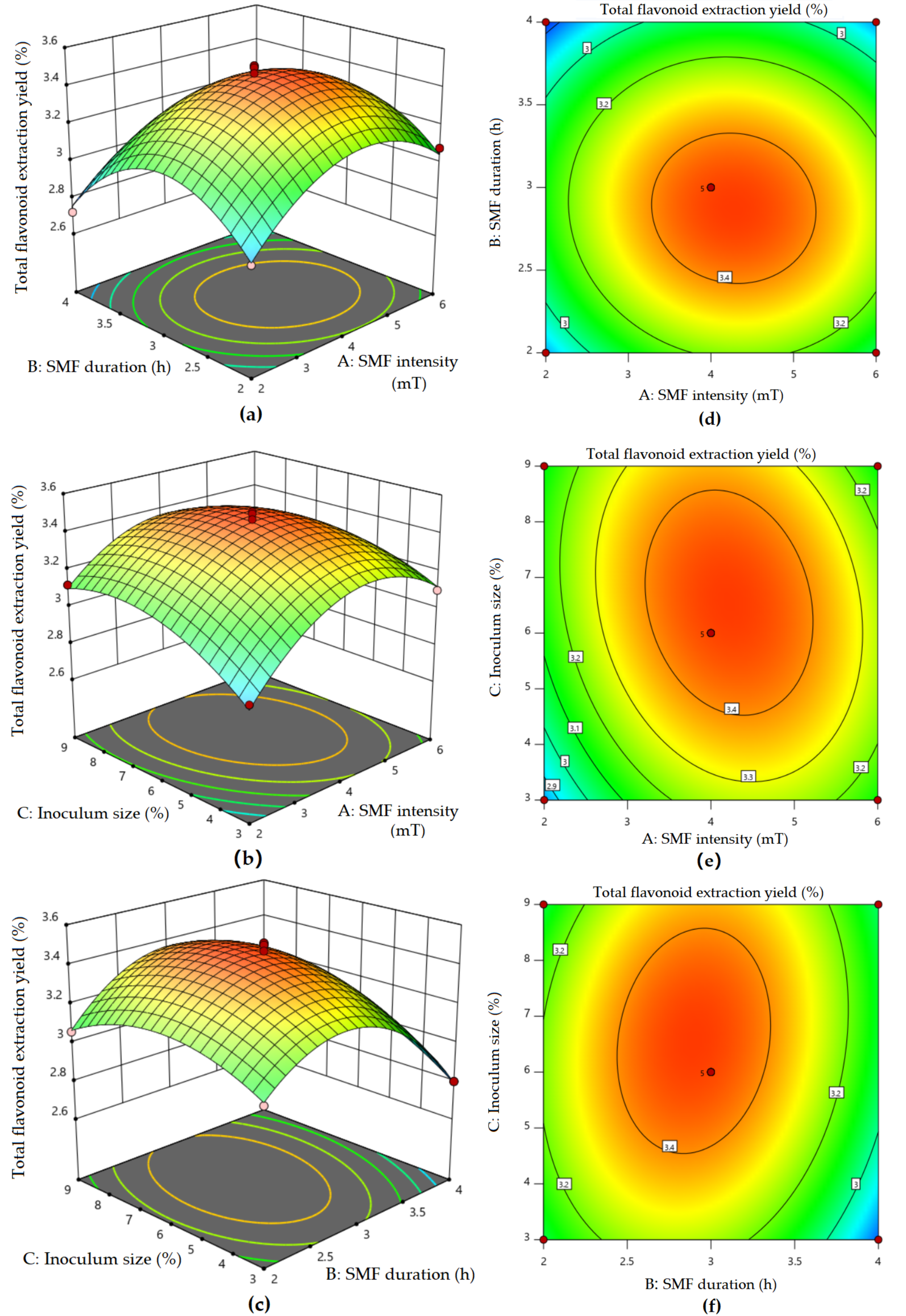
Seventeen sets of experiments were conducted to investigate the extraction of total flavonoids from Ginkgo biloba leaves fermented by L. paracasei with SMF assistance. The coded levels of each factor and corresponding experimental results are listed in Table 1. Design-Expert 11 software was adopted to perform quadratic polynomial regression on the experimental data, obtaining the following equation: Y = 3.46 + 0.0700A − 0.0762B + 0.0613C − 0.0275AB − 0.0675AC + 0.0600BC − 0.2655A2 − 0.3180B2 − 0.1480C2, where Y is the total flavonoid extraction yield, and A, B, and C are the coded values of SMF intensity, duration, and inoculum size, respectively. Analysis of variance (ANOVA) results in Table 2 reveal that the model exhibited an extremely significant difference (p < 0.01). The lack-of-fit term had a p-value of 0.6643, demonstrating satisfactory correlation. The adjusted coefficient of determination R2 was 96.30%, indicating high reliability. For linear terms, the order of significance was B (SMF duration) > A (SMF intensity) > C (inoculum size); all of the quadratic terms (A2, B2, and C2) were extremely significant (p < 0.01); as for the interaction terms, AC and BC showed significant effects (p < 0.05), while AB had no significant influence (p > 0.05).
Three-dimensional surface plots can intuitively depict the interactive effects of different investigated factors on response values [23]. As depicted in Figure 2, each experimental factor exhibits a parabolic relationship with the response value when the two factors are fixed. The steeper parabolic slopes of SMF intensity (A) and duration (B) indicate that they exert stronger influences on the extraction yield of total flavonoids. Meanwhile, the contour plots of AC and BC exhibit obvious elliptical shapes, demonstrating significant interactions that align with the ANOVA results. Applying Design-Expert software, the optimal process parameters were determined as follows: an SMF intensity of 4.236 mT, an SMF duration of 2.890 h, and an inoculum size of 6.474%, with a predicted total flavonoid extraction yield of 3.469%. Considering the feasibility of practical operations, these conditions were adjusted to an SMF intensity of 4.2 mT, an SMF duration of 2.9 h, and an inoculum size of 6.5%. Three parallel verification experiments were carried out, and the measured total flavonoid extraction yield was (3.41 ± 0.11)%—essentially consistent with the predicted value. This verifies that the established model is reliable and accurate in predicting the process parameters for SMF-assisted fermentation of Ginkgo biloba leaves with L. paracasei.

3.3. Nutritional Components Analysis

3.3.1. Basic Nutritional Components

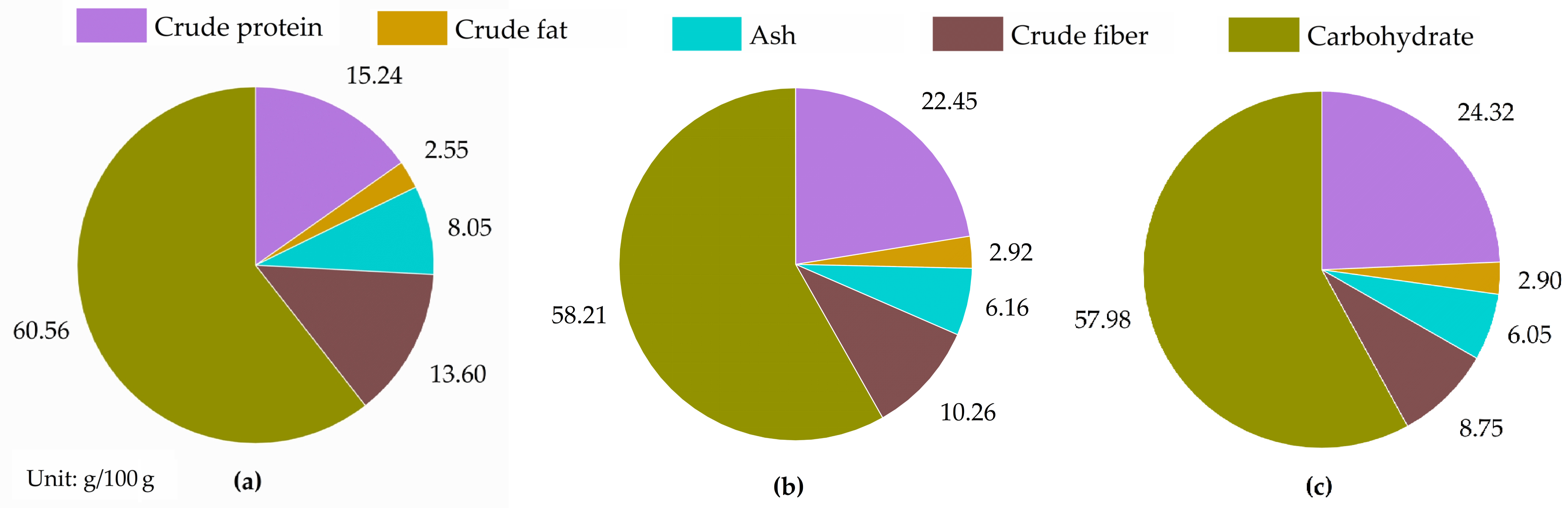
The basic nutritional components of the Ginkgo biloba leaves before and after fermentation are presented in Figure 3. Compared with the unfermented group, fermented leaves exhibited significantly higher contents of crude protein and crude fat (p < 0.05); meanwhile, the contents of ash and crude fiber fell significantly (p < 0.05). The crude protein content reached 22.45 g/100 g in the conventional fermentation group, and 24.32 g/100 g in the SMF-assisted fermentation group, representing respective increases of 47.31% and 59.58% relative to the unfermented group (15.24 g/100 g). This result is explained by the fact that the proliferating microbial cells were rich in single-cell protein; moreover, partial organic substrates in the Ginkgo biloba leaves were catabolized during microbial fermentation, leading to a protein concentration effect [24]. Notably, no significant difference was observed between the SMF-assisted group and the conventional fermentation group (p > 0.05). In keeping with our results, previous studies on SMF-assisted fermentation indicated that SMF hardly raised the total content of crude protein once microbial biomass reached a relatively stable level after fermentation; however, it could alter protein spatial conformation and free sulfhydryl groups, thereby improving protein solubility and emulsifying capacity [25]. The variation trend of crude fat was consistent with that of crude protein, showing a 13.72% elevation following SMF-assisted fermentation. Additionally, the ash content decreased by 23.48% and 24.84% in the conventional fermentation group and SMF-assisted group, respectively, which is likely due to leaching of inorganic minerals from the leaf matrix into the fermentation broth, facilitated by organic acids produced by L. paracasei [26]. The crude fiber content of the raw leaves was 13.60 g/100 g. After fermentation, this value significantly declined to 10.26 g/100 g (in the case of conventional fermentation) and 8.75 g/100 g (SMF-assisted fermentation), corresponding to reductions of 24.56% and 35.66%, respectively (p < 0.05). During fermentation, moderate SMF-assistance promoted L. paracasei to secrete cellulolytic enzymes that degraded crude fiber into small-molecule saccharides for microbial utilization [27]. The carbohydrate content exhibited no significant differences among the three groups (p > 0.05). Although moderate SMF-assistance facilitated the degradation of crude fiber, the released small-molecule saccharides were rapidly assimilated by L. paracasei, resulting in comparable total carbohydrate levels in fermented samples [28]. In summary, conventional fermentation dominated the overall nutritional enhancement of the Ginkgo biloba leaves, while moderate SMF-assistance further optimized fermentation performance by increasing fiber degradation and regulating the structural properties of nutrients, rather than drastically changing the total macronutrient contents.

3.3.2. Anti-Nutritional Factors

Ginkgo biloba leaves also contain various anti-nutritional factors that hinder digestion and absorption of nutrients in the body. Among them, ginkgolic acids are particularly hazardous; their excessive intake can induce toxic side effects such as nerve paralysis, vomiting, and even coma and convulsions [29]. As shown in Table 3, the contents of typical anti-nutritional factors in Ginkgo biloba leaves—namely phytic acid, tannins, phenolic acids, and ginkgolic acids—were observed to decrease significantly following fermentation (p < 0.05). After conventional fermentation, the phytic acid content dropped from 4.60 mg/g in the unfermented group to 3.35 mg/g (a reduction of 27.17%). Although the phytic acid content in the SMF-assisted group (3.24 mg/g) appeared lower than that in the conventional fermentation group (3.35 mg/g), this difference was not significant (p > 0.05), which may be ascribable to the relatively low SMF intensity applied in this study [30]. The tannin content dropped by 26.41%, and SMF-assisted fermentation significantly increased the reduction rate to 43.10%. During fermentation, L. paracasei secreted phytase and organic acids to facilitate the degradation or transformation of phytic acid and tannins [31]. The content of phenolic acids was reduced by 9.27% in the conventional fermentation group, and SMF-assistance showed a promotional effect on the elimination of phenolic acids, leading to a reduction of 23.42%. Moreover, the ginkgolic acid content was decreased by 35.05% following conventional fermentation. It is understood that SMF assistance can enhance the cell membrane permeability of lactic acid bacteria and elevate extracellular enzyme activity, thereby moderately facilitating adsorption, enzymatic hydrolysis, and transformation of ginkgolic acids [32,33]; as expected, an additional increase of 15.32% in removal efficiency was observed for ginkgolic acids in the SMF-assisted group. Overall, SMF-assisted probiotic fermentation delivers dual advantages: it simultaneously enriches bioactive total flavonoids and degrades toxic anti-nutritional factors. This one-step bioconversion improves the safety and utilization value of Ginkgo biloba leaves, offering a feasible and environmentally-friendly green industrial processing approach. To futurer validate this methodology, future work may explore the impact of hydrolytic enzyme activities on anti-nutritional factors.

3.4. Hypoglycemic Activity In Vitro

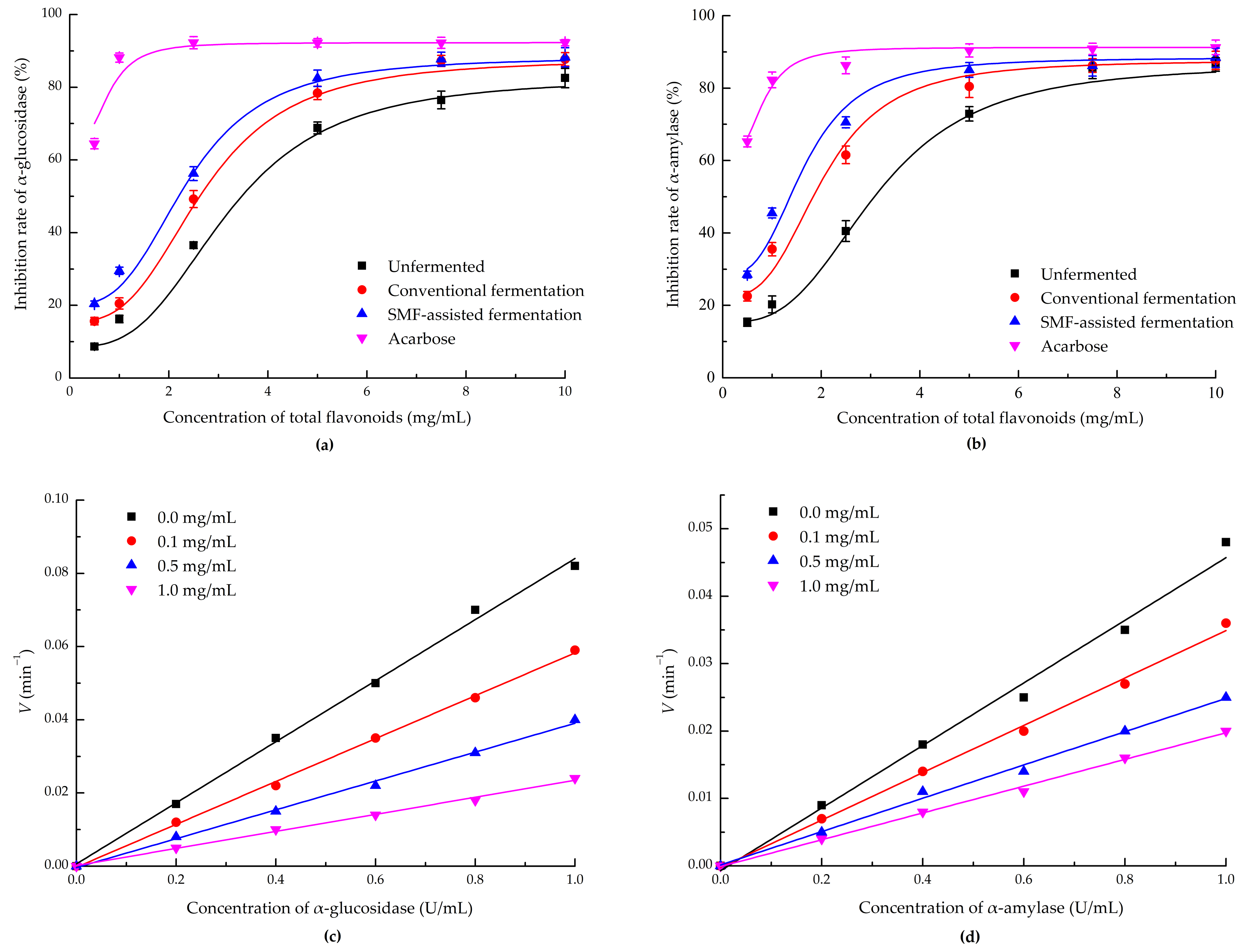
As shown in Figure 4a,b, within the tested concentration range of 0.5–10.0 mg/mL, the total flavonoids of all groups exerted inhibitory effects on α-glucosidase and α-amylase activity, without an obvious linear correlation. The logistic equation was adopted for nonlinear fitting of inhibition curves of samples from the unfermented, conventional fermentation, and SMF-assisted fermentation groups, and their corresponding IC50 (half-maximal inhibitory concentration) values were calculated as 3.21, 2.72, and 2.40 mg/mL for α-glucosidase, and 3.13, 2.03, and 1.65 mg/mL for α-amylase. When the mass concentration exceeded 7.5 mg/mL, both the α-glucosidase and α-amylase inhibition rates plateaued and remained nearly constant. It is important to note that here the inhibitory effects were normalized by flavonoid concentration in order to compare the intrinsic bioactivity of flavonoids from different groups. Further studies incorporating crude-extract dosage and extraction-yield data would enable a more holistic evaluation of the hypoglycemic potential of fermented Ginkgo biloba leaves.
Observing Figure 4c,d, the curves corresponding to the fitted linear equations for enzyme concentration–reaction rate all passed through the origin. Notably, the slope of the straight line for the flavonoid group was markedly lower than that of the inhibitor-free control group, and the slope gradually declined with greater flavonoid concentration. This trend conforms to the fundamental kinetic pattern of reversible inhibition [34]. Accordingly, we can conclude that the inhibition of total flavonoids against α-glucosidase and α-amylase is a process of reversible enzyme inhibition. Thus, total flavonoids from Ginkgo biloba leaves appear not to change the effective concentration of enzymes, but instead suppress their catalytic efficiency, thereby lowering their capacity for substrate degradation. Previous studies [35,36] have also demonstrated that fermentation of plant materials significantly elevates the contents of flavonoids such as quercetin and linarin. These flavonoids competitively bind to the active sites of α-glucosidase and α-amylase, suppress glucose production, and substantially raise hypoglycemic efficacy. At the same time, SMF-assisted fermentation improves flavonoid inhibitory activity against α-glucosidase and α-amylase. The proposed mechanism behind this is that applying a moderate SMF increases the membrane permeability of L. paracasei and boosts secretion of cell-wall-degrading hydrolase, releasing free flavonoid aglycones and facilitating flavonoid deglycosylation. Additionally, exposed phenolic hydroxyl groups effectively interfere with enzyme catalytic reactions to inhibit carbohydrate hydrolysis [37,38]. In future work, Lineweaver–Burk plotting will be required to classify the specific inhibition subtype (competitive, non-competitive, or mixed inhibition).

4. Conclusions

This study explored the effects of SMF-assisted liquid fermentation by L. paracasei on total flavonoid extraction, nutritional components, and hypoglycemic activity for Ginkgo biloba leaves. The results verified that SMF application significantly improved the total flavonoid extraction efficiency. The optimal fermentation conditions were a 3-fold-diluted MRS medium, an SMF intensity of 4.2 mT, an SMF duration of 2.9 h, and a 6.5% inoculum size. Under these conditions, the total flavonoid extraction yield reached (3.41 ± 0.11)%, which was 3.33 times that of the unfermented case.
After fermentation with SMF assistance, the contents of crude protein and crude fat increased by 59.58% and 13.72%, while the ash and crude fiber contents decreased by 24.84% and 35.66%, respectively. Application of the SMF noticeably affected the crude fiber content (p < 0.05). Additionally, the contents of phytic acid, tannins, phenolic acids, and ginkgolic acids were reduced by 29.56%, 43.10%, 23.42%, and 50.37%, respectively, and the SMF further promoted the removal of anti-nutritional factors. Fermentation markedly enhanced the inhibitory effects of flavonoids on α-glucosidase and α-amylase, and the SMF-assisted fermentation specifically strengthened the inhibition of both hydrolases. This work provides a theoretical and technical basis for the efficient utilization of Ginkgo biloba leaves via SMF-assisted probiotic fermentation. Further molecular-level research should be pursued to reveal the underlying mechanism acting between SMFs and L. paracasei.

Author Contributions

Conceptualization, Z.S.; methodology, Z.S. and L.N.; Writing—original draft preparation, Z.S.; fermentation, Z.S. and D.Z.; data analysis, L.N.; writing—review and editing, Z.S. and Y.Z.; supervision, Y.Z. All authors have read and agreed to the published version of the manuscript.

Funding

This research was funded by the Major Basic Science (Natural Science) Research Project of Universities in Jiangsu Province (No. 23KJA360001).

Institutional Review Board Statement

Not applicable.

Informed Consent Statement

Not applicable.

Data Availability Statement

The original contributions presented in this study are included in the article. Further inquiries can be directed to the corresponding author.

Conflicts of Interest

The authors declare no conflicts of interest.

Abbreviations

The following abbreviations are used in this manuscript:
SMFStatic magnetic field
MRSDe Man, Rogosa, and Sharpe
PNPGp-Nitrophenyl-β-D-galactopyranoside
NaOHSodium hydroxide
Al(NO3)3Aluminum nitrate
NaNO2Sodium nitrite
TPCTotal plate count
UPLCUltra-performance liquid chromatography
IC50Half-maximal inhibitory concentration

References

  1. Biernacka, P.; Felisiak, K.; Adamska, I.; Tokarczyk, G.; Bienkiewicz, G.; Przybylska, S.; Wiereo, W.; Lener, M.R.; Jaroszewska, A.; Baejczak, D.; et al. Ultrasound pre-treatment of Ginkgo biloba leaves enhances the functional and sensory properties of the fermented beverage. Food Chem. 2026, 499, 147329. [Google Scholar] [CrossRef] [Scilit]
  2. Nie, S.; Zhang, S.; Wang, Y.; Zhu, M.; Chen, X.; Wang, X.; Huang, P. Extraction, purification, structural characterization, and bioactivities of Ginkgo biloba leaf polysaccharides: A review. Int. J. Biol. Macromol. 2024, 281, 136280. [Google Scholar] [CrossRef] [Scilit]
  3. Kumari, K.; Kashyap, P.; Chakrabarti, P. Germination and probiotic fermentation: A way to enhance nutritional and biochemical properties of cereals and millets. Food Sci. Biotechnol. 2024, 33, 505–518. [Google Scholar] [CrossRef] [Scilit]
  4. Sandya, B.K.; Gowthami, G.A.; Harish Nayaka, M.A.; Gunashree, B.S. Lactobacillus plantarum fermentation to reduce anti-nutritional contents in peanut, mustard and sesame. Biomedicine 2021, 41, 611–615. [Google Scholar] [CrossRef] [Scilit]
  5. Yousfi, N.; Merbahi, N.; Bouajila, J.; Taillandier, P.; Debouba, M. Microbial fermentation assisted by pulsed electric fields, magnetic fields and cold atmospheric plasma: State of the art. Fermentation 2025, 11, 417. [Google Scholar] [CrossRef] [Scilit]
  6. Zhao, Y.; Wu, R.; Wu, J.; Shi, L.; Xu, J.; Hu, X.; Yao, L.; Qiao, K.; Shi, H.; Wang, W. Static magnetic field-assisted co-fermentation of lactic acid bacteria consortium in Platycodon grandiflorum root powder for process optimization and antioxidant property enhancement. Food Bioprocess. Technol. 2025, 18, 7561–7579. [Google Scholar]
  7. Yao, H.; Jin, Y.; Zhang, L.; Wu, S.; Zheng, Z.; Wang, T.; Yang, N.; Xu, X. Progress in application of magnetic field technology in the processing of agricultural products. Food Sci. 2024, 45, 306–316. [Google Scholar]
  8. Jian, D.; Jiang, J.; Miao, Q.; Liu, M. Research on technology of preparing bio-feed additives using solid-state fermentation of Ginkgo biloba leaves. Feed. Ind. 2025, 46, 142–149. (In Chinese) [Google Scholar]
  9. Li, Q.; Gao, S.F.; Zhang, F.Q.; Chen, R.; Zhang, X.; Zhao, L. Solid state fermentation of Ginkgo biloba leaves and its effects on antioxidant capacity and breast muscle quality of broilers. Bioprocess. Biosyst. Eng. 2026, 49, 651–665. [Google Scholar] [CrossRef] [Scilit]
  10. Peerajan, S.; Chaiyasut, C.; Sirilun, S.; Chaiyasut, K.; Kesika, P.; Sivamaruthi, B.S. Enrichment of nutritional value of Phyllanthus emblica fruit juice using the probiotic bacterium, Lactobacillus paracasei HII01 mediated fermentation. Food Sci. Technol. 2016, 36, 116–123. [Google Scholar] [CrossRef] [Scilit]
  11. Gu, J.; Zhu, Q.; Du, J.; Guo, J.; Wu, Y.; Yang, S.; Jiang, J. Enhancement of nutritional and flavor properties of red-fleshed pitaya juice via Lacticaseibacillus paracasei fermentation. Food Chem. X 2025, 30, 102915. [Google Scholar] [CrossRef] [Scilit]
  12. Yadoung, S.; Praphawilai, P.; Yana, P.; Jeeno, P.; Chuljerm, H.; Danmek, K.; Wu, M.-C.; Jung, C.; Chuttong, B.; Hongsibsong, S. Compositional and biofunctional properties of Xyris spp. and Mimosa spp. bee pollen from Thailand. Foods 2026, 15, 1990. [Google Scholar] [CrossRef] [Scilit]
  13. AOAC. Official Methods of Analysis, 21st ed.; Association of Official Analytical Chemists: Washington, DC, USA, 2019. [Google Scholar]
  14. GB/T 6434-2022; Determination of Crude Fiber Content in Feeds. Standardization Administration of the People’s Republic of China. China Standards Press: Beijing, China, 2022.
  15. GB 5009.153-2016; Determination of Phytic Acid Content in Food. Standardization Administration of the People’s Republic of China. China Standards Press: Beijing, China, 2016.
  16. Makkar, H.P.; Blümmel, M.; Borowy, N.K.; Becker, K. Gravimetric determination of tannins and their correlations with chemical and protein precipitation methods. J. Sci. Food Agric. 1993, 61, 161–165. [Google Scholar] [CrossRef] [Scilit]
  17. Parlindungan, E.; May, B.K.; Jones, O.A.H. Metabolic insights into the effects of nutrient stress on Lactobacillus plantarum B21. Front. Mol. Biosci. 2019, 6, 75. [Google Scholar] [CrossRef] [Scilit]
  18. Zieliński, M.; Zielińska, M.; Cydzik-Kwiatkowska, A.; Rusanowska, P.; Dębowski, M. Effect of static magnetic field on microbial community during anaerobic digestion. Bioresour. Technol. 2020, 323, 124600. [Google Scholar] [CrossRef] [Scilit]
  19. Teodori, L.; Albertini, M.C.; Uguccioni, F.; Falcieri, E.; Rocchi, M.B.L.; Battistelli, M.; Coluzza, C.; Piantanida, G.; Bergamaschi, A.; Magrini, A.; et al. Static magnetic fields affect cell size, shape, orientation, and membrane surface of human glioblastoma cells, as demonstrated by electron, optic, and atomic force microscopy. Cytom. A 2010, 69A, 75–85. [Google Scholar]
  20. Wang, P.; Du, L.; Li, Y.; Xu, Z.; Ye, L.; Dai, S.; Xu, L.; Yan, J.; Xie, X.; Cao, Q.; et al. High-intensity pulse magnetic fields affect redox homeostasis and survival rate of Escherichia coli according to initial level of intracellular glucose. Biomolecules 2025, 15, 1550. [Google Scholar] [CrossRef] [Scilit]
  21. König, A.; Sadova, N.; Dornmayr, M.; Schwarzinger, B.; Neuhauser, C.; Stadlbauer, V.; Wallner, M.; Woischitzschläger, J.; Müller, A.; Tona, R.; et al. Combined acid hydrolysis and fermentation improves bioactivity of citrus flavonoids in vitro and in vivo. Commun. Biol. 2023, 6, 1083. [Google Scholar] [CrossRef] [Scilit]
  22. Kthiri, A.; Hidouri, S.; Wiem, T.; Jeridi, R.; Sheehan, D.; Landouls, A.; Shahid, M. Biochemical and biomolecular effects induced by a static magnetic field in Saccharomyces cerevisiae: Evidence for oxidative stress. PLoS ONE 2019, 14, e0209843. [Google Scholar] [CrossRef] [Scilit]
  23. Bezerra, M.A.; Santelli, R.E.; Oliveira, E.P.; Villar, L.S.; Escaleira, L.A. Response surface methodology (RSM) as a tool for optimization in analytical chemistry. Talanta 2008, 76, 965–977. [Google Scholar] [CrossRef] [Scilit]
  24. Zou, M.; Guo, X.; Huang, Y.; Cao, F.; Wang, J. Improvement of the quality of Ginkgo biloba leaves fermented by Eurotium cristatum as high value-added feed. Processes 2019, 7, 627. [Google Scholar] [CrossRef] [Scilit]
  25. Betchem, G.; Dabbour, M.; Tuly, J.A.; Billong, L.F.; Ma, H. Experimental investigation into the implications of low-intensity magnetic field treatment on the structural and functional properties of rapeseed meal. Food Chem. 2024, 446, 13885. [Google Scholar] [CrossRef] [Scilit]
  26. Gupta, R.K.; Gangoliya, S.S.; Singh, N.K. Reduction of phytic acid and enhancement of bioavailable micronutrients in food grains. J. Food Sci. Technol. 2015, 52, 676–684. [Google Scholar] [CrossRef] [Scilit]
  27. Ran, J.; Jia, S.; Yong, L.; Wu, S. Characterization of cellulase under various intensities of static magnetic fields. Catal. Commun. 2010, 11, 91–95. [Google Scholar] [CrossRef] [Scilit]
  28. Koirala, P.; Maina, N.; Mojzita, D.; Coda, R. Regulation of sugar metabolism during fermentation of brewers’ spent grain by Leuconostoc pseudomesenteroides DSM20193. Microb. Biotechnol. 2025, 18, e70116. [Google Scholar] [CrossRef] [Scilit]
  29. Dong, Q.; Cao, J.; Wu, R.; Shi, T.; Su, E. Efficient removal of ginkgolic acids from Ginkgo biloba leaves crude extract by using hydrophobic deep eutectic solvents. Ind. Crops Prod. 2021, 166, 113462. [Google Scholar] [CrossRef] [Scilit]
  30. Ruiz-Gómez, M.J.; Prieto-Barcia, M.I.; Ristori-Bogajo, E.; Martínez-Morillo, M. Static and 50 Hz magnetic fields of 0.35 and 2.45 mT have no effect on the growth of Saccharomyces cerevisiae. Bioelectrochemistry 2004, 64, 151–155. [Google Scholar] [CrossRef] [Scilit]
  31. García-Mantrana, I.; Yebra, M.J.; Haros, M.; Monedero, V. Expression of bifidobacterial phytases in Lactobacillus casei and their application in a food model of whole-grain sourdough bread. Int. J. Food Microbiol. 2015, 216, 18–24. [Google Scholar] [CrossRef] [Scilit]
  32. Zieliński, M.; Cydzik-Kwiatkowska, A.; Zielińska, M.; Dębowski, M.; Rusanowska, P.; Kopańska, J. Nitrification in activated sludge exposed to static magnetic field. Water Air Soil Pollut. 2017, 228, 126. [Google Scholar] [CrossRef] [Scilit]
  33. Sun, Y.; Zhao, J.; Manickam, S.; He, J.; Li, D.; Han, Y.; Jiang, X.; Tao, Y. Biochemical and physical investigations on detoxification of ginkgo kernel juice using probiotic fermentation with macroporous resin addition. Food Innov. Adv. 2023, 2, 324–339. [Google Scholar] [CrossRef] [Scilit]
  34. Xiao, J.; Kai, G.; Yamamoto, K.; Chen, X. Advances in dietary polyphenols as α-glucosidases inhibitors: A review on structure–activity relationship aspect. Crit. Rev. Food Sci. Nutr. 2013, 53, 818–836. [Google Scholar] [CrossRef] [Scilit]
  35. Deng, Y.; Qu, F.; Wang, Z.; Xia, W.; Xing, Y. Effect of Eurotium cristatum fermentation on the α-glucosidase inhibitory activity of mulberry leaves flavonoid extract. Acad. J. Med. Health Sci. 2021, 2, 55–61. [Google Scholar] [CrossRef] [Scilit]
  36. Xu, C.; He, M.; Jiang, Z.; Yang, Q. Inhibition mechanism of buckwheat hulls polyphenols on α-amylase and α-glucosidase using kinetics, spectroscopics and molecular docking approaches. Int. J. Biol. Macromol. 2024, 280, 136046. [Google Scholar] [CrossRef] [Scilit]
  37. Park, C.M.; Kim, G.M.; Cha, G.S. Biotransformation of flavonoids by newly isolated and characterized Lactobacillus pentosus NGI01 strain from Kimchi. Microorganisms 2021, 9, 1075. [Google Scholar] [CrossRef] [Scilit]
  38. Wang, Y.; Wang, D.; Xing, M.; Ji, M.; Jiang, X.; Jia, L.; Li, L.; Song, G.; Yuan, T.; Gong, J. Effect and mechanism of chlorogenic acid inhibition of starch enzymatic hydrolysis: Comparison of different processing methods. Food Chem. X 2025, 29, 102796. [Google Scholar] [CrossRef] [Scilit]
Figure 1. The effects of different experimental factors on total flavonoid extraction yield from Ginkgo biloba leaves: (a) Dilution factor; (b) SMF intensity; (c) SMF duration; (d) Inoculum size. Different letters above the bars indicate statistically significant differences (p < 0.05).
Fermentation 12 00439 g001
Figure 2. Response surface plots and contour plots of the interactions between pairs of experimental factors: (a) SMF intensity vs. SMF duration (response surface plot); (b) SMF intensity vs. inoculum size (response surface plot); (c) SMF duration vs. inoculum size (response surface plot); (d) SMF intensity vs. SMF duration (corresponding contour plot); (e) SMF intensity vs. inoculum size (corresponding contour plot); (f) SMF duration vs. inoculum size (corresponding contour plot).
Fermentation 12 00439 g002
Figure 3. Basic nutritional components analysis of (a) unfermented, (b) conventionally fermented, and (c) SMF-assisted fermented Ginkgo biloba leaves.
Fermentation 12 00439 g003
Figure 4. The inhibitory effects of total flavonoids from Ginkgo biloba leaves against (a) α-glucosidase and (b) α-amylase; Inhibition types of total flavonoids on (c) α-glucosidase (d) and α-amylase.
Fermentation 12 00439 g004
Table 1. Box-Behnken design and results.
RunA
SMF Intensity
(mT)
B
SMF Duration (h)
C
Inoculum Size (%)
Y Total Flavonoid
Extraction Yield (%)
11 (6)0 (3)−1 (3)3.10
2−1 (2)1 (4)0 (6)2.72
31 (6)0 (3)1 (9)3.08
4−1 (2)0 (3)−1 (3)2.87
50 (4)0 (3)0 (6)3.47
60 (4)1 (4)−1 (3)2.80
70 (4)−1 (2)−1 (3)3.05
8−1 (2)−1 (2)0 (6)2.84
90 (4)0 (3)0 (6)3.38
100 (4)1 (4)1 (9)3.05
111 (6)−1 (2)0 (6)3.08
120 (4)0 (3)0 (6)3.50
130 (4)0 (3)0 (6)3.42
140 (4)0 (3)0 (6)3.51
151 (6)1 (4)0 (6)2.85
160 (4)−1 (2)1 (9)3.06
17−1 (2)0 (3)1 (9)3.12
Table 2. Analysis of variance and significance in the regression model.
SourceSum of SquaresDfMean SquareF-Valuep-ValueSignificance
Model1.0590.116847.28<0.0001**
A0.039210.039215.870.0053**
B0.046510.046518.830.0034**
C0.030010.030012.150.0102*
AB0.003010.00301.220.3051
AC0.018210.01827.380.0299*
BC0.014410.01445.830.0465*
A20.296810.2968120.13<0.0001**
B20.425810.4258172.33<0.0001**
C20.092210.092237.330.0005**
Residual0.017370.0025
Lack of Fit0.005230.00170.56930.6643
Pure Error0.012140.0030
Cor Total1.0716
Note: ** indicates extreme significant difference (p < 0.01); * indicates significant difference (p < 0.05). Df, degrees of freedom.
Table 3. Anti-nutritional factor analysis in fermented Ginkgo biloba leaves (mg/g dry matter).
GroupsPhytic AcidTanninsPhenolic AcidsGinkgolic Acids
unfermented4.60 ± 0.25 a15.22 ± 0.98 a32.05 ± 1.91 a8.16 ± 0.55 a
conventional fermentation3.35 ± 0.16 b11.20 ± 1.03 b29.08 ± 2.02 a5.30 ± 0.50 b
SMF-assisted fermentation3.24 ± 0.19 b8.66 ± 0.55 c24.54 ± 1.25 b4.05 ± 0.35 c
Different lowercase letters within the same column indicate significant differences (p < 0.05).
Disclaimer/Publisher’s Note: The statements, opinions and data contained in all publications are solely those of the individual author(s) and contributor(s) and not of MDPI and/or the editor(s). MDPI and/or the editor(s) disclaim responsibility for any injury to people or property resulting from any ideas, methods, instructions or products referred to in the content.