Article

Selection of Lactic Acid Bacteria Based on Antagonistic Activity and Mycotoxin Mitigation Capacity to Develop a Multistrain Consortium for Wheat Bran Fermentation


by
Anara Yeleussizova
1,
Modestas Ruzauskas
2,
Gulnur Aliyeva
1,
Saniya Tyshtykbayeva
1,
Rita Šiugždinienė
2,
Vytaute Starkute
3,4,
Yablochkova Gulmira
5,
Elena Bartkiene
3,4 and
Nurlan Kaumenov
1,*
1
Department of Veterinary Sanitation, Faculty of Agricultural Sciences, Akhmet Baitursynuly Kostanay Regional University, Baytursynov Str. 47, Kostanay 110000, Kazakhstan
2
Institute of Microbiology and Virology, Faculty of Veterinary Medicine, Lithuanian University of Health Sciences, Tilzes Str. 18, 47181 Kaunas, Lithuania
3
Institute of Animal Rearing Technologies, Faculty of Animal Sciences, Lithuanian University of Health Sciences, Tilzes Str. 18, 47181 Kaunas, Lithuania
4
Department of Food Safety and Quality, Faculty of Veterinary Medicine, Lithuanian University of Health Sciences, Tilzes Str. 18, 47181 Kaunas, Lithuania
5
Department of Veterinary Medicine, Faculty of Agricultural Sciences, Akhmet Baitursynuly Kostanay Regional University, Baytursynov Str. 47, Kostanay 110000, Kazakhstan
*
Author to whom correspondence should be addressed.
Fermentation 2026, 12(8), 371; https://doi.org/10.3390/fermentation12080371
Submission received: 22 June 2026 / Revised: 29 July 2026 / Accepted: 1 August 2026 / Published: 7 August 2026
(This article belongs to the Section Animal and Feed Fermentation)

Abstract

This study aimed to select lactic acid bacteria (LAB) strains based on their antagonistic activity and mycotoxin mitigation capacity, develop a biocompatible multistrain consortium, and evaluate its application for the fermentation of extruded wheat bran. The selected strains were assessed for biocompatibility, and a four-strain consortium comprising Lacticaseibacillus casei, Levilactobacillus brevis, Pediococcus acidilactici, and Latilactobacillus curvatus was established. The consortium was propagated in supplemented sour whey and subsequently applied to wheat bran fermentation. Compared with the two-strain formulations, the four-strain consortium reduced the total bacterial count by 1.6-fold and the yeast and mold count by 1.75-fold after 36 h of fermentation. The consortium also produced the most pronounced acidification of the substrate, reducing the pH from 5.65 to 3.82 and increasing titratable acidity from 0.20 to 4.40 °N (22.0-fold). In contrast, fermentation with the two-strain formulations reduced the pH to 4.02 ± 0.01, while titratable acidity increased by 20.5–20.6-fold. After 36 h of fermentation, the viable LAB count in the substrate fermented with the four-strain consortium reached 7.23 ± 0.11 log10 CFU/g, compared with 6.23 ± 0.02 and 6.42 ± 0.01 log10 CFU/g in the substrates fermented with the two-strain formulations. These findings indicate that the developed four-strain consortium represents a promising starter culture for wheat bran fermentation and highlight its potential for the valorization of cereal by-products into value-added feed material through LAB fermentation. All experiments were conducted in vitro, and the effectiveness of the proposed approach under animal feeding conditions requires further experimental validation.

1. Introduction

A balanced intestinal microbiota is essential for maintaining animal health, productivity, and overall well-being [1]. Growing restrictions on the use of antibiotic growth promoters have stimulated increasing interest in probiotic feed ingredients based on lactic acid bacteria (LAB), which contribute to gastrointestinal health through pathogen inhibition, modulation of the immune response, production of beneficial metabolites, and competitive exclusion of undesirable microorganisms [2,3]. LAB belonging to the genera Lactobacillus, Bifidobacterium, Pediococcus, and Leuconostoc are generally recognized as safe (GRAS) and are widely used in food and feed production because of their documented probiotic and antimicrobial properties [4,5,6,7]. However, despite extensive research, most commercial probiotic preparations are still based on individual strains, whereas multistrain consortia remain considerably less explored, although they may provide complementary biological functions and synergistic interactions [8,9].
The biological activity of LAB is highly strain-dependent. Different strains vary substantially in their ability to produce organic acids, bacteriocins, exopolysaccharides, and other antimicrobial metabolites, as well as in their capacity to adsorb or biotransform mycotoxins [10,11,12]. These functional differences are largely determined by cell wall composition, surface characteristics, metabolic activity, and fermentation conditions [13]. Therefore, rational selection of strains possessing complementary functional properties represents a critical step in the development of effective multistrain probiotic consortia [14].
Fermentation is an effective approach for improving the microbiological safety, nutritional value, and functional characteristics of feed materials [15,16]. Wheat bran, one of the major by-products of cereal processing, is an abundant source of dietary fiber and prebiotic compounds that can serve as an excellent substrate for LAB fermentation [17]. Within the framework of the circular economy concept, the utilization of cereal processing by-products, including wheat bran, as raw materials for the production of functional foods and high-value feed ingredients has become increasingly important and is a promising sustainable strategy [18,19]. Fermentation of wheat bran has been shown to enhance its nutritional quality, increase the bioavailability of biologically active compounds, improve intestinal health, suppress undesirable microorganisms, and reduce the concentrations of certain mycotoxins [20,21,22,23].
Another promising by-product suitable for sustainable biotechnology is sour whey. Produced in large quantities by the dairy industry, sour whey represents an inexpensive nutrient source for LAB cultivation and may serve as an environmentally friendly alternative to conventional laboratory media [24,25]. Consequently, propagation of selected LAB consortia in supplemented acid whey before feed fermentation may substantially improve the economic feasibility and sustainability of industrial-scale production.
We hypothesized that a multistrain LAB consortium composed of microorganisms with complementary biological functions would provide broader antimicrobial and mycotoxin-mitigating activity than individual strains. Such functional complementarity may integrate different mechanisms of pathogen inhibition and mycotoxin mitigation, including toxin adsorption by cell wall components, enzymatic biotransformation, organic acid production, synthesis of antimicrobial metabolites, and competitive exclusion of undesirable microorganisms [26]. Previous studies have shown that Lacticaseibacillus casei is primarily associated with organic acid production and pathogen inhibition, Levilactobacillus brevis with bacteriocin production, Pediococcus acidilactici with pediocin-like antimicrobial compounds and antimycotoxin activity, and Latilactobacillus curvatus with additional antifungal and detoxification properties [27,28]. Consequently, combining strains with complementary functional characteristics may improve fermentation stability and broaden the spectrum of biological activity compared with individual cultures.
Synergistic fermentation of extruded wheat bran using multistrain lactic acid bacteria (LAB) consortia has been shown to outperform single-strain cultures by significantly increasing the concentrations of phenolic compounds (up to 3.4-fold) and antioxidant activity (up to 2.64-fold). This approach also promotes more effective detoxification, reducing phytate content by up to 95% and mycotoxin concentrations to as low as 29.8 μg/kg, which has been attributed to the enhanced activities of β-glucosidase and feruloyl esterase [29].
In addition to microbial selection, substrate pretreatment plays an important role in determining fermentation efficiency. Extrusion partially disrupts the lignocellulosic structure of wheat bran, increases the availability of soluble dietary fiber and nutrients, and improves substrate accessibility for microbial colonization [30]. Combined with LAB fermentation, these structural modifications may promote bacterial growth, enhance organic acid production, and facilitate mycotoxin binding or biotransformation [31].
Despite growing interest in LAB-mediated fermentation of cereal by-products, relatively few studies have systematically developed multistrain consortia through the sequential evaluation of antimicrobial activity, mycotoxin mitigation capacity, strain compatibility, biomass propagation, and subsequent application in wheat bran fermentation [32]. Moreover, information regarding the cultivation of selected LAB consortia in supplemented sour whey before fermentation remains limited, although this approach offers considerable economic and environmental advantages [33].
To the best of our knowledge, this is the first study to integrate sequential antimicrobial and antifungal compatibility with sour whey-based cultivation for the development of a LAB consortium intended for wheat bran fermentation.
Therefore, the aim of the present study was to develop a multistrain LAB consortium through sequential functional screening based on antagonistic activity and mycotoxin mitigation capacity and to evaluate its application in the fermentation of extruded wheat bran. Ten LAB strains (Lacticaseibacillus casei LUHS210, Latilactobacillus curvatus LUHS51, Levilactobacillus brevis LUHS140, Pediococcus acidilactici LUHS29, Pediococcus pentosaceus LUHS183, Lactiplantibacillus plantarum LUHS135, Liquorilactobacillus uvarum LUHS245, Lacticaseibacillus paracasei LUHS244, Pediococcus pentosaceus LUHS22, and Pediococcus pentosaceus LUHS100) were comparatively evaluated. The selected consortium was subsequently propagated in supplemented sour whey and applied to the fermentation of extruded wheat bran, with fermentation efficiency assessed by pH, titratable acidity, viable LAB counts, total bacterial counts, Enterobacteriaceae, and yeast and mold populations.
The present study contributes to the field in three principal ways. First, it proposes a systematic strategy for selecting LAB strains through the combined assessment of antagonistic activity and mycotoxin mitigation capacity. Second, it establishes a functionally complementary four-strain consortium integrating microorganisms with distinct antimicrobial and antifungal activity. Third, it demonstrates the feasibility of propagating the selected consortium in supplemented sour whey and applying it to the fermentation of extruded wheat bran, thereby providing an integrated and cost-effective, environmentally sustainable approach for producing microbiologically safe valorization of wheat bran as a cereal by-product into value-added feed material. Collectively, these findings provide a practical framework for the rational development of multifunctional LAB consortia and establish a foundation for future in vivo investigations.

2. Materials and Methods

2.1. Strains of LAB Used for Consortium Formation

The LAB strains: (Lacticaseibacillus casei (Lc. casei) LUHS210, Latilactobacillus curvatus (Lat. curvatus) LUHS51, Levilactobacillus brevis (Lev. brevis) LUHS140, Pediococcus acidilactici (P. acidilactici) LUHS29, Pediococcus pentosaceus (P. pentosaceus) LUHS183, Lactiplantibacillus plantarum (Lp. plantarum) LUHS135, Liquorilactobacillus uvarum (Liq. uvarum) LUHS245, Lacticaseibacillus paracasei (Lc. paracasei) LUHS244, Pediococcus pentosaceus (P. pentosaceus) LUHS22, Pediococcus pentosaceus (P. pentosaceus) LUHS100) were obtained from the collection of the Lithuanian University of Health Sciences (Kaunas, Lithuania).
The studied LAB strains were inoculated into MRS broth (NICF LLC, Saint Petersburg, Russian Federation) and incubated at 30 ± 1 °C for 24 h. After incubation, 3 mL of culture was transferred onto solid MRS agar and incubated under anaerobic conditions for 24 h. Morphological and cultural characteristics of LAB were determined according to standard methods [34,35]. Identification of LAB strains was performed using the standard direct MALDI-TOF MS (MALDI Biotyper sirius, Bruker Daltonics, Bremen, Germany) protocol [36], with isolated colonies. The identification results are presented in Supplementary File S1, Table S1.

2.2. Determination of Antimicrobial Activity and Minimum Inhibitory Concentration Determination of LAB Strains

Antimicrobial activity was determined using the agar well diffusion method described by E. Bartkiene et al. [37]. Test microorganisms, including antimicrobial-resistant isolates obtained from diseased animals, mostly horses obtained from the collection of the Research Institute of Applied Biotechnology of Kostanay Regional University (Kostanay, Kazakhstan): Klebsiella pneumoniae 229 (K. pneumoniae 229), Salmonella sp., Proteus vulgaris 1226 (P. vulgaris 1226), Escherichia coli 654 (E. coli 654), Staphylococcus aureus (MRSA) 701 (S. aureus (MRSA) 701), Listeria monocytogenes 795 (L. monocytogenes 795).
A bacterial suspension of each pathogenic strain (0.5 McFarland standard (SIA Biosan, Riga, Latvia), ~108 CFU/mL) was spread onto cooled Mueller–Hinton agar (Oxoid Limited, Basingstoke, UK) using sterile cotton swabs. Wells (6 mm diameter) were cut into the agar and filled with 50 µL of individual LAB strains or their combinations cultured in MRS broth. Plates were incubated at 30 °C for 24 h. Results were evaluated by measuring inhibition zone diameters (mm). All experiments were performed in triplicate.
The minimum inhibitory concentration (MIC) of LAB metabolites against opportunistic pathogenic bacteria was determined according to standard protocols [38,39]. The ability of LAB strains to inhibit pathogen growth in liquid medium was assessed as follows: 0.1 mL of pathogen suspension (adjusted to 0.5 McFarland standard) was added to tubes containing 4.4 mL of Mueller–Hinton broth (Oxoid Limited, Basingstoke, UK), followed by 0.5 mL of LAB suspension (8.0 log10 CFU/mL). A parallel test using 1.0 mL LAB suspension was conducted to evaluate the effect of different concentrations. Tubes were incubated at 35 °C for 24 h, then plated onto selective solid media. Results were assessed based on the presence or absence of visible pathogen growth. Experiments were performed in triplicate.

2.3. Fungicidal Activity Evaluation of LAB Strains

Antifungal activity of LAB strains was determined using the standard agar diffusion method [40]. The fungicidal properties of LAB were evaluated against eight species of pathogenic molds: Aspergillus niger (F-22/269), Penicillium verruculosum (BIM-177), Alternaria alternata (A12), Alternaria tenuissima (No. 5), Fusarium graminearum (FG No. 13), Fusarium graminearum (F1).
The strains of test cultures of phytopathogenic mold fungi were obtained from the museum collection of LLP “Republican Collection of Microorganisms” (‘QazBioPharm” NHC, Astana, Kazakhstan).
LAB cultures grown in MRS broth were inoculated to assess their inhibitory properties against fungi [41]. Mold strains were cultivated on Sabouraud agar (SSC AMB, Moscow, Russia) and Czapek medium (SSC AMB, Moscow, Russia) at 22 °C for 5 days.
Inhibition of fungal growth and sporulation was evaluated by the presence of a clear zone around the wells. Experiments were conducted in triplicate, and mean inhibition zone diameters were calculated.

2.4. Mycotoxin Mitigation Capacity of LAB Strains

The mycotoxin mitigation capacity of individual LAB strains was evaluated at the Mycotoxicology Laboratory, Department of Food Safety and Quality, Veterinary Academy, Lithuanian University of Health Sciences (Kaunas, Lithuania). The concentrations of mycotoxins in the samples were determined using modified quantitative thin-layer chromatography (TLC) methods. Aflatoxin B1 (AFB1) and zearalenone (ZEN) concentrations were analyzed using the modified quantitative TLC method for aflatoxin B1 and zearalenone (ROMER™ Labs, Inc., Washington, MO, USA, method code: A/Z-TI-01-00.2). Deoxynivalenol (DON) and T-2 toxin concentrations were determined using the modified quantitative TLC method for Type A and B trichothecenes (ROMER™ Labs, Inc., method code: CAM-000031-1). Ochratoxin A (OTA) concentration was determined using a modified quantitative TLC method (ROMER™ Labs, Inc.). The complete analytical protocol and results for mycotoxin mitigation capacity are provided in Supplementary File S2 (Table S2). Because the analyses were performed by an external academic laboratory using laboratory-modified TLC protocols, laboratory-specific validation data (LOD, LOQ, recovery, repeatability, and reproducibility) were not available to the authors. This limitation is acknowledged in the present study and further discussed in Supplementary File S2 (Table S3). The results of the analysis of variance (ANOVA) for mycotoxin mitigation capacity and the Pearson correlation analysis are presented in Tables S4, S5 and S6, respectively.

2.5. Compatibility of Selected LAB Strains and Consortium Formation

Biosafety compatibility of the selected LAB strains was assessed using the drop method described by G.S. Volkova [42]. The method of direct co-cultivation of test and indicator strains on solid MRS-4 medium was employed. A 24 h bacterial culture was applied onto the agar surface using a 3 mm loop and allowed to absorb in a room temperature environment. A second drop of another culture was applied 2 mm from the first, allowing partial overlap. The free areas of the drop serve as controls. After drying, plates were incubated at 37 °C for 24 h. Results were evaluated at the interface of merging drops. Compatible strains exhibited joint growth in the overlapping zone.
Antagonism was identified visually after 24 h by inhibition of one culture by another. Clear boundaries indicated incompatibility. Partial overgrowth indicated weak/moderate antagonism, while distinct inhibition zones indicated strong antagonism [43].
The inhibitory effect of the LAB consortium against opportunistic pathogenic microorganisms (bacteria and molds) was determined using the delayed antagonism method via diffusion of LAB metabolites in agar. The antimicrobial activity of the LAB consortium was assessed by measuring the diameter of inhibition zones around agar wells.

2.6. Propagation of the LAB Consortium in Sour Whey and Solid-State Fermentation of Wheat Bran

Whey was obtained from LP “Zenchenko and Co.” (Petropavlovsk, Kazakhstan). The main chemical composition provided by the manufacturer was as follows: lactose 4.31%, protein 0.8%, lactic acid 0.4%, minerals 0.61%, total solids 6.0%.
Before LAB propagation, the sour whey was sterilized by autoclaving at 121 °C for 20 min, then cooled to 20–22 °C. After cooling, glucose (LLP “Kelun-Kazpharm”, Almaty, Kazakhstan), yeast extract (Titan Biotech Ltd., Rajasthan, India), and sucrose (“Armakhim” LLC, Saint-Petersburg, Russia) were added at concentrations of 2.5%, 2.0%, and 0.5%, respectively. The optimal component concentrations and fermentation duration were followed as previously described by Bartkiene et al. [24]. The fermentation duration data are presented in Table S7 (Supplementary File S3).
Wheat bran (nonprocessed and extruded) was obtained from the ME “Best Kostanay LTD” (Kostanay, Kazakhstan). Wheat bran was processed using a single-screw grain extruder (Grain extruder PE, LLP “Agrotechnservice 12”, Kostanay, Kazakhstan) at an extrusion temperature of 130 °C and a screw speed of 20 rpm. Prior to extrusion, the wheat bran had an initial moisture of 22%. The temperature profile across the extrusion zones was as follows: zone I (feeding zone), 40 °C; zone II (compression and plasticization zone), 85 °C; and zone III (metering zone), 130 °C. The feed rate was 7.0 kg/h (F = 7.0 kg/h), the die diameter was 4 mm, and the average residence time in the barrel was approximately 40 s. The residual moisture content of the final wheat bran samples (after extrusion) was 11%.
The extruded wheat bran was ground and moistened with sterile water to achieve a final moisture content of 60% (60% w/w; 60 mL of water per 100 g of wheat bran).
LAB biomass propagated in supplemented sour whey for 28 h was inoculated into the extruded wheat bran at a 1:1:1:1 ratio (3% of each strain; 3% v/v). Prior to consortium preparation, each LAB starter culture propagated in supplemented sour whey contained 8.3 ± 0.06 log10 CFU/mL viable cells.
The inoculated extruded wheat bran was transferred into sterile glass jars and fermented under anaerobic static conditions at 30 ± 2 °C for 36 h. The substrate moisture content was adjusted to 60% prior to inoculation and was not further regulated during fermentation.
The determination of viable LAB, total bacteria (TBC), Enterobacteriaceae (TEC), and molds/yeasts (M/Y) in the samples was carried out according to Bartkiene et al. [22]. Total viable bacterial counts were determined using the standard plate count method [44] and expressed as log10 CFU/g. All phenotypic analyses were performed in triplicate.
The pH was measured using a pH electrode (PP-15; Sartorius, Göttingen, Germany). Total titratable acidity (TTA, °N) was determined using a 10 g sample homogenized with 90 mL of distilled water, and the results were expressed as the volume (mL) of 0.1 mol/L NaOH required to reach pH 8.2.

2.7. Statistical Analysis

All experiments were conducted in triplicate, and the results were expressed as mean ± standard error (SE). Statistical analysis was performed using IBM SPSS Statistics 27.0 software program (SPSS Inc., Chicago, IL, USA). Differences in accuracy between means were evaluated using Student’s t-test (p ≤ 0.05). Differences were considered statistically significant at p ≤ 0.05.

3. Results and Discussion

The experimental workflow was designed as a sequential screening strategy. Only strains meeting the predefined selection criteria at each stage were advanced to subsequent experiments, ultimately leading to the development of the final four-strain consortium.

3.1. Antimicrobial Activity and Minimum Inhibitory Concentration Determination of LAB Strains

The results of growth inhibition of pathogenic and opportunistic bacteria by LAB strains on solid media are presented in Table 1.
All tested LAB strains exhibited inhibitory activity against opportunistic pathogenic bacterial strains (K. pneumoniae 229, Salmonella spp., P. vulgaris 1226, E. coli 654, S. aureus (MRSA) 701, L. monocytogenes 795). The growth inhibition zone ranged from a minimum of 6.7 ± 2.8 mm to a maximum of 15.9 ± 0.2 mm (P. pentosaceus LUHS183 against Salmonella spp. and P. acidilactici LUHS29 against L. monocytogenes 795, respectively).
High antimicrobial activity against all six tested pathogens was demonstrated by Lat. curvatus LUHS51 (10.3 ± 1.2 to 13.8 ± 1.0 mm), Lev. brevis LUHS140 (10.7 ± 1.4 to 14.3 ± 1.4 mm), Lp. plantarum LUHS135 (10.8 ± 0.7 to 13.9 ± 0.2 mm), and Lc. paracasei LUHS244 (10.2 ± 1.0 to 11.3 ± 1.2 mm).
The largest inhibition zones were observed against Listeria monocytogenes 795 for the following LAB strains: Lc. casei LUHS210, Lat. curvatus LUHS51, Lev. brevis LUHS140, and P. acidilactici LUHS29 (14.8 ± 0.8, 13.8 ± 1.0, 14.3 ± 1.4, and 15.9 ± 0.2 mm, respectively). A similar trend was observed for Klebsiella pneumoniae 229, where Lev. brevis LUHS140, P. acidilactici LUHS29, P. pentosaceus LUHS183, and Lp. plantarum LUHS135 showed inhibition zones of 12.5 ± 0.1, 12.8 ± 0.8, 12.9 ± 0.5, and 13.9 ± 0.2 mm, respectively.
The lowest inhibition zones were recorded against Salmonella sp. for P. pentosaceus LUHS183 and P. acidilactici LUHS29; the inhibition zones were 6.7 ± 2.8 mm and 7.9 ± 0.4 mm, respectively. Weak antimicrobial activity of Pediococcus spp. against Salmonella spp. was observed, with inhibition zones ranging from 6.7 ± 2.8 to 9.9 ± 0.5 mm.
The antimicrobial activity of LAB strains at two different concentrations (0.5 mL or 1.0 mL of LAB suspension at 8.0 log10 CFU/mL) against 0.1 mL (0.5 McFarland units) of opportunistic pathogenic bacteria in liquid medium is presented in Table 2.
Using a lower LAB volume (0.5 mL LAB + 0.1 mL pathogen), 4 out of 10 LAB strains (Lat. curvatus LUHS51, Lev. brevis LUHS140, Lp. plantarum LUHS135, Lc. paracasei LUHS244) inhibited all tested pathogens. Additionally, Lc. casei LUHS210, P. acidilactici LUHS29, and P. pentosaceus LUHS100 inhibited 6 out of 7 tested pathogens but did not inhibit Salmonella spp. The lowest spectrum of antimicrobial activity was observed for P. pentosaceus LUHS183, Liq. uvarum LUHS245, and P. pentosaceus LUHS22, which inhibited 5 pathogens but did not suppress the growth of Salmonella sp., Staphylococcus aureus (MRSA), and Klebsiella pneumoniae. Notably, 6 out of 10 LAB strains at the 0.5 mL volume did not inhibit Salmonella spp.
When the LAB inoculum volume was increased (1.0 mL LAB + 0.1 mL pathogen), the number of strains inhibiting all pathogens increased to 7 out of 10 tested. However, the increased concentration of P. pentosaceus LUHS22 demonstrated insufficient activity against Salmonella spp. and Klebsiella pneumoniae. Similarly, P. pentosaceus LUHS183 and Liq. uvarum LUHS245 did not inhibit Salmonella spp., even at higher concentrations.
Thus, the results demonstrated that increasing LAB volume (concentration) enhances their ability to inhibit a broader spectrum of pathogens.
Numerous studies have demonstrated the advantages of LAB application in various sectors of the food industry and, more recently, in feed production [45,46]. Improved microbiological safety of feed materials is associated with the presence of LAB, which produce antimicrobial compounds such as organic acids, hydrogen peroxide, and bacteriocins [25,47]. The antimicrobial action of LAB bacteriocins involves disrupting cell membrane integrity and inhibiting protein and nucleic acid synthesis, making the development of resistance challenging [48,49]. Lactic, propionic, and acetic acids produced by Lactobacillus species exhibit broad-spectrum antimicrobial activity against spoilage microflora and inhibit bacterial growth, in addition to producing protease-sensitive bacteriocins [50,51]. The antimicrobial activity of the LAB strains against pathogenic bacterial isolates obtained from animals was reported [52,53].
Antimicrobial activity differed among the evaluated strains and is consistent with previous studies demonstrating that different Lactobacillus species possess pronounced antimicrobial activity against both Gram-positive and Gram-negative pathogens, including Listeria monocytogenes [54], Staphylococcus aureus [55], Escherichia coli [56], Salmonella typhimurium [57], and Pseudomonas aeruginosa [58].

3.2. Antifungal Activity of LAB Strains

The antifungal activity of the tested LAB against mold fungi is presented in Table 3.
The qualitative classification was assigned according to the diameter of the inhibition zone and the degree of inhibition of mycelial growth and sporulation.
The most sensitive molds to the presence of LAB were Alternaria alternata (A12) and Fusarium graminearum (FG No. 13). All ten LAB strains inhibited spore formation in these fungi. However, the degree of fungicidal activity varied among LAB isolates, with 5 out of 10 strains showing lower activity and not inhibiting mycelial growth.
The most effective inhibition of both mycelial growth and sporulation (characterized by the formation of a clear inhibition zone around the well) for all six fungal strains was observed for Lc. casei LUHS210 and Lev. brevis LUHS140. The remaining LAB strains demonstrated species-specific selectivity in mold inhibition.
High fungicidal activity against Penicillium verruculosum (BIM-177) was observed in four LAB strains (Lc. casei LUHS210, Lev. brevis LUHS140, P. acidilactici LUHS29, P. pentosaceus LUHS183), whereas Lp. plantarum LUHS135 showed no sensitivity to this mold.
No inhibition of Aspergillus niger (F-22/269) was observed for Liq. uvarum LUHS245, P. pentosaceus LUHS22, and P. pentosaceus LUHS100.
Several LAB strains (Lc. casei LUHS210, Lat. curvatus LUHS51, Lev. brevis LUHS140, P. acidilactici LUHS29) actively suppressed both growth and sporulation of Alternaria alternata (A12), Alternaria tenuissima (No. 5), Fusarium graminearum (F1), and Fusarium graminearum (FG No. 13).
Partial inhibition of sporulation, accompanied by small zones of growth suppression around the well, was observed for Lp. plantarum LUHS135, Liq. uvarum LUHS245, Lc. paracasei LUHS244, P. pentosaceus LUHS22, and P. pentosaceus LUHS100.
These findings are consistent with studies of other scientists. Magnusson et al. reported that 37 LAB isolates, including Lp. plantarum and P. pentosaceus, exhibited strong or moderate antimicrobial activity against Aspergillus fumigatus, Aspergillus nidulans, Penicillium commune, among others, and identified antifungal cyclic dipeptides produced by LAB [59]. Qiao et al. [60] demonstrated that Lactobacillus strains produce several antifungal compounds (lactic acid, acetic acid, 3-phenyllactic acid, and p-hydroxyphenyllactic acid) that act synergistically to inhibit Penicillium growth. According to Shehata et al. [61], despite variability in growth conditions and the chemical composition of fermentation media, the antifungal activity of LAB remains relatively stable and consistent, being regulated by parameters such as temperature, pH, and incubation time.
Thus, LAB strains exhibiting antifungal activity and recognized as microbiologically safe represent an important tool for controlling or inhibiting mold growth across a wide range of applications.

3.3. Mycotoxin Mitigation Capacity of the Tested Lactic Acid Bacteria Strains

The mycotoxin mitigation capacity of lactic acid bacteria varies considerably depending on the bacterial strain and the specific mycotoxin. The analysis of the mycotoxin-reducing properties of LAB strains is presented in Table 4. Complete correlation matrices and the results of the analysis of variance (ANOVA), including the interactions among LAB strain, incubation time, mycotoxin type, and LAB concentration, are provided in the Supplementary File S2 (Tables S4–S6). Demonstrated higher concentration reduction results were achieved for ZEN, DON, and T-2 toxin.
The results demonstrated that the mycotoxin mitigation capacity of the tested LAB strains was influenced by incubation duration; however, this effect was both strain- and mycotoxin-dependent. While several LAB strains exhibited measurable mycotoxin reduction after 24 h of incubation, prolonged incubation to 48 h enhanced the mitigation capacity only for specific strain–mycotoxin combinations. In contrast, for some mycotoxins and LAB strains, no additional reduction was observed after extending the incubation period.
These findings indicate that prolonged fermentation does not universally improve mycotoxin mitigation but rather depends on the interaction between the bacterial strain and the target mycotoxin. This behavior may be associated with gradual adsorption of mycotoxins onto bacterial cell wall components (e.g., peptidoglycans and polysaccharides) and, for selected strains, slower metabolic biotransformation processes. Similar strain- and time-dependent patterns have been reported by Chlebicz and Śliżewska [62].
Although the observed decrease in mycotoxin concentrations indicates that the selected LAB strains possess mycotoxin mitigation capacity, the present experimental design does not allow discrimination between physical adsorption to bacterial cell-wall components and metabolic or enzymatic biotransformation. Heat-inactivated bacterial cells, cell-free supernatants, desorption assays, and identification of transformation products were not included in this study. Therefore, the contribution of each mechanism cannot be quantified, and the observed detoxification should be interpreted as the overall reduction in analytically detectable mycotoxin concentration rather than confirmation of a specific detoxification pathway. Similar limitations have been emphasized in recent reviews addressing LAB-mediated mycotoxin detoxification.
The effect of the LAB strain on mycotoxin mitigation capacity was significant (p < 0.05) but varied depending on the specific mycotoxin.
Among the tested microorganisms, the mycotoxin mitigation capacity varied considerably among LAB strains and depended on the target mycotoxin. Several strains, including P. acidilactici LUHS29, Lc. casei LUHS210, P. pentosaceus LUHS22, Lp. plantarum LUHS135, Lat. curvatus LUHS51, Lc. paracasei LUHS244, and Liq. uvarum LUHS245, exhibited comparatively higher mitigation capacity against specific mycotoxins, particularly ZEA and T-2 toxin, whereas no single strain demonstrated consistently high mitigation capacity across all tested mycotoxins.
The observed variability can be explained by differences in cell wall composition, surface charge and hydrophobicity, and the presence of binding sites for mycotoxins [63,64,65]. The pronounced strain-dependent differences observed in the present study agree well with previous reports demonstrating that the ability of LAB to reduce mycotoxins is largely determined by the structural characteristics of the bacterial cell envelope. Variations in peptidoglycan architecture, teichoic acids, exopolysaccharide production, cell-surface proteins, hydrophobicity, and surface charge influence both the number and accessibility of toxin-binding sites. Consequently, mycotoxin mitigation capacity cannot be generalized at the species level and should instead be regarded as a strain-specific characteristic. Furthermore, differences in the metabolic activity of LAB strains may contribute to variations in their biotransformation capacity, although this aspect was not directly investigated in the present study.
These findings are consistent with previous studies by Adunphatcharaphon, which demonstrated that the detoxification ability of LAB is highly strain-dependent and linked to structural properties of the bacterial cell envelope [66].
The type of mycotoxin markedly influenced the mycotoxin mitigation capacity of the tested LAB strains (p < 0.05). Marked differences were observed in the susceptibility of individual mycotoxins to LAB treatment. ZEN, deoxynivalenol, and T-2 toxin displayed the greatest reduction for selected strain–mycotoxin combinations, whereas AFB1 exhibited only minimal changes in concentration under the tested conditions, irrespective of the LAB strain or inoculum concentration. Similarly, OTA showed limited susceptibility to LAB treatment, with measurable reduction observed only for specific strain–concentration combinations. These findings indicate that the mycotoxin mitigation capacity of LAB is strongly dependent on the chemical nature of the target mycotoxin.
Such results contrast with some reports of higher removal efficiency but align with studies indicating that AFB1 binding is often reversible and condition-dependent [67]. According to Chlebicz and Śliżewska [62], Aiko et al. [68], and Wang et al. [69], effective AFB1 removal requires optimized environmental conditions, including pH and bacterial density. The very limited reduction in AFB1 observed in the present study is consistent with reports demonstrating that adsorption of aflatoxin by LAB is often weak, reversible, and highly dependent on experimental conditions. Efficient AFB1 removal requires an appropriate combination of bacterial biomass, physiological state of cells, pH, incubation time, and toxin-to-cell ratio. Moreover, only selected LAB strains possess sufficient affinity of their cell-wall components for stable AFB1 binding. Since the present work was designed primarily as a comparative strain screening rather than a mechanistic study, these parameters were not specifically optimized for maximal AFB1 removal, which probably contributed to the low reduction efficiency observed.
The mycotoxin mitigation capacity against ZEN differed markedly among the tested LAB strains and was influenced by the inoculum concentration. The highest mitigation capacity was demonstrated by Lc. paracasei LUHS244, which reduced the residual ZEA concentration to 3.763 µg/mL at the higher inoculum concentration after 48 h of incubation. Relatively high mitigation capacity was also observed for Liq. uvarum LUHS245 and Lp. plantarum LUHS135, whereas P. acidilactici LUHS29, Lc. casei LUHS210, and P. pentosaceus LUHS22 resulted in comparatively lower mitigation capacity under the tested conditions.
These results indicate moderate susceptibility of ZEA to LAB treatment, likely due to hydrophobic interactions with bacterial cell surfaces. Comparable findings have been reported by Adunphatcharaphon [66], who observed efficient binding of ZEA by Lp. plantarum strains.
The mycotoxin mitigation capacity against DON varied considerably among the tested LAB strains and depended on both incubation duration and inoculum concentration. Demonstrated higher values of mitigation capacity were observed for P. pentosaceus LUHS100, which reduced the residual DON concentration to 1.501 µg/mL after 48 h of incubation. In contrast, several other strains exhibited only moderate or limited mitigation capacity, indicating that the ability to reduce DON was highly strain-dependent under the applied experimental conditions. This confirms that DON is one of the most resistant mycotoxins to LAB-mediated removal, likely due to its hydrophilic structure and limited affinity for bacterial cell walls [70,71].
The mycotoxin mitigation capacity against T-2 toxin varied among the tested LAB strains and depended on both the inoculum concentration and incubation duration. A higher value for mitigation capacity was observed for P. pentosaceus LUHS100, which reduced the residual T-2 toxin concentration to 1.881 µg/mL after 48 h at the lower inoculum concentration. High mitigation capacity was also demonstrated by P. pentosaceus LUHS183 and Liq. uvarum LUHS245 under specific experimental conditions, whereas the remaining strains exhibited more limited reductions in T-2 toxin concentration. These findings indicate that the mitigation of T-2 toxin by LAB is strain-dependent and may involve adsorption to bacterial cell wall components and, potentially, other strain-specific mechanisms, as previously reported [72,73]. Previous studies have also confirmed that LAB can reduce T-2 toxin concentrations, although their mycotoxin mitigation capacity strongly depends on the experimental conditions [74,75].
The mycotoxin mitigation capacity against OTA varied considerably among the tested LAB strains and depended on the inoculum concentration. At the lower inoculum concentration, Lp. plantarum LUHS135 did not affect OTA concentration, with the residual toxin concentration remaining unchanged at the initial level (1.500 µg/mL) after both 24 and 48 h of incubation. In contrast, at the higher inoculum concentration, the same strain reduced the residual OTA concentration from 1.500 to 1.125 ± 0.232 µg/mL after 24 h, while after 48 h the OTA concentration decreased to below the analytical limit of detection. A similar result was obtained for P. pentosaceus LUHS22, which reduced the residual OTA concentration from 1.500 µg/mL to below the analytical limit of detection after 24 h of incubation at the higher inoculum concentration, and this effect was maintained after 48 h. This suggests negligible interaction between LAB and OTA under the tested conditions; however, it should be noted that LAB may be more effective in preventing OTA production rather than removing the toxin once formed, as reported in other studies [76].
Toxin binding (particularly ZEN) is most effective at pH 4.0–6.0. This is the natural range for LAB, making them ideal for fermentation processes (e.g., in silage or dough production) [77].
For Pediococcus and Lactobacillus strains, the optimal temperature range is 30–37 °C. Maintaining a stable temperature is critical for the synthesis of biodegradation enzymes. Enzymes such as laccase or peroxidase cleave the chemical ring of the toxin, rendering it non-toxic [78,79]. However, this process requires a sufficient concentration of live and metabolically active LAB cells within the medium [80].
Analysis of variance demonstrated that mycotoxin mitigation capacity was affected by LAB strain, incubation time, mycotoxin type, and LAB suspension volume (all p < 0.001; Supplementary File S2, Tables S4 and S5). Significant two-way and higher-order interactions among these factors (all p < 0.001) indicated that the efficiency of mycotoxin mitigation was determined by the combined effects of the experimental variables rather than by any single factor alone. Pearson correlation analysis (Table S6) further characterized the linear relationships among residual mycotoxin concentration, percentage reduction, incubation time, and LAB suspension volume, revealing variable correlation strengths ranging from weak to strong depending on the analyzed variable pair. These results indicate that the mycotoxin mitigation capacity of LAB was strain- and mycotoxin-dependent, while the effects of incubation time and LAB suspension volume varied according to the specific strain–mycotoxin combination.
Overall, the present results demonstrate that mycotoxin mitigation by LAB represents a complex, multifactorial process influenced by bacterial strain, toxin structure, incubation time, and experimental conditions. The observed reductions most likely result from the combined contribution of adsorption and possible metabolic transformation rather than from a single mechanism. Although the observed reductions for some mycotoxins were modest, these results were obtained after a single in vitro incubation under standardized conditions. The present study should therefore be considered as an initial screening of strain functionality rather than as evidence of complete detoxification. Optimization of fermentation parameters, strain ratios, and incubation conditions may further improve mycotoxin mitigation efficiency.

3.4. Compatibility of LAB Strains for Consortium Development

To formulate a consortium of LAB for wheat bran fermentation, cultures exhibiting the most pronounced antimicrobial activity were selected: Lc. casei LUHS210, Lat. curvatus LUHS51, Lev. brevis LUHS140, P. acidilactici LUHS29, Lp. plantarum LUHS135, P. pentosaceus LUHS183, and Lc. paracasei LUHS244.
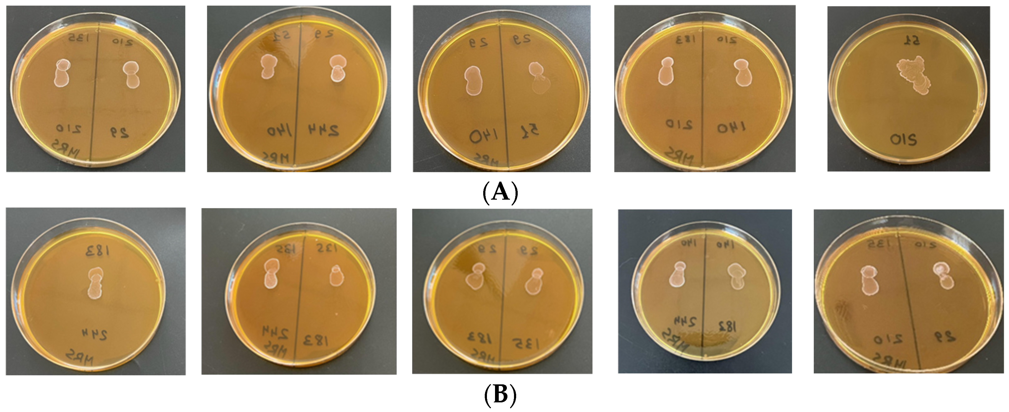
The compatibility study was conducted on seven promising strains Table 5.
The results allow us to conclude that strain No. 2 (Lat. curvatus LUHS51) has demonstrated higher likelihood values for a successful combination. It is compatible with 4 out of 7 strains and does not exhibit strong antagonism with any (only moderate antagonism with No. 1—Lc. casei LUHS210 and No. 6—P. pentosaceus LUHS183) other strains.
Strains No. 2, No. 3, and No. 4 demonstrate good compatibility with each other and with other cultures. Moderate antagonism was observed in certain pairs: between strains No. 1 and No. 2, and No. 2 and No. 6. This indicates partial competition, but not at a critical level.
Most strains appeared to be appropriate for use in combination; however, there were some with pronounced antagonism. For example, strains No. 5 (Lp. plantarum LUHS135) and No. 7 (Lc. paracasei LUHS244) appeared to be the least suitable for co-cultivation. Strain No. 5 demonstrated antagonism toward five cultures (No. 1, No. 3, No. 4, No. 6, and No. 7), whereas strain No. 7 was also incompatible in most cases (especially with No. 1, No. 3, No. 4, and No. 5).
The qualitative compatibility assay indicated the absence of strong antagonistic interactions among the four selected LAB strains (Lc. casei LUHS210, Lev. brevis LUHS140, P. acidilactici LUHS29, Lat. curvatus LUHS51), supporting their selection for inclusion in the multistrain consortium (Figure 1).
It should be noted that the absence of a visible inhibition zone in the drop co-culture assay should not be interpreted as evidence of equal growth of all strains, long-term population stability, or persistence of each strain within the consortium. Rather, this assay provides a qualitative indication of the absence of detectable strong antagonism under the experimental conditions used.
Based on the qualitative assessment of LAB strain compatibility and the absence of detectable strong antagonism under the experimental conditions, two LAB combinations were selected for subsequent consortium development: Lev. brevis LUHS140 + Lc. casei LUHS210 (Composition No. 1) and Lat. curvatus LUHS51 + P. acidilactici LUHS29 (Composition No. 2). The strains were selected through a screening procedure based on their growth in supplemented acid whey, antagonistic activity, acidification capacity, and qualitative compatibility. The results of co-cultivation of individual strains and the selected two-strain combinations are presented in Supplementary File S3 (Table S8). After 24 h of incubation, the multistrain consortium (1:1:1:1; 3%, v/v) exhibited the highest viable cell concentration (8.16 ± 0.05 log10 CFU/mL), whereas P. pentosaceus LUHS183 consistently showed the lowest growth throughout the incubation period, reaching 7.10 ± 0.01 log10 CFU/mL. Overall, the mixed cultures achieved total viable LAB counts comparable to or higher than those of the corresponding monocultures. The four-strain consortium reached the highest total viable LAB count after cultivation in supplemented acid whey. However, these results represent total LAB populations and should not be interpreted as evidence that all constituent strains proliferated equally or that quantitative strain compatibility was confirmed.
The selected two-strain consortia and the multistrain LAB consortium were subsequently evaluated for antagonistic activity against pathogenic microorganisms. The inhibitory effects against the tested opportunistic bacterial isolates are presented in Table 6.
Comparing the two paired compositions of LAB, it was observed that Composition No. 2 (Lat. curvatus LUHS51 + P. acidilactici LUHS29) (second column) in most cases demonstrates higher activity in inhibiting Staphylococcus aureus (No. 6)—25.0 ± 0.5 mm and Salmonella spp. (No. 7)—26.0 ± 1.6 mm. At the same time, weaker activity was observed against Proteus vulgaris (No. 2), where the inhibition zone was 20.1 ± 0.6 mm. Composition No. 1 (Lev. brevis LUHS140 + Lc. casei LUHS210) was more effective in suppressing Pseudomonas aeruginosa (No. 4)—23.5 mm (compared to 21.7 mm in the second group). For L. monocytogenes and S. aureus, the inhibition zone was approximately 24.0 mm.
The consortium (a mixture of all four strains in a 1:1:1:1 ratio) demonstrates a synergistic effect when strains are combined (the combined action of the components exceeds the sum of their individual effects). In all cases (No. 1–7), it showed higher results than the paired bacterial combinations alone. Demonstrated higher activity was observed against Salmonella spp. (DIZ—27.0 ± 0.4 mm).
The consortium compensates for the limitations of individual bacterial compositions. For example, in Composition No. 2 (Lat. curvatus LUHS51 and P. acidilactici LUHS29), antagonism against Proteus vulgaris reached 20.1 mm. Meanwhile, in Composition No. 1 (Lev. brevis LUHS140 and Lc. casei LUHS210), low antagonism was observed against E. coli, with an inhibition zone diameter of 21.0 mm. The LAB consortium, in both cases, balanced the results to 22.2–23.0 mm, preventing a decrease in effectiveness below a certain threshold.
In this study, inhibition of spore formation was assessed by the presence of a clear inhibition zone around the punctured well. The inhibitory effect of the selected LAB compositions against mold fungi is provided in Table 7. During the evaluation of the fungistatic potential of LAB, it was established that mold fungi exhibit a moderate sensitivity threshold to the tested compositions compared to bacterial pathogens. The zones of mycelial growth inhibition with delayed sporulation ranged from 7.3 to 8.9 mm.
Among the tested LAB pairs, the most pronounced antifungal activity was demonstrated by Composition No. 1 against Fusarium graminearum FG No. 13 (8.6 ± 1.4 mm), P. verruculosum BIM-177 (8.0 ± 2.4 mm), and A. tenuissima No. 5 (8.3 ± 1.4 mm), while moderate antagonism (7.6 ± 1.4 mm) was observed against A. niger (F-22/269) and Fusarium graminearum (F1).
Differential sensitivity among fungal strains was identified. The Fusarium graminearum FG No. 13 strain was the most sensitive to the Lev. brevis LUHS140 + Lc. casei LUHS210 composition (8.6 ± 1.4 mm), whereas the F. graminearum F1 strain exhibited greater resistance (7.6 ± 1.4 mm). The LAB consortium yielded the most stable and highest results against four out of five test cultures (A. niger, A. tenuissima, P. verruculosum, F. graminearum FG No. 13), with an inhibition in the range of 8.7 ± 1.4 to 8.9 ± 2.1 mm, respectively. The use of the 1:1:1:1 (each LAB suspension 3% v., containing 109 CFU/mL) consortium resulted in statistically homogeneous outcomes (index “a” for all test cultures), indicating stabilization of the antifungal effect when combining the four strains.
All mold species produced similar and pronounced sensitivity to the LAB consortium, indicating the broad-spectrum effectiveness of the multicomponent mixture against common raw material contaminants. The relatively small inhibition zones (7–8 mm) for fungi correlate with findings by Crowley et al. [81], who noted that effective mold suppression requires high concentrations of organic acids (lactic, acetic) and specific phenylcarboxylic acids. The antifungal effect of LAB is often associated with the production of low-molecular-weight compounds and cyclic dipeptides [82,83].
According to Schnürer and Magnusson [84], strains such as L. coryniformis and L. plantarum are among the most active against fungi of the Fusarium and Aspergillus genera. The relative resistance of Fusarium graminearum to the tested LAB compositions is consistent with the findings of Dalie et al. [85], who reported high adaptability of mycotoxigenic fungi to acidic environments. The comparatively low activity of the Lb. curvatus + P. acidilactici pair against F. graminearum (7.3 ± 2.8 mm) may be explained by this fungus’s ability to metabolize certain organic acids produced by pediococci.
The superiority of the LAB consortium over paired compositions can be explained by an expanded metabolite profile and the accumulation of synergistic concentrations of organic acids (lactic and acetic) [86,87].
According to Mateo et al. [88], it is the combined effect of acid mixtures, rather than their total concentration alone, that determines the fungistatic effect against Aspergillus and Fusarium. Bhattacharya et al. [89] also indicate that combining strains enables stable inhibition even when fungi exhibit low sensitivity to individual bacteriocins. The contribution of bacteriocin-like inhibitory substances (BLIS) should also be considered [90,91,92]. As noted by Dalie et al. [85], although LAB bacteriocins are primarily active against bacteria, some cyclic dipeptides can affect the morphology of fungal hyphae, slowing their radial growth, which corresponds to the observed inhibition zones of up to 8.6 mm.
Considerable variation was observed; results indicate the feasibility of co-cultivating selected pairs of LAB strains to develop a multicomponent probiotic consortium with a defined composition. Different LAB strains (Lev. brevis, L. casei, Lat. curvatus, P. acidilactici) produce different types of organic acids and bacteriocins [93,94]. Within a consortium, they act against pathogens through multiple mechanisms simultaneously, reducing the likelihood of pathogen survival [95,96]. The obtained results support the selection of these strains for further evaluation as a multistrain consortium. Under the experimental conditions of the present study, the consortium exhibited broader and generally stronger antagonistic activity against the tested pathogens than the paired LAB combinations.
Our findings are consistent with those of Semenova et al. [97]: the consortium grown on MRS medium exhibited a high cell titer (at least 1012 CFU/g) and demonstrated greater antagonistic activity compared to the individual LAB strains included in it.
According to our results, the tested LAB consortium is a promising candidate for starter culture development and broader application in the feed industry. When a 3% mixture of the four strains was used, the levels of all tested pathogens were markedly reduced. Cultures of Lactobacillus and Pediococcus exhibited pronounced antibacterial activity and effectively inhibited the growth of mold fungi, including phytopathogenic species associated with cereal crops.
The compatibility assessment performed in this study was intended as a preliminary qualitative screening to identify LAB strains suitable for consortium development. Although the drop co-culture assay enabled discrimination between compatible and antagonistic strain combinations, it does not provide quantitative information on the dynamics of strain interactions during co-cultivation. Future studies will include quantitative evaluation of strain interactions during co-cultivation, such as growth kinetics (OD600) and viable cell counts (CFU), to provide a more comprehensive validation of the compatibility and stability of the developed LAB consortium.

3.5. Acidity and Microbiological Parameters of Fermented Wheat Bran

Acidity parameters and microbiological indicators of wheat bran are presented in Table 8, and pH and viable lactic acid bacteria count in fermented wheat bran in Table 9.
The physicochemical and microbiological characteristics of fermented wheat bran are presented in Table 8 and Table 9. Fermentation with the selected four-strain LAB consortium resulted in progressive acidification of the substrate throughout the incubation period. The pH decreased from 5.65 ± 0.02 before fermentation to 4.07 ± 0.03 after 24 h and reached 3.82 ± 0.04 after 36 h (p ≤ 0.05). At the same time, total titratable acidity (TTA) increased from 0.20 ± 0.02 to 4.40 ± 0.12 °N, indicating active acid production during fermentation [98].
The viable LAB population increased throughout fermentation, reaching 7.23 ± 0.11 log10 CFU/g after 36 h (Table 9). Compared with the non-fermented wheat bran (control), the fermented substrate contained approximately 3 log10 CFU/g more LAB (7.23 vs. 4.20 log10 CFU/g). Higher LAB count values were observed after 36 h of fermentation, whereas a slight decrease to 6.93 ± 0.06 log10 CFU/g was recorded after 48 h.
Fermentation also affected the indigenous microbiota of wheat bran. Total bacterial counts decreased from 7.35 ± 0.10 to 6.67 ± 0.13 log10 CFU/g, while Enterobacteriaceae were not detected after fermentation. The numbers of molds and yeasts were reduced from 3.19 ± 0.11 to 2.75 ± 0.12 log10 CFU/g. Similar acidification patterns were observed for both two-strain LAB combinations; however, the four-strain consortium produced the lowest final pH (3.82 ± 0.04) and demonstrated higher TTA (4.40 ± 0.12 °N), accompanied by higher viable LAB counts.
The decrease in pH together with the increase in titratable acidity indicates active fermentation of wheat bran by the selected LAB strains. Acidification is one of the principal technological effects of LAB fermentation because it suppresses the growth of acid-sensitive microorganisms and contributes to improved microbiological quality of fermented substrates.
The absence of detectable Enterobacteriaceae after fermentation and the reduction in total bacterial, mold, and yeast counts are consistent with the inhibitory environment created during LAB fermentation.
The concurrent increase in viable LAB counts together with progressive substrate acidification suggests that these processes collectively contributed to the suppression of undesirable microorganisms during fermentation. Besides lowering the pH through the accumulation of organic acids, the growing LAB population may also have limited the proliferation of competing microorganisms by competing for available nutrients and ecological niches. In addition, previous studies have shown that some LAB strains are capable of producing bacteriocins and other antimicrobial metabolites that may further enhance microbial inhibition. However, these compounds were not determined in the present study; therefore, their contribution to the observed reduction in Enterobacteriaceae can only be discussed in the context of previously published literature.
Although antimicrobial metabolites such as organic acids, bacteriocins, and other low-molecular-weight compounds have been reported as important contributors to the antimicrobial activity of LAB in previous studies [99,100,101], these compounds were not determined in the present work. Therefore, the relative contribution of individual metabolites to the observed microbial changes cannot be established.
The four-strain consortium produced lower pH values and higher LAB counts than either of the two-strain combinations, suggesting that this formulation performed more efficiently under the experimental conditions used. However, because the study did not include metabolomic characterization or strain-specific monitoring during wheat bran fermentation, the mechanisms underlying the observed differences between the consortium and the paired cultures remain to be clarified.
Our findings are generally consistent with previous reports describing LAB fermentation of cereal substrates. Prücker et al. [102] and Arte et al. [103] reported substantial suppression of enterobacteria and molds during wheat bran fermentation by Lactiplantibacillus plantarum, Levilactobacillus brevis, and Companilactobacillus humilis. Similarly, Bartkiene et al. [2] and Vadopalas et al. [104] observed viable LAB counts of approximately 7.5 log10 CFU/mL and final pH values close to 3.9 after fermentation of dairy by-products with LAB strains. Previous studies have also reported progressive acidification of cereal substrates during fermentation, accompanied by an increase in LAB populations and a concomitant reduction in Enterobacteriaceae, coliforms, molds, and yeasts, thereby improving the microbiological quality of fermented cereal products [105,106]. Although direct comparisons between studies should be made with caution because of differences in substrates, inoculum composition, and fermentation conditions, the trends observed in the present study are consistent with previously published data.
The nutritional characteristics of the fermented wheat bran, including its chemical composition (e.g., crude protein, fiber fractions, metabolizable energy, and bioactive compounds), were not evaluated in the present study. Therefore, the nutritional and functional value of the fermented product requires further investigation before its application as a functional feed ingredient can be substantiated.
The obtained results demonstrate the potential of the developed LAB consortium for wheat bran fermentation and highlight its potential for the valorization of cereal by-products into value-added feed material. Further studies under in vivo conditions are required to confirm its efficacy and practical applicability.

4. Conclusions

This study demonstrated the feasibility of systematically selecting a multistrain lactic acid bacteria (LAB) consortium based on antimicrobial activity, mycotoxin mitigation capacity, and strain compatibility, followed by its propagation in supplemented sour whey and application to the fermentation of extruded wheat bran. Compared with individual LAB strains and two-strain combinations, the selected four-strain consortium provided more effective substrate acidification, maintained high populations of viable LAB, and improved the microbiological characteristics of the fermented wheat bran. These findings support the potential application of the developed consortium as a starter culture for the valorization of cereal by-products into value-added feed material.
Nevertheless, the results should be interpreted within the limitations of the present study. All experiments were performed in vitro, and the mycotoxin mitigation capacity of the consortium was evaluated only in liquid model systems rather than directly in the fermented wheat bran matrix. In addition, metabolomic profiling was not performed; therefore, the compounds responsible for the observed antimicrobial activity and mycotoxin mitigation were not identified. Future studies should include controlled solid-state fermentation experiments with appropriate control treatments comprising different strain combinations, validated chromatographic assessment of mycotoxin degradation, strain-specific monitoring of bacterial growth, and both targeted and untargeted metabolomic analyses. Further research will also focus on scaling up cultivation of the selected consortium in supplemented sour whey under pilot- and industrial-scale conditions, optimizing the technological process, and evaluating its reproducibility. In addition, controlled feeding trials in horses are planned to assess the safety of the developed feed additive, its effects on nutrient digestibility, animal performance, intestinal microbiota composition, immune responses, and biomarkers of mycotoxin exposure. These studies will provide a more comprehensive evaluation of the practical applicability of the developed technology and support its potential use in the valorization of cereal by-products into value-added feed material.

Supplementary Materials

The following supporting information can be downloaded at: https://www.mdpi.com/article/10.3390/fermentation12080371/s1, Table S1. Identification of LAB strains by direct MALDI-TOF MS; Table S2. Analytical protocol and results mycotoxin mitigation capacity. Table S3. European Union performance criteria for validated analytical methods for mycotoxin determination according to Commission Regulation (EU) No. 519/2014. Table S4. Residual mycotoxin concentrations and percentage reduction. Table S5. Four-way ANOVA for residual mycotoxin concentration. Table S6. Pearson correlation matrices; Table S7. LAB multiplication in sour whey by time exposure (t = 0, 6, 12, 24, 48 h); Table S8. LAB multiplication in sour whey by time exposure (t = 6, 12, 24 h); Supplementary File S1. Table S1_Identification of LAB strains by direct MALDI-TOF MS; Supplementary File S2. Table S2–S6_Analytical protocol and results mycotoxin mitigation capacity; Supplementary File S3. Tables S7 and S8_LAB multiplication in sour whey by time exposure.

Author Contributions

Conceptualization, A.Y. and E.B.; methodology, A.Y. and E.B.; formal analysis, N.K., A.Y., S.T. and V.S.; investigation, R.Š., G.A., S.T., N.K., Y.G. and A.Y.; writing—original draft preparation, A.Y., S.T., G.A. and N.K.; writing—review and editing, E.B. and M.R.; supervision, E.B., A.Y. and N.K. All authors have read and agreed to the published version of the manuscript.

Funding

This research has been funded by the Science Committee of the Ministry of Science and Higher Education of the Republic of Kazakhstan (Grant No. AP23489520).

Institutional Review Board Statement

No experiments involving animals or human-derived samples were conducted in the present study.

Informed Consent Statement

Not applicable.

Data Availability Statement

The original contributions presented in this study are included in the article/Supplementary Materials. Further inquiries can be directed to the corresponding author.

Conflicts of Interest

The authors declare no conflicts of interest.

References

  1. Markowiak, P.; Śliżewska, K. The role of probiotics, prebiotics and synbiotics in animal nutrition. Gut Pathog. 2018, 10, 21. [Google Scholar] [CrossRef] [PubMed]
  2. Bartkiene, E.; Bartkevics, V.; Krungleviciute, V.; Juodeikiene, G.; Zadeike, D.; Baliukoniene, V.; Bakutis, B.; Zelvyte, R.; Santini, A.; Cizeikiene, D. Application of hydrolases and probiotic Pediococcus acidilactici BaltBio01 strain for cereal by-products conversion to bioproduct for food/feed. Int. J. Food Sci. Nutr. 2018, 69, 165–175. [Google Scholar] [CrossRef] [PubMed]
  3. Yue, T.; Lu, Y.; Ding, W.; Xu, B.; Zhang, C.; Li, L.; Jian, F.; Huang, S. The Role of Probiotics, Prebiotics, Synbiotics, and Postbiotics in Livestock and Poultry Gut Health: A Review. Metabolites 2025, 15, 478. [Google Scholar] [CrossRef] [PubMed]
  4. Moretti, A.F.; Brisuela, N.S.; Bravo-Ferrada, B.M.; Tymczyszyn, E.E.; Golovchik, M.A. Current Applications and Future Trends of Dehydrated Lactic Acid Bacteria for Incorporation in Animal Feed Products. Fermentation 2023, 9, 742. [Google Scholar] [CrossRef]
  5. Jiang, S.; Cai, L.; Lv, L.; Li, L. Pediococcus pentosaceus: A future additive or probiotic candidate. Microb. Cell Fact. 2021, 20, 45. [Google Scholar] [CrossRef] [PubMed]
  6. Pasolli, E.; De Filippis, F.; Mauriello, I.E.; Cumbo, F.; Walsh, A.M.; Leech, J.; Cotter, P.D.; Segata, N.; Ercolini, D. Large-scale genome analysis links lactic acid bacteria from food to the gut microbiome. Nat. Commun. 2020, 11, 2610. [Google Scholar] [CrossRef] [PubMed]
  7. Zavistanaviciute, P.; Ruzauskas, M.; Antanaitis, R.; Televicius, M.; Lele, V.; Santini, A.; Bartkiene, E. Antimicrobial and mycotoxin-reducing properties of lactic acid bacteria and their influence on blood and feces parameters of newborn calves. Animals 2023, 13, 3345. [Google Scholar] [CrossRef] [PubMed]
  8. Liu, H.; Ren, X.; Li, Y.; Cao, Q.; Yang, L.; Jiang, S. Effects of different wheat bran fermentation sources on growth performance, nutrient digestibility, serum antioxidant capacity and fecal microbiota in growing pigs. Front. Vet. Sci. 2023, 10, 1289010. [Google Scholar] [CrossRef] [PubMed]
  9. Ghamry, M.; Zhao, W.; Li, L. Impact of Lactobacillus apis on the antioxidant activity, phytic acid degradation, nutraceutical value and flavor properties of fermented wheat bran, compared to Saccharomyces cerevisiae and Lactobacillus plantarum. Food Res. Int. 2023, 163, 112142. [Google Scholar] [CrossRef] [PubMed]
  10. Krishnan, S.V.; Anaswara, P.A.; Nampoothiri, K.M.; Kovács, S.; Adácsi, C.; Szarvas, P.; Király, S.; Pócsi, I.; Pusztahelyi, T. Biocontrol Activity of New Lactic Acid Bacteria Isolates Against Fusaria and Fusarium Mycotoxins. Toxins 2025, 17, 68. [Google Scholar] [CrossRef] [PubMed]
  11. Mahjoory, Y.; Mohammadi, R.; Hejazi, M.A.; Nami, Y. Antifungal activity of potential probiotic Limosilactobacillus fermentum strains and their role against toxigenic aflatoxin-producing aspergilli. Sci. Rep. 2023, 13, 388. [Google Scholar] [CrossRef] [PubMed]
  12. Azevedo, P.O.d.S.d.; Gierus, M. Lactic Acid Bacteria and Bacteriocins in Feed Preservation: Mechanisms and Antifungal Properties. Grass Forage Sci. 2025, 80, e12711. [Google Scholar] [CrossRef]
  13. Abraham, N.; Chan, E.T.S.; Zhou, T.; Seah, S.Y.K. Microbial Detoxification of Mycotoxins in Food. Front. Microbiol. 2022, 13, 957148. [Google Scholar] [CrossRef] [PubMed]
  14. Lee, C.; Kim, S.W.; Verma, R.; Noh, J.; Park, J.C.; Park, S.; Lee, H.; Park, H.E.; Kim, C.J.; Byun, S.; et al. Probiotic Consortium Confers Synergistic Anti-Inflammatory Effects in Inflammatory Disorders. Nutrients 2024, 16, 790. [Google Scholar] [CrossRef] [PubMed]
  15. Ayivi, R.D.; Gyawali, R.; Krastanov, A.; Aljaloud, S.O.; Worku, M.; Tahergorabi, R.; Silva, R.C.d.; Ibrahim, S.A. Lactic Acid Bacteria: Food Safety and Human Health Applications. Dairy 2020, 1, 202–232. [Google Scholar] [CrossRef]
  16. Mao, M.; Wang, P.; Shi, K.; Lu, Z.; Bie, X.; Zhao, H.; Zhan, C.; Lv, F. Effect of solid state fermentation by Enterococcus faecalis M2 on antioxidant and nutritional properties of wheat bran. J. Cereal Sci. 2020, 94, 102997. [Google Scholar] [CrossRef]
  17. Gibson, G.R.; Roberfroid, M.B. Dietary modulation of the human colonic microflora: Introducing the concept of prebiotics. J. Nutr. 1995, 125, 1401–1412. [Google Scholar] [CrossRef] [PubMed]
  18. Prueckler, M.; Siebenhandl-Ehn, S.; Apprich, S.; Hoeltinger, S.; Haas, C.; Schmid, E.; Kneifel, W. Wheat bran-based biorefinery 1: Composition of wheat bran and strategies of functionalization. LWT Food Sci. Technol. 2014, 56, 211–221. [Google Scholar] [CrossRef]
  19. Soukoulis, C.; Aprea, E. Cereal bran fractionation: Processing techniques for the recovery of functional components and their applications to the food industry. Recent Pat. Food Nutr. Agric. 2012, 4, 61–77. [Google Scholar] [CrossRef] [PubMed]
  20. Sun, W.; Bian, Z.; Yin, C.; Yang, H.; San, W.; Li, Y.; Li, X.; Li, Q.; Bontempo, V.; Jiang, X. Effects of fermented wheat bran on growth performance, diarrhoea incidence, antioxidant capacity and faecal microbiota in weaned piglets. Ital. J. Anim. Sci. 2026, 25, 595–605. [Google Scholar] [CrossRef]
  21. Spaggiari, M.; Ricci, A.; Calani, L.; Bresciani, L.; Neviani, E.; Dall’Asta, C.; Lazzi, C.; Galaverna, G. Solid state lactic acid fermentation: A strategy to improve wheat bran functionality. LWT 2020, 118, 108668. [Google Scholar] [CrossRef]
  22. Bartkiene, E.; Zokaityte, E.; Lele, V.; Starkute, V.; Zavistanaviciute, P.; Klupsaite, D.; Cernauskas, D.; Ruzauskas, M.; Bartkevics, V.; Pugajeva, I.; et al. Combination of extrusion and fermentation with Lactobacillus plantarum and L. uvarum strains for improving the safety characteristics of wheat bran. Toxins 2021, 13, 163. [Google Scholar] [CrossRef] [PubMed]
  23. Yang, S.; Xu, X.; Peng, Q.; Ma, L.; Qiao, Y.; Shi, B. Exopolysaccharides from lactic acid bacteria as an alternative to antibiotics: Regulation of gut health and immune system. Anim. Nutr. 2023, 13, 78–89. [Google Scholar] [CrossRef] [PubMed]
  24. Bartkiene, E.; Zavistanaviciute, P.; Lele, V.; Ruzauskas, M.; Bartkevics, V.; Bernatoniene, J.; Gallo, P.; Carlo Tenore, G.; Santini, A. Lactobacillus plantarum LUHS135 and paracasei LUHS244 as functional starter cultures for the food fermentation industry: Characterisation, mycotoxin-reducing properties, optimisation of biomass growth and sustainable encapsulation by using dairy by-products. LWT 2018, 93, 649–658. [Google Scholar] [CrossRef]
  25. Zokaityte, E.; Cernauskas, D.; Klupsaite, D.; Lele, V.; Starkute, V.; Zavistanaviciute, P.; Ruzauskas, M.; Gruzauskas, R.; Juodeikiene, G.; Rocha, J.M.; et al. Bioconversion of Milk Permeate with Selected Lactic Acid Bacteria Strains and Apple By-Products into Beverages with Antimicrobial Properties and Enriched with Galactooligosaccharides. Microorganisms 2020, 8, 1182. [Google Scholar] [CrossRef] [PubMed]
  26. Canon, F.; Maillard, M.-B.; Henry, G.; Thierry, A.; Gagnaire, V. Positive Interactions between Lactic Acid Bacteria Promoted by Nitrogen-Based Nutritional Dependencies. Appl. Environ. Microbiol. 2021, 87, e01055-21. [Google Scholar] [CrossRef] [PubMed]
  27. Rahman, M.S.; Soltani, S.; LaPointe, G.; Karboune, S.; Fliss, I. Lactic acid bacteria: Beyond fermentation to bio-protection against fungal spoilage and mycotoxins in food systems. Front. Microbiol. 2025, 16, 1580670. [Google Scholar] [CrossRef] [PubMed]
  28. Ponzio, A.; Rebecchi, A.; Zivoli, R.; Morelli, L. Reuterin, Phenyllactic Acid, and Exopolysaccharides as Main Antifungal Molecules Produced by Lactic Acid Bacteria: A Scoping Review. Foods 2024, 13, 752. [Google Scholar] [CrossRef] [PubMed]
  29. Huang, C.; Zhang, B.; Huang, J.; Liu, Y.; Chen, C.; Omedi, J.O.; Liang, L.; Zhou, Z.; Huang, W.; Li, N. The Effects of Single- or Mixed-Strain Fermentation of Red Bean Sourdough, with or without Wheat Bran, on Bread Making Performance and Its Potential Health Benefits in Mice Model. Foods 2024, 13, 2856. [Google Scholar] [CrossRef] [PubMed]
  30. Roye, C.; Henrion, M.; Chanvrier, H.; De Roeck, K.; De Bondt, Y.; Liberloo, I.; King, R.; Courtin, C.M. Extrusion-Cooking Modifies Physicochemical and Nutrition-Related Properties of Wheat Bran. Foods 2020, 9, 738. [Google Scholar] [CrossRef] [PubMed]
  31. Wu, Q.; San, Y.; Wu, S. A Further Understanding of Changes of Wheat Bran Functionality Induced by Different Types of Probiotics Fermentation: From Molecules to Regulation Mechanism. Food Chem. 2025, 463, 141231. [Google Scholar] [CrossRef] [PubMed]
  32. Huang, J.; Ling, F.; Sen, M.; Li, L. Fermentation Biotechnology Applied to Wheat Bran for the Degradation of Cell Wall Fiber and Its Potential Health Benefits: A Review. Int. J. Biol. Macromol. 2024, 272, 133529. [Google Scholar] [CrossRef] [PubMed]
  33. Rocha-Mendoza, D.; Kosmerl, E.; Krentz, A.; Zhang, L.; Badiger, S.; Miyagusuku-Cruzado, G.; Mayta-Apaza, A.; Giusti, M.; Jiménez-Flores, R.; García-Cano, I. Invited Review: Acid Whey Trends and Health Benefits. J. Dairy Sci. 2021, 104, 1262–1275. [Google Scholar] [CrossRef] [PubMed]
  34. Misganaw, W.; Teketay, W. Isolation and Identification of Lactic Acid Bacteria from Raw Cow Milk. Int. J. Adv. Res. Biol. Sci. 2016, 3, 44–49. Available online: http://s-o-i.org/1.15/ijarbs-2016-3-8-8 (accessed on 22 June 2026).
  35. Bartkiene, E.; Lele, V.; Sakiene, V.; Zavistanaviciute, P.; Ruzauskas, M.; Bernatoniene, J.; Jakstas, V.; Viskelis, P.; Zadeike, D.; Juodeikiene, G. Improvement of the antimicrobial activity of lactic acid bacteria in combination with berries/fruits and dairy industry by-products. J. Sci. Food Agric. 2019, 99, 3992–4002. [Google Scholar] [CrossRef] [PubMed]
  36. Li, D.; Yi, J.; Han, G.; Qiao, L. MALDI-TOF Mass Spectrometry in Clinical Analysis and Research. ACS Meas. Sci. Au 2022, 2, 385–404. [Google Scholar] [CrossRef] [PubMed]
  37. Cizeikiene, D.; Juodeikiene, G.; Paskevicius, A.; Bartkiene, E. Antimicrobial activity of lactic acid bacteria against pathogenic and spoilage microorganism isolated from food and their control in wheat bread. Food Control 2013, 31, 539–545. [Google Scholar] [CrossRef]
  38. Clinical and Laboratory Standards Institute (CLSI). Methods for Dilution Antimicrobial Susceptibility Tests for Bacteria That Grow Aerobically: M07-A10, Approved Standard, 10th ed.; Clinical and Laboratory Standards Institute: Wayne, PA, USA, 2015; ISBN 978-1-56238-987-1. [Google Scholar]
  39. EFSA Panel on Additives and Products or Substances used in Animal Feed (FEEDAP). Guidance on the Assessment of Bacterial Susceptibility to Antimicrobials of Human and Veterinary Importance. EFSA J. 2012, 10, 2740. [CrossRef]
  40. Clinical and Laboratory Standards Institute (CLSI). Performance Standards for Antimicrobial Susceptibility Testing, 29th ed.; CLSI supplement M100; Clinical and Laboratory Standards Institute: Wayne, PA, USA, 2018; Available online: https://webstore.ansi.org/standards/clsi/clsim100ed29 (accessed on 14 March 2022).
  41. Bartkiene, E.; Lele, V.; Ruzauskas, M.; Domig, K.J.; Starkute, V.; Zavistanaviciute, P.; Bartkevics, V.; Pugajeva, I.; Klupsaite, D.; Juodeikiene, G.; et al. Lactic Acid Bacteria Isolation from Spontaneous Sourdough and Their Characterization Including Antimicrobial and Antifungal Properties Evaluation. Microorganisms 2020, 8, 64. [Google Scholar] [CrossRef] [PubMed]
  42. Volkova, G.S.; Kuksova, E.V.; Serba, E.M. Study of biological inter-strain interactions and growth properties of production strains of lactic acid bacteria. Actual Issues Dairy Ind. Intersect. Technol. Qual. Manag. Syst. 2020, 1, 104–109. [Google Scholar] [CrossRef]
  43. Irkitova, A.N.; Yatsenko, E.S. Optimization of the method for determining the antagonistic activity of probiotic bacteria. Technol. Food Process. Ind. Agro-Ind. Complex—Healthy Food Prod. 2017, 5, 113–117. [Google Scholar] [CrossRef] [PubMed]
  44. O’Toole, G.A. Classic Spotlight: Plate Counting You Can Count On. J. Bacteriol. 2016, 198, 3127. [Google Scholar] [CrossRef] [PubMed]
  45. Silva, J.; Carvalho, A.S.; Teixeira, P.; Gibbs, P.A. Bacteriocin production by spray-dried lactic acid bacteria. Lett. Appl. Microbiol. 2002, 34, 77–81. [Google Scholar] [CrossRef] [PubMed]
  46. Bajagai, Y.S.; Klieve, A.V.; Dart, P.J.; Bryden, W.L. Probiotics in Animal Nutrition: Production, Impact and Regulation; FAO: Rome, Italy, 2016. [Google Scholar]
  47. Aboukhalaf, A.; El Amraoui, B.; Tabatou, M.; Rocha, J.M.; Belahsen, R. Screening of the antimicrobial activity of some extracts of edible wild plants in Morocco. Funct. Foods Health Dis. 2020, 10, 265–273. [Google Scholar] [CrossRef]
  48. Şanlier, N.; Gökcen, B.B.; Sezgin, A.C. Health benefits of fermented foods. Crit. Rev. Food Sci. Nutr. 2019, 59, 506–527. [Google Scholar] [CrossRef] [PubMed]
  49. Venema, K.; Haverkort, R.E.; Abee, T.; Haandrikman, A.J.; Leenhouts, K.J.; de Leij, L.; Venema, G.; Kok, J. Mode of action of LciA, the lactococcin A immunity protein. Mol. Microbiol. 1994, 14, 521–532. [Google Scholar] [CrossRef] [PubMed]
  50. Arena, M.P.; Silvain, A.; Normanno, G.; Grieco, F.; Drider, D.; Spano, G.; Fiocco, D. Use of Lactobacillus plantarum Strains as a Bio-Control Strategy against Food-Borne Pathogenic Microorganisms. Front. Microbiol. 2016, 7, 464. [Google Scholar] [CrossRef] [PubMed]
  51. Ibrahim, S.A.; Ayivi, R.D.; Zimmerman, T.; Siddiqui, S.A.; Altemimi, A.B.; Fidan, H.; Esatbeyoglu, T.; Bakhshayesh, R.V. Lactic Acid Bacteria as Antimicrobial Agents: Food Safety and Microbial Food Spoilage Prevention. Foods 2021, 10, 3131. [Google Scholar] [CrossRef] [PubMed]
  52. Yeleussizova, A.; Aleshina, Y.; Mendybayeva, A.; Khassanova, M.; Rychshanova, R. Resistance to Antibacterial Agents in Escherichia coli Isolated from Domestic Cats and Dogs in the Northern Region of the Republic of Kazakhstan. Am. J. Anim. Vet. Sci. 2023, 18, 190–198. [Google Scholar] [CrossRef]
  53. Aleshina, Y.; Yeleussizova, A.; Mendybayeva, A.; Shevchenko, P.; Rychshanova, R. Prevalence and antimicrobial resistance of Enterobacteriaceae in the north of Kazakhstan. Open Vet. J. 2024, 14, 604–616. [Google Scholar] [CrossRef] [PubMed]
  54. Yeleussizova, A.; Sobiech, P.; Kaumenov, N.; Batyrbekov, A.; Błażejak-Grabowska, J.; Isabaev, A.; Platt-Samoraj, A. Influence of lactic bacteria on the viability of Listeria monocytogenes in plant foods. Pol. J. Vet. Sci. 2023, 26, 713–719. [Google Scholar] [CrossRef] [PubMed]
  55. Muhammad, Z.; Ramzan, R.; Abdelazez, A.; Amjad, A.; Afzaal, M.; Zhang, S.; Pan, S. Assessment of the Antimicrobial Potentiality and Functionality of Lactobacillus plantarum Strains Isolated from the Conventional Inner Mongolian Fermented Cheese Against Foodborne Pathogens. Pathogens 2019, 8, 71. [Google Scholar] [CrossRef] [PubMed]
  56. Carvalho, F.M.; Teixeira-Santos, R.; Mergulhão, F.J.M.; Gomes, L.C. Effect of Lactobacillus plantarum Biofilms on the Adhesion of Escherichia coli to Urinary Tract Devices. Antibiotics 2021, 10, 966. [Google Scholar] [CrossRef] [PubMed]
  57. Potočnjak, M.; Pušić, P.; Frece, J.; Abram, M.; Janković, T.; Gobin, I. Three New Lactobacillus plantarum Strains in the Probiotic Toolbox against Gut Pathogen Salmonella enterica Serotype Typhimurium. Food Technol. Biotechnol. 2017, 55, 48–54. [Google Scholar] [CrossRef] [PubMed]
  58. Chappell, T.C.; Nair, N.U. Engineered Lactobacilli Display Anti-Biofilm and Growth Suppressing Activities against Pseudomonas aeruginosa. npj Biofilms Microbiomes 2020, 6, 48. [Google Scholar] [CrossRef] [PubMed]
  59. Magnusson, J.; Ström, K.; Roos, S.; Sjögren, J.; Schnürer, J. Broad and complex antifungal activity among environmental isolates of lactic acid bacteria. FEMS Microbiol. Lett. 2003, 219, 129–135. [Google Scholar] [CrossRef] [PubMed]
  60. Qiao, N.; Yu, L.; Zhang, C.; Wei, C.; Zhao, J.; Zhang, H.; Tian, F.; Zhai, Q.; Chen, W. A comparison of the inhibitory activities of Lactobacillus and Bifidobacterium against Penicillium expansum and an analysis of potential antifungal metabolites. FEMS Microbiol. Lett. 2020, 367, fnaa130. [Google Scholar] [CrossRef] [PubMed]
  61. Shehata, M.; Badr, A.; El-Sohaimy, S.; Asker, D.; Awad, T. Characterization of antifungal metabolites produced by novel lactic acid bacterium and their potential application as food biopreservatives. Ann. Agric. Sci. 2019, 64, 71–78. [Google Scholar] [CrossRef]
  62. Chlebicz, A.; Śliżewska, K. In vitro detoxification of aflatoxin B1, deoxynivalenol, fumonisins, T-2 toxin and zearalenone by probiotic bacteria from genus Lactobacillus and Saccharomyces cerevisiae yeast. Probiotics Antimicrob. Proteins. 2020, 12, 289–301. [Google Scholar] [CrossRef] [PubMed]
  63. Danchik, C.; Casadevall, A. Role of Cell Surface Hydrophobicity in the Pathogenesis of Medically-Significant Fungi. Front. Cell. Infect. Microbiol. 2021, 10, 594973. [Google Scholar] [CrossRef] [PubMed]
  64. Joannis-Cassan, C.; Tozlovanu, M.; Hadjeba-Medjdoub, K.; Ballet, N.; Pfohl-Leszkowicz, A. Binding of Zearalenone, Aflatoxin B1, and Ochratoxin A by Yeast-Based Products: A Method for Quantification of Adsorption Performance. J. Food Prot. 2011, 74, 1175–1185. [Google Scholar] [CrossRef] [PubMed]
  65. Niderkorn, V.; Morgavi, D.P.; Aboab, B.; Lemaire, M.; Boudra, H. Cell wall component and mycotoxin moieties involved in the binding of fumonisin B1 and B2 by lactic acid bacteria. J. Appl. Microbiol. 2009, 106, 977–985. [Google Scholar] [CrossRef] [PubMed]
  66. Adunphatcharaphon, S.; Petchkongkaew, A.; Visessanguan, W. In Vitro Mechanism Assessment of Zearalenone Removal by Plant-Derived Lactobacillus plantarum BCC47723. Toxins 2021, 13, 286. [Google Scholar] [CrossRef] [PubMed]
  67. Kosztik, J.; Mörtl, M.; Székács, A.; Kukolya, J.; Bata-Vidács, I. Aflatoxin B1 and Sterigmatocystin Binding Potential of Lactobacilli. Toxins 2020, 12, 756. [Google Scholar] [CrossRef] [PubMed]
  68. Aiko, V.; Edamana, P.; Mehta, M. Decomposition and detoxification of aflatoxin B1 by lactic acid. J. Sci. Food Agric. 2016, 96, 1959–1966. [Google Scholar] [CrossRef] [PubMed]
  69. Wang, Y.; Jiang, L.; Zhang, Y.; Ran, R.; Meng, X.; Liu, S. Research advances in the degradation of aflatoxin by lactic acid bacteria. J. Venom. Anim. Toxins Incl. Trop. Dis. 2023, 29, e20230029. [Google Scholar] [CrossRef] [PubMed]
  70. Yu, C.; Plaizier, P.; Gong, J.; Yang, C.; Liu, S. A Comprehensive Review: Current Strategies for Detoxification of Deoxynivalenol in Feedstuffs for Pigs. Animals 2025, 15, 2739. [Google Scholar] [CrossRef] [PubMed]
  71. Debevere, S.; Schatzmayr, D.; Reisinger, N.; Aleschko, M.; Haesaert, G.; Rychlik, M.; Croubels, S.; Fievez, V. Evaluation of the Efficacy of Mycotoxin Modifiers and Mycotoxin Binders by Using an In Vitro Rumen Model as a First Screening Tool. Toxins 2020, 12, 405. [Google Scholar] [CrossRef] [PubMed]
  72. Ragoubi, C.; Quintieri, L.; Greco, D.; Mehrez, A.; Maatouk, I.; D’Ascanio, V.; Landoulsi, A.; Avantaggiato, G. Mycotoxin Removal by Lactobacillus spp. and Their Application in Animal Liquid Feed. Toxins 2021, 13, 185. [Google Scholar] [CrossRef] [PubMed]
  73. Krishnan, S.V.; Nampoothiri, K.M.; Suresh, A.; Linh, N.T.; Balakumaran, P.A.; Pócsi, I.; Pusztahelyi, T. Fusarium biocontrol: Antagonism and mycotoxin elimination by lactic acid bacteria. Front. Microbiol. 2024, 14, 1260166. [Google Scholar] [CrossRef] [PubMed]
  74. Juodeikiene, G.; Bartkiene, E.; Cernauskas, D.; Cizeikiene, D.; Zadeike, D.; Lele, V.; Bartkevics, V. Antifungal activity of lactic acid bacteria and their application for Fusarium mycotoxin reduction in malting wheat grains. LWT 2017, 89, 307–314. [Google Scholar] [CrossRef]
  75. Zhang, J.; Liu, X.; Su, Y.; Li, T. An update on T2-toxins: Metabolism, immunotoxicity mechanism and human assessment exposure of intestinal microbiota. Heliyon 2022, 8, 1–9. [Google Scholar] [CrossRef] [PubMed]
  76. Wang, Y.; Wang, L.; Liu, F.; Wang, Q.; Selvaraj, J.N.; Xing, F.; Zhao, Y.; Liu, Y. Ochratoxin A Producing Fungi, Biosynthetic Pathway and Regulatory Mechanisms. Toxins 2016, 8, 83. [Google Scholar] [CrossRef] [PubMed]
  77. Murtaza, B.; Guo, L.; Wang, L.; Li, X.; Zeb, L.; Jin, B.; Li, J.; Xu, Y. Innovative probiotic fermentation approach for zearalenone detoxification in dried distiller’s grains. Front. Microbiol. 2025, 16, 1533515. [Google Scholar] [CrossRef] [PubMed]
  78. Harčárová, M.; Čonková, E.; Naď, P.; Proškovcová, M. Zearalenone Biodegradation by the Lactobacillus spp. and Bacillus spp. In Vitro. Folia Vet. 2022, 66, 70–74. [Google Scholar] [CrossRef]
  79. Monroy, I.; Olmeda, I.; Ferrer, S.; Pardo, I. Biochemical Characterization of Three Heterologous Lactic Acid Bacteria Laccases from Pediococcus, Lactobacillus, and Lactococcus Genus and Their Potential to Degrade Biogenic Amines Using ABTS and Epicatechin as Mediators. Fermentation 2024, 10, 32. [Google Scholar] [CrossRef]
  80. Niderkorn, V.; Morgavi, D.P.; Pujos, E.; Tissandier, A.; Boudra, H. Screening of fermentative bacteria for their ability to bind and biotransform deoxynivalenol, zearalenone and fumonisins in an in vitro simulated corn silage model. Food Addit. Contam. 2007, 24, 406–415. [Google Scholar] [CrossRef] [PubMed]
  81. Crowley, S.; Mahony, J.; van Sinderen, D. Current perspectives on antifungal lactic acid bacteria as biological preservatives. Trends Food Sci. Technol. 2013, 33, 93–109. [Google Scholar] [CrossRef]
  82. Nasrollahzadeh, A.; Mokhtari, S.; Khomeiri, M.; Saris, P.E.J. Antifungal preservation of food by lactic acid bacteria. Foods 2022, 11, 395. [Google Scholar] [CrossRef] [PubMed]
  83. Ebrahimi, M.; Sadeghi, A.; Mortazavi, S.A. The use of cyclic dipeptide producing LAB with potent anti-aflatoxigenic capability to improve techno-functional properties of clean-label bread. Ann. Microbiol. 2020, 70, 24. [Google Scholar] [CrossRef]
  84. Schnürer, J.; Magnusson, J. Antifungal lactic acid bacteria as biopreservatives. Trends Food Sci. Technol. 2005, 16, 70–78. [Google Scholar] [CrossRef]
  85. Dalie, D.; Deschamps, A.; Richard-Forget, F. Lactic acid bacteria—Potential for control of mould growth and mycotoxins: A review. Food Control. 2010, 21, 370–380. [Google Scholar] [CrossRef]
  86. Sun, L.; Xue, Y.; Xiao, Y.; Te, R.; Wu, X.; Na, N.; Wu, N.; Qili, M.; Zhao, Y.; Cai, Y. Community Synergy of Lactic Acid Bacteria and Cleaner Fermentation of Oat Silage Prepared with a Multispecies Microbial Inoculant. Microbiol. Spectr. 2023, 11, e00705-23. [Google Scholar] [CrossRef] [PubMed]
  87. Ji, Q.Y.; Wang, W.; Yan, H.; Qu, H.; Liu, Y.; Qian, Y.; Gu, R. The Effect of Different Organic Acids and Their Combination on the Cell Barrier and Biofilm of Escherichia coli. Foods 2023, 12, 3011. [Google Scholar] [CrossRef] [PubMed]
  88. Mateo, E.M.; Tarazona, A.; Aznar, R.; Mateo, F. Exploring the Impact of Lactic Acid Bacteria on the Biocontrol of Toxigenic Fusarium spp. and Their Main Mycotoxins. Int. J. Food Microbiol. 2023, 387, 110054. [Google Scholar] [CrossRef] [PubMed]
  89. Bhattacharya, S.; Das, A.; Kohari, K.; Pradhan, S. Bacteriocin-based synergetic consortia: A promising strategy for enhanced antimicrobial activity. J. Appl. Microbiol. 2022, 132, 2415–2429. [Google Scholar] [CrossRef] [PubMed]
  90. Wei, Y.; Chen, F.; Xia, W.; Song, J.; Liang, J.; Yang, X. Characterization and antimicrobial potential of bacteriocin-producing lactic acid bacteria isolated from the gut of Blattella germanica. Microbiol. Spectr. 2025, 13, e01203-25. [Google Scholar] [CrossRef] [PubMed]
  91. Muhialdin, B.J.; Saari, N.; Meor Hussin, A.S. Review on the Biological Detoxification of Mycotoxins Using Lactic Acid Bacteria to Enhance the Sustainability of Foods Supply. Molecules 2020, 25, 2655. [Google Scholar] [CrossRef] [PubMed]
  92. Thuy, T.T.D.; Lu, H.F.; Bregente, C.J.B.; Huang, F.A.; Tu, P.C.; Kao, C.Y. Characterization of the broad-spectrum antibacterial activity of bacteriocin-like inhibitory substance-producing probiotics isolated from fermented foods. BMC Microbiol. 2024, 24, 85. [Google Scholar] [CrossRef] [PubMed]
  93. Liang, Q.; Zhou, W.; Peng, S.; Liang, Z.; Liu, Z.; Zhu, C.; Mou, H. Current status and potential of bacteriocin-producing lactic acid bacteria applied in the food industry. Curr. Res. Food Sci. 2025, 10, 100997. [Google Scholar] [CrossRef] [PubMed]
  94. Chen, X.; Bai, H.; Mo, W.; Zheng, X.; Chen, H.; Yin, Y.; Liao, Y.; Chen, Z.; Shi, Q.; Zuo, Z.; et al. Lactic Acid Bacteria Bacteriocins: Safe and Effective Antimicrobial Agents. Int. J. Mol. Sci. 2025, 26, 4124. [Google Scholar] [CrossRef] [PubMed]
  95. Vougiouklaki, D.; Tsironi, T.; Papaparaskevas, J.; Halvatsiotis, P.; Houhoula, D. Characterization of Lacticaseibacillus rhamnosus, Levilactobacillus brevis and Lactiplantibacillus plantarum Metabolites and Evaluation of Their Antimicrobial Activity against Food Pathogens. Appl. Sci. 2022, 12, 660. [Google Scholar] [CrossRef]
  96. Anumudu, C.; Miri, T.; Onyeaka, H. Multifunctional Applications of Lactic Acid Bacteria: Enhancing Safety, Quality, and Nutritional Value in Foods and Fermented Beverages. Foods 2024, 13, 3714. [Google Scholar] [CrossRef] [PubMed]
  97. Semenova, V.A.; Kishilova, S.A.; Leonova, V.A.; Mitrova, V.A.; Rozhkova, I.V.; Kosareva, A.V.; Semipyatnyi, V.K.; Pryanichnikova, N.S.; Galstyan, A.G. Selection of Lactobacillus Strains to Form Production-Significant Consortia. Fermentation 2026, 12, 216. [Google Scholar] [CrossRef]
  98. Bertsch, A.; Roy, D.; LaPointe, G. Fermentation of Wheat Bran and Whey Permeate by Mono-Cultures of Lacticaseibacillus rhamnosus Strains and Co-culture With Yeast Enhances Bioactive Properties. Front. Bioeng. Biotechnol. 2020, 8, 956. [Google Scholar] [CrossRef] [PubMed]
  99. Todorov, S.D.; Dioso, C.M.; Liong, M.T.; Nero, L.A.; Khosravi-Darani, K.; Ivanova, I.V. Beneficial features of pediococcus: From starter cultures and inhibitory activities to probiotic benefits. World J. Microbiol. Biotechnol. 2022, 39, 4. [Google Scholar] [CrossRef] [PubMed]
  100. Niu, H.; Nair, J.; Yang, H.-E.; McAllister, T.A.; Chevaux, E.; Wang, Y. The Effects of a Heterofermentative Lactic Acid Bacterial Inoculant Containing Lentilactobacillus hilgardii and Lentilactobacillus buchneri with or Without Chitinases on the Ensiling, Aerobic Stability, and In Vitro Ruminal Fermentation of Whole-Crop Corn Silages. Fermentation 2026, 12, 29. [Google Scholar] [CrossRef]
  101. Bartkiene, E.; Mozuriene, E.; Lele, V.; Zokaityte, E.; Gruzauskas, R.; Jakobsone, I.; Juodeikiene, G.; Ruibys, R.; Bartkevics, V. Changes of bioactive compounds in barley industry by-products during submerged and solid state fermentation with antimicrobial Pediococcus acidilactici strain LUHS29. Food Sci. Nutr. 2020, 8, 340–350. [Google Scholar] [CrossRef] [PubMed]
  102. Prückler, M.; Lorenz, C.; Endo, A.; Kraler, M.; Dürrschmid, K.; Hendriks, K.; Soares da Silva, F.; Auterith, E.; Kneifel, W.; Michlmayr, H. Comparison of Homo- and Heterofermentative Lactic Acid Bacteria for Implementation of Fermented Wheat Bran in Bread. Food Microbiol. 2015, 49, 211–219. [Google Scholar] [CrossRef] [PubMed]
  103. Arte, E.; Rizzello, C.G.; Verni, M.; Nordlund, E.; Katina, K.; Coda, R. Impact of Enzymatic and Microbial Bioprocessing on Protein Modification and Nutritional Properties of Wheat Bran. J. Agric. Food Chem. 2015, 63, 8685–8693. [Google Scholar] [CrossRef] [PubMed]
  104. Vadopalas, L.; Ruzauskas, M.; Lele, V.; Starkute, V.; Zavistanaviciute, P.; Zokaityte, E.; Bartkevics, V.; Badaras, S.; Klupsaite, D.; Mozuriene, E.; et al. Pigs’ Feed Fermentation Model with Antimicrobial Lactic Acid Bacteria Strains Combination by Changing Extruded Soya to Biomodified Local Feed Stock. Animals 2020, 10, 783. [Google Scholar] [CrossRef] [PubMed]
  105. Houngbédji, M.; Padonou, S.W.; Parkouda, C.; Johansen, P.G.; Hounsou, M.; Agbobatinkpo, B.P.; Sawadogo-Lingani, H.; Jespersen, L.; Hounhouigan, D.J. Multifunctional Properties and Safety Evaluation of Lactic Acid Bacteria and Yeasts Associated with Fermented Cereal Doughs. World J. Microbiol. Biotechnol. 2021, 37, 34. [Google Scholar] [CrossRef] [PubMed]
  106. Li, X.; Yang, Y.; Fan, X.; Hu, X. Microbial Community Dynamics and Metabolite Changes during Wheat Starch Slurry Fermentation. Foods 2024, 13, 2586. [Google Scholar] [CrossRef] [PubMed]
Figure 1. Pictures of the co-cultivation of LAB strains on MRS medium to assess compatibility: (A) mutually compatible strains Levilactobacillus brevis LUHS140, Lactobacillus casei LUHS210, Pediococcus acidilactici LUHS29, and Lactiplantibacillus curvatus LUHS51 (when droplets merge, cultures are considered compatible); (B) strains with weak antagonism and incompatibility. Note: Weak/moderate antagonism—in the confluence zone, one culture comes to the surface of the other, without suppressing the growth of the second culture. Along the edge of the crop droplets, the border is clearly visible—biologically incompatible (strong antagonism). The free areas of the drop serve as controls.
Fermentation 12 00371 g001
Table 1. Antagonistic activity of lactic acid bacteria against Pathogenic and opportunistic bacterial strains.
LAB
Strains
Diameter of Inhibition Zone (DIZ), mm
Pathogenic and Opportunistic Bacterial Strains
Escherichia coli 654Proteus vulgaris 1226Klebsiella pneumoniae 229Staphylococcus aureus (MRSA) 701Listeria monocytogenes 795Salmonella spp.
Lc. casei LUHS21011.4 ± 0.7 b11.3 ± 1.4 b11.4 ± 0.5 b11.9 ± 0.4 b14.8 ± 0.8 c8.90 ± 0.2 a
Lat. curvatus LUHS5111.3 ± 1.4 a11.4 ± 0.7 a11.0 ± 0.8 a11.0 ± 0.1 a13.8 ± 1.0 b10.3 ± 1.2 a
Lev. brevis LUHS14011.9 ± 0.5 a11.8 ± 0.8 a12.5 ± 0.1 b10.1 ± 0.4 a14.3 ± 1.4 c10.7 ± 1.4 a
P. acidilactici LUHS2911.9 ± 0.5 b12.8 ± 0.7 b12.8 ± 0.8 b11.8 ± 1.0 b15.9 ± 0.2 c7.90 ± 0.4 a
P. pentosaceus LUHS18313.0 ± 0.1 c10.0 ± 0.2 b12.9 ± 0.5 c12.9 ± 0.4 c10.8 ± 0.7 b6.70 ± 2.8 a
Lp. plantarum LUHS13510.8 ± 0.7 a10.0 ± 0.1 a13.9 ± 0.2 c11.8 ± 0.8 b11.9 ± 0.5 b10.9 ± 0.4 a
Liq. uvarum LUHS24510.8 ± 0.7 b10.9 ± 0.4 b9.10 ± 0.5 a10.8 ± 1.0 b12.0 ± 0.1 c8.9 ± 0.5 a
Lc. paracasei LUHS24411.1 ± 0.2 a11.2 ± 0.7 a10.1 ± 0.5 a11.3 ± 1.2 a10.9 ± 0.5 a10.2 ± 1.0 a
P. pentosaceus LUHS2210.9 ± 0.2 c10.0 ± 0.1 b8.90 ± 0.4 a10.1 ± 0.5 b11.2 ± 0.7 c9.90 ± 0.5 b
P. pentosaceus LUHS10011.2 ± 0.7 c10.1 ± 0.5 b10.0 ± 0.1 b10.1 ± 0.4 b11.3 ± 1.1 c9.20 ± 1.0 a
LAB, lactic acid bacteria; DIZ, diameter of the inhibition zone. Lc. casei, Lacticaseibacillus casei; Lat. curvatus, Latilactobacillus curvatus; Lev. brevis, Levilactobacillus brevis; P. acidilactici, Pediococcus acidilactici; P. pentosaceus, Pediococcus pentosaceus; Lp. plantarum, Lactiplantibacillus plantarum; Liq. uvarum, Liquorilactobacillus uvarum; Lc. paracasei, Lacticaseibacillus paracasei. Data are presented as mean ± standard error of the mean (SEM) from three independent experiments (n = 3). Different superscript letters indicate statistically significant differences among LAB strains (p ≤ 0.05).
Table 2. Antimicrobial activity of the tested lactic acid bacteria strains and the number of inhibited pathogenic and opportunistic microorganisms in liquid medium.
Growth (+) or Growth Absence (−) of Pathogenic and Opportunistic Bacteria
Pathogenic and Opportunistic Bacterial Strains
0.51.00.51.00.51.00.51.00.51.00.51.00.51.00.51.0
LAB
Strains
1234567Number of the Inhibited Pathogens, Units.
Lc. casei
LUHS210
+67
Lat. curvatus LUHS5177
Lev. brevis LUHS14077
P. acidilactici LUHS29+67
P. pentosaceus LUHS183 + ++56
Lp. plantarum LUHS13577
Liq. uvarum LUHS245 + ++56
Lc. paracasei LUHS24477
P. pentosaceus LUHS22 + + +56
P. pentosaceus LUHS100+67
Pathogen control++++++++++++++
LAB control++++++++++++++
LAB, lactic acid bacteria; Lc. casei, Lacticaseibacillus casei; Lat. curvatus, Latilactobacillus curvatus; Lev. brevis, Levilactobacillus brevis; P. acidilactici, Pediococcus acidilactici; P. pentosaceus, Pediococcus pentosaceus; Lp. plantarum, Lactiplantibacillus plantarum; Liq. uvarum, Liquorilactobacillus uvarum; Lc. paracasei, Lacticaseibacillus paracasei. 0.5, Experiment 1 (0.5 mL LAB culture + 0.1 mL pathogen suspension); 1.0, Experiment 2 (1.0 mL LAB culture + 0.1 mL pathogen suspension). “+” indicates pathogen growth; “−” indicates complete inhibition of pathogen growth. Pathogenic test microorganisms: 1, Escherichia coli 654; 2, Proteus vulgaris 1226; 3, Klebsiella pneumoniae 229; 4, methicillin-resistant Staphylococcus aureus (MRSA) 701; 5, Listeria monocytogenes 795; 6, Salmonella spp.; 7, Pseudomonas aeruginosa. Pathogen control: pathogen cultured without LAB; LAB control: LAB cultured without pathogen.
Table 3. Antagonistic activity of LAB strains against microscopic molds.
Diameter of Inhibition Zone (DIZ), mm
LAB
Strains
Mold Fungi
Penicillium verruculosum BIM−177Aspergillus niger
F22/269
Alternaria alternata A12Alternaria tenuissima No. 5Fusarium graminearum FG 13Fusarium graminearum F1
Lc. casei LUHS2108.67 ± 1.43 c
+++
8.33 ± 1.43 c
+++
8.67 ± 1.43 c
+++
8.33 ± 1.43 c
+++
8.67 ± 1.43 c
+++
8.33 ± 1.43 c
+++
Lat. curvatus LUHS510.67 ± 2.87 a
+
4.00 ± 4.97 b
++
8.67 ± 1.43 c
+++
9.17 ± 0.72 c
+++
4.33 ± 5.17 b
++
8.33 ± 1.43 c
+++
Lev. brevis LUHS1407.67 ± 1.43 c
+++
8.67 ± 1.43 c
+++
8.33 ± 1.43 c
+++
8.17 ± 0.72 c
+++
8.67 ± 1.43 c
+++
7.67 ± 2.87 c
+++
P. acidilactici LUHS298.33 ± 1.43 c
+++
4.67 ± 2.87 b
++
8.67 ± 1.43 c
+++
1.67 ± 2.87 a
+
8.33 ± 1.43 c
+++
8.83 ± 0.72 c
+++
P. pentosaceus LUHS1837.67 ± 1.43 c
+++
1.67 ± 1.43 a
+
1.33 ± 1.43 a
+
4.33 ± 1.43 b
++
1.67 ± 2.87 a
+
4.33 ± 5.17 b
++
Lp. plantarum LUHS135nd
2.67 ± 1.43 a
++
1.33 ± 2.87 a
+
3.67 ± 3.79 b
++
1.33 ± 2.87 a
+
4.0 ± 4.30 b
++
Liq. uvarum LUHS2450.67 ± 1.43 a
+
nd
3.17 ± 1.90 b
++
2.17 ± 3.99 a
+
2.00 ± 4.30 a
+
1.00 ± 2.48 a
+
Lc. paracasei LUHS2444.33 ± 1.43 b
++
1.33 ± 2.87 a
+
nd
4.67 ± 5.74 b
++
1.33 ± 2.87 a
+
1.33 ± 2.87 a
+
P. pentosaceus LUHS221.17 ± 0.72 a
+
nd
3.33 ± 2.87 b
++
1.33 ± 2.87 a
+
4.33 ± 1.43 b
++
4.67 ± 1.43 b
++
P. pentosaceus LUHS1001.33 ± 1.43 a
+
nd
4.0 ± 4.97 b
++
1.83 ± 0.72 a
+
4.67 ± 2.87 b
++
nd
LAB, lactic acid bacteria; nd, no inhibition zone detected. Lc. casei, Lacticaseibacillus casei; Lat. curvatus, Latilactobacillus curvatus; Lev. brevis, Levilactobacillus brevis; P. acidilactici, Pediococcus acidilactici; P. pentosaceus, Pediococcus pentosaceus; Lp. plantarum, Lactiplantibacillus plantarum; Liq. uvarum, Liquorilactobacillus uvarum; Lc. paracasei, Lacticaseibacillus paracasei; Numerical values represent the diameter of the inhibition zone (mm). Data are presented as mean ± standard error of the mean (SEM) from three independent experiments (n = 3). Different superscript letters indicate statistically significant differences among LAB strains (p ≤ 0.05). Antifungal activity was classified as follows: (−) no inhibition; (+) delay of spore formation; (++) delay of spore formation accompanied by a small clear inhibition zone surrounding the agar well; (+++) strong inhibition of mycelial growth and sporulation with a large clear inhibition zone surrounding the agar well.
Table 4. Mycotoxin concentration reduction (in %) in media after 24 and 48 h.
C of LAB (v/v)LAB
Strains
Incubation Duration, h
2448
Mycotoxin Concentration Reduction, %
Aflatoxin B1
0.3 µg/mL0.3 µg/mL
I concentrationLc. paracasei LUHS2440.30.15 ± 0.12
II concentrationLp. plantarum LUHS1350.30.15 ± 0.12
Lc. paracasei LUHS2440.30.15 ± 0.12
Zearalenone
15.015 µg/mL15.015 µg/mL
I concentrationP. acidilactici LUHS2915.01513.514 ± 0.040 b
Lc. paracasei LUHS24415.0157.510 ± 0.017 a
Lc. casei LUHS21015.01513.514 ± 0.030 b
Liq. uvarum LUHS24515.0157.510 ± 0.138 a
II concentrationP. acidilactici LUHS2915.01513.514 ± 0.040 d
Lp. plantarum LUHS13515.0157.510 ± 0.043 b
P. pentosaceus LUHS18315.01511.288 ± 0.122 c
Lc. paracasei LUHS24415.0153.763 ± 0.149 a
Lev. brevis LUHS14015.0157.510 ± 0.837 b
P. pentosaceus LUHS2215.01514.298 ± 0.255 e
Deoxynivalenol
15.015 µg/mL15.015 µg/mL
I concentrationP. pentosaceus LUHS18315.0157.510 ± 0.187 b
P. pentosaceus LUHS1007.510 ± 0.021 a1.501 ± 0.011 a
Liq. uvarum LUHS2457.510 ± 0.113 a7.510 ± 0.138 b
Lev. brevis LUHS14013.550 ± 0.113 b7.510 ± 0.212 b
II concentrationP. pentosaceus LUHS1837.510 ± 0.187 a7.510 ± 0.187 b
Lc. casei LUHS21011.260 ± 0.089 b11.290 ± 0.065 c
P. pentosaceus LUHS1007.510 ± 0.021 a3.763 ± 0.012 a
Liq. uvarum LUHS2457.510 ± 0.113 a11.290 ± 0.215 c
Lev. brevis LUHS14015.0157.510 ± 0.212 b
T-2 toxin
15.015 µg/mL15.015 µg/mL
I concentrationLp. plantarum LUHS13511.261 ± 0.086 c11.261 ± 0.086 c
P. pentosaceus LUHS1833.753 ± 0.135 a3.753 ± 0.135 b
P. pentosaceus LUHS1007.058 ± 0.022 b1.881 ± 0.012 a
II concentrationLat. curvatus LUHS5115.01511.288 ± 0.029 c
P. pentosaceus LUHS1837.058 ± 0.082 b7.058 ± 0.082 b
P. pentosaceus LUHS1007.058 ± 0.022 b3.753 ± 0.077 a
Liq. uvarum LUHS2453.753 ± 0.135 a3.753 ± 0.135 a
Ochratoxin A
1.5 µg/mL1.5 µg/mL
I concentrationLp. plantarum LUHS13500
II concentrationLp. plantarum LUHS1351.125 ± 0.2320
P. pentosaceus LUHS2200
LAB, lactic acid bacteria; C, concentration; v/v, volume/volume. Lc. casei, Lacticaseibacillus casei; Lat. curvatus, Latilactobacillus curvatus; Lev. brevis, Levilactobacillus brevis; P. acidilactici, Pediococcus acidilactici; P. pentosaceus, Pediococcus pentosaceus; Lp. plantarum, Lactiplantibacillus plantarum; Liq. uvarum, Liquorilactobacillus uvarum; Lc. paracasei, Lacticaseibacillus paracasei. Data are presented as mean ± standard error of the mean (SEM) from three independent experiments (n = 3). Different superscript letters indicate statistically significant differences among LAB strains within the same mycotoxin, inoculum concentration, and incubation time (p ≤ 0.05).
Table 5. Compatibility of selected LAB strains.
LAB
Strains
Lc.
casei LUHS210
Lat.
curvatus LUHS51
Lev. brevis LUHS140P.
acidilactici LUHS29
Lp.
plantarum LUHS135
P.
pentosaceus LUHS183
Lc.
paracasei LUHS244
Lc. casei LUHS210 ±+++
Lat. curvatus LUHS51± +++±+
Lev. brevis LUHS140++ ++
P. acidilactici LUHS29+++
Lp. plantarum LUHS135 +
P. pentosaceus LUHS183 + ± + +
Lc. paracasei LUHS244 + +
LAB, lactic acid bacteria; Lc. casei, Lacticaseibacillus casei; Lat. curvatus, Latilactobacillus curvatus; Lev. brevis, Levilactobacillus brevis; P. acidilactici, Pediococcus acidilactici; Lp. plantarum, Lactiplantibacillus plantarum; P. pentosaceus, Pediococcus pentosaceus; Lc. paracasei, Lacticaseibacillus paracasei. Compatibility was evaluated based on the interaction between paired LAB strains as follows: (+), compatible; (±), moderate antagonism; (−), incompatible (strong antagonism).
Table 6. Antagonistic activity of the tested lactic acid bacteria compositions against opportunistic and pathogenic bacterial isolates.
Test
Organism
Diameter of Inhibition Zone (DIZ), mm
Lev. brevis LUHS140 + Lc. casei LUHS210 (1:1)Lat. curvatus LUHS51 + P. acidilactici LUHS29 (1:1)Consortium
(1:1:1:1; 3%, v/v)
E. coli21.0 ± 0.6 a22.6 ± 1.0 c23.0 ± 0.7 b
P. vulgaris21.0 ± 0.5 a20.1 ± 0.6 a22.2 ± 0.5 a
Kl. pneumoniae22.0 ± 0.6 b22.5 ± 1.4 c23.1 ± 0.5 b
P. aeruginosa23.5 ± 0.5 c21.7 ± 1.5 b24.4 ± 1.2 c
L. monocytogenes24.0 ± 0.4 c23.4 ± 0.7 c25.0 ± 0.2 d
S. aureus24.0 ± 0.7 c25.0 ± 0.5 d25.5 ± 0.5 d
Salmonella spp.25.0 ± 0.3 d26.0 ± 1.6 d27.0 ± 0.4 e
LAB, lactic acid bacteria; DIZ, diameter of the inhibition zone; Lev. brevis, Levilactobacillus brevis; Lc. casei, Lacticaseibacillus casei; Lat. curvatus, Latilactobacillus curvatus; P. acidilactici, Pediococcus acidilactici. Consortium consisted of Lev. brevis LUHS140, Lc. casei LUHS210, Lat. curvatus LUHS51, and P. acidilactici LUHS29 mixed at a ratio of 1:1:1:1 and applied at 3% (v/v). Data are presented as mean ± standard error of the mean (SEM) from three independent experiments (n = 3). Different superscript letters indicate statistically significant differences between laboratory strains (p < 0.05).
Table 7. Antagonistic activity of lactic acid bacteria compositions against mold fungi.
Test
Organism
Diameter of Inhibition Zone (DIZ), mm
Lev. brevis LUHS140 + Lc. casei LUHS210 (1:1) Lat. curvatus LUHS51 + P. acidilactici LUHS29 (1:1)Consortium
(1:1:1:1; 3%, v/v)
Aspergillus niger (F-22/269)7.6 ± 1.4 a7.6 ± 2.8 a8.7 ± 1.4 a
Alternaria tenuissima (No. 5)8.3 ± 1.4 b8.3 ± 2.8 b8.8 ± 1.7 a
Penicillium verruculosum (BIM-177)8.0 ± 2.4 b7.6 ± 1.4 a8.9 ± 0.7 a
Fusarium graminearum (FG. No. 13)8.6 ± 1.4 b7.3 ± 2.8 a8.9 ± 2.1 a
Fusarium graminearum (F1)7.6 ± 1.4 a7.6 ± 2.8 a8.6 ± 1.7 a
LAB, lactic acid bacteria; DIZ, diameter of the inhibition zone; Lev. brevis, Levilactobacillus brevis; Lc. casei, Lacticaseibacillus casei; Lat. curvatus, Latilactobacillus curvatus; P. acidilactici, Pediococcus acidilactici. Consortium consisted of Lev. brevis LUHS140, Lc. casei LUHS210, Lat. curvatus LUHS51, and P. acidilactici LUHS29 mixed at a ratio of 1:1:1:1 and applied at 3% (v/v). Data are presented as mean ± standard error of the mean (SEM) from three independent experiments (n = 3). Different superscript letters indicate statistically significant differences between laboratory strains (p < 0.05).
Table 8. Acidity parameters and microbiological indicators during the fermentation of wheat bran.
Duration of Fermentation, hNumber of Microorganisms, log10 CFU/g
Tested
Samples
0243602436LABTBCTECM/Y
pHTTA, °NMicrobiological Characteristics After 36 h Fermentation
Non-fermented wheat bran (control)5.6 ±
0.02 a
0.20 ± 0.02 a4.20 ± 0.12 a7.35 ± 0.1 b3.29 ± 0.123.19 ± 0.11 b
FWb Lev. brevis LUHS140 + Lc. casei LUHS210 (1:1)5.57 ±
0.03 a
4.39 ± 0.01 b4.02 ± 0.01 a0.20 ± 0.02 a3.72 ± 0.01 b4.11 ± 0.01 a6.42 ± 0.01 b6.88 ± 0.06 and2.91 ± 0.12 a
FWb Lat. curvatus LUHS51 + P. acidilactici LUHS29 (1:1)5.57 ±
0.03 a
4.40 ±
0.01 b
4.01 ± 0.01 a0.20 ± 0.02 a3.71 ± 0.01 b4.10 ± 0.01 a6.23 ± 0.02 b6.79 ± 0.02 and2.99 ± 0.11 a
FWb Consortium
(1:1:1:1; 3%, v/v)
5.65 ± 0.02 a4.07 ± 0.03 b3.82 ± 0.04 a0.20 ± 0.02 a3.90 ± 0.10 b4.40 ± 0.12 b7.23 ± 0.11 b6.67 ± 0.13 and2.75 ± 0.12 a
LAB, lactic acid bacteria; FWb, fermented wheat bran; TTA, total titratable acidity; °N, Neiman degree; TBC, total bacterial count; TEC, total Enterobacteriaceae count; M/Y, mold and yeast count; CFU, colony-forming units; nd, not detected; –, not analyzed. Lc. casei, Lacticaseibacillus casei LUHS210; Lev. brevis, Levilactobacillus brevis LUHS140; Lat. curvatus, Latilactobacillus curvatus LUHS51; P. acidilactici, Pediococcus acidilactici LUHS29; Data are presented as mean ± standard error of the mean (SEM) from five independent experiments (n = 5). Different superscript letters indicate statistically significant differences among fermentation times or treatments (p ≤ 0.05).
Table 9. pH and viable lactic acid bacteria count in fermented wheat bran.
Lactic Acid
Bacteria (LAB) Strains
Duration of Fermentation, h
12243648
Viable LAB Count (log10 CFU/g)
FWb Consortium (1:1:1:1; 3%, v/v)5.01 ± 0.08 c6.11 ± 0.05 b7.23 ± 0.11 a6.93 ± 0.06 a
pH
FWb Consortium (1:1:1:1; 3%, v/v)4.48 ± 0.02 c4.07 ± 0.03 b3.82 ± 0.04 a4.02 ± 0.02 a
LAB, lactic acid bacteria; FWb, fermented wheat bran; Lc. casei, Lacticaseibacillus casei LUHS210; Lev. brevis, Levilactobacillus brevis LUHS140; Lat. curvatus, Latilactobacillus curvatus LUHS51; P. acidilactici, Pediococcus acidilactici LUHS29. LAB counts are expressed as log10 CFU/g. Data are presented as mean ± standard error of the mean (SEM) from three independent experiments (n = 3). Different superscript letters indicate statistically significant differences among fermentation times or treatments (p ≤ 0.05).
Disclaimer/Publisher’s Note: The statements, opinions and data contained in all publications are solely those of the individual author(s) and contributor(s) and not of MDPI and/or the editor(s). MDPI and/or the editor(s) disclaim responsibility for any injury to people or property resulting from any ideas, methods, instructions or products referred to in the content.