Article

Novel Silicone–Polyol Antifoam Emulsions: Impact on Foam Control and Physiology of Diverse Microbial Cultures


by
Mikhail Frolov
1,*,
Trofim A. Lozhkarev
1,
Elmira A. Vasilieva
2,
Leysan A. Vasileva
2,
Almaz A. Zagidullin
2,
Lucia Ya. Zakharova
2,
Galim A. Kungurov
1,
Natalia V. Trachtmann
1 and
Shamil Z. Validov
1,*
1
Federal Research Center, Kazan Scientific Center of Russian Academy of Science, ul. Lobachevskogo, 2/31, Kazan 420111, Russia
2
Arbuzov Institute of Organic and Physical Chemistry, Kazan Scientific Center of Russian Academy of Sciences, Arbuzov Str. 8, Kazan 420088, Russia
*
Authors to whom correspondence should be addressed.
Fermentation 2026, 12(2), 78; https://doi.org/10.3390/fermentation12020078
Submission received: 24 December 2025 / Revised: 23 January 2026 / Accepted: 28 January 2026 / Published: 1 February 2026
(This article belongs to the Section Fermentation Process Design)

Abstract

The selection of an optimal antifoam is critical for efficient fermentation, as industrial agents often have detrimental side effects like growth inhibition, while some can enhance productivity. We studied the efficacy of novel silicone–polyol antifoam emulsions for use in fermentation as defoamers. Except for agent 3L10, all antifoams tested did not show inhibition on six bacterial and one fungal culture. Interestingly, agent 3L10 strongly inhibited Gram-positive bacteria (especially Corynebacterium glutamicum) but not Gram-negative strains. A comprehensive evaluation protocol—combining chemical design, cytotoxicity screening across diverse microorganisms, the determination of minimum effective concentrations (MECs), and validation in model bioreactor fermentations—was established. Through this process, 6T80 was identified as a promising antifoam agent for fermentation. It exhibited a low MEC, high emulsion stability, and no cytotoxicity and did not impair growth or recombinant protein production in Bacillus subtilis or Pseudomonas putida fermentations. This study concludes that agent 6T80 is suitable for further application in processes involving Gram-negative and certain Gram-positive hosts. The developed methodology enables the targeted selection of highly efficient and biocompatible antifoams for specific biotechnological processes.

1. Introduction

Foam formation in industrial microbial cultivation is a significant challenge, particularly during fermentation scale-up, where excessive foaming can reduce efficiency until equipment failure [1]. Uncontrolled foaming contributes substantially to production costs by reducing the effective bioreactor volume, compromising sterility, and decreasing substrate conversion and batch yield [1,2,3,4]. This can explain the sustained global demand for fermentation defoamers, whose market reached USD 2.1 billion in 2025 and is projected to grow up to USD 3.4 billion by 2035 [5].
Adequate dissolved oxygen is essential for microbial growth, and foaming severely disrupts the volumetric oxygen transfer coefficient (kLa) [4,6,7]. Foam in bioreactors is a gas-in-liquid dispersion stabilized by surface-active agents (e.g., microbial proteins, polysaccharides) produced during cultivation [2,8]. Exopolysaccharides stabilize foam by forming gels that slow film drainage and through electrostatic complexation with proteins [9]. The bubble formation rate is also crucial; microfluidic studies show that formation times from 0.01 to 2.8 ms affect the time available for stabilizer adsorption, influencing final foam stability and coalescence [10].
Different measures can be undertaken to decrease foam formation. For example, optimization on industrial data has demonstrated improved antifoam control, reducing foam volume by 53% [11].
Since foam is formed by cells and metabolites of growing cultures, foam reduction can be achieved by using engineered strains with lower cell surface hydrophobicity, which decreases the adhesion of cells to foam by 46% and improves foam control without compromising productivity [12,13,14].
Another approach is the use of antifoaming compounds. For example, vegetable oils (e.g., sunflower, soybean) suppress foam via film penetration but can reduce oxygen transfer and induce metabolic stress at high doses, complicating downstream processing [1,15,16,17]. Laboratory-scale foam quenching often uses accessible food-grade vegetable oil [15].
Polyether antifoams (e.g., PPG, PEO-PPO block copolymers) disrupt the interfacial film to promote bubble rupture [18,19]. Industrial antifoams, primarily based on polydimethylsiloxane (PDMS, silicone) or polyether systems, act by entering and spreading on foam films to induce bubble coalescence [20]. Silicone-based defoamers are widely used due to their food-grade status and environmental safety [21]. Silicone antifoams (e.g., PDMS oils) have low surface tension and high efficacy but require dose control to avoid cellular stress and reduced mass transfer [1,20,22]. Since pure PDMS is poorly effective in aqueous systems, commercial formulations combine it with hydrophobic silica (SiO2) to disrupt films and emulsifiers for dispersion [22,23,24].
Defoamers can negatively affect fermentation. A study on the Brazilian ethanol industry found that industrial defoamers at 60 mg/L reduced Saccharomyces cerevisiae growth by 4–6%, with transcriptomic analysis revealing induced stress responses and suppressed lipid biosynthesis genes [20]. At the same time, some commercial defoamers used in the mentioned research were shown to be less inhibitory for S. cerevisiae at similar concentrations, highlighting formulation differences [20].
Given the literature and prior experience with organic defoamers [25], this study implements a new approach for silicone-based defoamers using nonionic surfactants (Tween 80 and Laureth-10 and a higher aliphatic fatty alcohol). A number of formulations of silicone-based defoamers with nonionic surfactants were tested on different bacterial strains and one fungal culture to select the defoamer with the least negative effect on the growth of microbial cultures. The selected defoamer was tested in a fermentation mixture. The impact of the antifoam agent on glycerol dehydrogenase biosynthesis was studied in cells of Pseudomonas putida strain LN6160 , which was used as a model system [14].

2. Materials and Methods

2.1. Synthesis of Antifoam Agents

Nonionic surfactants Tween 80 (Tw80) and Laureth-10 (L10) were purchased from Sigma-Aldrich (St. Louis, MO, USA) and NORCHEM LLC (Nizhny Novgorod, Russia), respectively, and fatty alcohol 2-hexyl-1-decanol (2-HDol, 95%) was purchased from Macklin (Shanghai, China). Organosilicon compounds with cyclic (hexamethylcyclotrisiloxane (D3), octamethylcyclo-tetrasiloxane (D4), decamethylcyclopentasiloxane (D5), dodecamethylcyclohexasiloxane (D6)) and linear structures (polydimethylsiloxane with a viscosity of 200 cSt (PDMS200)) were purchased from Shandong Baolongda Industrial Group Co., Ltd. (Weifang, China). Emulsions were prepared using ultra-purified water (18.2 MΩ⋅cm resistivity at 25 °C) (Simplicity® UV, Millipore SAS, Molsheim, France). The structures of the compounds used are shown in Figure 1.
Emulsions were prepared by mixing the nonionic surfactants Tw80 and L10, PDMS200, and 2-HDol in a glass vial at various component ratios. The required amounts of each compound (in wt%) were weighed using a DV215CD analytical balance (Ohaus, Greifensee, Germany). After combining all components, the mixture was homogenized using a ZX3 vortex mixer (VELP Scientifica, Usmate, Italy) for 5 min. Purified water was then added to the system, resulting in an increase in both the viscosity and homogeneity of the emulsion. The stability of the emulsions was evaluated visually over time. Emulsions that did not exhibit phase separation for 24 h were considered stable. The component ratios for the different formulations are presented in Table 1.

2.2. Inhibition of Growth on Plates

To assess the cytotoxicity of the synthesized antifoams against different groups of biotechnologically relevant microorganisms, the strains listed in Table 2 were selected.
The cited sources show that each of these strains can be used either as a cell factory for the production of industrially important enzymes or as an independent biopreparation. Thus, optimizing the cultivation conditions for these strains, including the choice of antifoam, is a relevant task.
The microbial strains were cultivated in suitable liquid growth media at a set temperature for 16 h. A 100 µL aliquot of culture broth was taken and spread on a Petri dish containing an appropriate agarized nutrient medium. Sterile filter paper disks were then placed on the plate, and 10 µL of antifoam at a concentration of 1% stock solution was applied to each disk. The inhibition zone was assessed after 48 h of incubation.

2.3. Comparison of Growth Rate

The resulting cell suspension of the used strains (Section 2.2) was diluted with fresh growth medium to an optical density of approximately OD600 ≈ 0.1. Cell cultures were prepared in a volume of 200 μL with the addition of 10 μL of undiluted antifoam mixture (100%) and incubated in 96-well plates (Costar, New York, NY, USA) with continuous shaking for 24 h at 30 °C. The growth rate was assessed by measuring the optical density (OD600) of each culture twice per hour per well at a wavelength (λ) of 600 nm using a spectrophotometer (Feyond A400, Allsheng, Hangzhou, China).

2.4. The Determination of the Effective Concentration of Antifoams in a Conical Tube

A conical tube (Costar, Arlington, VA, USA) was filled with 30 mL of KB culture medium and vigorously shaken by hand to establish a normal level of foam formation. Next, we added 20 µL of defoamer and repeated shaking, assessing the foam level, adding 20 µL of defoamer as needed. The effective defoamer concentration was considered to be the volume at which the foam level did not exceed 3 mm. We also tested agent 6T80 under different pH (5.0, 7.0, 9.0) and temperature (25 °C, 30 °C, 37 °C) conditions.

2.5. Comparative Analysis of Culture Growth in Small Parallel Bioreactors

An evaluation of antifoam performance in laboratory fermenters was carried out in two parallel KV-108 bioreactors (Prointech Bio, Moscow, Russia) with a maximum working volume of 2000 mL. The working volume was 1600 mL. To obtain a preculture, a single colony from an agar plate was inoculated into 50 mL of the appropriate nutrient medium and incubated for 16 h at the appropriate temperature with agitation at 180 rpm.
Before sterilization (120 °C, 20 min), the reactor was filled with complete nutrient medium and cooled to the operating temperature (25–37 °C). For inoculation, 50 mL of the preculture was transferred from a sterile flask into the reactor. During cultivation, pH was maintained at the desired level by adding 0.1 M NaOH or 0.1 M HCl. The dissolved oxygen concentration was maintained above 20% air saturation by adjusting the agitation speed (180–300 min−1). The aeration rate was maintained at 3 L·min−1. Culture samples were collected from the reactor 5 min after inoculation and then at 2, 4, 6, 8, 10, 12, and 24 h of the fermentation process. Serial dilutions and plating were performed to determine the number of viable cells (CFU/mL) using an EasySpiral device (Interscience, Saint-Nom-la-Bretèche, France). Colony counting was carried out according to the manufacturer’s instructions using the Scan 1200 system (Interscience, Saint-Nom-la-Bretèche, France).

2.6. An Assessment of the Antifoam Agent’s Impact on Glycerol Dehydrogenase Biosynthesis in P. putida Cells as a Model

To study the effect of a defoaming agent on the efficiency of heterologous protein expression, a model recombinant plasmid system was employed. The construct was created based on the shuttle vector pJem2 [25], into which the gene encoding glycerol dehydrogenase (gldA) was cloned under the control of a rhamnose-inducible promoter. The target protein, with a predicted molecular mass of ~38 kDa, exhibits a high expression level, ensuring its reliable detection and visualization within the total cellular protein mixture. A strain of P. putida LN6160 [14] transformed with the pJem2-gld plasmid was used for this experiment. Target protein expression was induced with 0.2% rhamnose, followed by 16 h cultivation at room temperature. During the subsequent fermentation of this induced culture, the antifoam’s effect was determined by measuring the recombinant protein yield.
The conditions for preparing and operating the parallel bioreactors were similar to those described in Section 2.5. Luria–Bertani was used as a nutrient medium. The expression of the target protein was induced by the addition of 0.2% rhamnose, followed by 16 h of cultivation at room temperature. Plant oil and 6T80 were used as antifoam agents. Antifoam addition was performed automatically upon sensor activation. During the subsequent fermentation of this induced culture, the antifoam’s effect was determined by measuring the recombinant protein yield.
Cultures of 15 mL were pelleted by centrifugation at 5000× g for 10 min at 4 °C. The pellet was thoroughly resuspended in 5 mL Tris-HCl buffer (pH 7.5) to obtain a homogeneous suspension while keeping the cells on ice. Cells were lysed at 4 °C using an ultrasonic homogenizer (Bandelin electronic, Berlin, Germany). Cell debris and large molecular complexes were sedimented in a Gyrozen 2236R ultracentrifuge (Gyrozen, Daejeon, Republic of Korea) at 20,000× g for 30 min at 4 °C. The protein concentration was analyzed spectrophotometrically at 280 nm using a NanoDrop (Thermo Fisher Scientific, Waltham, MA, USA). Before electrophoresis, the samples (80 µg) were mixed with loading buffer containing β-mercaptoethanol and denatured at 95 °C for 10 min. The protein fractions were analyzed using 12% Tris–glycine SDS-PAGE. Protein bands were visualized by staining the gel with a solution of 2% Coomassie Brilliant Blue R-250 dissolved in 20% ethanol, 10% acetic acid, and 70% water. The gel was documented with a GelDoc Go Gel Imaging System (Bio-Rad, Hercules, CA, USA). The quantification of the synthesized target protein content in the total protein mixture of the cell lysate, as well as in the soluble and insoluble protein fractions, was performed by densitometry using the gel analysis software Image Lab 6.1.0 build 7 Standard Edition, 2020 (BioRad Laboratories Inc., Hercules, CA, USA).

3. Results

3.1. Formation of Antifoam Agents

The emulsified form of silicone compounds was prepared using the nonionic surfactants Tw80 and L10, which are widely employed as stabilizers and emulsifiers in the pharmaceutical, food, and cosmetic industries. As a co-surfactant, the long-chain alcohol 2-HDol was selected. The most common and effective form of defoamers is an emulsion, in which the active components are stabilized by surfactants, thickeners, preservatives, and co-emulsifiers to ensure good dispersibility, storage stability, and long-lasting antifoaming performance [26]. Although the individual contribution of 2-HDol to the defoaming process was not isolated experimentally, its proposed role is supported by literature reports demonstrating that structurally related branched long-chain alcohols, such as 2-butyloctanol, exhibit interfacial activity and auxiliary defoaming effects [27,28]. In addition, 2-HDol can act as a co-emulsifier by reducing the oil–water interfacial tension and improving the dispersion of the hydrophobic defoaming phase without inducing excessive foam stabilization, making its contribution to defoaming indirect but functionally relevant. Linear polydimethylsiloxane PDMS200 and cyclic silicone compounds (D3, D4, D5, D6, and their mixture) were used as defoaming agents. The component ratios for the different formulations are presented in Table 1. The resulting emulsions were milky-white in appearance and exhibited moderate viscosity. All formulations demonstrated high stability without delamination of mixtures during storage for more than three months.

3.2. Inhibition of Growth on Plates

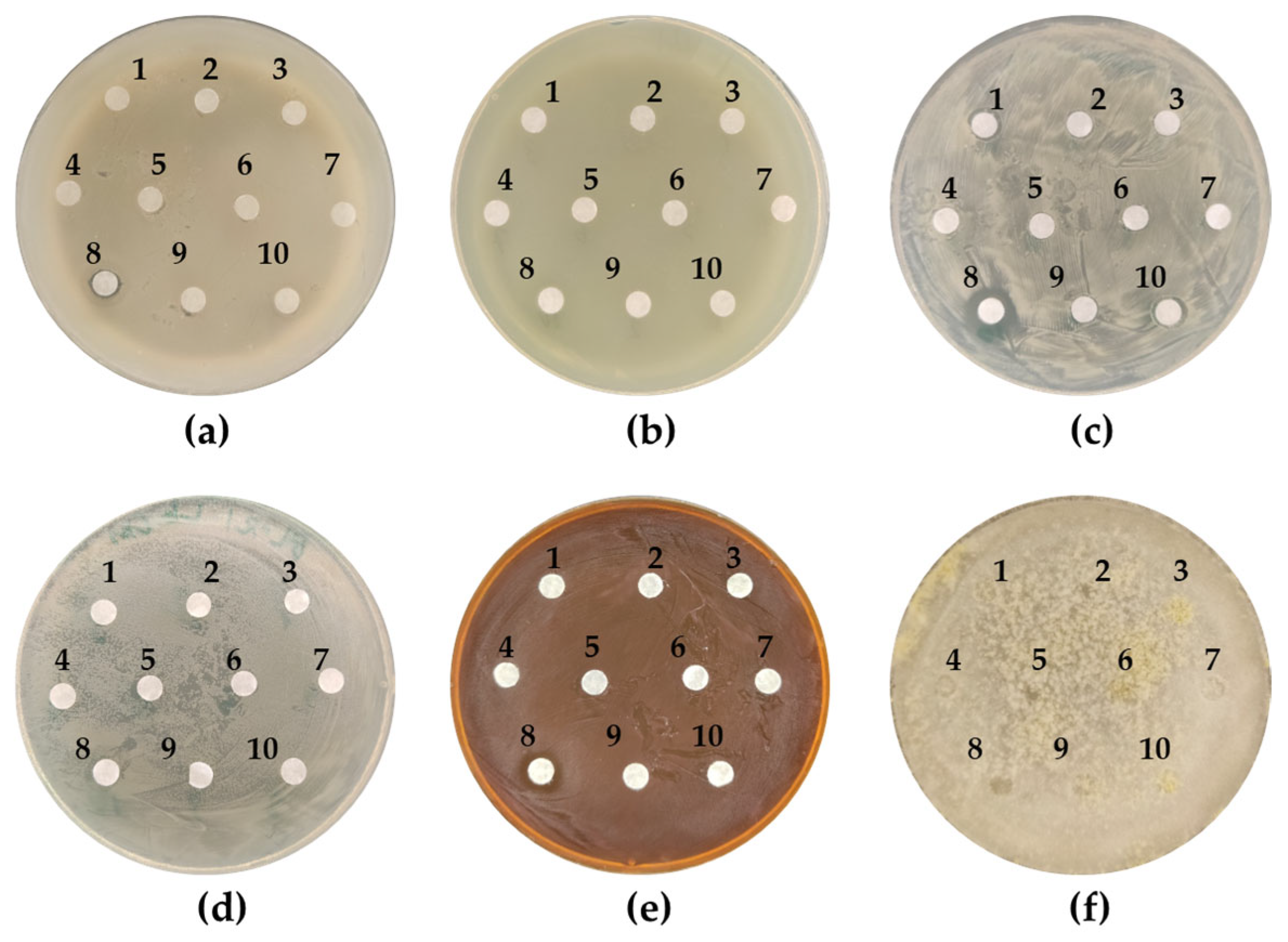
In experiments assessing the growth inhibitory effect, agent 3L10 demonstrated the formation of inhibition zones on lawns of B. subtilis MGMM36 (clear zone 1 mm), C. glutamicum MGMM638 (clear zone 4 mm), and L. plantarum MGMM126 (clear zone 3 mm) (Figure 2). All other agents showed no inhibitory effect against any of the tested strains.
The variation in inhibition zone diameters indicates that the suppression of growth in B. subtilis, C. glutamicum, and L. plantarum follows a concentration-dependent manner. B. subtilis seems to be inhibited only at high concentrations of 3L10, since a small-diameter halo was formed at the edge of the filter paper where the concentration of the defoamer is higher. C. glutamicum and L. plantarum formed bigger growth inhibition zones exhibiting sensitivity at lower concentrations of 3L10, taking into account that defoamer concentration is gradually decreasing along the diffusion gradient.

3.3. An Assessment of the Effect of Antifoam Agents on the Growth of the Tested Bacterial Strains

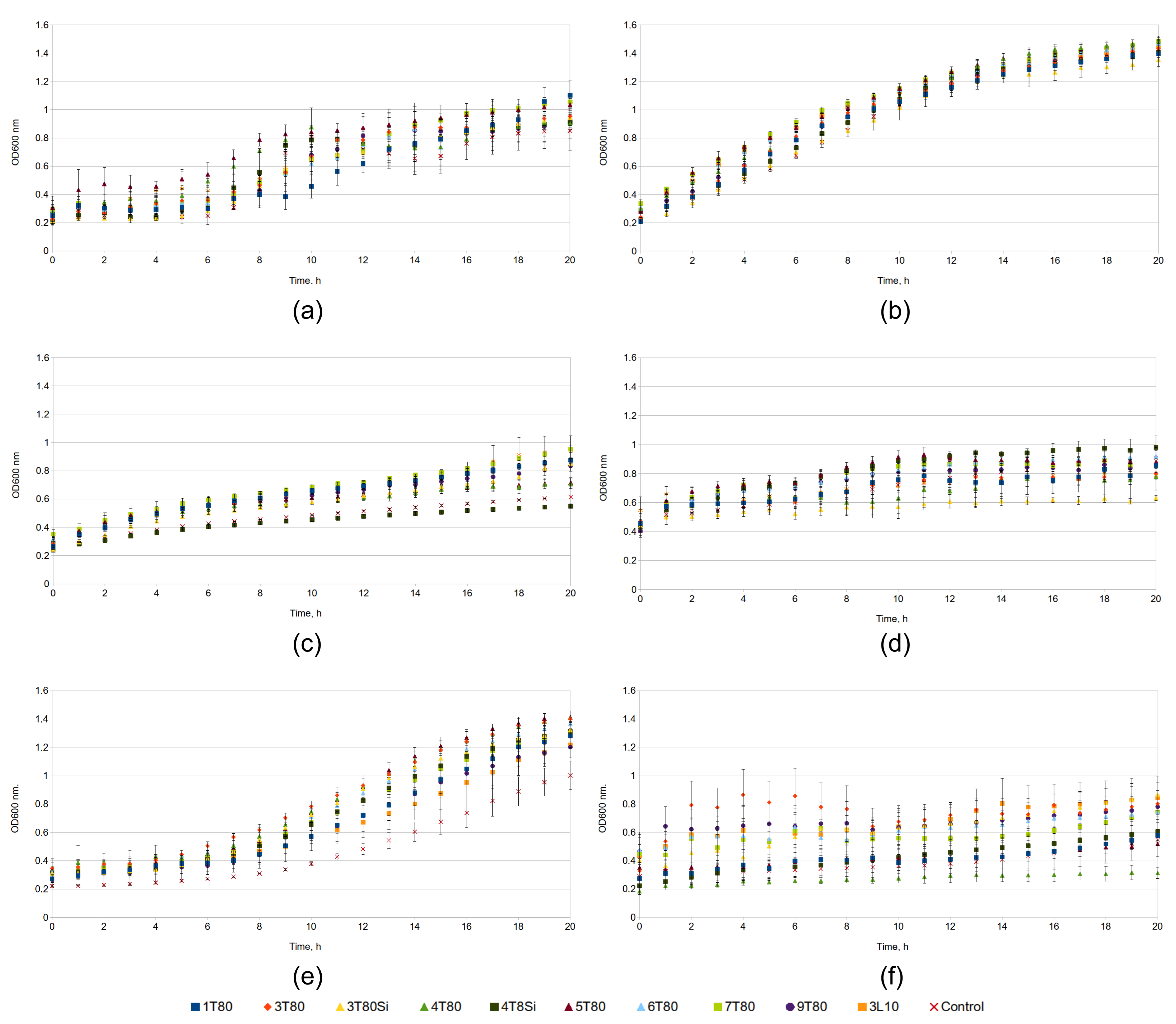
We also evaluated the effects of various agents at a fixed concentration (33.7 µg/mL) on microbial growth in liquid medium. Optical density was measured every 1 h over a 20 h period (Figure 3). This experiment demonstrated that the growth rates of P. fluorescens MGMM121 (Figure 3b) and E. coli BL21 (Figure 3d) remained largely unaffected by all tested agents. Conversely, we observed significant growth inhibition of C. glutamicum MGMM638 (Figure 3c) in the presence of the antifoam agent 3T80Si. This result contradicts the data obtained from the disk diffusion sensitivity test, where the agents were applied to a bacterial lawn using 3MM paper disks. In that test, the C. glutamicum strain showed no sensitivity to this agent (labeled as 3 in Figure 2c). A clear inhibitory effect on T. viride (Figure 3f) cells was demonstrated for antifoam agent 4T80. However, the optical density of this culture could not be measured reliably due to the plate wells being overgrown with T. viride mycelium, thus limiting data interpretation.
Additionally, this experiment demonstrated that some of the tested agents have a growth-promoting effect on L. plantarum MGMM126 and E. coli BL21.

3.4. The Determination of the Effective Concentration of Antifoams in a Conical Tube

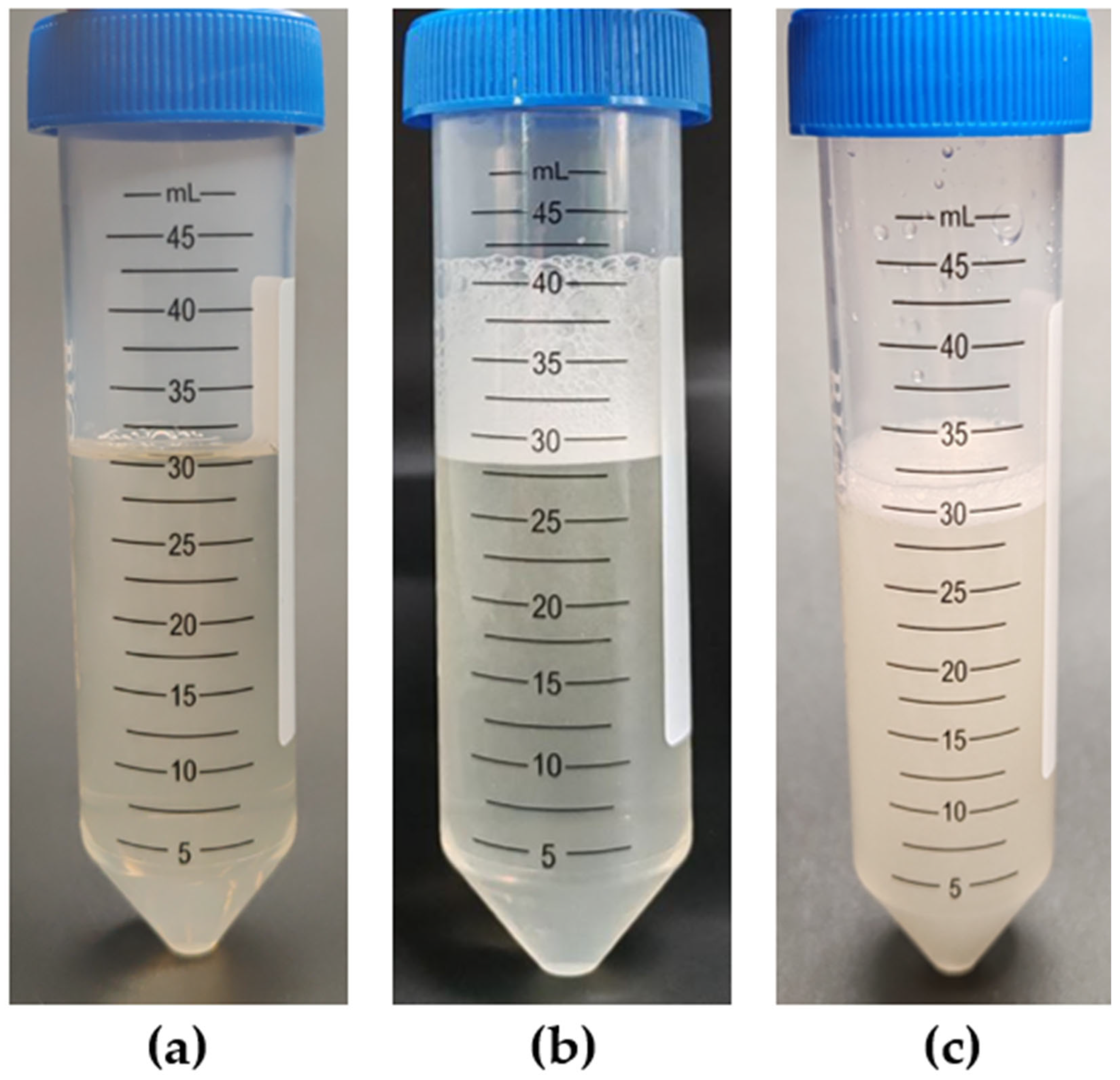
To assess the antifoaming (defoaming) properties of the agents, a shake test was employed using tubes containing the nutrient medium and the test compound. A representative result of this test is shown in Figure 4. When the nutrient medium in conical tubes (Figure 4a) was subjected to vigorous shaking, it generated a foam layer that occupied approximately one-third of the tube volume (Figure 4b). The addition of the defoamer markedly reduced this foam layer (Figure 4c).
The minimum concentration required to achieve complete foam suppression was defined as the minimum effective concentration (MEC) for each defoamer. The results of this experiment are summarized in Table 3.
As a reference, the addition of 80 µL of vegetable oil to 30 mL of medium (final concentration: 0.267%) was necessary to produce the foam suppression effect shown in Figure 4c. In contrast, the antifoam agents 1T80, 6T80, and 3L10 demonstrated the lowest MEC. A volume of 20 µL per 30 mL of medium (final concentration: 0.067%) was sufficient for these agents to suppress foam formation. Foam suppression required 40 µL (final concentration: 0.133%) for agents 3T80, 3T80-SiO2, 4T80, 4T80-SiO2, 5T80, and 9T80. Finally, agent 7T80 showed efficacy at a volume of 60 µL, resulting in a final concentration of 0.200%. The testing of 6T80 under different pH (5.0, 7.0, 9.0) and temperature (25 °C, 30 °C, 37 °C) conditions resulted in the same MEC. Based on these observations, antifoam 6T80 was chosen for subsequent use and scale-up to bioreactors. This selection was based on its absence of growth inhibitory effects on the test cultures and its low minimum effective concentration required for foam suppression.

3.5. Assessment of Effect of Antifoam Agents on Cultivation of B. subtilis MGMM38 Strain in Small Parallel Bioreactors

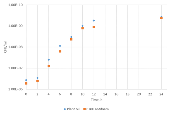
A cultivation experiment on the B. subtilis MGMM36 strain was conducted in parallel bioreactors with the addition of vegetable oil and an antifoam agent. As can be seen from Figure 5, no differences in CFU/mL values were observed throughout the entire fermentation process.
The final CFU titer after 24 h of incubation was similar under both conditions, at 2.69 × 10⁹ CFU/mL for plant oil and 2.39 × 10⁹ CFU/mL for the 6T80 antifoam agent.

3.6. Assessment of Antifoam Agent’s Impact on Glycerol Dehydrogenase Biosynthesis in Cells from P. putida LN6160 Strain as Model System

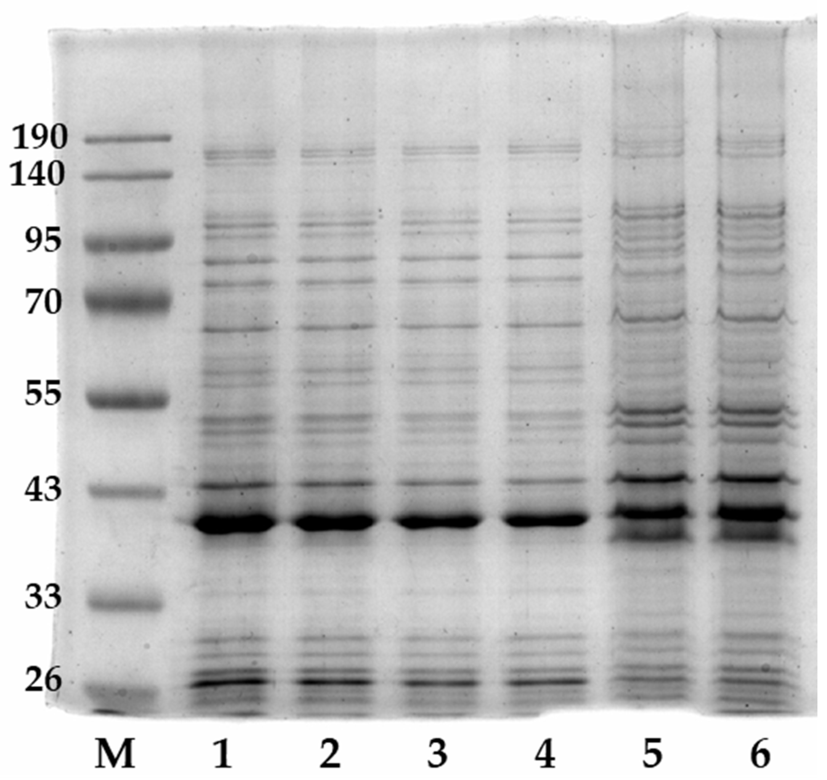
The experiments conducted to assess the impact of the antifoam agent on the protein synthesis process in bacteria using SDS-PAGE electrophoresis showed that the applied antifoam agent 6T80 does not exert an inhibitory or negative effect on the efficiency of target protein synthesis (Figure 6). The performed quantification of the synthesized glycerol dehydrogenase content in the total protein mixture of the cell lysate, as well as in the soluble and insoluble protein fractions, did not reveal statistically significant differences in the target protein content between samples obtained from cell suspensions cultured in the presence and in the absence of an antifoaming agent; the observed differences did not exceed the level of experimental error.

4. Discussion

The selection of an optimal antifoam is a critical step in the development and scale-up of fermentation processes. Despite their widespread use, many industrial antifoams exhibit detrimental side effects, including the inhibition of culture growth, induction of cellular stress, and reduction in productivity [20,29]. Notably, several cases also report a positive effect on the yield of target products, such as recombinant proteins. For instance, in the production of a fluorescent protein in P. pastoris cells, the addition of antifoams P2000, SB2121, and J673A doubled the yield of the synthesized protein [1]. This indicates a complex, system-dependent interaction between antifoams and cells and underscores the need for empirical screening and a physicochemical rationale for their action. Consequently, the development of novel formulations that combine high foam-suppressing efficacy with minimal impact on producer physiology remains highly relevant. In the present work, new silicone–polyol emulsions were created and subjected to comprehensive biological testing on a range of taxonomically diverse microorganisms. A key result of this study is the identification of selective antibacterial activity in one of the developed agents (3L10). This formulation exhibited pronounced inhibitory action against Gram-positive test strains (B. subtilis, L. plantarum, C. glutamicum) while showing no significant effect on the growth of Gram-negative bacteria (E. coli, P. fluorescens). The most sensitive culture was C. glutamicum, for which growth suppression was observed even at low agent concentrations [30]. This effect is likely due to fundamental differences in cell envelope structure. Gram-negative bacteria are protected by an outer membrane containing lipopolysaccharides, which serves as an effective barrier against many hydrophobic and amphiphilic compounds. In contrast, the single cytoplasmic membrane of Gram-positive bacteria is more accessible for interaction with amphiphilic molecules. C. glutamicum possesses a distinctive cell wall structure, containing mycolic acids that might form an external hydrophobic layer [31]. We hypothesize that the nonionic amphiphilic component L10, part of agent 3L10, can specifically interact with this lipid layer. This interaction likely disrupts the lipid bilayer’s organization, increases its permeability, and ultimately leads to growth inhibition or cell death. This specific selectivity makes agent 3L10 unsuitable for fermentations involving Gram-positive producers. However, this also indicates its potential value as a model compound for studying the mechanisms of interaction of surfactants with bacterial membranes. Although experiments with growth on 96-well plates revealed both the inhibitory and stimulating effects of certain agents (for example, on E. coli and L. plantarum), this analysis format has significant limitations associated with poor aeration and insufficient mixing of samples [32]. For further investigation, we determined the minimum effective concentrations (MECs) of defoamers necessary to completely suppress foaming under model conditions. For the most promising formulations, the MEC was approximately 0.067% (by volume). This parameter is key for subsequent process optimization, as it allows for the minimization of both potential toxic effects and negative impacts on oxygen mass transfer. High concentrations of antifoams, especially oil-based ones, can increase liquid viscosity and alter the surface tension at the phase interface, leading to a decrease in kLa and, consequently, limiting the oxygen supply to the cells [28]. This, in turn, can suppress culture growth and productivity. In this context, the low MEC (0.067% v/v) of agent 6T80, identified in our study, represents a significant practical advantage. To achieve the same level of foam suppression as with standard plant oil (MEC ≈ 0.267% v/v), approximately four times less volume of the active substance is required. This minimizes the amount of hydrophobic phase introduced into the system, potentially reducing interference with the gas exchange process [33].
Based on a comprehensive analysis, which considered the absence of inhibition in plate and microplate tests, a low MEC, and emulsion stability, agent 6T80 was selected for further study. Its neutrality toward cell physiology was confirmed in two model fermentation systems. First, in parallel cultivations of B. subtilis in laboratory mini-bioreactors, the dynamics of viable cell accumulation (CFU/mL) when using agent 6T80 showed no statistical difference from the control, where standard vegetable oil served as the defoamer. Second, in a heterologous expression system for glycerol dehydrogenase in P. putida, the addition of antifoam 6T80 had no negative effect on the accumulation level of the target recombinant protein, as evidenced by electrophoretic analysis.

5. Conclusions

This study demonstrates that the influence of antifoams on the fermentation process is multifaceted, being determined by both their chemical composition and the biological characteristics of the producer. Our developed approach, which combines the chemical design of emulsions, cytotoxicity screening that accounts for the taxonomic diversity of test cultures, the determination of MECs, and validation under conditions close to industrial scale, enables the rational selection of highly efficient and biocompatible formulations. The discovered selective activity of agent 3L10 underlines the necessity for the thorough preliminary testing of antifoams for the strains in question. Agent 6T80, which exhibited no inhibitory properties and demonstrated effectiveness in model fermentations, represents a promising candidate for further trials in targeted biotechnological processes utilizing both Gram-negative and Gram-positive bacteria.

Author Contributions

Conceptualization, N.V.T. and M.F.; methodology, M.F., N.V.T. and S.Z.V.; validation, T.A.L., N.V.T. and G.A.K.; formal analysis, M.F.; investigation, A.A.Z., E.A.V., L.A.V., M.F., T.A.L. and G.A.K.; resources, A.A.Z., E.A.V. and L.A.V.; data curation, N.V.T.; writing—original draft preparation, M.F., L.Y.Z. and N.V.T.; writing—review and editing, N.V.T. and S.Z.V.; visualization, T.A.L.; supervision, S.Z.V. All authors have read and agreed to the published version of the manuscript.

Funding

This research was funded by the government assignment for the FRC Kazan Scientific Center of RAS.

Institutional Review Board Statement

Not applicable.

Informed Consent Statement

Not applicable.

Data Availability Statement

The original contributions presented in this study are included in the article. Further inquiries can be directed to the corresponding authors.

Conflicts of Interest

The authors declare no conflicts of interest.

References

  1. Routledge, S.J. Beyond de-foaming: The effects of antifoams on bioprocess productivity. Comput. Struct. Biotechnol. J. 2012, 3, e201210014. [Google Scholar] [CrossRef] [Scilit]
  2. Gong, Z.; Yang, G.; Che, C.; Liu, J.; Si, M.; He, Q. Foaming of rhamnolipids fermentation: Impact factors and fermentation strategies. Microb. Cell Factories 2021, 20, 77. [Google Scholar] [CrossRef] [Scilit]
  3. Delvigne, F.; Takors, R.; Mudde, R.; Gulik, W.; Noorman, H. Bioprocess scale-up/down as integrative enabling technology: From fluid mechanics to systems biology and beyond. Microbal Biotech. 2017, 10, 1267–1274. [Google Scholar] [CrossRef] [Scilit]
  4. Vonester, D.; Hoffman, K.; Palmen, T.; Regestein, L.; Richter, U.; Altenoff, A.; Hoffman, M.; Radeva, Y.; Buchs, J.; Magnus, J.B. Foam Formation in Shake Flasks and Its Consequences. Eng. Life Sci. 2025, 25, e70057. [Google Scholar] [CrossRef] [Scilit]
  5. Global Fermentation Defoamer Market—Industry Trends and Forecast to 2028. Available online: https://www.databridgemarketresearch.com/reports/global-fermentation-defoamer-market (accessed on 20 December 2025).
  6. Takesono, S.; Onodera, M.; Toda, K.; Yoshida, M.; Yamagiwa, K.; Ohkawa, A. Improvement of foam breaking and oxygen-transfer performance in a stirred-tank fermenter. Bioprocess Biosyst. Eng. 2006, 28, 235–242. [Google Scholar] [CrossRef] [Scilit] [PubMed]
  7. Seidel, S.; Maschke, R.W.; Werner, S.; Jossen, V.; Eibl, D. Oxygen mass transfer in biopharmaceutical processes: Numerical and experimental approaches. Chem. Ing. Tech. 2021, 93, 42–61. [Google Scholar] [CrossRef] [Scilit]
  8. Rezaee, M.; Aider, M. Potential Use of Nonanimal-Based Biopolymers as Gelling/Emulsifying Stabilizing Agents to Reduce the Fat Content in Foods: A Review. ACS Food Sci. Technol. 2022, 2, 751–762. [Google Scholar] [CrossRef] [Scilit]
  9. Deng, B.; Neef, T.; Schroën, K.; de Ruiter, J. Mapping Bubble Formation and Coalescence in a Tubular Cross-Flow Membrane Foaming System. Membranes 2021, 11, 710. [Google Scholar] [CrossRef] [Scilit]
  10. Chen, J.; Huang, X.; He, L.; Luo, X. Foaming of Oils: Effect of Poly(dimethylsiloxanes) and Silica Nanoparticles. ACS Omega 2019, 4, 6502–6510. [Google Scholar] [CrossRef] [Scilit]
  11. Agarwal, A.; Liu, Y.A.; Dooley, L.; McDowell, C.; Thaysen, M. Large-Scale Industrial Fermenter Foaming Control: Automated Machine Learning for Antifoam Prediction and Defoaming Process Implementation. Ind. Eng. Chem. Res. 2022, 61, 5227–5238. [Google Scholar] [CrossRef] [Scilit]
  12. Blesken, C.C.; Bator, I.; Eberlein, C.; Heipieper, H.J.; Tiso, T.; Blank, L.M. Genetic cell-surface modification for optimized foam fractionation. Front. Bioeng. Biotechnol. 2020, 8, 572892. [Google Scholar] [CrossRef] [Scilit]
  13. Kumari, R.; Singha, L.P.; Shukla, P. Biotechnological potential of microbial bio-surfactants, their significance, and diverse applications. FEMS Microbes 2023, 4, xtad015. [Google Scholar] [CrossRef] [Scilit] [PubMed]
  14. Frolov, M.; Kungurov, G.A.; Valiakhmetov, E.E.; Gogov, A.S.; Trachtmann, N.V.; Validov, S.Z. Construction of the Pseudomonas putida Strain with Low Motility and Reduced Biofilm Formation for Application in Fermentation. Fermentation 2024, 10, 606. [Google Scholar] [CrossRef] [Scilit]
  15. Efthimiou, G.; Thumser, A.E.; Avignone-Rossa, C.A. A novel finding that Streptomyces clavuligerus can produce the antibiotic clavulanic acid using olive oil as a sole carbon source. J. Appl. Microbiol. 2008, 105, 2058–2064. [Google Scholar] [CrossRef] [Scilit] [PubMed]
  16. Andrede, C.J.; Coelho, A.L.S.; Feuser, P.E.; Andrade, L.M.; Carciofi, B.A.M.; Oliveira, D. Mannosylerythritol lipids: Production, downstream processing, and potential applications. Curr. Opin. Biotechnol. 2022, 77, 102769. [Google Scholar] [CrossRef] [Scilit]
  17. Tiso, T.; Demling, F.; Karmainski, T.; Oraby, A.; Eiken, J.; Liu, L.; Bongartz, P.; Wessling, M.; Desmond, P.; Schmitz, S.; et al. Foam control in biotechnological processes—Challenges and opportunities. Discov. Chem. Eng. 2024, 4, 2. [Google Scholar] [CrossRef] [Scilit]
  18. Zhao, Y.; Xue, C.; Ji, D.; Gong, W.; Liu, Y.; Li, Y. Microscopic Understanding of Interfacial Performance and Antifoaming Mechanism of REP Type Block Polyether Nonionic Surfactants. Molecules 2024, 29, 1816. [Google Scholar] [CrossRef] [Scilit]
  19. Zhang, T.; Sanguramath, R.A.; Israel, S.; Silverstein, M.S. Emulsion Templating: Porous Polymers and Beyond. Macromolecules 2019, 52, 5445–5479. [Google Scholar] [CrossRef] [Scilit]
  20. Nielsen, J.C.; Senne de Oliveira Lino, F.; Rasmussen, T.G.; Thykær, J.; Workman, C.T.; Basso, T.O. Industrial antifoam agents impair ethanol fermentation and induce stress responses in yeast cells. Appl. Microbiol. Biotechnol. 2017, 101, 8237–8248. [Google Scholar] [CrossRef] [Scilit]
  21. Etoc, A.; Delvigne, F.; Lecomte, J.P.; Thonart, P. Foam Control in Fermentation Bioprocess. In Twenty-Seventh Symposium on Biotechnology for Fuels and Chemicals; McMillan, J.D., Adney, W.S., Mielenz, J.R., Klasson, K.T., Eds.; ABAB Symposium; Humana Press: Totowa, NJ, USA, 2006. [Google Scholar] [CrossRef] [Scilit]
  22. Rezende, D.A.; Bittencourt, R.R.; Mansur, C.R.E. Evaluation of the efficiency of polyether-based antifoams for crude oil. J. Pet. Sci. Eng. 2011, 76, 172–177. [Google Scholar] [CrossRef] [Scilit]
  23. Ghildyal, N.P.; Lonsane, B.K.; Karanth, N.G. Foam control in submerged fer-mentation: State of the art. Adv. Appl. Microbiol. 1988, 33, 173–222. [Google Scholar] [CrossRef] [Scilit]
  24. Vasilieva, E.A.; Vasileva, L.A.; Zagidullin, A.A.; Davlitova, E.M.; Valeeva, F.G.; Krylov, F.D.; Ardabevskaia, S.N.; Milenin, S.A.; Nizameev, I.R.; Zakharova, L.Y. Structure–property correlation of amphiphilic alkylthio derivatives of carboxylic acids: From self-assembly to functional activity. Surf Internet 2025, 77, 107996. [Google Scholar] [CrossRef] [Scilit]
  25. Jeske, M.; Altenbuchner, J. The Escherichia coli rhamnose promoter rhaP BAD is in Pseudomonas putida KT2440 independent of Crp–cAMP activation. Appl. Microbiol. Biotechnol. 2010, 85, 1923–1933. [Google Scholar] [CrossRef] [Scilit]
  26. Koczo, K.; Leatherman, M.D.; Wylde, J.J. Chapter 4: Foam Control. In Surface Process, Transportation, and Storage; Wang, Q., Ed.; Oil and Gas Chemistry Management Series; Gulf Professional Publishing: Houston, TX, USA, 2023; Volume 4, pp. 153–226. [Google Scholar] [CrossRef] [Scilit]
  27. Arnaudov, L.; Denkov, N.D.; Surcheva, I.; Durbut, P.; Broze, G.; Mehreteab, A. Effect of oily additives on foamability and foam stability. 1. Role of interfacial properties. Langmuir 2001, 17, 6999–7010. [Google Scholar] [CrossRef] [Scilit]
  28. Taddeo, F.; Vitiello, R.; Ruocco, M.; Turco, R.; Russo, V.; Tesser, R.; Di Serio, M. Synthesis of new defoamer agents and characterization of cementitious formulations. Heliyon 2024, 10, e33164. [Google Scholar] [CrossRef] [Scilit] [PubMed]
  29. Kawase, Y.; Moo-Young, M. The effect of antifoam agents on mass transfer in bioreactors. Bioprocess Eng. 1990, 5, 169–173. [Google Scholar] [CrossRef] [Scilit]
  30. Worch, K.; Krause, M.; Burse, A. Effects of antifoam agents on Spodoptera frugiperda 9 cell growth and baculovirus infection dynamics. J. Biol. Eng. 2025, 19, 43. [Google Scholar] [CrossRef] [Scilit] [PubMed]
  31. Lanéelle, M.A.; Tropis, M.; Daffé, M. Current knowledge on mycolic acids in Corynebacterium glutamicum and their relevance for biotechnological processes. Appl. Microbiol. Biotechnol. 2013, 97, 9923–9930. [Google Scholar] [CrossRef] [Scilit]
  32. Neuss, A.; Steimann, T.; Tomas Borges, J.S.; Dinger, R.; Magnus, J.B. Scale-up of CHO cell cultures: From 96-well-microtiter plates to stirred tank reactors across three orders of magnitude. J. Biol. Eng. 2025, 19, 5. [Google Scholar] [CrossRef] [Scilit]
  33. Rols, J.L.; Condoret, J.S.; Fonade, C.; Goma, G. Mechanism of enhanced oxygen transfer in fermentation using emulsified oxygen-vectors. Biotechnol. Bioeng. 1990, 35, 427–435. [Google Scholar] [CrossRef] [Scilit]
Figure 1. The structures of the compounds used. The structural formulas of Tween80 and Laureth-10.
Fermentation 12 00078 g001
Figure 2. Inhibition of microbial growth on Petri dishes: (a) B. subtilis MGMM36, (b) P. fluorescens MGMM121, (c) C. glutamicum MGMM638, (d) E. coli BL21, (e) L. plantarum MGMM126, (f) T. viride MGMMF32. 1. 1T80; 2. 3T80; 3. 3T80Si; 4. 4T80; 5. 4T80Si; 6. 5T80; 7. 6T80; 8. 3L10; 9. 9T80; 10. 7T80.
Fermentation 12 00078 g002
Figure 3. The growth curves of the different strains in the presence of antifoam agents (a) B. subtilis MGMM36, (b) P. fluorescens MGMM121, (c) C. glutamicum MGMM638, (d) E.coli BL21, (e) L. plantarum MGMM126, (f) T. viride MGMMF32.
Fermentation 12 00078 g003
Figure 4. The foam volume of (a) LB nutrient medium, (b) foam formation upon shaking, and (c) foam formation upon shaking with the added antifoam agent.
Fermentation 12 00078 g004
Figure 5. The growth curve of the B. subtilis strain in parallel small-volume bioreactors in LB medium. The X-scale shows the absolute amount of live cells in mL (CFU/mL).
Fermentation 12 00078 g005
Figure 6. Staining 12% SDS-PAGE gel with Coomassie Brilliant Blue R250. M–Blue Plus® V Protein Marker (10–190 kDa) (TransGen Biotech, Beijing, China). Fractions of P. putida LN6160:pJeM2:gld: 1—P. putida LN6160:pJeM2:gld lysate (with plant oil); 2—P. putida LN6160:pJeM2:gld lysate (with antifoam 6T80); 3—P. putida LN6160:pJeM2:gld supernatant (with plant oil); 4—P. putida LN6160:pJeM2:gld supernatant (with antifoam 6T80); 5—P. putida LN6160:pJeM2:gld cell debris (with plant oil); 6—P. putida LN6160:pJeM2:gld cell debris (with antifoam 6T80).
Fermentation 12 00078 g006
Table 1. The component ratios for the different formulations.
CompositionWaterTw80PDMS2002-HDolD3D4D5D6DSiO2
wt%
1T802510103520-----
3T80-20----
3T80-SiO230-----1
4T8010-20----
4T80-SiO2-20---1
5T80--20---
6T8025101035--20--1
7T80---20--
9T80----20-
3L102510 (L-10)1035-20----
Table 2. Strains used in experiment.
StrainCultivation MediumCultivation ConditionsSource Link
Escherichia coli BL21Luria–Bertani37 °C[17]
Corynebacterium glutamicum MGMM638King’s B 30 °C[18]
Trichoderma viride MGMMF32King’s B30 °C[19]
Pseudomonas fluorescens MGMM121King’s B25 °C[20]
Pseudomonas putida LN6160King’s B25 °C[12]
Bacillus subtilis MGMM36King’s B30 °C[21]
Lactiplantibacillus plantarum MGMM126MRS 37 °C[22]
Table 3. Minimum effective concentration (MEC) of defoamer to suppress foam formation.
Antifoam AgentAdded Volume, µLConcentration in Solution, % (v/v)
Plant oil800.267
1T80200.067
3T80400.133
3T80-SiO2400.133
4T80400.133
4T80-SiO2400.133
5T80400.133
6T80200.067
7T80600.200
9T80400.133
3L10200.067
Disclaimer/Publisher’s Note: The statements, opinions and data contained in all publications are solely those of the individual author(s) and contributor(s) and not of MDPI and/or the editor(s). MDPI and/or the editor(s) disclaim responsibility for any injury to people or property resulting from any ideas, methods, instructions or products referred to in the content.