Article

Isolation and Characterization of Low-Temperature and High-Salinity Amylase from Halomonas sp. KS41843

1
Division of Life Sciences, Korea Polar Research Institute, Incheon 21990, Republic of Korea
2
Department of Marine Science, College of Natural Sciences, Incheon National University, Incheon 22012, Republic of Korea
3
Department of Polar Sciences, University of Science and Technology, Daejeon 34113, Republic of Korea
*
Author to whom correspondence should be addressed.
These authors contributed equally to this work.
Fermentation 2025, 11(8), 465; https://doi.org/10.3390/fermentation11080465
Submission received: 19 June 2025 / Revised: 8 August 2025 / Accepted: 11 August 2025 / Published: 13 August 2025
(This article belongs to the Section Microbial Metabolism, Physiology & Genetics)

Abstract

The polar regions harbor uniquely diverse organisms adapted to low temperatures. Strains obtained from these regions are likely to produce enzymes that are industrially useful at low temperatures. In this study, a Halomonas sp. strain isolated from the Antarctic Sea was cultured at 15 °C to obtain partially purified α-amylase. This enzyme exhibited maximum activity at 30 °C and pH 5.0, retaining over 80% of its maximum activity even at 5 °C. Its activity was >1.5-fold in the presence of Co2+, Mn2+, Mg2+, Fe2+, and Na+, indicating enhancement by most metal ions. Halophilic strain-derived enzyme maintained up to 95% of its maximum activity even at 4 M NaCl, highlighting its potential for industrial applications and possible cost savings. In this study, the low-temperature and high-salinity active amylase produced by Antarctic Halomonas sp. KS41843 was identified as a promising candidate for future biotechnology applications.

1. Introduction

Starch, one of the most abundant storage carbohydrates, has been widely used in industries as a cost-effective and renewable raw material [1]. It typically consists of 20–25% amylose and 75–80% amylopectin [2]. Amylose is a linear polysaccharide of α-D-glucose units linked by α-1,4-glycosidic bonds, whereas amylopectin is a highly branched polysaccharide with α-1,4-linked glucose chains branched by α-1,6-linkages [3,4]. Starch is primarily broken down into smaller molecules by enzymes and is subsequently used in various processes. Amylase is the primary enzyme that hydrolyzes starch into sugars. All amylases are classified as glycoside hydrolases that target α-1,4-glycosidic bonds. α-Amylase randomly breaks down long-chain carbohydrates along the starch chain, producing maltotriose and maltose from amylose, and glucose, maltose, and limit dextrins from amylopectin [5].
In industrial and biotechnological processes, cold-active enzymes can significantly reduce energy costs owing to structural adaptations that confer greater molecular flexibility than that of mesophilic and thermophilic enzymes [6,7]. The polar regions are characterized by low temperatures, summer ultraviolet exposure, low nutrient availability, and freeze–thaw cycles [8,9]. Microorganisms are the dominant life forms in polar regions and have several adaptive strategies that support their survival [10]. Owing to their ecological role as decomposers, these microorganisms likely produce cold-active enzymes of industrial interest [11,12]. These enzymes can exhibit up to 10-fold greater activity at low temperatures than that of mesophilic enzymes [13]. The Antarctic bacteria demonstrate amylolytic activity that facilitates the hydrolysis of complex polysaccharides derived from phytoplankton, macroalgae, and particulate organic detritus found in polar marine environments. This hydrolytic capability is essential for utilizing available carbon sources and supports microbial food webs in nutrient-limited, low-temperature habitats [14].
Salt-active amylases, which retain enzymatic activity under hypersaline conditions, are considered highly valuable for industrial applications owing to their stability and functionality in environments where conventional enzymes typically denature or become inactive. These enzymes are of particular interest in industries such as food processing, pulp and paper manufacturing, and pharmaceuticals, where high-salt raw materials or process conditions are commonly encountered. Consequently, the development and application of salt-tolerant and salt-activated amylases have emerged as an active area of research [15,16]. Previous studies have demonstrated that microorganisms isolated from hypersaline environments—particularly halophilic niches, such as marine habitats and salt lakes—exhibit enhanced halotolerance compared to non-halophilic microbes. The amylolytic enzymes produced by these halophilic microorganisms often have robust catalytic activity under elevated NaCl concentrations. Notably, halophilic genera such as Halomonas, Bacillus [17], Marinobacter [18,19], and Chromohalobacter [20] reportedly produce salt-active amylases. These enzymes often maintain their activity, or in some cases, enhance, their activity when exposed to ≥1 M NaCl, with certain enzymes demonstrating functional stability even at salt concentrations as high as 3–4 M [21,22,23]. In contrast to conventional enzymes, for which tertiary structures are destabilized under high-salinity conditions, salt-active amylases exhibit remarkable structural resilience. This stability is primarily attributed to an increased abundance of surface-exposed, negatively charged amino acid residues and highly hydrated side chains, which collectively mitigate salt-induced denaturation. Furthermore, enzyme activity in hypersaline environments reportedly contribute to stabilization of the tertiary structure and modulation of the hydrophilicity of substrate-binding regions, thereby enhancing catalytic performance under extreme conditions [24,25,26].
To identify novel low-temperature-active and high-salinity-tolerant amylases with diverse structures and properties, we screened bacterial strains isolated from Antarctic Ocean samples collected over several years. This study compared the amylase productivity of these strains and characterized the partially purified enzyme from Halomonas sp. KS41843—that was selected as the most productive strain.

2. Materials and Methods

2.1. Amylase-Producing Bacteria Screening and Identification

A total of 687 colonies were isolated from samples obtained by filtering seawater (0.2 μm pore size; Membrane filter porafil®, Macherey-Nagel, Düren, Germany) collected near the King Sejong Station in Antarctica. To identify cold-active amylase-producing bacteria, the colonies were incubated at 15 °C for 72 h on agar plates containing 10 g/L soluble starch (Junsei, Kyoto, Japan) in Zobell medium (0.5% [w/v] peptone, and 0.1% [w/v] yeast extract in 75% [v/v] sea water). Subsequently, six bacterial strains exhibiting amylase activity were identified using Lugol’s solution [27]. These strains were further distinguished using universal primers 27-F ([AGAGTTTGATCCTGGCTCAG] and 1492-R [GGTTACCTTGTTACGACTT]) and identified using Macrogen© (Macrogen Online Sequencing Order System, Seoul, Republic of Korea).

2.2. Amylase Production and Activity Quantification of Six Strains

The selected bacteria were cultured in Zobell medium at 15 °C for 48 h and used as seeds. The initial cell concentration was adjusted to OD600 = 0.01, and the amylase activity in the cell culture broth was measured after 72 h of incubation in Zobell medium containing 10 g/L soluble starch at 15 °C. The culture solution was centrifuged (9000× g, 30 min), supernatant was collected, and amylase activity was measured. To quantify amylase activity, 0.1 mL of each supernatant sample (enzyme solution) was added to 0.45 mL of phosphate-buffered saline (PBS) with 0.05 mL of 1% soluble starch. The mixture was then incubated at 15 °C for 1 h. The reaction was terminated by adding 0.6 mL of 3,5-dinitrosalicylic acid (DNS) reagent and boiling for 10 min. Absorbance was measured at OD580 using a spectrophotometer (Evolution 350; Thermo Fisher Scientific, Waltham, MA, USA). One unit of enzyme activity per milligram (U mg−1) was defined as the amount of enzyme that produced 1 μmol of reducing sugar per minute per milligram of protein. The protein concentration was determined by performing the Bradford assay with a protein assay reagent (5000006, Bio-Rad, Hercules, CA, USA), and bovine serum albumin was used as a standard [28]. For comparison, Amplify® Prime 100 L (Novozyme, Bagsvaerd, Denmark), a commercial amylase, was used as the standard.

2.3. Liquid Culture of Halomonas sp. KS41843

Halomonas sp. KS41843 was selected from six amylase-producing bacteria and cultured in a liquid medium. A seed culture was prepared by inoculating one colony from an agar plate into Zobell medium and incubating at 15 °C for 48 h. For the primary culture, the seed culture was inoculated into Zobell medium containing 10 g/L soluble starch based on an initial cell concentration of OD600 = 0.01. Cell growth and amylase production were monitored over time while culturing the sample at 15 °C, with 120 rpm of shaking, for 132 h. To assess the effect of salt on the halophilic strain Halomonas sp. KS41843, it was cultured in Zobell medium (1% [w/v] soluble starch, 0.5% [w/v] peptone, and 0.1% [w/v] yeast extract in 75% [v/v] sea water) supplemented with 0, 20, 40, and 60 g/L NaCl, resulting in total NaCl concentrations of 20, 40, 60, and 80 g/L, respectively, for 72 h. Thereafter, the cell density and enzyme activity were assessed.

2.4. Purification of Amylase

The target strain Halomonas sp. KS41843 was cultured at 15 °C and 120 rpm for 72 h. The culture was centrifuged (4 °C, 9000× g, 20 min) to obtain the supernatant for protein purification (RC-5C Plus; Sorvall, Waltham, MA, USA). The supernatant was desalted and buffer-exchanged (100 mM Tris-HCl, pH 7.0) using a 10 kDa molecular weight cut-off membrane (Z615366; Sartorius, Göttingen, Germany). The sample was freeze-dried for 3 days, and distilled water was added to adjust the protein concentration to 20 mg/mL. The crude enzyme was loaded onto a 5 mL prepacked ion-exchange column (Diethylaminoethyl [DEAE] column, Bio-Rad, USA). The enzyme was eluted at a flow rate of 2 mL/min using buffer A (100 mM Tris-HCl [pH 7.0]), buffer B (100 mM Tris-HCl [pH 7.0], 1 M NaCl), with a gradual increase in the proportion of buffer B. To increase the purity, the active fraction was loaded into a Superdex 75 (GE, Chicago, IL, USA) column and eluted at a flow rate of 0.4 mL/min with 100 mM Tris-HCl (pH 7.0) buffer. All processes were conducted at 4 °C. The active fraction was collected and concentrated using Vivaspin 20 (10 kDa cut-off) (GE, USA).
The molecular weight and purity of the starch-decomposing enzyme were confirmed using sodium dodecyl sulfate-polyacrylamide gel electrophoresis (SDS-PAGE). The samples were treated at 95 °C for 5 min and separated using a 4–15% polyacrylamide gel (4561084; Bio-Rad, USA). Protein bands were visualized after staining the gel with the EZ-Gel staining solution (DoGenBio, Seoul, Republic of Korea) [29]. The proteins separated via SDS-PAGE were transferred onto a PVDF (polyvinylidene fluoride) membrane at 100 V for 60 min. Prior to transfer, the PVDF membrane was activated by soaking it in 100% methanol for 1 min. The membrane was stained with Ponceau-S (0.1%) [30].
For zymography, non-heat-treated broth samples were separated using a 4–15% SDS-PAGE gel, and electrophoresis was performed at 4 °C, after which, the gel was washed with 50 mM Tris buffer. The gel was placed onto a 15% [w/v] agar plate containing 1% [w/v] soluble starch and incubated at 15 °C overnight. After incubation, the agar plate was stained with Lugol’s solution (iodine–potassium iodide solution). Enzyme activity was determined based on the presence of clear unstained zones, indicating starch degradation [31].

2.5. Characterization of Purified Amylase Enzyme

2.5.1. Effect of Temperature

Enzyme activity was assessed at 5–10 °C intervals from 5 to 70 °C. At each temperature, 5 μL of enzyme (0.143 μg protein), 50 μL soluble starch (1% w/v), and 545 μL PBS were incubated for 30 min, and the enzyme activity was measured. To assess thermal stability, the enzyme was pre-incubated at a specific temperature for 1 h, after which the substrate and buffer were added, and the reaction was performed at 25 °C for 30 min. Subsequently, 600 μL of DNS solution was added, boiled for 10 min, and the absorbance was measured at 580 nm to quantify reducing sugars.

2.5.2. Effect of pH

Enzyme activity was measured at 1-unit intervals at pH values ranging from 4.0 to 10.0. The reaction mixture contained 5 μL enzyme solution, 50 μL soluble starch (1% w/v), and 545 μL buffer for each pH (sodium acetate buffer for pH 4.0–6.0 and Tris-HCl buffer for pH 6.0–10.0) that was incubated at 25 °C for 30 min. To assess pH stability, the enzyme was incubated at pH values ranging from 4.0 to 10.0 for 1 h at 25 °C, followed by activity measurement.

2.5.3. Effect of Metal Ions

To assess the effect of metal ions on enzyme activity, various salts (CoCl2, MnCl2, CuSO4, CaCl2, ZnSO4, BaSO4, MgSO4, FeSO4, and Na2SO4) were added to the reaction solution [32] at a final concentration of 1 mM. Enzyme activity was measured, with the relative activity in the absence of metal ions defined as 100% for comparison.

2.5.4. Effect of NaCl

To assess the effect of salt concentration on enzyme activity, NaCl was added to the reaction mixture in 0.5 M intervals from 0 to 4.0 M, and the enzyme activity was measured.

3. Results

3.1. Identification and Selection of Bacterial Strains Capable of Enzyme Production

Table 1 presents the 16S rRNA identification results for the six strains exhibiting high amylase activity. These included two Alteromonas sp. species, two Halomonas sp. species, one Pseudoalteromonas sp., and one Subtercola sp. species. Among the six strains cultured in liquid medium, KS41843 exhibited the highest enzyme activity at 36.3 U/mg (Figure 1) and was selected for further investigation.

3.2. Effect of Salt Concentration on Cell Growth and Amylase Production by KS41843

Halomonas sp. KS41843 was cultured at 15 °C for 132 h, and the cell concentration and enzyme activity were measured. Cell growth increased until 90 h after inoculation, followed by a decline (Figure 2A). Enzyme activity started to increase rapidly at the beginning of culture and continued to increase until 90 h, after which, it sharply declined. The effects of NaCl on cell growth and amylase production were assessed. Because the Zobell medium contains seawater, it initially includes approximately 20 g/L NaCl. Additional NaCl was added at concentrations of 0, 20, 40, and 60 g/L. Cell growth and enzyme activity were highest at 20 g/L NaCl. Even at 40 g/L, the enzyme activity remained above 80% of the maximum value (Figure 2B).

3.3. Production and Purification of Amylase KS41843

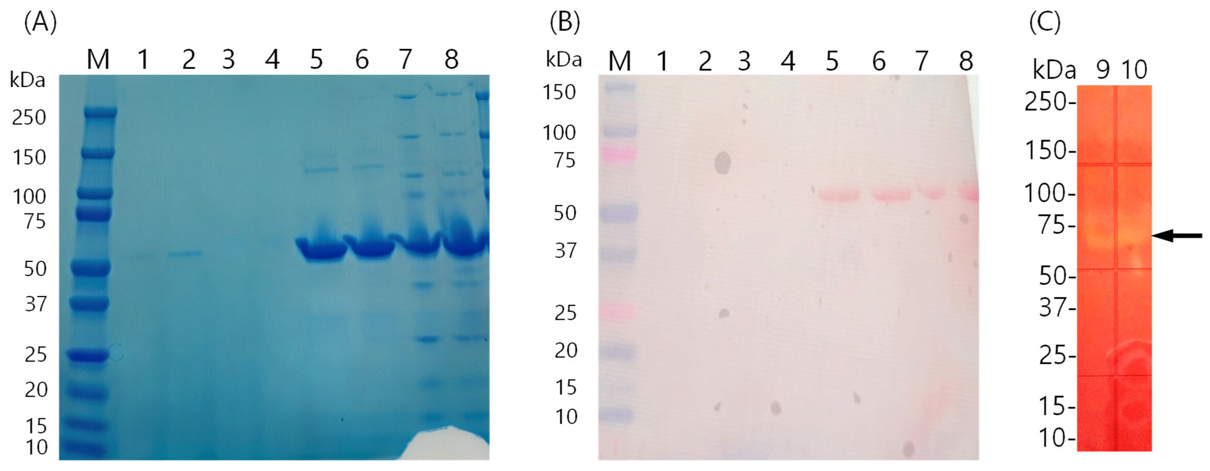
The culture broth was collected for enzyme purification. The cell culture medium was concentrated using a 10 kDa molecular weight cut-off membrane, and the primary active fraction was isolated using an ion-exchange column (diethylaminoethyl [DEAE] column). This sample was further separated using size-exclusion chromatography (Superdex 75) to obtain a partially purified sample (Figure 3). Although some loss occurred during concentration, the specific activity of the enzyme obtained after the DEAE column increased by 2-fold. Finally, the enzyme was purified using Superdex 75, resulting in a specific activity of 110.32 U/mg, 9.35-fold purification, and 6.03% yield. Table 2 summarizes the detailed results of each purification step. SDS-PAGE and zymography analyses indicated that KS41843 amylase has a molecular weight of approximately 60 kDa (Figure 3).

3.4. Effect of Temperature and pH on Amylase Activity and Stability

KS41843 amylase exhibited the highest activity at 30 °C, with activity gradually decreasing above 40 °C (Figure 4A). The enzyme retained over 95% of the maximum activity between 15 and 30 °C, and 80% at 5 °C. The commercial enzyme Amplify exhibited maximum activity at 70 °C, with reduced activity at lower temperatures. At 25 °C, the absolute activity was comparable to that of KS41843 amylase, but KS41843 amylase exhibited higher activity at 5 °C. The enzyme retained over 80% of its activity from 5 to 25 °C and maintained 60% of its maximum activity at 70 °C (Figure 4B).
Figure 4C,D illustrate the results for optimal pH. KS41843 amylase exhibited the highest activity at pH 5.0 and was inactivated at pH ≤ 3.0. Under basic alkaline conditions using Tris buffer, its activity gradually declined. In contrast, the commercial Amplify enzyme exhibited optimal activity at pH 8.0. The stability was maintained at >80% between pH 5 and 7, and reduced to <60% at pH < 5.0 or >8.0. In summary, KS41843 amylase exhibited optimal activity at pH 5.0 and remained stable for 1 h within the pH range of 5.0–7.0—its activity was reduced under alkaline conditions, indicating sensitivity to high pH.

3.5. Effect of Metal Ions on Amylase Activity

Amylase activity increased when exposed to ions, demonstrating > 1.5-fold enhancement in the presence of Co2+, Mn2+, Mg2+, Fe2+, and Na+ (Figure 5). These results demonstrate that KS41843 amylase activity can be affected by specific metal ions. The effect of metal ions on amylase activity has been reported to vary based on the microbial source.

3.6. Salt Tolerance of Amylase

To assess the effect of salinity on amylase activity, the relative activity of the enzyme was measured in buffers containing 0–4 M NaCl. KS41843 amylase exhibited maximum activity at 2.5 M NaCl and remained almost constant up to 4 M, whereas the control exhibited maximum activity at 0.5 M, followed by a rapid decline (Figure 6).

4. Discussion

We isolated cold-active amylase-producing bacterial strains from the Antarctic Sea and identified the most promising strain as halophilic. Additionally, we characterized its amylase production and assessed its stability under varying temperatures, pH values, metal ion concentrations, and salinity conditions.
Halophilic bacteria belonging to the genus Halomonas are known for their ability to survive at high salt concentrations [33]. These species are primarily isolated from saline habitats, including solar salt facilities, industrial brine solutions, intertidal estuaries, open oceans, and high-salinity lakes [34]. Studies on halophiles are ongoing, with researchers consistently identifying innovative methods to use their distinctive traits for technological advancements [35,36]. Halomonas species are highly halophilic, and several strains can survive in environments with NaCl concentrations ranging from 0.1 to 32.5% [37].
KS41843 secreted substantial amounts of amylase during the early stages of cultivation, presumably to hydrolyze starch and thereby secure a readily utilizable carbon source. This phenomenon was also observed by Rathour et al. [38], whereas Rathod et al. [39] reported a pattern in which amylase activity increased and subsequently decreased in parallel with cell growth. Notably, a marked decline in amylase activity was observed when cell growth plateaued and the carbon sources were depleted. The sharp decrease in activity observed during the death phase can be attributed to substrate exhaustion, increased cell death, and proteolytic degradation. These findings highlight the need for comprehensive future investigations, particularly in terms of regulating amylase-encoding gene expression.
KS41843 exhibited a maximum activity of 110.3 U/mg, higher than that of the cold-adapted α-amylase derived from Pseudoalteromonas sp. 2-3 (51.7 U/mg) [40] or Bacillus sp. dsh19-1 (16.4 U/mg) [26], but lower than that of the low-temperature active amylase derived from Alteromonas sp. KS7913 (200.34 U/mg) [27] or Zunongwangia profunda (284.9 U/mg) [41]. SDS-PAGE and zymography analyses indicated that KS41843 amylase had a molecular weight of 60 kDa—consistent with previous findings (56 kDa [42], 57 kDa [43], 50.1 kDa [26], 55 kDa [44], 55 kDa [45], and 66 kDa [41])—reporting amylase sizes in the range of 50–66 kDa (Table 3).
KS41843 amylase retained > 80% of its maximum activity even at 5 °C. Compared with other Antarctic amylase-producing bacteria, such as Arthrobacter sp. and Carnobacterium iners, the enzymatic activity at 5 °C was less than one-third that of KS41843 [46]. It exhibited greater activity at low temperatures than the cold-active enzyme derived from the Arctic strain Alteromonas sp. KS7913 that retained 68% of its maximum activity at 5 °C [27]. At 5 °C, Bacillus sp. dsh19-1, Zunongwangia profunda, Pseudoalteromonas sp. M175, and Microbacterium foliorum GA2 exhibited activity levels not exceeding 40%, 50%, 60%, and 10% of their maximum capacities, respectively [26,41,47,48].
Table 3. Enzymatic characteristics of KS41843 and other cold-active amylases.
EnzymeMicrobial SourceOptimum Temperature (°C)Optimum pHSpecific Activity (U/mg)Molecular Weight (kDa)Cations ActivatorsReference
KS41843Halomonas sp. KS41843305.0110.3260Co2+, Mn2+, Mb2+, Fe2+, Na+This study
AmyD-1Bacillus sp. dsh19-1206.016.450.1Na+, Ca2+[26]
KS7913Alteromonas sp. KS7913257.0200.3470Mn2+, Ba2+, Ca2+[27]
α-AmylasePseudoalteromonas sp. 2-3208.051.768.8Ca2+[40]
AmyZZunongwangia profunda357.0284.466Sr2+, Fe3+, Mg2+, Ba2+, NH4+, K+[41]
Ef-Amy IEisenia fedida405.517457-[43]
Ef-Amy IIEisenia fedida355.06557Ca2+[43]
amylaseNocardiopsis sp. 732635854855Ca2+, Mn2+, Mg2+, Cu2+, Co2+[44]
ParAmyPseudoalteromonas
arctica GS230
307.525.555Mn2+, K+, Na+[45]
GA2Microbacterium foliorum209--Mg2+[47]
GA6Bacillus cereus2010--Ca2+[47]
wtAmy175Pseudoalteromonas sp. M175307.5289.7961-[48]
Although previous studies have reported various results, KS41843 amylase exhibited optimal activity at a slightly acidic pH of 5. AmyD-1 exhibited maximum activity at pH 6 and remained stable between pH 5–7 [26]. In contrast, AmyZ1 from Pontibacillus sp. ZY exhibited maximum activity at pH 7.0 [1]. Amylases from the halotolerant strain Halomonas sp. Y2 and halophilic Bacilli (AT2RP4, HL1RS13, NRS4HaP9, and LK3HaP7) exhibited maximum activity at pH 8 [49,50]. Additionally, amylases from the Antarctic bacterium Pseudoalteromonas sp. 2-3 exhibited maximum activity at pH 8 [40].
Metal ions affect enzyme activity, producing varying results. In this study, KS41843 amylase was positively affected by metal ions. The activity of α-amylase from the halophilic bacterium Bacillus halodurans 38C-2-1 was enhanced by Ca2+ and Mg2+ ions, whereas Cu2+, Fe2+, Mn2+, Ba2+, and Zn2+ ions reduced its activity [51]. In Nocardiopsis sp. 7326, amylase activity increased in the presence of Ca2+, Mn2+, Mg2+, Cu2+, and Co2+ ions [44]. The amylase activity of Shewanella sp. ISTPL2 was reduced in the presence of Mn2+, Cd2+, Zn2+, Na+, and Co2+ ions; however, Cu2+ was required for efficient activity [38]. In Salinispora arenicola CNP193, Ca2+, Na+, and K+ ions significantly increased α-amylase activity, whereas other metal ions, such as Hg+, Cu2+, Zn2+, Pb2+, and Fe3+ inhibited the activity [52]. The activities of cold-active amylases from Alteromonas sp. KS7913 [27], Pseudoalteromonas sp. 2-3 [40], Bacillus sp. dsh19-1 [26], and Zunongwangia profunda [41] increased in the presence of Ca2+ ions. Cu2+ and Zn2+ ions inhibited enzyme activity [41]; however, KS41843 amylase was hardly affected.
In addition to enzymatic activity, the stability of amylase in the presence of metal ions is an important consideration for industrial applications. Several industrial processes, such as those of the starch, textile, and detergent industries, often involve environments where metal ions such as Ca2+ are present either naturally or as additives. Calcium ions are known to stabilize α-amylase structures, enhancing thermal tolerance [53]. Therefore, understanding the behavior of the enzyme in the presence of various metal ions contributes to its biochemical characterization and provides insight into its potential robustness and efficiency under practical process conditions. Specifically, the presence of Co2+, Mn2+, Mg2+, Fe2+, and Na+ ions resulted in a >1.5-fold increase in activity, suggesting a potential role in stabilizing the enzyme structure or facilitating the catalytic process. These findings indicate that KS41843 amylase could potentially be a metalloenzyme or be modulated by metal cofactors. The differential responses of amylases to metal ions indicate structural variability [44], indicating that the amylase from KS41843 has a distinct protein structure compared to that of other bacterial counterparts.
The KS41843 amylase remained active even in a high-salt environment, indicating its potential use in high-salt-based reactions, such as starch processing in the food industry [54], degradation of algal polysaccharides for marine environment restoration [55], and biofuel production [39]. Nevertheless, the characterization was performed using a partially purified enzyme. This approach does not fully account for the potential effects of minor impurities, indicating the need for experiments using more highly purified samples. This issue is expected to be addressed by producing the enzyme in a recombinant host [26,41,43].

5. Conclusions

In this study, we isolated Halomonas sp. KS41843, an amylase-producing bacterium from the Antarctic marine environment, and characterized its enzyme activity under low-temperature and high-salt conditions. The strain exhibited maximum amylase activity even at 5 °C and 4 M salinity, highlighting its potential use in various fields, such as salted food processing, low-temperature starch processing, and marine waste treatment. This study is significant because it discovered a novel source of functional industrial enzymes from the polar marine environment. In the future, gene cloning, enzyme mass production, and optimization studies under industrial conditions should be pursued.

Author Contributions

J.A.K. and M.J.K. performed experiments and drafted the manuscript. J.H.Y., I.-C.K. and J.-S.R. provided feedback and revised the manuscript. S.J.H. conceptualized, designed, and revised the study. All authors have read and agreed to the published version of the manuscript.

Funding

This work was supported by the Korea Polar Research Institute (KOPRI) grant funded by the Ministry of Oceans and Fisheries (KOPRI PE25160).

Institutional Review Board Statement

Not applicable.

Informed Consent Statement

Not applicable.

Data Availability Statement

The original contributions presented in this study are included in the article. Further inquiries can be directed to the corresponding author.

Conflicts of Interest

The authors declare no conflicts of interest.

References

  1. Fang, W.; Xue, S.; Deng, P.; Zhang, X.; Wang, X.; Xiao, Y.; Fang, Z. AmyZ1: A novel alpha-amylase from marine bacterium Pontibacillus sp. ZY with high activity toward raw starches. Biotechnol. Biofuels 2019, 12, 95. [Google Scholar] [CrossRef] [PubMed]
  2. Chang, M.; Chu, X.; Lv, J.; Li, Q.; Tian, J.; Wu, N. Improving the thermostability of acidic pullulanase from Bacillus naganoensis by rational design. PLoS ONE 2016, 11, e0165006. [Google Scholar] [CrossRef]
  3. Thakur, M.; Sharma, N.; Rai, A.K.; Singh, S.P. A novel cold-active type I pullulanase from a hot-spring metagenome for effective debranching and production of resistant starch. Bioresour. Technol. 2021, 320, 124288. [Google Scholar] [CrossRef]
  4. Zhang, S.Y.; Guo, Z.W.; Wu, X.L.; Ou, X.Y.; Zong, M.H.; Lou, W.Y. Recombinant expression and characterization of a novel cold-adapted type I pullulanase for efficient amylopectin hydrolysis. J. Biotechnol. 2020, 313, 39–47. [Google Scholar] [CrossRef]
  5. Mahmoudnia, F. Comparison of the synthesis of the alpha-amylase enzyme by the native strain Bacillus licheniformis in immobilized and immersed cells. Iran. J. Microbiol. 2024, 16, 827–834. [Google Scholar] [CrossRef]
  6. Siddiqui, K.S.; Cavicchioli, R. Cold-adapted enzymes. Ann. Rev. Biochem. 2006, 75, 403–433. [Google Scholar] [CrossRef] [PubMed]
  7. Javed, A.; Qazi, J.I. Psychrophilic microbial enzymes implications in coming biotechnological processes. Am. Sci. Res. J. Eng. Technol. Sci. 2016, 23, 103–120. [Google Scholar]
  8. Gocheva, Y.G.; Krumova, E.T.; Slokoska, L.S.; Miteva, J.G.; Vassilev, S.V.; Angelova, M.B. Cell response of Antarctic and temperate strains of Penicillium spp. to different growth temperature. Mycol. Res. 2006, 110 Pt 11, 1347–1354. [Google Scholar] [CrossRef]
  9. Goncalves, V.N.; Vaz, A.B.; Rosa, C.A.; Rosa, L.H. Diversity and distribution of fungal communities in lakes of Antarctica. FEMS Microbiol. Ecol. 2012, 82, 459–471. [Google Scholar] [CrossRef]
  10. de Souza, L.M.D.; Ogaki, M.B.; Teixeira, E.A.A.; de Menezes, G.C.A.; Convey, P.; Rosa, C.A.; Rosa, L.H. Communities of culturable freshwater fungi present in Antarctic lakes and detection of their low-temperature-active enzymes. Braz. J. Microbiol. 2023, 54, 1923–1933. [Google Scholar] [CrossRef] [PubMed]
  11. Cavicchioli, R.; Charlton, T.; Ertan, H.; Mohd Omar, S.; Siddiqui, K.S.; Williams, T.J. Biotechnological uses of enzymes from psychrophiles. Microb. Biotechnol. 2011, 4, 449–460. [Google Scholar] [CrossRef]
  12. Zucconi, L.; Canini, F.; Temporiti, M.E.; Tosi, S. Extracellular enzymes and bioactive compounds from Antarctic terrestrial fungi for bioprospecting. Int. J. Environ. Res. Public Health 2020, 17, 6459. [Google Scholar] [CrossRef]
  13. Margesin, R.; Feller, G. Biotechnological applications of psychrophiles. Environ. Technol. 2010, 31, 835–844. [Google Scholar] [CrossRef]
  14. Ramasamy, K.P.; Mahawar, L.; Rajasabapathy, R.; Rajeshwari, K.; Miceli, C.; Pucciarelli, S. Comprehensive insights on environmental adaptation strategies in Antarctic bacteria and biotechnological applications of cold adapted molecules. Front. Microbiol. 2023, 14, 1197797. [Google Scholar] [CrossRef]
  15. Yavari-Bafghi, M.; Amoozegar, M.A. Pharmaceutical applications of halophilic enzymes. Heliyon 2025, 11, e42754. [Google Scholar] [CrossRef]
  16. Wang, G.; Luo, M.; Lin, J.; Lin, Y.; Yan, R.; Streit, W.R.; Ye, X. A new extremely halophilic, calcium-independent and surfactant-resistant alpha-amylase from Alkalibacterium sp. SL3. J. Microbiol. Biotechnol. 2019, 29, 765–775. [Google Scholar] [CrossRef] [PubMed]
  17. Bandal, J.N.; Tile, V.A.; Sayyed, R.Z.; Jadhav, H.P.; Azelee, N.I.W.; Danish, S.; Datta, R. Statistical based bioprocess design for improved production of amylase from halophilic Bacillus sp. H7 isolated from marine water. Molecules 2021, 26, 2833. [Google Scholar] [CrossRef] [PubMed]
  18. Kumar, S.; Khare, S.K. Purification and characterization of maltooligosaccharide-forming alpha-amylase from moderately halophilic Marinobacter sp. EMB8. Bioresour. Technol. 2012, 116, 247–251. [Google Scholar] [CrossRef] [PubMed]
  19. Kumar, S.; Khare, S.K. Chloride activated halophilic alpha-amylase from Marinobacter sp. EMB8: Production optimization and nanoimmobilization for efficient starch hydrolysis. Enzym. Res. 2015, 2015, 859485. [Google Scholar] [CrossRef]
  20. Kumar, S.; Karan, R.; Kapoor, S.; Singh, S.P.; Khare, S.K. Screening and isolation of halophilic bacteria producing industrially important enzymes. Braz. J. Microbiol. 2012, 43, 1595–1603. [Google Scholar] [CrossRef]
  21. Dalmaso, G.Z.; Ferreira, D.; Vermelho, A.B. Marine extremophiles: A source of hydrolases for biotechnological applications. Mar. Drugs 2015, 13, 1925–1965. [Google Scholar] [CrossRef] [PubMed]
  22. Oliva, G.; Di Stasio, L.; Vigliotta, G.; Guarino, F.; Cicatelli, A.; Castiglione, S. Exploring the potential of four novel halotolerant bacterial strains as Plant-Growth-Promoting Rhizobacteria (PGPR) under saline conditions. Appl. Sci. 2023, 13, 4320. [Google Scholar] [CrossRef]
  23. Dutta, B.; Bandopadhyay, R. Biotechnological potentials of halophilic microorganisms and their impact on mankind. Beni Suef Univ. J. Basic Appl. Sci. 2022, 11, 75. [Google Scholar] [CrossRef]
  24. Ben Hmad, I.; Gargouri, A. Halophilic filamentous fungi and their enzymes: Potential biotechnological applications. J. Biotechnol. 2024, 381, 11–18. [Google Scholar] [CrossRef]
  25. Ahmad, A.; Rahamtullah; Mishra, R. Structural and functional adaptation in extremophilic microbial alpha-amylases. Biophys. Rev. 2022, 14, 499–515. [Google Scholar] [CrossRef]
  26. Dou, S.; Chi, N.; Zhou, X.; Zhang, Q.; Pang, F.; Xiu, Z. Molecular cloning, expression, and biochemical characterization of a novel cold-active alpha-amylase from Bacillus sp. dsh19-1. Extremophiles 2018, 22, 739–749. [Google Scholar] [CrossRef]
  27. Kim, M.J.; Kim, J.A.; Park, H.J.; Yim, J.H.; Kim, I.C.; Han, S.J. Novel cold-active amylase-producing bacterium from Chukchi Sea and its enzyme properties. Pol. Polar Res. 2024, 45, 215–230. [Google Scholar] [CrossRef]
  28. Sarnthima, R.; Khammaung, S.; Sa-Ard, P. Culture broth of Ganoderma lucidum exhibited antioxidant, antibacterial and alpha-amylase inhibitory activities. J. Food Sci. Technol. 2017, 54, 3724–3730. [Google Scholar] [CrossRef] [PubMed]
  29. Jalil, V.; Khan, M.; Haider, S.Z.; Shamim, S. Investigation of the Antibacterial, Anti-Biofilm, and Antioxidative Effect of Piper betle Leaf Extract against Bacillus gaemokensis MW067143 Isolated from Dental Caries, an In Vitro-In Silico Approach. Microorganisms 2022, 10, 2485. [Google Scholar] [CrossRef] [PubMed]
  30. Zottich, U.; Da Cunha, M.; Carvalho, A.O.; Dias, G.B.; Silva, N.C.; Santos, I.S.; do Nacimento, V.V.; Miguel, E.C.; Machado, O.L.; Gomes, V.M. Purification, biochemical characterization and antifungal activity of a new lipid transfer protein (LTP) from Coffea canephora seeds with alpha-amylase inhibitor properties. Biochim. Biophys. Acta 2011, 1810, 375–383. [Google Scholar] [CrossRef]
  31. Bhanja, T.; Rout, S.; Banerjee, R.; Bhattacharyya, B.C. Comparative profiles of alpha-amylase production in conventional tray reactor and GROWTEK bioreactor. Bioprocess Biosyst. Eng. 2007, 30, 369–376. [Google Scholar] [CrossRef]
  32. Xian, L.; Wang, F.; Luo, X.; Feng, Y.L.; Feng, J.X. Purification and characterization of a highly efficient calcium-independent alpha-amylase from Talaromyces pinophilus 1-95. PLoS ONE 2015, 10, e0121531. [Google Scholar] [CrossRef]
  33. Zeb, A.; Khan, Y.; He, H.; Zhang, D.; Shen, S. Molecular identification of Halomonas AZ07 and its multifunctional enzymatic activities to degrade Pyropia yezoensis under high-temperature condition. Mol. Biol. Rep. 2024, 51, 816. [Google Scholar] [CrossRef] [PubMed]
  34. Carlson, R.P.; Oshota, O.; Shipman, M.; Caserta, J.A.; Hu, P.; Saunders, C.W.; Xu, J.; Jay, Z.J.; Reeder, N.; Richards, A.; et al. Integrated molecular, physiological and in silico characterization of two Halomonas isolates from industrial brine. Extremophiles 2016, 20, 261–274. [Google Scholar] [CrossRef]
  35. Abosamaha, A.; Williamson, M.P.; Gilmour, D.J. Utilization and accumulation of compatible solutes in Halomonas pacifica: A species of moderately halophilic bacteria isolated from a saline lake in South Libya. Access Microbiol. 2022, 4, 000359. [Google Scholar] [CrossRef]
  36. Zhang, T.; Cui, T.; Cao, Y.; Li, Y.; Li, F.; Zhu, D.; Xing, J. Whole genome sequencing of the halophilic Halomonas qaidamensis XH36, a novel species strain with high ectoine production. Antonie Leeuwenhoek 2022, 115, 545–559. [Google Scholar] [CrossRef]
  37. Vreeland, R.H.; Litchfield, C.D.; Martin, E.L.; Elliot, E. Halomonas elongata, a new genus and species of extremely salt-tolerant bacteria. Int. J. Syst. Bacteriol. 1980, 30, 485–495. [Google Scholar] [CrossRef]
  38. Rathour, R.; Gupta, J.; Tyagi, B.; Thakur, I.S. Production and characterization of psychrophilic α-amylase from a psychrophilic bacterium, Shewanella sp. ISTPL2. Amylase 2020, 4, 1–10. [Google Scholar] [CrossRef]
  39. Rathod, B.G.; Pandala, S.; Poosarla, V.G. A novel halo-acid-alkali-tolerant and surfactant stable amylase secreted from halophile Bacillus siamensis F2 and Its application in waste valorization by bioethanol production and food industry. Appl. Biochem. Biotechnol. 2023, 195, 4775–4795. [Google Scholar] [CrossRef]
  40. Sanchez, A.C.; Ravanal, M.C.; Andrews, B.A.; Asenjo, J.A. Heterologous expression and biochemical characterization of a novel cold-active alpha-amylase from the Antarctic bacteria Pseudoalteromonas sp. 2-3. Protein Expr. Purif. 2019, 155, 78–85. [Google Scholar] [CrossRef] [PubMed]
  41. Qin, Y.; Huang, Z.; Liu, Z. A novel cold-active and salt-tolerant alpha-amylase from marine bacterium Zunongwangia profunda: Molecular cloning, heterologous expression and biochemical characterization. Extremophiles 2014, 18, 271–281. [Google Scholar] [CrossRef] [PubMed]
  42. Akbulut, K.; Taranaci, S.; Ozkok, S.; Varan, N.E.; Yildirim, D.; Binay, B. Heterologous expression of calcium-independent mesophilic alpha-amylase from Priestia megaterium: Immobilization on genipin-modified multi-walled carbon nanotubes and silica supports to enhance thermostability and catalytic activity. Bioorg. Chem. 2025, 155, 108151. [Google Scholar] [CrossRef]
  43. Tsukamoto, K.; Ariki, S.; Nakazawa, M.; Sakamoto, T.; Ueda, M. Novel cold-adapted raw-starch digesting alpha-amylases from Eisenia fetida: Gene cloning, expression, and characterization. Biotechnol. Rep. 2021, 31, e00662. [Google Scholar] [CrossRef]
  44. Zhang, J.W.; Zeng, R.Y. Purification and characterization of a cold-adapted alpha-amylase produced by Nocardiopsis sp. 7326 isolated from Prydz Bay, Antarctic. Mar. Biotechnol. 2008, 10, 75–82. [Google Scholar] [CrossRef]
  45. Lu, M.; Wang, S.; Fang, Y.; Li, H.; Liu, S.; Liu, H. Cloning, expression, purification, and characterization of cold-adapted alpha-amylase from Pseudoalteromonas arctica GS230. Protein J. 2010, 29, 591–597. [Google Scholar] [CrossRef] [PubMed]
  46. Ottoni, J.R.; Rodrigues e Silva, T.; Maia de Oliveira, V.; Zambrano Passarini, M.R. Characterization of amylase produced by cold-adapted bacteria from Antarctic samples. Biocatal. Agric. Biotechnol. 2020, 23, 101452. [Google Scholar] [CrossRef]
  47. Kuddus, M.; Roohi; Saima; Ahmad, I.Z. Cold-active extracellular α-amylase production from novel bacteria Microbacterium foliorum GA2 and Bacillus cereus GA6 isolated from Gangotri glacier, Western Himalaya. J. Gen. Eng. Biotechnol. 2012, 10, 151–159. [Google Scholar] [CrossRef]
  48. Wang, X.; Kan, G.; Shi, C.; Xie, Q.; Ju, Y.; Wang, R.; Qiao, Y.; Ren, X. Purification and characterization of a novel wild-type alpha-amylase from Antarctic sea ice bacterium Pseudoalteromonas sp. M175. Protein Expr. Purif. 2019, 164, 105444. [Google Scholar] [CrossRef]
  49. Yang, C.; Wang, Z.; Li, Y.; Niu, Y.; Du, M.; He, X.; Ma, C.; Tang, H.; Xu, P. Metabolic versatility of halotolerant and alkaliphilic strains of Halomonas isolated from alkaline black liquor. Bioresour. Technol. 2010, 101, 6778–6784. [Google Scholar] [CrossRef]
  50. Mukhtar, S.; Mehnaz, S.; Mirza, M.S.; Malik, K.A. Isolation and characterization of bacteria associated with the rhizosphere of halophytes (Salsola stocksii and Atriplex amnicola) for production of hydrolytic enzymes. Braz. J. Microbiol. 2019, 50, 85–97. [Google Scholar] [CrossRef]
  51. Murakami, S.; Nishimoto, H.; Toyama, Y.; Shimamoto, E.; Takenaka, S.; Kaulpiboon, J.; Prousoontorn, M.; Limpaseni, T.; Pongsawasdi, P.; Aoki, K. Purification and characterization of two alkaline, thermotolerant alpha-amylases from Bacillus halodurans 38C-2-1 and expression of the cloned gene in Escherichia coli. Biosci. Biotechnol. Biochem. 2007, 71, 2393–2401. [Google Scholar] [CrossRef] [PubMed]
  52. Liu, S.; Ahmed, S.; Fang, Y. Cloning, expression and characterization of a novel alpha-amylase from Salinispora arenicola CNP193. Protein J. 2019, 38, 716–722. [Google Scholar] [CrossRef]
  53. Jaiswal, N.; Jaiswal, P. Thermostable α-amylases and laccases: Paving the way for sustainable industrial applications. Processes 2024, 12, 1341. [Google Scholar] [CrossRef]
  54. Gomez-Villegas, P.; Vigara, J.; Romero, L.; Gotor, C.; Raposo, S.; Goncalves, B.; Leon, R. Biochemical characterization of the amylase activity from the new haloarchaeal strain Haloarcula sp. HS isolated in the Odiel Marshlands. Biology 2021, 10, 337. [Google Scholar] [CrossRef] [PubMed]
  55. Masasa, M.; Kushmaro, A.; Chernova, H.; Shashar, N.; Guttman, L. Carbohydrate-active enzymes of a novel halotolerant Alkalihalobacillus species for hydrolysis of starch and other algal polysaccharides. Microbiol. Spectr. 2022, 10, e0107822. [Google Scholar] [CrossRef] [PubMed]
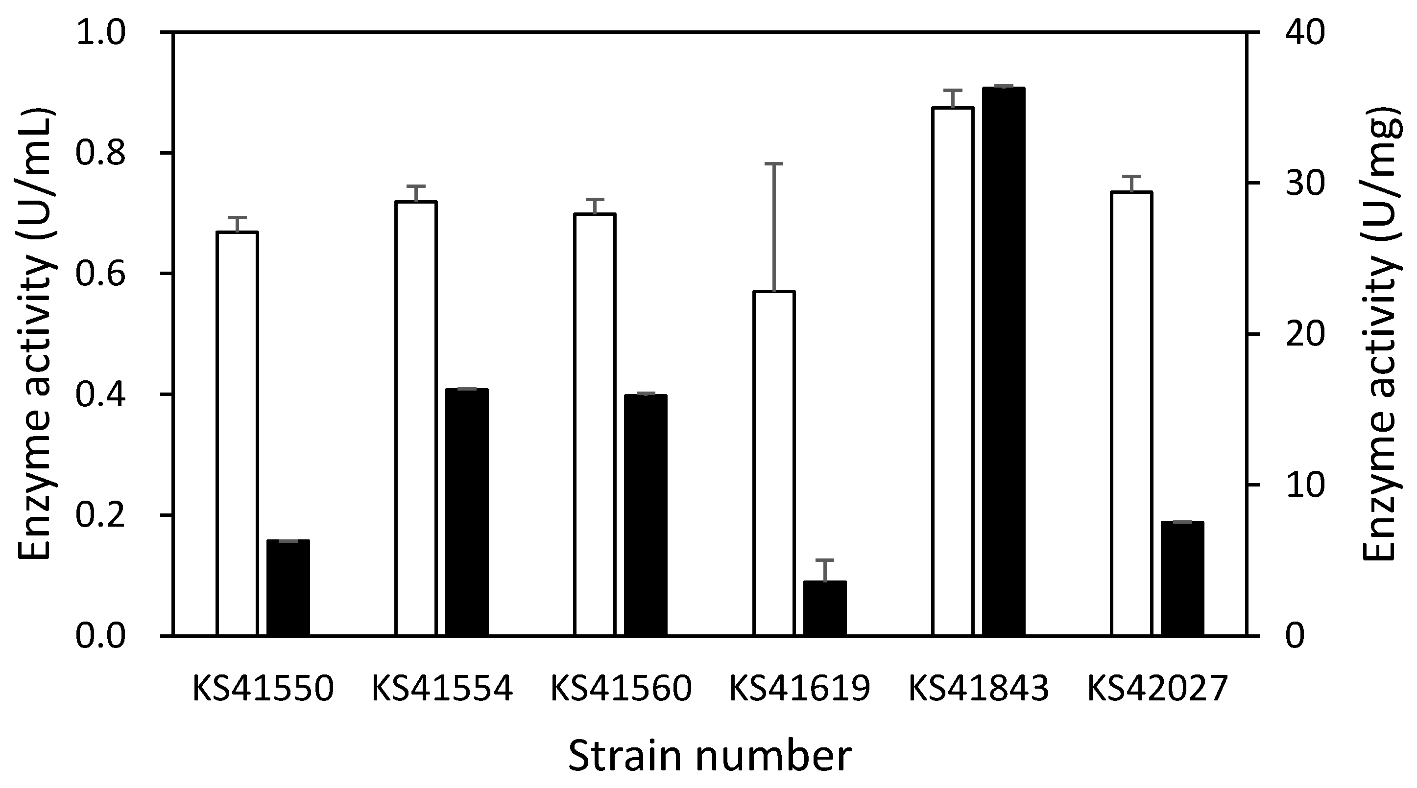
Figure 1. Quantitative comparison of amylase activity of six preliminarily selected strains. The strains were cultured in Zobell medium at 15 °C for 3 days, and the activity was measured using the broth. Enzyme activity was calculated per unit volume (white bars) and per unit mass (black bars). Means and standard deviations are shown.
Fermentation 11 00465 g001
Figure 2. (A) Cell growth and amylase production of Halomonas sp. KS41843. Cells were cultured in Zobell medium containing 1% soluble starch at 15 °C for 132 h, and alterations in cell density (○) and enzyme activity (□) over time were measured. (B) Effect of salt on cell growth and amylase production of Halomonas sp. KS41843. Cells were cultured for 72 h in Zobell medium with the addition of 0, 20, 40, and 60 g/L of NaCl, resulting in total NaCl concentrations of 20, 40, 60, and 80 g/L, respectively, and subsequently, the cell density (white bar) and enzyme activity (black bar) were measured. Means and standard deviations are shown.
Fermentation 11 00465 g002
Figure 3. Visualization of KS41843 amylase purification and confirmation of activity. (A) Sodium dodecyl sulfate-polyacrylamide gel electrophoresis of KS41843 amylase and Amplify. (B) KS41843 was transferred to a PVDF membrane. (C) Confirmation of amylase activity via zymography analysis. The black arrow indicates KS41843 amylase. M: size marker; Lanes 1–2, Amplify (0.3 μg); Lanes 3–4, KS41843 amylase (0.3 μg); Lanes 5–6, Amplify (1.5 μg); Lanes 7–8, KS41843 amylase (1.5 μg); Lanes 9–10, KS41843 amylase (0.25 μg).
Fermentation 11 00465 g003
Figure 4. Effect of temperature and pH on the amylase activities and stabilities (A) Relative starcholytic activities of KS41843 amylase (○) and the reference enzyme (Amplify), which is commercially available (□) at different temperatures ranging from 5 to 70 °C. (B) Thermostability of amylase derived from KS41843 (●) and Amplify (■). (C) Relative starcholytic activities of KS41843 amylase (○) and Amplify (□) at various pH values ranging from 4.0 to 10.0. (D) pH stabilities of KS41843 amylase (●) and Amplify (■). Relative values are expressed as percentages, with the maximum value normalized to 100%. Means and standard deviations are shown.
Fermentation 11 00465 g004
Figure 5. Effect of metal ions on the amylase activity. Each ion was added at a concentration of 1 mM and activity was measured. Enzyme activity under standard conditions (enzyme reactions without additives) was demonstrated as the rightmost bar (control). Means and standard deviations are shown.
Fermentation 11 00465 g005
Figure 6. Effect of salinity on enzyme activity. The activities of KA41843 amylase (●) and Amplify (■) are measured at NaCl concentrations ranging from 0 to 4 M. Means and standard deviations are shown.
Fermentation 11 00465 g006
Table 1. Identification of six bacterial strains exhibiting high amylase activity from the Antarctic Sea near the King Sejong station.
Sample No.Closest MatchIdentification
KS41550Alteromonas additaAlteromonas KS41550
KS41554Alteromonas naphthalenivoransAlteromonas KS41554
KS41560Halomonas titanicaeHalomonas KS41560
KS41619Pseudoalteromonas agrivoransPseudoalteromonas KS41619
KS41843Halomonas titanicaeHalomonas sp. KS41843
KS42027Subtercola vilaeSubtercola KS42027
Table 2. Purification summary for KS41843 amylase.
StageTotal ActivityTotal Protein Content (mg)Specific Activity (U/mg)FoldYield (%)
Cell culture broth162.8913.8011.801.00100.00
10 kDa cut-off38.894.219.230.7823.87
DEAE column22.500.9324.192.0513.81
Superdex 759.820.09110.329.356.03
Disclaimer/Publisher’s Note: The statements, opinions and data contained in all publications are solely those of the individual author(s) and contributor(s) and not of MDPI and/or the editor(s). MDPI and/or the editor(s) disclaim responsibility for any injury to people or property resulting from any ideas, methods, instructions or products referred to in the content.