Article

The Role of Quorum Sensing in Enhancing Lovastatin and Pigment Production in Monascus purpureus C322

Department of Biotechnology, School of Life Sciences, University of Westminster, London W1W 6XH, UK
*
Author to whom correspondence should be addressed.
Fermentation 2025, 11(8), 461; https://doi.org/10.3390/fermentation11080461
Submission received: 30 May 2025 / Revised: 31 July 2025 / Accepted: 7 August 2025 / Published: 11 August 2025
(This article belongs to the Special Issue Scale-Up Challenges in Microbial Fermentation)

Abstract

Monascus purpureus is a filamentous fungus known for producing pharmaceutically valuable secondary metabolites, including azaphilone pigments and lovastatin. Lovastatin is an HMG-CoA reductase inhibitor widely used to manage hypercholesterolaemia, while Monascus pigments serve as natural colourants with antioxidant and antimicrobial properties. This study evaluated the impact of quorum-sensing molecules (QSMs)—tyrosol (0.3 mM), farnesol (0.2 mM) and linoleic acid (0.4 mM)—on pigment and lovastatin yields in shake flasks and 2.5 L stirred-tank bioreactors. QSMs were introduced 48 h post-inoculation in shake flasks and 24 h in bioreactors. All QSMs increased yellow (OD400), orange (OD470), and red (OD510) pigments and lovastatin concentration relative to the control, with scale-up further enhancing yields. Farnesol produced the most pronounced effect: in flasks, OD400 7.10 (1.86-fold), OD470 8.00 (2.12-fold), OD510 7.80 (2.08-fold), and 74.6 mg/L lovastatin (2.05-fold); in bioreactors, OD400 11.9 (2.06-fold), OD470 15.1 (2.71-fold), OD510 13.7 (2.47-fold), and 97.2 mg/L lovastatin (2.48-fold). This was followed by tyrosol treatment and then linoleic acid. These findings demonstrate that QSMs—particularly farnesol—significantly (p < 0.01) stimulate pigment and lovastatin biosynthesis in M. purpureus. Quorum sensing modulation represents a promising, scalable strategy to optimise fungal fermentation for industrial metabolite production.

1. Introduction

Microbial fermentation is a key industrial process for producing bioactive secondary metabolites, including antibiotics, pigments, and statins. However, the yields of these valuable compounds are often limited by metabolic regulation and environmental factors [1,2,3]. To enhance metabolite production, various strategies have been explored, such as genetic manipulation, bioprocess optimisation, and the use of signalling molecules that modulate microbial metabolism [4]. One such approach involves quorum sensing (QS), a cell-to-cell communication mechanism that regulates gene expression based on microbial population density [5,6].
QS is mediated by small diffusible signalling molecules known as autoinducers, which accumulate in the extracellular environment as microbial populations grow. Once a threshold concentration is reached, these molecules trigger coordinated metabolic responses, influencing biofilm formation, morphogenesis, and secondary metabolism [1,6,7]. While extensively studied in bacteria, QS has also been identified in fungi, where it plays a crucial role in regulating secondary metabolite biosynthesis [1,3,7,8]. In filamentous fungi, several quorum-sensing molecules (QSMs), including farnesol, tyrosol, butyrolactone-I, and linoleic acid, have been shown to influence metabolic processes and enhance secondary metabolite production [7,9,10,11].
M. purpureus is a well-established fungal species widely used in the food and pharmaceutical industries due to its ability to produce secondary metabolites such as pigments and lovastatin. The pigments—red, orange, and yellow azaphilones—are of particular interest as natural colorants with antimicrobial and antioxidant properties [2,4,12,13,14,15]. Meanwhile, lovastatin, a polyketide-derived statin, is a potent inhibitor of 3-hydroxy-3-methylglutaryl coenzyme A (HMG-CoA) reductase and is used clinically for cholesterol management [16,17]. Despite their industrial significance, the regulation of pigment and lovastatin biosynthesis in Monascus remains complex and requires further investigation.
Recent studies suggest that QSMs may serve as metabolic regulators in M. purpureus, modulating its secondary metabolism through intricate transcriptional pathways [4,13,18]. Specifically, farnesol, tyrosol, and linoleic acid have been identified as key QS molecules that influence fungal growth and metabolite production [6,7,8,10,11,18,19]. These molecules interact with fungal signalling pathways, affecting morphogenesis, stress responses, and the biosynthetic gene clusters responsible for pigment and statin production. However, their precise role in Monascus metabolism remains underexplored.
This study aims to investigate the effects of farnesol, tyrosol, and linoleic acid on pigment and lovastatin production in M. purpureus C322. By comparing metabolite yields in shake flask cultures and stirred-tank bioreactors, this research seeks to elucidate the role of QS in metabolic regulation and assess its potential for industrial-scale bioprocess optimisation. Understanding how these signalling molecules influence Monascus fermentation could provide new insights into fungal QS and offer innovative strategies for enhancing secondary metabolite production.

2. Materials and Methods

2.1. Microorganism and Culture Maintenance

The filamentous fungus Monascus purpureus C322 was obtained from the culture collection at the University of Westminster. The strain was maintained on Yeast Malt Extract (YME) agar at pH 6.2 and subcultured every two weeks onto Hiroi Potato Dextrose Agar (PDA), as described in [20]. Hiroi PDA is composed of sucrose (100 g/L), yeast extract (3 g/L), casamino acids (5 g/L), NaNO3 (2 g/L), KH2PO4 (1 g/L), MgSO4·7H2O (0.5 g/L), KCl (0.5 g/L), FeSO4 (0.01 g/L), potato starch (4 g/L), dextrose (20 g/L), and agar (15 g/L) (All chemicals and reagents were acquired from VWR International Ltd., Lutterworth, UK). The agar plates were inoculated with 106 spores/mL prior to incubation at 28 °C for 10–15 days before storage at 4 °C for future use. The spores were harvested aseptically by gently scraping the culture from plates with a sterile inoculation loop in the presence of 0.1% Tween 80 solution. The collected spore suspension was centrifuged at high speed for 10 min, and the supernatant was discarded. Sterile glass beads were added to the remaining pellet, followed by the addition of sterile saline solution (0.9% w/v NaCl). The pellet was gently flicked to release spores while trapping mycelial fragments within the beads. The spore suspension above the glass beads was carefully pipetted into a fresh Falcon tube and stored at 4 °C for future use.

2.2. Fermentation Setup and Culture Conditions

Submerged fermentation was performed using a chemically defined growth medium, prepared according to the protocol outlined in [21]. The growth medium consisted of glucose (20 g/L), KH2PO4 (5 g/L), K2HPO4 (5 g/L), MgSO4·7H2O (0.5 g/L), FeSO4·7H2O (0.1 g/L), ZnSO4·7H2O (0.01 g/L), MnSO4·H2O (0.03 g/L), KCl (0.5 g/L), and monosodium glutamate (MSG) (5 g/L) (All chemicals and reagents were acquired from VWR International Ltd., Lutterworth, UK). The pH was adjusted to 6.5 prior to sterilisation and it was not controlled during fermentation, in order to observe changes associated with microbial metabolism and secondary metabolite production. Two fermentation systems were employed: 500 mL Erlenmeyer flasks for small-scale, and 2.5 L stirred-tank bioreactors (Fermac 310/60, Electrolab Biotech, Tewkesbury, UK) for large-scale fermentation. The shake flask cultures contained 100 mL of medium inoculated with 106 spores/mL prior to incubation at 25 °C with a shaking speed of 120 rpm for 18 days. The stirred-tank bioreactors were inoculated with 150 mL of actively growing Day-7 seed culture into 1350 mL of defined medium to make up a working volume of 1.5 L. The bioreactors were operated at 32 °C with an agitation speed of 300 rpm using a dual-impeller system. The setup included a Rushton turbine mounted at the base for radial flow and a pitched-blade impeller near the surface for foam disruption. The impellers were spaced vertically at approximately H/2, where H represents the working liquid height, to optimise mixing and minimise foam accumulation. Aeration was maintained at 0.5 vvm, and fermentation was carried out over a 5-day period.

2.3. Preparation and Supplementation of Quorum-Sensing Molecules

The quorum-sensing molecules—farnesol, tyrosol, and linoleic acid—were supplemented to assess their effects on M. purpureus C322 metabolism (The QSMs were acquired from VWR International Ltd., Lutterworth, UK). Tyrosol was dissolved in sterile deionized water, while farnesol and linoleic acid were dissolved in 1% dimethyl sulfoxide (DMSO). All QSMs were filter-sterilised using a 0.22 µm syringe filter before supplementation. Based on previous in-house studies, the shake flasks (100 mL working volume) and stirred-tank bioreactors (1.5 L working volume) were supplemented with QSMs at the following final concentrations: tyrosol at 0.3 mM, farnesol at 0.2 mM, and linoleic acid at 0.4 mM. The QSMs were added 48 h post-inoculation in shake flasks and 24 h post-inoculation in bioreactors. The timing of QSM addition in our study was chosen based on in-house optimisation and prior studies [14,19,22,23,24,25,26] and reflects the difference in lag phase duration between systems—being longer in shake flasks and shorter in bioreactors—to ensure addition during a comparable physiological stage. Control cultures were carried out without the supplementation of QSMs.

2.4. Analytical Methods

Throughout the fermentation, key parameters including pH, residual carbohydrate concentration, and pigment production were monitored at regular intervals. Biomass accumulation and lovastatin concentration were measured at the end of the fermentation cycle. The data presented in this study correspond to the samples harvested during the late exponential phase, just before the culture entered the stationary phase, in order to evaluate the impact of QSMs on metabolite production. Biomass quantification followed a modified gravimetric protocol [22]. The total biomass was filtered using a Merck™ Vacuum Flask with Büchner Funnels and a Fischerbrand FB70155 pump (Merck, Darmstadt, Germany), then transferred onto Whatman No. 1 filter paper and oven-dried at 100 °C for 12 h before CDW measurement. Pigment production was measured spectrophotometrically, with absorbance readings taken at 400 nm for yellow pigments, 470 nm for orange pigments, and 510 nm for red pigments [27]. Total carbohydrate consumption was determined using the phenol-sulfuric acid method to quantify residual glucose in the fermentation broth [28]. Lovastatin production was analysed using High-Performance Liquid Chromatography (Dionex HPLC 3000) (Thermo Fisher Scientific, Waltham, MA, USA) with a Lichrospher RP-18 Endcapped column at 25 °C. Samples were processed using an isocratic method, with a mobile phase of 55:45 (vol/vol) HPLC-grade acetonitrile and aqueous 0.1% phosphoric acid. The injection volume was 25 μL, and the flow rate was 1 mL/min. Detection was performed at 238 nm [24,29].

2.5. Statistical Analysis

All experiments were carried out in three independent replicates, with results reported as mean ± standard deviation. Differences among treatment groups were evaluated by one-way ANOVA with Tukey’s post hoc test (significance threshold p < 0.05). Statistical analyses and figure generation were performed using GraphPad Prism 10 and OriginPro 2025 software.

3. Results

3.1. Impact of Quorum-Sensing Molecules on Biomass Concentration in Shake Flasks and Bioreactors

The biomass concentration measured for the control group (C) in shake flasks was 5.35 g/L. QSM-supplemented groups showed comparable values, ranging between 5.33 and 5.44 g/L. Under bioreactor conditions, biomass accumulation increased across all treatments, with values ranging from 7.51 g/L to 7.79 g/L (Figure 1). One-way ANOVA revealed no statistically significant differences among groups (p > 0.05), indicating that QSM supplementation did not affect cell growth under the tested conditions. Fold increases in CDW from shake flasks to bioreactors were 1.46 in the control group and 1.43, 1.41, and 1.42 in the tyrosol (0.3 mM), farnesol (0.2 mM), and linoleic acid (0.4 mM) groups, respectively, demonstrating an increase in biomass concentration under bioreactor cultivation independent of QSM treatment.

3.2. Quorum-Sensing-Mediated Modulation of Monascus Pigment Biosynthesis in Shake Flask and Bioreactor Systems

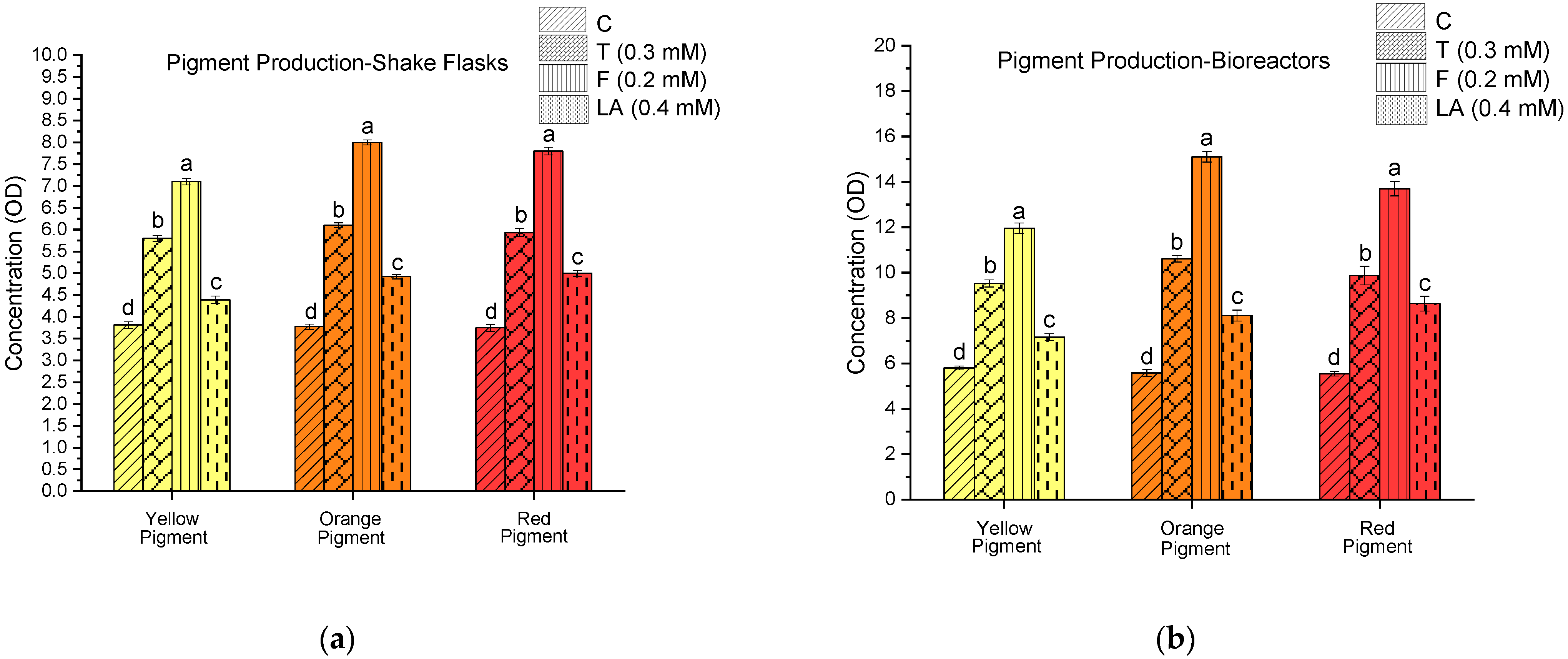
The production of yellow, orange, and red pigments was quantified under both shake flask and stirred-tank bioreactor conditions. In both systems, all QSM-supplemented groups consistently produced higher pigment concentrations than the control. Among them, farnesol (0.2 mM) produced the highest levels, followed by tyrosol (0.3 mM) and linoleic acid (0.4 mM) (Figure 2). This trend was observed across all three pigment types. In the shake flasks, pigment concentrations with farnesol reached OD510 7.80 (red), OD470 8.00 (orange), and OD400 7.10 (yellow). In the bioreactors, the pigment production was further increased to OD510 13.7, OD470 15.1, and OD400 11.9, respectively. One-way ANOVA revealed statistically significant differences among treatments for all pigments (p < 0.01). Tukey’s post hoc test confirmed that farnesol (0.2 mM) significantly enhanced yellow, orange, and red pigment production compared to other treatments (p < 0.01), followed by tyrosol (0.3 mM) and linoleic acid (0.4 mM), with the control showing the lowest levels.
To account for differences in biomass accumulation, pigment yields were also expressed as optical density per gram of dry cell weight (OD/g CDW). In shake-flask cultures, farnesol supplementation (0.2 mM) gave the highest yields—1.33, 1.50, and 1.46—followed by tyrosol (0.3 mM) at 1.07, 1.12, and 1.09; linoleic acid (0.4 mM) at 0.81, 0.91, and 0.92; and the control at 0.71, 0.70, and 0.70—for yellow, orange, and red pigments, respectively. Under bioreactor conditions, yields further improved and followed a similar trend: farnesol reached 1.59, 2.01, and 1.26; tyrosol reached 1.23, 1.37, and 1.11; linoleic acid reached 0.93, 1.06, and 1.13; and the control reached 0.74, 0.72, and 0.96—for yellow, orange and red pigments, respectively.
In addition to pigment concentration and yield, fold increases in production relative to the control group (C) were also calculated under each fermentation setup (Table 1, Figure 3). In the shake flasks, yellow pigment production increased by 1.52-fold in tyrosol (T, 0.3 mM), 1.86-fold in farnesol (F, 0.2 mM), and 1.15-fold in linoleic acid (LA, 0.4 mM). Orange pigment increased 1.62-fold (T), 2.12-fold (F), and 1.30-fold (LA), while red pigment increased by 1.58-fold (T), 2.08-fold (F), and 1.33-fold (LA) compared to the control. In bioreactors, yellow pigment production increased by 1.64-fold (T), 2.06-fold (F), and 1.23-fold (LA); orange pigment increased by 1.90-fold (T), 2.71-fold (F), and 1.45-fold (LA); and red pigment increased by 1.78-fold (T), 2.47-fold (F), and 1.55-fold (LA), relative to the control. Among the QSMs tested, farnesol yielded the highest fold increase in all pigment types across both shake flask and bioreactor conditions. To complement these comparisons, fold increases in pigment concentration were also evaluated during scale-up, comparing bioreactors to shake flasks within each treatment. These results are summarised in Table 1.

3.3. Enhancement of Lovastatin Biosynthesis by Quorum-Sensing Molecules

Lovastatin concentrations were quantified under both shake flask and stirred-tank bioreactor conditions (Figure 4). In both systems, all QSM-supplemented groups produced higher concentrations than the control. Among the treatments, farnesol (0.2 mM) yielded the highest levels, followed by tyrosol (0.3 mM) and linoleic acid (0.4 mM), mirroring the trend observed for pigment production. With farnesol, the lovastatin concentration reached 74.6 mg/L in shake flasks and 97.2 mg/L in bioreactors. One-way ANOVA revealed a statistically significant difference in lovastatin concentrations among the treatment groups, including with respect to the control (p < 0.01). Tukey’s post hoc test showed that cultures supplemented with farnesol (0.2 mM) produced significantly higher lovastatin compared to other treatments. Tyrosol (0.3 mM) also led to a significant increase compared to the control. Linoleic acid (0.4 mM) yielded higher concentrations than the control but was significantly lower than both the farnesol and tyrosol treatments. The control group exhibited the lowest lovastatin production across all conditions.
To evaluate production efficiency, lovastatin yields were calculated as mg/g CDW. In the shake flasks, farnesol achieved the highest yield (14.0 mg/g), followed by tyrosol (12.5 mg/g), linoleic acid (9.46 mg/g), and the control (6.80 mg/g). Under bioreactor conditions, farnesol again resulted in the highest yield (12.9 mg/g), with tyrosol and linoleic acid yielding 10.1 mg/g and 7.26 mg/g, respectively, compared to 5.03 mg/g in the control. Across both systems, farnesol treatment resulted in the greatest production efficiency relative to biomass. Fold increases in lovastatin concentration relative to the control were also assessed. In the shake flasks, tyrosol, farnesol, and linoleic acid resulted in 1.88-, 2.05-, and 1.41-fold increases, respectively. Under bioreactor conditions, these values increased further to 2.00-fold (T), 2.48-fold (F), and 1.42-fold (LA), confirming that farnesol consistently produced the greatest enhancement across both scales (Figure 3d). To compare the impact of scale-up, fold increases in lovastatin concentration from shake flasks to bioreactors were calculated within each treatment group. These values are summarised in Table 2.

3.4. pH Variation and Carbohydrate Consumption in Shake Flask and Bioreactor Cultures

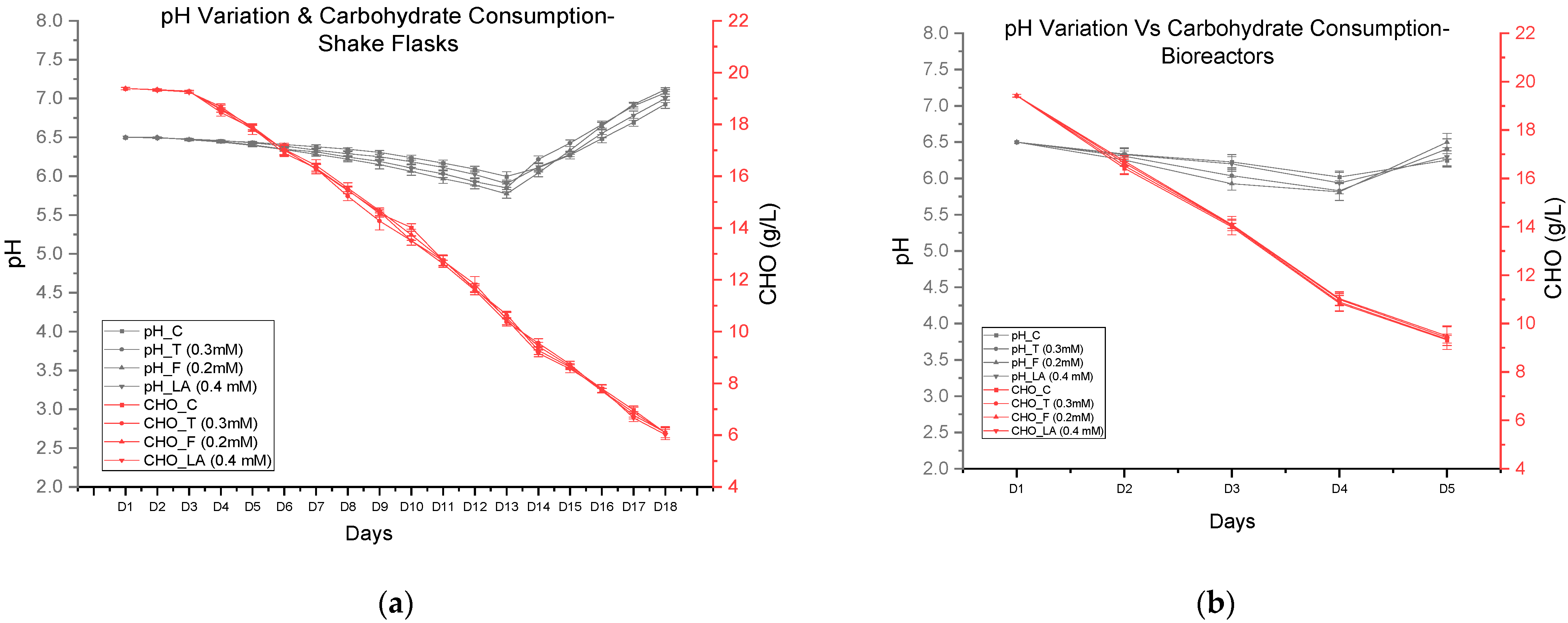
pH and residual carbohydrate concentrations were monitored throughout the fermentation period in both shake flasks and bioreactors (Figure 5). In all conditions, pH followed a similar trend, beginning with an initial value of 6.5 across all groups. A gradual decline was observed until Day 4, followed by a slight increase by Day 5. In shake flasks, the lowest pH values ranged from 5.81 to 6.02; the final values on Day 18 ranged from 6.25 in the control to 6.50 in the farnesol-treated group (F, 0.2 mM). Similar patterns were recorded under bioreactor conditions, where the pH dropped to 5.81–6.02 by Day 4, before increasing to between 6.25 and 6.50 by Day 5. One-way ANOVA showed no statistically significant differences in pH values among the treatment groups (p > 0.05) in either shake flasks or bioreactors.
Carbohydrate consumption exhibited a consistent downward trend throughout the fermentation process across all conditions (Figure 5). In shake flasks, the initial carbohydrate concentrations were 19.4 g/L across all groups. These declined steadily to 9.33 g/L (C), 9.42 g/L (T), 9.48 g/L (F), and 9.35 g/L (LA) by Day 18. The corresponding average consumption rates in shake flasks ranged from 0.74 to 0.75 g/L/day across the treatment groups. In bioreactors, residual carbohydrate concentrations were lower by Day 5, recorded as 9.33 g/L (C), 9.42 g/L (T), 9.48 g/L (F), and 9.35 g/L (LA), with an average consumption rate between 1.99 and 2.02 g/L/day. The final residual values on Day 5 were comparable across treatments. Statistical analysis revealed no significant differences in carbohydrate consumption between groups (p > 0.05) in both shake flasks and bioreactors, indicating that QSM supplementation did not significantly alter substrate utilisation dynamics.

4. Discussion

This study demonstrates that quorum-sensing molecules modulate secondary metabolism in Monascus purpureus C322, enhancing azaphilone pigment and lovastatin production. The consistent effects observed with farnesol, tyrosol, and linoleic acid across both fermentation scales suggest that these molecules interact with intrinsic regulatory systems, influencing secondary metabolite fluxes independently of growth.
All three QSMs enhanced metabolite output relative to the control, with farnesol yielding the highest response, followed by tyrosol and linoleic acid. Farnesol’s pronounced effect reinforces its role as a conserved fungal signalling molecule capable of modulating oxidative stress pathways, membrane dynamics, and biosynthetic gene clusters [6,7,11,14,19,22,30,31,32,33]. In Monascus purpureus, this may involve upstream activation of polyketide synthase genes within the mok and mpp clusters, as previously implicated in pigment and lovastatin biosynthesis [13,34,35,36]. Farnesol’s influence likely extends beyond transcription, potentially altering intracellular trafficking or membrane transporter activity, which are known to affect metabolite secretion in fungi. This is supported by studies indicating that farnesol, through its role in protein prenylation, can modulate the function of small GTPases involved in vesicular transport and membrane dynamics, thereby influencing the secretion pathways of secondary metabolites [37].
While tyrosol has been associated with density-dependent morphogenesis and oxidative stress tolerance in Candida albicans [24,38,39], its role in filamentous fungi such as M. purpureus remains unclear. The observed enhancements in pigments and lovastatin levels, suggest that tyrosol may modulate intracellular stress or signalling responses, possibly influencing secondary metabolism through conserved regulatory pathways. Linoleic acid also contributed to pigment and lovastatin enhancement across conditions. As a known oxylipin precursor, it may act by modulating lipid-derived signalling cascades or influencing membrane-associated enzyme activity [40,41,42,43]. Although its specific function in Monascus is not well understood, its consistent effects point to a signalling role rather than a nutritional one. It is possible that linoleic-acid-derived oxylipins engage GPCR-based sensing systems, as described in other fungal models [40,44]. The coordinated stimulation of both pigment and lovastatin biosynthetic pathways suggests shared upstream control, likely involving acetyl-CoA and malonyl-CoA as central precursors [13,45]. The convergence between pigment and lovastatin pathways observed in this study suggests a quorum-regulated control mechanism, supporting the idea that QSMs trigger a systemic metabolic shift rather than isolated pathway activation. To the best of our knowledge, this is the first study to report a significant (p < 0.01) increase in lovastatin production using M. purpureus following supplementation with the quorum-sensing molecules farnesol, tyrosol and linoleic acid in submerged cultures (both shake flasks and 2.5 L stirred-tank bioreactors). Prior research has explored this concept primarily in Aspergillus terreus, where QSMs, such as butyrolactone-I and linoleic acid, have been shown to increase lovastatin titres and activate biosynthetic gene expression [6,14,19,25,26,46]. The present findings extend this principle to M. purpureus C322, revealing that exogenous QSMs can act as metabolic modulators to co-stimulate pigment and lovastatin biosynthesis in a scalable fungal fermentation system.
Bioreactor conditions further amplified QSM responses, indicating that environmental parameters—such as oxygen transfer, shear, and pH stability—may stabilise or potentiate intracellular signalling. This aligns with findings in other filamentous fungi where hydrodynamic cues and quorum signals jointly regulate gene expression [47,48,49,50,51,52,53,54,55]. The scalability and robustness of the observed effects highlight the feasibility of applying QSM-based modulation in industrial fermentation settings. Interestingly, the addition of quorum-sensing molecules (QSMs) did not result in a significant increase in biomass, indicating that the enhanced production of secondary metabolites was not due to increased fungal growth. This suggests that QSMs may directly influence the metabolic pathways responsible for pigment and lovastatin biosynthesis, rather than promoting overall biomass accumulation. Such a decoupling of biomass and metabolite production is advantageous in industrial fermentation processes, where maximising product yield without excessive biomass can improve efficiency and reduce downstream processing costs [13,18,56,57].
Interestingly, pH and carbohydrate utilisation patterns also remained consistent across all QSM-treated groups, further supporting the conclusion that QSMs redirected metabolic activity without altering core physiological functions. The characteristic pH shift—an initial drop during early growth followed by gradual alkalinisation—reflects known trends linked to organic acid production and the subsequent accumulation of alkaline byproducts like ammonia [12,58,59,60,61]. Carbohydrate depletion followed a biphasic pattern, with rapid utilisation during the log phase and slower consumption thereafter, consistent with previously reported metabolic transitions in filamentous fungi [12,62,63]. This metabolic stability reinforces the view that QSMs specifically modulated secondary metabolism while maintaining primary metabolic homeostasis.
Despite the phenotypic clarity observed, the molecular basis of QSM signalling in M. purpureus remains largely unknown. Future studies using transcriptomics, proteomics, and chromatin accessibility profiling could help identify quorum-responsive elements and uncover the regulatory architecture governing pigment and lovastatin biosynthesis. Such insights would support more precise engineering of quorum-sensing pathways for targeted metabolic control.

5. Conclusions

This study demonstrates that quorum-sensing molecule (QSM) supplementation is an effective approach for enhancing secondary metabolite production in M. purpureus C322. The observed improvements in pigment and lovastatin titres under both shake flask and bioreactor fermentation highlight the applicability of QSM in scalable fermentation processes. Farnesol, in particular, significantly enhanced production across all pigments and yielded the highest increase in lovastatin, indicating its potential as a powerful regulatory molecule for metabolic activation. The findings confirm that quorum sensing plays a crucial role in modulating fungal biosynthetic pathways, and that external supplementation of signalling molecules can be harnessed to steer metabolite output. This strategy offers a level of metabolic fine-tuning that is valuable for optimising industrial fermentations, where both yield and process efficiency are critical. Future investigations into the signalling pathways and transcriptional regulators influenced by these QSMs could provide deeper insights into their mode of action. Such knowledge would pave the way for targeted metabolic engineering and the development of fermentation strategies that exploit cell-to-cell communication mechanisms. Overall, this work positions quorum-sensing-based modulation as a promising and underutilised tool in fungal biotechnology. Its integration into fermentation workflows could improve product yields, support cost-effective production, and contribute to the sustainable biomanufacturing of high-value fungal metabolites at an industrial scale.

Author Contributions

Conceptualisation, T.K. and S.Y.; methodology, S.Y.; validation, T.K. and S.Y.; formal analysis, S.Y.; investigation, S.Y.; resources, T.K. and S.Y.; data curation, S.Y.; writing—original draft preparation, S.Y.; writing—review and editing, S.Y., T.K., G.K. and S.J.G.; visualisation, S.Y.; supervision, T.K., G.K. and S.J.G.; project administration, S.Y.; funding acquisition, S.Y. All authors have read and agreed to the published version of the manuscript.

Funding

This research was supported by the 125 Fund at the University of Westminster, London, UK.

Institutional Review Board Statement

Not applicable.

Informed Consent Statement

Not applicable.

Data Availability Statement

The original contributions presented in this study are included in the article. Further inquiries can be directed to the corresponding author.

Conflicts of Interest

The authors declare no conflicts of interest.

Abbreviations

The following abbreviations are used in this manuscript:
QSQuorum Sensing
QSMQuorum-Sensing Molecule
FFarnesol
TTyrosol
LALinoleic Acid
CHOCarbohydrate
CDWCell Dry Weight
SFShake Flasks
BBioreactor
ODOptical Density
AUAbsorbance Units
ANOVAAnalysis of Variance
HPLCHigh-Performance Liquid Chromatography

References

  1. Reshi, Z.A.; Ahmad, W.; Lukatkin, A.S.; Javed, S. Bin From Nature to Lab: A Review of Secondary Metabolite Biosynthetic Pathways, Environmental Influences, and In Vitro Approaches. Metabolites 2023, 13, 895. [Google Scholar] [CrossRef]
  2. Liu, J.; Du, Y.; Ma, H.; Pei, X.; Li, M. Enhancement of Monascus Yellow Pigments Production by Activating the CAMP Signalling Pathway in Monascus purpureus HJ11. Microb. Cell Fact. 2020, 19, 224. [Google Scholar] [CrossRef]
  3. Prajapati, P.A.; Andhare, P.; Upadhyay, D.; Kumari, S. Microbial secondary metabolites. Int. J. Biol. Pharm. Allied Sci. 2021, 10, 488–496. [Google Scholar] [CrossRef]
  4. Shi, J.; Qin, X.; Zhao, Y.; Sun, X.; Yu, X.; Feng, Y. Strategies to Enhance the Production Efficiency of Monascus Pigments and Control Citrinin Contamination. Process Biochem. 2022, 117, 19–29. [Google Scholar] [CrossRef]
  5. Miller, M.B.; Bassler, B.L. Quorum Sensing in Bacteria. Annu. Rev. Microbiol. 2001, 55, 165–199. [Google Scholar] [CrossRef] [PubMed]
  6. Albuquerque, P.; Casadevall, A. Quorum Sensing in Fungi—A Review. Med. Mycol. 2012, 50, 337–345. [Google Scholar] [CrossRef]
  7. Barriuso, J.; Hogan, D.A.; Keshavarz, T.; Martínez, M.J. Role of Quorum Sensing and Chemical Communication in Fungal Biotechnology and Pathogenesis. FEMS Microbiol. Rev. 2018, 42, 627–638. [Google Scholar] [CrossRef] [PubMed]
  8. Berrocal, A.; Navarrete, J.; Oviedo, C.; Nickerson, K.W. Quorum Sensing Activity in Ophiostoma ulmi: Effects of Fusel Oils and Branched Chain Amino Acids on Yeast-Mycelial Dimorphism. J. Appl. Microbiol. 2012, 113, 126–134. [Google Scholar] [CrossRef]
  9. Amache, R.; Yerramalli, S.; Giovanni, S.; Keshavarz, T. Quorum Sensing Involvement in Response Surface Methodology for Optimisation of Sclerotiorin Production by Penicillium sclerotiorum in Shaken Flasks and Bioreactors. Ann. Microbiol. 2019, 69, 1415–1423. [Google Scholar] [CrossRef]
  10. Tian, X.; Ding, H.; Ke, W.; Wang, L. Quorum Sensing in Fungal Species. Annu. Rev. Microbiol. 2021, 75, 449–469. [Google Scholar] [CrossRef]
  11. Berrocal, A.; Oviedo, C.; Nickerson, K.W.; Navarrete, J. Quorum Sensing Activity and Control of Yeast-Mycelium Dimorphism in Ophiostoma floccosum. Biotechnol. Lett. 2014, 36, 1503–1513. [Google Scholar] [CrossRef] [PubMed]
  12. Agboyibor, C.; Kong, W.B.; Chen, D.; Zhang, A.M.; Niu, S.Q. Monascus Pigments Production, Composition, Bioactivity and Its Application: A Review. Biocatal. Agric. Biotechnol. 2018, 16, 433–447. [Google Scholar] [CrossRef]
  13. Gong, P.; Shi, R.; Liu, Y.; Luo, Q.; Wang, C.; Chen, W. Recent Advances in Monascus Pigments Produced by Monascus purpureus: Biosynthesis, Fermentation, Function, and Application. LWT 2023, 185, 115162. [Google Scholar] [CrossRef]
  14. Raina, S.; De Vizio, D.; Palonen, E.K.; Odell, M.; Brandt, A.M.; Soini, J.T.; Keshavarz, T. Is Quorum Sensing Involved in Lovastatin Production in the Filamentous Fungus Aspergillus terreus? Process Biochem. 2012, 47, 843–852. [Google Scholar] [CrossRef]
  15. Chaudhary, V.; Katyal, P.; Poonia, A.K.; Kaur, J.; Puniya, A.K.; Panwar, H. Natural Pigment from Monascus: The Production and Therapeutic Significance. J. Appl. Microbiol. 2022, 133, 18–38. [Google Scholar] [CrossRef] [PubMed]
  16. Alberts, A.W.; Chen, J.; Kuron, G.; Hunt, V.; Huff, J.; Hoffman, C.; Rothrock, J.; Lopez, M.; Joshua, H.; Harris, E.; et al. Mevinolin: A Highly Potent Competitive Inhibitor of Hydroxymethylglutaryl-Coenzyme A Reductase and a Cholesterol-Lowering Agent. Proc. Natl. Acad. Sci. USA 1980, 77, 3957–3961. [Google Scholar] [CrossRef]
  17. Valentovic, M. Lovastatin. In xPharm: The Comprehensive Pharmacology Reference; Enna, S.J., Bylund, D.B., Eds.; Elsevier: Amsterdam, The Netherlands, 2007; pp. 1–5. ISBN 9780080552323. [Google Scholar] [CrossRef]
  18. Shi, R.; Luo, Q.; Liu, Y.; Meng, G.; Chen, W.; Wang, C. Effect of γ-Butyrolactone, a Quorum Sensing Molecule, on Morphology and Secondary Metabolism in Monascus. LWT 2022, 172, 114225. [Google Scholar] [CrossRef]
  19. Raina, S.; Odell, M.; Keshavarz, T. Quorum Sensing as a Method for Improving Sclerotiorin Production in Penicillium sclerotiorum. J. Biotechnol. 2010, 148, 91–98. [Google Scholar] [CrossRef]
  20. Ajdari, Z.; Ebrahimpour, A.; Abdul Manan, M.; Hamid, M.; Mohamad, R.; Ariff, A.B. Nutritional Requirements for the Improvement of Growth and Sporulation of Several Strains of Monascus purpureus on Solid State Cultivation. Biomed. Res. Int. 2011, 2011, 487329. [Google Scholar] [CrossRef]
  21. Chatterjee, S.; Maity, S.; Chattopadhyay, P.; Sarkar, A.; Laskar, S.; Sen, S.K. Characterization of Red Pigment from Monascus in Submerged Culture Red Pigment from Monascus purpureus. J. Appl. Sci. Res. 2009, 5, 2102–2108. [Google Scholar]
  22. Amache, R. Quorum Sensing for Improved Production of Industrially Useful Products from Filamentous Fungi. Ph.D. Thesis, University of Westminster, London, UK, 2014. [Google Scholar]
  23. Tamimi, R. Effects of Quorum Quenchers on Aspergillus Fumigatus Conidia Aggregation, Adhesion to Surfaces, and Biofilm Formation. Ph.D. Thesis, University of Westminster, London, UK, 2020. [Google Scholar]
  24. Merchant, M. Investigating Physical and Chemical Interaction of Aspergillus terreus Spores for Changes in Morphology and Physiology. Ph.D. Thesis, University of Westminster, London, UK, 2020. [Google Scholar]
  25. Raina, S.; Vizio, D.D.; Odell, M.; Clements, M.; Vanhulle, S.; Keshavarz, T. Microbial Quorum Sensing: A Tool or a Target for Antimicrobial Therapy? Biotechnol. Appl. Biochem. 2009, 54, 65–84. [Google Scholar] [CrossRef]
  26. Sorrentino, F.; Roy, I.; Keshavarz, T. Impact of Linoleic Acid Supplementation on Lovastatin Production in Aspergillus terreus Cultures. Appl. Microbiol. Biotechnol. 2010, 88, 65–73. [Google Scholar] [CrossRef]
  27. Bühler, R.M.M.; Dutra, A.C.; Vendruscolo, F.; Moritz, D.E.; Ninow, J.L. Monascus Pigment Production in Bioreactor Using a Co-Product of Biodiesel as Substrate. Food Sci. Technol. 2013, 33, 9–13. [Google Scholar] [CrossRef]
  28. Chaplin, M.F.; Kennedy, J.F. Carbohydrate Analysis: A Practical Approach; Oxford University Press: Oxford, UK, 1987; p. 228. [Google Scholar]
  29. Nichols, L. 4.6: Step-by-Step Procedures For Extractions—Chemistry LibreTexts 2025. Available online: https://www.citethisforme.com/citation-generator/multidisciplinary-digital-publishing-institute (accessed on 25 December 2024).
  30. Esmaeilishirazifard, E. Investigation of a quorum sensing peptide in bacillus licheniformis and its novel antifungal property. Ph.D. Thesis, University of Westminster, London, UK, 2016. [Google Scholar]
  31. Rodrigues, C.F.; Černáková, L. Farnesol and Tyrosol: Secondary Metabolites with a Crucial Quorum-Sensing Role in Candida Biofilm Development. Genes 2020, 11, 444. [Google Scholar] [CrossRef] [PubMed]
  32. Calvo, A.M.; Wilson, R.A.; Bok, J.W.; Keller, N.P. Relationship between Secondary Metabolism and Fungal Development. Microbiol. Mol. Biol. Rev. 2002, 66, 447. [Google Scholar] [CrossRef] [PubMed]
  33. Kiziler, M.E.; Orak, T.; Doymus, M.; Arslan, N.P.; Adiguzel, A.; Taskin, M. Farnesol and Tyrosol: Novel Inducers for Microbial Production of Carotenoids and Prodigiosin. Arch. Microbiol. 2022, 204, 107. [Google Scholar] [CrossRef]
  34. Zhu, B.; Qi, F.; Wu, J.; Yin, G.; Hua, J.; Zhang, Q.; Qin, L. Red Yeast Rice: A Systematic Review of the Traditional Uses, Chemistry, Pharmacology, and Quality Control of an Important Chinese Folk Medicine. Front. Pharmacol. 2019, 10, 1449. [Google Scholar] [CrossRef] [PubMed]
  35. Zhang, C.; Liang, J.; Zhang, A.; Hao, S.; Zhang, H.; Zhu, Q.; Sun, B.; Wang, C. Overexpression of Monacolin K Biosynthesis Genes in the Monascus purpureus Azaphilone Polyketide Pathway. J. Agric. Food Chem. 2019, 67, 2563–2569. [Google Scholar] [CrossRef]
  36. Balakrishnan, B.; Lim, Y.J.; Hwang, S.H.; Lee, D.W.; Park, S.H.; Kwon, H.J. Selective Production of Red Azaphilone Pigments in a Monascus purpureus MppDEG Deletion Mutant. J. Appl. Biol. Chem. 2017, 60, 249–256. [Google Scholar] [CrossRef]
  37. Verdaguer, I.B.; Crispim, M.; Hernández, A.; Katzin, A.M. The Biomedical Importance of the Missing Pathway for Farnesol and Geranylgeraniol Salvage. Molecules 2022, 27, 8691. [Google Scholar] [CrossRef]
  38. Kruppa, M. Quorum Sensing and Candida Albicans. Mycoses 2009, 52, 1–10. [Google Scholar] [CrossRef]
  39. Han, T.L.; Cannon, R.D.; Villas-Bôas, S.G. The Metabolic Basis of Candida Albicans Morphogenesis and Quorum Sensing. Fungal Genet. Biol. 2011, 48, 747–763. [Google Scholar] [CrossRef]
  40. Tsitsigiannis, D.I.; Keller, N.P. Oxylipins as Developmental and Host–Fungal Communication Signals. Trends Microbiol. 2007, 15, 109–118. [Google Scholar] [CrossRef]
  41. Tsitsigiannis, D.I.; Kowieski, T.M.; Zarnowski, R.; Keller, N.P. Endogenous Lipogenic Regulators of Spore Balance in Aspergillus nidulans. Eukaryot. Cell 2004, 3, 1398–1411. [Google Scholar] [CrossRef]
  42. Tsitsigiannis, D.I.; Zarnowski, R.; Keller, N.P. The Lipid Body Protein, PpoA, Coordinates Sexual and Asexual Sporulation in Aspergillus nidulans. J. Biol. Chem. 2004, 279, 11344–11353. [Google Scholar] [CrossRef]
  43. Kim, O.Y.; Song, J. Important Roles of Linoleic Acid and α-Linolenic Acid in Regulating Cognitive Impairment and Neuropsychiatric Issues in Metabolic-Related Dementia. Life Sci. 2024, 337, 122356. [Google Scholar] [CrossRef]
  44. Beccaccioli, M.; Pucci, N.; Salustri, M.; Scortichini, M.; Zaccaria, M.; Momeni, B.; Loreti, S.; Reverberi, M.; Scala, V. Fungal and bacterial oxylipins are signals for intra- and inter-cellular communication within plant disease. Front. Plant Sci. 2022, 13, 823233. [Google Scholar] [CrossRef]
  45. Song, J.; Luo, J.; Ma, Z.; Sun, Q.; Wu, C.; Li, X. Quality and Authenticity Control of Functional Red Yeast Rice—A Review. Molecules 2019, 24, 1944. [Google Scholar] [CrossRef] [PubMed]
  46. Palonen, E.K.; Neffling, M.R.; Raina, S.; Brandt, A.; Keshavarz, T.; Meriluoto, J.; Soini, J. Butyrolactone I Quantification from Lovastatin Producing Aspergillus terreus Using Tandem Mass Spectrometry—Evidence of Signalling Functions. Microorganisms 2014, 2, 111–127. [Google Scholar] [CrossRef] [PubMed]
  47. Lin, L.; Wang, C.; Li, Z.; Wu, H.; Chen, M. Effect of Temperature-Shift and Temperature-Constant Cultivation on the Monacolin K Biosynthetic Gene Cluster Expression in Monascus sp. Food Technol. Biotechnol. 2017, 55, 40. [Google Scholar] [CrossRef] [PubMed]
  48. Lind, A.L.; Smith, T.D.; Saterlee, T.; Calvo, A.M.; Rokas, A. Regulation of Secondary Metabolism by the Velvet Complex Is Temperature-Responsive in Aspergillus. G3 Genes Genomes Genet. 2016, 6, 4023–4033. [Google Scholar] [CrossRef]
  49. Félix, C.; Meneses, R.; Gonçalves, M.F.M.; Duarte, A.S.; Jorrín-Novo, J.V.; van de Peer, Y.; Deforce, D.; Van Nieuwerburgh, F.; Alves, A.; Esteves, A.C. How Temperature Modulates the Expression of Pathogenesis-Related Molecules of the Cross-Kingdom Pathogen Lasiodiplodia hormozganensis. Sci. Total Environ. 2024, 927, 171917. [Google Scholar] [CrossRef] [PubMed]
  50. Lu, Z.; Chen, Z.; Liu, Y.; Hua, X.; Gao, C.; Liu, J. Morphological Engineering of Filamentous Fungi: Research Progress and Perspectives. J. Microbiol. Biotechnol. 2024, 34, 1197–1205. [Google Scholar] [CrossRef] [PubMed]
  51. Jiang, C.; Guo, D.; Li, Z.; Lei, S.; Shi, J.; Shaoa, D. Clinostat Rotation Affects Metabolite Transportation and Increases Organic Acid Production by Aspergillus carbonarius, as Revealed by Differential Metabolomic Analysis. Appl. Environ. Microbiol. 2019, 85, e01023-19. [Google Scholar] [CrossRef]
  52. Ibrahim, D.; Weloosamy, H.; Lim, S.-H. Effect of Agitation Speed on the Morphology of Aspergillus niger HFD5A-1 Hyphae and Its Pectinase Production in Submerged Fermentation. World J. Biol. Chem. 2015, 6, 265. [Google Scholar] [CrossRef] [PubMed]
  53. Peñalva, M.A.; Arst Jr, H.N. Regulation of Gene Expression by Ambient PH in Filamentous Fungi and Yeasts. Microbiol. Mol. Biol. Rev. 2002, 66, 426. [Google Scholar] [CrossRef]
  54. Banti, D.C.; Tsali, A.; Mitrakas, M.; Samaras, P. The Dissolved Oxygen Effect on the Controlled Growth of Filamentous Microorganisms in Membrane Bioreactors. Environ. Sci. Proc. 2020, 2, 39. [Google Scholar] [CrossRef]
  55. García-Soto, M.; Botello-Alvarez, E.; Jiménez-Islas, H.; Navarrete-Bolaños, J.L.; Barajas-Conde, E.; Rico-Martínez, R.; Guevara-González, R.; Torres-Pacheco, I. Growth Morphology and Hydrodynamics of Filamentous Fungi in Submerged Cultures. Crit. Rev. Biotechnol. 2000, 20, 17–48. [Google Scholar] [CrossRef]
  56. Kumar, R.; Ghosh, A.K.; Dhurandhar, R.; Chakrabortty, S. Downstream Process: Toward Cost/Energy Effectiveness. In Handbook of Biofuels; Sahay, S., Ed.; Academic Press: Cambridge, MA, USA, 2022; pp. 249–260. ISBN 9780128228104. [Google Scholar] [CrossRef]
  57. Makepa, D.C.; Chihobo, C.H. Sustainable Pathways for Biomass Production and Utilization in Carbon Capture and Storage—A Review. Biomass Convers. Biorefin. 2024, 15, 11397–11419. [Google Scholar] [CrossRef]
  58. Punia Bangar, S.; Suri, S.; Trif, M.; Ozogul, F. Organic Acids Production from Lactic Acid Bacteria: A Preservation Approach. Food Biosci. 2022, 46, 101615. [Google Scholar] [CrossRef]
  59. Kubicek, C.P.; Punt, P.; Visser, J. Production of Organic Acids by Filamentous Fungi. Ind. Appl. 2011, 10, 215–234. [Google Scholar] [CrossRef]
  60. Liaud, N.; Giniés, C.; Navarro, D.; Fabre, N.; Crapart, S.; Gimbert, I.H.; Levasseur, A.; Raouche, S.; Sigoillot, J.-C. Exploring Fungal Biodiversity: Organic Acid Production by 66 Strains of Filamentous Fungi. Fungal Biol. Biotechnol. 2014, 1, 1. [Google Scholar] [CrossRef]
  61. Nie, M.; Li, K.; Li, Z. β-Alanine Metabolism Leads to Increased Extracellular PH during the Heterotrophic Ammonia Oxidation of Pseudomonas putida Y-9. Microorganisms 2023, 11, 356. [Google Scholar] [CrossRef] [PubMed]
  62. Wong, H.C.; Lin, Y.C.; Koehler, P.E. Regulation of Growth and Pigmentation of Monascus purpureus by Carbon and Nitrogen Concentrations. Mycologia 1981, 73, 649–654. [Google Scholar] [CrossRef]
  63. Mata, F.; Valenzuela, P.L.; Gimenez, J.; Tur, C.; Ferreria, D.; Domínguez, R.; Sanchez-Oliver, A.J.; Sanz, J.M.M. Carbohydrate Availability and Physical Performance: Physiological Overview and Practical Recommendations. Nutrients 2019, 11, 1084. [Google Scholar] [CrossRef] [PubMed]
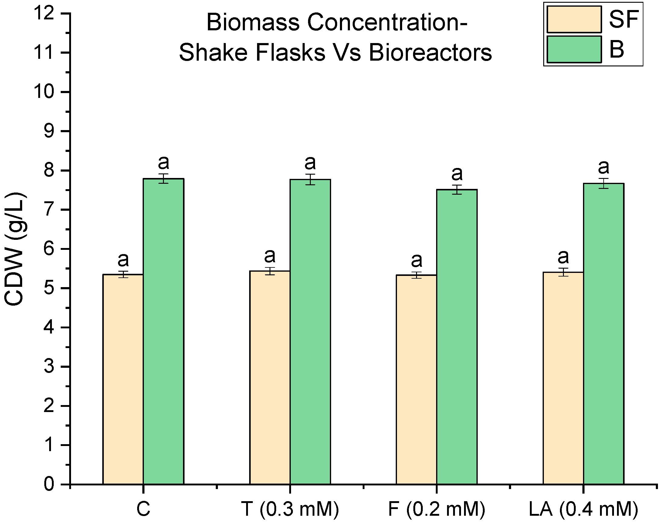
Figure 1. Biomass concentrations in M. purpureus C322 cultures cultivated in shake flasks (SF) and stirred-tank bioreactors (B). QSMs were added at the following concentrations: tyrosol (T, 0.3 mM), farnesol (F, 0.2 mM), and linoleic acid (LA, 0.4 mM). Control cultures (C) were not supplemented with QSMs. Data represent mean values from three biological replicates. Error bars indicate standard deviation. Statistical significance was determined using one-way ANOVA followed by Tukey’s post hoc test (p > 0.05).
Fermentation 11 00461 g001
Figure 2. Effect of quorum-sensing molecules on pigment production in M. purpureus C322, where (a) pigment production (OD) in shake flask cultures and (b) pigment production (OD) in stirred-tank bioreactors. Yellow, orange, and red pigments were quantified as OD400, OD470, and OD510, respectively. Treatments include control (C), tyrosol (T, 0.3 mM), farnesol (F, 0.2 mM), and linoleic acid (LA, 0.4 mM). Data represent mean values from triplicate cultures. Error bars indicate standard deviation (p < 0.01).
Fermentation 11 00461 g002
Figure 3. Three-dimensional surface plots depicting the fold increase in pigment and lovastatin production relative to the control (C) in M. purpureus C322 cultures under shake flask and stirred-tank bioreactor conditions, where (a) yellow pigment, (b) orange pigment, (c) red pigment, and (d) lovastatin. Each data point represents the mean of three replicates (p < 0.01).
Fermentation 11 00461 g003
Figure 4. Lovastatin production by M. purpureus C322 under shake flask (SF) and stirred-tank bioreactor (B) conditions. Lovastatin concentrations (mg/L) were measured for control cultures (C) and those treated with tyrosol (T, 0.3 mM), farnesol (F, 0.2 mM), and linoleic acid (LA, 0.4 mM). All experiments were performed in triplicate. Error bars show standard deviation (p < 0.01).
Fermentation 11 00461 g004
Figure 5. pH variation and carbohydrate consumption during fermentation of M. purpureus C322 under QSM treatments, where (a) shake flask and (b) 2.5 L stirred-tank bioreactors. Carbohydrate concentrations (CHO, g/L) are represented on the left y-axis and pH values on the right y-axis. Error bars indicate standard deviation from triplicate samples (p > 0.05).
Fermentation 11 00461 g005
Table 1. Fold increase in pigment concentration during scale-up from shake flasks to bioreactors in M. purpureus C322 cultures. Values represent within-treatment increases in OD for yellow (OD400), orange (OD470), and red (OD510) pigments (p < 0.01).
QSMYellow PigmentOrange PigmentRed Pigment
Control (C)1.521.481.48
Tyrosol (T)1.641.741.66
Farnesol (F)1.681.891.76
Linoleic Acid (LA)1.631.651.73
Table 2. Fold increase in lovastatin concentration during scale-up from shake flasks to bioreactors in M. purpureus C322 cultures (p < 0.01).
QSMFold Increase (Bioreactor/Flask)
Control (C)1.08
Tyrosol (T)1.15
Farnesol (F)1.30
Linoleic acid (LA)1.09
Disclaimer/Publisher’s Note: The statements, opinions and data contained in all publications are solely those of the individual author(s) and contributor(s) and not of MDPI and/or the editor(s). MDPI and/or the editor(s) disclaim responsibility for any injury to people or property resulting from any ideas, methods, instructions or products referred to in the content.