Article

Isolation and Characterization of Bacteriocin-like-Producing Companilactobacillus farciminis YLR-1 and the Inhibitory Activity of Bacteriocin Against Staphylococcus aureus

1
College of Animal Science and Technology, Jilin Agricultural Science and Technology University, Jilin 132101, China
2
Jilin Provincial Key Laboratory of Preventive Veterinary Medicine, Jilin 132101, China
3
Jilin Province Cross Regional Cooperation Technology Innovation Center of Porcine Main Disease Prevention and Control, Jilin 132101, China
4
Jilin Province Technology Innovation Center of Pig Ecological Breeding and Disease Prevention and Control, Jilin 132101, China
5
Jilin Sino-ROK Institute of Animal Science, Changchun Sci-Tech University, Changchun 130600, China
*
Authors to whom correspondence should be addressed.
Fermentation 2025, 11(8), 460; https://doi.org/10.3390/fermentation11080460
Submission received: 22 June 2025 / Revised: 30 July 2025 / Accepted: 7 August 2025 / Published: 11 August 2025
(This article belongs to the Section Probiotic Strains and Fermentation)

Abstract

This study aimed to identify a probiotic bacterium with antagonistic activity against the foodborne pathogen Staphylococcus aureus (S. aureus) and investigate the mechanism of its antibacterial components. Growth kinetics were analyzed to assess bacterial proliferation. Acid and bile salt tolerance are vital indicators for evaluating probiotic survival in the gastrointestinal tract. The results indicated that Companilactobacillus farciminis (C. farciminis) YLR-1 not only had high tolerance to salt conditions (0.03%, 0.3%, and 0.5%) but also has a high survival rate at pH 3–4. The bacteriocin-like inhibitory substance (BLIS) isolated from C. farciminis YLR-1 was dialyzed using a membrane with a molecular weight cut-off (MWCO) of 500 Da, followed by sodium dodecyl sulfate polyacrylamide gel electrophoresis (SDS-PAGE) analysis. The results indicate that the BLIS produced by C. farciminis YLR-1 is a small-molecule peptide. BLIS displayed pH tolerance within acidic and neutral environments (4–8) and exhibited thermostability. When treated with proteinase K, the antibacterial action of BLIS was found to be inactivated. Membrane disruption mechanisms were examined using fluorescence imaging and scanning electron microscopy (SEM). SEM and fluorescence imaging revealed that BLIS-induced membrane damage in S. aureus ATCC 25923 causes cytoplasmic leakage and cell death.

1. Introduction

Staphylococcus aureus is a leading foodborne pathogen responsible for widespread food poisoning and human infections. Its remarkable desiccation resistance enables survival in harsh environments, facilitating contamination across diverse food products, many of which provide nutrient-rich growth substrates for the bacterium [1]. The primary virulence factors of S. aureus, Staphylococcal enterotoxins, are associated with numerous foodborne illnesses and pose significant public health risks due to their potential infiltration into food chains during production, processing, or distribution [2]. Recent years have seen heightened concerns over S. aureus-related foodborne outbreaks, particularly in meat products. Symptoms of S. aureus foodborne illnesses—including nausea, abdominal cramps, vomiting, and hypersalivation—typically manifest acutely, with or without diarrhea [1,2,3]. Such outbreaks can escalate rapidly, affecting thousands of individuals. Annually, an estimated 76 million illnesses, 325,000 hospitalizations, and 5000 deaths are caused by foodborne diseases in the United States [1,4].
Food preservation remains a critical global challenge, with synthetic preservatives playing an essential role in extending shelf life and maintaining product quality. These additives inhibit oxidative degradation and microbial growth, thereby preserving sensory attributes such as flavor, texture, and appearance in processed foods. However, excessive reliance on synthetic preservatives has raised health concerns, including associations with carcinogenicity, allergic reactions, and cardiac arrhythmias [5]. Growing consumer awareness of the adverse effects of synthetic additives has driven demand for natural and traditional alternatives, perceived as safer and healthier [6]. Due to customer demand and their biological efficacy, natural preservatives are being increasing used in the food business to guarantee product safety and quality.
Lactic acid bacteria (LAB) play a pivotal role in fermentation by metabolizing nutrients in food substrates to produce diverse metabolites, including organic acids, hydrogen peroxide, acetoin, diacetyl, and antimicrobial agents such as bacteriocins [7]. Bacteriocins, which are ribosomally synthesized antimicrobial peptides or proteins secreted by specific bacterial strains, exhibit inhibitory or bactericidal activity against both closely and distantly related microorganisms [8]. Most LAB-derived bacteriocins exert their effects by degrading the peptidoglycan layer of cell walls or inducing pore formation in cytoplasmic membranes, ultimately leading to microbial cell death [9].
Bacteriocins are increasingly employed in the food industry due to their broad-spectrum antimicrobial activity, non-toxicity, and susceptibility to enzymatic degradation without adverse effects on human physiology [10,11]. Their potent antibacterial and antibiofilm properties have garnered significant interest as sustainable alternatives to synthetic preservatives [10,11,12]. Studies demonstrate that bacteriocins effectively inhibit pathogenic and spoilage microorganisms in model food systems, validating their utility as biopreservatives [11,12]. Notably, LAB-derived bacteriocins exhibit exceptional efficacy against diverse foodborne pathogens and spoilage organisms, achieving bactericidal effects at nanomolar concentrations while maintaining the sensory quality of food products [10,11].
Fermented foods are important parts of traditional food culture with a long history worldwide. Pickle or Paocai, as a Chinese traditional fermented vegetable, is rich in LAB [13]; the microbial community in Pickle is akin to an immense library of microorganisms. Its nutrient-rich substrates and open fermentation process foster microbial diversity, ultimately shaping its distinctive quality and flavor profiles [14]. This study successfully isolated bacteriocin-producing LAB from a wide variety of fermented foods. Notably, the selected strain, C. farciminis YLR-1, exhibited significant inhibitory activity against S. aureus. Furthermore, we evaluated the antibacterial mechanism and activity of a BLIS generated by strain C. farciminis YLR-1. Therefore, in response to the prevailing food safety risks in the current social environment, this study aims to identify a food preservative alternative that effectively inhibits foodborne pathogens while minimizing potential side effects.

2. Materials and Methods

2.1. Sample Collection and Bacterial Isolation

Samples of fermented foods, including pickled Chinese cabbage, pickled chili, pickled cabbage, pickled cowpea, pickled bamboo shoots, and pickled ginger, were procured from various local markets in Jilin, China, for the isolation of LAB. The samples were placed in sterile sampling tubes and transported to the laboratory on the same day. The isolation was performed using the method described previously [15]. Following serial dilution, a 2 g sample of each pickle was spread onto a De Man–Rogosa–Sharpe (MRS) agar culture medium (Hopobio, Qingdao, China) and incubated at 37 °C for 24 h. Among the isolated strains, the supernatant produced by the LAB strain YLR-1 exhibited the highest antibacterial activity against S. aureus ATCC 25923 and was selected as the subject of further research. The isolated LAB strain was preserved in MRS broth containing glycerol (25%, v/v) at −80 °C.

2.2. Lactic Acid Bacteria Identification Using 16S rRNA Sequence Analyses

The total genomic DNA of the isolated LAB strain YLR-1 was extracted using the rapid extraction kit (Aidlab, Beijing, China). A portion of the 16S rRNA gene was amplified using purified DNA as a template, the Polymerase Chain Reaction (PCR) technique, and the universal primer set, which consists of 27F (5′-AGAGTTTGATCCTG-GCTCAG-3′) and 1492R (5′-GGTTACCTTGTTACGAC TT-3′) [15]. An initial denaturation stage at 94 °C for 5 min, 30 cycles of denaturation at 94 °C for 1 min, annealing at 50 °C for 1 min, extension at 72 °C for 1 min, and a final extension step at 72 °C for 10 min comprised the amplification conditions of the 16S rRNA gene. After separating the PCR product using 1% agarose gel electrophoresis (Sangon Biotech, Shanghai, China), the gels were stained with 0.5 mg/mL ethidium bromide (Macklin, Shanghai, China) and captured on a camera using ultraviolet light (THBC-470, Shanghai, China) [8]. The PCR products were sent to Sangon Biotech, Changchun, China, for sequencing and were compared with those deposited in the GenBank database using the online BLAST program (http://www.ncbi.nlm.nih.gov/BLAST/) [16], accessed on 20 September 2023.

2.3. Sequencing and Sequence Assembly of the Genome

Using the Oxford Nanopore Technique, the whole genome of C. farciminis YLR-1 was sequenced. Canu v1.5 software was used to assemble the filtered reads for genome assembly. After masking putative functional genes, the whole genomes were scanned using the GenBlastA v1.0.4 software. Racon v3.4.3 software was used to further correct the Canu v1.5 assembly results, utilizing third-generation reads for increased accuracy.

2.4. Bacterial Growth Curve

To analyze the growth kinetics of C. farciminis YLR-1, a 1 mL aliquot of the C. farciminis YLR-1 overnight culture was centrifuged, resuspended in phosphate-buffered saline (PBS) (Sangon Biotech, Shanghai, China), inoculated into 300 mL of autoclaved MRS liquid medium (Hopobio, Qingdao, China), and cultured at 37 °C. Samples were collected at regular 2 h intervals to monitor bacterial growth and antimicrobial activity [17]. Antimicrobial activity against S. aureus ATCC 25923 was evaluated using the agar well diffusion method [18]. Briefly, an overnight culture (12 h) of S. aureus ATCC 25923 was adjusted to 1 × 10⁸ CFU/mL, then inoculated in 100 μL of this suspension and into 20 mL of soft agar medium. Aliquots (200 μL) of C. farciminis YLR-1 cell-free supernatants were loaded into pre-formed wells. After 24 h of incubation at 37 °C, zones of inhibition around the wells were measured and interpreted as evidence of antimicrobial production. All experiments were conducted in triplicate.

2.5. Tolerance to Low pH Conditions of C. farciminis YLR-1

The assay was adapted from [19] with minor modifications. The cells were suspended in PBS (OD600 ≈ 0.5) after 1 mL of the overnight culture was extracted by centrifugation at 10,000× g for 5 min. The suspension was adjusted to pH 2, 3, and 4 using HCl (SINOPEC, Nanjing, China) and incubated statically at 37 °C for 3 h. Samples were collected hourly (0–3 h) during incubation for viability analysis. Viable cells were enumerated on autoclaved MRS agar plates to assess the acid tolerance of C. farciminis YLR-1. All experiments were conducted in triplicate. Survival rates were calculated using the following formula:
Survival   rate   ( % ) = Final   ( Log   CFU / mL ) Initial   ( Log   CFU / mL ) ×   100 %

2.6. Determination of Bile Tolerance of C. farciminis YLR-1

The assay was adapted from [20] with minor modifications. Briefly, overnight cultures were transferred to fresh medium and incubated at 37 °C for 6 h. Cells were washed three times with sterile PBS and resuspended to an optical density of 0.5 at OD600. MRS liquid medium supplemented with bile salts (0.03%, 0.3%, and 0.5%) (Hongrun Baoshun, Beijing, China) was autoclaved at 121 °C for 15 min. After cooling, the bacterial suspension was inoculated into the medium and incubated at 37 °C. Samples were collected hourly (0–3 h) during incubation, and survival rates were calculated using Equation (1). All experiments were conducted in triplicate.

2.7. BLIS Production

The assay was adapted from [21] with minor modifications. C. farciminis YLR-1 was cultured in MRS broth at 37 °C for 24 h to produce BLIS. Cells were removed by centrifugation at 6000× g for 15 min, and the supernatant was filtered through a 0.22 μm sterile membrane. The filtrate was concentrated tenfold using a rotary evaporator (50 °C), re-filtered, and stored at 4 °C for further use.

2.8. Preliminary Extraction of YLR-1 BLIS

The BLIS was dialyzed against distilled water for 24 h using a 500 Da MWCO dialysis membrane (Sangon Biotech, Shanghai, China) [22,23]. Antimicrobial activity of the intra- and extra-dialysate fractions was assessed via the agar well diffusion assay, as described previously, using S. aureus ATCC 25923 as the indicator strain. All experiments were conducted in triplicate.

2.9. Effect of Temperature, pH, and Enzymes on Antimicrobial Activity

To evaluate pH stability, YLR-1 BLIS was incubated at pH 4–11 (in 1-unit increments) for 1 h at 37 °C, neutralized to pH 7, and held at room temperature for 30 min [8]. For enzymatic sensitivity, BLIS was treated with papain, trypsin, proteinase K, pepsin, and catalase (1 mg/mL; Sangon Biotech, Shanghai, China) at 37 °C for 2 h, followed by 10 min of boiling at 100 °C. C. farciminis YLR-1 cell-free supernatants served as controls. BLIS was exposed to temperatures ranging from 40 °C to 121 °C (15 min at 121 °C). Antimicrobial activity against S. aureus ATCC 25923 was assessed using the agar well diffusion assay, with untreated BLIS as the control [18]. All experiments were performed in triplicate.

2.10. SDS-PAGE and In Situ Activity Assay of YLR-1 BLIS

The molecular weight of YLR-1 BLIS was determined using SDS-PAGE coupled with an in situ antibacterial activity assay. Electrophoresis was performed using a discontinuous gel system: 4% stacking gel, 10% spacer gel, and 18% separating gel. Protein molecular weight markers (2.7–40 kDa; Sangon Biotech, Shanghai, China) were included for calibration. The gel was run at 80 V for 30 min, followed by 120 V for 1 h, and subsequently stained with Coomassie Brilliant Blue (Sangon Biotech, Shanghai, China) [24]. For in situ activity analysis, the unstained gel segment was overlaid onto Mueller–Hinton (MH) agar (Hopobio, Qingdao, China) inoculated with 50 μL of the S. aureus ATCC 25923 (1 × 10⁸ CFU/mL) culture. After 24 h incubation at 37 °C, inhibitory zones confirmed the presence of antimicrobial activity corresponding to YLR-1 BLIS [21]. The gels for Coomassie Brilliant Blue staining and the in situ activity assay were prepared simultaneously under identical conditions on the same apparatus.

2.11. Electron Microscopy Observation

The effect of YLR-1 BLIS on S. aureus ATCC 25923 membrane integrity was analyzed via SEM (hitachi SU8100, Tokyo, Japan). An overnight culture (12 h) of S. aureus ATCC 25923 was adjusted to 1 × 10⁸ CFU/mL, harvested by centrifugation, and resuspended in PBS. The bacterial suspension was mixed 1:1 (v/v) with YLR-1 BLIS in a 2 mL centrifuge tube and incubated for 30 min. Cells were washed with PBS and fixed overnight at 4 °C with 2.5% glutaraldehyde. Samples were collected, fixed, and dried, then mounted on carbon-coated conductive tape. Following sputter-coating with gold, specimens were imaged using SEM. Untreated S. aureus ATCC 25923 served as the negative control [25,26].

2.12. Live–Dead Staining Assay

Live–Dead staining gives an indication of possible membrane damage by the YLR-1 BLIS. In brief, S. aureus ATCC 25923 was prepared as described in the antimicrobial assay and resuspended in 85% Nacl to approximately 6 × 10⁷ CFU/mL. The assay was carried out following the protocol of the SYTO-9/PI dual staining Bacterial Viability Kit (Servicebio, Wuhan, China). These suspensions were then incubated with YLR-1 BLIS at 37 °C for 2 h. Among them, S. aureus ATCC 25923 without YLR-1 BLIS was used as a positive control. Following the incubation, the cells underwent three washes with 85% Nacl and were subsequently stained with SYTO 9 and PI for 15 min in the absence of light. The status of the bacteria was examined under dark-field conditions using a fluorescence microscope (Nikon, Tokyo, Japan).

3. Results

3.1. Identification of Bacterial Strain

The 16S rRNA sequence of C. farciminis YLR-1 was determined and compared against the reference sequences in the NCBI database, demonstrating 100% similarity with the C. farciminis strain PR13.

3.2. Genome Assembly Results Statistics

The complete genome sequence of C. farciminis YLR-1 was computationally analyzed to determine its key genomic characteristics. The genome comprises 2273 genes, scaffold and contig lengths of 2,379,900 bp each, and a GC content of 36.61%. These sequence data were deposited in GenBank under accession numbers CP162899 (chromosome) and CP162900 (plasmid).

3.3. Growth Kinetics of C. farciminis YLR-1

Growth data for C. farciminis YLR-1 were collected through 18 samplings over 36 h. Based on OD600 measurements (Figure 1), the strain reached peak growth at 24 h and transitioned into the decline phase after 32 h. Acidogenic activity, reflected by pH changes, increased significantly at 8 h, with the final pH stabilizing at approximately 3.1. Analysis of OD600 and pH trends identified the logarithmic growth phase at 8 h. The bacteriostatic zone diameter peaked at 28 h (23 mm), while antibacterial compound synthesis began at 18 h (18 mm), as indicated by the inhibition zone measurements. These findings enable targeted optimization of cultivation duration based on specific objectives. The original data for this experiment are provided in Supplementary Materials, Table S1.

3.4. Tolerance to Low pH Conditions of the Isolate

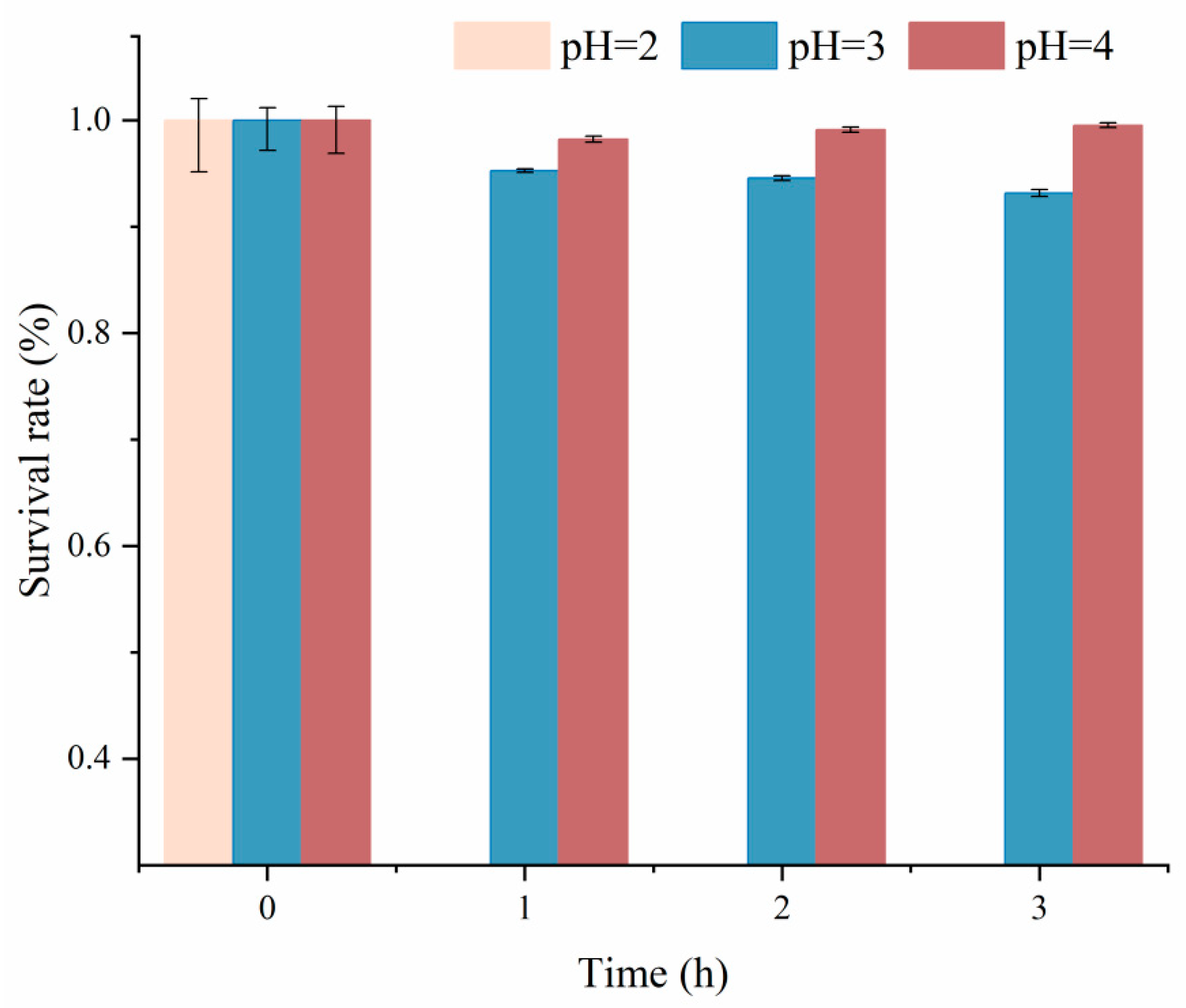
The survival rate of C. farciminis YLR-1 under simulated gastric juice conditions was evaluated. As shown in Figure 2, the strain exhibited no viability at pH 2 but demonstrated notable survival rates of 94.34 ± 0.34% and 98.97 ± 0.26% after 3 h at pH 3 and pH 4, respectively. These results indicate that C. farciminis YLR-1 tolerates moderate gastric acidity (pH 3–4) but cannot survive under highly acidic conditions (pH 2). The original data for this experiment are provided in Supplementary Materials, Table S2.

3.5. Bile Salt Tolerance Test

The bile salt tolerance of C. farciminis YLR-1 was assessed at concentrations of 0.03%, 0.3%, and 0.5%. Following 3 h exposure (Figure 3), the strain had high survival rates of 95.57 ± 0.76%, 92.45 ± 1.00%, and 87.50 ± 1.50%, respectively. These results demonstrate that C. farciminis YLR-1 exhibits robust tolerance to bile salts at the tested concentrations. The original data for this experiment are provided in Supplementary Materials, Table S3.

3.6. Rough Determination of Cutoff Membrane

As shown in Figure 4, the agar well diffusion assay was employed to estimate the molecular size range of antibacterial compounds in C. farciminis YLR-1 BLIS after dialysis. The results revealed strong antibacterial activity in compounds outside the dialysis membrane (Figure 4B), suggesting that active antibacterial molecules have a low molecular mass. Conversely, compounds retained within the membrane exhibited no inhibitory effects (Figure 4A). The original data for this experiment are provided in Supplementary Materials, Figure S1.

3.7. Heat, pH, and Enzymatic Stability of YLR-1 BLIS

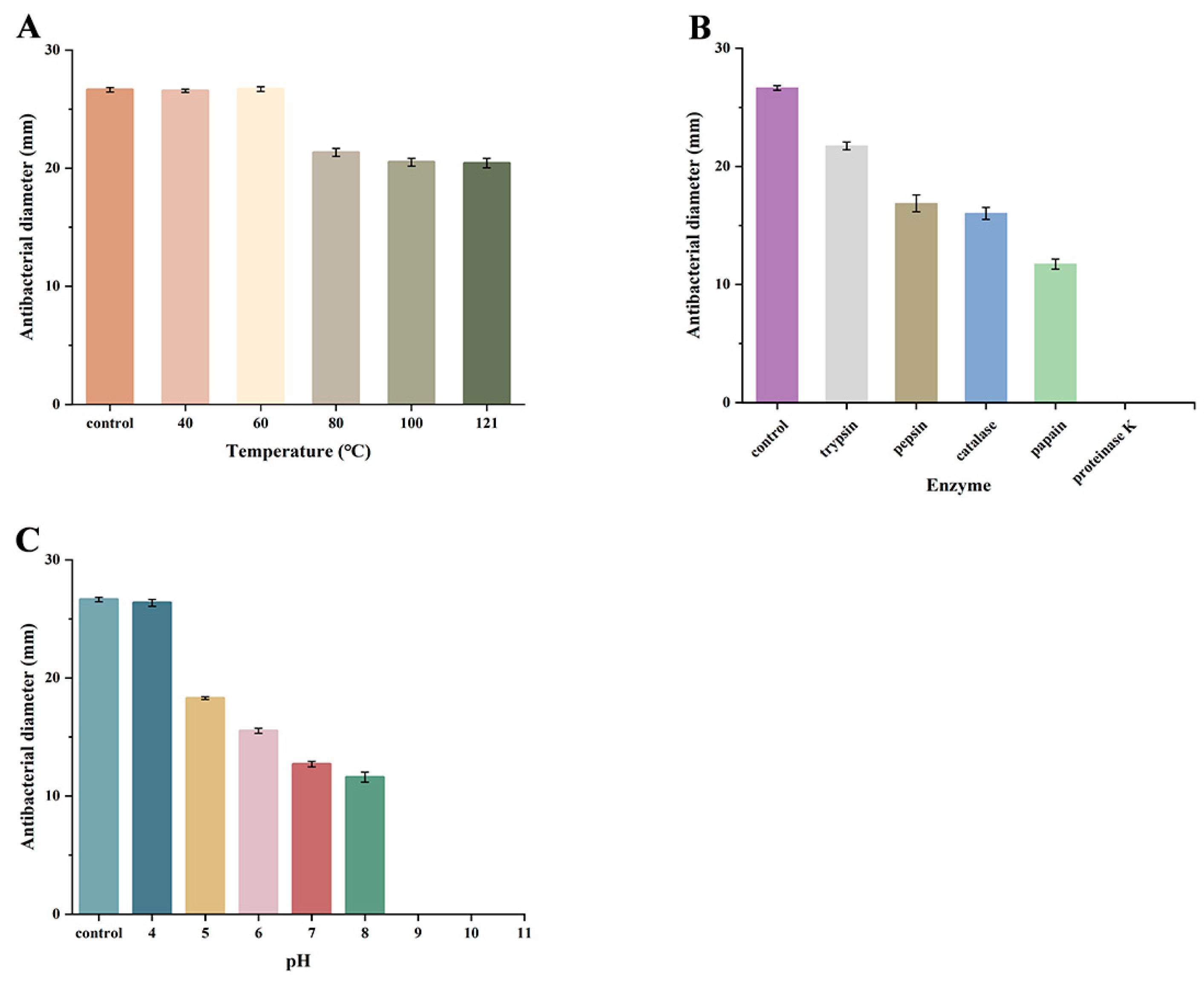
Figure 5 summarizes the stability characteristics of C. farciminis YLR-1 BLIS. Thermal stability was assessed by exposing YLR-1 BLIS to temperatures ranging from 40 °C to 121 °C. Remarkably, antibacterial activity remained fully intact even at 121 °C for 15 min (Figure 5A). Enzyme sensitivity assays demonstrated that YLR-1 BLISs were entirely inactivated by proteinase K, while pepsin, catalase, papain, and trypsin only partially reduced their activity (Figure 5B). To evaluate pH effects on antibacterial activity, YLR-1 BLIS was adjusted to pH 4–11 and compared to untreated controls. The substance retained greater activity under acidic conditions (pH 4–6) than at neutral pH (7–8), but this activity was terminated under alkaline conditions (pH 9–11) (Figure 5C). The original data for this experiment are provided in Supplementary Materials, Table S4.

3.8. SDS-PAGE Analysis of BLIS Produced by C. farciminis YLR-1

SDS-PAGE analysis of the crude YLR-1 BLIS extract revealed a single protein band with a molecular mass below 2.7 kDa (Figure 6). In situ activity assays demonstrated clear inhibition zones against S. aureus ATCC 25923, corresponding to the same molecular mass range (<2.7 kDa). These findings collectively confirm that YLR-1 BLIS comprises low-molecular-mass antimicrobial peptides. The original data for this experiment are provided in Supplementary Materials, Figure S2.

3.9. SEM Analysis

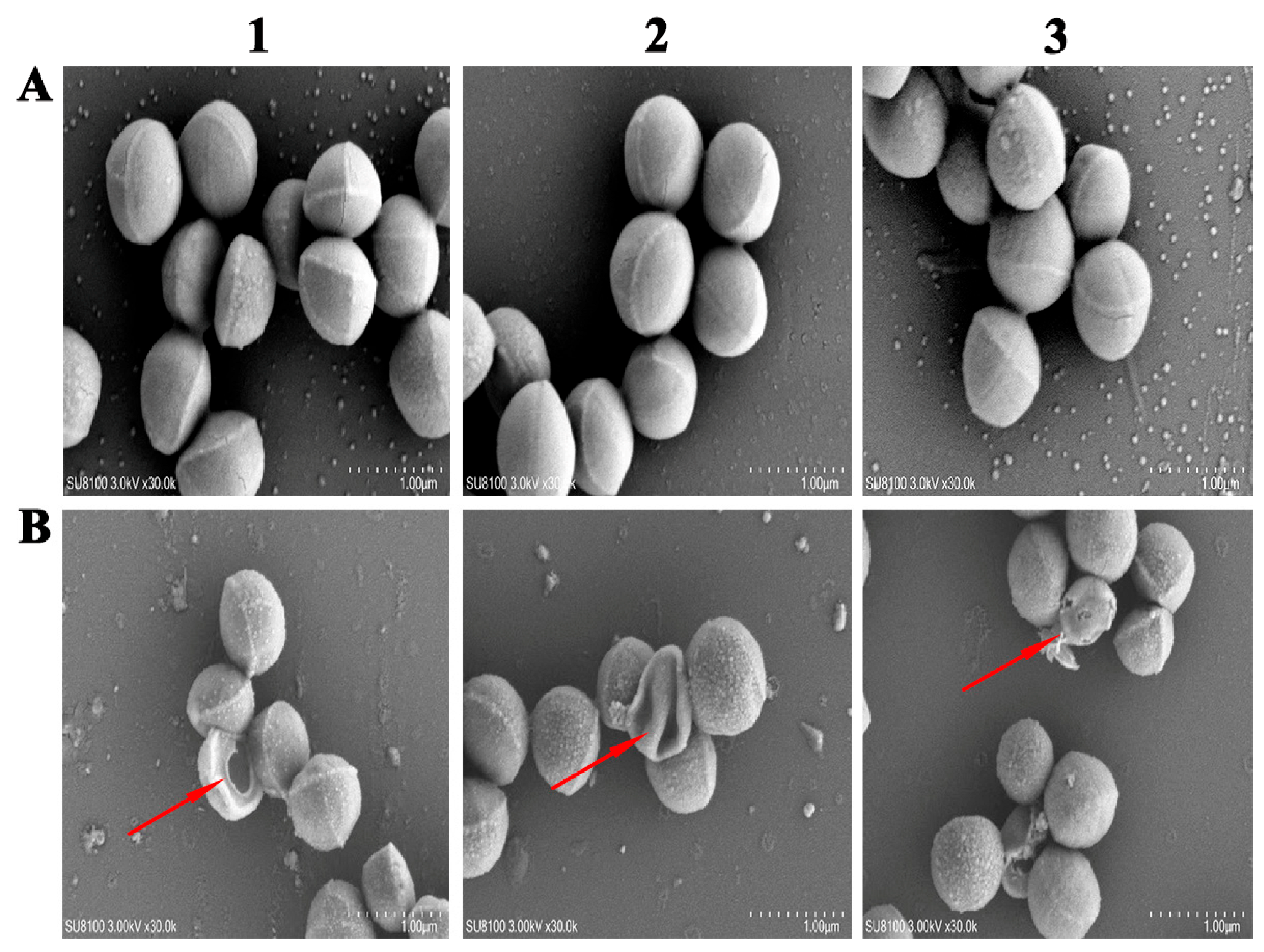
SEM analysis revealed stark morphological differences between untreated S. aureus ATCC 25923 controls and cells exposed to YLR-1 BLIS (Figure 7). Untreated cells (panels A1–A3) exhibited intact, smooth surfaces, whereas YLR-1 BLIS-treated S. aureus ATCC 25923 displayed significant surface degradation, morphological distortion, and cell collapse. High-magnification micrographs (panels B1–B3) further demonstrated perforations, extensive surface roughness, cellular shrinkage, and lysed cell debris (The region highlighted by the red arrow in Figure 7). These structural disruptions confirm the bactericidal effects of YLR-1 BLIS against S. aureus ATCC 25923. The original data for this experiment are provided in Supplementary Materials, Figure S3.

3.10. Bactericidal Effect Observed Using Fluorescence Microscope

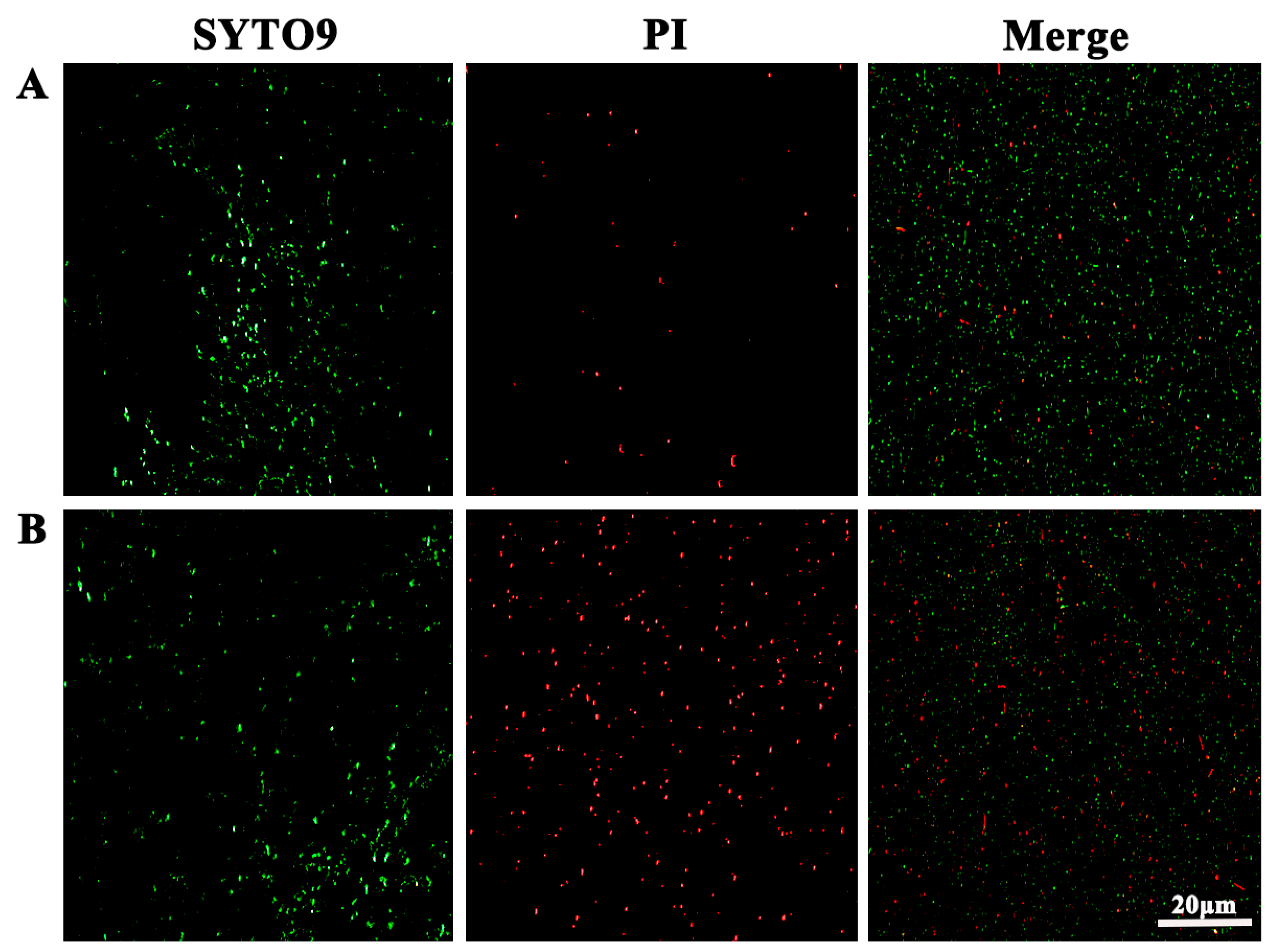
The bactericidal effect of YLR-1 BLIS on S. aureus ATCC 25923 was assessed via fluorescence microscopy using PI and SYTO 9 (Figure 8). Notably, SYTO 9, a membrane-permeable green, fluorescent nucleic acid stain, labels all cells regardless of membrane integrity, whereas PI, a red fluorescent counterstain, selectively penetrates cells with compromised membranes. In the control group (Figure 8A), cells exhibited predominantly green fluorescence (SYTO 9, labeling both viable and non-viable cells) and minimal red fluorescence (PI, specific to membrane-damaged cells), confirming high bacterial viability. In contrast, YLR-1 BLIS-treated samples (Figure 8B) displayed a marked increase in red fluorescence intensity, indicating the significant disruption of bacterial inner membrane permeability. The original data for this experiment are provided in Supplementary Materials, Figure S4.

4. Discussion

S. aureus-mediated food poisoning persists as a widespread yet underdiagnosed public health concern, imposing significant socioeconomic burdens due to healthcare expenditures, productivity decline, and financial losses in food production and hospitality sectors [2,27]. This study sought to isolate C. farciminis YLR-1 from fermented pickle samples and characterize its BLIS production as a natural antimicrobial agent against S. aureus. The strain was successfully isolated and identified; subsequently, the key physicochemical properties and antibacterial effects of its BLIS were evaluated. This study identified a bacteriocin from YLR-1 that exhibits inhibitory activity against S. aureus. Similar results were recorded for other bacteriocins [28,29]. Currently, there is a limited number of research articles focusing on the bacteriocin associated with this strain.
The pH changes and BLIS production of strain YLR-1 are shown in Figure 1. With S. aureus ATCC 25923 as the indicator organism, BLIS production by YLR-1 began at 18 h and peaked at 28 h. Also, the changes in pH values decreased from 4.83 (at the beginning) to 3.1 (at the end of the screening), reaching their minimum (pH 3.1) after 28 h of growth. The growth-phase-dependent production and subsequent decline of BLIS antibacterial activity suggest that this bacteriocin may function as a secondary metabolite [17,30]. To assess the probiotic potential of YLR-1, we evaluated its tolerance to acidic conditions and bile salts. The first criterion evaluated was the ability of YLR-1 to tolerate low pH conditions. In 3 h of simulated gastric digestion, significant reductions were observed at pH 3 and 4, with no survival at pH 2. However, considering the buffering capacity of food matrices, these harsh conditions do not fully represent actual gastric environments in animals and humans [19,31]. Thus, good probiotic strains should be able to thrive at pH levels of at least 3.0 in the stomach [32]. Therefore, in previous studies, most in vitro assays have tested tolerance to acidity at pH 3.0 and above [15,33]. Beyond the extreme acidity of the gastric environment, the high bile salt concentration in the intestinal tract represents another critical factor influencing probiotic survival [27]. To function effectively, probiotics must tolerate bile acids synthesized from cholesterol in the hepatobiliary system and secreted into the duodenum via the gallbladder [19]. Previous studies indicate that a bile salt concentration of 0.3% serves as the threshold for selecting resistant probiotic isolates [31]. YLR-1 demonstrated excellent bile salt resistance, with survival rates of 95.57 ± 0.76%, 92.45 ± 1.0%, and 87.5 ± 1.5% at bile salt concentrations of 0.03%, 0.3%, and 0.5%, respectively. In this study, a satisfactorily acceptable survival rate was obtained in pH 4–5 and 0.03–0.5% bile salt conditions. Similar results were observed for nine strains of LAB isolated from Kefir samples, with Lactobacillus pallantarum, Lactobacillus harbinensis, and Lactobacillus paracasei being the predominant isolates [19]. These results indicate that YLR-1 has promising probiotic potential. In this study, preliminary extraction was carried out using the dialysis membrane method as described in the previous study [23], followed by molecular weight determination via SDS-PAGE and in situ activity assays, which revealed that YLR-1 BLISs are tiny antimicrobial peptides. The molecular size is lower than that of most previously identified bacteriocins, such as a 9 kDa bacteriocin produced by Pseudomonas aeruginosa UCM B-353 [34], a 24 kDa bacteriocin produced by Latilactobacillus sakei 205 [30], and a 68 kDa bacteriocin produced by Lactobacillus casei TA0021 [35].
Stability assays indicated that antimicrobial activity was retained across pH 4–8, though activity was lost under alkaline conditions (pH 9–11), highlighting its adaptability to acidic and neutral environments. Complete inactivation of antimicrobial activity was observed following treatment with the proteolytic enzyme proteinase K, confirming its protein-based composition (Figure 5). A smaller reduction in activity occurred with other proteases (papain, trypsin, and pepsin), and following catalase treatment, it was revealed that the active moiety of the antimicrobial substance seemed to be protein in nature. These findings align with prior studies reporting the inhibition of various bacteriocins by proteinase K [17,30,36], as well as pepsin and papain [8]. Notably, YLR-1 BLIS exhibited exceptional thermal stability, maintaining efficacy across the tested temperature range (55–121 °C). The high-temperature stability of YLR-1 BLIS enables it to resist conventional pasteurization or sterilization heat treatments commonly employed in the food industry [37]. Similar results were observed for other bacteriocins produced by Lactobacillus strains [8,17]. Although bacteriocins often show resistance to heat, it is important to note that this property is not uniformly present in all types [36]. SEM and SYTO-9/PI dual staining revealed that YLR-1 BLIS induced severe morphological damage to S. aureus ATCC 25923, including membrane shrinkage, perforations, and an increased number of PI-positive cells. These findings suggest that the bactericidal mechanism of YLR-1 BLIS may involve binding to bacterial membranes, where it disrupts membrane integrity and induces cytoplasmic content efflux, ultimately resulting in the cell death of S. aureus ATCC 25923. Other studies have also demonstrated the killing effect of bacteriocins [26,38,39]. The YLR-1 BLIS identified in this study exhibited a molecular weight below 2.7 kDa, thermal stability, susceptibility to enzymatic hydrolysis, and a bactericidal mechanism involving cell membrane disruption. These findings suggest that YLR-1 BLIS may be an unconventional bacteriocin.

5. Conclusions

In conclusion, C. farciminis YLR-1—a bacteriocin-producing strain—demonstrates promising potential for development as a probiotic. Acid tolerance (pH 3–4) and bile salt resistance (up to 0.5%) assays support its candidacy for gastrointestinal survival. The BLIS from YLR-1 demonstrated robust physicochemical stability and induced S. aureus lysis and death by disrupting outer membrane integrity. These findings highlight YLR-1 BLIS as a potential antibacterial agent for clinical or food-industry applications.

Supplementary Materials

The following supporting information can be downloaded at https://www.mdpi.com/article/10.3390/fermentation11080460/s1.

Author Contributions

L.Y.: Writing—review and editing, Writing—original draft, Conceptualization, Investigation, Resources, Information analysis, Visualization, and Data curation. H.S.: Investigation, Data curation, and Formal analysis. J.W.: Formal analysis, Investigation. S.S.: Information analysis, and Data curation. S.L.: Information analysis. B.Y.: Writing—review, Methodology, Supervision, and Data curation. W.D.: Writing—review, Project administration, and Methodology. G.L.: Writing—review, Project administration, and Methodology. All authors have read and agreed to the published version of the manuscript.

Funding

This research received no external funding.

Institutional Review Board Statement

Not applicable.

Informed Consent Statement

Not applicable.

Data Availability Statement

The original contributions presented in this study are included in the article and Supplementary Materials. Further inquiries can be directed to the corresponding authors.

Acknowledgments

We extend our sincere gratitude to our supervisor for their expert guidance and unwavering encouragement throughout this research. Special thanks are also due to our dedicated laboratory colleagues for their invaluable technical assistance and insightful discussions. Their collective contributions were pivotal in advancing this work and significantly enhanced the quality and efficiency of our experimental procedures and data analysis.

Conflicts of Interest

The authors declare no conflicts of interest.

References

  1. Kadariya, J.; Smith, T.C.; Thapaliya, D. Staphylococcus aureus and Staphylococcal Food-Borne Disease: An Ongoing Challenge in Public Health. BioMed Res. Int. 2014, 1, 827965. [Google Scholar] [CrossRef]
  2. Argudín, M.Á.; Mendoza, M.C.; Rodicio, M.R. Food Poisoning and Staphylococcus aureus Enterotoxins. Toxins 2010, 2, 1751–1773. [Google Scholar] [CrossRef]
  3. Mancusi, A.; Egidio, M.; Marrone, R.; Scotti, L.; Paludi, D.; Dini, I.; Proroga, Y.T.R. The In Vitro Antibacterial Activity of Argirium SUNc against Most Common Pathogenic and Spoilage Food Bacteria. Antibiotics 2024, 13, 109. [Google Scholar] [CrossRef]
  4. Newell, D.G.; Koopmans, M.; Verhoef, L.; Duizer, E.; Aidara-Kane, A.; Sprong, H.; Opsteegh, M.; Langelaar, M.; Threfall, J.; Scheutz, F.; et al. Food-borne diseases—The challenges of 20years ago still persist while new ones continue to emerge. Int. J. Food Microbiol. 2010, 139, S3–S15. [Google Scholar] [CrossRef]
  5. Mavani, N.R.; Mohd Ali, J.; Hussain, M.A.; Rahman, N.A.; Hashim, H. Determining food safety in canned food using fuzzy logic based on sulphur dioxide, benzoic acid and sorbic acid concentration. Heliyon 2024, 10, e26273. [Google Scholar] [CrossRef]
  6. Settanni, L.; Corsetti, A. Application of bacteriocins in vegetable food biopreservation. Int. J. Food Microbiol. 2008, 121, 123–138. [Google Scholar] [CrossRef]
  7. O’sullivan, L.; Ross, R.; Hill, C.J.B. Potential of bacteriocin-producing lactic acid bacteria for improvements in food safety and quality. Biochimie 2002, 84, 593–604. [Google Scholar] [CrossRef]
  8. Odah, K.A.; Dong, W.-L.; Lei, L.; Atiah, L.A.; Wang, Y.-m.; Kong, L.-C.; Ma, H.-X. Isolation, Identification, and Characterization of a Novel Bacteriocin Produced by Brevibacillus laterosporus DS-3 Against Methicillin-Resistant Staphylococcus aureus (MRSA). Int. J. Pept. Res. Ther. 2019, 26, 709–715. [Google Scholar] [CrossRef]
  9. Ahmad, V.; Khan, M.S.; Jamal, Q.M.S.; Alzohairy, M.A.; Al Karaawi, M.A.; Siddiqui, M.U. Antimicrobial potential of bacteriocins: In therapy, agriculture and food preservation. Int. J. Antimicrob. Agents 2017, 49, 1–11. [Google Scholar] [CrossRef] [PubMed]
  10. Field, D.; Ross, R.P.; Hill, C. Developing bacteriocins of lactic acid bacteria into next generation biopreservatives. Curr. Opin. Food Sci. 2018, 20, 1–6. [Google Scholar] [CrossRef]
  11. O’Connor, P.M.; Ross, R.P.; Hill, C.; Cotter, P.D. Antimicrobial antagonists against food pathogens: A bacteriocin perspective. Curr. Opin. Food Sci. 2015, 2, 51–57. [Google Scholar] [CrossRef]
  12. Mathur, H.; Field, D.; Rea, M.C.; Cotter, P.D.; Hill, C.; Ross, R.P. Bacteriocin-Antimicrobial Synergy: A Medical and Food Perspective. Front. Microbiol. 2017, 8, 1205. [Google Scholar] [CrossRef] [PubMed]
  13. Liu, T.; Zhou, K.; Yin, S.; Liu, S.; Zhu, Y.; Yang, Y.; Wang, C. Purification and characterization of an exopolysaccharide produced by Lactobacillus plantarum HY isolated from home-made Sichuan Pickle. Int. J. Biol. Macromol. 2019, 134, 516–526. [Google Scholar] [CrossRef] [PubMed]
  14. Wang, Y.; Chenhao, Z.; Fengsong, L.; Zhengyu, J.; Xia, X. Ecological succession and functional characteristics of lactic acid bacteria in traditional fermented foods. Crit. Rev. Food Sci. Nutr. 2023, 63, 5841–5855. [Google Scholar] [CrossRef]
  15. Thao, T.T.P.; Thoa, L.T.K.; Ngoc, L.M.T.; Lan, T.T.P.; Phuong, T.V.; Truong, H.T.H.; Khoo, K.S.; Manickam, S.; Hoa, T.T.; Tram, N.D.Q.; et al. Characterization halotolerant lactic acid bacteria Pediococcus pentosaceus HN10 and in vivo evaluation for bacterial pathogens inhibition. Chem. Eng. Process. Process Intensif. 2021, 168, 108576. [Google Scholar] [CrossRef]
  16. Leite, A.M.O.; Miguel, M.A.L.; Peixoto, R.S.; Ruas-Madiedo, P.; Paschoalin, V.M.F.; Mayo, B.; Delgado, S. Probiotic potential of selected lactic acid bacteria strains isolated from Brazilian kefir grains. J. Dairy Sci. 2015, 98, 3622–3632. [Google Scholar] [CrossRef]
  17. Chen, C.; Chen, X.; Jiang, M.; Rui, X.; Li, W.; Dong, M. A newly discovered bacteriocin from Weissella hellenica D1501 associated with Chinese Dong fermented meat (Nanx Wudl). Food Control 2014, 42, 116–124. [Google Scholar] [CrossRef]
  18. Cloeckaert, A.; Miljkovic, M.; Jovanovic, S.; O’Connor, P.M.; Mirkovic, N.; Jovcic, B.; Filipic, B.; Dinic, M.; Studholme, D.J.; Fira, D.; et al. Brevibacillus laterosporus strains BGSP7, BGSP9 and BGSP11 isolated from silage produce broad spectrum multi-antimicrobials. PLoS ONE 2019, 14, e0216773. [Google Scholar] [CrossRef]
  19. Talib, N.; Mohamad, N.E.; Yeap, S.K.; Hussin, Y.; Aziz, M.N.M.; Masarudin, M.J.; Sharifuddin, S.A.; Hui, Y.W.; Ho, C.L.; Alitheen, N.B. Isolation and Characterization of Lactobacillus spp. from Kefir Samples in Malaysia. Molecules 2019, 24, 2606. [Google Scholar] [CrossRef]
  20. Lu, H.; Yang, P.; Zhong, M.; Bilal, M.; Xu, H.; Zhang, Q.; Xu, J.; Liang, N.; Liu, S.; Zhao, L.; et al. Isolation of a potential probiotic strain Bacillus amyloliquefaciens LPB-18 and identification of antimicrobial compounds responsible for inhibition of food-borne pathogens. Food Sci. Nutr. 2022, 11, 2186–2196. [Google Scholar] [CrossRef]
  21. Arakawa, K.; Yoshida, S.; Aikawa, H.; Hano, C.; Bolormaa, T.; Burenjargal, S.; Miyamoto, T. Production of a bacteriocin-like inhibitory substance by Leuconostoc mesenteroides subsp. dextranicum 213M0 isolated from Mongolian fermented mare milk, airag. Anim. Sci. J. 2015, 87, 449–456. [Google Scholar] [CrossRef]
  22. Mandal, V.; Sen, S.K.; Mandal, N.C. Optimized culture conditions for bacteriocin production by Pediococcus acidilactici LAB 5 and its characterization. Indian J. Biochem. Biophys. 2008, 45, 106–110. [Google Scholar] [PubMed]
  23. Janes, M.E.; Nannapaneni, R.; Proctor, A.; Johnson, M.G. Rice Hull Ash and Silicic Acid as Adsorbents for Concentration of Bacteriocins. Appl. Environ. Microbiol. 1998, 64, 4403–4409. [Google Scholar] [CrossRef] [PubMed]
  24. Wang, Y.; Haqmal, M.A.; Liang, Y.d.; Muhammad, I.; Zhao, X.O.; Elken, E.M.; Gao, Y.H.; Jia, Y.; He, C.g.; Wang, Y.M.; et al. Antibacterial activity and cytotoxicity of a novel bacteriocin isolated from Pseudomonas sp. strain 166. Microb. Biotechnol. 2022, 15, 2337–2350. [Google Scholar] [CrossRef] [PubMed]
  25. Polat, T.; Soyhan, İ.; Cebeci, S.; İldeniz, T.A.Ö.; Gök, Ö.; Elmas, M.A.; Mozioğlu, E.; Ünübol, N. New-generation biofilm effective antimicrobial peptides and a real-time anti-biofilm activity assay: CoMIC. Appl. Microbiol. Biotechnol. 2024, 108, 316. [Google Scholar] [CrossRef]
  26. Zhang, W.; An, Z.; Bai, Y.; Zhou, Y.; Chen, F.; Wang, K.-J. A novel antimicrobial peptide Scyreptin1-30 from Scylla paramamosain exhibiting potential therapy of Pseudomonas aeruginosa early infection in a mouse burn wound model. Biochem. Pharmacol. 2023, 218, 115917. [Google Scholar] [CrossRef]
  27. Wieneke, A.A.; Roberts, D.; Gilbert, R.J. Staphylococcal food poisoning in the United Kingdom, 1969–1990. Epidemiol. Infect. 2009, 110, 519–531. [Google Scholar] [CrossRef]
  28. Andersson, R.J.I.J.o.F.M. Inhibition of Staphylococcus aureus and spheroplasts of Gram-negative bacteria by an antagonistic compound produced by a strain of Lactobacillus plantarum. Int. J. Food Microbiol. 1986, 3, 149–160. [Google Scholar] [CrossRef]
  29. Fagheei Aghmiyuni, Z.; Saderi, H.; Owlia, P.; Saidi, N.; Laranjo, M. Evaluation of the Effect of Lactobacillus acidophilus ATCC 4356 Bacteriocin against Staphylococcus aureus. BioMed Res. Int. 2024, 2024, 4119960. [Google Scholar] [CrossRef]
  30. Martín, I.; Barbosa, J.; Pereira, S.I.A.; Rodríguez, A.; Córdoba, J.J.; Teixeira, P. Study of lactic acid bacteria isolated from traditional ripened foods and partial characterization of their bacteriocins. LWT 2023, 173, 114300. [Google Scholar] [CrossRef]
  31. Yasar, S.; Yerlikaya, S.; Sen Arslan, H.; Akgul, K.; Simsek, H. Determination of probiotic properties of lactic acid bacteria and yeasts isolated from three lacto-fermented cereals mixed with whey, citrus, and tomato pomace. J. Food Process. Preserv. 2022, 46, e17231. [Google Scholar] [CrossRef]
  32. Albene, D.; Lema, N.K.; Tesfaye, G.; Andeta, A.F.; Ali, K.; Guadie, A. Probiotic potential of lactic acid bacteria isolated from Ethiopian traditional fermented Cheka beverage. Ann. Microbiol. 2024, 74, 25. [Google Scholar] [CrossRef]
  33. Kato, K.; Serata, M.; Nakamura, M.; Ando, M.; Suzuki, T.; Okumura, T. Cell wall polysaccharide enhances Lacticaseibacillus paracasei strain Shirota growth in milk and contributes to acid and bile tolerance. Int. J. Food Microbiol. 2024, 422, 110811. [Google Scholar] [CrossRef] [PubMed]
  34. Balko, O.B. Low Molecular Weight Pseudomonas aeruginosa Bacteriocins. Mikrobiolohichnyi Zhurnal 2019, 81, 97–109. [Google Scholar] [CrossRef]
  35. Noroozi, E.; Mojgani, N.; Motevaseli, E.; Modarressi, M.H.; Tebianian, M.J.I.J.o.M. Physico-chemical and cytotoxic analysis of a novel large molecular weight bacteriocin produced by Lactobacillus casei TA0021. Iran. J. Microbiol. 2019, 11, 397. [Google Scholar] [CrossRef] [PubMed]
  36. Wang, Y.; Fu, X.; Wang, Y.; Wang, J.; Kong, L.; Guo, H. Antibacterial Activity and Cytotoxicity of the Novel Bacteriocin Pkmh. Int. J. Mol. Sci. 2024, 25, 9153. [Google Scholar] [CrossRef]
  37. Bédard, F.; Biron, E. Recent Progress in the Chemical Synthesis of Class II and S-Glycosylated Bacteriocins. Front. Microbiol. 2018, 9, 1048. [Google Scholar] [CrossRef]
  38. Zhang, S.; Yang, N.; Mao, R.; Hao, Y.; Teng, D.; Wang, J. In Vitro/Vivo Mechanisms of Antibacterial Peptide NZ2114 against Staphylococcus pseudintermedius and Its Biofilms. Antibiotics 2024, 13, 341. [Google Scholar] [CrossRef]
  39. Thuy, T.T.D.; Lu, H.-F.; Bregente, C.J.B.; Huang, F.-C.A.; Tu, P.-C.; Kao, C.-Y. Characterization of the broad-spectrum antibacterial activity of bacteriocin-like inhibitory substance-producing probiotics isolated from fermented foods. BMC Microbiol. 2024, 24, 85. [Google Scholar] [CrossRef]
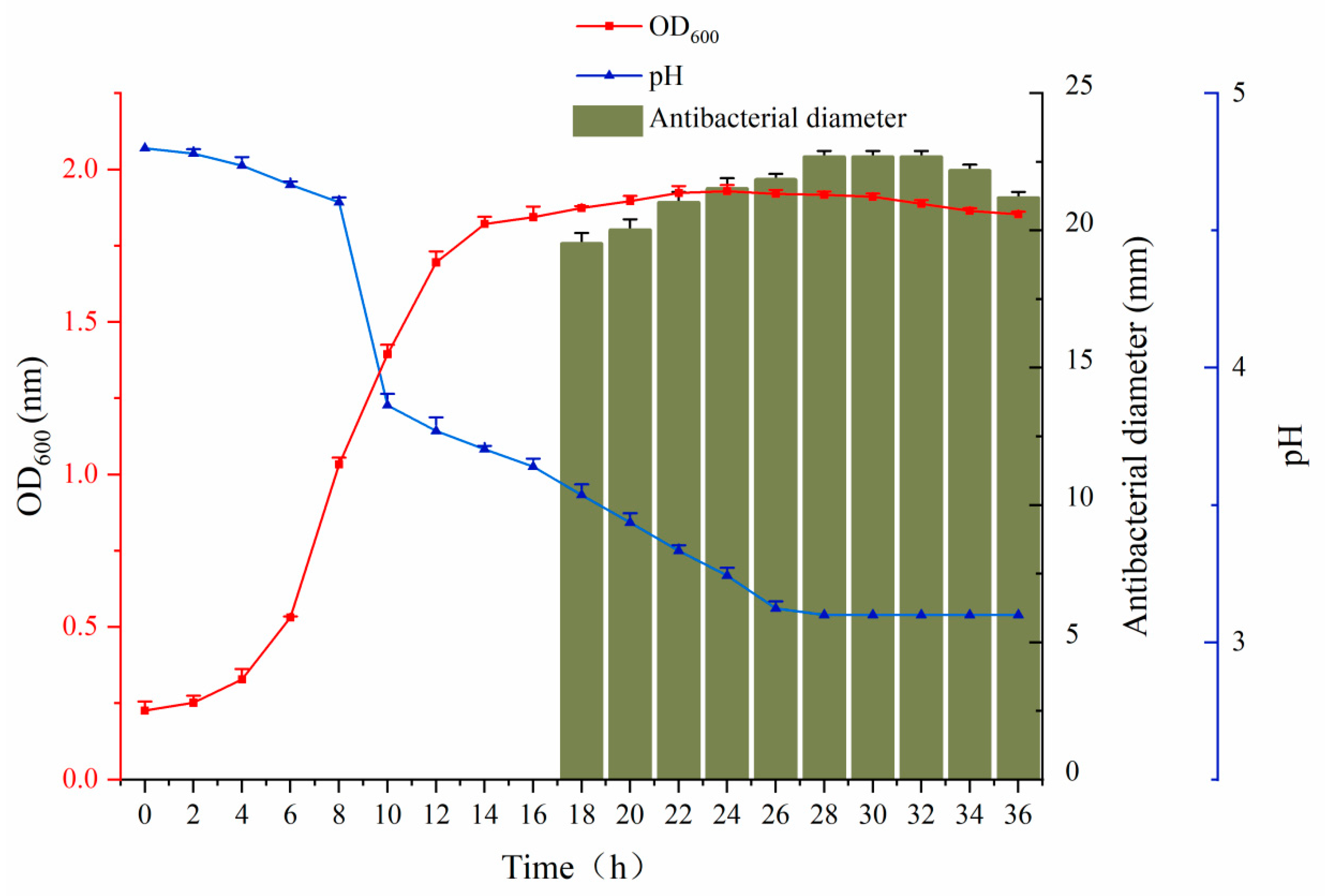
Figure 1. The growth kinetics of C. farciminis YLR-1. The red line represents the growth curve of strain C. farciminis YLR-1. The blue line represents the changing value of pH. The rectangular graph shows the size of the antibacterial diameter. The data are the average of three replicates, and error bars indicate standard deviations.
Fermentation 11 00460 g001
Figure 2. The survival rate of C. farciminis YLR-1 after incubation at acidic conditions (pH 2–4). The data are the average of three replicates, and error bars indicate standard deviations.
Fermentation 11 00460 g002
Figure 3. The resistance of C. farciminis YLR-1 to bile salts. The data are the average of three replicates, and error bars indicate standard deviations.
Fermentation 11 00460 g003
Figure 4. The inhibition zone of YLR-1 BLIS on MH agar. Region A shows the antibacterial results of substances in the dialysis membrane; Region B shows the antibacterial results of substances outside the dialysis membrane.
Fermentation 11 00460 g004
Figure 5. Stability analysis of YLR-1 BLIS against temperature, pH, and enzymes. (A) The effect of temperature on the stability of YLR-1 BLIS; (B) the effect of enzymes on the stability of YLR-1 BLIS; (C) the effect of pH on the stability of YLR-1 BLIS. The data are the average of three replicates, and error bars indicate standard deviations.
Fermentation 11 00460 g005
Figure 6. Inhibitory zones on the in situ antibacterial activity assay after sodium dodecyl SDS-PAGE was performed on cell-free culture supernatants of C. farciminis YLR-1. Lane 1: molecular weight markers; lane 2: YLR-1 BLIS loaded on the gel (the peptide found is indicated by an arrow); lane 3: gel with the same samples overlayed with MH-soft agar containing S. aureus ATCC 25923 after incubation overnight at 37 °C.
Fermentation 11 00460 g006
Figure 7. Electron microscopy images of S. aureus ATCC 25923 treated with YLR-1 BLIS. Panels (A1A3): control, untreated S. aureus ATCC 25923. Panels (B1B3): S. aureus ATCC 25923 treated with YLR-1 BLIS.
Fermentation 11 00460 g007
Figure 8. Fluorescence microscopy of S. aureus ATCC 25923 bacteria treated with YLR-1 BLIS. Panels (A): control, untreated bacteria. Panels (B): S. aureus ATCC 25923 was treated with YLR-1 BLIS for 2 h. The cells were stained with SYTO 9 and PI. Fluorescence images for SYTO 9 (green fluorescence; left panels) and PI (red fluorescence; middle panels); merged images (right panels) are shown.
Fermentation 11 00460 g008
Disclaimer/Publisher’s Note: The statements, opinions and data contained in all publications are solely those of the individual author(s) and contributor(s) and not of MDPI and/or the editor(s). MDPI and/or the editor(s) disclaim responsibility for any injury to people or property resulting from any ideas, methods, instructions or products referred to in the content.