Article

From Lebanese Soil to Antimicrobials: A Novel Streptomyces Species with Antimicrobial Potential

by
Razane Hamiyeh
1,2,†,
Aya Hanna
1,2,† and
Antoine Abou Fayad
1,2,*
1
Department of Experimental Pathology, Immunology and Microbiology, Faculty of Medicine, American University of Beirut, 1107-2020 Beirut, Lebanon
2
Center for Drug Discovery, American University of Beirut, 1107-2020 Beirut, Lebanon
*
Author to whom correspondence should be addressed.
These authors contributed equally to this work.
Fermentation 2025, 11(7), 406; https://doi.org/10.3390/fermentation11070406
Submission received: 28 May 2025 / Revised: 27 June 2025 / Accepted: 28 June 2025 / Published: 15 July 2025
(This article belongs to the Section Microbial Metabolism, Physiology & Genetics)

Abstract

The ongoing threat of antimicrobial-resistant pathogens has intensified the need for new antimicrobial agents, making the discovery of novel natural products crucial. This study focuses on the isolation and characterization of a novel Streptomyces species from the Anjar region in Lebanon, an area rich in microbial diversity that is largely unexplored for its biotechnological potential. Soil samples were collected and processed, leading to the isolation of Streptomyces strain ANJ10. Comprehensive morphological, physiological, and genomic analyses were conducted, including whole-genome sequencing (WGS) to identify biosynthetic gene clusters (BGCs) and broth microdilution (BMD) assays to evaluate antimicrobial activity. The ANJ10 genome revealed 42 BGCs, significantly more than the average number in Streptomyces species, suggesting a high potential for secondary metabolite production. Phylogenetic analysis confirmed ANJ10 as a novel species, and BMD assays demonstrated its strong antimicrobial activity against several gram-negative pathogens, specifically, Acinetobacter baumannii. These findings underscore the potential of this strain as a significant source of new antimicrobial compounds, reinforcing the importance of exploring underexploited environments like Lebanon for microbial bioprospecting.

1. Introduction

Despite significant advances in microbiology, the vast majority of bacterial species remain uncharacterized, with environmental microbiologists estimating that only around 2% can be cultivated in a controlled laboratory setting [1]. This limited culture capability restricts our understanding of microbial diversity and the potential applications of these microorganisms. Identifying and culturing environmental bacteria is crucial as it allows for the exploration of novel biochemical pathways and secondary metabolites, and plays a role in deepening our understanding of nutrient cycling, ecosystem functioning, and environmental sustainability [2,3]. The integration of genomics, metabolomics, and bioinformatics accelerates the identification and characterization of previously uncultured microorganisms and novel secondary metabolites, revealing the immense diversity and biotechnological potential of bacterial natural products [4,5].
However, pathogenic bacteria have increasingly developed antimicrobial resistance, posing a major public health challenge. The slow development of new antimicrobials lags behind the rapid emergence of resistant bacterial strains, creating a critical treatment gap [6]. In response to these challenges, there has been an increasing focus on natural sources, such as environmental microorganisms, for the discovery of novel bioactive compounds with therapeutic potential [7]. Environmental bacteria, which adapt to diverse habitats, from soil and water to extreme conditions, produce an abundance of secondary metabolites, making them promising targets for drug development [8,9].
Streptomycetes are Gram-positive bacteria characterized by significantly elevated guanine and cytosine (G + C) genomic content. They are classified under the family Streptomycetaceae in the order Actinomycetales [10]. These bacteria are frequently encountered in various environments, including marine and freshwater habitats, rhizosphere soil, compost, and vermicompost. Remarkably, Streptomycetes represent 50% of the total population of soil actinobacteria [11]. This genus is known to produce a variety of bioactive compounds, including antifungal, antibacterial, antiviral, and anticancer agents, as well as insecticides, herbicides, immunosuppressants, antioxidants, and enzyme inhibitors. They account for 39% of these secondary metabolites and have been extensively studied for their potential [8].
Notably, the majority of known antimicrobials are produced by Streptomyces species. Despite the extensive characterization of metabolites from this genus over the past century, genome sequencing initiatives have revealed that the chemical diversity identified using conventional culture-based methods represents only a fraction of the total potential of this genus. [12]. A notable number of cryptic BGCs responsible for the production of various metabolites remain undiscovered in Streptomyces genomes. This holds promise for the discovery of novel natural products that may lead to the development of next-generation antimicrobials [13]. The average genome size and BGC content in Streptomyces are 8.5 Mbp and 33 BGCs per genome, respectively, whereas the majority of groups display a broad spectrum of BGC abundance, varying from 20 to 45 BGCs per genome [14].
During investigations of soil samples collected from Anjar, Lebanon, bacterial isolation revealed the presence of a novel Streptomyces species isolated from desiccated soil samples. This strain exhibited a higher-than-average BGC count and produced secondary metabolites with antimicrobial activity. This successful cultivation of the previously undiscovered strain highlights the diverse microbial population present in Lebanese ecosystems and its potential for novel bacterial discovery.

1.1. Sample Processing and Bacterial Isolation

1.1.1. Sample Collection

Soil samples were collected from Anjar, Lebanon (33°43′33″ N, 35°55′47″ E) in sterile containers and transported to the Bacteriology and Molecular Microbiology Laboratory at the American University of Beirut, Lebanon, for analysis. Details regarding the isolated environmental bacterium, including its origin, order of isolation, and target pathogens, are provided in Supplementary Table S1.

1.1.2. Sample Processing and Bacterial Isolation

Sample processing was performed as described by Awada et al. [15]. Briefly, soil samples were dehydrated, dissolved in water, and heated to enrich for spore-forming microorganisms. The soil suspensions were then diluted and inoculated on soil (30 g dried soil sample, 18 g bacteriological agar, 10 g corn starch in 1 L distilled water) and ISP3 (20 g oats, 18 g bacteriological agar, 2.5 mL ISP3 trace elements—each 1 mL trace salts solution contains 0.1 g FeSO4 × 7H2O + MnCl2 × 4H2O + ZnSO4 × 7H2O and 100 mL dH2O-, in 1 L of distilled water, pH = 7.8) agars. After a 14-day incubation period, the formed colonies were sub-cultured, isolated, purified, and banked for further manipulation (Supplementary Figure S1). The name ANJ10 was derived from the name of the Lebanese region (Anjar) from which the 10th isolate was obtained.

1.2. Morphological, Biochemical, and Physiological Characterization

The shape and color of the bacteria were observed on ISP2 (10 g malt extract, 4 g yeast extract, 4 g glucose, 15 g bacteriological agar, in 1 L distilled water) and ISP3. Gram staining was performed to phenotypically classify the isolates. Furthermore, the optimal conditions for growth were first evaluated for Sodium Chloride (NaCl) resistance by inoculating on agar 5339 (10 g casein peptone, 5 g yeast extract, 20 g agar, in 1 L distilled water, pH = 7) with varying NaCl concentrations (0, 2.5, 5, 7.5, and 10%). In addition, a pH tolerance test was performed by inoculating ISP2 agar at different pH levels (3, 4, 5, 6, 7, 8, 9, and 10). For biochemical and physiological properties, API 20 E was employed according to the manufacturer’s instructions (BioMérieux, Lyon, France).

1.3. Scanning Electron Microscopy (SEM)

SEM was employed to examine the detailed morphology of ANJ10. In summary, overnight bacterial cultures were centrifuged at 5000× g for 2 min at 4 °C. After discarding the supernatant, the pellet was washed with 1X PBS and centrifuged again at 6000× g for 2 min at 4 °C. The resulting pellet was resuspended in 50 μL of 2% formaldehyde (FA). A 10 μL portion of the FA/bacterial mixture was placed on a carbon adhesive tab affixed to an aluminum specimen mount. The samples were incubated at room temperature for 25 min for primary fixation, washed with 10 μL of 1X PBS, and treated with 2.5% glutaraldehyde (GA). After another 25 min of incubation, the samples were washed with 20 μL of autoclaved distilled water. Once dry, the fixed samples were coated with a 5 nm layer of gold and observed using SEM (MIRA3 LMU with OXFORD EDX detector).

1.4. Genomic Characterization

1.4.1. DNA Extraction and WGS

Bacterial strains were cultivated in TSB broth, after which the cultures were centrifuged at 4000 rpm and 4 °C for 5 min to pellet the cells. The supernatant was discarded, and the pellet was resuspended in 200 µL PBS. Following the manufacturer’s instructions, DNA was extracted and purified to achieve ultra-pure DNA using the Zymo Research Quick-DNA Fungal/Bacterial Miniprep Kit and the Zymo DNA Clean & Concentrator®-5 kit, respectively. The obtained gDNA from the extraction and purification processes was assessed for both purity and yield using a Nanodrop spectrophotometer. gDNA was used for library preparation, whereas for short-read sequencing, the Illumina DNA Prep kit was used according to the manufacturer’s instructions with 500 ng of gDNA for library preparation, and the prepared libraries were then sequenced using the MiSeq Reagent Kit v2 (500-cycles) on a MiSeq device over 36 h. For long-read sequencing, library preparation was performed using the Rapid Barcoding Kit V14 from Oxford Nanopore Technologies, following the manufacturer’s instructions, with 50 ng of gDNA as input. Sequencing was performed on an MK1B device, a rapid barcoding 96 v14 flow cell, with data acquisition and base calling conducted in real time over 72 h. For assembly and annotation, the FASTQ files, both short and long reads (which were concatenated into one file in advance), were processed through the nf-core/bacass pipeline version 2.1.0 [16], where the hybrid assembly step used Unicycler, specifying the assembly type as hybrid in the pipeline parameters, and BUSCO [17] was used to assess the genome completeness. A genomic map was drawn using the DNAPlotter function in Artemis version 18.2.0 [18].

1.4.2. Phylogenetic Classification

Two methods were employed for the taxonomic classification of Streptomyces. The first method used the Saffrontree tool [19] for phylogenetic analysis based on whole-genome sequencing data, comparing 602 Streptomyces reference genomes in the NCBI database, and performing Average Nucleotide Identity (ANI) calculations using pyANI [20] and DNA-DNA hybridization using the GGDC tool [21]. The second method involved the use of Type Strain Genome Server (TYGS), accessed at https://tygs.dsmz.de, accessed on 22 June 2024, for comprehensive taxonomic analysis. The TYGS uses the MASH algorithm to determine closely related type strains based on genome similarity. Additionally, 16S rDNA sequences extracted from user genomes using RNAmmer were BLASTed against a database of 21,278 type strains to refine taxonomic distances using Genome BLAST Distance Phylogeny (GBDP). Phylogenomic inference was conducted via pairwise genome comparisons using GBDP, with branch support inferred through FASTME 2.1.6.1, and rooted midpoint trees visualized using PhyD3. Type-based species clustering utilized a 70% dDDH radius around 18 type strains, with subspecies clustering employing a 79% dDDH threshold [21,22,23,24,25,26,27,28,29,30]. All trees were visualized using MEGA software v11.0.13.

1.4.3. Functional Annotation

Biosynthetic gene cluster (BGC) identification was performed using the Unicycler assembly FASTA file as an input for the antiSMASH software [31]. All available search features in antiSMASH were enabled to analyze genomic data and identify potential BGCs. Additionally resistance markers and potential antimicrobial targets were screened using the ARTS web server [32]. For BGC networking, BiG-SCAPE [33] was used to analyze the diversity of BGCs in our sample dataset. The ‘mix’ option was applied, combining all BGC classes into a single network file for comprehensive analysis. BiG-SCAPE constructed sequence similarity networks among the BGCs with a 0.75 cut-off, and the files were visualized using Cytoscape v3.10.1. Furthermore, prokaryotic protein sequences were classified into Cluster of Orthologous Genes (COG) functional categories using COGClassifier [34].

1.5. Small-Scale Secondary Metabolite Production and Extraction

The first seed was prepared by removing multiple colonies, spores, or a sizable portion of ANJ10 mycelia cultured on ISP3 agar and inoculating them into 5 mL of ISP3 broth (ISP3 medium without the addition of bacteriological agar). This was then incubated at 28 °C and 130 rpm for 48 h, and 1 mL was transferred into 10 mL of ISP3 broth to constitute the second seed. Subsequently, 1 mL of the second seed was used to inoculate 50 mL of each of the 14 different production media (Supplementary Table S2). The cultures were then fermented for 7 days to allow the isolates to produce secondary metabolites (SMs). Each of the 14 production media offered a unique combination of stimuli that activated specific cryptic pathways encoded in the bacterial genomes. Thus, exposing the same bacterium to different conditions enables it to synthesize an inimitable assortment of compounds in each medium.
Following this, SMs were extracted from small-scale bacterial cultures by adding 1 mL of Amberlite XAD 16N resins to each culture (Supplementary Figure S2). The resins were incubated with the bacterial suspension for 4 h on a shaking platform at 130 rpm to allow the SMs to adsorb onto them. Afterward, the resins and cell masses were allowed to precipitate at the bottom of the flasks before the supernatant (media) was discarded. Consequently, the SMs were extracted from the resins by adding a mixture of organic solvents (30 mL of acetone and 10 mL of methanol) to the pellet of resins and cell mass. The SMs were then dried using a rotary evaporator before being dissolved in Dimethyl Sulfoxide (DMSO) to prepare 10 mg/mL stocks of crude extracts, which were stored at −20 °C until further use.

1.6. Antibacterial Bioactivity Screening

1.6.1. Bacterial Strains

Both Gram-positive and Gram-negative ESKAPE pathogens were used to screen the crude extracts for antibacterial activity. The Gram-positive microorganisms encompassed three strains of Staphylococcus aureus (Methicillin-sensitive S. aureus (MSSA) strains, S. aureus ATCC 29213 and S. aureus Newman, and Methicillin-resistant S. aureus (MRSA) strain S. aureus N315) as well as Vancomycin-sensitive Enterococcus faecalis strain (E. faecalis ATCC 19433). In contrast, the Gram-negative bacteria comprised one Escherichia coli strain (E. coli ATCC 25922), two Klebsiella pneumoniae strains (K. pneumoniae DSM and K. pneumoniae ATCC 13883), one Acinetobacter baumannii strain (A. baumannii DSM 30008), and two Pseudomonas aeruginosa strains (P. aeruginosa mexAB and P. aeruginosa ATCC 27853).

1.6.2. Broth Microdilution (BMD) Assay

To evaluate the antibacterial activity of the crude extracts, a Broth Microdilution (BMD) assay was performed against the ESKAPE pathogens. In a 96-well plate, columns 2 to 12 were filled with 100 μL of Mueller Hinton Cation-Adjusted Broth (MHCAB), while column 1 received 195 μL of MHCAB plus 5 μL of the crude extracts. A two-fold serial dilution was performed from columns 1 to 11, creating a concentration gradient of 250 to 0.24 µg/mL. Column 12 served as the positive (PC) and negative (NC) controls. Each crude extract was tested in duplicate rows (Supplementary Figure S3).
A bacterial suspension with an optical density of 0.5 McF (108 CFU/mL) was prepared from overnight cultures. This was diluted to 5 × 10⁶ CFU/mL and then to 5 × 10⁵ CFU/mL. Next, 10 μL of this final dilution was added to columns 1–11 and PC wells, while NC wells received no bacteria. The plates were incubated overnight at 37 °C and 160 rpm.
Bacterial growth inhibition was visually assessed, and optical density (OD) measurements were normalized to the NC and plotted using GraphPad Prism v10.5.0. The Minimum Inhibitory Concentration (MIC) was defined as the lowest concentration that showed visual growth inhibition.

2. Results

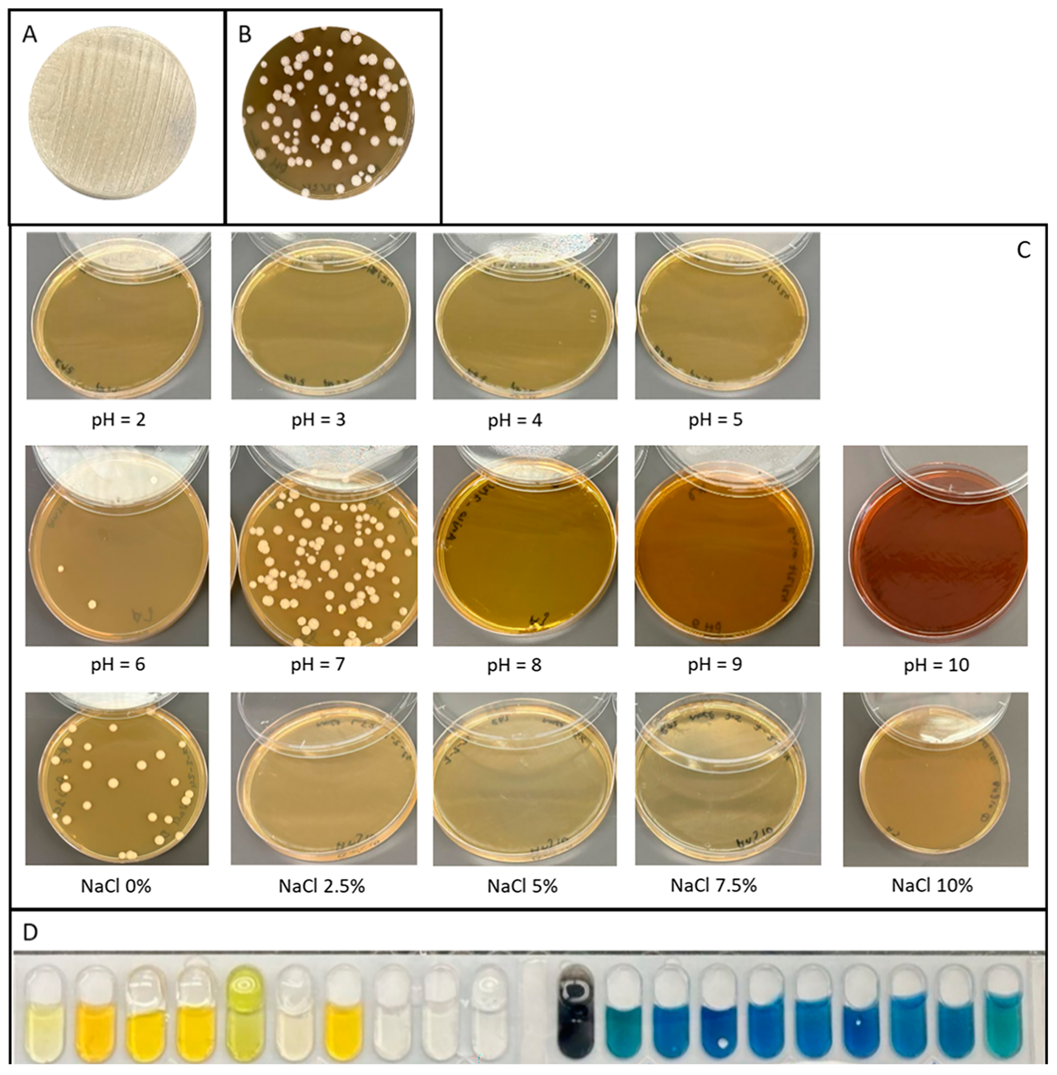
2.1. Morphological, Physiological, and Biochemical Characterization

2.1.1. Morphology

Scan Electron Microscopy of ANJ10 presented dense aerial mycelia and long chains of spore formation (Figure 1).
On ISP3 agar, ANJ10 appeared as spore-forming, circular, raised, white colonies with a slight pink tint. (Figure 2A,B).

2.1.2. Gram Staining

Gram-positive, filamentous.

2.1.3. Salt Tolerance

ANJ10 grew only at 0% NaCl concentration (Figure 2C).

2.1.4. pH Tolerance

ANJ10 grew at pH levels 6 and 7 (Figure 2C).

2.1.5. Biochemical Properties

ONPG and PNPG positive: β-galactosidase positive; ANJ10 is a lactose fermenter. (Figure 2D).

2.2. Genomic Characterization

2.2.1. Genome Features

The complete genome of ANJ10 is 9,939,599 base pairs (bp) in length, with a GC content of 70.68%. Genome annotation revealed 8670 coding sequences (CDS), 18 ribosomal RNA (rRNA) genes, 87 transfer RNA (tRNA) genes, and 1 transfer-messenger RNA (tmRNA) (Figure 3). BUSCO assessment showed a completeness of 99.8% (Supplementary Figure S4).

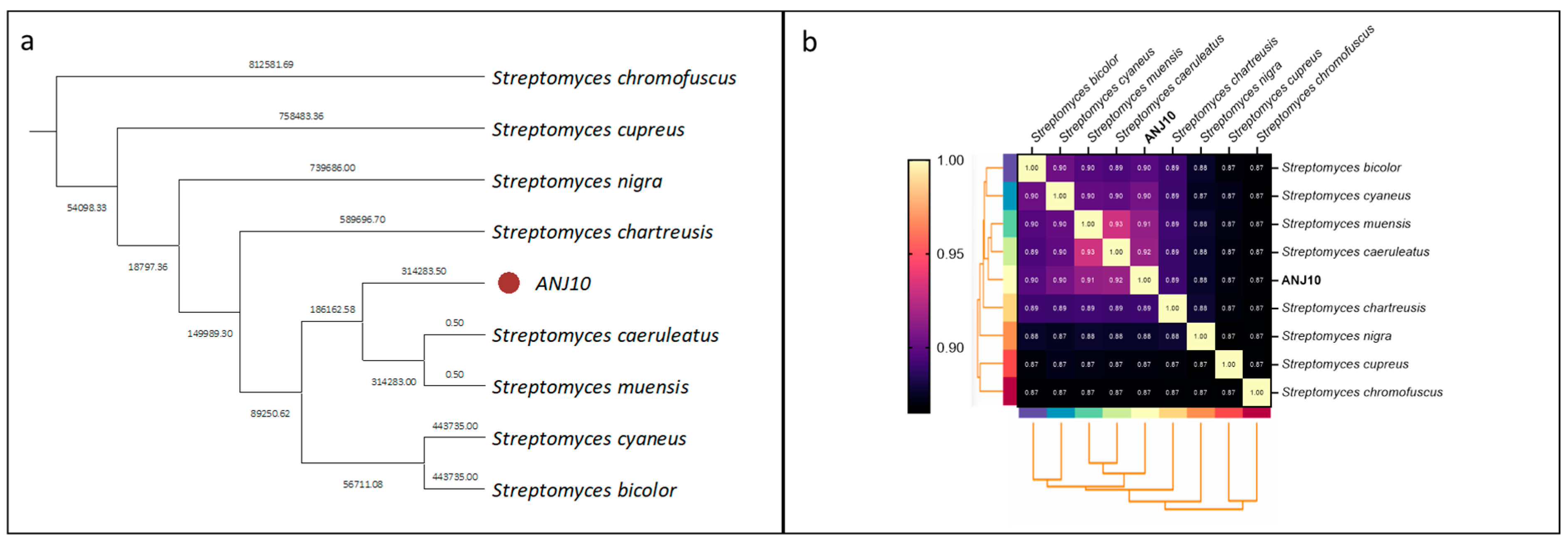
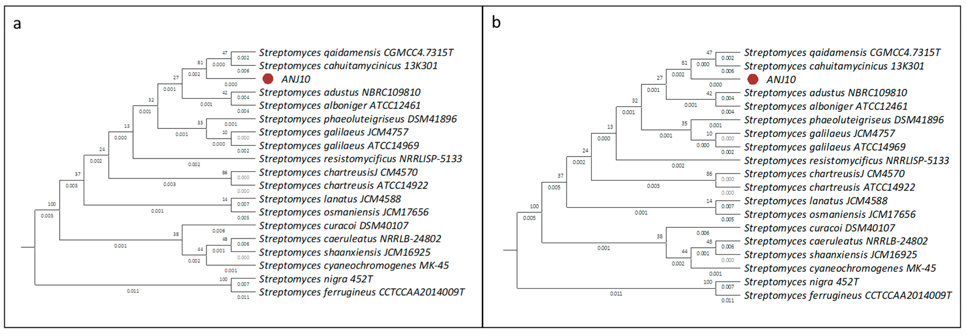
2.2.2. Phylogenetic Classification

The Saffrontree phylogenetic tree construction based on ANJ10 WGS data and comparison with all reference Streptomyces species demonstrated similarities to several Streptomyces species, including S. bicolor, S. caeruleatus, S. nigra, S. ceaneus, S. cupreus, S. chromofuscus, and S. muensis. Tree construction using these genomes showed that ANJ10 was most closely related to S. caeruleatus and S. muensis (Figure 4a). The ANI results showed that none of the comparisons exceeded the 95% threshold, which is commonly used to define species boundaries [35], and less than 70% DNA–DNA hybridization (dDDH) in all formulas and compared to all references (Figure 4b, Supplementary Table S3). As for the TYGS results, the tree inferred with FastME 2.1.6.1 from GBDP distances calculated from 16S rDNA gene sequences showed similarity to S. qaidamensis and S. cahuitamycinicus, whereas WGS showed the highest similarity to S. curaco. dDDH had the highest value of 51.1% using formula 1 when compared to S. caeruleatus, lower than the 70% threshold (Figure 5). In summary, these results indicate that ANJ10 represents a novel Streptomyces species.

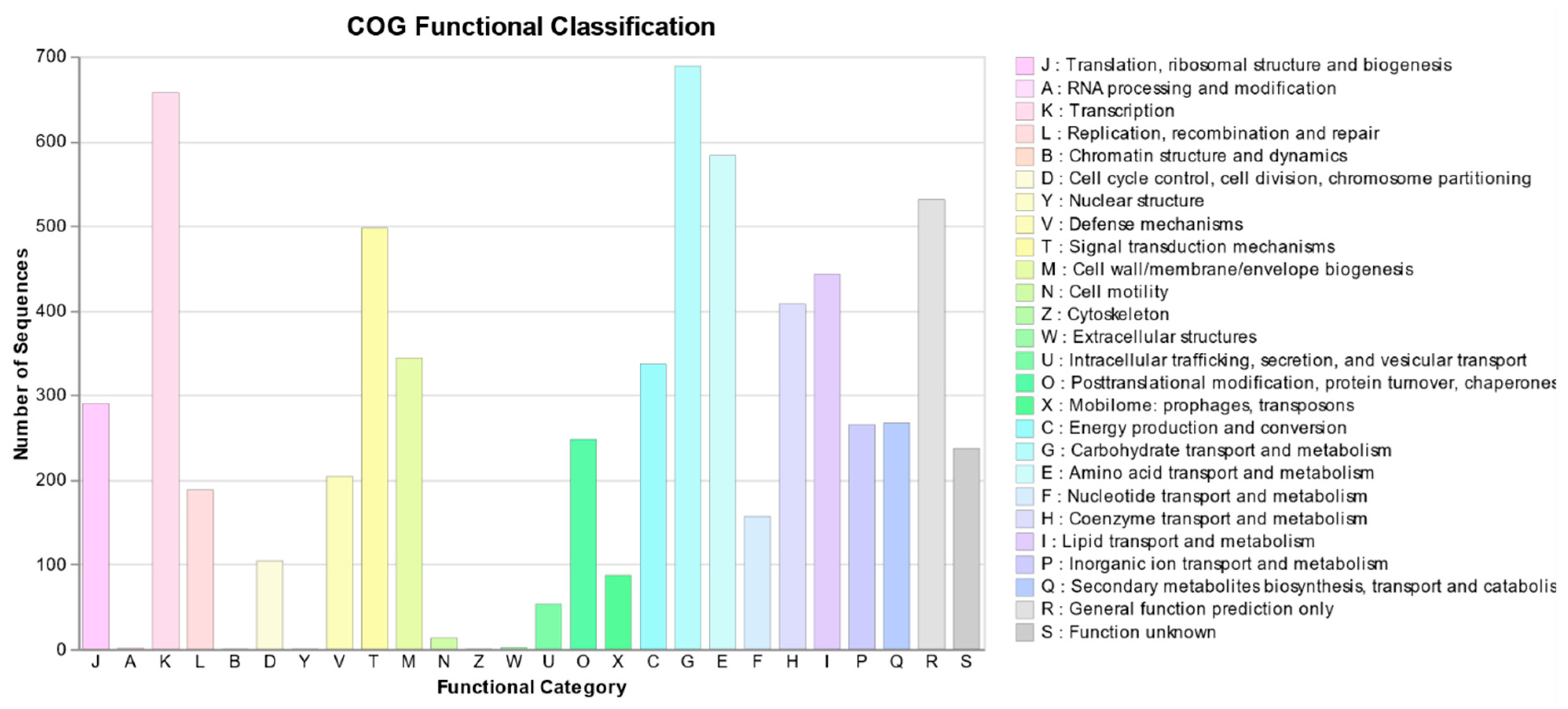
2.2.3. Functional Annotation

antiSMASH analysis revealed a total of 44 BGCs, among which 11 exhibited over 75% similarity to known compounds. Notably, three of the BGCs were positioned at the contig edges, rendering them incomplete. Further examination revealed that the last three Type I polyketide synthase (T1PKS) clusters, with the latter two situated at the beginning of their respective contigs, were identical BGCs spanning three contigs. Consequently, the total number of discernible BGCs was adjusted to 42. The distribution of BGC types was as follows: Terpene—13, Nonribosomal Peptide Synthetase (NRPS)—12, Polyketide Synthase (PKS)—14, Ribosomally synthesized and post-translationally modified Peptides (RiPPs)—5, Siderophore—5, Lactone—4, Lanthipeptide—3, Melanin, Ectoine, Arylpolyene, β-lactam, and hydrogen cyanide. Additionally, three BGCs showed proximity to resistance markers. Specifically, ANJ10’s second BGC contained a carboxyl transferase domain, BGC 15 included OTCace and Asp/Orn binding domains, and BGC 36 was associated with a biotin/lipoyl attachment domain according to the ARTS results (Table 1, Figure 6). BGC network analysis revealed that while some regions were networked with BGCs from the miBIG repository, 28 regions showed no genomic similarities to known BGCs, highlighting the potential for novel secondary metabolite discovery (Figure 6, Supplementary Table S4). COG analysis revealed a diverse distribution of functional genes, highlighting significant clusters in transcription (K), carbohydrate metabolism (G), and amino acid metabolism (E). Notably, secondary metabolite biosynthesis, transport, and catabolism (Q) featured prominently, with 267 genes, suggesting a robust potential for secondary metabolite production. Additional notable categories included cell wall biogenesis (M), protein modification and turnover (O), and ion transport (P), underscoring the metabolic versatility of the organism and its potential for ecological interactions through secondary metabolites (Figure 7).

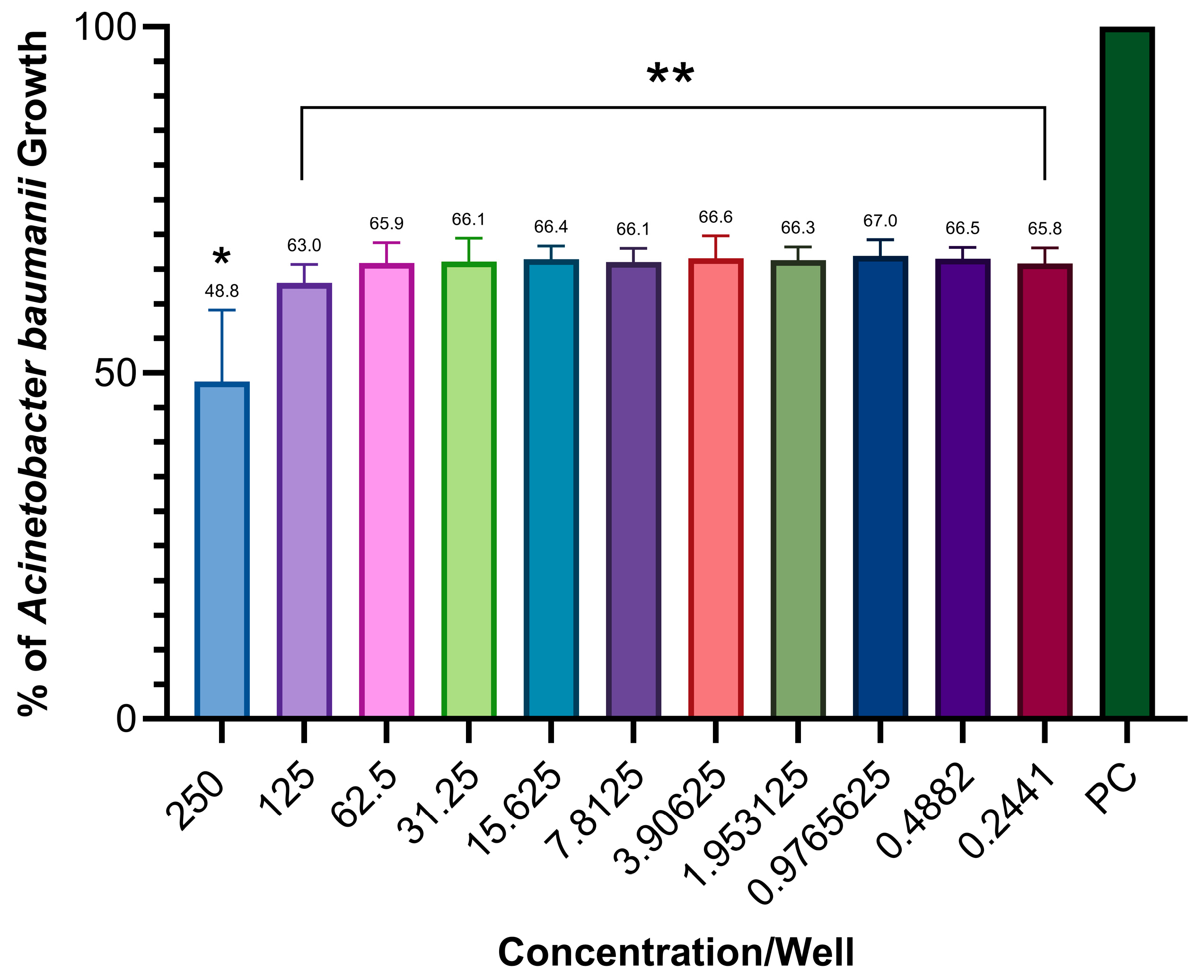
2.3. Antimicrobial Activity Testing

The antimicrobial potential of ANJ10 crude extracts produced in 14 different media was assessed in small-scale production against known ESKAPE pathogens. Based on this, the antimicrobial activity was evaluated. Most of the tested crude extracts showed relatively low activity against a panel of ESKAPE pathogens. However, ANJ10’s crude extract produced in Media V demonstrated visual inhibition against five pathogens, specifically E. coli ATCC 25922 and A. baumannii DSM 30008, with the lowest MIC values (Table 2). Based on these results, we conducted large-scale production in medium V and tested it against A. baumannii DSM 30008. For a better assessment, we used OD measurements to assess the turbidity of the media, reflecting bacterial growth. The results showed a significant reduction in OD across all tested concentrations, highlighting the antimicrobial activity of ANJ10 crude extract (Figure 8).

3. Discussion

The genus Streptomyces plays a critical role in natural product drug discovery, particularly due to its extensive repertoire of biosynthetic gene clusters (BGCs) [36]. The novel Streptomyces species isolated from Anjar, Lebanon, stands out in this regard, displaying a higher-than-average number of BGCs compared to typical Streptomyces genomes, which generally harbor 33 BGCs per genome [14]. This highlights the strain’s strong potential to produce a diverse array of secondary metabolites, including therapeutically valuable compounds such as cyclosporine, a crucial immunosuppressant in organ transplantation and autoimmune disease treatment [37], and rapamycin, which has applications in immunosuppression and cancer therapy [38].
Phylogenetic analysis not only revealed significant divergence from known Streptomyces species but also confirmed that the strain represents a novel species. This is supported by the identification of numerous unique genes within its biosynthetic gene clusters, with 28 failing to cluster confidently with any entries in the miBiG database, indicating that these loci likely encode previously uncharacterized biosynthetic pathways [33]. These poorly clustered regions vary widely in type and genomic length and have less than 5% similarity to known BGCs. For example, short clusters such as Region 1.16, classified as a RiPP-like biosynthetic gene cluster and spanning approximately 11.4 kilobases, and Region 2.1, another RiPP-like cluster of similar size, are typically associated with a range of bioactivities [39]. Other regions are larger and more complex, such as Region 4.2, a hybrid terpene and PKS-like cluster approximately 48.9 kb in length, and Region 2.2, a terpene cluster spanning approximately 20.9 kb. Several clusters also fall into high-interest classes, such as NRPS, PKS, and lanthipeptides, including Region 1.1 and Region 2.3, enzyme systems often responsible for the biosynthesis of bioactive natural products, such as antibiotics, cytotoxins, and immunosuppressants [40]. The structural and enzymatic diversity observed among these BGCs, combined with their failure to form strong clustering with any known reference pathways, strongly suggests that they may direct the biosynthesis of entirely novel chemical scaffolds.
In addition to these unclustered regions, several BGCs exhibited low similarity to known clusters. For instance, Region 1.9 showed 16% similarity to the clavulanic acid biosynthetic cluster, Region 1.18 shared 20% similarity with the rubiginone cluster, and Region 3.5 shared 20% similarity with the rakicidin biosynthetic pathway. These cases may represent cryptic or divergent pathways, where limited domain-level similarity is insufficient to support confident clustering, but may hint at the production of structurally or functionally related analogs.
In contrast, the genome also harbors several biosynthetic gene clusters that are one hundred percent identical to well-characterized reference pathways. These include those responsible for the production of albaflavenone, ectoine, gamma-butyrolactone, desferrioxamine, althiomycin, ε-poly-L-lysine, informatipeptin, and citrulassin D. Their presence supports the accuracy and robustness of the genome mining approach, while also demonstrating the strain’s capacity to produce well-established bioactive compounds with known functions, such as antibacterial activity, metal chelation, or cytoprotection [41,42,43,44,45]. Altogether, this dual landscape of known and unclustered or low-similarity BGCs underscores both the evolutionary novelty and biosynthetic richness of this strain and highlights the value of genome mining for the discovery of natural products.
The ecological significance of Streptomyces is immense. As prolific producers of secondary metabolites, these bacteria play vital roles in soil ecosystems, influencing microbial community dynamics [46]. The discovery of unique genes with ANJ10 is not entirely unexpected, given Lebanon’s rich and varied ecosystem. Such ecological diversity fosters unique microbial niches and evolutionary pressures, providing fertile ground for the emergence of novel microbial species with specialized metabolic capabilities, as shown by Awada et al. [47].
Given the novelty of the isolated Streptomyces species, we evaluated its antimicrobial potential. The urgent need for new antibiotics, particularly against ESKAPE pathogens, which are major contributors to antimicrobial resistance (AMR), further motivated this investigation [6]. To assess bioactivity, the strain was cultured in 14 different production media, each offering distinct nutritional conditions and stressors known to influence the activation of specific biosynthetic pathways [48]. Notably, variations in antimicrobial activity were observed across different media, underscoring the importance of environmental factors in modulating secondary metabolite expression.
Among the tested media, Medium V exhibited the broadest and most potent antimicrobial activity. Consequently, the crude extract was selected for further testing against Acinetobacter baumannii, a clinically relevant ESKAPE pathogen. Remarkably, significant bacterial inhibition was observed even at low extract concentrations. This sustained activity at reduced concentrations may be attributed to the complex nature of the crude extract, which likely contains a mixture of secondary metabolites that act synergistically or exhibit overlapping antimicrobial spectra at varying MICs.
Future work will focus on the purification and characterization of individual bioactive compounds in this extract. This may uncover novel antimicrobial agents with the potential for development into clinically applicable therapeutics, an essential step in combating the escalating challenge of multidrug-resistant pathogens.

4. Conclusions

In conclusion, Lebanon’s geographical location and diverse ecosystems make it a crucial area for exploring microbial diversity. The discovery of this new Streptomyces species not only adds to the growing repository of bioactive compounds but also reinforces the potential of Lebanon as a significant source of microorganisms with promising applications in drug discovery. The high number of BGCs identified in this strain suggests vast, unexplored chemical diversity that could lead to the development of next-generation antimicrobials and other therapeutic agents. Moreover, the isolation and characterization of this novel Streptomyces species from Anjar, Lebanon, highlights the pivotal role of this genus in natural product drug discovery and underscores the ecological and biotechnological significance of exploring diverse environments. The unique genetic and biochemical properties of this strain position it as a valuable candidate for further research, particularly in the development of new antimicrobial drugs.

Supplementary Materials

The following supporting information can be downloaded at: https://www.mdpi.com/article/10.3390/fermentation11070406/s1, Supplementary Figure S1: The Process of Sample Processing and Bacterial Isolation. The soil samples underwent a series of steps including dehydration, dissolution, incubation, dilution, and inoculation onto different agar plates. Bacterial colonies that developed were subsequently cultured. Supplementary Figure S2: The process of small-scale secondary metabolite production and extraction. Colonies are transferred from solid culture agar into a liquid first seed, then a second seed, and last 14 different production media. SMs produced are adsorbed on resins and extracted using acetone/methanol mixture then dried to obtain a crude extract. Supplementary Figure S3: The broth microdilution (BMD) assay. On the left, the serial dilution of the crude extracts in the MHCAB is seen. While, on the right, the addition of an equal volume of the bacterial inoculum can be observed. Supplementary Figure S4: ANJ10 Genome Completeness. Assessment was done using BUSCO, which evaluates the presence of universal single-copy orthologs. The analysis was conducted based on the order of Streptomycetales database, to determine the completeness and quality of the assembly.; Supplementary Table S1: The name of the environmental bacteria with bioactive secondary metabolites, its region of origin, its order of isolation, and its target pathogens. Supplementary Table S2: Composition of the 14 different production media. The first row represents the name of the media described. Column 1 shows media components. Values are listed in g/L. Dashes indicate a value of 0 g/L. Supplementary Table S3: DNA-DNA Hybridization. Genome to Genome distance calculator software was used. Distances are inferred using three distinct formulas (Formula: 1 (HSP length / total length), Formula: 2 (identities / HSP length), Formula: 3 (identities / total length)) from the set of HSPs MUMs obtained by comparing each pair of genomes with the chosen software. These distances are transformed to values analogous to DDH using a generalized linear model (GLM) inferred from an empirical reference dataset comprising real DDH values and genome sequences. Model-based confidence intervals are specified in square brackets but can also be obtained via bootstrapping. Logistic regression (a special type of GLM) is used for reporting the probabilities that DDH is >=70% and >=79%. Percent G+C content cannot differ by >1 within a single species but by <=1 between distinct species Supplementary Table S4: ANJ10 BGC similarity. BGCs were annotated using antiSMASH. This table details each BGC found, and most similar known cluster along with their type, degree of similarity, and known activity.

Author Contributions

R.H. and A.H. contributed equally to this work. The order of authors was determined based on seniority. Conceptualization R.H. and A.H.; methodology R.H. and A.H.; software R.H. and A.H.; validation, A.A.F.; formal analysis, R.H. and A.H.; investigation, R.H. and A.H.; data curation, R.H. and A.H.; writing—original draft preparation, R.H. and A.H.; writing—review and editing, R.H. and A.H.; visualization R.H. and A.H.; supervision, A.A.F.; project administration, funding acquisition. All authors have read and agreed to the published version of the manuscript.

Funding

This work was funded by the Medical Practice Plan (MPP) of AUB/AUBMC.

Institutional Review Board Statement

Not applicable.

Informed Consent Statement

Not applicable.

Data Availability Statement

The contig sequences associated with the genome have been deposited in GenBank and are publicly accessible under the accession number SAMN44756297.

Acknowledgments

We thank the Kamal A. Shair Central Research Science Laboratory (KAS CRSL) at the American University of Beirut for providing access to SEM and the AUBMC Division of Infectious Diseases for facilitating access to sequencing.

Conflicts of Interest

The authors declare no conflicts of interest.

Abbreviations

The following abbreviations are used in this manuscript:
BGCBiosynthetic Gene Cluster
WGSWhole-Genome Sequencing
BMDBroth Microdilution
NaClSodium Chloride
SEMScanning Electron Microscopy
PBSPhosphate-Buffered Saline
FAFormaldehyde
GAGlutaraldehyde
TSBTryptic Soy Broth
gDNAGenomic DNA
CDSCoding Sequences
rRNARibosomal RNA
tRNATransfer RNA
tmRNATransfer-messenger RNA
ANIAverage Nucleotide Identity
dDDHDigital DNA–DNA Hybridization
TYGSType Strain Genome Server
GBDPGenome BLAST Distance Phylogeny
PCPositive Control
NCNegative Control
ODOptical Density
MHCABMueller Hinton Cation-Adjusted Broth
SMsSecondary Metabolites
NRPSNonribosomal Peptide Synthetase
PKSPolyketide Synthase
T1PKSType I Polyketide Synthase
T2PKSType II Polyketide Synthase
RiPPsRibosomally synthesized and post-translationally modified Peptides
NRPNonribosomal Peptide
COGCluster of Orthologous Genes
ARTSAntibiotic Resistant Target Seeker
MRSAMethicillin-Resistant Staphylococcus aureus
MSSAMethicillin-Sensitive Staphylococcus aureus
WHOWorld Health Organization
MDRMulti-Drug Resistant
miBIGMinimum Information about a Biosynthetic Gene cluster

References

  1. Wade, W. Unculturable bacteria—The uncharacterized organisms that cause oral infections. J. R. Soc. Med. 2002, 95, 81–83. [Google Scholar] [PubMed]
  2. Hug, L.A. Sizing Up the Uncultured Microbial Majority. mSystems 2018, 3, e00185-18. [Google Scholar] [CrossRef] [PubMed]
  3. Lloyd, K.G.; Steen, A.D.; Ladau, J.; Yin, J.; Crosby, L. Phylogenetically Novel Uncultured Microbial Cells Dominate Earth Microbiomes. mSystems 2018, 3, e00055-18. [Google Scholar] [CrossRef] [PubMed]
  4. Baltz, R.H. Marcel Faber Roundtable: Is our antibiotic pipeline unproductive because of starvation, constipation or lack of inspiration? J. Ind. Microbiol. Biotechnol. 2006, 33, 507–513. [Google Scholar] [CrossRef]
  5. Newman, D.J.; Cragg, G.M. Natural Products as Sources of New Drugs from 1981 to 2014. J. Nat. Prod. 2016, 79, 629–661. [Google Scholar] [CrossRef]
  6. Fair, R.J.; Tor, Y. Antibiotics and bacterial resistance in the 21st century. Perspect. Med. Chem. 2014, 6, 25–64. [Google Scholar] [CrossRef]
  7. Genilloud, O. Actinomycetes: Still a source of novel antibiotics. Nat. Prod. Rep. 2017, 34, 1203–1232. [Google Scholar] [CrossRef]
  8. Bérdy, J. Thoughts and facts about antibiotics: Where we are now and where we are heading. J. Antibiot. 2012, 65, 385–395. [Google Scholar] [CrossRef]
  9. Demain, A.L.; Sanchez, S. Microbial drug discovery: 80 years of progress. J. Antibiot. 2009, 62, 5–16. [Google Scholar] [CrossRef]
  10. Kämpfer, P.; Glaeser, S.P.; Parkes, L.; van Keulen, G.; Dyson, P. The Family Streptomycetaceae. In The Prokaryotes: Actinobacteria; Rosenberg, E., DeLong, E.F., Lory, S., Stackebrandt, E., Thompson, F., Eds.; Springer: Berlin/Heidelberg, Germany, 2014; pp. 889–1010. [Google Scholar]
  11. Sathya, A.; Vijayabharathi, R.; Gopalakrishnan, S. Plant growth-promoting actinobacteria: A new strategy for enhancing sustainable production and protection of grain legumes. 3 Biotech 2017, 7, 102. [Google Scholar] [CrossRef]
  12. Cimermancic, P.; Medema, M.H.; Claesen, J.; Kurita, K.; Wieland Brown, L.C.; Mavrommatis, K.; Pati, A.; Godfrey, P.A.; Koehrsen, M.; Clardy, J.; et al. Insights into secondary metabolism from a global analysis of prokaryotic biosynthetic gene clusters. Cell 2014, 158, 412–421. [Google Scholar] [CrossRef]
  13. Doroghazi, J.R.; Albright, J.C.; Goering, A.W.; Ju, K.S.; Haines, R.R.; Tchalukov, K.A.; Labeda, D.P.; Kelleher, N.L.; Metcalf, W.W. A roadmap for natural product discovery based on large-scale genomics and metabolomics. Nat. Chem. Biol. 2014, 10, 963–968. [Google Scholar] [CrossRef] [PubMed]
  14. Chung, Y.-H.; Kim, H.; Ji, C.-H.; Je, H.-W.; Lee, D.; Shim, S.H.; Joo, H.-S.; Kang, H.-S.; Gutierrez, M. Comparative Genomics Reveals a Remarkable Biosynthetic Potential of the Streptomyces Phylogenetic Lineage Associated with Rugose-Ornamented Spores. mSystems 2021, 6, e0048921. [Google Scholar] [CrossRef] [PubMed]
  15. Awada, B.; Hamie, M.; El Hajj, R.; Derbaj, G.; Najm, R.; Makhoul, P.; Ali, D.H.; Fayad, A.G.A.; El Hajj, H. HAS 1: A natural product from soil-isolated Streptomyces species with potent activity against cutaneous leishmaniasis caused by Leishmania tropica. Front. Pharmacol. 2022, 13, 1023114. [Google Scholar] [CrossRef]
  16. Ewels, P.A.; Peltzer, A.; Fillinger, S.; Patel, H.; Alneberg, J.; Wilm, A.; Garcia, M.U.; Di Tommaso, P.; Nahnsen, S. The nf-core framework for community-curated bioinformatics pipelines. Nat. Biotechnol. 2020, 38, 276–278. [Google Scholar] [CrossRef]
  17. Manni, M.; Berkeley, M.R.; Seppey, M.; Simão, F.A.; Zdobnov, E.M. BUSCO Update: Novel and Streamlined Workflows along with Broader and Deeper Phylogenetic Coverage for Scoring of Eukaryotic, Prokaryotic, and Viral Genomes. Mol. Biol. Evol. 2021, 38, 4647–4654. [Google Scholar] [CrossRef]
  18. Carver, T.; Harris, S.R.; Berriman, M.; Parkhill, J.; McQuillan, J.A. Artemis: An integrated platform for visualization and analysis of high-throughput sequence-based experimental data. Bioinformatics 2011, 28, 464–469. [Google Scholar] [CrossRef]
  19. Page, A.J.; Hunt, M.; Seemann, T.; Keane, J.A. SaffronTree: Fast, reference-free pseudo-phylogenomic trees from reads or contigs. J. Open Source Softw. 2017, 2, 243. [Google Scholar] [CrossRef]
  20. Pritchard, L.; Glover, R.H.; Humphris, S.; Elphinstone, J.G.; Toth, I.K. Genomics and taxonomy in diagnostics for food security: Soft-rotting enterobacterial plant pathogens. Anal. Methods 2016, 8, 12–24. [Google Scholar] [CrossRef]
  21. Meier-Kolthoff, J.P.; Carbasse, J.S.; Peinado-Olarte, R.L.; Göker, M. TYGS and LPSN: A database tandem for fast and reliable genome-based classification and nomenclature of prokaryotes. Nucleic Acids Res. 2021, 50, D801–D807. [Google Scholar] [CrossRef]
  22. Lagesen, K.; Hallin, P.; Rødland, E.A.; Stærfeldt, H.-H.; Rognes, T.; Ussery, D.W. RNAmmer: Consistent and rapid annotation of ribosomal RNA genes. Nucleic Acids Res. 2007, 35, 3100–3108. [Google Scholar] [CrossRef] [PubMed]
  23. Meier-Kolthoff, J.P.; Auch, A.F.; Klenk, H.-P.; Göker, M. Genome sequence-based species delimitation with confidence intervals and improved distance functions. BMC Bioinform. 2013, 14, 60. [Google Scholar] [CrossRef] [PubMed]
  24. Camacho, C.; Coulouris, G.; Avagyan, V.; Ma, N.; Papadopoulos, J.; Bealer, K.; Madden, T.L. BLAST+: Architecture and applications. BMC Bioinform. 2009, 10, 421. [Google Scholar] [CrossRef] [PubMed]
  25. Meier-Kolthoff, J.P.; Hahnke, R.L.; Petersen, J.; Scheuner, C.; Michael, V.; Fiebig, A.; Rohde, C.; Rohde, M.; Fartmann, B.; Goodwin, L.A.; et al. Complete genome sequence of DSM 30083T, the type strain (U5/41T) of Escherichia coli, and a proposal for delineating subspecies in microbial taxonomy. Stand. Genom. Sci. 2014, 9, 2. [Google Scholar] [CrossRef]
  26. Lefort, V.; Desper, R.; Gascuel, O. FastME 2.0: A Comprehensive, Accurate, and Fast Distance-Based Phylogeny Inference Program. Mol. Biol. Evol. 2015, 32, 2798–2800. [Google Scholar] [CrossRef]
  27. Kreft, Ł.; Botzki, A.; Coppens, F.; Vandepoele, K.; Van Bel, M. PhyD3: A phylogenetic tree viewer with extended phyloXML support for functional genomics data visualization. Bioinformatics 2017, 33, 2946–2947. [Google Scholar] [CrossRef]
  28. Ondov, B.D.; Treangen, T.J.; Melsted, P.; Mallonee, A.B.; Bergman, N.H.; Koren, S.; Phillippy, A.M. Mash: Fast genome and metagenome distance estimation using MinHash. Genome Biol. 2016, 17, 132. [Google Scholar] [CrossRef]
  29. Meier-Kolthoff, J.P.; Göker, M. TYGS is an automated high-throughput platform for state-of-the-art genome-based taxonomy. Nat. Commun. 2019, 10, 2182. [Google Scholar] [CrossRef]
  30. Farris, J.S. Estimating Phylogenetic Trees from Distance Matrices. Am. Nat. 1972, 106, 645–668. [Google Scholar] [CrossRef]
  31. Blin, K.; Shaw, S.; Augustijn, H.E.; Reitz, Z.L.; Biermann, F.; Alanjary, M.; Fetter, A.; Terlouw, B.R.; Metcalf, W.W.; Helfrich, E.J.N.; et al. antiSMASH 7.0: New and improved predictions for detection, regulation, chemical structures and visualisation. Nucleic Acids Res. 2023, 51, W46–W50. [Google Scholar] [CrossRef]
  32. Mungan, M.D.; Alanjary, M.; Blin, K.; Weber, T.; Medema, M.H.; Ziemert, N. ARTS 2.0: Feature updates and expansion of the Antibiotic Resistant Target Seeker for comparative genome mining. Nucleic Acids Res. 2020, 48, W546–W552. [Google Scholar] [CrossRef] [PubMed]
  33. Navarro-Muñoz, J.C.; Selem-Mojica, N.; Mullowney, M.W.; Kautsar, S.A.; Tryon, J.H.; Parkinson, E.I.; De Los Santos, E.L.C.; Yeong, M.; Cruz-Morales, P.; Abubucker, S.; et al. A computational framework to explore large-scale biosynthetic diversity. Nat. Chem. Biol. 2020, 16, 60–68. [Google Scholar] [CrossRef]
  34. Shimoyama, Y. COGclassifier: A Tool for Classifying Prokaryote Protein Sequences into COG Functional Category 2022. Available online: https://github.com/moshi4/COGclassifier (accessed on 15 April 2024).
  35. Jain, C.; Rodriguez-R, L.M.; Phillippy, A.M.; Konstantinidis, K.T.; Aluru, S. High throughput ANI analysis of 90K prokaryotic genomes reveals clear species boundaries. Nat. Commun. 2018, 9, 5114. [Google Scholar] [CrossRef] [PubMed]
  36. Donald, L.; Pipite, A.; Subramani, R.; Owen, J.; Keyzers, R.A.; Taufa, T. Streptomyces: Still the Biggest Producer of New Natural Secondary Metabolites, a Current Perspective. Microbiol. Res. 2022, 13, 418–465. [Google Scholar] [CrossRef]
  37. Tribe, H.T. The discovery and development of cyclosporin. Mycologist 1998, 12, 20–22. [Google Scholar] [CrossRef]
  38. Seto, B. Rapamycin and mTOR: A serendipitous discovery and implications for breast cancer. Clin. Transl. Med. 2012, 1, 29. [Google Scholar] [CrossRef]
  39. Russell, A.H.; Truman, A.W. Genome Mining Strategies for Ribosomally Synthesised and Post-Translationally Modified Peptides. Comput. Struct. Biotechnol. J. 2020, 18, 1838–1851. [Google Scholar] [CrossRef]
  40. Scherlach, K.; Hertweck, C. Mining and unearthing hidden biosynthetic potential. Nat. Commun. 2021, 12, 3864. [Google Scholar] [CrossRef]
  41. Liu, W.; Sun, F.; Hu, Y. Genome Mining-Mediated Discovery of a New Avermipeptin Analogue in Streptomyces Actuosus ATCC 25421. ChemistryOpen 2018, 7, 558–561. [Google Scholar] [CrossRef]
  42. Zheng, D.; Ding, N.; Jiang, Y.; Zhang, J.; Ma, J.; Chen, X.; Liu, J.; Han, L.; Huang, X. Albaflavenoid, a New Tricyclic Sesquiterpenoid from Streptomyces Violascens. J. Antibiot. 2016, 69, 773–775. [Google Scholar] [CrossRef]
  43. Hur, J.; Jang, J.; Sim, J. A Review of the Pharmacological Activities and Recent Synthetic Advances of γ-Butyrolactones. IJMS 2021, 22, 2769. [Google Scholar] [CrossRef] [PubMed]
  44. Purev, E.; Kondo, T.; Takemoto, D.; Niones, J.T.; Ojika, M. Identification of ε-Poly-L-Lysine as an Antimicrobial Product from an Epichloë Endophyte and Isolation of Fungal ε-PL Synthetase Gene. Molecules 2020, 25, 1032. [Google Scholar] [CrossRef] [PubMed]
  45. Liu, Z.; Zhang, Y.; Sun, J.; Huang, W.-C.; Xue, C.; Mao, X. A Novel Soluble Squalene-Hopene Cyclase and Its Application in Efficient Synthesis of Hopene. Front. Bioeng. Biotechnol. 2020, 8, 426. [Google Scholar] [CrossRef] [PubMed]
  46. Chater, K.F. Recent advances in understanding Streptomyces. F1000Res 2016, 5, 2795. [Google Scholar] [CrossRef]
  47. Awada, B.; Chahine, D.; Derbaj, G.; Abdel Khalek, P.; Kallassy, M.; Abou Fayad, A. Antimicrobial Natural Products Derived from Microorganisms Inhabiting the MENA Region. Nat. Prod. Commun. 2023, 18, 1–27. [Google Scholar] [CrossRef]
  48. Liu, Z.; Zhao, Y.; Huang, C.; Luo, Y. Recent Advances in Silent Gene Cluster Activation in Streptomyces. Front. Bioeng. Biotechnol. 2021, 9, 632230. [Google Scholar] [CrossRef]
Figure 1. SEM of ANJ10. (A) Overview at 20 µm magnification, showing dense mycelial growth. (B) At 10 µm magnification, the presence of aerial mycelia is more distinct. (C) Higher magnification at 5 µm reveals long chains of spore formation, clearly visible among the aerial mycelia.
Fermentation 11 00406 g001
Figure 2. Morphological, physiological, and biochemical characterization of bacterial isolate ANJ10. (A). Growth, shape, and color of isolate ANJ10 on ISP3 agar 10 days after bacterial inoculation at 28 °C. (B). Growth, shape, and color of the isolate ANJ10 and spore formation on ISP2 agar 10 days after bacterial inoculation at 28 °C. (C). Physiological characterization of ANJ10 isolate on ISP2 agar with different pH (3, 4, 5, 6, 7, 8, 9, and 10) after 10 days at 28 °C and on 5339 agar with different NaCl concentrations (0, 2.5, 5, 7.5, and 10%). (D). Biochemical testing of ANJ10 was performed using the API 20E kit, in order from left to right, ONPG, ADH, LDC, ODC, CIT, H2S, URE, TDA, IND, VP, GEL, GLU, MAN, INO, SOR, RHA, SAC, MEL, AMY, ARA.
Fermentation 11 00406 g002
Figure 3. ANJ10 Genomic Map. From the outer to inner circle: Map Backbone (Unicycler FASTA file), CDS annotation (PROKKA GBK file), antiSMASH annotation, GC% plot, and GC Skew. The ANJ10 genome spans 9,939,599 bp with a GC content of 70.68%, 8670 coding sequences (CDS), 118 rRNA genes, 87 tRNA genes, and 1 tmRNA. A total of 42 biosynthetic gene clusters were identified.
Fermentation 11 00406 g003
Figure 4. (a). ANJ10 WGS Phylogenetic Tree. Phylogenetic analysis was performed using SaffronTree. The data was visualized with MEGA software. SaffronTree uses k-mer analysis to build a phylogenetic Neighbor-Joining tree by generating k-mer profiles for each sequence from WGS data, calculating pairwise distances based on these profiles, and constructing the tree using the Neighbor-Joining algorithm. Branch lengths represent the extent of evolutionary change or genetic distance between nodes. (b). Average Nucleotide Identity (ANI). ANI was calculated using PyANI. This heatmap illustrates the ANI between S. bicolor, S. caeruleatus, S. nigra, S. ceaneus, S. cupreus, S. chromofuscus, and S. muensis, and ANJ10. A lighter shade indicates a closer relationship. The branching pattern also reflects the relatedness among genomes.
Fermentation 11 00406 g004
Figure 5. (a). ANJ10 16S Phylogenetic Tree from TYGS. Tree inferred with FastME 2.1.6.1 from GBDP distances calculated from 16S rDNA gene sequences. The branch lengths were scaled using the GBDP distance formula d5. The numbers above the branches are GBDP pseudo-bootstrap support values > 60% from 100 replications, with an average branch support of 42.4%. The tree was rooted at its midpoint. ANJ10 is shown by red symbol. (b). ANJ10 WGS Phylogenetic Tree from TYGS. Tree inferred with FastME 2.1.6.1 from GBDP distances calculated from the genome sequences. The branch lengths were scaled using the GBDP distance formula d5. The numbers above the branches are GBDP pseudo-bootstrap support values > 60% from 100 replications, with an average branch support of 93.8%. The tree was rooted at its midpoint. ANJ10 is shown by red symbol.
Fermentation 11 00406 g005
Figure 6. Network of Biosynthetic Gene Clusters (BGCs). Identified within ANJ10 (pink), labeled with their region numbers. BGCs from the miBIG database that show similarities to those in ANJ10 are represented in grey. White lines indicate genomic similarities between BGCs. Network analysis revealed that while some regions in ANJ10 are similar to known miBIG BGCs, 28 regions exhibited no significant genomic similarity to any known BGCs.
Fermentation 11 00406 g006
Figure 7. ANJ10 COG Classification. The figure illustrates the total number of COG-classified genes, which is 5759, including 255 genes specifically involved in secondary metabolite biosynthesis, transport, and catabolism. These genes were classified using the COGclassifier. Each color in the figure represents a distinct functional category.
Fermentation 11 00406 g007
Figure 8. ANJ10 media V BMD Results. The graph shows Acinetobacter bacterial growth inhibition in ANJ10 media V at different concentrations (µg/mL). Data are represented as a percentage of PC. One-way ANOVA was performed, followed by Dunnett’s multiple comparison test. * for p value < 0.05, ** for p value < 0.01.
Fermentation 11 00406 g008
Table 1. ANJ10 BGC annotation. BGCs were annotated using antiSMASH. This table details each BGC found, its type, location, and the most similar known cluster, along with its type, degree of similarity, and known activity. Each colored region represents a different type of BGC; red represents BGCs on a contig edge, and green represents clusters with more than 75% similarity to known BGCs.
RegionTypeFromToMost Similar Known ClusterTypeSimilarity
Region 1.1lanthipeptide-class-i270,498296,733
Region 1.2NRPS, NRPS-like, arylpolyene320,092418,525o-dialkylbenzene 1/o-dialkylbenzene 2Polyketide+NRP96%
Region 1.3T2PKS443,846516,361spore pigmentPolyketide75%
Region 1.4NRPS537,820594,302arginomycinOther20%
Region 1.5terpene662,834683,847albaflavenoneTerpene100%
Region 1.6CDPS1,675,4371,696,183nocardiopsistin A/nocardiopsistin B/nocardiopsistin CPolyketide9%
Region 1.7furan1,848,9291,869,942methylenomycin AOther9%
Region 1.8butyrolactone1,891,2101,902,202triacsin COther6%
Region 1.9blactam2,025,3852,046,998clavulanic acidOther:Non-NRP beta-lactam16%
Region 1.10NI-siderophore3,395,9333,425,705desferrioxamin B/desferrioxamine EOther100%
Region 1.11melanin3,522,1013,532,601istamycinSaccharide4%
Region 1.12ectoine4,844,6714,855,075ectoineOther100%
Region 1.13NAPAA5,081,4395,115,026ε-Poly-L-lysineNRP100%
Region 1.14NI-siderophore5,302,6745,332,887kinamycinPolyketide16%
Region 1.15betalactone5,567,3655,593,487JBIR-34/JBIR-35NRP8%
Region 1.16RiPP-like5,681,7515,693,103
Region 1.17butyrolactone, terpene5,768,9855,792,627γ-butyrolactoneOther100%
Region 1.18terpene5,918,1955,939,997rubiginone A2/rubiginone J/rubiginone K/rubiginone L/rubiginone M/rubiginone N/ochromycinone/rubiginone B2Polyketide20%
Region 1.19NI-siderophore6,035,5206,066,813paulomycinOther13%
Region 1.20lassopeptide6,088,8096,111,354citrulassin DRiPP100%
Region 2.1RiPP-like116,451128,376
Region 2.2terpene187,177208,133
Region 2.3NRPS, T1PKS339,857390,604Sch-47554/Sch-47555Polyketide3%
Region 2.4NI-siderophore424,258456,845peucechelinNRP30%
Region 2.5lanthipeptide-class-iii, RiPP-like467,702494,891informatipeptinRiPP:Lanthipeptide100%
Region 2.6terpene527,615548,646cyphomycinPolyketide2%
Region 2.7T1PKS, NRPS-like, NRPS626,113713,163antimycinNRP+Polyketide93%
Region 2.8NRP-metallophore, NRPS840,099907,141cahuitamycin A/cahuitamycin B/cahuitamycin CNRP62%
Region 2.9NRPS-like997,8431,040,518deoxyhangtaimycinPolyketide+NRP11%
Region 2.10T1PKS1,080,4401,125,278herbimycin APolyketide30%
Region 2.11linaridin, lanthipeptide-class-iv1,466,7401,516,503Sch-47554/Sch-47555Polyketide3%
Region 3.1terpene12,26733,244rimomycin A/rimomycin B/rimomycin CNRP6%
Region 3.2terpene, T1PKS130,006180,149malacidin A/malacidin BNRP:Lipopeptide:Ca+-dependent lipopeptide5%
Region 3.3T1PKS, NRPS181,637269,950foxicin A/foxicin B/foxicin C/foxicinNRP+Polyketide14%
Region 3.4terpene331,138352,196ebelactonePolyketide5%
Region 3.5hydrogen-cyanide, betalactone, NRPS, T1PKS739,801807,911rakicidin A/rakicidin BNRP:Cyclic depsipeptide+Polyketide:Modular type I polyketide20%
Region 3.6T1PKS1,001,4241,032,706niphimycins C-EPolyketide29%
Region 4.1PKS-like40,61381,626sanglifehrin ANRP+Polyketide6%
Region 4.2terpene, PKS-like118,451167,319sanglifehrin ANRP+Polyketide4%
Region 4.3terpene182,354209,122hopeneTerpene92%
Region 4.4NRPS, T1PKS512,984576,593althiomycinNRP+Polyketide:Modular type I polyketide100%
Region 4.5T1PKS760,445800,778niphimycins C-EPolyketide35%
Region 5.1T1PKS120,202griseochelinPolyketide46%
Region 6.1T1PKS114,357ECO-0501Polyketide14%
Table 2. Broth Microdilution (BMD) Assay Results for ANJ10. Pathogenic strains were tested using crude extracts from different media. The values, expressed in µg/mL, indicate the MIC (minimum inhibitory concentration), defined as the lowest concentration that shows growth inhibition. (-) represents MIC > 250 µg/mL.
Name of Producing StrainMedia
BacteriaVVegABCINARA3GPMYV6AF/MSGYMM8COMNL2
S. aureus ATCC 29213----250--250------
S. aureus Newman125125125125-------250--
S. aureus N315----250---------
E. feacalis ATCC 19433--------125-----
K. pneumonaie DSM----250-----250---
A. baumannii DSM 3000812525025025062.5250250250125250250250250125
K. pneumonaie ATCC 13883 --------------
P. aeruginosa ATCC 27853250---250----250250250250250
P. aeruginosa mexAB250-250-----250250250125250250
E. coli ATCC 2592262.512562.5250-----125----
Disclaimer/Publisher’s Note: The statements, opinions and data contained in all publications are solely those of the individual author(s) and contributor(s) and not of MDPI and/or the editor(s). MDPI and/or the editor(s) disclaim responsibility for any injury to people or property resulting from any ideas, methods, instructions or products referred to in the content.

