Article

Neuroprotective Effect of Lactobacillus gasseri MG4247 and Lacticaseibacillus rhamnosus MG4644 Against Oxidative Damage via NF-κB Signaling Pathway

1
MEDIOGEN Co., Ltd., Biovalley 1-ro, Jecheon 27159, Republic of Korea
2
Division of Applied Life Science (BK21), Institute of Agriculture and Life Science, Gyeongsang National University, Jinju 52828, Republic of Korea
*
Authors to whom correspondence should be addressed.
Fermentation 2025, 11(7), 385; https://doi.org/10.3390/fermentation11070385
Submission received: 20 May 2025 / Revised: 25 June 2025 / Accepted: 1 July 2025 / Published: 3 July 2025

Abstract

Probiotics have recently gained attention as modulators of the gut–brain axis in neurodegenerative diseases such as Alzheimer’s disease. In this study, we identified probiotic strains with neuroprotective effects and investigated their mechanisms and safety. We screened strains based on their ability to inhibit acetylcholinesterase (AChE) activity and protect cells against H2O2-induced damage. The cell-free supernatants (CFS) of Lactobacillus gasseri MG4247 and Lacticaseibacillus rhamnosus MG4644 inhibited AChE activity and reduced cell damage and reactive oxygen species generation. These effects were mediated through inhibition of the MyD88/NF-κB pathway and modulation of the JNK/Bax-dependent apoptotic pathway in neuronal cells treated with H2O2. Whole-genome sequencing and antibiotic susceptibility testing confirmed the identity and safety of both strains. These findings suggest that MG4247 and MG4644, as probiotics, may help protect neuronal cells from oxidative stress and inflammation.

1. Introduction

Oxidative stress is related to an imbalance between the generation and defense system of reactive oxygen species (ROS), including free radicals, which negatively affect the systemic, circulatory, and nervous systems [1]. In particular, the brain contains fewer antioxidant enzymes than other organs, rendering it vulnerable to oxidative stress. [2]. Excessive generation of ROS in the nervous system can lead to apoptosis by damaging mitochondrial DNA and triggering neuro-inflammation through the release of inflammatory mediators [3,4]. Although the exact relationship between ROS and the cholinergic system is unclear, ROS accumulation during aging may be indirectly detrimental to synaptic plasticity [2]. An imbalance between ROS production and antioxidant defenses leads to oxidative damage that accelerates aging and contributes to neurodegenerative diseases (NDs) such as mild cognitive impairment, Parkinson’s disease, and Alzheimer’s disease [5]. Several studies have established hydrogen peroxide (H2O2)-induced neuronal cell lines, including pheochromocytoma 12 (PC12), hippocampal HT-22, and neuroblastoma SK-N-MC cells, as in vitro models for studying NDs [6,7]. Thus, confirming the protective effects against oxidative stress in neuronal cells may be a method for indirectly evaluating cognitive dysfunction due to aging in an in vitro model.
Probiotics are live microorganisms that provide health benefits when consumed in appropriate amounts [8]. The gut–brain axis theory refers to the interaction between the brain and the gut microbiome, and it is well-established that the microbiota significantly affects the brain [9]. Probiotics regulate the gut–brain axis by modulating the gut microflora and immune system, improving the intestinal barrier, and producing various metabolites [8]. Therefore, probiotic therapeutics have been developed to treat NDs [10]. Recently, probiotics with beneficial effects on NDs have been defined as psychobiotics [8]. It has been reported that single species or consortia of probiotics, including L. rhamnosus, L. paracasei, L. plantarum, and L. gasseri, are being developed as live biotherapeutic products and functional foods targeting ND [11,12].
Therefore, we examined the protective effects on oxidative damage markers in neuronal cells using single strains of L. rhamnosus, L. paracasei, L. plantarum, and L. gasseri. Additionally, strains predicted to have a beneficial effect on NDs were analyzed by whole-genome sequencing (WGS) to identify taxonomic relatedness and to determine the presence of acquired antibiotic-resistance genes.

2. Materials and Methods

2.1. Preparation of Probiotic Samples

All probiotic strains (MEDIOGEN, Jecheon, Republic of Korea) used in this study originated from humans and were identified using 16S rRNA gene sequencing (SolGent Co., Ltd., Daejeon, Republic of Korea). The 16S rRNA gene sequences in the GenBank database and the origins of all strains are shown in Table 1. The strains were cultivated in De Man, Rogosa, and Sharpe (MRS) broth (BD Biosciences, Franklin Lakes, NJ, USA) for 18 h at 37 °C, followed by centrifugation at 4000× g for 15 min at 4 °C. The cell-free supernatants (CFS) were collected, filtered using a 0.2 μm polytetrafluoroethylene membrane (ADVANTEC, Tokyo, Japan), and adjusted to pH 7.2 (neutral) using 1 N NaOH [13].

2.2. Acetylcholinesterase (AChE) Inhibitory Activity

AChE inhibitory activity was assessed using a PicoSens™ assay kit (Biomax, Gyeonggi-do, Republic of Korea) according to the manufacturer’s instructions. Briefly, CFS (50 µL) and assay buffer (50 µL) were added to each well of a 96-well plate. Subsequently, 50 µL of the reaction mix, prepared according to the manufacturer’s instructions, was added to each well. The plate was incubated at 37 °C for 30 min, and the absorbance was measured at 570 nm using a microplate reader (BioTek, Winooski, VT, USA).

2.3. Cell Culture

PC12 cells were purchased from the American Type Culture Collection (ATCC, Manassas, VA, USA). The mouse hippocampal neuronal HT-22 cells were obtained from the histology laboratory of the College of Medicine of Gyeongsang National University (Jinju, Republic of Korea). Human neuroblastoma SK-N-MC cells were purchased from the Korea Cell Line Bank (KCBL, Seoul, Republic of Korea). HT-22 cells were incubated in Dulbecco’s modified Eagle’s medium (Gibco, Waltham, MA, USA) with 10% fetal bovine serum (Gibco), 50 units/mL penicillin, and 100 μg/mL streptomycin (Gibco). PC12 and SK-N-MC cells were incubated in DMEM medium with 10% fetal bovine serum, 50 units/mL penicillin, and 100 μg/mL streptomycin under 5% CO2 at 37 °C.

2.4. Measurement of Cell Viability and Damage

Cell viability was assessed using the 3-(4,5-dimethylthiazol-2-yl)-2,5-diphenyltetrazolium bromide (MTT) assay [14]. PC12, HT-22, and SK-N-MC cells were seeded in 96-well plates at a density of 1 × 10⁴ cells per well and incubated for 24 h. The cells were then treated with 0–10% cell-free supernatant (CFS) and incubated for another 24 h. Following this, H2O2 was added, and the cells were incubated for 3 h. After treatment, 5 mg/mL MTT solution was added to each well, and the cells were incubated for 3 h. The resulting formazan crystals were dissolved in dimethyl sulfoxide (DMSO), and absorbance was measured at 570 nm with a reference wavelength of 655 nm using a microplate reader (BioTek).
Lactate Dehydrogenase (LDH) release in PC12 cells was evaluated using the Quanti-LDH™ PLUS cytotoxicity assay kit (Biomax) according to the manufacturer’s instructions [15]. The LDH release was measured at 490 nm using a microplate reader.

2.5. Intracellular ROS Levels

Intracellular ROS levels were measured using the 2′,7′-dichlorofluorescein diacetate (DCF-DA) assay [16]. HT-22 and SK-N-MC cells were seeded in 96-well black plates (1 × 10⁴ cells/well) and incubated for 24 h. The cells were treated with 2.5 and 5% CFS from selected strains. After 24 h, H2O2 was treated and incubated for 3 h. Finally, the cells were reacted with 10 μM of DCF-DA dissolved in phosphate-buffered saline for 50 min. ROS production was measured using a fluorescence microplate reader (Infinite 200, Tecan Co., San Jose, CA, USA) at excitation and emission wavelengths of 485 and 535 nm, respectively.

2.6. Protein Extraction and Western Blotting

Cells were lysed in RIPA buffer containing a 1% protease inhibitor cocktail (Thermo Fisher Scientific Inc., Waltham, MA, USA) for 10 min on ice. The lysed cells were centrifuged at 13,000× g for 10 min, and the supernatant containing the protein was collected. Protein content was measured using the Bradford reagent (Bio-Rad, Hercules, CA, USA). The protein samples were prepared using Laemmli sample buffer (Bio-Rad) and denatured at 95 °C. Protein samples were separated using 8–12% sodium dodecyl sulfate-polyacrylamide gel electrophoresis and transferred to polyvinylidene difluoride membranes (Millipore, Billerica, MA, USA). The membranes were blocked using 5% skim milk in Tris-buffered saline with 0.1% Tween 20 for 1 h and immunoblotted overnight at 4 °C with primary antibodies (Table S1). The membranes were then incubated with secondary antibodies for 1 h at ambient temperature. The membrane bands were visualized using an iBright Imager (Thermo Fisher Scientific Inc.), and the band intensity was analyzed using ImageJ software (https://imagej.net/ij/index.html, National Institutes of Health, Bethesda, MD, USA).

2.7. Genomic DNA (gDNA) Isolation

The gDNA of L. rhamnosus MG4644 and L. gasseri MG4247 were extracted using a PureLink™ microbiome DNA purification kit (Invitrogen, Waltham, MA, USA), according to the manufacturer’s instructions. The purity and quantity of DNA were measured using a NanoDrop ND-1000 spectrophotometer (Thermo Fisher Scientific). Intact, high-purity gDNA with an OD260/OD280 ratio of 1.8–2.0 and an OD260/OD230 ratio of 2.0–2.2 was used for WGS [17].

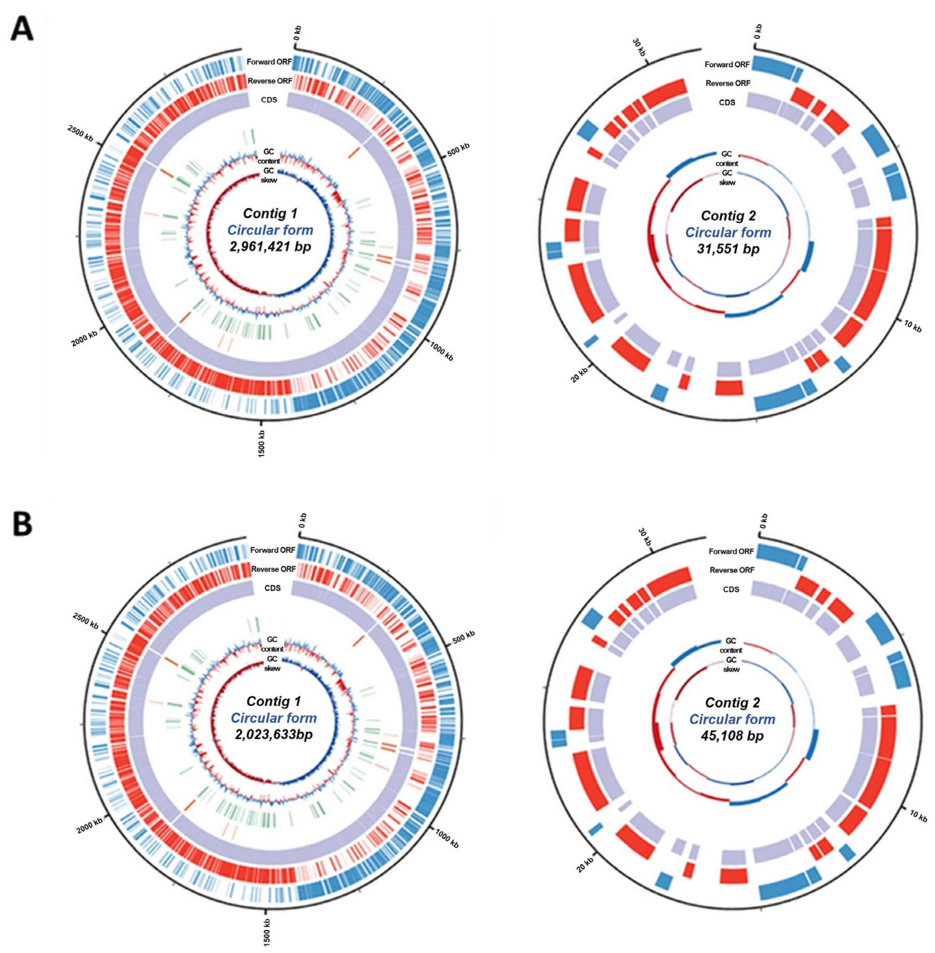
2.8. WGS and Genome Annotation

Genome sequencing was performed on two complementary next-generation sequencing platforms and analyzed by a certified service provider (DNA Link, Inc., Seoul, Republic of Korea) using a Sequel II (Pacific Biosciences, Menlo Park, CA, USA) equipped with an Illumina NovaSeq 6000 system (Illumina, San Diego, CA, USA). Gene prediction of the coding sequences, ribosomal RNA (rRNA), and transfer RNA (tRNA) in the assembled gene were performed using Prokka v1.13. Functional gene annotation was performed using BLAST2GO (BioBam Bioinformatics, Valencia, Spain), and the results were divided into three gene ontology categories [18].

2.9. Morphological Analysis

The strain morphology was analyzed using field-emission scanning electron microscopy (SU5000, Hitachi, Tokyo, Japan) [19]. The samples were vacuum-coated with a double layer of platinum before visualization. The observations were performed at acceleration voltages of 5.0 and 3.0 kV.

2.10. Antibiotic Susceptibility

Antibiotic susceptibility to nine antibiotics was measured using E-strips according to the manufacturer’s instructions (bioMérieux, Marcy-l’Étoile, France). The two bacterial strains were adjusted to a McFarland turbidity of 0.5 and swabbed onto Brain Heart Infusion agar (Difco, BD Bioscience, Bergen County, NJ, USA) plates using a sterilized cotton swab. Then, antibiotic strips were placed on the surface of the plate and incubated at 37 °C for 48 h. Antibiotic resistance was confirmed by the minimum inhibitory concentration (MIC) according to the European Food Safety Authority (EFSA) guidelines [20]. Antibiotic resistance genes in the WGS of each strain were analyzed using the ResFinder database (4.5.0 version; http://genepi.food.dtu.dk/resfinder). The acquired antimicrobial resistance genes were read under a threshold for identity of 90% and a minimum length of 60%.

2.11. Statistical Analysis

The results were analyzed by one-way analysis of variance (ANOVA) followed by Dunnett’s multiple comparison test, which was performed using GraphPad Prism 10 (GraphPad Software Inc., Boston, MA, USA). The results were shown as the mean ± standard error (SE). Differences were considered statistically significant at p < 0.05.

3. Results

3.1. Screening of Strains Based on AChE Inhibition and Neuroprotective Potential

We screened AChE activity, viability, and cytotoxicity in PC12 cells to select candidate strains that could benefit neuronal cells (Table 2). All tested strains showed AChE inhibition of more than 50%. In terms of cell viability and LDH release, only L. rhamnosus and L. gasseri exerted a significant protective effect against oxidative damage in PC12 cells. Therefore, L. rhamnosus and L. gasseri strains were used in subsequent experiments.

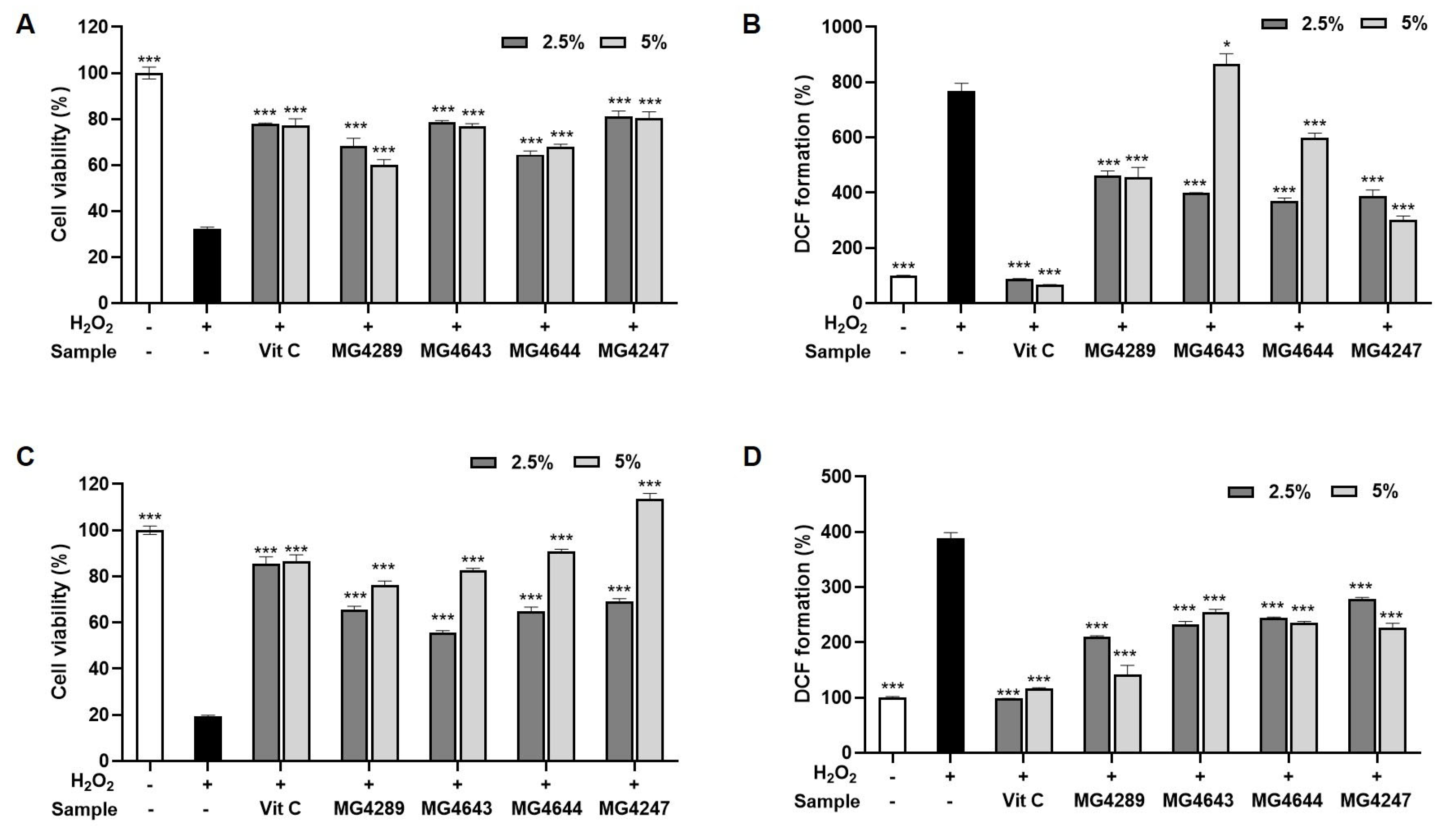
3.2. CFS Improves Cell Viability and Reduces ROS in H2O2-Treated HT-22 and SK-N-MC Cells

Cell viability and ROS generation by CFS treatment were evaluated in HT-22 and SK-N-MC cells treated with H2O2 (Figure 1). Treatment of H2O2 in HT-22 and SK-N-MC cells markedly decreased cell viability and increased ROS generation. In contrast, cell viability was significantly recovered by all CFS treatments. In particular, greater viability was observed with L. gasseri MG4247 than with vitamin C, the positive control. As indicated by the DCF-DA assay, most strains reduced ROS generation, except for L. rhamnosus MG4643 in HT-22 cells. Thus, three strains, excluding L. rhamnosus MG4643, were chosen to evaluate their effects on inflammation, apoptosis, and cholinergic signaling pathways in HT-22 and SK-N-MC cells.

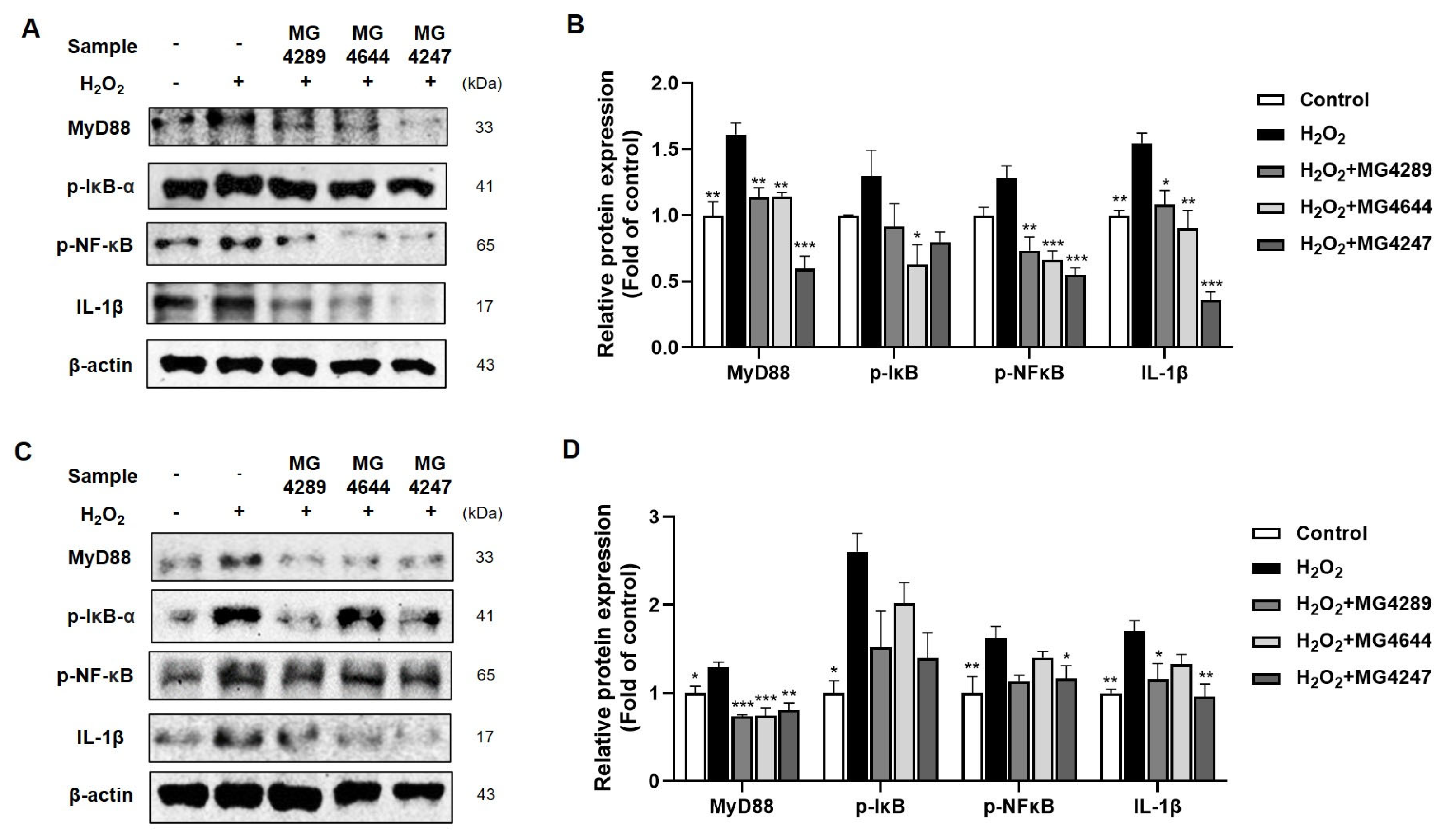
3.3. Anti-Inflammatory Effects of Selected Strains in H2O2-Treated HT-22 and SK-N-MC Cells

The anti-inflammatory effects of L. rhamnosus MG4289, MG4644, and L. gasseri MG4247 were analyzed by Western blot in HT-22 and SK-N-MC cells treated with H2O2 (Figure 2). All strains significantly reduced the protein expression levels of myeloid differentiation primary response 88 (MyD88), which were increased by treatment with H2O2 in both neuronal cell lines. In addition, the expression of phosphorylated-nuclear factor kappa-light-chain-enhancer of activated B cells (p-NF-κB) and interleukin (IL)-1β was remarkably suppressed by treatment of all strains in HT-22 cells (Figure 2A,B). Among the L. rhamnosus strains, MG4644 showed slightly greater suppression of IL-1β expression than MG4289 in HT-22 cells, suggesting a strain-specific difference in anti-inflammatory potency.; however, only L. gasseri MG4247 decreased their expression levels significantly in SK-N-MC cells (Figure 2C,D).

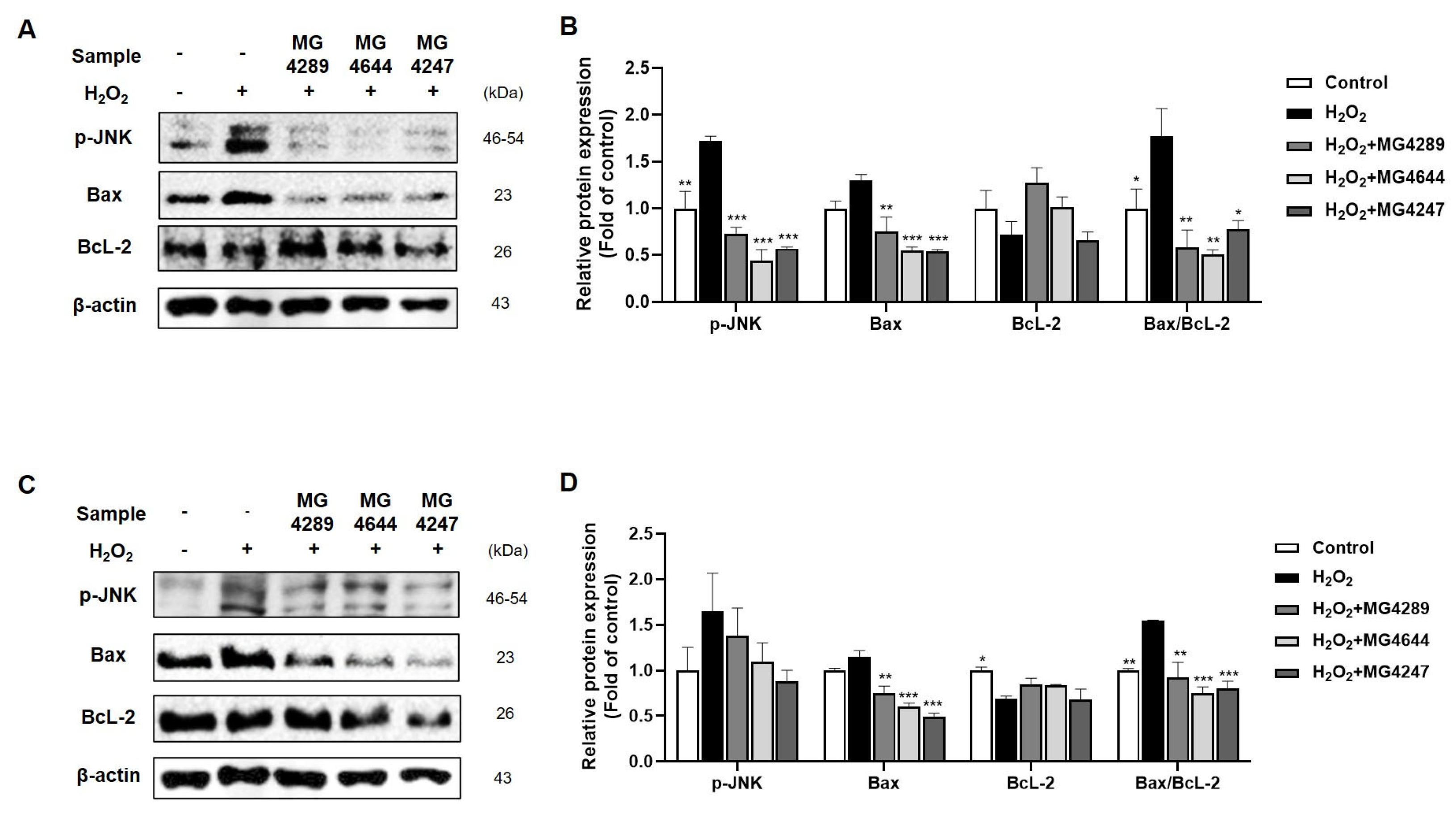
3.4. Modulation of Apoptotic Pathways by Selected Strainsin H2O2-Treated HT-22 and SK-N-MC Cells

The effects of L. rhamnosus MG4289, MG4644, and L. gasseri MG4247 on apoptosis in HT-22 and SK-N-MC cells treated with H2O2 were confirmed by Western blotting (Figure 3). The treatment with all strains considerably downregulated phosphorylated-c-Jun N-terminal kinases (p-JNK) activation in HT-22 cells (Figure 3A,B) and upregulated phosphorylated-protein kinase B (p-Akt) activation in SK-N-MC cells (Figure 3C,D). Additionally, although there was no significant change in B-cell lymphoma (BcL)-2 expressions, BcL-2-associated X (Bax) expression significantly decreased under all strains.

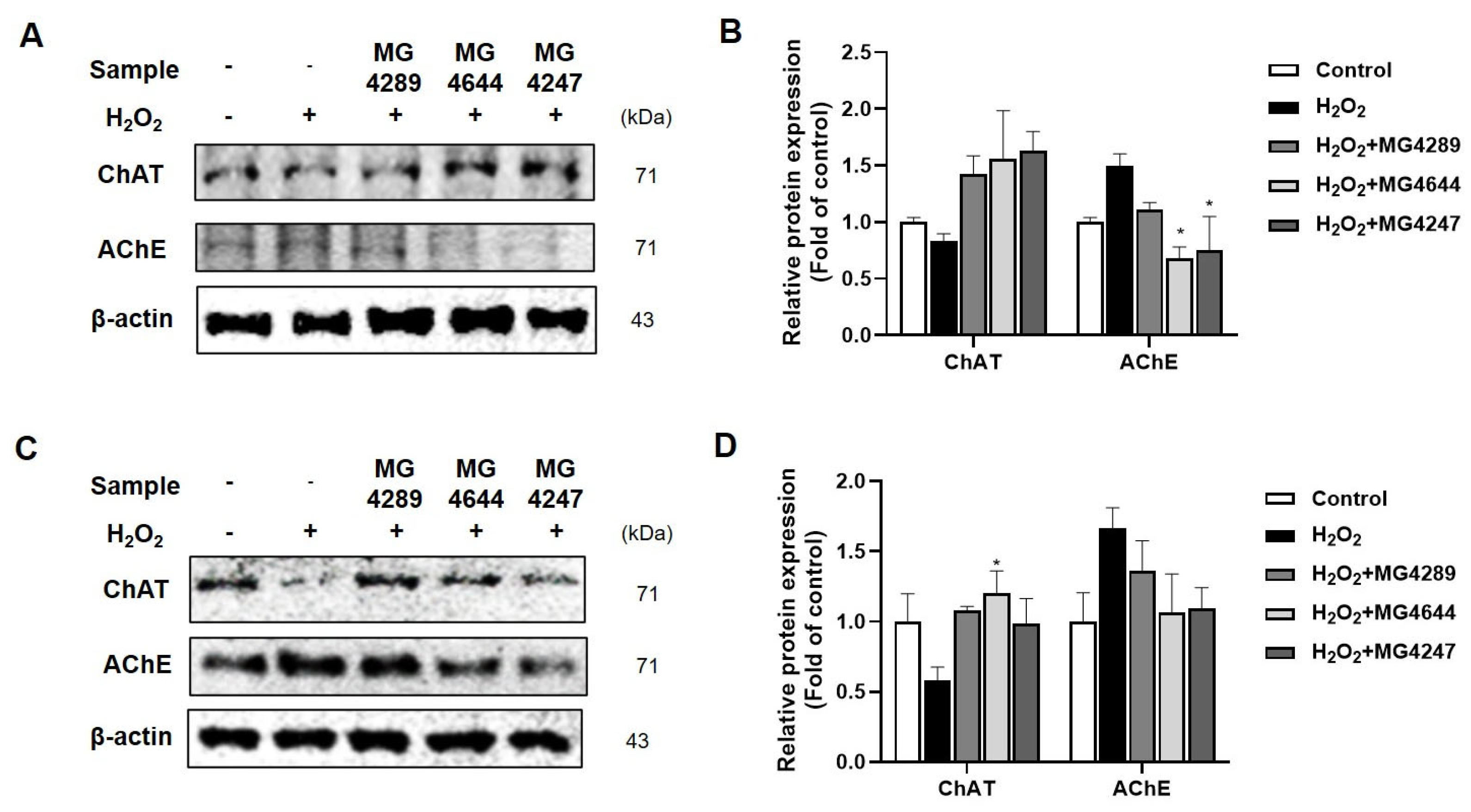
3.5. Regulation of Cholinergic System by Probiotic Strains in H2O2-Treated HT-22 and SK-N-MC Cells

The specific effects of L. rhamnosus MG4289, MG4644, and L. gasseri MG4247 on the cholinergic system in H2O2-exposed neuronal cells were examined using Western blotting (Figure 4). All strains demonstrated modulation of choline-acetyltransferase (ChAT) and AChE in H2O2-treated neuronal cells. Notably, L. rhamnosus MG4644 and L. gasseri MG4247 significantly suppressed H2O2-induced AChE activation in HT-22 cells (Figure 4A,B). Only L. rhamnosus MG4644 increased ChAT activation in H2O2-treated SK-N-MC cells (Figure 4C,D).

3.6. General Features of L. rhamnosus MG4644 and L. gasseri MG4247

Genomic analyses of L. rhamnosus MG4644 and L. gasseri MG4247 revealed two circular contigs (Figure 5). The sizes of the entire genome sequences of L. rhamnosus MG4644 and L. gasseri MG4247 were 2,992,972 bp and 2,068,741 bp, respectively. The GC content in the whole genome sequence was 46.81% (contig 1) and 42.52% (contig 2) in L. rhamnosus MG4644 and 34.77% (contig 1) and 37.86% (contig 2) in L. gasseri MG4247. Contigs 1 and 2 of the coding sequences were identified in 2729 and 38 in L. rhamnosus MG4644 and 1887 and 52 in L. gasseri MG4247, respectively. L. rhamnosus MG4644 contained 15 rRNA and 59 tRNA genes, whereas L. gasseri MG4247 contained 12 rRNA and 59 tRNA genes. All the rRNA and tRNA genes of L. rhamnosus MG4644 and L. gasseri MG4247 were found only in contig 1.
The annotated genes of L. rhamnosus MG4644 and L. gasseri MG4247 were divided into three gene ontology categories (Figure S2). The number of transcripts in L. rhamnosus MG4644 was 3307 for biological processes, 1317 for cellular components, and 3071 for molecular functions, based on gene ontology categories. The number of transcripts in L. gasseri MG4247 was 2683 for biological processes, 1246 for cellular components, and 2314 for molecular functions, based on multiple gene ontologies. The morphological characteristics of L. rhamnosus MG4644 and L. gasseri MG4247 were also observed. The representative image shows that both strains are short, rod-shaped, and have a general bacilli-like form (Figure S3).

3.7. Antibiotic Susceptibility of L. rhamnosus MG4644 and L. gasseri MG4247

The MIC of L. rhamnosus MG4644 and L. gasseri MG4247 for nine antibiotics, including ampicillin, gentamicin, kanamycin, streptomycin, tetracycline, chloramphenicol, erythromycin, vancomycin, and clindamycin, were tested using the E-strip test. As listed in Table 3, L. rhamnosus MG4644 and L. gasseri MG4247 were resistant only to kanamycin and chloramphenicol, according to the EFSA guidelines. According to ResFinder DB, L. rhamnosus MG4644 and L. gasseri MG4247 did not acquire antimicrobial resistance genes for kanamycin and chloramphenicol.

4. Discussion

AChE inhibitors play an important role in cognition and memory by preventing the hydrolysis of the neurotransmitter acetylcholine. It is well-established that AChE analysis can be utilized as a screening method [21]. In addition, H2O2 has high cell membrane permeability in PC12 cells; therefore, measuring viability, LDH release, and cell damage in PC12 cells using H2O2 as an inducer is generally considered a screening method for neurotoxicity [22]. In our study, L. rhamnosus and L. gasseri were selected as candidates to alleviate AChE activity, improve viability, and reduce cell damage in PC12 cells.
Oxidative damage by ROS in neuronal cells is closely related to the cellular damage observed in NDs [23]. Therefore, the estimation of ROS formation is regarded as a direct indicator of NDs [6]. In this study, L. rhamnosus MG4289, L. rhamnosus MG4644, and L. gasseri MG4247 were found to significantly reduce ROS generation in neuronal cells. We investigated the mode of action of these three strains in mitigating oxidative damage caused by ROS in neuronal cells.
ROS affects inflammation, apoptosis, and the cholinergic system in neuronal cells [5,6]. ROS generation by H2O2 oxidizes MyD88, a mediator of the NF-κB signaling pathway [24]. NF-κB, a major inflammation inducer, is activated by the phosphorylation of IκB and translocates into the nucleus, producing various inflammatory cytokines [4]. IL-1β, a pro-inflammatory cytokine, has been proven to cause NDs by exacerbating neuroinflammation [25]. In our study, L. rhamnosus MG4289, L. rhamnosus MG4644, and L. gasseri MG4247 reduced IL-1β via the MyD88/NF-κB pathway in neuronal cells. The activation of NF-κB by ROS-induced apoptosis contributes to aging [1,5]. ROS inhibit BcL-2 by suppressing the phosphorylation of JNK and inducing apoptosis by damaging mitochondrial DNA, which activates Bax [26]. In this study, L. rhamnosus MG4289, L. rhamnosus MG4644, and L. gasseri MG4247 suppressed apoptosis in neuronal cells by modulating the JNK/Bax-dependent pathway.
Numerous studies have reported that NDs are induced when the cholinergic system is dysregulated. Acetylcholine, which regulates cholinergic signaling, is synthesized from choline and acetyl-CoA [27]. In addition, AChE is an enzyme that hydrolyzes acetylcholine, breaking it down into choline and acetate. It binds to acetylcholine receptors in the extracellular space and is a primary target in the treatment of NDs [28]. In Alzheimer’s disease, the overexpression of cytokines by NF-κB activation leads to deficient cholinergic neurotransmission [29]. While both L. rhamnosus strains reduced ROS levels and inflammatory markers, MG4644 exhibited a more substantial effect on modulating cholinergic signaling and suppressing apoptosis, indicating that functional differences exist even within the same species. In this study, L. rhamnosus MG4644 and L. gasseri MG4247 modulated ChAT and AChE in response to H2O2-induced oxidative damage in neuronal cells, thereby affecting the cholinergic system, which plays a crucial role in cognitive function.
Taxonomic misidentification of probiotics is frequently reported due to the limitations of traditional microbiological identification and detection methods; however, these problems can be addressed using WGS [30]. Therefore, we performed WGS to identify the two strains that were predicted to benefit NDs. The average nucleotide identity (ANI) of the whole genome is used for bacterial identification, with an ANI greater than 95% indicating that the bacterial species are identical [31]. Each strain was identified as L. rhamnosus and L. gasseri compared to the type strains L. rhamnosus DSM 20021 (ANI of 97.16%) and L. gasseri ATCC 33323 (ANI of 97.21%), respectively.
Antibiotic resistance poses a significant threat to human health and can be determined using the MIC [32]. In our study, L. rhamnosus MG4644 and L. gasseri MG4247 were phenotypically resistant to kanamycin and chloramphenicol, according to the EFSA guidelines. When the MIC surpasses the defined threshold for a specific antimicrobial agent, it is necessary to distinguish between acquired and intrinsic resistance that can be transmitted to the host through WGS [33]. However, upon examining the antimicrobial resistance genes using WGS and the ResFinder DB, we did not detect any in L. rhamnosus MG4644 and L. gasseri MG4247. These results are consistent with previous reports indicating the absence of antibiotic acquisition genes in most Lactobacillus strains [31]. In addition, previous studies have shown that L. rhamnosus MG4644 and L. gasseri MG4247 were observed to survive under simulated gastrointestinal tract conditions and adhered to intestinal epithelial cells without exhibiting hemolysis or bile salt hydrolase activity. Additionally, they did not compromise the cell viability of intestinal epithelial cells [19,34]. Therefore, because L. rhamnosus MG4644 and L. gasseri MG4247 meet all the safety requirements of the FAO/WHO and EFSA guidelines, they can be used as commercial functional food [35]. This study demonstrated the efficacy and safety of L. gasseri MG4247 and L. rhamnosus MG4644. Based on these findings, L. rhamnosus MG4644 and L. gasseri MG4247 may serve as potential neuroprotective probiotics. Given their dual ability to modulate inflammation and cholinergic signaling, this study may lay a foundation in other neurodegenerative conditions characterized by oxidative stress and neuroinflammation; however, further in vivo studies are needed to evaluate their systemic efficacy and blood–brain barrier permeability.

5. Conclusions

Overall, L. rhamnosus MG4644 and L. gasseri MG4247 were found to inhibit inflammation, apoptosis, and cholinergic system impairment caused by oxidative damage through the NF-κB signaling pathway in vitro. The results of this study suggest that L. rhamnosus MG4644 and L. gasseri MG4247 have potential applications in future research related to neurodegenerative diseases (NDs). However, these findings are based on in vitro data, and further in vivo and clinical studies are necessary to validate their therapeutic effects. Furthermore, additional research is required to investigate the practical application of these strains as probiotics in functional foods and microbiome-targeted therapies.

Supplementary Materials

The following supporting information can be downloaded at: https://www.mdpi.com/article/10.3390/fermentation11070385/s1, Table S1: List of primary antibody information used in this study. Table S2: Cell viability and LDH releases of 10% CFS in PC12 cells treated with H2O2. Figure S1: The predicted functional genes were divided into three gene ontology categories: biological process (yellow), cellular component (orange), and molecular function (green). Figure S2: The field emission-scanning electron microscope (FE-SEM) images (magnification 10,000×).

Author Contributions

Conceptualization, S.-I.C. and H.J.H.; methodology, J.Y.L., J.H.K. and J.-Y.P.; formal analysis, J.H.K. and J.-Y.P.; investigation, J.H.K. and J.-Y.P.; resources, B.-K.K.; data curation, J.Y.L., S.-I.C. and H.J.H.; writing—original draft preparation, J.Y.L.; writing—review and editing, S.-I.C.; visualization, J.Y.L.; supervision, S.-I.C. and H.J.H.; project administration, B.-K.K.; funding acquisition, S.-I.C. and H.J.H.; All authors have read and agreed to the published version of the manuscript.

Funding

This research was financially supported by the Ministry of Small and Medium-sized Enterprises (SMEs) and Startups (MSS), Korea, under the “Regional Specialized Industry Development Plus Program (R&D, S3366186)” supervised by the Korea Technology and Information Promotion Agency for SMEs (TIPA).

Institutional Review Board Statement

Not applicable.

Informed Consent Statement

Not applicable.

Data Availability Statement

The data presented in this study are openly available in NCBI repository, L. gasseri MG4247 (CP161921-CP161922) and L. rhamnosus MG4644 (CP161813-CP161814).

Conflicts of Interest

Authors Ji Yeon Lee, Jeong-Yong Park, Byoung-Kook Kim and Soo-Im Choi were employed by the company MEDIOGEN Co., Ltd. The remaining authors declare that the research was conducted in the absence of any commercial or financial relationships that could be construed as a potential conflict of interest.

References

  1. Jakubczyk, K.; Dec, K.; Kałduńska, J.; Kawczuga, D.; Kochman, J.; Janda, K. Reactive oxygen species-sources, functions, oxidative damage. Pol Merkur Lekarski. 2020, 48, 124–127. [Google Scholar]
  2. Beckhauser, T.F.; Francis-Oliveira, J.; De Pasquale, R. Reactive oxygen species: Physiological and physiopathological effects on synaptic plasticity: Supplementary issue: Brain plasticity and repair. J. Exp. Neurosci. 2016, 10, S39887. [Google Scholar] [CrossRef] [PubMed]
  3. Massaad, C.A.; Klann, E. Reactive oxygen species in the regulation of synaptic plasticity and memory. Antioxid. Redox Signal. 2011, 14, 2013–2054. [Google Scholar] [CrossRef]
  4. Hsieh, H.-L.; Yang, C.-M. Role of redox signaling in neuroinflammation and neurodegenerative diseases. BioMed Res. Int. 2013, 2013, 484613. [Google Scholar] [CrossRef] [PubMed]
  5. Liguori, I.; Russo, G.; Curcio, F.; Bulli, G.; Aran, L.; Della-Morte, D.; Gargiulo, G.; Testa, G.; Cacciatore, F.; Bonaduce, D. Oxidative stress, aging, and diseases. Clin. Interv. Aging 2018, 13, 757–772. [Google Scholar] [CrossRef] [PubMed]
  6. Feitosa, C.M.; da Silva Oliveira, G.L.; do Nascimento Cavalcante, A.; Morais Chaves, S.K.; Rai, M. Determination of parameters of oxidative stress in vitro models of neurodegenerative diseases-A review. Curr. Clin. Pharmacol. 2018, 13, 100–109. [Google Scholar] [CrossRef]
  7. Aksenova, M.V.; Aksenov, M.Y.; Mactutus, C.F.; Booze, R.M. Cell culture models of oxidative stress and injury in the central nervous system. Curr. Neurovasc. Res. 2005, 2, 73–89. [Google Scholar] [CrossRef]
  8. Bermúdez-Humarán, L.G.; Salinas, E.; Ortiz, G.G.; Ramirez-Jirano, L.J.; Morales, J.A.; Bitzer-Quintero, O.K. From probiotics to psychobiotics: Live beneficial bacteria which act on the brain-gut axis. Nutrients 2019, 11, 890. [Google Scholar] [CrossRef]
  9. Toledo, A.R.L.; Monroy, G.R.; Salazar, F.E.; Lee, J.-Y.; Jain, S.; Yadav, H.; Borlongan, C.V. Gut–brain axis as a pathological and therapeutic target for neurodegenerative disorders. Int. J. Mol. Sci. 2022, 23, 1184. [Google Scholar] [CrossRef]
  10. Bi, M.; Liu, C.; Wang, Y.; Liu, S.-J. Therapeutic Prospect of New Probiotics in Neurodegenerative Diseases. Microorganisms 2023, 11, 1527. [Google Scholar] [CrossRef]
  11. Xiang, S.; Ji, J.-L.; Li, S.; Cao, X.-P.; Xu, W.; Tan, L.; Tan, C.-C. Efficacy and safety of probiotics for the treatment of Alzheimer’s disease, mild cognitive impairment, and Parkinson’s Disease: A systematic review and meta-analysis. Front. Aging neurosci. 2022, 14, 730036. [Google Scholar] [CrossRef]
  12. Aghamohammad, S.; Hafezi, A.; Rohani, M. Probiotics as functional foods: How probiotics can alleviate the symptoms of neurological disabilities. Biomed. Pharmacother. 2023, 163, 114816. [Google Scholar] [CrossRef] [PubMed]
  13. Salek, F.; Mirzaei, H.; Khandaghi, J.; Javadi, A.; Nami, Y. Apoptosis induction in cancer cell lines and anti-inflammatory and anti-pathogenic properties of proteinaceous metabolites secreted from potential probiotic Enterococcus faecalis KUMS-T48. Sci. Rep. 2023, 13, 7813. [Google Scholar] [CrossRef]
  14. Kumar, P.; Nagarajan, A.; Uchil, P.D. Analysis of cell viability by the MTT assay. Cold Spring Harb. Protoc. 2018, 2018, pdb. prot095505. [Google Scholar] [CrossRef] [PubMed]
  15. Kumar, P.; Nagarajan, A.; Uchil, P.D. Analysis of cell viability by the lactate dehydrogenase assay. Cold Spring Harb. Protoc. 2018, 2018, pdb. prot095497. [Google Scholar] [CrossRef]
  16. Heo, S.-J.; Cha, S.-H.; Kim, K.-N.; Lee, S.-H.; Ahn, G.; Kang, D.-H.; Oh, C.; Choi, Y.-U.; Affan, A.; Kim, D. Neuroprotective effect of phlorotannin isolated from Ishige okamurae against H2O2-induced oxidative stress in murine hippocampal neuronal cells, HT22. Appl. Biochem. Biotechnol. 2012, 166, 1520–1532. [Google Scholar] [CrossRef]
  17. Stergiou, O.S.; Tegopoulos, K.; Kiousi, D.E.; Tsifintaris, M.; Papageorgiou, A.C.; Tassou, C.C.; Chorianopoulos, N.; Kolovos, P.; Galanis, A. Whole-genome sequencing, phylogenetic and genomic analysis of Lactiplantibacillus pentosus L33, a potential probiotic strain isolated from fermented sausages. Front. Microbiol. 2021, 12, 746659. [Google Scholar] [CrossRef]
  18. Bae, W.-Y.; Lee, Y.J.; Jung, W.-H.; Shin, S.L.; Kim, T.-R.; Sohn, M. Draft genome sequence and probiotic functional property analysis of Lactobacillus gasseri LM1065 for food industry applications. Sci. Rep. 2023, 13, 12212. [Google Scholar] [CrossRef]
  19. Park, J.-Y.; Lee, J.Y.; Kim, Y.; Kang, C.-H. Limosilactobacillus fermentum MG5091 and Lactococcus lactis MG4668 and MG5474 Suppress Muscle Atrophy by Regulating Apoptosis in C2C12 Cells. Fermentation 2023, 9, 659. [Google Scholar] [CrossRef]
  20. Zielińska, D.; Sionek, B.; Kołożyn-Krajewska, D. Safety of probiotics. In Diet, Microbiome and Health; Elsevier: Amsterdam, The Netherlands, 2018; pp. 131–161. [Google Scholar]
  21. Liu, D.-M.; Xu, B.; Dong, C. Recent advances in colorimetric strategies for acetylcholinesterase assay and their applications. Trends Anal. Chem. 2021, 142, 116320. [Google Scholar] [CrossRef]
  22. Wang, J.; Xiao, S.; Cai, Q.; Miao, J.; Li, J. Antioxidant Capacity and Protective Effects on H2O2-Induced Oxidative Damage in PC12 Cells of the Active Fraction of Brassica rapa L. Foods 2023, 12, 2075. [Google Scholar] [CrossRef] [PubMed]
  23. Oswald, M.C.; Garnham, N.; Sweeney, S.T.; Landgraf, M. Regulation of neuronal development and function by ROS. FEBS Lett. 2018, 592, 679–691. [Google Scholar] [CrossRef] [PubMed]
  24. Stottmeier, B.; Dick, T.P. Redox sensitivity of the MyD88 immune signaling adapter. Free Radic. Biol. Med. 2016, 101, 93–101. [Google Scholar] [CrossRef]
  25. Mendiola, A.S.; Cardona, A.E. The IL-1β phenomena in neuroinflammatory diseases. J. Neural Transm. 2018, 125, 781–795. [Google Scholar] [CrossRef]
  26. Redza-Dutordoir, M.; Averill-Bates, D.A. Activation of apoptosis signalling pathways by reactive oxygen species. BBA Mol. Cell Res. 2016, 1863, 2977–2992. [Google Scholar] [CrossRef]
  27. Bekdash, R.A. The cholinergic system, the adrenergic system and the neuropathology of Alzheimer’s disease. Int. J. Mol. Sci. 2021, 22, 1273. [Google Scholar] [CrossRef]
  28. Picciotto, M.R.; Higley, M.J.; Mineur, Y.S. Acetylcholine as a neuromodulator: Cholinergic signaling shapes nervous system function and behavior. Neuron 2012, 76, 116–129. [Google Scholar] [CrossRef]
  29. Hafiz, Z.Z.; Amin, M.A.M.; Johari James, R.M.; Teh, L.K.; Salleh, M.Z.; Adenan, M.I. Inhibitory effects of raw-extract Centella asiatica (RECA) on acetylcholinesterase, inflammations, and oxidative stress activities via in vitro and in vivo. Molecules 2020, 25, 892. [Google Scholar] [CrossRef]
  30. Wang, Y.; Liang, Q.; Lu, B.; Shen, H.; Liu, S.; Shi, Y.; Leptihn, S.; Li, H.; Wei, J.; Liu, C. Whole-genome analysis of probiotic product isolates reveals the presence of genes related to antimicrobial resistance, virulence factors, and toxic metabolites, posing potential health risks. BMC Genom. 2021, 22, 210. [Google Scholar] [CrossRef]
  31. Kim, M.-H.; Kim, D.; Sung, J.H. A Gut-Brain Axis-on-a-Chip for studying transport across epithelial and endothelial barriers. J. Ind. Eng. Chem. 2021, 101, 126–134. [Google Scholar] [CrossRef]
  32. Karapetkov, N.; Georgieva, R.; Rumyan, N.; Karaivanova, E. Antibiotic susceptibility of different lactic acid bacteria strains. Benef. Microbes 2011, 2, 335–339. [Google Scholar] [CrossRef] [PubMed]
  33. EFSA Panel on Additives and Products or Substances used in Animal Feed (FEEDAP). Guidance on the assessment of bacterial susceptibility to antimicrobials of human and veterinary importance. EFSA J. 2012, 10, 2740. [Google Scholar]
  34. Lee, J.Y.; Park, Y.; Jeong, Y.; Kang, H. Anti-Inflammatory Response in TNFα/IFNγ-Induced HaCaT Keratinocytes and Probiotic Properties of Lacticaseibacillus rhamnosus MG4644, Lacticaseibacillus paracasei MG4693, and Lactococcus lactis MG5474. J. Microbiol. Biotechn. 2023, 33, 1039. [Google Scholar] [CrossRef] [PubMed]
  35. FAO/WHO Work Group. Guidelines for the Evaluation of Probiotics in Food; FAO/WHO Work. Group: Rome, Italy, 2002; pp. 1–11. [Google Scholar]
Figure 1. The neuroprotective effects of CFS in HT-22 and SK-N-MC cells. The cell viability in (A) HT-22 and (C) SK-N-MC cells and DCF formation indicated ROS generation in (B) HT-22 and (D) SK-N-MC cells treated with H2O2. The results shown were mean ± SE (n = 3). The significance was determined by Dunnett’s multiple comparisons test; * p < 0.05, and *** p < 0.001 vs. H2O2-treated group.
Fermentation 11 00385 g001
Figure 2. The CFS reduces inflammation signaling pathways in neuronal cells treated with H2O2. The representative images and relative protein expressions of MyD88, p-IκB, p-NF-κB, and IL-1β in (A,B) HT-22 and (C,D) SK-N-MC by Western blot. The β-actin was used as a loading control. The results shown were mean ± SE (n = 3). The significance was determined by Dunnett’s multiple comparisons test; * p < 0.05, ** p < 0.01, and *** p < 0.001 vs. H2O2-treated group.
Fermentation 11 00385 g002
Figure 3. The CFS modulates apoptosis signaling pathways in neuronal cells treated with H2O2. The representative images and relative protein expressions of p-JNK, Bax, and BcL-2 in (A,B) HT-22 and (C,D) SK-N-MC by Western blot. The β-actin was used as a loading control. The results shown were mean ± SE (n = 3). The significance was determined by Dunnett’s multiple comparisons test; * p < 0.05, ** p < 0.01, and *** p < 0.001 vs. H2O2-treated group.
Fermentation 11 00385 g003
Figure 4. The CFS regulated cholinergic signaling pathways in neuronal cells treated with H2O2. The representative images and relative protein expressions of ChAT and AChE in HT-22 (A,B) and SK-N-MC (C,D) cells by Western blot. The β-actin was used as a loading control. The results shown were mean ± SE (n = 3). The significance was determined by Dunnett’s multiple comparisons test; * p < 0.05 vs. H2O2-treated group.
Fermentation 11 00385 g004
Figure 5. The circular gene map of two contigs from (A) L. rhamnosus MG4644 and (B) L. gasseri MG4247.
Fermentation 11 00385 g005
Table 1. Accession number in NCBI GenBank database of 16s rRNA gene and origin from all strains used in this study.
SpeciesStrainAccession NumberOrigin
Lacticaseibacillus rhamnosusMG4289MW947158.1Human (vagina)
MG4643ON668169.1Human (feces)
MG4644ON668170.1Human (feces)
Lacticaseibacillus paracaseiMG4267OP102572.1Human (vagina)
MG4272MW947164.Human (vagina)
MG4693OP077096.1Human (oral)
Lactiplantibacillus plantarumMG5106OP102458.1Food (fish)
MG4604OP102458.1Human (feces)
Lactobacillus gasseriMG4247MN069036.1Human (vagina)
Table 2. Inhibition of acetylcholinesterase (AChE), cell viability, and LDH releases in PC12 cells of CFS from probiotic strains.
SpeciesStrainAChE Inhibition (%)Cell Viability (%)LDH Releases (%)
Control 10.00 ± 1.46100.00 ± 1.33100.00± 3.41
L. rhamnosusMG428951.83 ± 0.12177.67 ± 1.05 ***21.46 ± 0.86 ***
MG464352.00 ± 1.07118.19 ± 3.38 **16.67 ± 1.00 ***
MG464460.08 ± 1.39121.85 ± 1.28 ***14.86 ± 0.27 ***
L. paracaseiMG426754.35 ± 1.9082.48 ± 2.36 **93.56 ± 2.42
MG427260.57 ± 1.5382.09 ± 3.79 *76.08 ± 4.44 *
MG469363.85 ± 2.0188.53 ± 2.57 *81.64 ± 7.18
L. plantarumMG510663.02 ± 0.6948.06 ± 1.00 **68.35 ± 1.55 **
MG460452.80 ± 1.7259.64 ± 8.19 **63.84 ± 2.98 **
L. gasseriMG424767.04 ± 0.28131.12 ± 5.13 **14.21 ± 0.79 ***
The cells were treated with 5% cell-free supernatant (CFS). The results shown were mean ± SE (standard error) of the triple experiments. The significance was determined by Dunnett’s multiple comparisons test; * p < 0.05, ** p < 0.01, and *** p < 0.001 vs. control group. 1 In AChE inhibition, MRS is used as a control; in cell viability and LDH releases, H2O2-treated cells are used as a control.
Table 3. The minimum inhibitory concentration (MIC) of L. rhamnosus MG4644 and L. gasseri MG4247.
AntimicrobialsL. gasseri MG4247L. rhamnosus MG4644
MIC (μg/mL)S/R 1MIC (μg/mL)S/R
Ampicillin0.125S0.5S
Gentamicin3S1.5S
Kanamycin48R24R
Streptomycin6S8S
Tetracycline0.75S0.75S
Chloramphenicol6R6R
Erythromycin0.047S0.09S
Vancomycin0.5Sn.r. 2
Clindamycin0.5S0.25S
1 S, susceptibility; R, resistance; 2 not required.
Disclaimer/Publisher’s Note: The statements, opinions and data contained in all publications are solely those of the individual author(s) and contributor(s) and not of MDPI and/or the editor(s). MDPI and/or the editor(s) disclaim responsibility for any injury to people or property resulting from any ideas, methods, instructions or products referred to in the content.

