Article

Antioxidant and ACE-Inhibition Activities After In Vitro Digestion of a Non-Fermented Dairy Beverage Enriched with Postbiotics of Lactobacillus spp.

by
Norma Angélica Bolivar-Jacobo
1,
Raúl Alberto Reyes-Villagrana
2,*,
Martha María Arévalos-Sánchez
1,
Ana Luisa Rentería-Monterrubio
1,
Eduardo Santellano-Estrada
1,
Nora Aidee Salas-Salazar
3 and
América Chávez-Martínez
1,*
1
Facultad de Zootécnica y Ecología, Universidad Autónoma de Chihuahua, Periférico Francisco R. Almada, Km 1, Chihuahua 31453, Mexico
2
Consejo Nacional de Ciencia y Tecnología, Av. Insurgentes Sur 1582, Col. Crédito Constructor, Col. Benito Juárez, Ciudad de Mexico 03940, Mexico
3
Facultad de Ciencias Agrotecnológicas, Universidad Autónoma de Chihuahua, Campus I, Circuito Universitario, Chihuahua 31310, Mexico
*
Authors to whom correspondence should be addressed.
Fermentation 2025, 11(4), 223; https://doi.org/10.3390/fermentation11040223
Submission received: 28 February 2025 / Revised: 8 April 2025 / Accepted: 14 April 2025 / Published: 16 April 2025
(This article belongs to the Special Issue Fermentation: 10th Anniversary)

Abstract

:
Postbiotics are recently gaining consumer attention for their potential health benefits. This study aimed to examine the effects of supplementation of a non-fermented dairy beverage with postbiotics derived from Lactobacillus acidophilus and Lactobacillus helveticus on antioxidant (DPPH, ABTS, FRAP, and ORAC), antimicrobial, and ACE-inhibition activities before and after in vitro digestion. Three dairy beverages were elaborated: without the addition of postbiotics (T0), with Lactobacillus acidophilus postbiotics (T1), and with Lactobacillus helveticus postbiotics (T2). Before in vitro digestion, T2 presented higher antioxidant activity (p < 0.05). And, after in vitro digestion, except by the ABTS method, T1 and T2 presented the highest antioxidant activities (p < 0.05) and bioaccessibility indexes (p < 0.05). Regarding ACE-inhibition activity, before in vitro digestion, there were no differences among treatments (p > 0.05), but after in vitro digestion, T1 and T2 presented the highest ACE-inhibition activities (p < 0.05) and bioaccessibility indexes (p < 0.05). An antimicrobial effect against Bacillus spp. and S. aureus was observed in Lactobacillus acidophilus and Lactobacillus helveticus postbiotics. However, L. acidophilus postbiotics did not present an antibacterial effect against E. coli. Such findings highlight the potential of postbiotics as functional ingredients to enhance the antioxidant and ACE-inhibition activities of non-fermented dairy beverages, further adding to their appeal as health-promoting dairy food.

1. Introduction

In recent years, the number of people with chronic conditions (diabetes, hypertension, cancer, gastrointestinal disorders, autoimmune diseases, etc.) associated with poor diet and lifestyle (sedentary lifestyle, smoking, etc.) has increased [1]. Nutrition and intestinal homeostasis are two fundamental factors for individuals to grow and develop healthily. The necessary nutrients are obtained from the first one, and the second contributes to approximately 70% of the total immunity of humans [2]. To help reduce the incidence of these diseases, the food industry developed functional foods [3,4]. These are defined as foods that, besides providing nutrients and energy, beneficially modulate one or more targeted functions in the body [5]. Functional foods can be separated into two categories: conventional and modified. Conventional foods are natural, whole-food ingredients that are rich in important nutrients such as vitamins, minerals, and heart-healthy fats, or non-nutrients such as phytochemicals, including polyphenols, prebiotics, probiotics, postbiotics, and dietary fibers. Modified foods are fortified with additional ingredients like vitamins, minerals, fiber, probiotics, prebiotics, and postbiotics, among others. Functional foods have physiological functions in the body to improve well-being and health, reduce disease risk, and/or improve disease outcomes.
Moreover, postbiotics are defined as the “preparation of inanimate microorganisms and/or their components that confers a health benefit on the host” [6]. These are produced from probiotic species, such as Lactobacillus spp., and are characterized as being simple to transport, store, and maintain [7]. These also have several significant advantages over probiotics, including being much more shelf-stable and posing less risks when used with vulnerable populations, such as very young subjects or those who are immunocompromised [8]. Their mechanisms of action include the alteration of the gut microbiome, immune system responses, and direct antimicrobial properties [9]. Lactobacillus spp. postbiotics include a wide range of compounds, each with its unique properties and potential health benefits. Among these are metabolites (byproducts of metabolic activities), such as vitamins, amino acids, peptides, and organic acids [10], or microbial cell fractions (parts of the bacterial cell that may have beneficial effects), such as lysates, teichoic acid, peptidoglycan-derived muropeptides, and pili-type structures [11], extracellular polysaccharides, secreted biosurfactants, and bacteriocins [10]. Therefore, both metabolites and microbial cell fractions are important for the human body. These compounds exhibit a range of bioactivities, including antioxidant, antimicrobial (against E. coli, L. monocytogenes, S. enterica, and Staphylococcus spp.), and anti-inflammatory properties [12,13,14,15].
Aside from that, while it has been shown that Lactobacillus probiotics can lower blood pressure [16,17], it has not yet been reported what effect Lactobacillus postbiotics have on stopping the angiotensin-converting enzyme (ACE). A recent study reported that acetate and butyrate, compounds derived from the gut microbiota, can reduce blood pressure in untreated hypertensive patients [18]. Then, since acetate and butyrate are two short fatty acids produced by probiotics, it could be assumed that postbiotics present ACE-inhibitory activity. The angiotensin-converting enzyme (ACE) contributes to blood pressure reduction primarily through its role in the renin-angiotensin system (RAS). Primarily, ACE is responsible for converting angiotensin I (Ang I) to angiotensin II (Ang II), a potent vasoconstrictor that increases blood pressure by constricting blood vessels and promoting sodium retention. When ACE is inhibited, angiotensin II formation is reduced, resulting in less vasoconstriction and sodium retention. Lower levels of angiotensin II result in less vasoconstriction, allowing blood vessels to relax and dilate. This dilation increases blood flow and lowers systemic blood pressure. ACE also breaks down bradykinin, a vasodilator that helps relax blood vessels. Therefore, by inhibiting ACE, bradykinin levels increase, which further contributes to vasodilation and blood pressure reduction. This translates into improved blood flow and reduces cardiac workload, thereby improving cardiac function and reducing the risk of heart failure [19].
Furthermore, postbiotics, like any other bioactive component, must maintain their biofunctionality, remain bioaccessible, and exhibit stability. Likewise, the amount of postbiotics released from the food matrix can be influenced by the processing conditions (pH, temperature, etc.) and the interaction that these may have with other components or ingredients in the food matrix [20].
Therefore, the objective of this work was to evaluate the antioxidant (DPPH, ABTS, and FRAP), antimicrobial, and ACE-inhibitory activity of a functional dairy drink with added postbiotics from Lactobacillus acidophilus and Lactobacillus helveticus before and after in vitro digestion.

2. Materials and Methods

2.1. Materials

UHT skim and whole milk (LALA®, Gomez Palacio, Durango, Mexico), powdered milk (NIDO®, Ocotlán, Jalisco, Mexico), cocoa (Hershey’s, Hershey, PA, USA), Avicel® (FMC Corporation, Philadelphia, PA, USA), and sugar (Zulca, Culiacán, Sinaloa, Mexico) were purchased in a local market. The starter cultures were donated by Hansen® (Hørsholm, Denmark). The chemicals and reagents used were all of analytical reagent grade.

2.2. Postbiotics Obtention

To obtain postbiotics from L. acidophilus (LA-5, CHR HANSEN) and L. helveticus (LH-B02, CHR HANSEN), one flask for each probiotic with 500 mL of skimmed milk (LALA Light®) was inoculated at 1% w/v (8.08 Log10 CFU/mL) with the corresponding culture. Then, ultrasound-assisted fermentation was performed. First, each flask containing the probiotic was subjected to sonication (30% amplitude-3 min) using an ultrasonic processor with a 1.3 cm diameter probe (20 kHz, GEX750, Sonic, Newtown, CT, USA) [21]; then, samples were incubated in aerobiosis at 37 ± 1 °C for 4 h. These parameters were selected based on previous results (Bolivar-Jacobo et al., 2023) [21]. Subsequently, samples were centrifuged (Avanti® J-26 XPI, Beckman Coluter®, Indianapolis, IN, USA) at 20,000× g for 30 min at 4°C, and the supernatant (aqueous extract) was filtered (Whatman™ No. 1, GE Healthcare, UK) and lyophilized (Labconco Niro Mobile Minor DK-2860, GEA Company, Spalding, UK) at −85°C and 0.035 mbar pressure.

2.3. Beverage Elaboration

The dairy beverage was prepared by combining 200 mL of UHT fluid whole milk, 10 g of dry milk, 30 g of sugar, 20 g of cocoa, and 1 g of avicel®. The mixture was slowly heated to a temperature of 34 ± 0.5 °C. Subsequently, it was cooled to 5 °C. Then, from this mixture, the following treatments were prepared: beverage without the addition of postbiotics (T0), beverage added with Lactobacillus acidophilus postbiotics (T1), and beverage added with Lactobacillus helveticus postbiotics (T2). To add postbiotics to T1 and T2, a solution was prepared by dissolving 1 g of lyophilized postbiotics in 10 mL of sterile water. Subsequently, 2 mL of each solution was added to the beverages (200 mL) to prepare the treatments. Treatments were prepared in triplicate under a completely randomized design.

2.4. In Vitro Digestion

Treatments were subjected to in vitro digestion according to the [22] methodology with some modifications. Briefly, 100 mL of each treatment was placed in an Erlenmeyer flask with 0.1 g of pepsin (Sigma-Aldrich, St. Louis, MO, USA). Subsequently, hydrochloric acid (HCl) 2M (Golden Bell, Mexico) was added until the pH was adjusted to 2. Then, the mixture was incubated at 37 ± 1°C (90 rpm) for 2 h (first phase of digestion). After this, 20 mL of the previous mixture was taken, and the pH was adjusted to 7 with 1 M sodium bicarbonate (NaHCO3). Then, 5 mL of a solution containing intestinal enzymes, which was made with 25 g of bile and 4 g of pancreatin mixed in a sodium bicarbonate solution, was added. Finally, the mixture was incubated at 37 ± 1°C (100 rpm) for 2 h (second phase of digestion). Samples were taken for each treatment before in vitro digestion and after the first and second phases of digestion. The samples were spun in a centrifuge at 12,000 rpm for 20 min, then passed through Whatman 2 paper, and the liquid collected was frozen for later tests (ABTS, DPPH, FRAP, ACE-inhibition activity). Each treatment’s digestion was performed in triplicate.

2.5. Antioxidant Activities

Antioxidant activity (AA) was investigated by the ABTS, DPPH, FRAP, and ORAC methodologies as outlined below.
ABTS method. AA by the ABTS methodology was conducted according to [23]. AA was reported as the mg Trolox equivalent (mg TE/100 mL). The determinations were performed in triplicate.
DPPH method. AA by the 2,2-Diphenyl-1-Picrylhydrazil (DPPH) methodology was conducted according to [23]. AA was reported as the mg Trolox equivalent (mgTE)/100 mL). The determinations were performed in triplicate.
FRAP method. AA by the ferric reducing antioxidant power (FRAP) assay was determined with a commercial kit (Sigma-Aldrich, St. Louis, MO, USA) following the manufacturer’s instructions and conducted according to [23]. Results were expressed as the mM ferrous equivalent (mM Fe2⁺ equivalents).
ORAC method. AA by the oxygen radical trapping capacity (ORAC) methodology was determined according to [24] with some modifications. First, a phosphate buffer solution was prepared by weighing 13.19 g of K2HPO4 and 10.26 g of KH2PO4 and dissolving it in 900 mL of distilled water, adjusting the pH to 7.4 ± 0.2, and making it up to 1 L. For the working solution of fluorescein disodium salt, 0.0376 mg of cobalt (II) fluoride tetrahydrate (fluorescein, Sigma Aldrich, St. Louis, MO, USA) 10 nM were weighed in 100 mL of phosphate buffer (pH 7.4), and for the 250 mM solution of 2,20-azobis (2-amidino-propane) dihydrochloride (APPH, Sigma Aldrich, St. Louis, MO, USA), 667.98 mg were weighed in 10 mL of phosphate buffer. To measure the antioxidant capacity of the samples, 150 μL of 10 nM fluorescein solution was placed in the wells of the microplate, 25 μL of the sample or the Trolox standards were added, and the phosphate buffer was used as a blank. The plate was subsequently incubated (Varioskan Flash, Thermo Scientific, Waltham, MA, USA) at 37 °C for 30 min, and at the end, 25 μL of 250 mM AAPH were added. Fluorescence (excitation at 485 nm and emission at 520 nm) was read every 90 s for 90 min; the plate was shaken before each reading. For the 6-Hydroxy-2,5,7,8-tetramethylchroman-2-carboxylic acid (Trolox, Sigma-Aldrich, St. Louis, MO, USA) calibration curve, solutions at 0.1, 0.3, 0.5, 0.8, and 1 mM concentrations, from a stock solution (31.3 mg of Trolox in 10 mL of phosphate buffer), were prepared to develop the standard curve, obtaining the following equation: y = 0.4987x + 0.1173, R2 = 0.9966. The antioxidant capacity was expressed in the mM Trolox equivalent (mM TE or mg TE/100 mL). The determinations were carried out in triplicate. The data was analyzed using SkanIt 2.4.5 RE for Variouskan Flash software.

2.6. Angiotensin Converting Enzyme-Inhibitory Activity

The inhibitory ACE activity was analyzed using the spectrophotometric method proposed by [25], with some modifications. An ACE solution (100 mU/mL) was prepared by diluting one unit of ACE (Sigma-Aldrich) in 10 mL of 50 mM Tris-HCl (pH = 7.5) with 0.3 M NaCl. Aliquots of 1 mL were taken and frozen at −20 °C until use. Likewise, a 2.5 mM Hippuril-L-Histidyl-L-Leucine (HHL, Sigma-Aldrich) solution was prepared by weighing 0.1073 g of HHL in 10 mL of distilled water. The samples, the HHL, the water, and the ECA were kept at 37 °C. Samples were prepared as shown in Table 1.
The samples and the HHL were put to react, incubating at 37 °C for 30 min. Subsequently, the ACE solution was added to the corresponding wells, and they were allowed to react again at 37 °C for 30 min with constant agitation. Finally, the reaction was stopped with the addition of 250 μL of 1 M HCl. The absorbance was measured in a spectrophotometer (Multiskan GO, Thermo Scientific, Vantaa, Finland) at 228 nm. The readings were performed in triplicate. The percentage of ACE inhibition was calculated as described below:
ACE inhibition (%) =1 − ((A − C))/((A-B)) × 100

2.7. Bioaccessibility Index for AA and Inhibitory ACE Activity

The bioaccessibility index (BI%) was calculated to evaluate the effect of the food matrix composition on the antioxidant compounds released during in vitro gastrointestinal digestion:
Bioaccessibility Index (%) = Af/Ai × 100
where Af is the antioxidant activity after in vitro gastrointestinal digestion and Ai is the initial antioxidant activity before the in vitro digestion.

2.8. Antimicrobial Activity

The antibacterial capacity was determined using the agar diffusion method described by [26] against the following pathogenic bacteria: E. coli, Bacillus spp., and S. aureus. A stock solution of each microorganism was prepared at a concentration of 1 × 10⁸ colony-forming units (CFU) per mL. Then, 0.1 mL of this solution was taken and seeded by the method of spreading on nutritive agar plates (Bioxon, Cuatitlan Izcalli, State of Mexico, Mexico). Subsequently, 0.1 mL of each treatment was taken (before and after in vitro digestion), as well as a postbiotic sample (1:10 w/w, lyophilized/water), and sterile discs were impregnated. Finally, the discs were placed on the nutrient agar and incubated at 37 °C for 24 h in aerobic conditions. The antimicrobial activity was reported in mm by measuring the clear zone formed around the discs or halo of inhibition.

2.9. Statistical Analysis

All data are presented as means ± SD of three replicates. Multiple comparisons were made with GraphPad Prism 8.0 (GraphPad Software, San Diego, CA, USA). A one-way analysis of variance (ANOVA) with Tukey’s post hoc test with a p < 0.05 was used to determine significant differences between means.

3. Results and Discussion

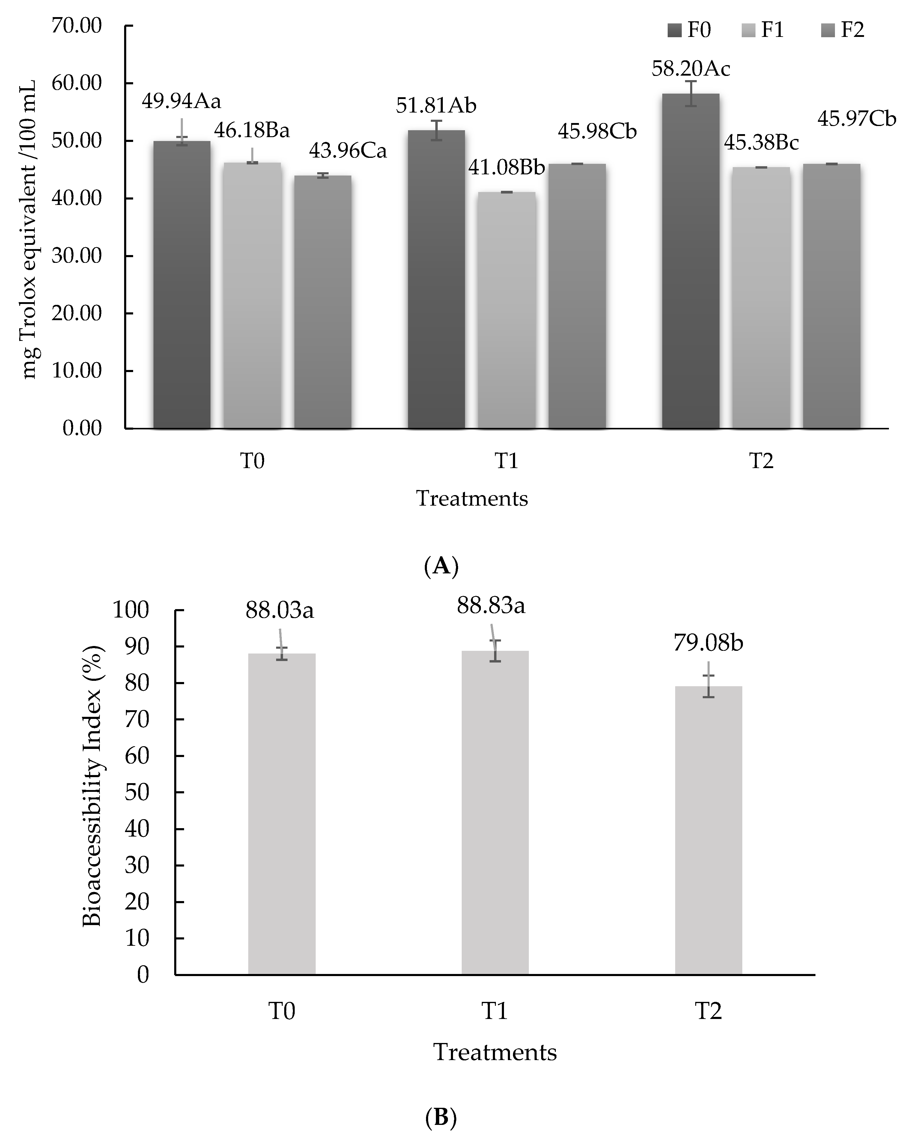
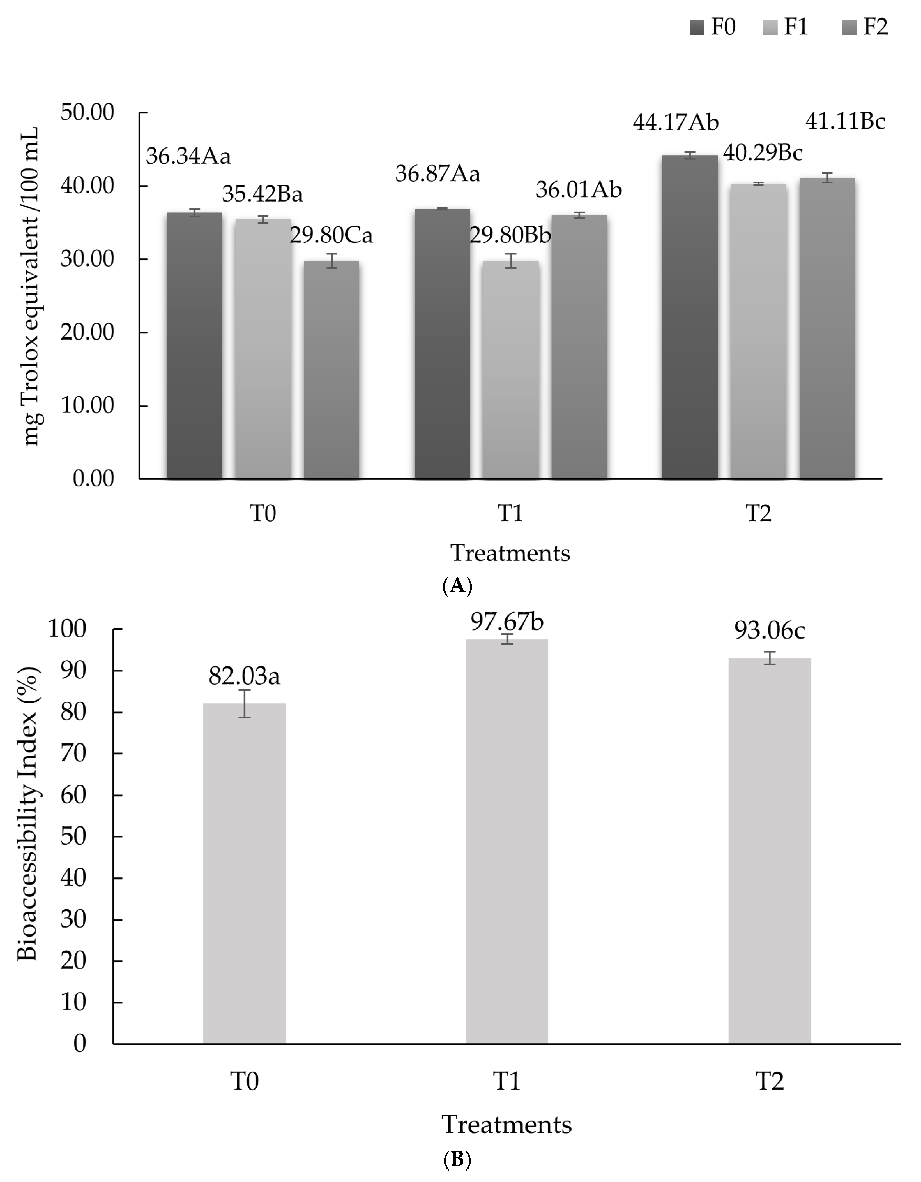
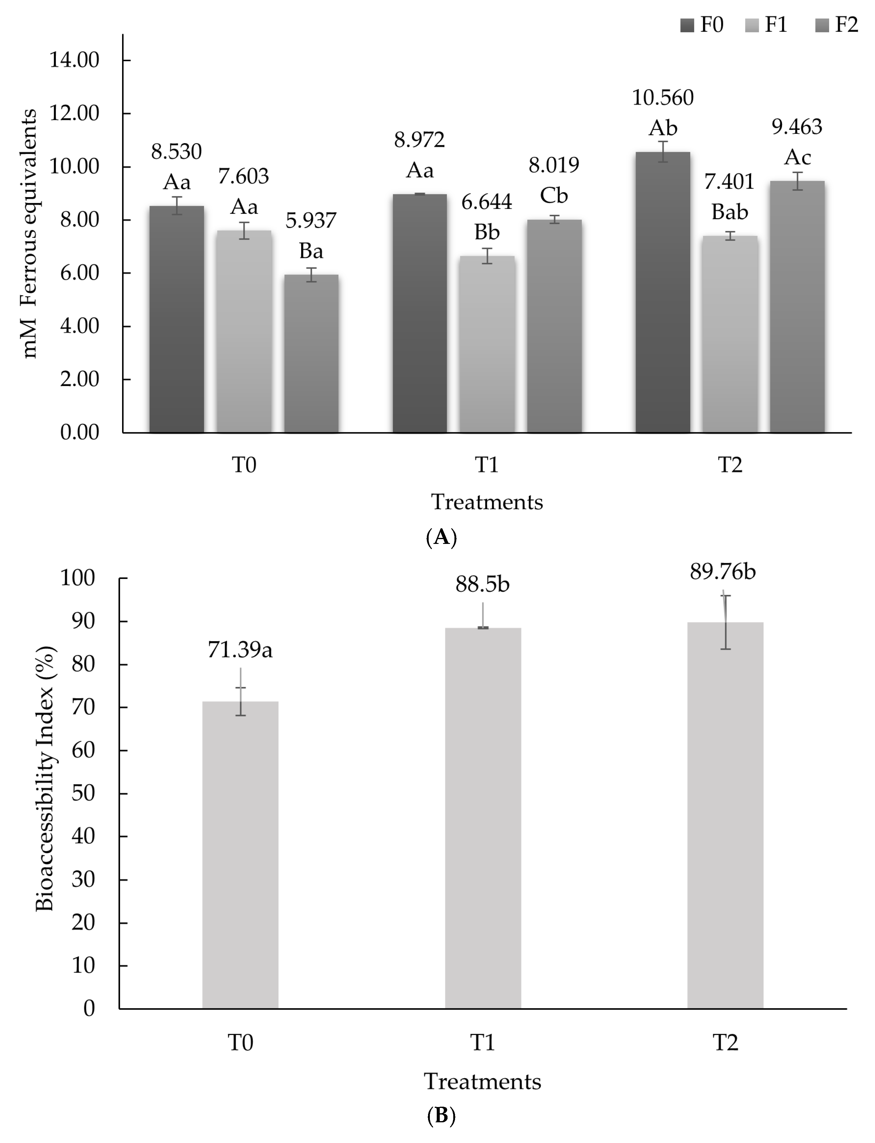
3.1. Antioxidant Activity

Treatments AA (ABTS, DPPH, FRAP, and ORAC) and bioaccessibility index, before and after in vitro digestion, are shown in Figure 1, Figure 2, Figure 3 and Figure 4.
Results of AA by the ABTS method are observed in Figure 1A. Statistically significant differences (p ˂ 0.05) were found between the digestion phases of each treatment (T0, T1, and T2). For T0 (drink without adding postbiotics), the highest value was observed in F0 (phase before digestion) with a value of 49.94 ± 0.003; it decreased in F1 (gastric phase) at 46.18 ± 0.000, and then it decreased again in F2 (intestinal phase) at 43.96 ± 0.002. In T1, significant differences (p < 0.05) were found between the phases. The highest AA was found in F0 with a value of 51.81 ± 0.007; then it decreased in F1 (41.08 ± 0.000) and increased in F2 (58.20 ± 0.000), the latter value being lower than that presented in F0 but higher than F1. The same behavior was observed for T2, where F0 (58.20 ± 0.009), F1 (45.38 ± 0.000), and F2 (45.97 ± 0.000) were statistically different (p ˂ 0.05). Regarding the digestion phases, in the F0 phases, all treatments were statistically significantly different (p ˃ 0.05), where T2 had (58.20 ± 0.009) the highest AA. In F1, significant differences (p ˂ 0.05) were observed between treatments, where T0 presented the highest AA (46.18 ± 0.000) and T1 (41.08 ± 0.000) the lowest. Finally, in F2, T1 (45.98 ± 0.000) and T2 (45.97 ± 0.000) did not present differences between them (p ˃ 0.05) but were different (p ˂ 0.05) from T0 (43.96 ± 0.002), which presented the lowest AA. In general, T1 and T2 presented similar behavior, starting in F0 with the highest AA, decreasing in F1, and increasing in F2. It is concluded that T1 and T2 presented the highest AA after in vitro digestion by the ABTS methodology.
Results of AA by the DPPH method are observed in Figure 2A. Statistically significant differences (p ˂ 0.05) were found between the digestion phases in each treatment. For T0, the highest value was observed in F0 with a value of 36.34 ± 0.003, then it decreased in F1 to 35.42 ± 0.000, and then it decreased again in F2 to 29.80 ± 0.002. In T1, no significant difference (p ˃ 0.05) was found between F0 (36.87 ± 0.001) and F2 (36.01 ± 0.002), and the lowest AA (p ˂ 0.05) occurred in F1 (29.80 ± 0.004). While in T2, F0 (44.17 ± 0.003) presented the highest AA, and it was statistically different (p ˂ 0.05) from F1 (40.29 ± 0.007) and F2 (41.11 ± 0.000). Regarding the phases, in F0, T2 (44.17 ± 0.002) presented the highest AA (p ˂ 0.05), which was statistically different from T1 (36.87 ± 0.001) and T0 (36.34 ± 0.003), which presented lower activities (p > 0.05). Regarding F1, treatments were significantly different (p ˂ 0.05); the highest AA occurred in T2 (40.29 ± 0.001), followed by T0 (35.42 ± 0.001) and finally T1 (29.80 ± 0.004), which presented the lowest AA (p ˂ 0.05). Finally, in F2, there was a significant difference (p ˂ 0.05) between treatments; the highest AA (p ˂ 0.05) was observed in T2 (41.11 ± 0.003), and the lowest (p ˂ 0.05) occurred in T0 (29.80 ± 0.004). In general, treatments T1 and T2 presented similar behavior. These started with the highest AA in F0; later, these decreased in F1 and increased in F2. It is concluded that T1 and T2 presented higher AA after in vitro digestion. This behavior was the same as that presented in the AA by the ABTS method.
The behavior of the AA by FRAP is observed in Figure 3A. All the treatments presented significant differences between their phases (p ˂ 0.05). For T0, the highest AA (p ˂ 0.05) occurred in F0 (8.530 ± 0.330) and F1 (7.603 ± 0.001), without showing statistically significant differences between them (p ˃ 0.05). In T1, the highest AA (p ˂ 0.05) was observed in F0 (8.972 ± 0.033); later, it decreased (p ˂ 0.05) in F1 (6.644 ± 0.291) and ended up increasing in F2 (8.019 ± 0.142). For T2, F0 (10.560 ± 0.389) and F2 (9.463 ± 0.328) presented the highest AA and were statistically different (p ˂ 0.05) from F1 (7.401 ± 0.160), which presented the lowest AA (p ˂ 0.05). In addition, a significant difference (p ˂ 0.05) was found between treatments for each digestion phase. In F0, T0 (8.530 ± 0.330) and T1 (8.972 ± 0.033) presented lower AA (p ˂ 0.05) than T2 (10.560 ± 0.389), which presented the greatest AA. Regarding F1, T0 (7.603 ± 0.315) presented the highest AA (p ˂ 0.05) and T1 (6.644 ± 0.291) the lowest (p ˂ 0.05); however, T2 (7.401 ± 0.160) did not present differences (p > 0.05) with respect to T0 and T1. Finally, in F2, there was a significant difference (p < 0.05) between treatments; the highest AA was presented by T2 (9.463 ± 0.328), followed by T1 (8.019 ± 0.142) and finally T0 (5.937 ± 0.258). In general, T1 and T2 presented similar behavior. These started with higher AA in F0, then decreased in F1, and later increased in F2. Likewise, T1 and T2 presented higher AA after in vitro digestion compared to T0.
The AA by ORAC methodology is observed in Figure 4A. All treatments presented significant differences between the gastric phases (p ˂ 0.05). For T0, the highest AA (p ˂ 0.05) occurred in F0 (20.33 ± 0.330), and the lowest (p ˂ 0.05) in F1 (15.41 ± 0.258) and F2 (15.28 ± 0.258). In T1, the highest AA (p ˂ 0.05) was observed in F2 (28.31 ± 0.033), and the lowest AA (p ˂ 0.05) in F1 (15.99 ± 0.142). For T2, F2 (30.56 ± 0.328) presented the highest AA (p ˂ 0.05), and F1 (14.96 ± 0.160) the lowest AA (p ˂ 0.05). Regarding digestion phases, in F0, T0 (20.33 ± 0.330), T1 (19.05 ± 0.033), and T2 (19.55 ± 0.142) were not statistically different (p > 0.05). In F1, T0 (15.41 ± 0.330), T1 (15.99 ± 0.033), and T2 (14.96 ± 0.142) were also not statistically different (p > 0.05). Finally, in F2 there was a significant difference (p ˂ 0.05) between treatments; T0 (15.28 ± 0.258) had the lowest AA (p ˂ 0.05) and T2 (30.56 ± 0.328) the highest. In general, T1 and T2 presented a similar behavior; they started with an AA like T0 in F0, decreased in F1, and later increased in F2. Likewise, T1 and T2 presented higher AA after in vitro digestion compared to T0.
The DPPH, ABTS, FRAP, and ORAC methods are widely used in research to evaluate the AA of various compounds. The differences found in AA when using these techniques are due to their chemical principles, mechanisms of action, and methods of quantifying AA. Each methodology offers specific advantages and presents limitations. Therefore, recent studies suggest that the combined use of several methodologies provides a more complete and accurate assessment of the total AA of the analyzed samples, especially in complex samples containing various antioxidant compounds with different mechanisms of action, as is the case with postbiotics. The DPPH and ABTS techniques measure free radical scavenging capacity but differ in the type of radical used and the reaction conditions. The FRAP technique specifically assesses iron-reducing capacity, which is an electron-transfer mechanism, while the ORAC method measures the protection capacity against peroxide radical-induced oxidation, which operates primarily through hydrogen atom transfer. These methodological differences explain why the same sample may present different AA values depending on the method used, as observed in several comparative studies [27,28,29].
The above coincides with the results obtained. DPPH and ABTS techniques showed similar results. T2 had the highest AA before digestion (p < 0.05), and T1 and T2 showed the highest AA after digestion (p < 0.05). The study in [30] reported that L. helveticus increased the AA of cheese, which was attributed to 15 peptides derived mainly from β- and αs1-casein with a size between 6 and 14 amino acid residues, where 10 of these contained an apolar residue (proline). However, despite this, all treatments showed a decrease in AA after digestion. By the ABTS method, T0 and T1 showed higher BI than T2. Compared to T0, T2 presented a fold change of 0.89. And, by the DPPH assay, T1 and T2 had the highest BI, although without showing significant differences between these (p > 0.05). Compared to T0, T1 and T2 presented a fold change of 1.19 and 1.13, respectively.
Firstly, the differences observed between these two methods may be due to the structure of the radicals used, which can react differently with the different compounds present in the postbiotics. Instead, the lower BI presented in T2, compared to T0 and T1, by ABTS method, may be because L. helveticus presents a high proteolytic activity [31] that results in the generation of peptides with AA, which could be degraded during digestion by the action of digestive enzymes, causing their loss and, therefore, lower AA, which in turn induced a decrease in BI.
Regarding the FRAP method, in general, all treatments showed ferrous ion chelating capacity, although with significant differences between them (p < 0.05). T2 presented the highest capacity before and after digestion (p < 0.05). Similar investigations have reported metal ion chelating activity in cell-free intracellular extracts obtained from Streptococcus thermophilus, Bifidobacterium longum, L. bulgaricus, and L. casei [32,33]. However, the compounds related to this property have not been identified, so the available information is scarce. Although a study carried out with L. helveticus MB2-1 showed that it exerted potent antioxidant effects such as the ability to eliminate hydroxyl radicals, superoxide, and DPPH, as well as ferrous ion chelating activity, which was attributed to an exopolysaccharide composed of galactose, glucose, and mannose [34]. Furthermore, in vitro AA results suggested that EPS exhibits strong scavenging activity against DPPH, superoxide, and hydroxyl radicals, as well as chelating activity against ferrous ions [34]. The above agrees with what was found in this study, where T2 presented the highest BI by FRAP and ORAC methodologies. Since Fe+2 generates free radicals through the gain or loss of electrons, metal ion chelating compounds could reduce the formation of reactive oxygen species, thus avoiding or reducing the oxidative damage that contributes to the appearance of neurodegenerative disorders.
The ORAC assay measures the protective effect of an antioxidant on a target chemical subjected to an oxidant. The measurement is based on the alteration in fluorescence resulting from the oxidation of the target chemical. The assay enables the quantification of the total antioxidant capacity of the tested sample. By this assay, T1 and T2 showed greater AA after in vitro digestion than before digestion. Therefore, these treatments also showed the highest BI. Compared to T0, T1 and T2 presented a fold change of 1.97 and 2.07, respectively. Then, it is hypothesized that ultrasound applied to probiotics helps to release AA molecules located in the cytoplasm of bacteria, and their bioaccessibility is improved by the harsh conditions presented in the gastrointestinal tract. Or, it may be because postbiotics exert a synergistic effect with the metabolites produced during digestion. Likewise, ref. [35] observed the largest delivery of antioxidants (ORAC method) in milk fermented with Bifidobacterium longum subsp. Longum after digestion. Also, ref. [36] studied the antioxidative activity under in vitro conditions of various species of lactobacilli used as yogurt starter cultures or as probiotic bacteria. Antioxidative activity was evaluated by the ability of the whole cells or the cell-free extracts from cultures to protect a protein from being attacked by free radicals. Findings showed that the highest antioxidant capacity was linked to the cell-free extracts of the cultures, indicating their potential role in providing antioxidants to the intestines. Cell-free supernatants generally outperformed intact cells or intracellular extracts [37,38].
In this study, the beverage was elaborated, in addition to postbiotics, with milk and cocoa. Dairy foods, including milk, are indeed a rich source of compounds exhibiting antioxidant properties. These foods account for approximately 25–30% of the average human diet [39]. The antioxidant capacity of milk and dairy products is mainly related to the presence of sulfur amino acids, whey proteins (especially β-lactoglobulin), vitamins A, E, and C, or β-carotene [39,40,41]. In whey proteins, the content of histidine and other hydrophobic amino acids determines their AA. Whey proteins form metal chelates, scavenging free radicals and retrieving thiol -SH groups from proteins [42]. On the other hand, lactoferrin chelates iron, thereby increasing its bioavailability and inhibiting pro-oxidant effects [43]. The AA of β-lactoglobulin has been attributed to its high content of sulfur-containing amino acids. In addition, antioxidant peptides are released during whey hydrolysis [40]. The above shows that milk and its derivatives are foods that have a recognized AA. Therefore, the addition of postbiotics to these foods represents an alternative to increase the benefit that these foods have on human health.
Additionally, cocoa has a rich profile of bioactive compounds. Among these, polyphenols—particularly flavanols such as epicatechin and catechin and their oligomeric derivatives, procyanidins—stand out for their potent antioxidant properties. These compounds neutralize reactive oxygen species (ROS), chelate metal ions, and modulate enzymatic activity, thereby offering protection against oxidative stress-linked pathologies such as cardiovascular diseases, metabolic disorders, and inflammation [44].
Disturbances in pro-oxidant/antioxidant homeostasis, as well as genetic and environmental factors, contribute to the development of chronic degenerative diseases. Endogenous and exogenous antioxidants deactivate reactive oxygen species (ROS) while maintaining homeostasis. However, intense and prolonged oxidative stress causes free radical processes to intensify, causing permanent changes in the structure of DNA, proteins, and lipids. These processes lead to damage to cellular structures and genes, which induce metabolic disorders and neoplastic transformation [26]. However, the human organism is not completely defenseless against these threats, since it has enzymatic and non-enzymatic defense systems that protect it from pro-oxidants [45]. However, this defense can be enhanced by incorporating biologically active foods rich in antioxidants (hydrophilic and lipophilic), as well as vitamins and minerals, into the diet [39].
Furthermore, several variables, including food microstructure, pH, light, and temperature, can affect the stability of postbiotics [34]. Also, these could interact with other chemical compounds found in foods, influencing their bioavailability [46]. Lastly, antioxidants produced and/or released by probiotic bacteria will need to be examined in vivo to ensure whether these are metabolized and absorbed in a similar manner to the in vitro digestion models employed.

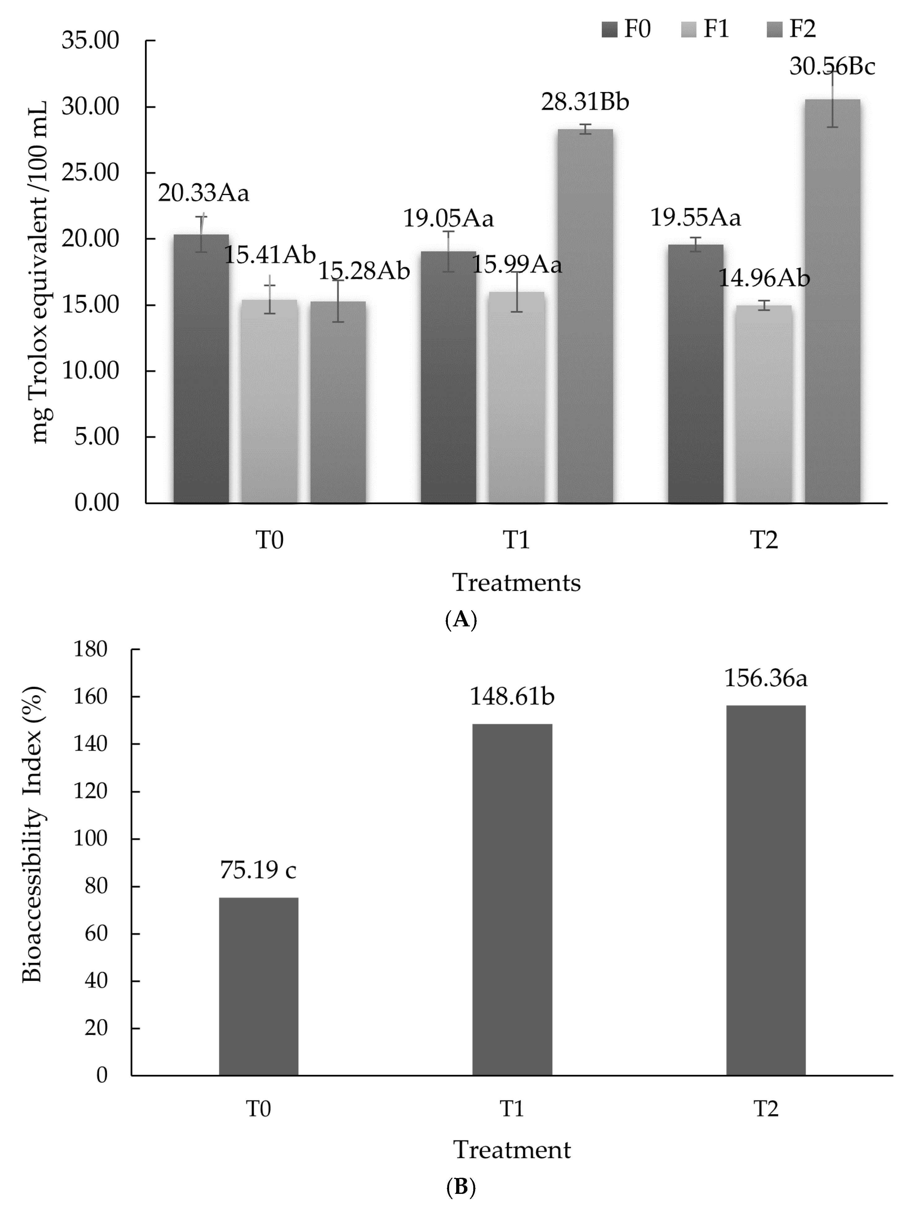
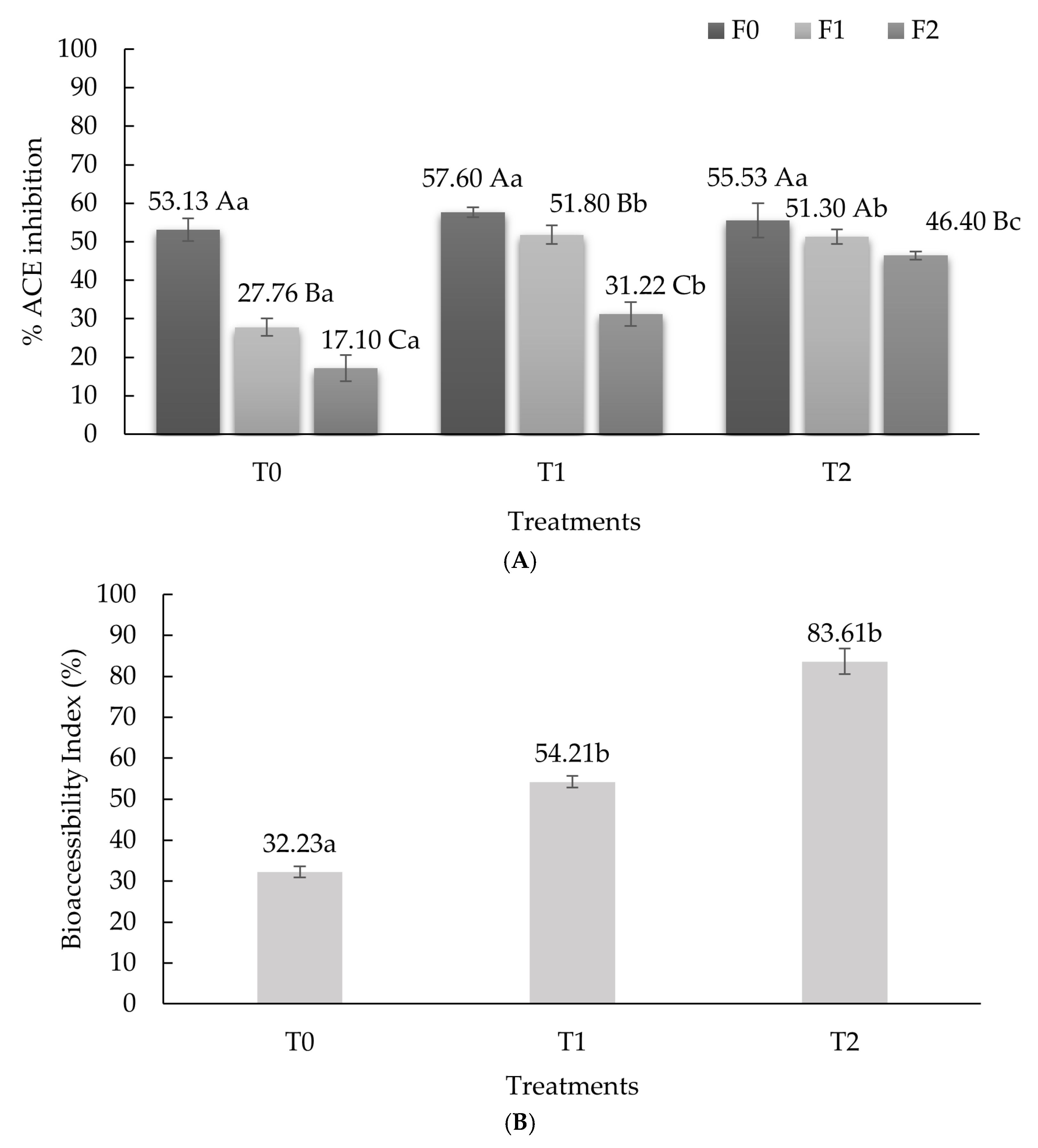
3.2. Angiotensin Converting Enzyme-Inhibitory Activity

The ACE-inhibitory activity of the beverages is observed in Figure 5A. Differences (p ˂ 0.05) were found between the digestion phases in each of the treatments. For T0 (drink without adding postbiotics), the highest percentage of inhibition (p ˂ 0.05) was found in F0 (53.13 ± 2.90), decreasing in the following two phases (p ˂ 0.05), being F2 (17.10 ± 0.23), where the lowest percentage was observed (p ˂ 0.05).
In T1, a significant difference (p ˂ 0.05) was found between F0 (57.60 ± 1.29), F1 (51.80 ± 0.59), and F2 (31.22 ± 31.15). While in T2, F0 (55.53 ± 1.56) and F1 (51.30 ± 0.68) did not present differences between them (p > 0.05) but were different (p ˂ 0.05) from F2 (46.40 ± 1.015), which presented the lowest percentage of inhibition.
The behavior of the treatments in each phase is described below. In F0 there was no significant difference (p ˃ 0.05) between the treatments. In F1, the treatments in T1 (51.80 ± 0.59) and T2 (51.30 ± 0.68) were significantly different (p ˂ 0.05) from T0, which presented the least percentage of inhibition (27.76 ± 0.68). Finally, in the F2 phase, there was a significant difference (p ˂ 0.05) between the treatments; the highest activity was observed in T2 (46.40 ± 1.01) and the lowest in T0 (17.10 ± 0.23). In general, all treatments (T0, T1, and T2) presented a similar behavior; they started with a higher percentage of inhibition, and after digestion, this percentage decreased. To enhance postbiotic stability in future formulations, strategies can be adapted from advanced drug delivery systems and polymer-based stabilization techniques. Polymers may play a critical role in enhancing postbiotic stability through structural reinforcement, environmental protection, and controlled release mechanisms. Regarding the BI, compared to T0, T1 and T2 presented a fold change of 1.68 and 2.60, respectively.
The production of bioactive peptides, such as ACE-inhibitory peptides, during food product fermentation has been linked to probiotics’ capacity to lower blood pressure [46]. L. helveticus has a highly efficient proteolytic system. During bacterial lysis, its internal peptidases and cell-envelope-associated proteases are released into the growing media [47,48]. Then, these generate peptides with ACE-inhibition activity [48]. In addition to this, L. helveticus produces oligopeptides, which during digestion are hydrolyzed by gastrointestinal enzymes to produce more bioactive peptides [49]. This may explain the higher BI found in T2 compared to T0. In addition, peptides from L. helveticus postbiotics retain stability during gastrointestinal digestion, making them suitable for oral delivery in functional foods [50,51].
Regarding the behavior in the percentage of ACE inhibition, all treatments began with a higher percentage of activity than that obtained after digestion. This may be due to the gastrointestinal enzymes, such as pepsin in the stomach and trypsin, chymotrypsin, and carboxypeptidases in the small intestine that degrade a proportion of the peptides found in postbiotics, which, in the case of L. helveticus, is lower due to what was mentioned above. Meanwhile, ACE-inhibitory peptides from L. helveticus have demonstrated distinct advantages in potency and structural features compared to other Lactobacillus species, as evidenced by studies on casein and whey fermentation [52,53]. In whey fermentation, produced peptides presented IC50 values as low as 5.3–7.8 μg/mL, representing some of the strongest ACE-inhibitory activity among tested species [52]. And, in casein hydrolysates, crude fractions showed IC50 values of 16–100 μg/mL, with synthetic peptides matching the activity of pharmaceutical ACE inhibitors like enalapril [53]. Studies also reported that L. helveticus generates pentapeptides (e.g., fractions H5 and H7) with a conserved N-terminal alanine and C-terminal hydrophobic/aromatic residues (e.g., Pro, Val, Leu, Phe), optimizing ACE active-site binding and the release of longer casein-derived peptides (e.g., αS1-casein f24–47, β-casein f58–76) with multi-site ACE inhibition [52,53]. The results of this study show that L. helveticus postbiotics can be used to create healthy foods (like cheese and fermented milk) because they stay stable during digestion and work well with dairy products, indicating potential health benefits. Meanwhile, whey fermentation with L. helveticus offers a cost-effective method to valorize dairy byproducts. In summary, L. helveticus outperforms related species in generating potent, structurally optimized ACE-inhibitory peptides, particularly from casein, while maintaining versatility across protein substrates. In summary, the stability of L. helveticus-derived ACE-inhibitory postbiotics during digestion positions them as viable candidates for managing hypertension and related cardiovascular conditions, with potential synergistic benefits for metabolic and immune health.

3.3. Antimicrobial Activity

The inhibitory zones on agar confirmed the antimicrobial activity of postbiotics on the tested pathogens (Figure 6 and Table 2). Antimicrobial effect against Bacillus spp. and S. aureus was observed in L. acidophilus and L. helveticus postbiotics. However, L. acidophilus postbiotics did not present an antibacterial effect against E. coli. And, none of the treatments showed antimicrobial activity against strains of Bacillus spp., S. aureus, and E. coli before and after in vitro digestion.
Antimicrobial activity of lactic acid bacteria (LAB) results from the release of compounds such as organic acids, bacteriocins, hydrogen peroxide, etc [54,55]. Although, the antimicrobial activity of L. acidophilus has not been correlated with the production of bacteriocins but with the production of organic acids [56]. A previous study found that postbiotics of L. acidophilus presented a high content of lactic, acetic, and citric acids [21]. Then, the antimicrobial effect of L. acidophilus postbiotics could be attributed to them. Regarding L. helveticus, it owes its antimicrobial activity to its ability to synthesize peptides with antimicrobial activity, bacteriocins, and organic acids. The bacteriocins produced can limit the growth of Gram-negative and Gram-positive bacteria, including S. aureus and E. coli [55].
Postbiotics derived from L. helveticus demonstrate significant antimicrobial activity against both Gram-positive and Gram-negative pathogens, with mechanisms involving the disruption of bacterial membranes, biofilm inhibition, and intracellular content leakage. These metabolites show promise for food preservation and combating antibiotic-resistant strains. Pathogens targeted include multidrug-resistant Staphylococcus aureus, Escherichia coli O157:H7, and Pseudomonas aeruginosa [57,58]. Regarding S. aureus, postbiotics from L. helveticus (LHPs) have exhibited strong inhibition, with a 32.76 mm minimum inhibition zone and a MIC of 36.00 μg/mL, which was attributed to disrupting membrane integrity, reducing biofilm formation, and impairing bacterial motility [57]. Concerning E. coli, LHPs have shown a 25.63 mm inhibition zone and MIC of 60.00 μg/mL. They alter bacterial surface charge and cause intracellular leakage [57].
Regarding postbiotics from L. acidophilus (LAPs), these have exhibited significant antimicrobial activity, particularly against Staphylococcus aureus. However, cell-free supernatants have demonstrated a concentration-dependent inhibitory effect, with higher concentrations showing greater efficacy [59]. The primary antimicrobial compounds identified include lactic acid and laurostearic acid, which contribute to their pathogen-suppressing properties. The MIC of LAPs against S. aureus was determined to be 100 mg/mL in both in vitro and food model (pasteurized milk) settings. However, its antimicrobial activity in food matrices required a higher minimum effective concentration (MEC) of 150 mg/mL, highlighting the influence of environmental factors on efficacy [59].
In this study, the antimicrobial activity of L. acidophilus and L. helveticus postbiotics was lost when these were incorporated into the beverages, since these did not show antimicrobial activity before undergoing in vitro digestion. We hypothesize that this could be due to the dilution factor, the interaction of postbiotics with other compounds, and the pH of the treatments. It has been reported that the antimicrobial activity of CFS from L. acidophilus against E. coli and S. aureus is lost when adjusting pH to 7 [60]; the treatments’ pH was close to 7.0. And, as mentioned before, the MEC of postbiotics against pathogens differs among different food models and the interactions of postbiotics with food ingredients, a behavior previously observed by [61].

4. Conclusions

The main advantage of adding postbiotics as a functional ingredient in food matrices is that there is no restriction for their incorporation since viability is not necessary for their bioeffect. However, the results obtained suggest that several factors, including the amount and form of postbiotic addition (lyophilized, diluted, encapsulated, etc.), the food matrix (pH, aqueous activity, physicochemical composition, etc.), shelf life, sensory acceptance, and, most importantly, the bioaccessibility index of the bioactivity to be demonstrated, can cause the bioactivities to disappear or change their efficacy. Overall, the functionality of bioactive compounds during the digestion process is complex and can be influenced by many factors. Understanding how these compounds function during digestion is important for optimizing their health benefits.
In this research, treatments added with postbiotics (T1 and T2) had a remarkably higher bioaccessibility of antioxidants and ACE-inhibition compounds. This confirms that during digestion, a large part of these maintain their bioactivity. Therefore, postbiotics from L. acidophilus and L. helveticus are an alternative to increasing the antioxidant and ACE-inhibitory capacity of non-fermented milk beverages. Then, scaling postbiotic production for industrial use requires further optimization of production methods. Overall, adding postbiotics to non-fermented beverages represents a promising innovation in the functional food industry, providing health benefits while maintaining convenience and safety for consumers.

Author Contributions

Conceptualization, A.C.-M. and R.A.R.-V.; data curation, E.S.-E.; formal analysis, N.A.B.-J. and E.S.-E.; funding acquisition, A.C.-M.; investigation, N.A.B.-J.; methodology, A.C.-M., A.L.R.-M., M.M.A.-S. and R.A.R.-V.; project administration, A.C.-M. and N.A.S.-S.; supervision, A.C.-M., M.M.A.-S., R.A.R.-V. and A.L.R.-M.; writing—original draft, N.A.B.-J.; writing—review and editing, A.C.-M., A.L.R.-M., N.A.S.-S., E.S.-E., M.M.A.-S. and R.A.R.-V. All authors have read and agreed to the published version of the manuscript.

Funding

The Science and Technology National Council of Mexico (CONACYT) provided a graduate study scholarship for Norma Angélica Bolivar-Jacobo.

Institutional Review Board Statement

Not applicable.

Informed Consent Statement

Not applicable.

Data Availability Statement

The original contributions presented in this study are included in the article. Further inquiries can be directed to the corresponding authors.

Acknowledgments

The authors acknowledge that the Universidad Autónoma de Chihuahua supported this investigation.

Conflicts of Interest

The authors declare no conflicts of interest.

References

  1. Li, H.Y.; Zhou, D.D.; Gan, R.Y.; Huang, S.Y.; Zhao, C.N.; Shang, A.; Xu, X.Y.; Li, H.B. Effects and mechanisms of probiotics, prebiotics, synbiotics, and postbiotics on metabolic diseases targeting gut microbiota: A narrative review. Nutrients 2021, 13, 3211. [Google Scholar] [CrossRef] [PubMed]
  2. Abbasi, A.; Hajipour, N.; Hasannezhad, P.; Baghbanzadeh, A.; Aghebati-Maleki, L. Potential in vivo delivery routes of postbiotics. Crit. Rev. Food Sci. Nutr. 2020, 62, 3345–3369. [Google Scholar] [CrossRef] [PubMed]
  3. Konstantinidi, M.; Koutelidakis, A.E. Functional Foods and Bioactive Compounds: A Review of Its Possible Role on Weight Management and Obesity’s Metabolic Consequences. Medicines 2019, 6, 94. [Google Scholar] [CrossRef] [PubMed]
  4. Miano, T. Functional Food—A Review. Eur. J. Res. 2016, IV, 5695–5702. [Google Scholar]
  5. Guimarães, J.T.; Balthazar, C.F.; Scudino, H.; Pimentel, T.C.; Esmerino, E.A.; Ashokkumar, M.; Freitas, M.Q.; Cruz, A.G. High-intensity ultrasound: A novel technology for the development of probiotic and prebiotic dairy products. Ultrason. Sonochem. 2019, 57, 12–21. [Google Scholar] [CrossRef]
  6. Homayouni Rad, A.; Aghebati Maleki, L.; Samadi Kafil, H.; Fathi Zavoshti, H.; Abbasi, A. Postbiotics as novel health-promoting ingredients in functional foods. Health Promot. Perspect. 2020, 10, 3–4. [Google Scholar] [CrossRef]
  7. Rafique, N.; Jan, S.Y.; Dar, A.H.; Dash, K.K.; Sarkar, A.; Shams, R.; Pandey, V.K.; Khan, S.A.; Amin, Q.A.; Hussain, S.Z. Promising bioactivities of postbiotics: A comprehensive review. J. Agric. Food Res. 2023, 14, 100708. [Google Scholar] [CrossRef]
  8. Mosca, A.; Abreu YAbreu, A.T.; Gwee, K.A.; Ianiro, G.; Tack, J.; Nguyen, T.V.H.; Hill, C. The clinical evidence for postbiotics as microbial therapeutics. Gut Microbes 2022, 14, 2117508. [Google Scholar] [CrossRef]
  9. Ma, L.; Tu, H.; Chen, T. Postbiotics in Human Health: A Narrative Review. Nutrients 2023, 15, 291. [Google Scholar] [CrossRef]
  10. Nataraj, B.H.; Ali, S.A.; Behare, P.V.; Yadav, H. Postbiotics-parabiotics: The new horizons in microbial biotherapy and functional foods. Microb. Cell Fact. 2020, 19, 1–22. [Google Scholar] [CrossRef]
  11. Wegh, C.A.M.; Geerlings, S.Y.; Knol, J.; Roeselers, G.; Belzer, C. Postbiotics and Their Potential Applications in Early Life Nutrition and Beyond. Int. J. Mol. Sci. 2019, 20, 4673. [Google Scholar] [CrossRef] [PubMed]
  12. Aguilar-Toalá, J.E.; Garcia-Varela, R.; Garcia, H.S.; Mata-Haro, V.; González-Córdova, A.F.; Vallejo-Cordoba, B.; Hernández-Mendoza, A. Postbiotics: An evolving term within the functional foods field. Trends Food Sci. Technol. 2018, 75, 105–114. [Google Scholar] [CrossRef]
  13. Noori, S.M.A.; Behfar, A.; Saadat, A.; Ameri, A.; Yazdi, S.S.A.; Saadat, A. Antimicrobial and Antioxidant Properties of Natural Postbiotics Derived from Five Lactic Acid Bacteria. Jundishapur J. Nat. Pharm. Prod. 2023, 18, e130785. [Google Scholar] [CrossRef]
  14. Khani, N.; Shkouhian, S.M.J.; Kafil, H.S.; Gilani, N.; Abbasi, A.; Rad, A.H. Assessing the growth-inhibitory activity of postbiotics of Lactobacillus spp. against Staphylococcus aureus under in vitro circumstances and food model. Lett. Appl. Microbiol. 2023, 76, ovac056. [Google Scholar] [CrossRef]
  15. Hosseini, S.; Homayouni-Rad, A.; Samadi Kafil, H.; Dorud, N. Evaluating the antimicrobial effect of postbiotic extract from Lactobacillus casei on Escherichia coli in commercial sterilized milk. J. Food Hyg. Saf. 2022, 8, 42–52. [Google Scholar] [CrossRef]
  16. Gómez-Guzmán, M.; Toral, M.; Romero, M.; Jiménez, R.; Galindo, P.; Sánchez, M.; Zarzuelo, M.J.; Olivares, M.; Gálvez, J.; Duarte, J. Antihypertensive effects of probiotics Lactobacillus strains in spontaneously hypertensive rats. Mol. Nutr. Food Res. 2015, 59, 2326–2336. [Google Scholar] [CrossRef]
  17. Robles-Vera, I.; Toral, M.; Romero, M.; Jiménez, R.; Sánchez, M.; Pérez-Vizcaíno, F.; Duarte, J. Antihypertensive Effects of Probiotics. Curr. Hypertens. Rep. 2017, 19, 26. [Google Scholar] [CrossRef]
  18. Jama, H.A.; Rhys-Jones, D.; Nakai, M.; Yao, C.K.; Climie, R.E.; Sata, Y.; Anderson, D.; Creek, D.J.; Head, G.A.; Kaye, D.M.; et al. Prebiotic intervention with HAMSAB in untreated essential hypertensive patients assessed in a phase II randomized trial. Nat. Cardiovasc. Res. 2023, 2, 35–43. [Google Scholar] [CrossRef]
  19. Ahmad, H.; Khan, H.; Haque, S.; Ahmad, S.; Srivastava, N.; Khan, A. Angiotensin-Converting Enzyme and Hypertension: A Systemic Analysis of Various ACE Inhibitors, Their Side Effects, and Bioactive Peptides as a Putative Therapy for Hypertension. JRAAS—J. Renin-Angiotensin-Aldosterone Syst. 2023, 2023, 7890188. [Google Scholar] [CrossRef]
  20. Santana Andrade, J.K.; Chagas Barros, R.G.; Pereira, U.C. Bioaccessibility of bioactive compounds after in vitro gastrointestinal digestion and probiotics fermentation of Brazilian fruits residues with antioxidant and antidiabetic potential. LWT-Food Sci. Technol. 2022, 153, 112469. [Google Scholar] [CrossRef]
  21. Bolivar-Jacobo, N.A.; Reyes-Villagrana, R.A.; Rentería-Monterrubio, A.L.; Sánchez-Vega, R.; Santellano-Estrada, E.; Tirado-Gallegos, J.M.; Chávez-Martínez, A. Culture Age, Growth Medium, Ultrasound Amplitude, and Time of Exposure Influence the Kinetic Growth of Lactobacillus acidophilus. Fermentation 2023, 9, 63. [Google Scholar] [CrossRef]
  22. Rodríguez-Roque, M.J.; Rojas-Graü, M.A.; Elez-Martínez, P.; Martín-Belloso, O. In vitro bioaccessibility of health-related compounds from a blended fruit juice–soymilk beverage: Influence of the food matrix. J. Funct. Foods 2014, 7, 161–169. [Google Scholar] [CrossRef]
  23. Chávez-Alzaga, G.; Reyes-Villagrana, R.A.; Espino-Solis, G.P.; Arévalos-Sánchez, M.M.; Rentería-Monterrubio, A.L.; Sánchez-Vega, R.; Santellano-Estrada, E.; Bolivar-Jacobo, N.A.; Tirado-Gallegos, J.M.; Chávez-Martínez, A. The Effects of Substrates and Sonication Methods on the Antioxidant Activity of Kefir Postbiotics. Fermentation 2024, 10, 492. [Google Scholar] [CrossRef]
  24. Číž, M.; Čížová, H.; Denev, P.; Kratchanova, M.; Slavov, A.; Lojek, A. Different Methods for Control and Comparison of the Antioxidant Properties of Vegetables. Food Control 2010, 21, 518–523. [Google Scholar] [CrossRef]
  25. Donkor, O.N.; Henriksson, A.; Vasiljevic, T.; Shah, N.P. Probiotic Strains as Starter Cultures Improve Angiotensin-converting Enzyme Inhibitory Activity in Soy Yogurt. J. Food Sci. 2005, 70, m375–m381. [Google Scholar] [CrossRef]
  26. Mushtaq, M.; Gani, A.; Masoodi, F.A. Himalayan cheese (Kalari/Kradi) fermented with different probiotic strains: In vitro investigation of nutraceutical properties. LWT 2019, 104, 53–60. [Google Scholar] [CrossRef]
  27. Dudonné, S.; Vitrac, X.; Coutiére, P.; Woillez, M.; Mérillon, J.M. Comparative study of antioxidant properties and total phenolic content of 30 plant extracts of industrial interest using DPPH, ABTS, FRAP, SOD, and ORAC assays. J. Agric. Food Chem. 2009, 57, 1768–1774. [Google Scholar] [CrossRef]
  28. Thaipong, K.; Boonprakob, U.; Crosby, K.; Cisneros-Zevallos, L.; Hawkins Byrne, D. Comparison of ABTS, DPPH, FRAP, and ORAC assays for estimating antioxidant activity from guava fruit extracts. J. Food Compos. Anal. 2006, 19, 669–675. [Google Scholar] [CrossRef]
  29. Thennakoon, T.M.E.S.; Abeysinghe1, P.D.; Ranasinghe, P.; Pathirana, R.N.; White, A.; Dilantha-Fernando, W.G.; Abeysinghe, S.; Premakumara, S. Total phenolic content, total flavonoid content and in vitro antioxidant activities measured by the FRAP, ABTS, DPPH and ORAC assays of Sri Lankan black and green tea (Camellia sinensis) infusions. Food Biol. 2022, 11, 1–10. [Google Scholar] [CrossRef]
  30. Yang, W.; Hao, X.; Zhang, X.; Zhang, G.; Li, X.; Liu, L.; Sun, Y.; Pan, Y. Identification of antioxidant peptides from cheddar cheese made with Lactobacillus helveticus. LWT 2021, 141, 110866. [Google Scholar] [CrossRef]
  31. Chelladhurai, K.; Ayyash, M.; Turner, M.; Kamal-Eldin, A. Lactobacillus helveticus: Health effects, current applications, and future trends in dairy fermentation. Trends Food Sci. Technol. 2023, 136, 159–168. [Google Scholar] [CrossRef]
  32. Lin, M.Y.; Yen, C.L. Antioxidative ability of lactic acid bacteria. J. Agric. Food Chem. 1999, 47, 1460–1466. [Google Scholar] [CrossRef] [PubMed]
  33. Lee, J.; Hwang, K.T.; Chung, M.Y.; Cho, D.H.; Park, C.S. Resistance of Lactobacillus casei KCTC 3260 to reactive oxygen species (ROS): Role for a metal ion chelating effect. J. Food Sci. 2005, 70, m388–m391. [Google Scholar] [CrossRef]
  34. Li, W.; Ji, J.; Chen, X.; Jiang, M.; Rui, X.; Dong, M. Structural elucidation and antioxidant activities of exopolysaccharides from Lactobacillus helveticus MB2-1. Carbohydr. Polym. 2014, 102, 351–359. [Google Scholar] [CrossRef]
  35. Gagnon, M.; Savard, P.; Rivière, A.; Lapointe, G.; Roy, D. Bioaccessible antioxidants in milk fermented by bifidobacterium longum subsp. longum strains. Biomed. Res. Int. 2015, 2015, 169381. [Google Scholar] [CrossRef]
  36. Saide, J.A.O.; Gilliland, S.E. Antioxidative activity of lactobacilli measured by oxygen radical absorbance capacity. J. Dairy Sci. 2005, 88, 1352–1357. [Google Scholar] [CrossRef]
  37. Liu, Y.; Gao, S.; Cui, Y.; Wang, L.; Duan, J.; Yang, X.; Liu, X.; Zhang, S.; Sun, B.; Yu, H.; et al. Characteristics of Lactic Acid Bacteria as Potential Probiotic Starters and Their Effects on the Quality of Fermented Sausages. Foods 2024, 13, 198. [Google Scholar] [CrossRef]
  38. Gu, X.; Wang, H.; Wang, L.; Zhang, K.; Tian, Y.; Wang, X.; Xu, G.; Guo, Z.; Ahmad, S.; Egide, H.; et al. The antioxidant activity and metabolomic analysis of the supernatant of Streptococcus alactolyticus strain FGM. Sci. Rep. 2024, 14, 8413. [Google Scholar] [CrossRef]
  39. Pizzino, G.; Irrera, N.; Cucinotta, M.; Pallio, G.; Mannino, F.; Arcoraci, V.; Squadrito, F.; Altavilla, D.; Bitto, A. Oxidative stress: Harms and benefits for human health. Oxid. Med. Cell. Longev. 2017, 2017, 8416763. [Google Scholar] [CrossRef]
  40. Khan, I.T.; Nadeem, M.; Imran, M.; Ullah, R.; Ajmal, M.; Jaspal, M.H. Antioxidant properties of Milk and dairy products: A comprehensive review of the current knowledge. Lipids Health Dis. 2019, 18, 41. [Google Scholar] [CrossRef]
  41. Stobiecka, M.; Król, J.; Brodziak, A. Antioxidant Activity of Milk and Dairy Products. Animals 2022, 12, 245. [Google Scholar] [CrossRef] [PubMed]
  42. Bielecka, M.; Cichosz, G.; Czeczot, H. Antioxidant, antimicrobial and anticarcinogenic activities of bovine milk proteins and their hydrolysates—A review. Int. Dairy J. 2022, 127, 105208. [Google Scholar] [CrossRef]
  43. Power, O.; Jakeman, P.; FitzGerald, R.J. Antioxidative peptides: Enzymatic production, in vitro and in vivo antioxidant activity and potential applications of milk-derived antioxidative peptides. Amino Acids 2013, 44, 797e820. [Google Scholar] [CrossRef]
  44. Katz, D.L.; Doughty, K.; Ali, A. Cocoa and chocolate in human health and disease. Antioxid. Redox Signal. 2011, 15, 2779–2811. [Google Scholar] [CrossRef]
  45. George, S.; Abrahamse, H. Redox potential of antioxidants in cancer progression and prevention. Antioxidants 2020, 9, 1156. [Google Scholar] [CrossRef]
  46. Isaac-Bamgboye, F.J.; Mgbechidinma, C.L.; Onyeaka, H.; Isaac-Bamgboye, I.T.; Chukwugozie, D.C. Exploring the Potential of Postbiotics for Food Safety and Human Health Improvement. J. Nutr. Metab. 2024, 2024, 1868161. [Google Scholar] [CrossRef]
  47. Aggarwal, S.; Sabharwal, V.; Kaushik, P.; Joshi, A.; Aayushi, A.; Suri, M. Probiotics: From emerging concept to application. Front. Sustain. Food Syst. 2022, 6, 887642. [Google Scholar] [CrossRef]
  48. Moser, A.; Schafroth, K.; Meile, L.; Egger, L.; Badertscher, R.; Irmler, S. Population dynamics of Lactobacillus helveticus in Swiss Gruyère-type cheese manufactured with natural whey cultures. Front. Microbiol. 2018, 9, 637. [Google Scholar] [CrossRef]
  49. Zhong, Z.; Hu, R.; Zhao, J.; Liu, W.; Kwok, L.Y.; Sun, Z.; Zhang, H.; Chen, Y. Acetate kinase and peptidases are associated with the proteolytic activity of Lactobacillus helveticus isolated from fermented food. Food Microbiol. 2021, 94, 103651. [Google Scholar] [CrossRef]
  50. Garbowska, M.; Berthold-Pluta, A.; Stasiak-Różańska, L.; Pluta, A. The Impact of the Adjunct Heat-Treated Starter Culture and Lb. helveticus LH-B01 on the Proteolysis and ACE Inhibitory Activity in Dutch-Type Cheese Model during Ripening. Animals 2021, 11, 2699. [Google Scholar] [CrossRef]
  51. Hati, S.; Patel, N.P.; Pipaliya, R. Bioactivities and production of antihypertensive peptides during fermentation of soy milk by lactic cultures. Rev. Res. Med. Microbiol. 2023, 34, 79–88. [Google Scholar] [CrossRef]
  52. Ahn, J.; Park, S.; Atwal, A.S.; Gibbs, B.F.; Lee, B. Angiotensin i-converting enzyme (ace) inhibitory peptides from whey fermented by lactobacillus species. J. Food Biochem. 2009, 33, 587–602. [Google Scholar] [CrossRef]
  53. Minervini, F.; Algaron, F.; Rizzello, C.G.; Fox, P.F.; Monnet, V.; Gobbetti, M. Angiotensin I-converting-enzyme-inhibitory and antibacterial peptides from Lactobacillus helveticus PR4 proteinase-hydrolyzed caseins of milk from six species. Appl. Environ. Microbiol. 2003, 69, 5297–5305. [Google Scholar] [CrossRef]
  54. Mengzhu, F.; Tingting, G.; Wanru, L.; Jing, C.; Fushuo, L.; Chao, W.; Shi, Y.; Li, D.X.; Shaohui, Z. Isolation and identification of novel casein-derived bioactive peptides and potential functions in fermented casein with Lactobacillus helveticus. Food Sci. Hum. Well. 2019, 8, 156–176. [Google Scholar] [CrossRef]
  55. Diop, M.B.; Alvarez, V.B.; Guiro, A.T.; Thonart, P. Efficiency of neutralized antibacterial culture supernatant from bacteriocinogenic lactic acid bacteria supplemented with salt in control of microorganisms present in Senegalese artisanally handled fish by immersion preservative technology during Guedj seafood processing at 10 °C and 30 °C. J. Food Microbiol. Saf. Hyg. 2016, 1, 102. [Google Scholar] [CrossRef]
  56. Abedi, D.; Feizizadeh, S.; Akbari, V.; Jafarian-dehkordi, A. In vitro anti-bacterial and anti-adherence effects of Lactobacillus delbrueckii subsp bulgaricus on Escherichia coli. Res. Pharm. Sci. 2013, 8, 260–268. [Google Scholar]
  57. Abbasi, A.; Hashemi, M.; Pourjafar, H.; Hosseini, S.M.; Kafil, H.S.; Rad, A.H.; Taghizadeh, M.; Hosseini, H. Chemical Characterization, Cell-Based Safety, and Antioxidant Assessments of Lactobacillus helveticus Postbiotics and Their Potential Antibacterial Effects and Mode of Action Against Food-Borne Multidrug-Resistant Staphylococcus aureus and Enterohaemorrhagic Escherichia coli O157:H7. J. Food Saf. 2024, 44, e13174. [Google Scholar] [CrossRef]
  58. Kishilova, S.A.; Kolokolova, A.Y.; Rozhkova, I.V. Antimicrobial Activity of Metabolite Complexes of Lactobacillus against Pseudomonas aeruginosa. Biofizika 2024, 69, 324–332. [Google Scholar] [CrossRef]
  59. Khani, N.; Abedi Soleimani, R.; Homayouni-Rad, A. Characterization and Antimicrobial Activity of Postbiotic from Lactobacillus Acidophilus LA5 on Staphylococcus Aureus in Food Model and In vitro. Curr. Nutr. Food Sci. 2025, 21, e15734013305717. [Google Scholar] [CrossRef]
  60. Mirnejad, R.; Vahdati, A.R.; Rashidiani, J.; Erfani, M.; Piranfar, V. The antimicrobial effect of Lactobacillus casei culture supernatant against multiple drug-resistant clinical isolates of Shigella sonnei and Shigella flexneri in vitro. Iran. Red Crescent Med. J. 2013, 15, 122–126. [Google Scholar] [CrossRef]
  61. Gálvez, A.; Abriouel, H.; López, R.L.; Ben Omar, N. Bacteriocin-based strategies for food biopreservation. Int. J. Food Microbiol. 2007, 120, 51–70. [Google Scholar] [CrossRef]
Figure 1. Antioxidant activity by ABTS method of the chocolate-flavored beverage added with postbiotics. (A) antioxidant activity (B) and bioaccessibility index. T0, beverage without the addition of postbiotics; T1, beverage added with Lactobacillus acidophilus postbiotics; T2, beverage added with Lactobacillus helveticus postbiotics. F0 (phase before digestion); F1 (gastric phase); F2 (intestinal phase). (A) ABC = uppercase superscript indicates a significant statistical difference between the stages of digestion for the same treatment (p < 0.05), abc = lowercase superscript indicates significant statistical difference between treatments in the same digestion phase (p < 0.05). (B) ab = lowercase superscript indicates significant statistical difference between treatments (p < 0.05).
Fermentation 11 00223 g001
Figure 2. Antioxidant activity by DPPH method of the chocolate-flavored beverage added with postbiotics. (A) antioxidant activity (B) and bioaccessibility index. T0, beverage without the addition of postbiotics; T1, beverage added with Lactobacillus acidophilus postbiotics; T2, beverage added with Lactobacillus helveticus postbiotics. F0 (phase before digestion); F1 (gastric phase); F2 (intestinal phase). (A)ABC = uppercase superscript indicates a significant statistical difference between the stages of digestion for the same treatment (p < 0.05), abc = lowercase superscript indicates significant statistical difference between treatments in the same digestion phase (p < 0.05). (B) abc = lowercase superscript indicates significant statistical difference between treatments (p < 0.05).
Fermentation 11 00223 g002
Figure 3. Antioxidant activity by FRAP method of the chocolate-flavored beverage added with postbiotics. (A) antioxidant activity (B) and bioaccessibility index. T0, beverage without the addition of postbiotics; T1, beverage added with Lactobacillus acidophilus postbiotics; T2, beverage added with Lactobacillus helveticus postbiotics. F0 (phase before digestion); F1 (gastric phase); F2 (intestinal phase). (A) ABC = uppercase superscript indicates a significant statistical difference between the stages of digestion for the same treatment (p < 0.05), abc = lowercase superscript indicates significant statistical difference between treatments in the same digestion phase (p < 0.05). (B) ab = lowercase superscript indicates significant statistical difference between treatments (p < 0.05).
Fermentation 11 00223 g003
Figure 4. Antioxidant activity by ORAC method of the chocolate-flavored beverage added with postbiotics. (A) antioxidant activity (B) and bioaccessibility index. T0, beverage without the addition of postbiotics; T1, beverage added with Lactobacillus acidophilus postbiotics; T2, beverage added with Lactobacillus helveticus postbiotics. F0 (phase before digestion); F1 (gastric phase); F2 (intestinal phase). (A) AB = uppercase superscript indicates a significant statistical difference between the stages of digestion for the same treatment (p < 0.05), abc = lowercase superscript indicates significant statistical difference between treatments in the same digestion phase (p < 0.05). (B) abc = lowercase superscript indicates significant statistical difference between treatments (p < 0.05).
Fermentation 11 00223 g004
Figure 5. Angiotensin-converting enzyme (ACE) inhibition of the chocolate-flavored beverage added with postbiotics (A) and bioaccessibility index (%) of antioxidant activity (B). T0, beverage without the addition of postbiotics; T1, beverage added with Lactobacillus acidophilus postbiotics; T2, beverage added with Lactobacillus helveticus postbiotics. F0 (phase before digestion); F1 (gastric phase); F2 (intestinal phase). (A) ABC = uppercase superscript indicates a significant statistical difference between the stages of digestion for the same treatment (p < 0.05), abc = lowercase superscript indicates significant statistical difference between treatments in the same digestion phase (p < 0.05). (B) ab = lowercase superscript indicates significant statistical difference between treatments (p < 0.05).
Fermentation 11 00223 g005
Figure 6. Antimicrobial activity of the chocolate-flavored beverage added with postbiotics. 1P, first phase of digestion; 2P, second phase of digestion. T0, beverage without the addition of postbiotics; T1, beverage added with Lactobacillus acidophilus postbiotics; T2, beverage added with Lactobacillus helveticus postbiotics.
Fermentation 11 00223 g006
Table 1. Reaction preparation for ACE activity.
MicrowellBuffer SolutionWaterSample ACE
A100 μL40 μL-------20 μL
B100 μL-------40 μL-------
C100 μL20 μL40 μL20 μL
Table 2. Antimicrobial effect of L. acidophilus and L. helveticus postbiotics. (Inhibition zone mm, mean ± standard deviation).
PostbioticsMicroorganism
Bacillus spp.S. aureusE. coli
Lactobacillus acidophilus11.66 ± 0.57 b12.33 ± 0.57 a--
Lactobacillus helveticus13.33 ± 0.57 a12.33 ± 0.57 a11.667 ± 0.57
ab indicates significant statistical difference between postbiotics.
Disclaimer/Publisher’s Note: The statements, opinions and data contained in all publications are solely those of the individual author(s) and contributor(s) and not of MDPI and/or the editor(s). MDPI and/or the editor(s) disclaim responsibility for any injury to people or property resulting from any ideas, methods, instructions or products referred to in the content.

