Article

Amylase Production by the New Strains of Kocuria rosea and Micrococcus endophyticus Isolated from Soil in the Guassa Community Conservation Area

1
National Agrobiodiversity Center, National Institute of Agricultural Sciences, Rural Development Administration, Jeonju 5487, Republic of Korea
2
Advanced Radiation Technology Institute, Korea Atomic Energy Research Institute, Jeongeup 56212, Republic of Korea
3
Department of Biology, Debre Berhan University, Debre Berhan P.O. Box 445, Ethiopia
4
Department of Biotechnology, Debre Berhan University, Debre Berhan P.O. Box 445, Ethiopia
*
Author to whom correspondence should be addressed.
These authors contributed equally to this work.
Fermentation 2025, 11(4), 211; https://doi.org/10.3390/fermentation11040211
Submission received: 23 February 2025 / Revised: 15 March 2025 / Accepted: 4 April 2025 / Published: 11 April 2025
(This article belongs to the Section Microbial Metabolism, Physiology & Genetics)

Abstract

:
Amylases facilitate the hydrolysis of starch into simpler sugars, thus playing a significant role in various industrial applications. This study aimed to isolate and characterize bacteria capable of producing amylase from soil samples collected from the Guassa Community Conservation Area (GCCA), Ethiopia. Comprehensive biochemical and morphological characterizations were performed on strains isolated from GCCA soil, followed by the optimization of amylase activity. Among the isolates, Kocuria rosea and Micrococcus endophyticus emerged as promising candidates because of their pronounced amylase activity. K. rosea exhibited a clear hydrolysis zone of 15 mm, while M. endophyticus demonstrated a zone of 20 mm, reflecting their efficiency in starch degradation. These two strains achieved optimal growth and produced maximum amylase at a pH of 6–7, temperatures ranging from 30 °C to 40 °C, and an incubation period of 36–72 h. Amylase activity reached its maximum efficiency at temperatures between 45 °C and 55 °C, 0.5 g/L MgCl2 and CaCl2, and a pH of 5–7. The amylase of M. endophyticus released 1.505 and 1.421 g/L sugar (highest activity) in acetate and phosphate buffer, respectively. Furthermore, crude amylase extracted from both isolates was used effectively in the dough leavening process, underscoring their applicability in the food industry. This study underscores the potential of K. rosea and M. endophyticus as novel sources of amylases.

1. Introduction

Amylases are pivotal enzymes that catalyze the hydrolysis of starch into simpler sugars, such as glucose, maltose, and dextrins, and play a crucial role in various industrial applications, including food processing, textiles, bioethanol production, and pharmaceuticals [1]. The increasing demand for sustainable and environmentally friendly industrial processes has intensified the search for efficient and robust amylase-producing microorganisms. Soil ecosystems, particularly those rich in organic matter, are prolific reservoirs of microbial diversity, harboring bacteria capable of producing a wide range of extracellular enzymes, such as amylases [2]. Researchers have explored various ecological niches to identify novel amylase producers, with soil environments proving especially promising. These microorganisms have immense potential to advance industrial applications through the development of efficient biotechnological solutions [3].
The exploration of unique environments, such as the Guassa Community Conservation Area (GCCA), for amylase-producing bacteria is not only scientifically intriguing but also has practical implications. There is no human intervention or activity in GCCA for a long period of time, as it is the coldest in the region; hence, the probability of obtaining novel bacteria that produce amylase or bacteria that have not been reported before to produce amylase is high. The soil in this region is a potential source of new amylase-producing bacteria that are valuable for industrial applications. Investigating these microorganisms contributes to the understanding of microbial diversity and enzymatic capabilities adapted to specific environmental conditions. Although previous studies have highlighted the potential of soil-dwelling bacteria for enzyme production, limited research has focused on bacterial isolates from the Guassa Community Conservation Area (GCCA). Unique environmental conditions, including vegetation and altitude, create a habitat that supports microbial communities with potentially novel biotechnological applications. Exploring these microbial communities can reveal bacteria with innovative enzymatic properties. Such research not only enriches our knowledge of microbial diversity but also holds significant promise for industrial and scientific advancements.
The isolation and characterization of amylase-producing bacteria from soil has been the focus of numerous studies. For example, research conducted in the Afar region of Ethiopia has successfully isolated thermostable amylase-producing bacteria from soil samples, underscoring the importance of exploring diverse environments for industrial enzyme sources [4].
K. rosea, a Gram-positive actinobacterium, is commonly found in soil and has been recognized for its enzymatic capabilities. Although K. rosea is recognized for its numerous applications in biotechnological processes, including the degradation of aflatoxin B1 [5], the production of lignin peroxidase enzymes that can decolorize various industrial dyes [6], the synthesis of exopolysaccharides under halophilic conditions [7], the generation of exocellular keratinase [8], and the decolorization and detoxification of sulfonated azo dyes [9], research focusing on the production of amylase by K. rosea is limited. However, recent studies have highlighted the potential of K. rosea to produce thermostable amylases suitable for industrial processes that require high-temperature operation [10].
M. endophyticus is a Gram-positive, coccus, nonmotile, and aerobic bacterium that was first isolated from surface-sterilized roots of Aquilaria sinensis trees in a tropical rainforest in China [11]. M. endophyticus has been isolated from various environments, including oligotrophic subsurface lateritic soil [12], cryoconite holes in high Arctic glaciers [13], soils contaminated by spilled crude oil [14], the medicinal plant Vernonia anthelmintica [15], and soils contaminated with dye and mangrove soil [16]. This bacterium is known for its ability to thrive in environments with limited nutrients, such as the interior of plant tissues. The role of this bacterium in enzyme production, in addition to its association with agarase [17], remains largely unexamined.
Optimizing the production and purification of extracellular amylases from soil bacteria is essential to enhance enzyme yield and activity. Studies have shown that factors such as pH, temperature, and nutrient availability significantly influence amylase production, which requires customized optimization strategies for each bacterial isolate [10].
Furthermore, understanding the genetic and biochemical pathways involved in amylase production by these bacteria can facilitate genetic engineering approaches to enhance enzyme yield and stability. Such advances could revolutionize industrial applications that rely on amylases, making processes more efficient and environmentally friendly. Additionally, trying to produce amylase from organisms that were not reported to produce amylase increases the probability of obtaining more stable amylase under different conditions [18] and multifunctional amylases [19], integrating unique properties in amylase [20], and expanding substrate specificity, meaning that amylases capable of targeting a wide variety of starches allow for the utilization of diverse raw materials, thus enhancing the adaptability and scope of industrial applications [19]. Furthermore, amylases of various organisms can exhibit optimal activity in specific pH ranges, varied temperature, and different cation requirements, making them suitable for applications in different catalytic conditions [21].
Recent progress in the production of amylase has been directed toward enhancing the enzyme’s stability and efficiency for application in industrial settings. Microbial sources, particularly bacteria and fungi, are favored due to their economic feasibility and their ability to produce enzymes with advantageous characteristics such as thermostability and pH tolerance [22]. Techniques in genetic and protein engineering, including rational design and directed evolution, have been utilized to refine these attributes, thereby facilitating the production of enzymes capable of enduring rigorous industrial environments [20]. The use of statistical optimization strategies has further enhanced production yields by optimizing fermentation conditions. In addition, novel approaches such as immobilizing α-amylase on chitosan-loaded barium ferrite nanoparticles have been explored, resulting in improved enzyme activity and reusability [23]. Amylases are widely used in multiple industries, such as textiles, biofuels, and food processing, due to their capacity to effectively hydrolyze starch into simpler sugars [24]. These advancements underscore the essential role in industrial processes and emphasize ongoing efforts to improve its production and application.
There is a growing need for a comprehensive study to evaluate the amylase production potential of microorganisms isolated from the soil in a new unique environment. This study aimed to identify the strains that perform the best, optimize the conditions for amylase production, and assess their suitability for industrial applications.

2. Materials and Methods

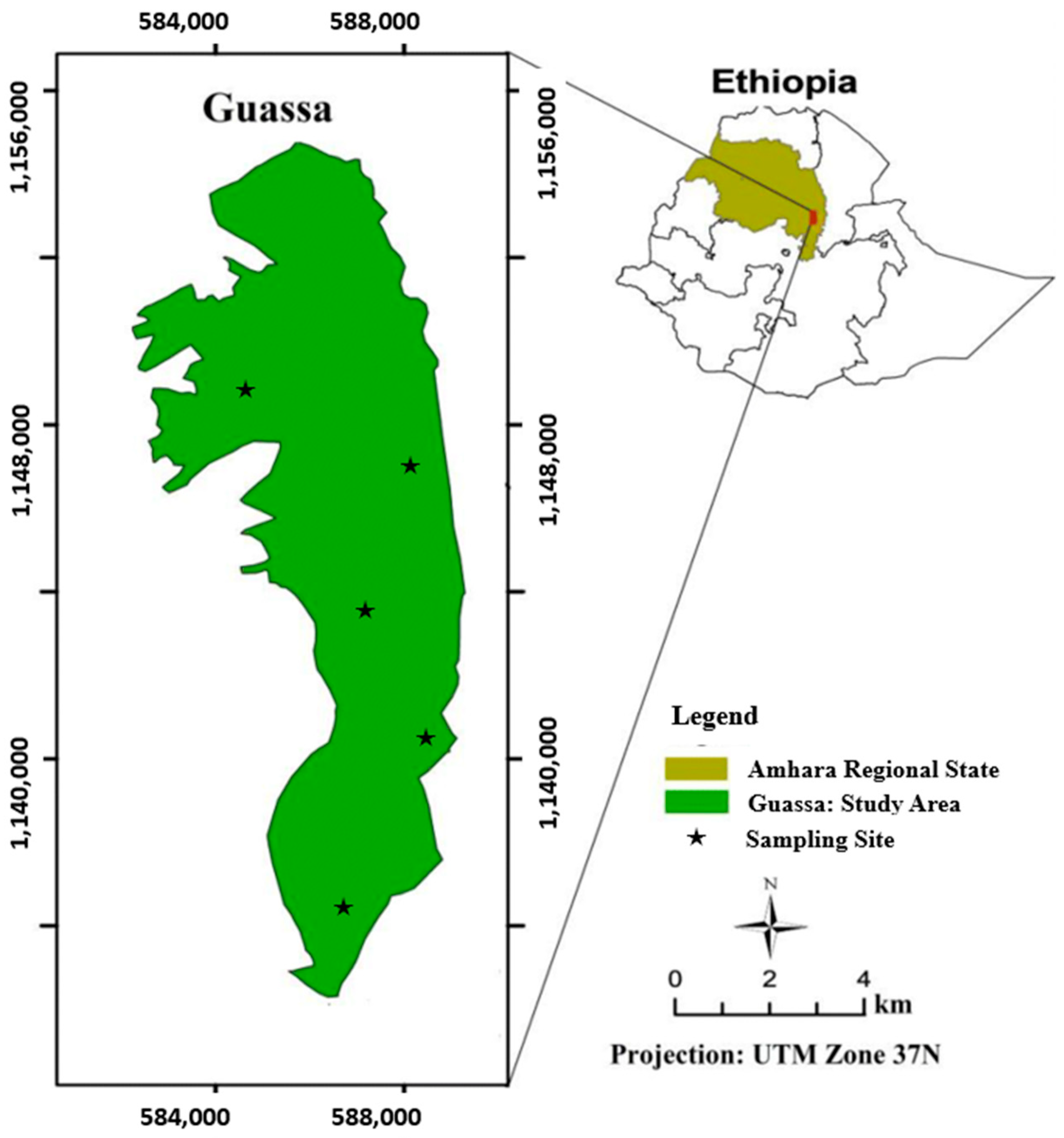
2.1. Description of the Sample Area

The soil was sampled from the Guassa Community Conservation Area (GCCA) (Figure 1). GCCA is located in the central highlands of Ethiopia, 260 km northeast of Addis Ababa, in the Amhara region. This area features an Afroalpine habitat along the western edge of the Great Rift Valley, with altitudes between 3200 and 3700 m asl [25]. GCCA, one of the oldest resource management systems in sub-Saharan Africa [26], receives 1200–1600 mm of annual rainfall.

2.2. Sample Collection

Soil samples were collected from the Guassa Community Conservation Area. The samples were extracted from the subsurface using a sterile spatula, transferred to sterile plastic bags, and transported to the laboratory under aseptic conditions.

2.3. Medium Preparation and Isolation of Amylase-Producing Bacteria

A minimal medium with 10 g starch as carbon source, 0.4 g K2HPO4, 0.2 g peptone, 0.2 g MgSO4∙.7H2O, 0.1 g CaCl2∙2H2O, 0.25 g NaCl, and 0.4 g yeast extract (Himedia, Maharashtra, India) per liter was used for the isolation of bacteria producing amylase. Sterilized water was diluted with 1 g of soil from 10 -1 to 10−5 concentrations. Aseptically, 0.1 mL of each dilution was placed on starch agar plates, spread on a glass rod, and incubated (Biobase, Jinan, China) for 24 h at 30 °C. Pure bacterial cultures were obtained by subculture and preserved at 4 °C [27].

2.4. Primary Screening of Strains

The isolated colonies were cultured in minimal media supplemented with soluble starch, and bacterial inoculation was carried out using the dot method. After incubation at 30 °C for 24 h, each plate was flooded with Gram’s iodine, resulting in a clear zone around the colonies for amylase positivity. The diameter of the clear zone was measured using a ruler [28].

2.5. Secondary Screening of Isolates Using Submerged Fermentation

Minimal broth medium (0.4 (g/L) yeast extract, 0.2 (g/L) peptone, 0.25 (g/L) NaCl, 0.4 (g/L) K2HPO4, 0.1 (g/L) KH2PO4, 0.2 (g/L) MgSO4∙7H2O, 0.1 (g/L) CaCl2∙2H2O, and 10 (g/L) soluble starch, which were supplied by Himedia, India) was prepared. In total, 50 mL of broth medium was dispersed in 250 mL Erlenmeyer flasks and sterilized at 121 °C for 15 min. Subsequently, 1 mL of active inoculum (approximately 8 log CFU/mL) was added to 50 mL of production medium. The medium was adjusted to pH 6 and incubated in a shaker incubator (BOECO, Hamburg, Germany) at 30 °C and 120 rpm. Subsequently, the biomass was separated by centrifugation, amylase was concentrated using cold acetone, and amylase assays were conducted.

2.6. Utilizing MALDI-TOF for the Identification of Isolates

Bacterial samples isolated from soil samples were characterized using matrix-assisted laser desorption/ionization–time-of-flight mass spectrometry (MALDI-TOF MS) (Autobio, Zhengzhou, China). Pure bacterial colonies were selected and transferred to 96-well microscout plates (Bruker Daltonics, Billerica, MA, USA). Each well was coated with a thin film of a colony and air-dried. Subsequently, 1 μL of α-cyano-4-hydroxycinnamic acid solution (12.5 mg) dissolved in 1 mL of a mixture containing 50% acetonitrile and 2.5% trifluoroacetic acid was added. After drying, MALDI 96 microscout plates were introduced into the MALDI-TOF MS device. The device was configured to operate in linear positive-ion mode, targeting a mass range of 2000–20,000 Daltons. The ionization was facilitated by a nitrogen laser with a wavelength of 337 nm operating at a frequency of 60 Hz. The measurement of each colony was carried out by delivering 40 bursts, each consisting of 240 laser pulses [29]. The two identified bacterial strains were subsequently used for further characterization and optimization, with Bacillus subtilis serving as the positive control during the optimization process.

2.7. Crude Amylase Separation and Extraction

A 1 mL bacterial suspension from a 24 h culture was centrifuged at 4500 rpm (Eppendorf, Hamburg, Germany) for 15 min to obtain the crude enzyme. Then, 2 mL of cold acetone was added to 1 mL of cell-free supernatant and refrigerated at −18 °C for 2 h. Subsequently, it was centrifuged again at 4500 rpm for 15 min, the suspensions were discarded, and the supernatant was used as a crude enzyme. Phosphate buffer was added to solubilize the filtrate and form crude amylase.

2.8. Amylase Activity Assay

In total, 1 mL of soluble starch (1%) and 0.5 mL of sodium phosphate buffer (0.05 M, pH) were incubated at 50 °C for 30 min in a water bath (Biobase, China) to assess amylase activity in the cell-free supernatant. After the 30 min incubation period, 2 mL of DNS solution (in g/L; 3,5-Dinitrosalicylic acid (Sigma Aldrich, Hamburg, Germany), 1; sodium hydroxide (Himedia, India), 1; sodium potassium tartrate (Sigma Aldrich, Germany), 2; sodium bisulfite (Himedia, India), 0.05; and phenol (Himedia, India), 0.2) was introduced for color development, followed by heating at 50 °C for 15 min in a boiling water bath. A volume of 0.5 mL of 1% (w/v) sodium potassium tartrate solution was added to end the reaction. The absorbance was recorded at 540 nm against a blank reagent after cooling in running water. Concentrations of dextrose (Sigma Aldrich, Germany) of 0.03, 0.07, 0.19, 0.21, 0.30, and 0.35 (g/L) were used to develop a standard curve to determine the amount of reducing sugars liberated by amylase. The amylase activity of Bacillus subtilis ATCC6051 served as a reference for evaluating the amylase activity of newly isolated bacterial strains.

2.9. Biochemical Characterization of Bacteria

2.9.1. Motility Test

Bacterial motility was evaluated in YEM broth formulated with 1 g of yeast extract, 0.5 g of K2HPO4, 0.2 g of MgSO4∙7H2O, 0.1 g of NaCl, and 10 g of mannitol (Himedia, India), supplemented with 0.2–0.5% agar per liter. Subsequently, the bacteria were inoculated by stabbing into test tubes, which were then incubated at 30 °C for 24 h, and the motility of the bacterial isolates in each test tube was assessed [30].

2.9.2. Gram Staining Test

Gram staining tests were performed on two isolates using separate sterile glass slides to create and fix pure bacterial colonies by heat application. The samples were initially stained with crystal violet for one minute, followed by rinsing with tap water. Subsequently, the specimens were treated with Gram’s iodine for one minute, after which they were washed again with tap water. The decolorization step was performed using 95% ethyl alcohol for 20 s. Finally, the samples were stained with safranin, rinsed with water, and examined under a microscope using an oil immersion objective for detailed observation [31].

2.9.3. Catalase Test

A catalase test was performed to assess the presence of catalase, which catalyzes the decomposition of hydrogen peroxide into water and oxygen. Bacterial colonies, aged for 24 h, were transferred to a clean glass slide and a single drop of 3% hydrogen peroxide was applied. Bubble formation was interpreted as an indication of catalase activity [31].

2.9.4. Citrate Utilization Test

The use of citrate as a carbon source was evaluated using Simmons citrate agar medium (Himedia, India). The medium was prepared in sterile test tubes into which a loopful of pure bacterial isolate was inoculated on the slant and incubated at 30 °C for 48 h. After the 48 h incubation period, a color change from green to blue was interpreted as a positive result [32].

2.10. Effect of Nitrogen and Carbon Sources

Different nitrogen sources (g/L) such as peptone (0.1), urea (0.2), yeast extract (1), ammonium sulfate (0.1), and sodium nitrate (0.1) were sterilized, cooled, inoculated, and incubated at 30 °C for 24 h. Minimal media containing 0.4 g K2HPO4, 0.2 g peptone, 0.2 g MgSO4∙7H2O, 0.1 g CaCl2∙2H2O, 0.25 g NaCl, 0.4 g yeast extract, and carbon sources glucose, starch, sucrose, and maltose 2.5 g per liter were used to evaluate carbon sources. Growth was measured at 600 nm using a spectrophotometer (Jenway 6405, Stone, UK) [33].

2.11. Process Optimization for Amylase Activity

2.11.1. Effect of Metal Ions

Varying concentrations (0, 0.1, 0.5, 1, 1.5, and 2 g/L) of NaCl, MgCl2, and CaCl2 were introduced into a solution consisting of 1 mL of 1% soluble starch, 0.5 mL of phosphate buffer at a pH of 6, and 2 mL of crude amylase. The mixture was incubated at 45 °C in a water bath (Biobase, China). Amylase activity was quantified using the DNS method [34].

2.11.2. Effects of Various Buffer Solutions on Enzyme Activity

Crude amylase was solubilized using acetate, phosphate, and citrate buffers in a pH range from 3.0 to 9.0. Enzymatic activity in 1% soluble starch was assessed using the DNS method [35].

2.11.3. Effect of Temperature on Amylase Activity

A mixture comprising 2 mL of crude amylase, 1 mL of 1% soluble starch, and 0.5 mL of phosphate buffer at pH 6 was prepared and incubated for a duration of 30 min in a water bath set at temperatures of 40, 45, 50, 55, 60, and 65 °C. Subsequently, 2 mL DNS solution was added to the mixture. The mixture was then heated with sodium potassium tartrate (0.5 mL). The test tubes were cooled to ambient temperature using running tap water. The color intensity in each test tube was quantified at a wavelength of 540 nm using a spectrophotometer [36].

2.12. Application of Amylase in Dough Leavening

The efficacy of the enzyme extracted from the isolates was evaluated during the dough leavening process using wheat flour acquired from a local market. To eliminate the yeast that is naturally present in wheat, the flour was subjected to a 15 min autoclaving process at 121 °C. Subsequently, 50 g of wheat flour, 1 g of table salt, 2 mL of yeast cells, 65 mL of sterile distilled water, 2 mL of amylase, and 1 mL of vegetable oil were amalgamated to prepare the dough in a 250 mL measuring cylinder. A film of vegetable oil was applied to prevent the dough from adhering to the sides of the flask. A control experiment was conducted following an identical procedure, but without the addition of amylase. The dough-containing flasks were sealed with foil paper, labeled, and incubated at 30 °C for 10 h. Finally, the alteration of volume between the initial (0 min) and final measurements of the dough was used to determine the effects of amylase on the leavening process [34].

2.13. Data Analysis

Process optimizations for amylase production and isolate growth were calculated using descriptive statistics, and the results are displayed in graphs. Each experiment was carried out in triplicate, and the results are expressed as mean ± SD. One-way ANOVA was performed using Microsoft Excel 2021.

3. Results

3.1. Isolation and Purification of Amylase Enzyme-Producing Bacteria

In the current study, 13 bacterial strains were successfully isolated from the soil of the Guassa Community Conservation Area. Each isolate demonstrated the ability to proliferate on agar medium containing starch, indicating its potential as an amylase producer. Subsequently, five of these isolates, identified through secondary screening, were selected for further detailed investigation and characterization.

3.2. Morphological and Biochemical Characteristics of Isolates

The morphological and biochemical characteristics of the isolates are shown in Table 1. The isolates exhibited various colony characteristics. Microscopic observations and colony morphology suggested that all isolates were Gram-positive and rod-shaped and displayed arrangement in singles, pairs, and chains upon microscopic examination. Furthermore, the majority lacked motility, and all exhibited catalase positivity.

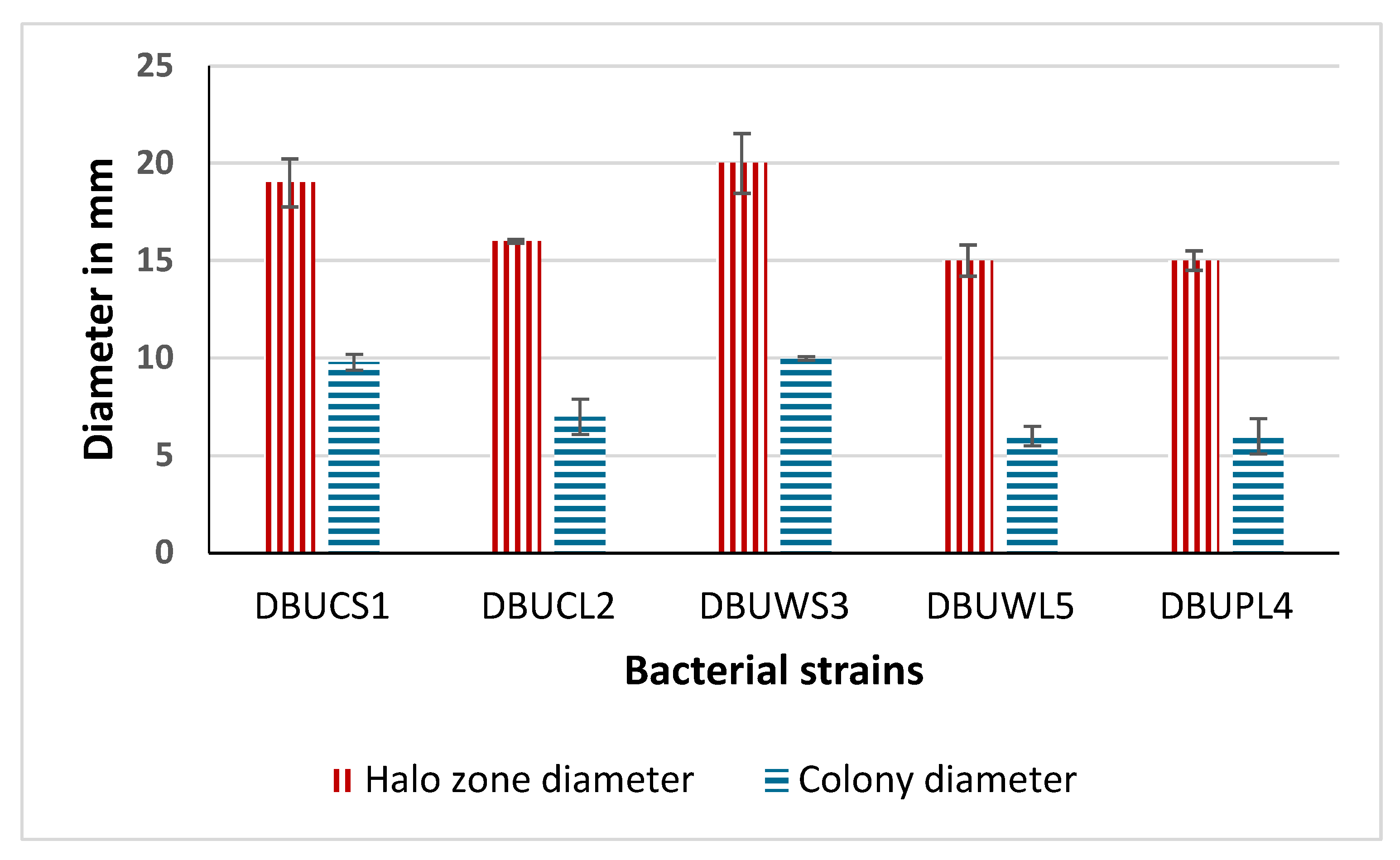
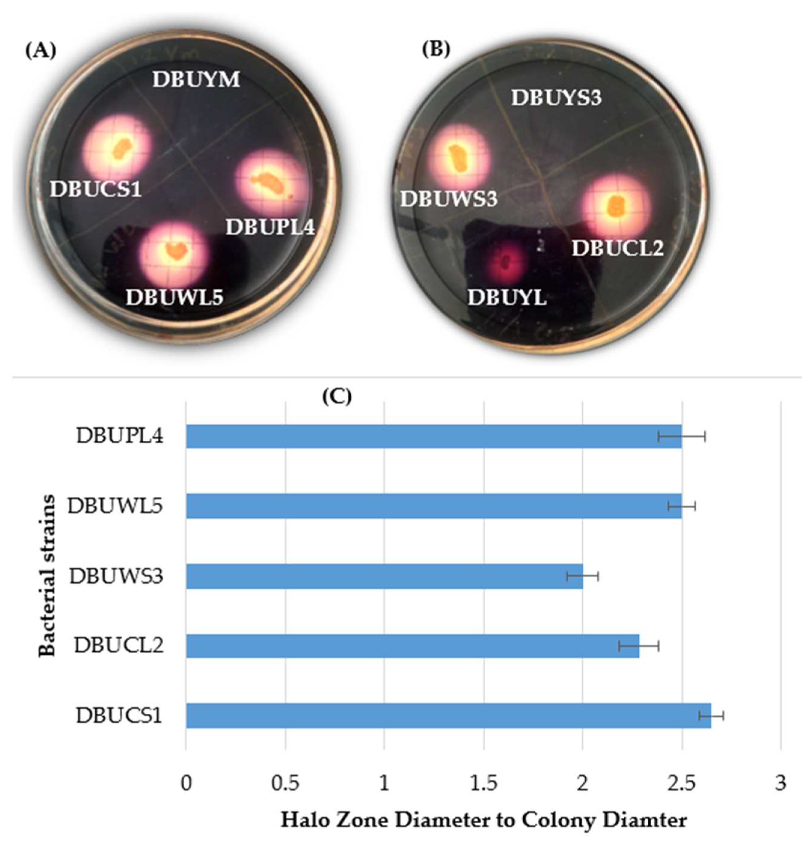
3.3. Primary Screening for Amylase Production

Among the 13 positive isolates, 5 isolates that demonstrated a substantial clear zone of hydrolysis measuring between 15 and 20 mm, together with notable characteristics of hydrolysis and growth, were selected for this study (Figure 2). The strain DBUWS3 has the largest diameter of the halo zone of 20 mm and the largest colony diameter of 10 mm. Both DBUWL5 and DBUPL4 exhibit an identical halo zone diameter of 15 mm and a colony diameter of 6 mm each. DBUCL2 presents with a halo zone diameter of 16 mm and a colony diameter of 7 mm, while DBUCS1 has a halo zone diameter of 19 mm and a colony diameter of 9.8 mm. In general, the diameters of the halo zone ranged from 15 to 20 mm, while the colony diameters ranged from 6 to 10 mm.
Figure 3 shows the starch hydrolysis results of five isolates based on iodine flooding tests. Isolates DBUPL4, DBUWL5, and DBUCS1 had a higher ratio of the diameter of the clear zone to the diameter of the colony (2.5, 2.5, and 2.65), respectively; this indicates that they were able to degrade more starch than DBUCL2 and DBUWS3. Therefore, these two strains were used for further studies.

3.4. Bacterial Identification

The result of bacterial identification by MALDI-TOF MS is presented in Table 2. The identification results for the five bacterial strains exhibit varying degrees of reliability depending on the respective scores obtained. The strains DBUCS1 (2.13) and DBUPL4 (2.48) achieved scores that suggest the probable identification of species such as M. endophyticus and K. rosea, respectively. In contrast, the strains DBUCL2 (1.69), DBUWS3 (1.46), and DBUWL5 (1.43) fell below the critical threshold of 1.7, making their species identification unreliable. Therefore, only two strains were conclusively identified at the species level and used for further characterization and optimization, while the remaining strains required further analysis.

3.5. Optimization of Growth of Bacteria

3.5.1. Effect of Incubation Period on Growth of Isolates

The growth of M. endophyticus DBUCS1 and K. rosea DBUPL4 was monitored over an incubation period of up to 108 h (Figure 4). Both isolates showed an initial lag phase, followed by exponential growth, and eventually reached a stationary phase. M. endophyticus DBUCS1 exhibited a higher growth compared to K. rosea DBUPL4, indicating better growth in minimal broth under the given conditions. The growth of both bacteria peaked around 48–72 h, after which the growth rate stabilized.

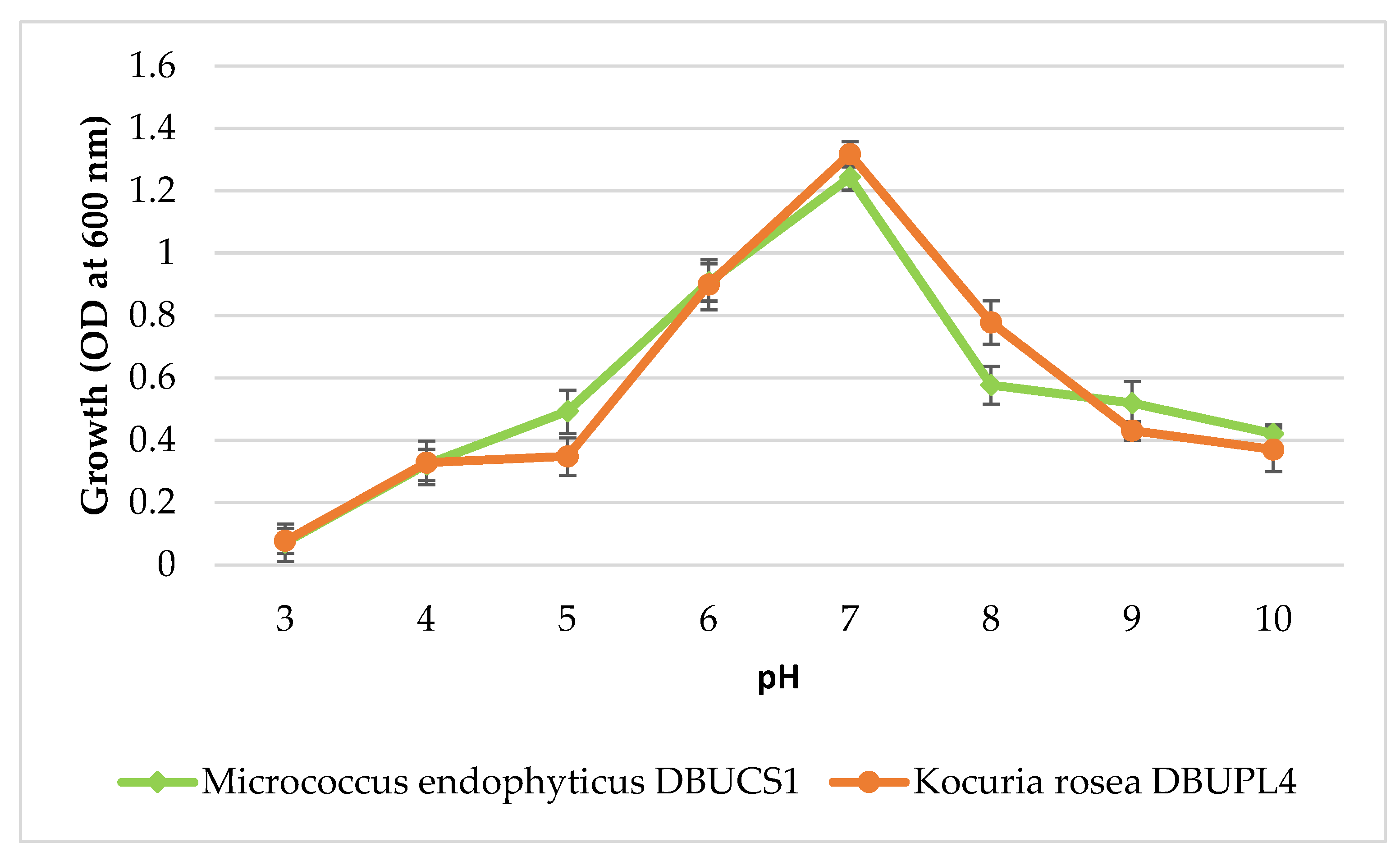
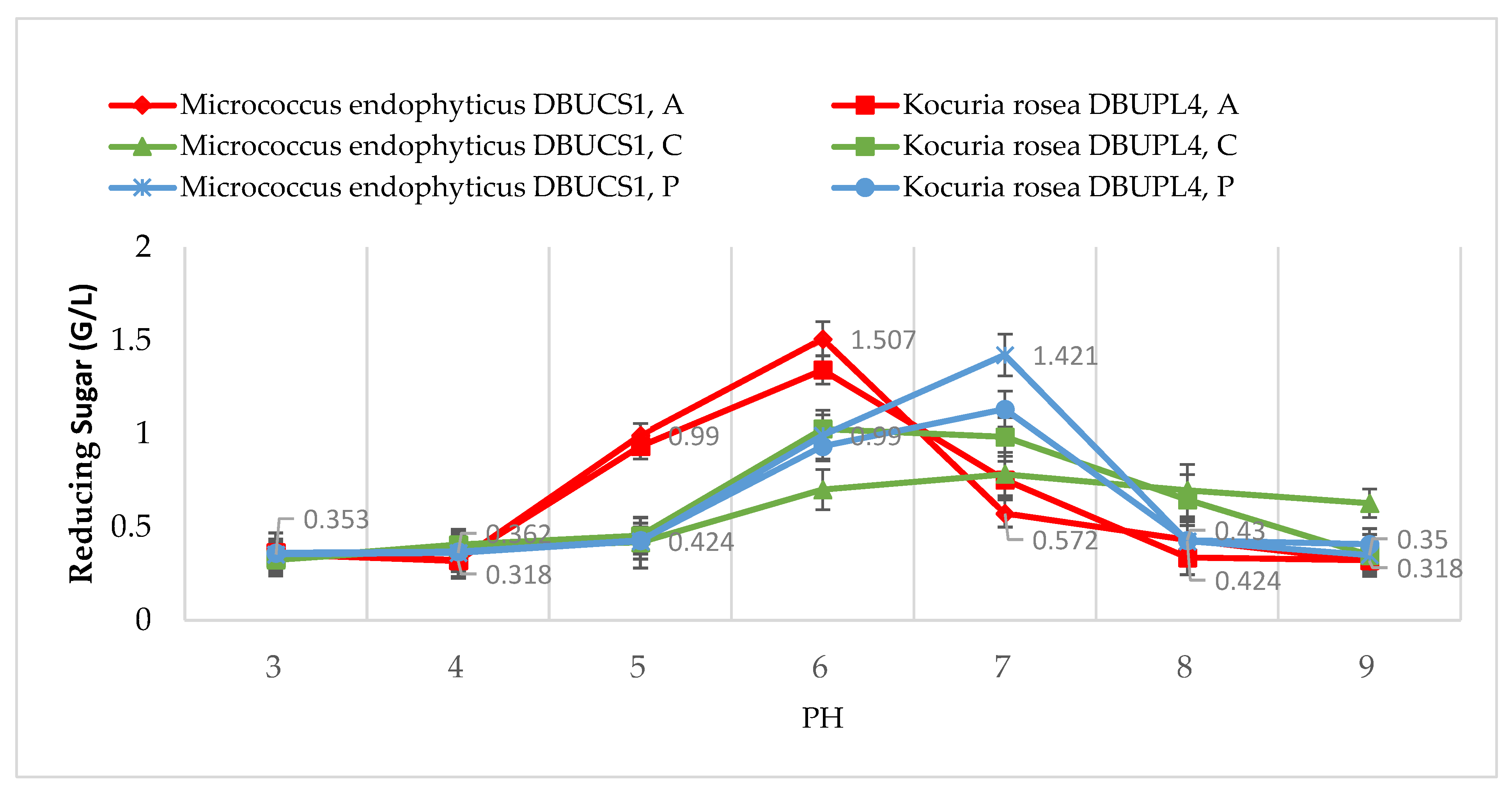
3.5.2. Effect of pH on Isolate Growth

The effect of pH on bacterial growth (Figure 5) demonstrated that both isolates showed maximum growth at neutral pH 7. Growth declined under more acidic (pH 3–5) and alkaline (pH 9–10) conditions. This indicates that the bacteria prefer a neutral environment for optimal proliferation, with K. rosea DBUPL4 displaying slightly higher tolerance to pH variations compared to M. endophyticus DBUCS1.

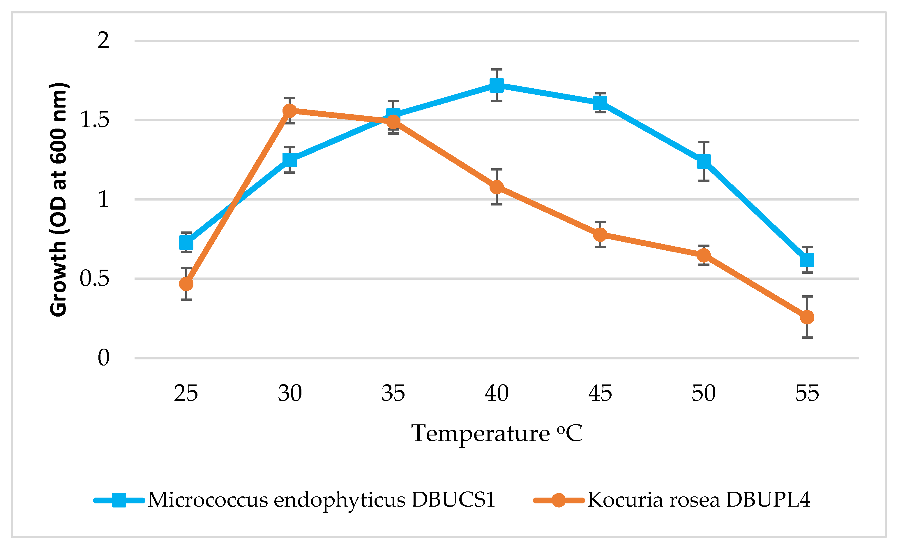
3.5.3. Effect of Temperature on Isolate Growth

The growth of M. endophyticus DBUCS1 and K. rosea DBUPL4 was evaluated at temperatures ranging from 25 °C to 55 °C. Both isolates exhibited optimal growth at 30 °C, with M. endophyticus DBUCS1 showing slightly higher OD values than K. rosea DBUPL4 (Figure 6). Growth decreased significantly at temperatures above 40 °C, indicating that both isolates are mesophilic and prefer moderate temperatures for optimal growth.
Figure 6 shows the growth patterns of two bacterial species, M. endophyticus DBUCS1 and K. rosea DBUPL4, in a temperature range of 25 °C to 55 °C. M. endophyticus DBUCS1 showed a steady increase in growth from 25 °C to an optimal temperature around 40 °C, followed by a gradual decline. K. rosea DBUPL4, on the other hand, exhibited a more rapid growth between 25 °C and 30 °C, reaching its peak at 30 °C. Its growth then decreased significantly, becoming negligible beyond 40 °C. This indicates that M. endophyticus DBUCS1 has a higher optimal growth temperature and a wider growth temperature range compared to K. rosea DBUPL4, which prefers lower temperatures and exhibits a narrower growth range.

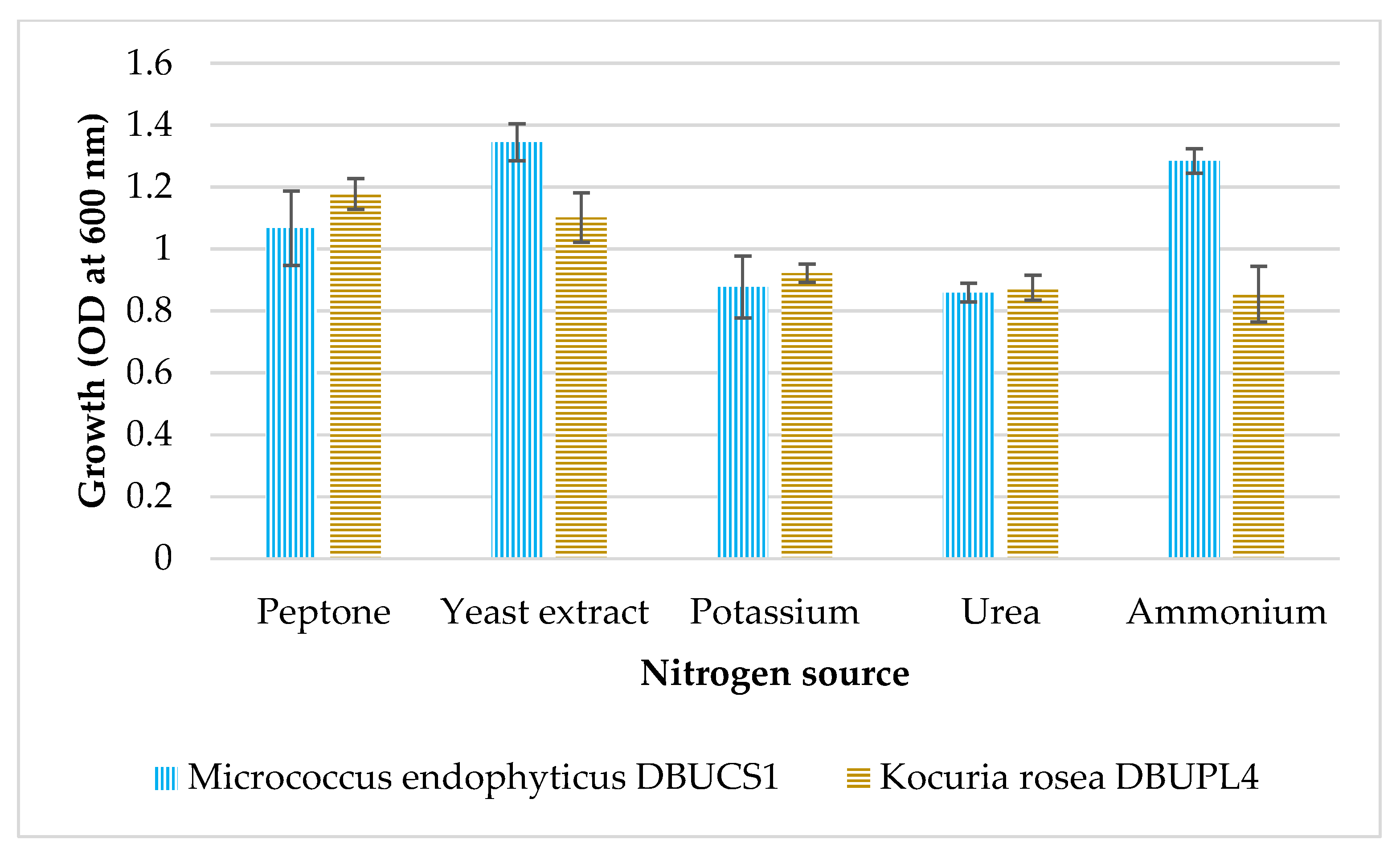
3.5.4. Effect of Nitrogen Source on Growth of Isolates

The growth of M. endophyticus DBUCS1 and K. rosea DBUPL4 was tested using different nitrogen sources, including peptone, yeast extract, potassium, urea, and ammonium. Both isolates showed the highest growth in the presence of peptone, followed by yeast extract (Figure 7). Urea and ammonium resulted in lower growth rates, and M. endophyticus DBUCS1 generally outperformed K. rosea DBUPL4 in all nitrogen sources. Organic nitrogen sources were highly preferred for maximum isolate growth over inorganic nitrogen sources (Figure 7).

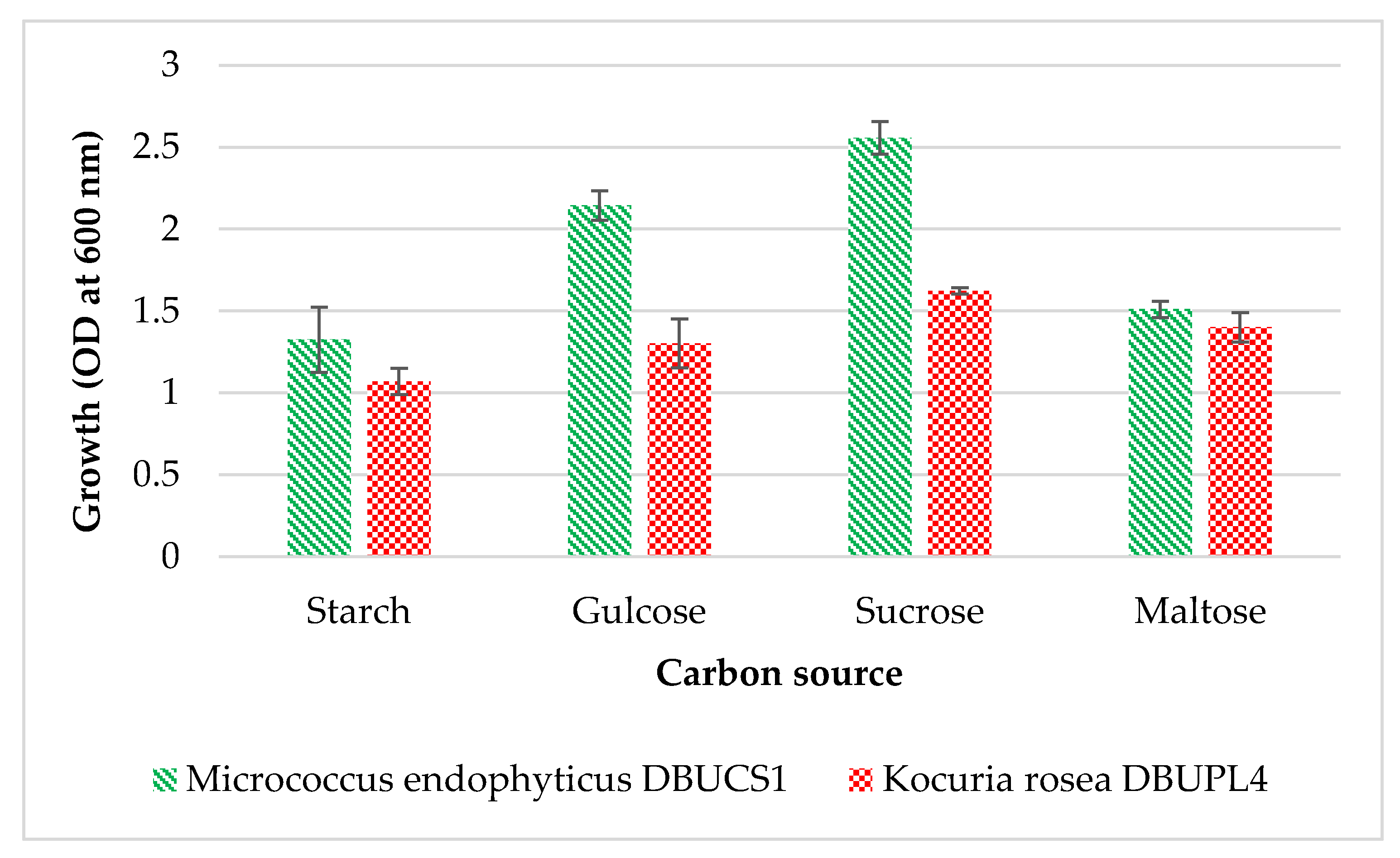
3.5.5. Effect of Carbon Source on Growth of Isolates

The growth of M. endophyticus DBUCS1 and K. rosea DBUPL4 was evaluated using different carbon sources, including starch, glucose, sucrose, and maltose. Both isolates showed the highest growth with glucose as a carbon source, followed by sucrose and maltose (Figure 8). Starch resulted in the lowest growth, with M. endophyticus DBUCS1 consistently showing higher growth compared to K. rosea DBUPL4 in all carbon sources.

3.6. Process Optimization for Amylase Activities

3.6.1. Effect of Temperature on Amylase Activity

The amylase activity of M. endophyticus DBUCS1 and K. rosea DBUPL4 was measured at temperatures ranging from 30 °C to 65 °C. Both isolates exhibited optimal amylase activity at 45 °C to 55 °C, with K. rosea DBUPL4 showing slightly higher activity compared to M. endophyticus DBUCS1. Activity decreased sharply at temperatures above 50 °C, indicating that the enzymes are thermolabile and function best at moderate temperatures (Figure 9).

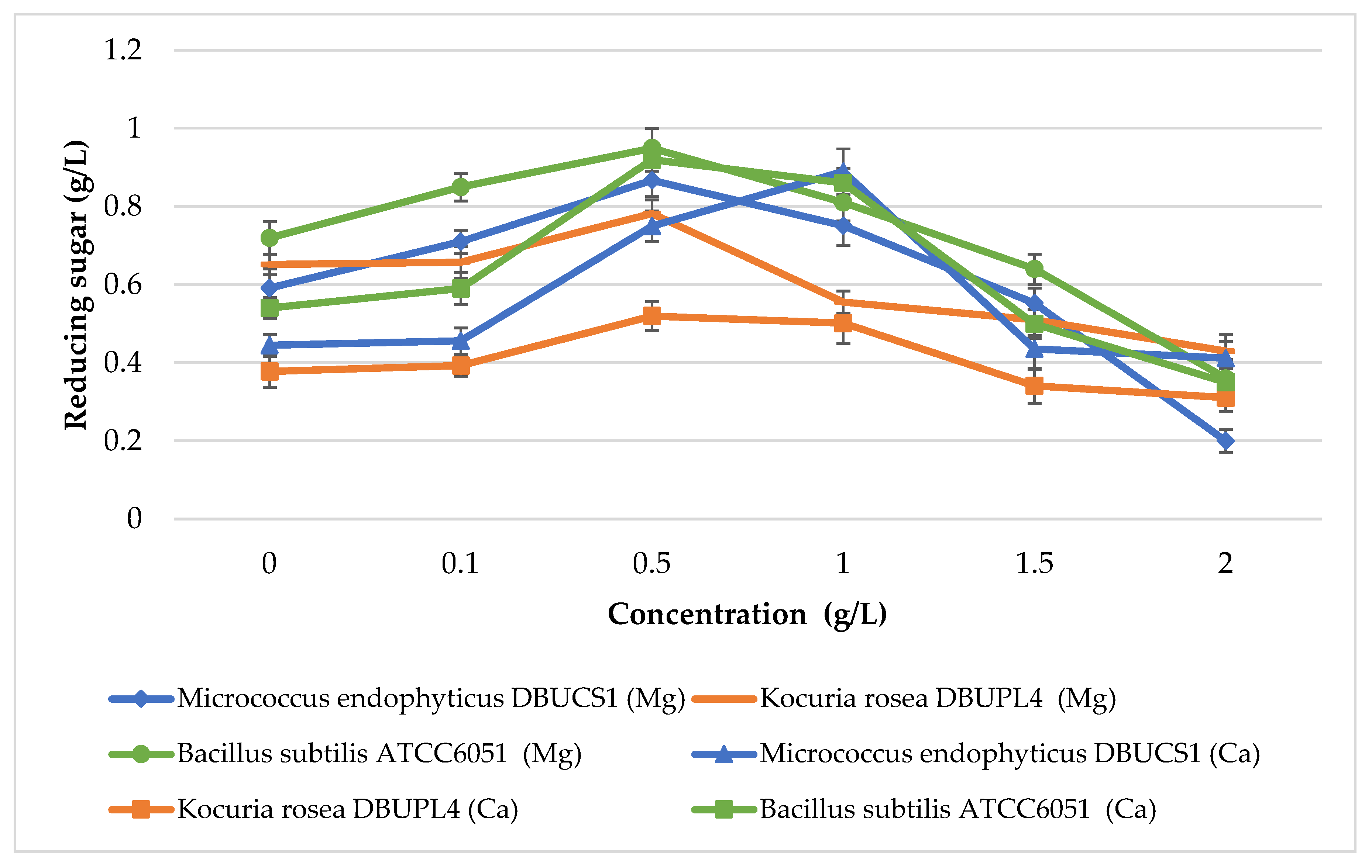
3.6.2. Effect of Metal Ions on Amylase Activity

The effect of metal ions (magnesium chloride and calcium chloride) on amylase activity (Figure 10) was investigated. Both M. endophyticus DBUCS1 and K. rosea DBUPL4 showed increased amylase activity in the presence of magnesium chloride, with K. rosea DBUPL4 exhibiting higher activity. Calcium chloride also enhanced amylase activity, but to a lesser extent compared to magnesium chloride, suggesting that magnesium ions are more effective in promoting enzyme activity. The concentration of the two metal chlorides ranged from 0.1 to 1 g/L, being optimal at 0.5 g/L.

3.6.3. Effect of Buffers on Amylase Activity

The amylase activity of M. endophyticus DBUCS1 and K. rosea DBUPL4 was tested in different buffer systems (acetate, citrate, and phosphate) in a pH range from 3 to 9. Both isolates showed optimal activity in phosphate buffer at pH 7, with K. rosea DBUPL4 exhibiting slightly higher activity (Figure 11). However, pH 6 was optimal for amylase in the acetate buffer of the two strains. Activity was lower under acidic (pH 3–5) and alkaline (pH 8–9) conditions, indicating that neutral pH is optimal for amylase activity.

3.7. Application of Amylase in Wheat Flour Dough Leavening Process

Figure 12 illustrates the application of amylase in the wheat flour dough leavening process over time, comparing the performance of Bacillus subtilis ATCC6051 (a standard), K. rosea DBUPL4, and M. endophyticus DBUCS1. The results show that Bacillus subtilis ATCC6051 consistently produced the highest dough volume, indicating superior amylase activity and leavening efficiency. M. endophyticus DBUCS1 also demonstrated a significant leavening capacity, although slightly lower than the standard. On the contrary, K. rosea DBUPL4 showed the least effectiveness in increasing dough volume, suggesting lower amylase activity. These findings highlight the potential of Bacillus subtilis ATCC6051 as a reliable standard for the application of amylase in dough leavening, while also indicating that M. endophyticus DBUCS1 could be a viable alternative.

4. Discussion

The process of bacterial isolation from soil samples and the subsequent identification of amylase-producing bacteria were successfully achieved. This was done by growing the isolates in a medium containing starch. This process allowed for the selection of bacteria capable of using starch as a carbon source, indicating their potential to produce amylase enzymes. Similar methods have been used in several studies to isolate and identify bacteria with the capacity to produce amylase [21].
The morphological, physiological, and biochemical characterization of the isolates revealed important information about their taxonomy. The results indicated that most of the isolates were Gram-positive, rod-shaped (other than DBUCS1 and DBUPL4), and catalase-positive. These characteristics are commonly associated with bacteria that produce amylase. This is consistent with the characteristics of many known amylase-producing bacteria, such as Bacillus and Micrococcus species [37,38]. Pandit, Moin, Mondal, Banik, and Alam [12] reported that Micrococcus sp. BirBP01, isolated from oligotrophic subsurface lateritic soil, was identified as a Gram-positive coccus and exhibited non-motility. As in this study, K. rosea was identified as a coccoid shape, lacking spores and mobility, and tested positive for both catalase and oxidase [39].
The halo zone diameter of the strains ranged from 15 to 20 mm (Figure 2 and Figure 3), showing the strains’ potential to produce amylase. In a study by Aarti et al. [40], different bacteria isolated from different districts of Tamil Nadu State of India resulted in a halo zone diameter between 10.6 and 26.6 mm, only three isolates showed more than 20 mm, and most were less than 15 mm. In another study, the zone of hydrolysis of bacteria isolated from contaminated soils varied between 1 and 11.6 mm [41], which is lower than this study.
The isolates were identified using MALDI-TOF MS, a technique that has become increasingly popular in recent years due to its precision and efficiency in identifying bacteria at the species level [42,43]. The results of this study indicate that two strains, DBUCS1 and DBUPL4, were reliably identified as M. endophyticus and K. rosea, respectively, which have been reported to be common inhabitants of soil and other environments [7,12,14,44].
The optimization of bacterial growth is a critical step in enzyme production, as it directly affects the yield and efficiency of the process. The results of this study showed that the isolates grew best in a neutral environment and in a medium containing peptone as a nitrogen source and glucose as a carbon source. The two strains in this study showed optimal growth in 48 to 72 h incubation and similarly the amylase producing Bacillus have grown optimally within 36 to 72 h of incubation [4]. As in this study, after 72 h of incubation, the growth and enzyme production by a variety of Bacillus species decreased [41]. M. endophyticus exhibited optimal growth at 40 °C, whereas K. rosea demonstrated a preference for 30 °C. Furthermore, M. endophyticus was able to thrive over a broader temperature range compared to K. rosea. L. rosea exhibited optimal growth at 30 °C [39], whereas Micrococcus sp. BirBP01 demonstrated optimal growth within a temperature range from 20 to 50°C, with peak growth occurring near 37 °C [12]. However, Chen, Zhao, Park, Zhang, Xu, Lee, Kim, and Li [11] isolated M. endophyticus from the root of the plant that showed maximum growth at 30 °C. However, most of the Bacillus species isolated from contaminated soil grew at temperatures between 25 and 45 °C, with the optimal temperature being 37 °C [41].
K. rosea can use a variety of carbon sources, including glucose, crude oil, and various organic compounds found in soil and environmental habitats, while common nitrogen sources can be peptone, yeast extract, and ammonium nitrate depending on strain and growth conditions [45]. These conditions provide the necessary nutrients and environmental factors for optimal bacterial growth and enzyme production.
The optimization of amylase activity revealed that both M. endophyticus DBUCS1 and K. rosea DBUPL4 exhibited optimal activity at temperatures between 45 °C and 55 °C, with activity declining rapidly above 50 °C. This thermolabile nature of the enzymes is consistent with the findings of Yassin, Jiru, and Indracanti [4], who reported similar temperature optima for amylases from Bacillus species. However, the amylase activity of Bacillus spp. isolated from soils of Afdera exhibited a wider temperature range, 60 to 80 °C [4]. In another study, detergent-stable amylase from different Bacillus species was optimal at a temperature of 50 °C [46].
On the one hand, the enhanced amylase activity in the presence of magnesium and calcium chlorides suggests that magnesium and calcium ions play a crucial role in stabilizing the enzyme structure, as previously observed in other amylase-producing bacteria [21]. These metal ions probably served as cofactors or modulators that facilitated the transcription and translation of genes associated with amylase synthesis, leading to an overall improvement in enzyme yield [46,47]. On the other hand, magnesium and calcium could have inhibitory effects on β-amylase [48].
The effect of buffer systems on amylase activity indicated that phosphate buffer at pH 7 was optimal for both isolates. This is consistent with the neutral pH preference observed during growth optimization, suggesting that enzymes are adapted to function in environments with neutral pH [20,49]. The lower activity under acidic and alkaline conditions further underscores the importance of pH control in industrial applications of amylases [50]. The Bacillus species were effectively growing and producing amylase when the pH was adjusted between 6 and 8 [41] and 6 and 9 [46].
In contrast, K. rosea DBUPL4 showed the least effective increase in dough volume, indicating lower amylase activity compared to the other two strains. This suggests that while K. rosea might produce some amylase, the amount or specific activity is insufficient to contribute significantly to dough leavening. This observation highlights the importance of strain selection when utilizing microbial amylases for baking applications. The efficiency of starch hydrolysis, a crucial step in the leavening process, varies significantly between different microbial species and even strains within the same species [51].
The observed differences in leavening performance emphasize the crucial role of amylase in providing fermentable sugars for yeast metabolism. Starch breakdown by amylase generates maltose and other oligosaccharides, which the yeast then converts to CO2, causing the dough to rise [52]. The rate and extent of this process are directly correlated with the increase in dough volume.
Generally, the application of amylase in the leavening of wheat flour dough showed promising results, with isolates demonstrating their ability to increase the volume of the dough. This indicates their potential use in the baking industry as a substitute for chemical additives. The use of microbial enzymes in bread making has gained attention in recent years due to its ability to improve dough properties and enhance the nutritional value of the final product [53].

5. Conclusions

This study successfully isolated and characterized amylase-producing bacteria from the soil of the Guassa Community Conservation Area, Ethiopia. Two strains, Kocuria rosea and Micrococcus endophyticus, were identified as efficient amylase producers, with optimal enzyme activity observed at moderate temperatures (45–55 °C) and neutral pH. The presence of magnesium and calcium ions significantly enhanced amylase activity, suggesting their role as enzyme stabilizers. The application of these enzymes in the leaving of wheat flour dough demonstrated their potential in the baking industry, and M. endophyticus showed promising results. These findings underscore the importance of exploring unique environments for novel microbial enzymes with industrial applications. This study contributes to the growing body of knowledge on sustainable enzyme production and highlights the potential of microbial amylases as eco-friendly alternatives to chemical additives in various industries. Further research could focus on the pathogenicity and toxicology of strains prior to application in the food industry.

Author Contributions

Conceptualization, S.-H.K. and W.-J.K.; methodology, S.-H.K., W.-J.K. and Y.Y.; software, J.R.; validation, J.R.; formal analysis, S.-H.K., W.-J.K. and J.R.; investigation, Y.Y.; resources, A.T.; data curation, J.R.; writing—original draft preparation, Y.Y.; writing—review and editing, A.T.; visualization, Y.Y.; supervision, A.T.; fund acquisition, S.-H.K. and W.-J.K.; project administration, A.T. All authors have read and agreed to the published version of the manuscript.

Funding

This work was supported by the research program of KAERI, Republic of Korea (Project Number 523410-25).

Institutional Review Board Statement

Not applicable.

Informed Consent Statement

Not applicable.

Data Availability Statement

The original contributions presented in this study are included in the article. Further inquiries can be directed to the corresponding author.

Acknowledgments

We thank Belayhun Tesfaye for technical assistance during the experimental work.

Conflicts of Interest

The authors declare no conflicts of interest.

References

  1. Farooq, M.A.; Ali, S.; Hassan, A.; Tahir, H.M.; Mumtaz, S.; Mumtaz, S. Biosynthesis and industrial applications of α-amylase: A review. Arch. Microbiol. 2021, 203, 1281–1292. [Google Scholar] [CrossRef] [PubMed]
  2. Morbia, M.; Pandey, A.; Mahla, P.; Gohil, S. Isolation of α-Amylase Producing Microorganisms from Soil of Kachchh, Gujarat. J. Pure Appl. Microbiol. 2024, 18, 9218. [Google Scholar] [CrossRef]
  3. Lim, S.J.; Oslan, S.N. Native to designed: Microbial-amylases for industrial applications. PeerJ 2021, 9, e11315. [Google Scholar] [CrossRef] [PubMed]
  4. Yassin, S.N.; Jiru, T.M.; Indracanti, M. Screening and Characterization of Thermostable Amylase-Producing Bacteria Isolated from Soil Samples of Afdera, Afar Region, and Molecular Detection of Amylase-Coding Gene. Int. J. Microbiol. 2021, 2021, 5592885. [Google Scholar] [CrossRef]
  5. Wang, J.; Chen, Q.; Yan, P.; Dong, C.; Shao, Z. Isolation and Optimization of Aflatoxin B(1) Degradation by Uniform Design and Complete Genome Sequencing of Novel Deep-Sea Kocuria rosea Strain 13. Toxins 2023, 15, 520. [Google Scholar] [CrossRef] [PubMed]
  6. Parshetti, G.K.; Parshetti, S.; Kalyani, D.C.; Doong, R.-a.; Govindwar, S.P. Industrial dye decolorizing lignin peroxidase from Kocuria rosea MTCC 1532. Ann. Microbiol. 2012, 62, 217–223. [Google Scholar] [CrossRef]
  7. Gu, D.; Jiao, Y.; Wu, J.; Liu, Z.; Chen, Q. Optimization of EPS Production and Characterization by a Halophilic Bacterium, Kocuria rosea ZJUQH from Chaka Salt Lake with Response Surface Methodology. Molecules 2017, 22, 814. [Google Scholar] [CrossRef] [PubMed]
  8. Bernal, C.; Cairó, J.; Coello, N. Purification and characterization of a novel exocellular keratinase from Kocuria rosea. Enzym. Microb. Technol. 2006, 38, 49–54. [Google Scholar] [CrossRef]
  9. Parshetti, G.K.; Telke, A.A.; Kalyani, D.C.; Govindwar, S.P. Decolorization and detoxification of sulfonated azo dye methyl orange by Kocuria rosea MTCC 1532. J. Hazard. Mater. 2010, 176, 503–509. [Google Scholar] [CrossRef]
  10. Elamary, R.; Salem, W.M. Optimizing and purifying extracellular amylase from soil bacteria to inhibit clinical biofilm-forming bacteria. PeerJ 2020, 8, e10288. [Google Scholar] [CrossRef]
  11. Chen, H.-H.; Zhao, G.-Z.; Park, D.-J.; Zhang, Y.-Q.; Xu, L.-H.; Lee, J.-C.; Kim, C.-J.; Li, W.-J. Micrococcus endophyticus sp. nov., isolated from surface-sterilized Aquilaria sinensis roots. Int. J. Syst. Evol. Microbiol. 2009, 59, 1070–1075. [Google Scholar] [CrossRef] [PubMed]
  12. Pandit, B.; Moin, A.; Mondal, A.; Banik, A.; Alam, M. Characterization of a biofilm-forming, amylase-producing, and heavy-metal-bioremediating strain Micrococcus sp. BirBP01 isolated from oligotrophic subsurface lateritic soil. Arch. Microbiol. 2023, 205, 351. [Google Scholar] [CrossRef] [PubMed]
  13. Singh, P.; Singh, S.M.; Dhakephalkar, P. Diversity, cold active enzymes and adaptation strategies of bacteria inhabiting glacier cryoconite holes of High Arctic. Extremophiles 2014, 18, 229–242. [Google Scholar] [CrossRef] [PubMed]
  14. Ali, N.; Dashti, N.; Khanafer, M.; Al-Awadhi, H.; Radwan, S. Bioremediation of soils saturated with spilled crude oil. Sci. Rep. 2020, 10, 1116. [Google Scholar] [CrossRef]
  15. Rustamova, N.; Wubulikasimu, A.; Mukhamedov, N.; Gao, Y.; Egamberdieva, D.; Yili, A. Endophytic Bacteria Associated with Medicinal Plant Vernonia anthelmintica: Diversity and Characterization. Curr. Microbiol. 2020, 77, 1457–1465. [Google Scholar] [CrossRef]
  16. Klinfoong, R.; Thummakasorn, C.; Ungwiwatkul, S.; Boontanom, P.; Chantarasiri, A. Diversity and activity of amylase-producing bacteria isolated from mangrove soil in Thailand. Biodiversitas J. Biol. Divers. 2022, 23, 5519–5531. [Google Scholar] [CrossRef]
  17. Choi, H.J.; Hong, J.B.; Park, J.J.; Chi, W.-J.; Kim, M.-C.; Chang, Y.-K.; Hong, S.-K. Production of agarase from a novel Micrococcus sp. GNUM-08124 strain isolated from the East Sea of Korea. Biotechnol. Bioprocess Eng. 2011, 16, 81–88. [Google Scholar] [CrossRef]
  18. Ariaeenejad, S.; Zolfaghari, B.; Sadeghian Motahar, S.F.; Kavousi, K.; Maleki, M.; Roy, S.; Hosseini Salekdeh, G. Highly Efficient Computationally Derived Novel Metagenome α-Amylase With Robust Stability Under Extreme Denaturing Conditions. Front. Microbiol. 2021, 12, 713125. [Google Scholar] [CrossRef]
  19. Ju, L.; Pan, Z.; Zhang, H.; Li, Q.; Liang, J.; Deng, G.; Yu, M.; Long, H. New insights into the origin and evolution of α-amylase genes in green plants. Sci. Rep. 2019, 9, 4929. [Google Scholar] [CrossRef]
  20. Far, B.E.; Ahmadi, Y.; Khosroshahi, A.Y.; Dilmaghani, A. Microbial alpha-amylase production: Progress, challenges and perspectives. Adv. Pharm. Bull. 2020, 10, 350. [Google Scholar]
  21. Gupta, R.; Gigras, P.; Mohapatra, H.; Goswami, V.K.; Chauhan, B. Microbial α-amylases: A biotechnological perspective. Process Biochem. 2003, 38, 1599–1616. [Google Scholar] [CrossRef]
  22. Souza, P.M.d. Application of microbial α-amylase in industry-A review. Braz. J. Microbiol. 2010, 41, 850–861. [Google Scholar] [CrossRef] [PubMed]
  23. Hallol, M.; Helmy, O.; Shawky, A.-E.; El-Batal, A.; Ramadan, M. Optimization of Alpha-Amylase Production by a Local Bacillus paramycoides Isolate and Immobilization on Chitosan-Loaded Barium Ferrite Nanoparticles. Fermentation 2022, 8, 241. [Google Scholar] [CrossRef]
  24. Shad, M.; Hussain, N.; Usman, M.; Akhtar, M.; Sajjad, M. Exploration of computational approaches to predict the structural features and recent trends in α-amylase production for industrial applications. Biotechnol. Bioeng. 2023, 120, 2092–2116. [Google Scholar] [CrossRef]
  25. Nigussie, G.; Fekadu, M.; Demissew, S.; Warkineh, B. Impact of conservation management on land change: A case study in Guassa Community Conservation Area for the last 31 years (1986–2015). Model. Earth Syst. Environ. 2019, 5, 1495–1504. [Google Scholar] [CrossRef]
  26. Tadesse, S.; Teketay, D. Contribution of the Guassa Community Eco-Lodge to the Conservation of Wild Mammals, Birds and Woody Plants in Menz-Gera Midir District, North Shewa Administrative Zone, Ethiopia. Int J Avian Wildl. Biol 2017, 2, 00029. [Google Scholar] [CrossRef]
  27. Abd-Elhalem, B.T.; El-Sawy, M.; Gamal, R.F.; Abou-Taleb, K.A. Production of amylases from Bacillus amyloliquefaciens under submerged fermentation using some agro-industrial by-products. Ann. Agric. Sci. 2015, 60, 193–202. [Google Scholar] [CrossRef]
  28. Rakaz, M.A.; Hussien, M.O.; Ibrahim, H.M. Isolation, extraction, purification, and molecular characterization for thermostable α-amylase from locally isolated bacillus species in Sudan. Biochem. Res. Int. 2021, 2021, 6670380. [Google Scholar] [CrossRef]
  29. Çevik, Y.N.; Ogutcu, H. Identification of Bacteria in Soil by MALDI-TOF MS and Analysis of Bacillus spp., Paenibacillus spp. and Pseudomonas spp. with PCA. Anal. Chem. Lett. 2020, 10, 784–797. [Google Scholar] [CrossRef]
  30. Archna Karel, A.K.; Anshu Karel, A.K.; Jat, B. Studies on vesicular-arbuscular mycorrhiza of trigonella foenum-graecum. Trends Biosci. 2017, 10, 5808–5817. [Google Scholar]
  31. Tyagi, A.; Kumar, V.; Tomar, A. Isolation, identification, biochemical and antibiotic sensitivity characterization of Rhizobium strains from Vigna mungo (L.) Hepper, Cicer arietinum (L.) and Vigna radiata (L.) Wilczek in Muzaffarnagar, Uttar Pradesh India. Int. J. Curr. Microbiol. Appl. Sci. 2017, 6, 2024–2035. [Google Scholar] [CrossRef]
  32. Padma, S.; Pallavi, K. Isolation and characterization of amylase producing Bacillus spp. from selected soil sample. Int. J. Res. Biosci. 2016, 5, 24–29. [Google Scholar]
  33. Lal, N.; Jyoti, J.; Sachan, P. Optimization of nitrogen source (s) for the growth and amylase production from Bacillus licheniformis JAR-26 under submerged fermentation. Indian J. Biol. 2016, 3, 127–132. [Google Scholar] [CrossRef]
  34. Lakew, E. Characterization of wild yeasts isolated from selected fruits for their bread leavening capacity. Master Dissertation, Addis Ababa University, Addis Ababa, Ethiopia, 2017. Available online: http://etd.aau.edu.et/handle/12345678/1091 (accessed on 10 January 2025).
  35. Alemayhu, F. Isolating Thermostable Amylase Producing Bacteria from Hot Spring Water in Senbete, Ethiopia and Optimization of Its Production. Master Dissertation, Debre Berhaan University, Debre Berhan, Ethiopia, 2019. [Google Scholar]
  36. Patel, G. Isolation and characterization of starch degrading bacteria from garden soil, Ganpat University, Gujarat, India. Indian J. Microbiol. Res. 2015, 2, 111–114. [Google Scholar]
  37. Pandey, A.; Soccol, C.R.; Nigam, P.; Soccol, V.T. Biotechnological potential of agro-industrial residues. I: Sugarcane bagasse. Bioresour. Technol. 2000, 74, 69–80. [Google Scholar] [CrossRef]
  38. Xu, X.; Cheng, Y.; Fang, Z.; Yin, J.; Shen, H.; Ma, D. Identification and utilization of a new Bacillus amyloliquefaciens XY-1 against Fusarium head blight. Front. Plant Sci. 2022, 13, 1055213. [Google Scholar] [CrossRef]
  39. Gholami, M.; Etemadifar, Z.; Bouzari, M. Isolation a new strain of Kocuria rosea capable of tolerating extreme conditions. J. Environ. Radioact. 2015, 144, 113–119. [Google Scholar] [CrossRef]
  40. Aarti, C.; Khusro, A.; Agastian, P.; Darwish, N.M.; Al Farraj, D.A. Molecular diversity and hydrolytic enzymes production abilities of soil bacteria. Saudi J. Biol. Sci. 2020, 27, 3235–3248. [Google Scholar] [CrossRef] [PubMed]
  41. Munawar, A.; Shaheen, M.; Ramzan, S.; Masih, S.A.; Jabeen, F.; Younis, T.; Aslam, M. DIVERISTY and enzymatic potential of indigenous bacteria from unexplored contaminted soils in Faisalabad. Heliyon 2023, 9, e15256. [Google Scholar] [CrossRef]
  42. Darie-Ion, L.; Whitham, D.; Jayathirtha, M.; Rai, Y.; Neagu, A.-N.; Darie, C.C.; Petre, B.A. Applications of MALDI-MS/MS-based proteomics in biomedical research. Molecules 2022, 27, 6196. [Google Scholar] [CrossRef]
  43. Sun, J.; Shi, H.; Xue, Y.; Cheng, W.; Yu, M.; Ding, C.; Xu, F.; Yu, S. Releasing bacteria from functional magnetic beads is beneficial to MALDI-TOF MS based identification. Talanta 2021, 225, 121968. [Google Scholar] [CrossRef] [PubMed]
  44. Wu, C.-Y.; Chen, N.; Li, H.; Li, Q.-F. Kocuria rosea HN01, a newly alkaliphilic humus-reducing bacterium isolated from cassava dreg compost. J. Soils Sediments 2014, 14, 423–431. [Google Scholar] [CrossRef]
  45. Akbari, E.; Rasekh, B.; Maal, K.B.; Karbasiun, F.; Yazdian, F.; Emami-Karvani, Z.; Peighami, R. A novel biosurfactant producing Kocuria rosea ABR6 as potential strain in oil sludge recovery and lubrication. AMB Express 2021, 11, 131. [Google Scholar] [CrossRef] [PubMed]
  46. Inan Bektas, K.; Nalcaoğlu, A.; Ceylan, E.; Colak, D.N.; Caglar, P.; Agirman, S.; Sivri, N.S.; Gunes, S.; Kaya, A.; Canakci, S.; et al. Isolation and characterization of detergent-compatible amylase-, protease-, lipase-, and cellulase-producing bacteria. Braz. J. Microbiol. 2023, 54, 725–737. [Google Scholar] [CrossRef] [PubMed]
  47. Ovais, M.; Khalil, A.T.; Ayaz, M.; Ahmad, I.; Nethi, S.K.; Mukherjee, S. Biosynthesis of metal nanoparticles via microbial enzymes: A mechanistic approach. Int. J. Mol. Sci. 2018, 19, 4100. [Google Scholar] [CrossRef]
  48. Umar Dahot, M.; Saboury, A.A.; Moosavi-Movahedi, A.A. Inhibition of β-Amylase Activity by Calcium, Magnesium and Zinc Ions Determined by Spectrophotometry and Isothermal Titration Calorimetry. J. Enzym. Inhib. Med. Chem. 2004, 19, 157–160. [Google Scholar] [CrossRef]
  49. Sundarram, A.; Murthy, T.P.K. α-amylase production and applications: A review. J. Appl. Environ. Microbiol. 2014, 2, 166–175. [Google Scholar]
  50. Pandey, A.; Nigam, P.; Soccol, C.R.; Soccol, V.T.; Singh, D.; Mohan, R. Advances in microbial amylases. Biotechnol. Appl. Biochem. 2000, 31, 135–152. [Google Scholar] [CrossRef]
  51. Armenta, S.; Sánchez-Cuapio, Z.; Munguia, M.E.; Pulido, N.O.; Farrés, A.; Manoutcharian, K.; Hernandez-Santoyo, A.; Moreno-Mendieta, S.; Sánchez, S.; Rodríguez-Sanoja, R. The role of conserved non-aromatic residues in the Lactobacillus amylovorus α-amylase CBM26-starch interaction. Int. J. Biol. Macromol. 2019, 121, 829–838. [Google Scholar] [CrossRef]
  52. Mietton, L.; Samson, M.-F.; Marlin, T.; Godet, T.; Nolleau, V.; Guezenec, S.; Segond, D.; Nidelet, T.; Desclaux, D.; Sicard, D. Impact of Leavening Agent and Wheat Variety on Bread Organoleptic and Nutritional Quality. Microorganisms 2022, 10, 1416. [Google Scholar] [CrossRef] [PubMed]
  53. Raveendran, S.; Parameswaran, B.; Ummalyma, S.B.; Abraham, A.; Mathew, A.K.; Madhavan, A.; Rebello, S.; Pandey, A. Applications of microbial enzymes in food industry. Food Technol. Biotechnol. 2018, 56, 16. [Google Scholar] [CrossRef] [PubMed]
Figure 1. Map of the Guassa Community Conservation Area [25].
Fermentation 11 00211 g001
Figure 2. Amylase-producing bacterial isolates with halo zone and colony diameter in mm; the error bar is the standard error of the mean.
Fermentation 11 00211 g002
Figure 3. (A,B) Halo zones of the isolates and (C) ratio of the halo zone diameter to the colony diameter; the error bar is the standard error of the mean.
Fermentation 11 00211 g003
Figure 4. Bacterial isolate growth curve in minimal broth at pH 7 and a temperature of 30 °C; the error bar is the standard error of the mean.
Fermentation 11 00211 g004
Figure 5. Effect of pH on the growth of isolates at 30 °C for 24 h; the error bar is the standard error of the mean.
Fermentation 11 00211 g005
Figure 6. Effect of temperature on the growth of isolates incubated in starch broth at pH 7.0 for 24 h; the error bar is the standard error of the mean.
Fermentation 11 00211 g006
Figure 7. Effect of the nitrogen source on the growth of isolates; the error bar is the standard error of the mean.
Fermentation 11 00211 g007
Figure 8. Effect of the carbon source on the growth of isolates; the error bar is the standard error of the mean.
Fermentation 11 00211 g008
Figure 9. Effect of temperature on the amylase activity; the error bar is the standard error of the mean.
Fermentation 11 00211 g009
Figure 10. Effect of magnesium chloride and calcium chloride on amylase activity; the error bar is the standard error of the mean.
Fermentation 11 00211 g010
Figure 11. Effect of the pH of the buffers on amylase activity (A = acetate, C = citrate, and P = phosphate buffers); the error bar is the standard error of the mean.
Fermentation 11 00211 g011
Figure 12. Application of amylase in the bread dough leavening process in hours; the error bar is the standard error of the mean.
Fermentation 11 00211 g012
Table 1. Morphological and biochemical characteristics of isolates grown on starch agar at 30 °C for 48 h (+ indicates positive biochemical tests or presence of morphology, and - indicates negative biochemical tests or absence of morphological parts).
IsolateColony MorphologyBiochemical Test
ShapeColorSizeMarginSurfaceShapeMotilityGramCitrate
e Test
Catalase
DBUCS1RoundCreamSmallEntireSmoothCoccus-+_+
DBUCL2RoundCreamLargeEntireSmoothRod++_+
DBUWS3RoundWhiteSmallEntireSmoothRod-+++
DBUPL4RoundPinkLargeEntireSmoothCoccus-+++
DBUWL5RoundWhiteLargeEntireSmoothRod-+++
Table 2. Identification of bacterial isolates using MALDI-TOF MS (scoring criteria: score < 1.7 Unreliable; 1.7–2.0 Probable Genus; ≥2.0 Probable Species).
Strain CodeScoreSuggested Probable Species
DBUCS12.13Micrococcus endophyticus
DBUCL21.69Cutibacterium granulosum
DBUWS31.46Pasteurella canis
DBUPL42.48Kocuria rosea
DBUWL51.43Cutibacterium granulosum
Disclaimer/Publisher’s Note: The statements, opinions and data contained in all publications are solely those of the individual author(s) and contributor(s) and not of MDPI and/or the editor(s). MDPI and/or the editor(s) disclaim responsibility for any injury to people or property resulting from any ideas, methods, instructions or products referred to in the content.

