Article

Release and Purification of Poly(3-Hydroxybutyrate) P(3HB) via the Combined Use of an Autolytic Strain of Azotobacter vinelandii OP-PhbP3+ and Non-Halogenated Solvents

1
Departamento de Ingeniería Celular y Biocatálisis, Instituto de Biotecnología, Universidad Nacional Autónoma de México (UNAM), Av. Universidad 2001, Col. Chamilpa, Cuernavaca 62210, Morelos, Mexico
2
Departamento de Microbiología Molecular, Instituto de Biotecnología, Universidad Nacional Autónoma de México (UNAM), Av. Universidad 2001, Col. Chamilpa, Cuernavaca 62210, Morelos, Mexico
*
Author to whom correspondence should be addressed.
Fermentation 2025, 11(10), 571; https://doi.org/10.3390/fermentation11100571
Submission received: 22 August 2025 / Revised: 19 September 2025 / Accepted: 21 September 2025 / Published: 2 October 2025
(This article belongs to the Section Fermentation Process Design)

Abstract

P(3HB) is a biodegradable and biocompatible polymer, which can replace petroleum-derived plastics. Previous studies have shown that Azotobacter vinelandii strain OP-PhbP3+, which overexpresses the phasin protein PhbP3, produces high concentrations of P(3HB) and undergoes early autolysis, facilitating polymer release. The aim of the present study was to evaluate the performance of this strain for P(3HB) production in 3 L bioreactors and assess the feasibility of a simplified recovery process. After 36 h of cultivation, rapid cell lysis was observed, resulting in a ~50% decrease in the protein content of the cell dry weight, without reducing P(3HB) concentration, which reached 4.6 g L−1. Flow cytometry analysis revealed significant morphological changes during cultivation, which was consistent with the strain’s lytic behavior. The biomass recovered at 36 h was washed with SDS, obtaining a yield of 92.5% (respect to P(3HB) initial) and a purity of 97.6%. An alternative extraction procedure using the non-halogenated solvent cyclohexanone (CYC) resulted in an even higher yield of 97.8% with a purity of 99.3% of P(3HB). Notably, the weight average molecular weight of the polymer remained stable at 8000 kDa during the entire process. Overall, the combination of PhbP3 over-expression and environmentally friendly solvents, such as CYC, enabled efficient P(3HB) production with high yield and purity while preserving polymer quality.

1. Introduction

Polyhydroxyalkanoates (PHAs) are biodegradable and biocompatible thermoplastics that exhibit properties similar to petrochemical plastics and have been proposed as substitute for these polymers. Among PHAs, poly(3-hydroxybutyrate) P(3HB) is the best known and is synthesized by strains of Azotobacter vinelandii, a bacterium capable of accumulating up to 90% of P(3HB) based on its dry weight. These biopolymers are produced from renewable resources and have properties ranging from brittle and rigid to rubber-like [1]. Structurally, they can be classified as short-chain, medium-chain, or long-chain PHAs based on the number of carbon atoms in the monomeric units polymerized [2]. The thermomechanical properties and biodegradability of PHAs are largely determined on their monomeric composition and molecular weight (MW). Being biodegradable, PHAs can be used as an alternative to petroleum-derived plastics, in addition to their use as food additives [2].
Numerous studies have investigated the fermentation-based production of PHAs, encompassing various strategies, such as batch, fed-batch and continuous process [3]. In addition, a wide range of carbon substrates has been explored, including industrial-grade sugars, molasses, and agro-industrial by-products derived from food processing wastes [4,5,6,7].
The development of a bioprocess that allows biopolymer recovery through a simple, efficient, and minimally polluting strategy positively impacts the viability of commercial production. In addition, the extraction process must be designed to ensure high yield at a low cost, as this stage can account for up to 35% of the total production cost [8]. PHAs are intracellular lipophilic products (insoluble in water); therefore, three stages are generally required to obtain the final product: (1) cell disruption, (2) extraction or separation of the polymer from cellular debris, and (3) polymer purification. At the industrial level, mechanical methods for cell disruption are the most widely used; however, due to the high investment cost of the equipment, long processing times, and difficulty in scaling up, they are not economically viable [8].
On the other hand, solvent extraction is a common method for the recovery of PHAs because it is highly efficient and allows the removal of endotoxins from the recovered biopolymer [9]. Currently, most extraction strategies are based on the use of halogenated solvents, such as chloroform, dichloroethane, chloropropane and methyl chloride, with chloroform being the most commonly used. However, the use of these solvents has disadvantages, as they are expensive, harmful to the environment, cause biopolymer degradation, and require large amounts of solvent [10,11,12,13].
To address this issue, our research group has developed various rupture and extraction strategies that employ environmentally friendly solvents [14,15]. These processes utilize solvents, such as ethanol and acetone, to disrupt the cell membrane and solubilize the polar fractions of the cell. This method yields up to 79% efficiency and results in a bioplastic purity of 93% through a simple two-stage process using cells from the OP strain of Azotobacter vinelandii, which contains P(3HB) levels ranging from 70 to 90% on a dry weight basis [14,15].
Additionally, the polymer resulting from these procedures exhibited a weight average molecular weight of over 5000 kDa. This is particularly relevant because mechanical properties, such as Young’s modulus, significantly increase with the weight average molecular weight of the polymer [2]. Recently, García-Cerna et al. [15] introduced a cell disruption method involving spray drying followed by washing with sodium dodecyl sulfate (SDS) for the purification of P(3HB). This technique delivers polymer yields of up to 68% and a product purity of 99% while preserving the weight average molecular weight of the biopolymer. Biological release methods have been explored based on the ability of microorganisms to self-lyse while expressing lytic proteins. Lysis systems derived from bacteriophages that infect bacteria and lysozymes from other models, have been proposed as an alternative approach for the release and purification of PHAs [16,17,18,19,20,21]. In contrast, nutrient limitation in the culture can trigger cell lysis processes that could facilitate PHB extraction [22].
The biotechnological potential of using a modified A. vinelandii strain that overexpresses the PhbP3 phasin (OP-PhbP3+ strain) has recently been reported. In shaken flask cultures, developed at 6.0 ± 0.04 mmol O2 L−1 h−1 of oxygen transfer rate (OTR), this strain exhibits a high growth rate and polymer production, obtaining a high concentration of P(3HB) with a high weight average molecular weight (close to 8000 kDa) [22]. Interestingly, a significant drop in the cellular protein content was observed after 36 and 48 h of cultivation, decreasing the protein concentration from 1.3 to 0.67 g L−1 at 36 h. It is important to note that this decrease in protein concentration was related to a decrease from 1.5 × 10⁸ to 0.8 × 10⁸ viable cells [22]. Recent studies have confirmed that the decrease in OTR, and therefore in the growth, was related to the limitation of phosphates in the culture medium (PYS), and when this nutrient was added, cell growth was kept constant, as well as OTR (data not published).
Despite the decline in cell viability, no reduction in the concentration of P(3HB) recovered was observed. This suggests that this strain may be a promising candidate for the development of facilitated extraction strategies, potentially eliminating the need for halogenated solvents such as chloroform.
For this reason, the aim of this study was to develop an extraction and purification strategy for P(3HB) through the combined use of strain (OP-Phb3+), which overproduces P(3HB), and environmentally friendly solvent extraction methods.

2. Materials and Methods

2.1. Microbial Strain, Culture Medium and Inoculum Generation

The strain used in this study, designated OP-PhbP3+, was derived from strain OP, which is considered a poly(3-hydroxybutyrate) P(3HB) overproducer [22,23]. The OP-PhbP3+ strain overexpresses the phasin protein PhbP3 by altering its gene dosage [23]. The strain was maintained and preserved using PYS medium containing sucrose (20 g L−1), yeast extract (3 g L−1), peptone (5 g L−1), agar (18 g L−1) for the solid medium and kanamycin (6 µg mL−1), adjusted to pH 7.2. For inoculum preparation, the PYS medium was adjusted to pH 7.2 using NaOH prior to sterilization. The inoculum was grown at 29 °C and 200 rpm for 20–24 h, until reaching an optical density of 0.16 ± 0.02 (λ = 540 nm) in a 1:50 dilution, equivalent to 0.8–1.0 g L−1 of biomass [22].

2.2. Scaling from Shake Flask to Bioreactor

The use of the oxygen transfer rate (OTR) was proposed, with the aim of developing a scale-up strategy that was able to reproduce, in the fermenter, the conditions prevailing in the shaken flasks. A maximum OTR value of 6 mmol O2 L−1 h−1 was obtained during fermentation at an agitation rate of 380 rpm.
The agitation rate was determined using the following equation:
O T R = k L a C * C
Equation (1) was used to determine a k L a based on the OTR, where OTR is the oxygen transfer rate (mmol O2 L−1 h−1), k L a is the mass transfer coefficient (h−1 or s−1), C* is the dissolved oxygen concentration at saturation and C is the dissolved oxygen concentration (mmol O2 L−1).
Subsequently, the gassed power was determined, using Equation (2) [24]:
k L a = 0.04 P g V 0 0.47 U g 0.6
where Pg is the power (W), V0 is the total volume of the tank (m3), U g is the superficial gas velocity (m/s), and 0.04 is a constant.
A value of 0.087 W was obtained. Finally, Equation (3) was used to determine the agitation rate [24]:
N i = P g N p × ( D t ) 5 3
where Pg is the gassed power (W), Np is the power number, which is 9 for two impellers [24], and Dt is the tank diameter (m).

2.3. Bioreactor Cultures

Bioreactor cultivations were performed using an Applikon system (The Netherlands) with a nominal volume of 3 L and a working volume of 2 L. The vessel was equipped with two Rushton-type turbine impellers with Di/DT = 0.35 cm (where Di = diameter of impeller Rushton for a bioreactor of 3 litters and DT = diameter tank of 3 litters) with six flat blades each, and an air sparger with seven orifices. The pH was adjusted to 7.2 using 2N NaOH and 2N HCl. Foam formation was controlled by the manual addition of a 10% (w/v) silicone-based antifoam. Dissolved oxygen tension (DOT) was monitored using a polarographic oxygen electrode.
To measure the concentrations of O2 and CO2 during fermentation, a gas analyzer (Teledyne Analytical Instruments, Model 7500) was used at the bioreactor gas outlet. The oxygen transfer rate (OTR) was calculated using Equation (4):
O T R = V G V L × V N X i n X o u t
where VG is the inlet gas flow rate (L h−1), VL is the working volume of the culture (L), VN is the molar volume of gas (L mol−1), and Xin and Xout are the molar fractions of oxygen at the gas inlet and outlet, respectively.

2.4. Analytical Methods

2.4.1. Microbial Growth

Microbial growth was evaluated using protein measurements by the Lowry method [25]. Total biomass dry weight was measured as described previously by [14]. The quantification of sucrose was evaluated by DNS reaction, as described by [26]. This method relies on the measurement of reducing sugars released after enzymatic hydrolysis of sucrose by β-fructosidase. The resulting sugars, glucose and fructose, subsequently react with the DNS reagent, enabling their colorimetric detection [26].
The experimentally obtained data on cell growth kinetics were described using logistic Equation (5).
X t = X 0 e μ t 1 X 0 X m a x 1 e μ t
By plotting Ln|(xt/xmax)/(1-(xt/xmax))| versus time, the specific growth rate was determined from the slope of the resulting linear graph, which corresponds to the growth kinetics.

2.4.2. P(3HB) Quantification

The P(3HB) content was determined using high-performance liquid chromatography (HPLC). To detect P(3HB), it was converted into crotonic acid by acidic hydrolysis with concentrated H2SO4. Samples of 3–5 mg of biomass or P(3HB) after extraction treatments were hydrolyzed with 1 mL of H2SO4 at 90 °C for 1 h. Hydrolyzed samples were diluted and injected (20 µL) into HPLC equipment (Separation module Alliance 2695 Waters, Milford, MA, USA) with an Aminex HPX-87H column (Bio-rad). The mobile phase was H2SO4 25 mM at 50 °C with a flow rate of 0.65 mL min−1. Crotonic acid was detected using a diode array detector at 220 nm [14].

2.4.3. Weight Average Molecular Weight (WAMW) Determination

The MMM of P(3HB) was determined by GPC coupled with HPLC using a SHODEX GPCK-806 M column (Shodex). The eluent was chloroform at a flow rate of 0.7 mL min−1 and 30 °C. The injection volume was 50 µL, and P(3HB) was detected using a refraction index detector (Waters 2487). Calibration was performed with polystyrene standards (Styragel) within a range of 2.9 × 10³ to 5.97 × 10⁶ kDa [27].

2.5. Electron Microscopy Assay

The visualization of P(3HB) granules was performed following a previously reported protocol with some modifications [28]. Cells grown for 3 days in liquid PYS medium were collected and washed three times with phosphate buffer (pH 7.2) at 4 °C. They were washed with 0.16 M sodium cacodylate buffer and fixed with a paraformaldehyde (4%), glutaraldehyde (2%) mixture for 1 h at room temperature. Subsequently, the cells were washed again with 0.16 M sodium cacodylate and fixed with 2% osmium tetroxide for 2 h at 4 °C. Following fixation, the cell suspensions were washed and dehydrated by passage through a graded ethanol series. After exposure to propylene oxide, the samples were embedded in Epon 812 resin, which was allowed to polymerize for 24 h at 65 °C. Ultrathin sections were cut, incubated with uranyl acetate, washed with distilled water, treated with lead citrate, washed again, and observed with a Zeiss transmission electron microscope (model Libra 120, at 80 kV).

2.6. P(3HB) Recovery and Washing Using SDS

Biomass was recovered from independent cultures at 36 and 72 h. For each time, one liter of culture broth was collected and centrifuged using a Beckman Model J2-21 centrifuge at 8000 rpm and 10 °C for 25 min. The supernatant was discarded, and the wet biomass was thermally dried at 80 °C for 6 h. The resulting dry biomass was resuspended at a ratio of 150 g L−1 biomass in distilled water and the pH was adjusted to 10 using 1 M SDS. A 1:4 dilution with distilled water was prepared, corresponding to a final concentration of 8 g L−1. The mixture was vortexed for 30 min to ensure proper dispersion. Subsequently, the mixture was centrifuged at 7000 rpm for 10 min. The supernatant was discarded, and the biomass pellet was subjected to a final drying step in an oven at 80 °C for 4 h (Figure 1).

2.7. P(3HB) Recovery and Purification Using Cyclohexanone (CYC)

From the biomass recovered by centrifugation and heat drying, 150 mg was ground using a mortar and transferred to a 100 mL round-bottom flask. The sample was then treated with 20 mL of cyclohexanone (Cat. 529, HYCEL) at 125 °C for 15 min under magnetic stirring at 700 rpm. Following extraction, the mixture was cooled to 70 °C, and 80 mL of methanol were added as a non-solvent to precipitate the polymer. The mixture was stirred for an additional 2 min and subsequently filtered under vacuum using a pre-weighed Whatman filter paper (90 mm diameter, 0.42 µm pore size). The purified P(3HB) was retained on the filter, while the cyclohexanone/methanol solvent mixture was recovered by simple distillation at 80 °C for potential reuse. The retained polymer was allowed to dry at room temperature until it solidified.
A general diagram illustrating the P(3HB) recovery processes using SDS and CYC, is presented in Figure 1.

2.8. Yield and Purity Determination of P(3HB) Extracted from Cultivations

The yield and purity of P(3HB) were determined using the mathematical expressions reported by [14].
R e c o v e r y y i e l d % = g P 3 H B s o l i d e x t r a c t e d g g P 3 H B i n b i o m a s s g × 100
P u r i t y % = P 3 H B g P 3 H B s o l i d s a f t e r S D S o r C Y C e x t r a c t i o n × 100

2.9. Preparation of Staining for Flow Cytometry Analysis

Samples (1 mL) were collected from the culture broth every 24 h during fermentation. Each sample was diluted in 1 mM phosphate-buffered saline (PBS) to achieve a final concentration of 2 × 10⁶ cells mL−1. The suspension was centrifuged at 11,000 rpm for 2 min, and the supernatant was discarded. The resulting pellet was resuspended in 1 mL of PBS-EDTA (1 mM) and centrifuged again under the same conditions. The supernatant was removed.
Finally, the cells were resuspended in 150 µL of 1 mM PBS-EDTA and stored at 4 °C in the dark until flow cytometric analysis [29].

3. Results and Discussion

3.1. Scaling-Up from Shake Flasks to Bioreactors

The scale-up of growth and P(3HB) production in a 3.0 L fermenter was evaluated. For this purpose, the OTRMAX obtained previously by Quiroz-Cardozo et al. [22] from the cultures conducted in shake flasks of 250 mL with 50 mL of filling volume at 200 rpm was reproduced in a stirred tank by manipulating of the agitation rate. The agitation rate in the bioreactor was maintained at 380 rpm, according to the algorithm described in the Materials and Methods section.
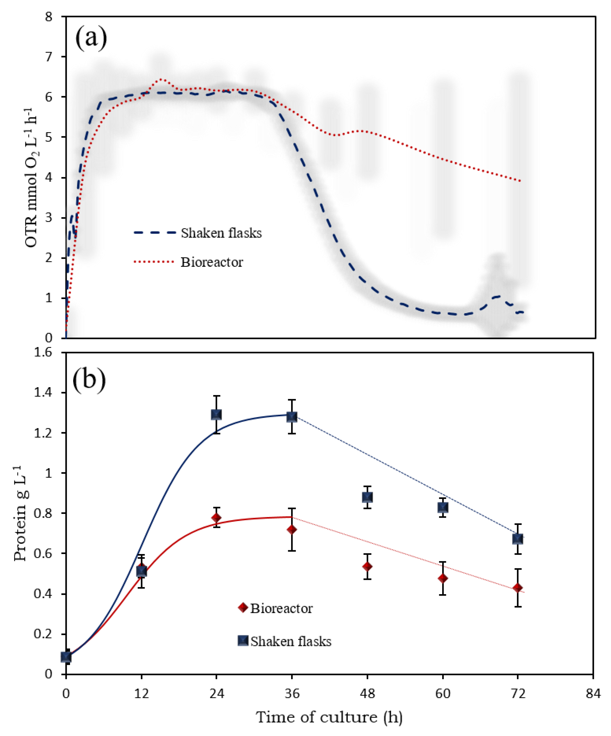
Figure 2a shows the OTR profiles of the cultures of the OP-PhbP3+ strain grown in shaken flasks and in the 3 L bioreactor. The lines represent the average of three independent experiments, and the shaded areas correspond to the standard deviation. It is important to mention that in the case of the bioreactor, after 36 h of culture, the variations in the OTR values were high due to instability in the readings of the oxygen sensor in the gas analyzer. In both cases (shake flask and bioreactor), the OTRMAX was obtained after 6 h of cultivation, reaching an average value of 6.00 ± 0.04 mmol O2 L−1 h−1. The maximum value of OTR remained constant until 36 h of culture, indicating that the cultures were limited by oxygen [30,31]. Although it was possible to replicate the OTR during the first 36 h of culture, significant differences in the OTR were observed after this time. In the case of cultures in shaken flasks, the OTR decreased to values close to zero, whereas in cultures in the bioreactor, the rate of decrease in OTR was lower, reaching a minimum value of 4 mmol L−1 h−1 after 72 h of culture. It is known that the decrease in OTR is related to a drop-in bacterial respiratory activity caused by the limitation of nutrients in the culture medium [31]. Because the medium still contained 8 g L−1 of sucrose after 36 h (Figure 3c), the limitation may be due to another nutrient, such as phosphates or trace elements.
Figure 2b shows the growth kinetics, measured as the protein concentration in the cultures, in the 3 L bioreactor compared with previous results reported for shaken flasks [13]. It can be observed that, although during the first 36 h of culture the OTR behavior was replicated in the 3 L bioreactor, the growth kinetics was significantly different from those achieved in the shaken flasks.
A specific growth rate (µ) of 0.085 ± 0.02 h−1 was obtained, which is significantly lower than the previous results reported by Quiroz et al. [22] for shaken flask cultures, where a µ of 0.12 ± 0.01 h−1 was determined. This observed behavior may be attributed to differences in the hydrodynamic conditions between the bioreactor and shaken flasks and/or variations in the carbon-to-nitrogen (C/N) ratio of the culture medium. However, further studies are required to fully elucidate the reasons for the growth discrepancies observed at the two scales.
After 36 h of cultivation, an important decrease in the cellular protein content was observed in the OP-PhbP3+ culture, decreasing from 0.8 to 0.4 g L−1 at 72 h of cultivation. It is important to note that, since protein determination is carried out on the cell pellet obtained after centrifugation of the medium, the measured protein content in the pellet decreases. This drop corresponded to the decrease in OTR observed after 36 h of culture (Figure 2a). Interestingly, this behavior was not due to the carbon source depletion. Recent studies from our group have confirmed that the reduction in OTR, and consequently in strain growth, is associated with phosphate limitation in the PYS culture medium.
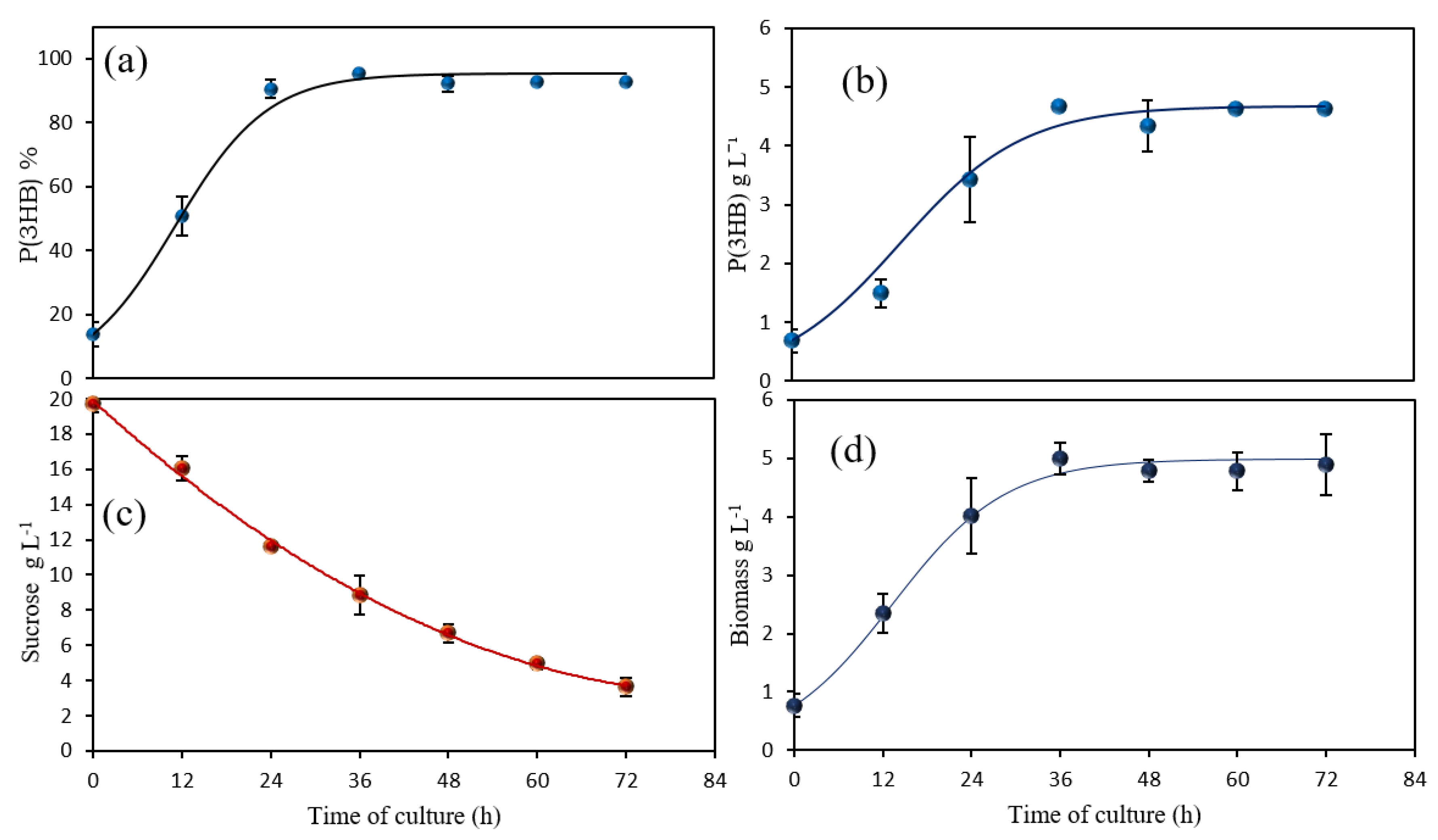
Despite the marked decrease in protein concentration—and consequently in strain growth—the P(3HB) concentration and accumulation remained unaffected (Figure 3a, b), reaching a maximum of 5.0 g L−1 after 72 h of cultivation, with an accumulation level higher exceeding 90%.
The decrease in protein concentration (0.6 g/L) had very little effect on the total biomass content (5.0 g/L) and therefore not reflected in the biomass concentration after 36 h of cultivation.
In addition, the figure shows that at the beginning of cultivation the P(3HB) content was 13.78 ± 3.89%, followed by a progressive accumulation that peaked at 36 h, reaching 95 ± 0.88%. This value is notably higher than the maximum accumulation reported in shake flask cultures by Quiroz et al. [22], which reached 89.7 ± 1.24%. It is likely that the increased accumulation of P(3HB) in the cells result from more severe oxygen limitation conditions in the bioreactor cultures, which stimulate enhanced polymer synthesis within the cells. Under these conditions, the cells redirect their carbon flux toward P(3HB) synthesis to maintain redox balance and store excess carbon, thereby promoting a higher polymer accumulation [32].
Figure 4 presents microscopy images of bacterial cells isolated at 36 and 72 h, revealing significant changes in the cell morphology. At 36 h of cultivation, the cells appear intact and exhibit a high intracellular accumulation of P(3HB), with the polymer clearly localized within the bacterial cytoplasm as distinct granules. The cellular envelope remains structurally preserved, suggesting active metabolism and polymer synthesis (Figure 4a). In contrast, the images at 72 h of cultivation show a marked shift in cell morphology. Many cells appear disrupted or lysed, with numerous P(3HB) granules observed freely dispersed in the extracellular medium (Figure 4b, black box). This suggests that extended cultivation may lead to cell lysis and subsequent release of the polymer, potentially due to nutrient depletion or autolytic processes.
These findings highlight the potential of the OP-PHB3+ strain to simplify the extraction of P(3HB), enabling the use of less complex and more cost-effective recovery methods, as discussed in the following sections.

3.2. Cell Population Analysis by Flow Cytometry

Flow cytometry analysis was performed to evaluate the population dynamics of A. vinelandii OP-PhbP3+ cultures over time, with particular focus on cell lysis beginning at 36 h of cultivation.
Flow cytometric analysis of the cell population enabled the characterization of structural and morphological changes undergone by A. vinelandii OP-PhbP3+ during cultivation. Using FSC-A vs. SSC-A dot plots, the relative distribution of cellular events was quantified based on size and morphology, providing indirect evidence of the physiological state of the strain.
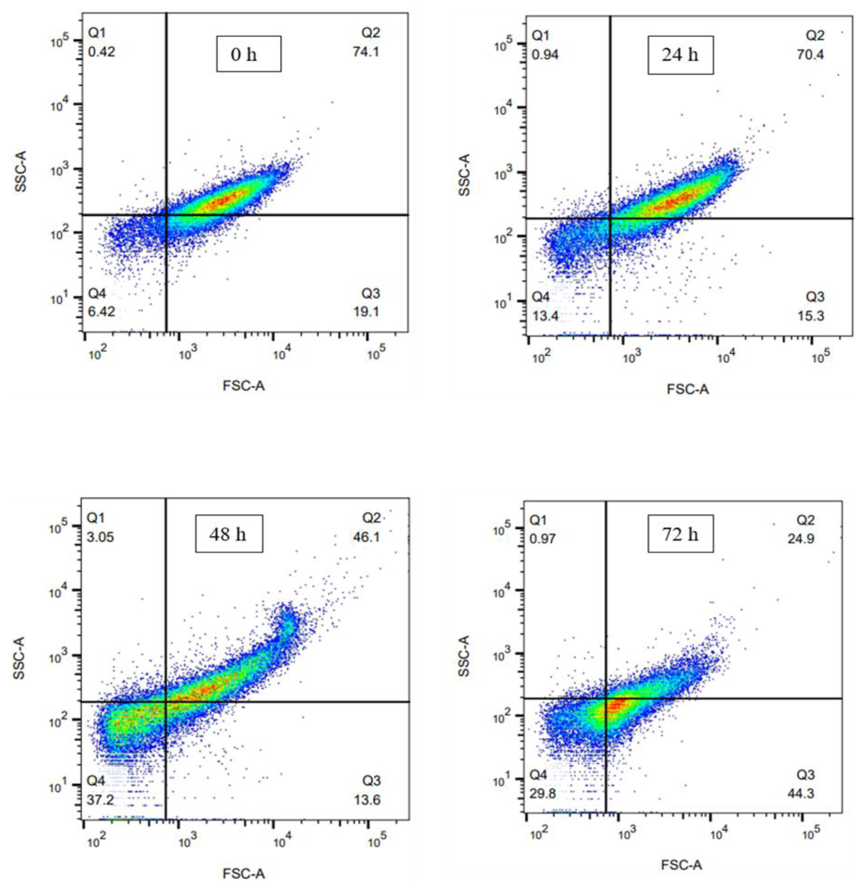
Figure 5 shows the grouping of objects across different quadrants, as well as the distribution within each quadrant.
At the beginning of the culture, the initial profile was predominantly composed of large, highly granular cells (Q2, 74.1%), likely representing an active inoculum with basal levels of P(3HB) accumulation. Additionally, 19.1% of the population was found in quadrant Q3, corresponding to large cells with low internal complexity. In contrast, the proportions of cells in quadrants Q1 and Q4 were minimal (0.42% and 6.42%, respectively), suggesting that the population was largely intact and relatively homogeneous at the onset of cultivation.
After 24 h of cultivation, a slight decrease in Q2 (70.4%) was observed, accompanied by increases in Q4 (13.4%) and Q1 (0.94%). This redistribution may indicate a metabolic transition phase during which some cells begin to fragment or release intracellular inclusions. The progressive emergence of events in Q4 suggests the onset of an early lytic process, characteristic of this strain.
At 48 h of cultivation, a notable shift in population distribution was observed. The cells in Q2 shifted to higher SSC-A values, possibly due to an increased content of P(3HB). However, their proportion decreased significantly to 46.1%, while the proportion in Q4 increased to 37.2%, likely indicating a substantial loss of cellular integrity in part of the population. The rise in events within Q4 corresponds to degraded cellular components or P(3HB) granules released into the extracellular medium. This time point may represent a critical threshold at which the culture transitions into a lytic phase.
Finally, at 72 h of cultivation, the population profile shifted markedly. The proportion in Q2 dropped to 24.9%, while Q3 (44.3%) and Q4 (29.8%) increased substantially. These changes indicate an advanced stage of lysis, during which many cells have lost internal complexity or undergone structural collapse. The important decrease in Q2 suggests that a large portion of the population containing intracellular granules has already been degraded.
In summary, it can be stated that during cultivation, there is an evolution of the morphology of the A. vinelandii OP-PhbP3+ population. Temporal analysis revealed a clear transition from an initially homogeneous and structurally intact inoculum to a highly degraded population, consistent with the lytic behavior previously reported for this strain [22].

3.3. Yield and Purity of the Products Obtained from OP-PhbP3+ Using SDS and CYC Method

In the present study, two environmentally friendly methods were employed for the extraction and purification of P(3HB), utilizing sustainable solvents. Figure 1 illustrates the distinct processes: one involving SDS (sodium dodecyl sulfate) and the other using CYC (cyclohexanone). These treatments were chosen due to the prior spontaneous cell rupture and the high polymer content within the cells.
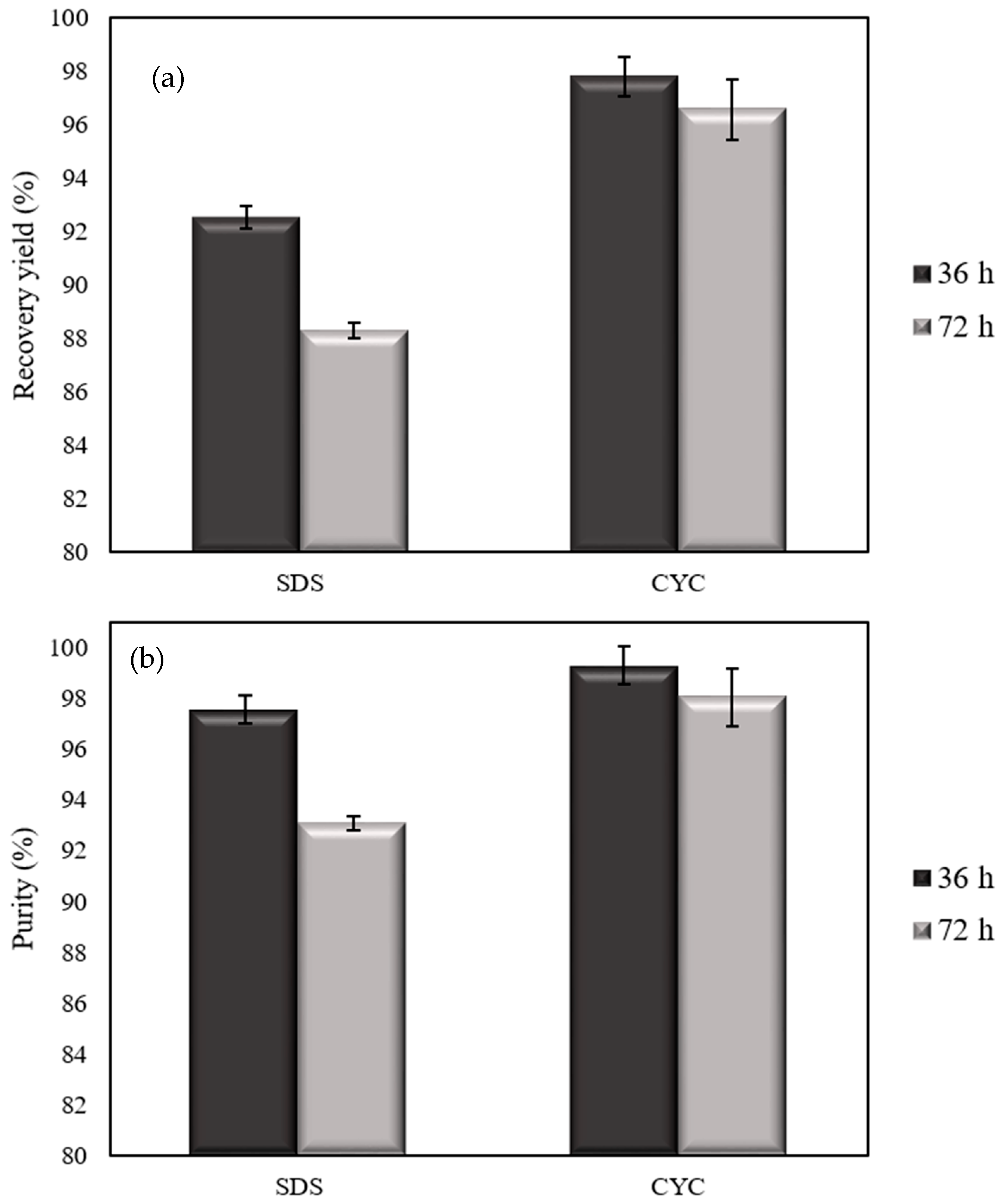
Figure 6 shows the results for yield and purity (calculated according to Equations (6) and (7) in Material and Methods section), with the cells harvested at 36 and 72 h of cultivation. Both yield and purity were higher in cells harvested at 36 h, using both extraction methods. Specifically, the SDS method achieved a yield of 92.52 ± 0.4%, while the CYC method resulted in an even higher yield of 97.77 ± 0.74%.
Using cells harvested after 72 h of cultivation, the yield obtained with SDS treatment was 88.29 ± 0.27%; whereas the yield with the CYC method reached 96.56 ± 0.15%, further demonstrating the superior effectiveness of the CYC treatment.
In a previous study, Rosengart et al. [33] utilized the CYC method to extract of the P(3HB) produced by Burkholderia sacchari DSM 17,165. That study reported an intracellular accumulation of 57.7% and an extraction yield of 93.2% [33]. In comparison, the results obtained in the present study using A. vinelandii OP-PhbP3+ demonstrate higher yield, highlighting the effectiveness of this method when applied to this biological system. On the other hand, regarding the SDS method, García et al. [11] reported an extraction yield of 68% using biomass from A. vinelandii OPNA—a value considerably lower than that obtained in the present work.
In addition, Figure 6a,b present the purity values obtained using both extraction methods. The highest purity was achieved with the CYC method using cells harvested at 36 h, reaching 99.3 ± 0.05%. This result slightly exceeds the value reported by Rosengart et al. [33] who employed the same extraction method and B. sacchari DSM 17,165 as the production strain, achieving a purity of 98.2 ± 1.6%.
On the other hand, when cells harvested at 72 h were used, the CYC method yielded purity of 98.06 ± 1.13%, indicate that the CYC method enables the production of a highly pure final product.
Using the SDS extraction method, the purity was 97.55 ± 0.55% in the cells isolated at 36 h; however, this difference may not be significant, given that the biomass at this point contained 95.34 ± 0.88% P(3HB), resulting a ~2% only about. Similarly, at 72 h, the P(3HB) content in the biomass was 92.76 ± 0.23%, and the purity following SDS extraction was 93.06 ± 0.29. Therefore, it can be said that treatment of cells harvested at 36 and 72 h with SDS did not have a positive effect on the purity of the polymer.
Table 1 presents a comparison of yields and purities obtained using CYC as the extraction method, as reported in the literature. The data clearly demonstrate that the combination of the OP-Phb3+ strain and cyclohexanone (CYC) allows yields and purities that are comparable to those achieved by other researchers using the same solvent. For instance, Jiang et al. [34] reported a yield of up to 99% and a purity of 95% using Cupriavidus necator H16, with CYC extraction performed at 120 °C for 3 min. Similarly, Rosengart et al. [33] achieved a maximum yield and purity of 98% using Burkholderia sacchari DSM 17,165, with CYC at 130 °C for 30 min [32]. In contrast, Van Walsem et al. [35], using E. coli as the production organism, reported lower values—80% yield and 92% purity—following CYC extraction at 90 °C for 35 min.
The findings are consistent with those previously reported in the literature. However, it is important to emphasize that the extraction yield obtained using a specific solvent depends on multiple factors, including the bacterial strain—and consequently, the composition and permeability of its cell membrane—the P(3HB) content, the biomass-to-solvent weight-to-volume ratio, extraction temperature, and extraction time.
Overall, the extraction using CYC presents a clear potential for large-scale industrial implementation, as it combines high extraction and purification efficiency with a markedly low environmental footprint. One of its main advantages lies on the use of CYC solvent, which not only exhibits a strong capacity to solubilize the polymer, but in addition can be efficiently recovered by distillation and reintroduced into subsequent extraction cycles.
All the above, reduces solvent consumption and, consequently, decreases the operational costs associated with raw materials, making the process more sustainable and economically attractive. Another critical factor is the biological system employed. The strain used is a well-established P(3HB) overproducer that, unlike other commonly studied strains such as the OP strain, naturally releases polymer granules into the culture supernatant. This intrinsic characteristic eliminates the need for energy-intensive cell disruption steps and simplifies downstream processing, thereby facilitating polymer recovery and ensuring high purity levels.
In this context, this study represents the first report demonstrating that the combination of an efficient solvent recovery strategy with a biologically optimized strain provides a platform that significantly may improves the cost-effectiveness of biopolymer production.
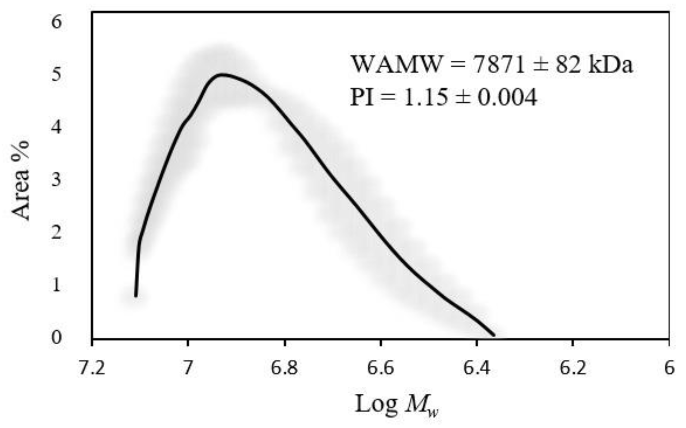
Figure 7 shows the molecular mass distribution of the polymer extracted from cells harvested at 72 h using the CYC method. The weight average molecular weight was 7871 ± 82 kDa, with a polydispersity index (PDI) of 1.15 ± 0.004, indicating a high and uniform molecular size distribution of P(3HB). In the present study, the values of weight average molecular weight obtained for P(3HB) films were comparable to those previously reported by Quiroz-Cardoso et al. [22].
According to these authors [22], the highest weight average molecular weight of P(3HB) produced by A. vinelandii OP-PhbP3+ in shaken flasks was achieved at 24 h of cultivation (8301 ± 381 kDa), whereas at 72 h of cultivation the weight average molecular weight decreased to 7904 ± 5 kDa. This behavior may be related to intracellular or extracellular polymer degradation processes due the action of depolymerases.
The similarity in the weight average molecular weight between the polymer isolated using the CYC extraction method from the polymer obtained in 3 L bioreactor and the values obtained from shaken flasks cultures, confirmed that the extraction method does not cause degradation of the biopolymer. This preserves a quality comparable to that reported by Quiroz-Cardoso et al. [22]. Additionally, the weight average molecular weight observed with the CYC method suggest that it may serve as a viable, less toxic, and potentially recyclable alternative for P(3HB) recovery, provided that extraction time and cultivation conditions are properly optimized.

4. Conclusions

Our results demonstrate, for the first time, that culturing the OP-PhbP3+ strain, which overexpresses the phasin PhbP3, in a 3 L bioreactor enables the production of a high P(3HB) content at the end of the exponential growth phase. Additionally, this strain exhibits early lysis, facilitating the release of P(3HB) granules and allowing for their extraction using low-toxicity solvents such as CYC. This approach results in high-purity polymer production with high yields, making it a promising strategy for generating PHB bioplastics.

Author Contributions

Conceptualization, C.P. and D.S.; methodology, C.A.-Z. and J.V.; formal analysis, C.P. and J.V.; investigation J.V. and C.A.-Z.; resources, C.P. and D.S.; writing—original draft preparation, C.P., J.V. and C.A.-Z.; writing—review and editing, C.P., D.S. and E.G.; supervision, C.P.; funding acquisition, C.P. and D.S. All authors have read and agreed to the published version of the manuscript.

Funding

This research was funded by DGAPA-UNAM, grant number IG200225.

Institutional Review Board Statement

Not applicable.

Informed Consent Statement

Not applicable.

Data Availability Statement

Data is unavailable.

Acknowledgments

The authors thank to Celia Flores and Guadalupe Zavala, IBt-UNAM for their technical support.

Conflicts of Interest

The authors declare no conflicts of interest.

References

  1. Sudesh, K.; Abe, H.; Doi, Y. Synthesis, structure and properties of polyhydroxyalkanoates: Biological polyesters. Prog. Polym. Sci. 2000, 25, 1503–1555. [Google Scholar] [CrossRef]
  2. Mai, J.; Kockler, K.; Parisi, E.; Chan, C.M.; Pratt, S.; Laycock, B. Synthesis and physical properties of polyhydroxyalkanoate (PHA)-based block copolymers: A review. Int. J. Biol. Macromol. 2024, 263 Pt 1, 320–327. [Google Scholar] [CrossRef] [PubMed]
  3. Koller, M. A review on established and emerging fermentation schemes for microbial production of polyhydroxyalkanoate (PHA) biopolyesters. Fermentation 2018, 4, 30. [Google Scholar] [CrossRef]
  4. Koller, M.; Maršálek, L.; de Sousa, M.; Gerhart, B. Producing microbial polyhydroxyalkanoate (PHA) biopolyesters in a sustainable manner. New Biotechnol. 2017, 37, 24–38. [Google Scholar] [CrossRef]
  5. Rodríguez-Contreras, A. Recent advances in the use of waste as raw materials for polyhydroxyalkanoates production. Biotechnol. Adv. 2019, 54, 107871. [Google Scholar] [CrossRef]
  6. Koller, M. Switching from petro-plastics to microbial polyhydroxyalkanoates (PHA): The biotechnological escape route of choice out of the plastic predicament. EuroBiotech J. 2019, 3, 32–44. [Google Scholar] [CrossRef]
  7. Wong, Y.-M.; Brigham, C.J.; Rha, C.; Sinskey, A.J.; Sudesh, K. Biosynthesis and characterization of polyhydroxyalkanoate containing high 3-Hydroxyhexanoate monomer fraction from crude palm kernel oil by recombinant Cupriavidus necator. Bioresour. Technol. 2012, 121, 320–327. [Google Scholar] [CrossRef]
  8. Abate, T.; Amabile, C.; Muñoz, R.; Chianese, S.; Musmarra, D. Polyhydroxyalkanoate recovery overview: Properties, characterizations, and extraction strategies. Chemosphere 2024, 356, 141950. [Google Scholar] [CrossRef]
  9. Tamer, I.M.; Moo-Young, M.; Chisti, Y. Optimization of poly(β-hydroxybutyric acid) recovery from Alcaligenes latus: Combined mechanical and chemical treatments. Bioprocess. Eng. 1998, 19, 459–468. [Google Scholar] [CrossRef]
  10. Kurian, N.S.; Das, B. Comparative analysis of various extraction processes based on economy, eco-friendly, purity and recovery of polyhydroxyalkanoate: A review. Int. J. Biol. Macromol. 2021, 183, 1881–1890. [Google Scholar] [CrossRef]
  11. Gumel, A.M.; Annuar, M.S.M.; Chisti, Y. Recent Advances in the Production, Recovery and Applications of Polyhydroxyalkanoates. J. Polym. Environ. 2012, 21, 580–605. [Google Scholar] [CrossRef]
  12. Hahn, S.K.; Chang, Y.K.; Kim, B.S.; Chang, H.N. Optimization of microbial poly(3-hydroxybutyrate) recover using dispersions of sodium hypochlorite solution and chloroform. Biotechnol. Bioeng. 1994, 44, 256–261. [Google Scholar] [CrossRef]
  13. Valappil, S.P.; Misra, S.K.; Boccaccini, A.R.; Keshavarz, T.; Bucke, C.; Roy, I. Large-scale production and efficient recovery of PHB with desirable material properties, from the newly characterised Bacillus cereus SPV. J. Biotechnol. 2007, 132, 251–258. [Google Scholar] [CrossRef] [PubMed]
  14. García, A.; Pérez, D.; Castro, M.; Utruvia, V.; Catillo, T.; Díaz-Barrera, A.; Espín, G.; Peña, C. Production and recovery of poly-3-hydroxybutyrate [P(3HB)] of ultra-high molecular weight using fed-batch cultures of Azotobacter vinelandii OPNA strain. J. Chem. Technol. Biotechnol. 2019, 94, 1853–1860. [Google Scholar] [CrossRef]
  15. García-Cerna, S.; Sánchez-Pacheco, U.; Meneses-Acosta, A.; Rojas-García, J.; Campillo-Illanes, B.; Segura-Gonzáles, D.; Peña-Malacara, C. Evaluation of Poly-3-Hydroxybutyrate (P3HB) Scaffolds Used for Epidermal Cells Growth as Potential Biomatrix. Polymers. 2022, 14, 4021. [Google Scholar] [CrossRef]
  16. Martínez, V.; García, P.; García, J.L.; Prieto, M.A. Controlled autolysis facilitates the polyhydroxyalkanoate recovery in Pseudomonas putida KT2440. Microb. Biotechnol. 2011, 4, 533–547. [Google Scholar] [CrossRef] [PubMed]
  17. Chen, Y.; Guo, W.; Wen, R.; Chen, G.-Q. An automatic lytic system for downstream purification of PHA produced by Halomonas. Chem. Eng. J. 2025, 517, 164425. [Google Scholar] [CrossRef]
  18. Borrero-de Acuña, J.M.; Hidalgo-Dumont, C.; Pacheco, N.; Cabrera, A.; Poblete-Castro, I. A novel programmable lysozyme-based lysis system in Pseudomonas putida for biopolymer production. Sci. Rep. 2017, 7, 4373. [Google Scholar] [CrossRef]
  19. Resch, S.; Gruber, K.; Wanner, G.; Slater, S.; Dennis, D.; Lubitz, W. Aqueous release and purification of poly(β-hydroxybutyrate) from Escherichia coli. J. Biotechnol. 1998, 65, 173–182. [Google Scholar] [CrossRef]
  20. Hori, K.; Kaneko, M.; Tanji, Y.; Xing, X.H.; Unno, H. Construction of self-disruptive Bacillus megaterium in response to substrate exhaustion for polyhydroxybutyrate production. Appl. Microbiol. Biotechnol. 2002, 59, 211–216. [Google Scholar] [CrossRef]
  21. Lacmata, S.T.; Yao, L.; Xian, M.; Liu, H.; Kuiate, J.-R.; Liu, H.; Feng, X.; Zhao, G. A novel autolysis system controlled by magnesium and its application to poly (3-hydroxypropionate) production in engineered Escherichia coli. Bioengineered 2017, 8, 594–599. [Google Scholar] [CrossRef]
  22. Quiroz-Cardoso, R.; Castillo, T.; Galindo, E.; Ruíz-Escobedo, J.; Segura, D.; Peña, C. Looking for improved strains of Azotobacter vineladii and favorable culture conditions yielding high-molecular-weight poly(3-hydoxybutyrate). J. Chem. Technol. Biotechnol. 2025, 100, 477–487. [Google Scholar] [CrossRef]
  23. Ruiz Escobedo, J. Estudio del Papel de las Proteínas Avin34710 y Avin34720 en el Metabolismo de Polihidroxibutirato (PHB) en la Bacteria Azotobacter vinelandii. Master’s Thesis, Universidad Nacional Autónoma de México, Ciudad de México, Mexico, 2020. [Google Scholar]
  24. Doran, P.M. Mass Transfer. In Bioprocess Engineering Principles, 1st ed.; Elsevier Science & Technology Books: San Diego, CA, USA, 1995; pp. 191–217. [Google Scholar]
  25. Lowry, O.H.; Rosebrough, N.J.; Farr, A.L.; Randall, R.J. Protein measurement with the Folin phenol reagent. J. Biol. Chem. 1951, 193, 265–275. [Google Scholar] [CrossRef]
  26. Miller, G.L. Use of Dinitrosalicylic Acid Reagent for Determination of Reducing Sugar. Anal. Chem. 1959, 31, 426–428. [Google Scholar] [CrossRef]
  27. Gómez-Hernández, E.; Salgado-Lugo, H.; Segura, D.; García, A.; Díaz-Barrera, A.; Peña, C. Production of Poly-3-Hydroxybutyrate (P3HB) with Ultra-High Molecular Weight (UHMW) by Mutant Strains of Azotobacter vinelandii Under Microaerophilic Conditions. Appl. Biochem. Biotechnol. 2021, 193, 79–95. [Google Scholar] [CrossRef] [PubMed]
  28. Mejía-Ruíz, H.; Moreno, S.; Guzmán, J.; Nájera, R.; León, R.; Soberón-Chávez, G.; Espín, G. Isolation and characterization of an Azotobacter vinelandii algK mutant. FEMS Microbiol. Lett. 1997, 156, 101–106. [Google Scholar] [CrossRef] [PubMed]
  29. Servain-Viel, S.; Aknin, M.L.; Domenichini, S.; Perlemuter, G.; Cassard, A.M.; Schlecht-Louf, G.; Lievin-Le Moal, V. A flow cytometry method for safe detection of bacterial viability. Cytom. Part A 2024, 105, 146–156. [Google Scholar] [CrossRef] [PubMed]
  30. Zimmermann, H.F.; Anderlei, T.; Büchs, J.; Binder, M. Oxygen limitation is a pitfall during screening for industrial strains. Appl. Microbiol. Biotechnol. 2006, 72, 1157–1160. [Google Scholar] [CrossRef]
  31. Anderlei, T.; Zang, W.; Papaspyrou, M.; Büchs, J. Online respiration activity measurement (OTR, CTR, RQ) in shake flasks. Biochem. Eng. J. 2004, 17, 187–194. [Google Scholar] [CrossRef]
  32. García, A.; Ferrer, P.; Albiol, J.; Castillo, T.; Segura, D.; Peña, C. Metabolic flux analysis and the NAD(P)H/NAD(P)+ ratios in chemostat cultures of Azotobacter vinelandii. Microb. Cell Fact. 2018, 17, 10. [Google Scholar] [CrossRef]
  33. Rosengart, A.; Cesário, M.T.; de Almeida, C.D.; Raposo, R.S.; Espert, A.; Díaz de Apodaca, E.; da Fonseca, M.R. Efficient P(3HB) extraction from Burkholderia sacchari cells using non-chlorinated solvents. Biochem. Eng. J. 2015, 103, 39–46. [Google Scholar] [CrossRef]
  34. Jiang, G.; Johnston, B.; Tonrow, D.E.; Radeka, I.; Koller, M.; Chaber, P.; Adamus, G.; Kowalczuk, M. Biomass Extraction Using Non-Chlorinated Solvents for Biocompatibility Improvement of Polyhydroxyalkanoates. Polymers 2018, 10, 731. [Google Scholar] [CrossRef]
  35. Van Walsem, J.; Zhong, L.; Shih, S.S. Polymer Extraction Methods 2010. U.S. Patent No. 7,713,720 B2, 7 August 2007. [Google Scholar]
  36. Filippi, S.; Cinelli, P.; Mezzetta, A.; Carlozzi, P.; Seggiani, M. Extraction of polyhydroxyalkanoates from purple non-sulfur bacteria by non-chlorinated solvents. Polymers 2021, 13, 4163. [Google Scholar] [CrossRef]
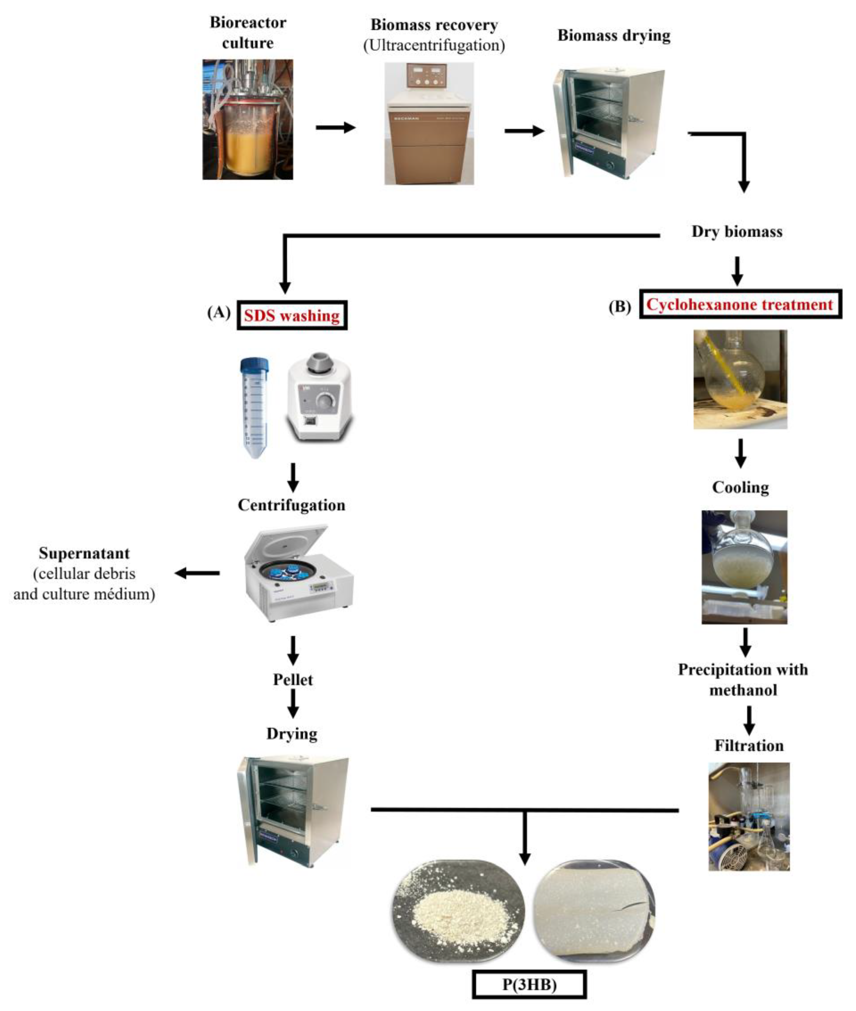
Figure 1. Schematic representation of the extraction processes involving SDS and CYC.
Fermentation 11 00571 g001
Figure 2. Comparison of OTR profiles and growth kinetics between cultures in bioreactors and shaken flasks. In (a) are shown the OTR profiles from an experimental triplicate. Panel (b) shows the growth kinetics from the protein concentration.
Fermentation 11 00571 g002
Figure 3. % P(3HB) accumulation on dry biomass (a), total concentration of P(3HB) (b), sucrose consumption (c), and biomass concentration (d) in cultures of the OP-PhbP3+ strain in a 3 L bioreactor.
Fermentation 11 00571 g003
Figure 4. Transmission electron micrographs of strain OP-PhbP3+. (a) Cell morphology at 36 h of cultivation, showing intact cell structure and intracellular P(3HB) granules. (b) Cell morphology at 72 h of cultivation. P(3HB) granules released into the medium are indicated within the black boxes.
Fermentation 11 00571 g004
Figure 5. Scatter plots showing cell size and complexity during the culture. The colors represent the event density in the analyzed sample. Blue or green colors, correspond to regions with a lower number of detected cells, whereas, yellow or red colors indicate areas with a higher concentration of events.
Fermentation 11 00571 g005
Figure 6. Yield and Purity of P(3HB) extracted from cultivations using SDS and CYC. Recovery yield at 36 and 72 h of culture (a). Purity at 36 and 72 h of culture (b).
Fermentation 11 00571 g006
Figure 7. Molecular mass distribution of P(3HB) from 72 h extracted with CYC. This shaded area represents the deviation observed in triplicate experiments.
Fermentation 11 00571 g007
Table 1. Comparison of yields and purities obtained using CYC as the extraction method, as reported in the literature.
Ref.Bacterial StrainContent of P(3HB)
(%)
Temperature of Extraction (°C)Time of Extraction (min)Yield of Extraction (%)Purity (%)
This researchA. vinelandii OP-PhbP3+95120–130159899
[36]Rhodovulum
sulfidophilum
DSM-1374
141255
10
20
43
95
98
N. A
[34]Cupriavidus
necator H16
82.380
100
120
1200
5
3
16
90
99
95
[33]Burkholderia
sacchari DSM 17,165
57.7120–13015
15
30
98
87
89
98.2
91.9
92.3
[35]E. coli8090358092
Disclaimer/Publisher’s Note: The statements, opinions and data contained in all publications are solely those of the individual author(s) and contributor(s) and not of MDPI and/or the editor(s). MDPI and/or the editor(s) disclaim responsibility for any injury to people or property resulting from any ideas, methods, instructions or products referred to in the content.